analysis and evaluation of segmental concrete tunnel linings
Upload
independentCategory
view
0download
0
Expression of Human Plasma Gelsolin in Escherichia coli and Dissection of Actin Binding Sites by Segmental Deletion Mutagenesis M. Way, J. Gooch , B. Pope, and A. G. Weeds Medical Research Council Laboratory of Molecular Biology, Cambridge CB2 2QH, England
Abstract. Human plasma gelsolin has been expressed in high yield and soluble form in Escherichia coli. The protein has nucleating and severing activities identical to those of plasma gelsolin and is fully cal- cium sensitive in its interactions with monomeric ac- tin. A number of deletion mutants have been ex- pressed to explore the function of the three actin binding sites. Their design is based on the sixfold seg- mental repeat in the protein sequence. (These sites are located in segment 1, segments 2-3, and segments 4-6.) Two mutants, S1-3 and $4-6, are equivalent to the NH:- and COOH-terminal halves of the molecule obtained by limited proteolysis. S1-3 binds two actin monomers in the presence or absence of calcium, it severs and caps filaments but does not nucleate poly- merization. $4-6 binds a single actin monomer but only in calcium. These observations confirm and ex-
tend current knowledge on the properties of the two halves of gelsolin. Two novel constructs have also been studied that provide a different pairwise juxtapo- sition of the three sites. $2-6, which lacks the high affinity site of segment 1 (equivalent to the 14,000-Mr proteolytic fragment) and S1,4-6, which lacks seg- ments 2-3 (the actin filament binding domain previ- ously identified using the 28,000-Mr proteolytic frag- ment). $2-6 binds two actin monomers in calcium and nucleates polymerization; it associates laterally with filaments in the presence or absence of calcium and has a weak calcium-dependent fragmenting activity. S1,4-6 also binds two actin monomers in calcium and one in EGTA, has weak severing activity but does not nucleate polymerization. A model is presented for the involvement of the three binding sites in the various activities of gelsolin.
ELSOLIN is an 82,000-Mr calcium-dependent actin severing and capping protein found universally in vertebrate tissues (Yin et al., 1981; Kwiatkowski et
al., 1988). A larger secreted form is readily isolated from blood plasma (Harris and Gooch, 1981). In addition to its severing activity, it accelerates actin polymerization by form- ing a stable nucleating complex with two actin monomers (Bryan and Kurth, 1984; Doi and Frieden, 1984; Weeds et al., 1986b).
Actin binding domains have been identified using limited proteolysis (Kwiatkowski et al., 1985; Bryan and Hwo, 1986). The protein is readily cleaved into two halves by a va- riety of proteases. The 45,000-M~ NH~-terminal domain (CT45N using the nomenclature of Yin et al., 1988) severs actin filaments as effectively as gelsolin, is calcium insensi- tive, and has only weak nucleating activity (Bryan and Hwo, 1986; Chaponnier et al., 1986). The COOH-terminal do- main of 38,000-Mr (CT38C) binds actin-Sepharose only in the presence of calcium, but neither severs nor nucleates (Kwiatkowski et al., 1985). Bryan (1988) has shown that this fragment binds a single actin monomer and inhibits actin po- lymerization at high molar ratios to actin, probably by se- questering monomers.
CT45N is further degraded to yield two smaller frag- ments, CTI5N and CT28N, the former being NH,-terminal (Chaponnier et al., 1986; Yin et al., 1988; Bryan, 1988). CT15N binds actin-Sepharose in a calcium-independent manner (Kwiatkowski et al., 1985) and forms a 1:1 high affinity complex with G-actin (Bryan, 1988). CT28N binds F-actin at a 1:1 stoichiometry with actin subunits (Yin et al., 1988; Bryan, 1988). Neither CT28N nor CT15N has sever- ing activity.
These studies show that gelsolin is composed of stable structural domains connected by proteolytically sensitive regions. The cDNA derived amino acid sequence shows a strong tandem repeat, suggesting evolution by gene duplica- tion (Kwiatkowski et al., 1986). Internal repeats have been reported for related actin severing proteins, fragmin (Ampe and Vanderkerckhove, 1987), severin (Andr6 et al., 1988), and villin (Bazari et al., 1988; Arpin et al., 1988). Our own analysis of all these proteins has revealed six large segmental repeats of '~15,000 M~ in gelsolin and villin and three simi- lar repeats in severin and fragmin. The evidence suggests that this family of proteins has evolved from a precursor of '~130-150 amino acids (Way and Weeds, 1988). The bound- aries of the six segments in gelsolin are consistent with the
© The Rockefeller University Press, 0021-9525/89/08/593/13 $2.00 The Journal of Cell Biology, Volume 109, August 1989 593-605 593

fragmentation patterns reported earlier, but facile proteolytic cleavage appears to be restricted to sites between segments I and 2 and segments 3 and 4. The three actin binding do- mains are located in proteolytic fragments corresponding to segment 1, segments 2-3, and segments 4-6.
To understand the role of individual segments in the vari- ous actin binding activities of gelsolin, we have constructed and expressed in Escherichia coil a number of deletion mu- tants as well as whole gelsolin (Fig. 1). In the absence of an x-ray crystal structure for gelsolin or more importantly gel- solin-actin complexes, the mutants have been designed based on the chymotryptic digestion pattern of human plasma gelsolin (Chaponnier et al., 1986) and the sixfold segmental repeat in gelsolin (Way and Weeds, 1988). Our nomenclature for the mutants is based on their segmental content. Thus mutants S1-3 and $4-6, corresponding to CT45N and CT38C, have been made essentially as controls. Two mutants are described that cannot be made by proteoly- sis: $2-6 in which segment 1 (CTI5N) is deleted and S1,4-6 which lacks segments 2 and 3 (CT28N). Both $2-6 and S1,4-6 have two actin binding sites but they behave very differently in severing and nucleation assays. Their proper- ties provide new information about the roles of individual ac- tin binding sites.
Materials and Methods
Construction of the Gelsolin Expression System T4 DNA ligase, polynucleotide kinase, all restriction enzymes, and the non- phosphorylated Hind III linker (CAAGCTTG) were obtained from New En- gland Biolabs (Beverly, MA), Klenow DNA polymerase from Boehringer Mannheim Diagnostics, Inc. (Houston, TX). Methods for preparation and analysis of recombinant DNA and culturing of bacteria were those de- scribed by Maniatis et al. (1982). All DNA sequences were determined by a series of oligonucleotides evenly spaced along the sequence using the dideoxy chain termination method (Sanger et al., 1977).
Two overlapping but incomplete eDNA clones, M1D and pU43a, which
1 2 3 ,I 5 6 GS
S1-3
S4-6
~ S:Z-6
GS i i
CT4ISN
CT3SC
CTI4N II III
CT28N
Figure 1. Schematic representation of the sixfold repeating se- quence of gelsolin and the design of the deletion mutants. The NH2- and COOH-terminal residues are indicated below in single letter code together with their positions in the amino acid sequence of human plasma gelsolin (Kwiatkowski et al., 1986). At the bottom of the figure the positions and notation ofproteolytic fragments have been given for comparison. The vertical arrow indicates the posi- tion of Ser 24, the NH2-terminal residue of CT14N (Yin et al., 1988).
together cover the entire coding region of human plasma gelsolin, were a kind gift from Drs. Kwiatkowski and Yin (Massachusetts General Hospital, Boston, MA; Kwiatkowski et al., 1986).
Nonphosphorylated Hind Ill linker (CAAGCTTG) was kinased and ligated into a unique Stu I site 64 bases 3' to the termination triplet of pU43a. Digestion with Barn HI and Hind Ill yielded a fragment containing the entire coding region of this clone. This fragment was ligatexl into the Barn HI and Hind II! sites of the expression vector pLclI (Nagai and ThCgersen, 1984) and transformed into JM101. One clone pLclI/pU43a was picked ready to accept the required coding region from MID.
A 907-bp Eco RI-Bal 1 fragment from the 5' end of MID, containing the coding region absent in pU43a as well as a 613-bp overlap, was isolated and cloned into Eco RI-Hinc ll-cleaved Ml3mpl9. Several clones containing the insert were selected to produce single stranded DNA according to Carter et al. (1985). An oligonuclootide BamFXMID. (TCGCTGCCCGTCCGCG- C GGGATCCATCGA~TAGGGCC AC TGCGGTC GCGGGGGGCGT) coding for Bam HI and Factor Xa recognition sites (underlined) was used to juxtapose the last codon of the Factor Xa sequence (Arg) and the first codon (Ala) of the mature human plasma gelsolin sequence. BamFXMID was kinased, annealed to the single stranded template, extended with Klenow DNA polymerase, and ligated according to Carter et al. (1985). The ligation mixture was transfected into calcium competent BMH 71-18 tontL cells and positive clones identified by hybridization with the mutagenic oli- gonuclcotide. These were plaque purified and sequenced along their entire length. Using the engineered Bam HI site and a unique Not I site at position 394 in the 907 Eco RI-Bal I insert, a 294-bp fragment was isolated and ligated into pLcll/pU43a. The ligation mixture was transfected into QY13 and clones selected on ampicillin plates before restriction mapping and in- duction of the expression construct pLclIFXGS (Fig. 2).
Construction of Mutants A Barn HI and Hind III digest of pLcIIFXGS was used to obtain the entire coding sequence of human plasma gelsolin. This fragment was subcloned into M13mpl9 and used to provide single stranded DNA for all subsequent mutagenesis. Mutagenesis was performed as described in the engineering of the Barn HI-Factor Xa recognition site in the 907-bp fragment from MID. A schematic representation of each gelsolin mutant along with positions changed is shown in Fig. 1. All mutant constructs were sequenced through- out their length before being excised from MI3mpl9 and engineered back into pLcII. Mutants with the correct construct were selected by restriction mapping before expression in QY13.
Induction, Expression, and Purification of Mutants Cultures of QY13 containing the vector pLcIIFXGS or mutated constructs were grown and induced as described in Nagai and Th0gersen (1987). Post-heat shock flasks were grown at 37°C for 3 h before centrifugation of cells at 4°C (accelerating up to 10,000 g and immediately turning off the centrifuge). Pellets were frozen at -20°C before resuspension at room tem- perature in 50 toM Tris HCI pH 8.0, 25% sucrose, and l mM EDTA (20 ml/liter original culture). 5 mg lysozytoe (10 mg/ml in distilled water) was added to 20 ml suspension to lyse the cells and digestion proceeded for 30 min at room temperature. The suspension was clarified by centrifugation at 150,000 g for 20 min and the supernatant dialyzed overnight at 4°C against 10 toM "Iris HCI pH 8.0, 100 mM NaCI, 0.1 mM CaCI2, and 3 mM NAN3. fxgelsolin was purified by actin-Sepharose a~nity chromatography (Weeds et al., 1986a). The fusion protein was subsequently digested with Factor Xa (kindly provided by Dr. K. Nagai [Medical Research Council Laboratory of Molecular Biology]) in 25 mM Tris I-ICI pH 8.0, 100 mM NaCI, 0.1 mM CaC12, and 1 mM NaN3 at a substrate to enzyme ratio of 50:i (wt/wt) for 5 h at room temperature or 100:1 (wt/wt) overnight at 4°C to yield native protein.
Unlike fxgelsolin, not all mutants were soluble but were found in the in- clusion body fraction. Inclusion body preparations were purified as de- scribed in Nagai and Th~gersen (1987) and solubilized in 8 M urea. The urea was removed by dialysis against 10 mM "Iris HCI pH 8.0, 100 mM NaCI, 0.1 toM CaCI2, and 3 mM NaN3. The dialysate was clarified by fur- ther centrifugation at 2,500 g for 10 rain followed by filtration through a 0.2 #m-filter Miilipore Continental Water Systems (Bedford, MA). Further purification of mutants was performed by actin-Sepharose chromatography (Weeds et al., 1986a). Calcium dependence was examined using EGTA- containing buffers. Mutant concentrations were calculated from the tyrosine and tryptophan content (Bryan, 1988) or using the Folin reaction as de- scribed previously (Weeds et al., 1986a).
The Journal of Cell Biology, Volume 109, 1989 594

FX
c z x ------~ I ~ 4 ~ - - GS 29 30 3~ I B a m ~ ~ 2 3
ACAGCGGA~GGGGGATCCATCGAGGGTAGqGCCAC TGC G ThrAlaGlu~lyGlySer I leGluGlyAr~laThrAla
cll
Bam HI
Not I
P L
p L c I I F X G S
Amp
Ori \ Hind III
Figure 2. Organization of the cleavable fusion protein expression vector, pLclIFXGS. The plasmid directs synthesis o f a hybrid pro- tein consist ing of the first 31 amino-terminal residues of lambda clI protein, the Factor Xa recognition sequence (FX), and human plasma gelsolin. PL, Lambda promotor; Amp, ampicillin-resistance gene; and Ori, the origin of replication of the vector.
Assay Methods All assays have been performed on mutant proteins purified on actin- Sepharose with the ell leader peptide present, fxgelsolin is as active as pig plasma gelsolin both with and without the ell leader peptide. There is no reason to believe that the fusion peptide is detrimental to the activity of gel- solin.
The DNase I inhibition assay (Harris et al., 1982) was used to compare fxgelsolin with pig plasma gelsolin and to analyze the mutants.
Preparation of Fluorescently Labeled Actin and Monomer Binding Assays 71trations with NBD-Actin and Pl-Actin. Preparation of Pl-actin n reacted on Cys 374 with N-(l-pyrenyl)iodoacetamide and NB-actin reacted with N-ethylmaleimide on Cys 374 then on Lys 373 with 7-chloro-4-nitrobenzo- 2-oxa-l,3-diazole were as described (Weeds et al., 1986b). Titrations with NBD-actin were carried out using a constant total protein concentration (200 or 400 nM) with continuous variation of both gelsolin and NBD-actin in G' buffer (10 mM Tris HCI pH 8.0, 0.2 mM ATP and dithiothreitol, 0.1
1. Abbreviations used in this paper: NBD-actin, actin reacted with N-ethyl- maleimide on Cys 374 then on Lys 373 with 7-chloro-4-nitrobenzeno-2-oxa- 1,3-diazole; PI-actin, actin reacted on Cys 374 with N-(l-pyrenyl)iodo- acetamide.
mM CaCI2, and 1 mM NAN3) as described previously (Weeds et al., 1986b). BSA (0.1 mg/ml) was added to all samples to minimize losses due to adsorption at surfaces. Samples were incubated at 4°C overnight in the dark before measuring the fluorescence at 21°C. Similar experiments were also carried out under polymerizing conditions (G' buffer + 2 mM MgCI2 and 0.1 M KCI, with either 0.1 mM CaCI2 or 1 mM EGTA). For experi- ments in the absence of calcium, both G-actin and mutant were preincubated at 2 /zM in 0.25 mM MgCl2, 1 mM EGTA before mixing.
Titrations were also carried out with 1/zM PI-actin and various concen- trations of S1-3.
Nucleation Assay. 4 #M PI G-actin in G' buffer (containing varying con- centrations of fxgelsolin in the range 10-120 nM) was polymerized by addi- tion of 1:20 vol of 40 mM MgCI2 and 2 M KCI. The fluorescence increased exponentially with time: a semi-Ln plot of the approach to equilibrium gives a straight line of slope k+ [N] where k+ is the association rate constant at the pointed end of filaments and [N] the concentration of gelsolin. (Under the ionic conditions used, [nuclei] = [gelsolin] [Janmey and Stossei, 1986; Selve and Wegner, 1986].) The nucleating efficiency of mutants is calculated relative to that of gelsolin.
Inhibition o f Polymerization. 2.8/~M PI-actin was polymerized in the presence of 30 nM gelsolin to give nucleated filament assembly. The con- centration of polymerized actin was determined from the final fluorescence and measurement of the critical concentration (method in Harris and Weeds, 1983). The effects of increasing mutant concentration on the maximum fluorescence enhancement were monitored and the concentration of actin bound to mutant was determined from the percent inhibition of fluorescence enhancement compared with controls.
AirfugeAssay. Assays carried out as described previously, using 18 ~M actin and different concentrations of mutants (Pope and Weeds, 1986). Sam- ples of supernatant and pellets resuspended to the original volume were ana- lyzed by SDS-PAGE and densitometry was performed using a densitometer (Comag Electrophoresis Scanner; Cambridge Instruments Ltd., Cam- bridge, UK).
Viscometry. A capillary flow viscometer was used at 22°C with 14.9/~M F-actin (Pope and Weeds, 1986). Flow times were 91.5 + 0.5 s for buffer and 138 + l s for 14.9/~M F-actin. Measurements were made initially in 0.2 mM EGTA after addition of fxgelsolin or mutant. The same samples were then assayed in 1.6 mM excess calcium. Results are expressed as specific viscosity. Aziditional assays were performed for $2-6 and S1,4-6 using "capped" filaments with an average length of ,~400 monomers pre- pared by polymerizing 54 ~,M G-actin in the presence of 13.6 nM fxgelsolin.
Severing Assay. The severing assay was adapted from that originally de- scribed for villin by Walsh et al. (1984) and used for gelsolin by Bryan and Coluccio (1985). Precapped filaments were used throughout to avoid prob- lems due to variation in the extent of capping and eliminate monomer dis- sociation from free barbed ends.
25 #M G-actin in G' buffer (containing 4.2 #M PI-actin) was polymer- ized in the presence of 0.125 or 0.25 nM gelsolin by addition of 5 mM MgCI2 and 100 mM NaCI to produce precapped filaments with an average length of • 200 or 100 monomers, respectively. (The length distribution is initially Poisson [Oosawa and Asakura, 1975].) Polymerization is complete in <2 rain. The filaments were diluted to 200 nM in l0 mM Tris HCI pH 8.0, 50 mM NaCI, 3 mM MgCl2, 0.2 mM ATE 0.2 mM dithiothreitol, and 0.1 mM CaCI2 (conditions used by Bryan and Coluccio, 1985) or G' buffer, using a cut Gilson tip to prevent shearing, and mixed by inverting the cuvette three times. Under these conditions, below the critical concentration, capped filaments depolymerize at a constant rate from their "pointed" ends, giving a linear fluorescence change over at least 10 min of the assay (the maximum time used, to minimize photobleaching). The rate of actin mono- mer dissociation is obtained by dividing this initial rate by the total fluores- cence change for depolymerization and the off-rate (k - ) estimated on the assumption that the filament concentration equals the gelsolin concentration (Janmey and Stossel, 1986; Selve and Wegner, 1986).
The severing capacity of fxgelsolin or mutant was measured from the fluorescence kinetics after addition of mutant (made "~30 s after dilution of the capped filaments to provide a control of the initial fluorescence of each diluted actin sample). Since filaments are cut at random, the resulting length distribution is exponential. Thus depolymerization follows exponential ki- netics according to the equation: A (t) = A°.e -k°bs't, where kobs = G/A%k-, A (t) is the concentration of F-actin at time t, A ° = 0.2 #M F-actin, G the gelsolin concentration, and k - the off-rate for monomers from the pointed end of filaments. A semi-Ln plot of [F(t) - F(oo)] gives a straight line with slope = kobs, where F(oo) = fluorescence at the end of the reac- tion and F(t) = that at time t. On the assumption that every gelsolin mole-
Way et al. Gelsolin Mutagenesis in E. coli 595

cule severs and caps an actin filament, a plot of kobs as a function of gelsolin concentration gives a slope = k-lA*. The value of k - should equal that obtained in the control depolymerization assays using capped filaments. A lower value indicates less efficient severing. If the extent of severing was small, the severing activity was determined by comparing the initial slope of the fluorescence decrease in the presence of mutant with controls.
Electron Microscopy. F-actin (2.4/~M) was incubated for various times with mutants at concentrations up to 2.4 #M in 2 mM TrisHCI pH 7.5, 0.5 mM MgCI2, 0.1 M KCI, 0.5 mM ATE and either 0.2 mM CaCI2 or 1 mM EGTA. Samples were prepared for microscopy as described by Valentine et al. (1968).
Calcium Binding. Experiments were done using a rapid calcium binding assay developed by Koch et al., 1986. Three 9-cm filler circles (Nos. 54 or 541; Whatman Inc., Clifton, NJ) were washed in a Bnchner funnel with 10 mM imidazole pH 7.0, 0.1 M NaCI, and 1.0 mM MgCI2 (washing buffer). All buffers were made up in BDH Analar water (<1.25 I~M Ca 2+) and stored in plastic to avoid contaminating calcium from glassware. A grid- ded nitroceUulose filter (BA 85/20 Schleicher & Schull, Inc., Keene, NH) was placed on top of the filter circles and rinsed with washing buffer. The presence of the three filter circles ensures that the vacuum is sufficient but not excessive. 50--200 pmol of protein in washing buffer were spotted on to the nitrocellulose with the system under vacuum. After applying protein, the nitrocellulose filter was overlaid in a rocking petri dish for 15 min with 5-10 ml of washing buffer containing 1-2 mCi/mpSCa 2+ and 0-40 #M car- rier 4°Ca2+. Surplus calcium buffer was removed from the filter by vacu- um. The nitrocellulose was washed in 50 ml washing buffer for 20 s in a petri dish on a rotary shaker and dried again. Although bound calcium can exchange during the rinsing stage, this procedure gave reproducible back- ground counts (,,,7,000 counts/min) without apparent loss of protein-bound calcium. After drying, the filter was cut into squares and radioactivity mea- sured in a scintillation counter. Negative controls (cytochrome c) and buffer blanks were included on each filter (these gave similar counts). Assays were carried out at different protein concentrations and free calcium concentra- tions between 0.1 and 40 ttM, but mostly at 10 or 40 ~tM. The free calcium concentration was based on Harafugi and Ogawa (1980).
Calcium binding was also measured by equilibrium dialysis as described previously (Weeds et al., 1986a), using 0.3-ml samples containing ,,o!0/~M protein and dialyzing against 50 ml calcium buffer containing 5-40/zM total calcium (0.131-20.3/zM free calcium). Protein concentrations were mea- sured after dialysis by absorbance at 280 nm (and calibrated using Bradford and Folin assays). Binding data were fitted using a nonlinear least squares fit (Press et al., 1988) to the equation B = nlCa]/(Kd + [CAD, where B is the bound calcium per tool mutant, [Ca] is the free calcium concentration, n the maximum number of binding sites, and Kd the dissociation constant.
Results
Expression and Purification of Gelsolin and Mutants
There were no obvious bands corresponding to gelsolin (or mutants) before induction (Fig. 3), but samples of postinduc- tion cell protein revealed a band that comigrated with plasma gelsolin. Western blots showed that only this band cross- reacted with an affinity-purified polyclonal antibody against plasma gelsolin (data not shown), fxgelsolin, synthesized at 10-15% of total cell protein, was found in the soluble frac- tion after cell lysis. It was purified on actin-Sepharose (Fig. 3) (Weeds et al., 1986a). NH2-terminal sequence analysis (by the method of Matsudaira [1987]) of fxgelsolin digested with factor Xa, confirmed that the first 12 amino acids were identical to those of human plasma gelsolin (Kwiatkowski et al., 1986).
Expression levels of mutants varied both between mutants and with different preparations (Fig. 4). However, the overall levels of expression were generally as good as that observed for fxgelsolin (compare Fig. 3 and Fig. 4). Unlike fxgelsolin, the mutants were found mainly in inclusion bodies, although $4-6 and S1,4-6 were also present in significant concentra- tions in the soluble fraction. These two mutants have been purified from both soluble and insoluble fractions and shown
Figure 3. Expression and purification of fxgelsolin. Lanes 1-7, SDS-PAGE. Lane 1, preinduction total cell protein; 2, 3-h postin- duction total cell protein; 3, soluble fraction; 4, protein not bound to actin-Sepharose; 5, actin-Sepharose-bound protein; 6, actin- Sepharose bound after washing with 1 M MgCI2; 7, purified fxgel- solin eluted with 4 M MgCI2. The arrow indicates the position of fxgelsolin and the asterisk indicates actin (also the position of a pro- tein in E. coli that comigrates with actin).
to behave identically. $2-6 was also present in very small amounts in the soluble fraction (Fig. 4). Inclusion bodies were readily solubilized in 8 M urea. Removal of urea by di- alysis precipitated much of the contaminating protein, leav- ing relatively pure mutant in the supernatant (Fig. 4).
Activities of fxgelsolin and Mutants
Actin-Sepharose Binding. fxgelsolin and all deletion mu- tants bound to actin-Sepharose in 20 mM Tris HCI pH 8.0, 150 mM NaCl, 1 mM MgCl2, 0.2 mM ATE 2 mM NAN3, and 0.1 mM CaCl2. $2-6 and $4-6 were eluted in the same buffer containing 0.5 M NaCl or in 1 M MgCl2 (a step in- cluded in the purification of plasma gelsolin to remove im- munoglobulins), but fxgelsolin, S1-3, and S1,4-6 required 4 M MgCl2 for elution.
The calcium dependence of binding to actin-Sepharose was also analyzed by loading columns in 0.1 mM CaC12 and washing with the same buffer plus 1 mM EGTA. $2-6 was completely eluted while $1,4-6 remained fully bound (Fig. 5). $4-6 behaved like $2-6 while $1-3 and fxgelsolin were like S1,4-6 (data not shown). When the mutants were applied to actin-Sepharose in the presence of 1 mM EGTA, S1,4-6 bound but $2-6 did not (Fig. 5).
Binding to Monomeric Actin NBD-Actin Titrations. The continuous variation method is the best means of determining the stoichiometry of com- plexes in NBD-actin titrations, provided the affinity is high and there is significant fluorescence enhancement on com- plex formation (Weeds et al., 1986b). Experiments using fxgelsolin showed maximum fluorescence enhancement in calcium at two actin monomers per gelsolin (Fig. 6 A). Simi-
The Journal of Cell Biology, Volume 109, 1989 596

Figure 4. Expression and distribution of gelsolin mutants 3-h postinduction in E. coli. Lanes 1, 1', 5', and 5, $2-6 (1, total cell protein; 1' soluble fraction, 5', solubilized after removal of 8 M urea; 5, insoluble after removal of urea). Lanes 2, 2', 6', and 6, S1,4-6 (lanes as described for $2-6 above). Lanes 3, Y, 7', and 7, $4-6 (lanes as above). Lanes 4, 4', 8', and & S1-3 (lanes as above). The arrows mark the positions of $2-6, S1,4-6, and $4-6 or S1-3, while the asterisk shows the E. coil protein that comigrates with actin (the intensity of this band shows that gel loadings are similar for all mutants).
lar experiments in the absence of calcium showed no fluores- cence enhancement. Comparable experiments with the mu- tants are also shown in Fig. 6. The inflexion point in Fig. 6 B indicated a binding stoichiometry of 1.8 actin per S1-3 both in calcium and EGTA, but curvature of the graph makes the accuracy less precise. $4-6 gave no fluorescence enhance- ment in calcium or EGTA (Fig. 6 D). $2-6 gave a profile similar to that of fxgelsolin, with maximum enhancement on binding two actin monomers in calcium (Fig. 6 D) and no enhancement in EGTA (data not shown). S1,4-6 gave a simi- lar fluorescence enhancement plus or minus calcium and a stoichiometry close to 1:1 may be extrapolated from Fig. 6 C. The maximum enhancement with this mutant in calcium was much less than that for all other mutants. A lower quan- tum yield associated with segment 1 was also inferred from
Figure 5. Binding to actin-Sepharose of $2-6 (lanes 1, 2, and 5) and SI,4-6 (lanes 3, 4, and 6). Lanes I and 3 show bound samples after loading in calcium; lanes 2 and 4 show material remaining bound after elution with 10 column volumes of buffer containing 1 mM EGTA; lanes 5 and 6 show bound mutant after loading column in EGTA. Asterisk indicates the position of actin.
titrations of S1,4-6 with NBD-actin using either fixed actin concentrations and variable S1,4-6 or vice versa (the mean value for maximum enhancement in eight different experi- ments under the three assay conditions was 51 + 4.5%).
Experiments carried out with S1-3, $4-6, and S1,4-6 un- der polymerizing conditions but below the critical concen- tration showed no differences in either the extent of fluores- cence enhancement or inflexion points of the titrations (data not shown).
The binding stoichiometry of S1-3 was further assessed from the fluorescence increase on mixing this mutant with 1 #M PI-actin in G'-buffer (an effect not observed with $4-6 or S1,4-6). Maximum enhancement occurred at •1.8 ac- tin/S1-3. The binding studies are summarized in Table I.
I n h i b i t i o n o f P o l y m e r i z a t i o n
Monomer binding by mutants was investigated by inhibition of actin polymerization as shown in Fig. 7. Increasing con- centrations of $4-6, S1-3, or S1,4-6 reduced the maximum fluorescence enhancement, but did not affect the rate con- stant for gelsolin-nucleated polymerization. By contrast, $2-6 increased the rate of polymerization (see below). The concentration of mutant-bound actin was calculated from the percent inhibition assuming a maximum concentration of monomeric actin available for polymerization equals 1.9 #M (based on a critical concentration of 0.9 ftM). Binding is very tight (Fig. 7). The binding stoichiometry was determined from the linear slope at low mutant concentrations, giving 1.0 for $4-6, 2.12 for S1-3, and 1.81 for S1,4-6. A Scatchard plot of the results shown in Fig. 7 for $4-6 suggests a dissoci- ation constant of '~ 25 nM.
N u c l e a t i o n
When 4/~M actin was polymerized in the presence of fxgel-
Way et al. Gelsolin Mutagenesis in E. coli 597

0
200
30O
0
20o
loo
I i i
50 1 O0 1 50 200 150 100 50 0
400 400 B
300 o
2oo
1 O0
0 i ! i
[Actin] 0 5 0 1 oo 1 50 2 0 0 [Gelsolin] 200 15o 1 oo 5o 0
[Acfin] [S1-3]
50(1
40O
O 3oo
20o
lO0
0 [actm]
400 [Mutant]
C
i i i
1 O0 200 300 400 300 200 100 0
600
50O
o 400
300
20o
lOO
o [Actin]
[S 1,4-61
D
! i !
0 100 200 300 400 400 300 200 100 0
Figure 6. Fluorescence titrations at constant total protein concentration (in nanomolar) with continuous variation of both NBD-actin and gelsolin (or mutant). In all cases, solid symbols represent analyses in the presence of 0.1 mM calcium, while open circles show experiments in the absence of calcium. (A) fxgelsolin; (B) S1-3; (C) S1,4-6; (D) triangles represent $2-6 and circles S4-6. Data for $2-6 and $4-6 in the absence of calcium are not shown: they were identical to those of fxgelsolin in EGTA or $4-6 in calcium.
solin, the fluorescence intensity increased exponentially with time after a lag of 20 s or less and reached a maximum within 3-10 min depending on gelsolin concentration. In the absence of gelsolin the fluorescence rise was <2 % of the total change on polymerization in the first 2 min and <15% in the first 10 min, showing that spontaneous nucleation was very slow. The rate constant obtained from semi-Ln plots in- creased linearly with gelsolin concentration (10-I00 nM), giving a value of k+ for monomer addition at the pointed ends of filaments of 0.179/#M/s (Fig. 8). $2-6 gave a similar value for k+ = 0.165/#M/s. By contrast, $4-6, S1-3, and SI,4-6 at concentrations up to 120 nM did not increase the rate of fluorescence rise above controls.
Table I. Binding of Monomeric Actin to fxgelsolin and Mutants
Activity fxgelsolin S1-3 $4-6 S2-6 SI ,4-6
A c t i n - S e p h a r o s e ca l c ium + + + + +
A c t i n - S e p h a r o s e E G T A - + - - +
NBD-ac t in c a l c ium 2 2 0 2 1
NBD-ac t in E G T A 0 2 0 0 1 Inh ib i t ion of po lymer i za t i on ND 2 1 N D 2 Nuc lea t ion + - - + -
Total s i tes c a l c ium 2 2 1" 2 2*
Tota l s i tes E G T A 0 2 0 0 1
Fluorescently "silent" sites.
Sedimentation Assay
The effects of mutants on F- and G-actin were studied using cosedimentation assays at various mutant:actin ratios (e.g., 1:2 in Fig. 9). fxgelsolin, S1,4-6, S1-3, and $4-6 were all found predominantly in the supernatant both in calcium and EGTA. By contrast, over 80% of $2-6 was in the pellet when sedimentation was carried out in calcium within a few min- utes of mixing, but after longer incubation (30 min), increas- ing amounts of this mutant were also found in the superna- tant. In EGTA $2-6 was always predominantly in the pellet.
The actin distribution between supernatant and pellet was measured by gel densitometry. Actin increased in the super- natant with all mutants except $2-6 (Fig. 9). Incubation of $4-6 with F-actin for <30 min in calcium gave a ratio of ac- tin/S4-6 = 0.4 this increased to >0.7 after 4-h incubation, showing the effects of monomer sequestration on filament depolymerization. In EGTA there was no increase in super- natant actin concentration. Incubations of S1,4--6 with F-actin for 30 min or 4 h gave a ratio of actin/S1,4-6 = 1.8 in calcium and '~0.7 in EGTA, consistent with the binding stoichiome- tries obtained by other methods. Experiments with $2-6 showed an increasing proportion of the actin in the superna- tant in calcium for incubation times >30 rain; no such in- crease occurred in EGTA.
Similar sedimentation experiments using G-actin under polymerizing conditions showed: (a) the ratio in the super- natant using $4-6 was 1.1 in calcium, but in EGTA all the actin polymerized; (b) with S1,4-6, the actin/mutant ratio in
The Journal of Cell Biology, Volume 109, 1989 598

o 1' :~
[Mutant] (i.tM)
Figure 7. Bound actin, calculated from inhibition of polymerization of 2.8 I~M PI-actin (containing 1.9 #M polymerizable actin) by $1,4-6 (open squares), S1-3 (solid circles), and $4-6 (solid squares). Values for stoichiometry obtained from linear portion of slopes (with concentration range of mutant used in parenthesis): $4-6 = 1.00 (0-1.4 ~M); SI-3 = 2.12 (0-0.75 t~M); $1,4-6 = 1.81 (0-0.8 #M).
the supernatant was 1.8 in calcium and 1.1 in EGTA; (c) when $2-6 was mixed with G-actin at low concentrations un- der polymerizing conditions, most of the actin and mutant were in the pellet.
DNase Inhibition Assay
The DNase inhibition assay showed that fxgelsolin is active in the cell supernatant. After purification on actin-Sepharose fxgelsolin has an activity of 27-29 U/nmol similar to that of plasma gelsolin (Weeds et al., 1986b). This activity is un- changed after digestion of the fusion protein with Factor Xa. S1-3 gave a similar activity, S1,4-6 an intermediate value of
2o
16
4
0 o ~o 4o ~'o " 8'o " , o o ,~o
[Mutant] (riM)
Figure 8. Effect of mutant concentration on rate constant for poly- merization of 4 ~M PI-actin in the presence of fxgelsolin (solid tri- angles) and $2-6 (open circles). Slope for fxgelsolin = 0.179/#M/s and $2-6 = 0.165/#M/s.
14-18 U/nmol, while $4-6 and $2-6 showed no activity in this assay.
Viscometry
The effects of fxgelsolin on the specific viscosity of F-actin in calcium and EGTA are shown in Fig. 10 A with compara- tive results for plasma gelsolin. The viscosity decreases steeply in calcium and reaches a minimum of fxgelso- lin/actin ratios of '~1:30. Assays using several different preparations, with or without the fusion peptide, gave simi- lar profiles to Fig. 10 A. fxgelsolin has no significant effect on the viscosity of actin filaments in EGTA.
Fig. 10, B-D shows the effects of the mutants on actin vis- cosity. ,54-6 had no effect on specific viscosity. S1-3 reduced the viscosity both in the presence and absence of calcium, but the decrease was not as marked as that observed with gel- solin (Fig. 10 B). The effect o fS2-6 was quantitatively simi- lar to gelsolin, but the final viscosity significantly higher (Fig. 10 C). The behavior of S1,4-6 was very different from the other mutants. The viscosity did not drop sharply at low con- centrations of S1,4-6, but decreased approximately linearly
Figure 9. Cosedimentation of mutants with F-actin, mixed at 1:2 molar ratio. Lanes 1-6, su- pernatant fractions; lanes 1'- 6', pellets rcsuspended to same volume as supernatants. Lanes 1 and 1', fxgelsolinl lanes 2 and 2; $2-6; lanes 3 and 3', S1, 4-6; lanes 4 and 4" SI-3; lanes 5 and 5', $4-6; lanes 6 and 6', control.
Way et al. Gelsolin Mutagenesis in E. coli 599

; > . ,
,q r , ¢3
0.5 []
0.4
0.3
0,2
A
o
r ,q
• I i , i • i 1 •
0.00 0.01 0.02 0.03 0.04 0.05 0.06
Gelsolin/actin ratio
",q
o.6 '~--r~ B o.5 ~ ~2 ~ = 041 0.3
0.2
OA
0.0 0.00 0.01 0.02 0.03 0.04 0.05
Mutant/actin ratio
0"6 ~ 0 . ~ C
0 .5
• ~" 0.4
• ~ o.3
~ 0.2
0.'1
0.0 ! ! !
0.00 0.01 0.02 0.03 0.04
Mutant/actin ratio
10
8
4
2
0
0 . 6 '
o.5t D
0.4
0 . 2 1 - ;- _- .
0.1
0.0 . ~ , j , , .
0.00 0.01 0,02 0.03 0.04 0.05 0.06
Mutant/aetin ratio
Figure 10. Effects of gelsolin and mutants on the viscosity of 15 #M F-actin. Measurements were made within 3 min of mixing in either 0.1 mM calcium (solid symbols) or 0.2 mM EGTA (open symbols). (A) Pig plasma gelsolin (circles) and fxgelsolin (squares); (B) SI-3 (circles) and $4-6 (squares); (C) $2-6 (circles) and SI,4-6 (squares); (D) $2-6 (circles) and S1,4-6 (squares) but with capped filaments prepared by nucleating actin in the presence of 0.25% gelsolin (on a molar basis).
with increasing concentration of mutant in a calcium-depen- dent manner (Fig. 10 C). This effect was reproducible over several different preparations.
The viscosity of F-actin may be reduced either by internal severing or by monomer dissociation from filament ends. To test the possible involvement of the "barbed" end of actin fila- ments on the effects observed for S1,4-6, viscometric assays were performed using capped filaments. These assays are less sensitive because the starting viscosity is much lower. However, it is clear from Fig. 10 D that $2-6 reduced the specific viscosity of capped filaments in a calcium-dependent manner, but S1,4-6 has little effect.
Severing Assay
The fluorescence decrease in the severing assay was ex- ponential with time giving a rate constant, kobs, which in- creased linearly with gelsolin concentration. A value of k - = 0.068/s was obtained from the plot in Fig. 11. These values compare with a mean value of 0.058 5: 0.014/s from the ini- tial depolymerization rate of capped filaments obtained from >20 control experiments using at least four different prepara- tions of PI-actin and seven of gelsolin.
$4-6 had no effect on the depolymerization rate even at a 1:2 molar ratio to actin. Assays using the other mutants are shown in Fig. 11. S1-3 had a severing activity similar to that of gelsolin based on the apparent k - value, while the other two mutants had activities 10-20% of this value. (Assays car-
ried out on three different preparations of each mutant confirmed these results.)
Electron Microscopy
The effects of all the mutants on F-actin were examined in
0 50 100 150
[Mutant] (nM) Figure 11. Effect of mutants on the rate constant for F-actin depoly- merization in the severing assay, using fxgelsolin (solid triangles), S1-3 (solid circles), $2-6 (open circles), and $1,4-6 (open squares). Values of k - calculated from the slopes are: fxgelsolin = 0.068 s-~; S1-3 = 0.059 s-~; $2-6 = 0.0115 s -t (17% of gelsolin value); SI,4-6 = 0.0082 s -~ (12% of gelsolin value).
The Journal of Cell Biology, Volume 109, 1989 600

Figure 12. Electron micrographs of (A) F-actin incubated with $2-6 at 1:1 molar ratio and sampled within 2 rain and (B) actin control. Bar, 100 nm.
the electron microscope, fxgelsolin and S1-3 rapidly severed filaments. $4-6 had no effect on filaments even at 1:1 molar ratios. Long filaments were clearly visible when samples of $2-6 and F-actin were mixed at 1:1 molar ratios in calcium, but within 5 min, there were only short filaments. The mean length was '~53 nm (n = 330, SD = 28 nm). These filaments were stable up to 3 h and did not reanneal. The most striking observation was that filaments had a diameter about twice that of controls and the helical pitch was less marked (Fig. 12). In EGTA filaments appeared broadened but no shorten- ing was observed.
S1,4-6 eliminated all filaments at high molar ratios, but at lower ratios (1:20 or 1:50) filaments appear to be shorter. Capped filaments (1:400 gelsolin/actin) treated with S1,4-6 are more stable, but at 1:1 molar ratios, they disappear within a few minutes.
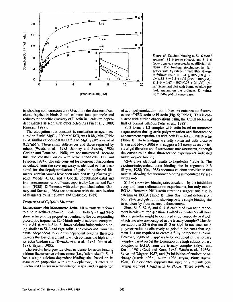
Calcium Binding Calcium binding values in the presence of 50-200 pmol of mutants were increased to between 10,000 and 35,000 counts/min compared with controls of 7,000. Table II shows the calcium-binding stoichiometry for fxgelsolin and the four mutants, fxgelsolin and $2-6 binds 2 mol of calcium each and S1-3 and $4-6 1 mol. Because S1,4-6 gave an inter- mediate value using the rapid binding assay, equilibrium di- alysis was carried out to assess the binding affinity (Fig. 13).
The binding stoichiometries of the three mutants tested (to- gether with Ko values in parenthesis from nonlinear least squares fit) were as follows: $4-6 = 1.24 (0.8 #M); $2-6 = 2.3 (0.55 #M); S1,4-6 = 1.67 (0.8 #M).
Discussion
Properties of PuriJied fxgelsolin
fxgelsolin is active in the cell supernatant as judged by the DNase I inhibition assay. It behaves like human platelet gel- solin (Bryan and Kurth, 1984; Kurth and Bryan, 1984), bo- vine plasma gelsolin (Cou6 and Korn, 1985), and pig stom- ach gelsolin (Hinssen, H., A. G. Weeds, unpublished data)
Table II. Calcium Binding to fxgelsolin and Deletion Mutants
Different Total Mean value P ro t e in p repara t ions samples SD (mol/mol protein)
n n
Fxgelsolin 7 36 0.36 1.96 $2-6 6 30 0.30 1.91 $4-6 9 42 0.19 0.99 S1-3 5 20 0.05 0.86 SI,4-6 8 36 0.16 1.61
Way et al. Gelsolin Mutagenesis in E. coli 601
_ _ _ _ _ . o i i c
2,0 S2-6 0 0
1.6 ~ n l I r"
O [ ] 81,4-6 "R
1.2 o [] s4-6 • • II
~ 0.8 2
.
m 0.4 1 = " " " "
0 0 0.1 0.2 0.3
0 2 4 6 8 10 I/
20
[Free calcium] (I.tM)
Figure 13. Calcium binding to $4-6 (solid squares), $2-6 (open circles), and S1,4-6 (open squares) measured by equilibrium di- alysis. The binding stoichiometries (to- gether with K0 values in parenthesis) were as follows: $4-6 = 1.24 5:0.05 (0.8 + 0.1 /~M); $2-6 = 2.3 + 0.06(0.55 + 0.05/~M); SI,4-6 = 1.67 -t- 0.07 (0.08 + 0.1/LM). (In- set) Scatchard plot with bound calcium per mole mutant on the ordinate. K0 values were ~0.6/LM in every case.
by showing no interaction with G-actin in the absence of cal- cium. fxgelsolin binds 2 mol calcium ions per mole and reduces the specific viscosity of F-actin in a calcium-depen- dent manner as seen with other gelsolins (Yin et al., 1980; Hinssen, 1987).
The elongation rate constant in nucleation assays, mea- sured in 2 mM MgCI2, 100 mM KCI, was 0.18/#M/s (Table I). A similar experiment using 5 mM MgCI2 gave a value of 0.22/#M/s. These small differences and those reported by others (Weeds et al., 1985; Janmey and Stossel, 1986; Carlier and Pantaloni, 1988) are not unexpected, because this rate constant varies with ionic conditions (Doi and Frieden, 1984). The rate constant for monomer dissociation calculated from the severing assay is identical to that mea- sured for the depolymerization of gelsolin-nucleated fila- ments. Similar values have been obtained using plasma gel- solin (Weeds, A. G., and J. Gooch, unpublished data) and from measurements of off-rates reported by Carlier and Pan- taloni (1988). Differences with other published values (Jan- mey and Stossel, 1986) are consistent with the stabilization of filaments by salt (Bryan and Coluccio, 1985).
Properties o f Gelsolin Mutants
Interactions with Monomeric Actin. All mutants were found to bind to actin-Sepharose in calcium. Both S1-3 and $4-6 show actin binding properties identical to the corresponding proteolytic fragments. $2-6 binds only in calcium, compara- ble to $4-6, while S1,4-6 shows calcium-independent bind- ing similar to S1-3 and fxgelsolin. The conversion from cal- cium-independent to calcium-dependent binding therefore mirrors the loss of segment 1, which contains the high affin- ity actin binding site (Kwiatkowski et al., 1985; Yin et al., 1988, Bryan, 1988).
The results here provide clear evidence for actin binding without fluorescence enhancement (i.e., "silent sites"). $4-6 has a single calcium-dependent binding site, based on its association properties with actin-Sepharose, its effects on F-actin and G-actin in sedimentation assays, and its inhibition
of actin polymerization, but it does not enhance the fluores- cence of NBD-actin or PI-actin (Fig. 6; Table I). This is con- sistent with earlier observations using the COOH-terminal half of plasma gelsolin (Way et al., 1988).
S1-3 forms a 1:2 complex with actin based on monomer sequestration during actin polymerization and fluorescence enhancement experiments with both PI-actin and NBD-actin (Table I). These findings are fully consistent with those of Bryan and Hwo (1986) who suggest a 1:2 complex on the ba- sis of gel filtration and fluorescence measurements, although the curvature in their fluorescence experiments indicated much weaker binding.
$2-6 gives identical results to fxgelsolin (Table I). The calcium-independent actin binding site in segments 2-3 (Bryan, 1988; Yin, 1988) becomes calcium sensitive in this mutant, showing that monomer binding is modulated by seg- ments 4-6.
S1,4-6 shows two binding sites in calcium by the inhibition assay and from sedimentation experiments, but only one in EGTA. However, NBD-actin titrations suggest one site in calcium or EGTA (Table I). Thus this mutant differs from both $2-6 and gelsolin in showing only a single binding site in calcium by fluorescence enhancement.
Since S1-3, $2-6, and S1,4-6 each bind two actin mono- mers in calcium, the question is raised as to whether all three sites in gelsolin might be occupied simultaneously or if not, which two sites are occupied in the ternary complex? The ob- servation that $2-6 (but not S1-3 or S1,4-6) nucleates actin polymerization as effectively as gelsolin indicates that seg- ment 1 is not required to create a fully competent nucleus. However, segment 1 appears to be occupied in the ternary complex based on (a) the formation of a high affinity binary complex in EGTA from the ternary complex (Bryan and Kurth, 1984; Cou6 and Korn, 1985; Weeds et al., 1986b; Selve and Wegner, 1987) and (b) inhibition of nucleotide ex- change (Harris, 1985; Tellam, 1986; Bryan, 1988; Harris, 1988). Our evidence supports this since only mutants con- taining segment 1 bind actin to EGTA. These results can
The Journal of Cell Biology, Volume 109, 1989 602

most easily be reconciled if the actin monomer bound to seg- ments 4-6 is also associated with segment 1.
The stoichiometry of actin binding by $2-6 in calcium suggests that the fluorescently silent site in $4-6 is occupied first, i.e., binding is cooperative (see Weeds et al., 1986b, for details of the arguments). By contrast the two binding sites in S1,4-6 are not coupled, since NBD-actin fluores- cence shows only a single site, corresponding to segment 1. Cooperative binding by gelsolin has been reported elsewhere (Janmey et al., 1986; Selve and Wegner, 1987). Thus it ap- pears that in the absence of calcium the high affinity sites are inaccessible to G-actin: calcium facilitates monomer binding initially on segments 4-6 and subsequently at the other sites.
Interactions with F-Actin
The methods used here differ in ionic conditions and the ac- tin concentrations. Differences in results may therefore reflect the actin affinities of the mutants and or differences in stabil- ity of the F-actin. Before considering the results, it is impor- tant to appreciate the differences between the methods.
Viscometry provides a very sensitive measure of severing because it is most strongly influenced by the longest fila- ments and these have the highest probability of being severed.
The DNase inhibition assay is carried out in 15 % glycerol, conditions under which actin filaments do not depolymerize below the critical concentration (Harris et al., 1982). Were depolymerization to occur, the extent of DNase inhibition would be much greater than that observed: an inhibitory ac- tivity equivalent to two actin monomers is produced by gel- solin (Weeds et al., 1986b). The most plausible explanation of this stoichiometry is that DNase binds to the two actin subunits at the pointed ends of filaments and is thereby in- hibited. In support of this Podolski and Steck (1988) have shown that DNase I binds to the pointed ends of protofila- ments in red cell cytoskeletons.
The severing assay measures the number concentration of free pointed ends. Because gelsolin severs filaments very rapidly and caps their barbed with high affinity (Selve and Wegner, 1986), there is a 1:1 correlation between the gelsolin concentration and number of filaments. The agreement in values of k - between severing assays and control depoly- merization experiments with nucleated filaments supports this conclusion. A lower activity may reflect weaker binding affinity, a defective severing mechanism or reduced mono- mer dissociation rate constant (e.g., actin filaments are stabi- lized by tropomyosin [Bernstein and Bamburg, 1982]).
$1-3 and S4-(x All assays show that these mutants behave identically to the NH2- and COOH-terminal proteolytic frag- ments of gelsolin (Kwiatkowski et al., 1985; Chaponnier et al., 1986; Bryan, 1988; Yin et al., 1988).
$2-~ Viscometric analysis suggests that $2-6 severs illa- ments in a calcium-dependent manner, but the magnitude of the effect is small (e.g., the fall in viscosity using at 1:200 mutant/actin ratio, where the assay is most sensitive, is only 36% the value obtained with gelsolin). The severing assay also suggests an activity <20% that of fxgelsolin (Fig. 11). Sedimentation experiments in calcium also showed binding to filaments and a time-dependent appearance of both actin and $2-6 in the supernatant. Electron microscopy showed a calcium-sensitive time-dependent reduction in filament length and prolonged incubation gave short filaments of 19
+ 10 monomers long at 1:1 molar ratios of S2-6/actin. The most striking effect of $2-6 was to increase the diameter of the filaments (Fig. 9), indicating lateral association along their entire length. The calcium dependence seen in the in- teraction of $2-6 with G-actin does not exist with F-actin, based on sedimentation and electron microscopy in EGTA. One possible explanation is that the affinity of the actin bind- ing site in segments 2-3 for F-actin subunits is much higher than that for G-actin monomers (Bryan, 1988; Yin et al., 1988).
These results show that $2-6 has a high affinity for the sides of filaments and that severing occurs relatively slowly as compared to gelsolin. One property of this mutant that may be relevant in understanding its severing mechanism is that it caps filament ends. This is based on its nucleating ac- tivity, the absence of reannealing of shortened filaments, and an inhibitory activity of elongation of actin protofilaments in red cell ghosts 77% that of gelsolin (unpublished observa- tions using the method of Pinder et al., [1986]). Association to the sides of filaments may affect their flexibility and facili- tate the formation of a cap, i.e., fragmentation. Such "capping-induced severing" is very different from the activity of gelsolin, because it occurs much more slowly and not at every binding site.
$1,4-(x All the assays show that S1,4-6 lacks the potent severing activity of gelsolin. Viscometry and the severing as- say suggest even lower activities than $2-6. One striking contrast between $2-6 and S1,4-6 is in the sedimentation as- say, in which $2-6 appears with Foactin in the pellet while S1,4-6 is always in the supernatant (complexed with two ac- tin monomers in calcium and one in EGTA). Thus S1,4-6 does not bind strongly to filaments. Nevertheless, electron microscopy showed clear evidence of shortened filaments at S1,4-6/actin ratios 1:50 or 1:20 and at higher ratios filaments disappeared almost instantaneously. However, when capped filaments were incubated with S1,4-6 they were more refrac- tile. This was clearly evident in electron microscopy, but also apparent in both viscometric and DNase inhibition assays. We are unable at present to explain these observations but monomer sequestration alone cannot account for the 20% reduction in specific viscosity when this mutant is added to F-actin at 1:100 molar ratio. Nor can it explain the results of the severing assay (Fig. 11) which is carried out below the critical concentration.
Localization of Calcium Binding Sites
Gelsolin binds 2 mol calcium per mole protein. One binding site is fully contained within segments 4-6; the other in seg- ments 1-3 (Table II). Binding studies with $2-6 suggest that the site in S1-3 is localized in segments 2-3. However, S1,4-6 gives an intermediate value of 1.6-1.7 both in the rapid assay and in equilibrium dialysis (Fig. 13). Binding affinities ap- pear to be identical at all sites based on Ka values of 0.6-0.8 /~M from equilibrium dialysis (similar to those previously reported for gelsolin [Weeds et al., 1986a]). The apparent paradox in these observations may be resolved if the addi- tional binding site in segment 1 of S1,4-6 corresponds to the cryptic binding site identified in a COOH-terminal truncated mutant containing the first 160 residues of gelsolin (Kwiat- kowski et al., 1989).
Way et al. Gelsolin Mutagenesis in E. coli 603
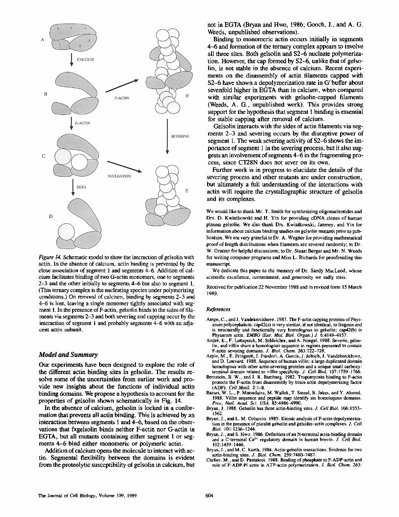
Figure 14. Schematic model to show the interaction of gelsolin with actin. In the absence of calcium, actin binding is prevented by the close association of segment 1 and segments 4-6. Addition of cal- cium facilitates binding of two G-actin monomers, one to segments 2-3 and the other initially to segments 4-6 but also to segment 1. (This ternary complex is the nucleating species under polymerizing conditions.) On removal of calcium, binding by segments 2-3 and 4-6 is lost, leaving a single monomer tightly associated with seg- ment 1. In the presence of F-actin, geisolin binds to the sides of illa- ments via segments 2-3 and both severing and capping occur by the interaction of segment 1 and probably segments 4-6 with an adja- cent actin subunit.
Model and Summary
Our experiments have been designed to explore the role of the different actin binding sites in gelsolin. The results re- solve some of the uncertainties from earlier work and pro- vide new insights about the functions of individual actin binding domains. We propose a hypothesis to account for the properties of gelsolin shown schematically in Fig. 14.
In the absence of calcium, gelsolin is locked in a confor- mation that prevents all actin binding. This is achieved by an interaction between segments 1 and 4-6, based on the obser- vations that fxgelsolin binds neither F-actin nor G-actin in EGTA, but all mutants containing either segment 1 or seg- ments 4-6 bind either monomeric or polymeric actin.
Addition of calcium opens the molecule to interact with ac- tin. Segmental flexibility between the domains is evident from the proteolytic susceptibility of gelsolin in calcium, but
not in EGTA (Bryan and Hwo, 1986; Gooch, J., and A. G. Weeds, unpublished observations).
Binding to monomeric actin occurs initially in segments 4-6 and formation of the ternary complex appears to involve all three sites. Both gelsolin and $2-6 nucleate polymeriza- tion. However, the cap formed by $2-6, unlike that of gelso- lin, is not stable in the absence of calcium. Recent experi- ments on the disassembly of actin filaments capped with $2-6 have shown a depolymerization rate in G' buffer about sevenfold higher in EGTA than in calcium, when compared with similar experiments with gelsolin-capped filaments (Weeds, A. G., unpublished work). This provides strong support for the hypothesis that segment I binding is essential for stable capping after removal of calcium.
Gelsolin interacts with the sides of actin filaments via seg- ments 2-3 and severing occurs by the disruptive power of segment 1. The weak severing activity of $2-6 shows the im- portance of segment 1 in the severing process, but it also sug- gests an involvement of segments 4-6 in the fragmenting pro- cess, since CT28N does not sever on its own.
Further work is in progress to elucidate the details of the severing process and other mutants are under construction, but ultimately a full understanding of the interactions with actin will require the crystallographic structure of gelsolin and its complexes.
We would like to thank Mr. T. Smith for synthesizing oligonucleotides and Drs. D. Kwiatkowski and H. Yin for providing cDNA clones of human plasma gelsolin. We also thank Drs. Kwiatkowski, Janmey, and Yin for information about calcium binding studies on gelsolin mutants prior to pub- lication. We are very grateful to Dr. A. Wegner for providing mathematical proof of length distributions when filaments are severed randomly; to Dr. W. Gratzer for helpful discussions; to Dr. Stuart Berger and Mr. N. Weeds for writing computer programs and Miss L. Richards for proofreading this manuscript.
We dedicate this paper to the memory of Dr. Sandy MacLeod, whose scientific excellence, commitment, and generosity we sadly miss.
Received for publication 22 November 1988 and in revised form 15 March 1989.
References
Ampe, C., and J. Vandekerckhove. 1987. The F-actin capping proteins of Phys- arnm polycephalum: cap42(a) is very similar, if not identical, to fragmin and is structurally and functionally very homologous to gelsolin; cap42(b) is Physarum actin. EMBO (Eur. Mol. Biol. Organ.) J. 6:4149-4157.
Andre, E., F. Louspeich, M. Schleicher, and A. Noegel. 1988. Severin, gelso- lin, and villin share a homologous sequence in regions presumed to contain F-actiu severing domains. J. Biol. Chem. 263:722-728.
Arpin, M., E. Pringau|t, J. Finidori, A. Garcia, J. Jeltsch, J. Vandekerckhove, and D. Louvard. 1988. Sequence of human villin: a large duplicated domain homologous with other actin-severing proteins and a unique small carboxy- terminal domain related to villin specificity. J. Cell Biol. 107:1759-1766.
Bernstein, B. W., and J. R. Bamburg. 1982. Tropomyosin binding to F-actin protects the F-actin from disassembly by brain actin depolymerizing factor (ADF). Cell Motil. 2:1-8.
Bazari, W. L., P. Matsudaira, M. Wailek, T. Smeal, R. Jakes, and Y. Ahmed. 1988. Villin sequence and peptide map identify six homologous domains. Proc. Natl. Acad. Sci. USA. 85:4986-4990.
Bryan, J. 1988. Gelsolin has three actin-binding sites. J. Cell Biol. 106:1553- 1562.
Bryan, J., and L. M. Coluccio. 1985. Kinetic analysis of F-actin depolymeriza- tion in the presence of platelet gelsolin and gelsolin-actin complexes. J. Cell Biol. 101:1236-1244.
Bryan, J., and S. Hwo. 1986. Definition of an N-terminal actin-binding domain and a C-terminal Ca 2+ regulatory domain in human brevin. J. Cell Biol. 102:1439-1446.
Bryan, J., and M. C. Kurth. 1984. Actin-gelsolin interactions. Evidence for two actin-binding sites. J. Biol. Chem. 259:7480-7487.
Carlier, M., and D. Pantaloni. 1988. Binding of phosphate to F-ADP-actin and role of F-ADP-Pi-actin in ATP-actin polymerization. J. Biol. Chem. 263:
The Journal of Cell Biology, Volume 109, 1989 604

817-825. Carter, P., H. Bedouelle, and G. Winter. 1985. Improved oligonucleotide site-
directed mutagenesis using M i 3 vectors. Nucleic Acids Res. 13:4431-4443. Chaponnier, C., P. A. Janmey, and H. L. Yin. 1986. The actin filament-sever-
ing domain of plasma gelsolin. J. Cell Biol. 103:1473-1481. Con6, M., and E. D. Korn. 1985. Interaction of plasma gelsolin with G-actin
and F-actin in the presence and absence of calcium ions. J. Biol. Chem. 260:15033-15041.
Doi, Y., and C. Frieden. 1984. Actin polymerization. The effect of hrevin on filament size and rate of polymerization. J. Biol. Chem. 259: I 1868-11875.
Harafugi, H., and Y. Ogawa, 1980. Re-examination of the apparent binding constant of EGTA with calcium around neutral pH. J. Biochem. (Tokyo). 87:1305-1312.
Harris, H. E. 1988. The binary complex of pig plasma gelsolin with Mg2 +-G- actin in ATP and ADP. FEBS (Fed. Fur. Biochem. Soc.) Lett. 233:359-362.
Harris, H. E. 1985. Lack of nucleotide exchange on the binding of G-actin-ATP to plasma gelsolin. FEBS (Fed. Eur. Biochem. Soc.) Lett. 190:81-83.
Harris, H. E., and J. Gooch. 1981. An actin depolymerizing protein from pig plasma. FEBS (Fed. Eur. Biochem. Soc.) Lett. 123:49-53.
Harris, H. E., and A. G. Weeds. 1983. Plasma actin depolymerizing factor has both calcium-dependent and calcium-independent effects on actin. Biochem- istry. 22:2728-2741.
Harris, H. E., J. R. Bamburg, B. W. Bernstein, and A. G. Weeds. 1982. The depolymerization of actin by specific proteins from plasma and brain: a quan- titative assay. Anal. Biochem. 119:102-114.
Hinssen, H. 1987. Actin-modulating proteins. Complex formation and calcium dependence of interaction with actin. Wohlfarth-Bottermann (Hsrg): Nature and function of cytoskeletal protein in motility and transport. Prog. Zool. 34:53-63.
Janmey, P. A., and T. P. Stossel. 1986. Kinetics of actin monomer exchange at the slow growing ends of actin filaments and their relationship to the elon- gation of filaments shortened by gelsolin. J. Muscle Res. Cell Motil. 7:446-454.
Janmey, P. A., T. P. Stossel, and S. E. Lind. 1986. Sequential binding of actin monomers to plasma gelsolin and its inhibition by vitamin D-binding protein. Biochem. Biophys. Res. Commun. 136:72-79.
Koch, G., M. Smith, D. Macer, P. Webster, and R. Mortara. 1986. Endoplas- mic reticulum contains a common, abundant calcium-binding glycoprotein, endoplasmin. J. Cell Sci. 86:217-232.
Kurth, M. C., and J. Bryan. 1984. Platelet activation induces the formation of a stable gelsolin-actin complex from monomeric gelsolin. J. Biol. Chem. 259:7473-7479.
Kwiatkowski, D. J., P. A. Janmey, and H. L. Yin. 1989. Identification of criti- cal functional and regulatory domains in gelsolin. J. Cell Biol. 108:1717- 1726.
Kwiatkowski, D. J., P. A. Janmey, J. E. Mole, and H. L. Yin. 1985. Isolation and properties of two actin-binding domains in gelsolin. J. Biol. Chem. 260:15232-15238.
Kwiatkowski, D. J., T. P. Stossel, S. H. Orkin, J. E. Mole, H. R. Colten, and H. L. Yin. 1986. Plasma and cytoplasmic gelsolins are encoded by a single gene and contain a duplicated actin-binding domain. Nature (Lond.). 323: 455-458.
Kwiatkowski, D. J., R. Mehl, S. Izumo, N. Nadal-Ginard, and H. L. Yin. 1988. Muscle is the major source of plasma gelsolin. J. Biol. Chem. 263: 8239-8243.
Maniatis, T., E. F. Fritsch, and J. Sambrook. 1982. Molecular Cloning: A Lab- oratory Manual. Cold Spring Harbor Laboratory, Cold Spring Harbor, NY. 507 pp.
Matsudaira, P. 1987. Sequence from picomole quantities of proteins electro- blotted onto polyvinylidene difluoride membranes. J. Biol. Chem. 262: 10035 - 10038.
Nagai, K., and H. C. Thegersen. 1987. Synthesis and sequence-specific pro- teolysis of hybrid proteins produced in Escherichia coli. Methods EnzymoL 153:461--481.
Nagai, K., and H. C. Thegersen. 1984. Generation of B-globin by sequence specific proteolysis of a hybrid protein produced in Escherichia coli. Nature (Lond.). 309:810-812.
Oosawa, F., and S. Asakura. 1975. Thermodynamics of the Polymerization of Proteins. Academic Press, London. 51-54.
Pinder, J. C., A. G. Weeds, and W. B. Gratzer. 1986. Study ofactin filaments in the human red cell membrane. J. MoL Biol. 191:461--468.
Podolski, J. L., and T. L. Steck. 1988. Association of deoxyribonuclease I with the pointed ends of actin filaments in human red blood cell membrane skele- tons. J. Biol. Chem. 263:638-645.
Pope, B., and A. G. Weeds. 1986. Binding of pig plasma geisolin to F-actin and partial fractionation into calcium-dependent and calcium-independent forms. Eur. J. Biochem. 161:85-93.
Press, W. H., B. P. Flannery, S. A. Teukolsky, and W. T. VeRerling. 1988. Numerical recipes in C. Cambridge University Press. 305-309.
Sanger, F., S. Nicklen, and A. R. Coulson. 1977. DNA sequencing with chain terminating inhibitors. Proc. Natl. Acad. Sci. USA. 74:5463-5467.
Selve, N., and A. Wegner. 1987. pH-dependent rate of formation of the gel- solin-actin complex from gelsolin and monomeric actin. Fur. J. Biochem. 161:111-115.
Selve, N., and A. Wegner. 1986. Rate oftreadmilling ofactin filaments in vitro. J. Mol. Biol. 187:627-631.
Tellam, R. L. 1986. Gelsolin inhibits nucleotide exchange from actin. Biochem- istry. 25:5799-5804.
Valentine, R. C., B. M. Shapiro, and E. R. Stadtman. 1968. Regulation of glutamine synthetase XII: electron microscopy of the enzyme from E. Coli. Biochemistry. 7:2143-2152.
Walsh, T. P., A. Weber, K. David, E. Bonder, and M. Mooseker. 1984. Cal- cium dependence of villin-induced actin depolymerization. Biochemistry. 23:6099-6102.
Way, M., and A. G. Weeds. 1988. Nucleotide sequence of pig plasma gelsolin. Comparison of protein sequence with human gelsolin and other actin- severing proteins shows strong homologies and evidence for large internal repeats. J. Mol. Biol. 203:1127-1133.
Way, M., J. Gooch, B. Pope, and A. G. Weeds. 1988. Regulation of actin fila- ment assembly and cytoskeletal structure by capping proteins. In Structure and Functions of the Cytoskeleton, Biological and Physiopatbological As- pects. B. Rousset, editor. Inserm-John Libbey Eurotext. 171:401-413.
Weeds, A. G., J. Gooch, B. P. Pope, and H. E. Harris. 1986a. Preparation and characterization of pig plasma and platelet gelsolins. Eur. J. Biochem. 161:69-76.
Weeds, A. G., H. Harris, W. B. Gratzer, and J. Gooch. 1986b. Interactions of pig plasma gelsolin with G-actin. Eur. J. Biochem. 161:77-84.
Weeds, A. G., H. E. Harris, J. Gooch, and B. Pope. 1985. Actin capping pro- teins. Hormones and Cell Regulation. Inserm European Symposium. J. E. Dumont, B. Hamprecht and J. Nunez, editors. Elsevier Science Publishing Co., Inc., New York. 9:143-165.
Yin, H. L. 1988. Gelsolin: calcium and polyphosphoinositide-regulated actin- modulating protein. Bioessays. 7:176-179.
Yin, H. L., J. H. Hartwig, K. Maruyama, and T. P. Stossel. 1981. Ca2+ con- trol of actin filament length. Effects of macrophage gelsolin on actin poly- merization. J. BioL Chem. 256:9693-9697.
Yin, H. L., K. lida, and P. A. Janmey. 1988. Identification ofa polyphospho- inositide-modulated domain in gelsolin which binds to the sides of actin fila- ments. J. Cell Biol. 106:805-812.
Yin, H. L., K. S. Zaner, and T. P. Stossel. 1980. Calcium control of actin gela- tion. J. Biol. Chem. 255:9494-9500.
Way et al. Gelsolin Mutagenesis in E. coli 605
Copyright © 2022 FDOKUMEN