The ALS8 protein VAPB interacts with the ER–Golgi recycling protein YIF1A and regulates membrane...
17
The ALS8 protein VAPB interacts with the ER–Golgi recycling protein YIF1A and regulates membrane delivery into dendrites Marijn Kuijpers 1,2 , Ka Lou Yu 1,2 , Eva Teuling 2 , Anna Akhmanova 1 , Dick Jaarsma 2 and Casper C Hoogenraad 1,2, * 1 Division of Cell Biology, Department of Biology, Faculty of Science, Utrecht University, Utrecht, The Netherlands and 2 Department of Neuroscience, Erasmus Medical Center, Rotterdam, The Netherlands The vesicle-associated membrane protein (VAMP) asso- ciated protein B (VAPB) is an integral membrane protein localized to the endoplasmic reticulum (ER). The P56S mutation in VAPB has been linked to motor neuron degen- eration in amyotrophic lateral sclerosis type 8 (ALS8) and forms ER-like inclusions in various model systems. However, the role of wild-type and mutant VAPB in neu- rons is poorly understood. Here, we identified Yip1-inter- acting factor homologue A (YIF1A) as a new VAPB binding partner and important component in the early secretory pathway. YIF1A interacts with VAPB via its transmem- brane regions, recycles between the ER and Golgi and is mainly localized to the ER–Golgi intermediate compart- ments (ERGICs) in rat hippocampal neurons. VAPB strongly affects the distribution of YIF1A and is required for intracellular membrane trafficking into dendrites and normal dendritic morphology. When VAPB-P56S is pre- sent, YIF1A is recruited to the VAPB-P56S clusters and loses its ERGIC localization. These data suggest that both VAPB and YIF1A are important for ER-to-Golgi transport and that missorting of YIF1A may contribute to VAPB- associated motor neuron disease. The EMBO Journal advance online publication, 4 June 2013; doi:10.1038/emboj.2013.131 Subject Categories: membranes & transport; neuroscience Keywords: amyotrophic lateral sclerosis (ALS); dendrite morphology; secretory pathway; VAMP-associated protein (VAP); Yip1 interacting factor (YIF1) Introduction The secretory pathway is responsible for the delivery of a variety of protein and lipid cargo, such as ion channels, receptors and lipid membranes, to their proper location. In the early secretory pathway, newly synthesized proteins that pass the quality control of the endoplasmic reticulum (ER) concentrate at specialized ER subdomains called the ER exit sites (ERES) (Lee et al, 2004; Dancourt and Barlowe, 2010). From here, cargo leaves the ER and is transported to the Golgi compartments to undergo further processing, sorting and subsequent delivery to the plasma membrane or other cellular organelles. ER-derived vesicles deliver their cargo either directly to the cis-Golgi membrane or pass a specialized membrane compartment called the ER–Golgi intermediate compartment (ERGIC) (Watson and Stephens, 2005; Appenzeller-Herzog and Hauri, 2006; Saraste et al, 2009; Lorente-Rodriguez and Barlowe, 2011). Whereas the early secretory pathway and its organization is well studied in a variety of cellular model systems such as yeast and fibroblasts, little information is available about this trafficking pathway in neuronal cells. A number of studies have demonstrated the presence of ER-to-Golgi transport components in neurons (Krijnse-Locker et al, 1995; Pierce et al, 2001; Horton and Ehlers, 2003) and indicated the importance of the early secretory trafficking for neuronal development and morphogenesis (Hanus and Ehlers, 2008; Tang, 2008; Aridorand Fish, 2009; Jan and Jan, 2010). Members of the highly conserved VAP family, including the mammalian vesicle-associated membrane protein associated protein A (VAPA) and VAPB proteins, are present in the ER and have been proposed to play a role in maintaining Golgi complex identity, ER morphology, lipid transfer and in reg- ulating ER and Golgi transport (Soussan et al, 1999; Skehel et al, 2000; Pennetta et al, 2002; Amarilio et al, 2005; Teuling et al, 2007; Lev et al, 2008; Peretti et al, 2008; Tsuda et al, 2008; Han et al, 2012). VAP proteins contain an N-terminal domain, which is highly homologous to the nematode major sperm protein (MSP), a central domain that forms a coiled- coil structure and the C-terminal transmembrane tail domain (Nishimura et al, 1999; Kaiser et al, 2005). Interest in VAP proteins has greatly increased after the discovery of a dominant missense mutation P56S in the MSP domain of VAPB, which causes a familial motor neuron disease, designated amyotrophic lateral sclerosis type 8 (ALS8) (Nishimura et al, 2004). P56S mutant VAPB accumulates in inclusions that contain abnormal organized ER (Teuling et al, 2007; Papiani et al, 2012) and recruit wild-type VAPA/B in these inclusions, suggesting that VAPB-P56S could have a dominant-negative effect on VAP function (Teuling et al, 2007). Indeed, a loss-of-function mechanism has been observed in several model systems (Chai et al, 2008; Ratnaparkhi et al, 2008; Tsuda et al, 2008; Suzuki et al, 2009; Forrest et al, 2013). Moreover, a reduction in VAP protein levels has been described in sporadic ALS patients, SOD1 mutant mice as well as ALS-derived motor neurons (Teuling et al, 2007; Anagnostou et al, 2010; Mitne-Neto et al, 2011). However, mechanisms leading to VAPB-linked motor neuron degeneration are still poorly understood. To gain moreinsight into the role of VAPB in neurons, we searched for new binding partners of VAPB. We identified *Corresponding author. Cell Biology, Faculty of Science, Utrecht University, Padualaan 8, 3584 CH Utrecht, The Netherlands. Tel.: þ31 30 2534585; Fax: þ31 30 2532837; E-mail [email protected] Received: 2 November 2012; accepted: 7 May 2013 The EMBO Journal (2013), 1–17 www.embojournal.org EMBO THE EMBO JOURNAL THE EMBO JOURNAL 1 & 2013 European Molecular Biology Organization The EMBO Journal
Transcript of The ALS8 protein VAPB interacts with the ER–Golgi recycling protein YIF1A and regulates membrane...

The ALS8 protein VAPB interacts with theER–Golgi recycling protein YIF1A and regulatesmembrane delivery into dendrites
Marijn Kuijpers1,2, Ka Lou Yu1,2,Eva Teuling2, Anna Akhmanova1,Dick Jaarsma2 and CasperC Hoogenraad1,2,*1Division of Cell Biology, Department of Biology, Faculty of Science,Utrecht University, Utrecht, The Netherlands and 2Department ofNeuroscience, Erasmus Medical Center, Rotterdam, The Netherlands
The vesicle-associated membrane protein (VAMP) asso-
ciated protein B (VAPB) is an integral membrane protein
localized to the endoplasmic reticulum (ER). The P56S
mutation in VAPB has been linked to motor neuron degen-
eration in amyotrophic lateral sclerosis type 8 (ALS8) and
forms ER-like inclusions in various model systems.
However, the role of wild-type and mutant VAPB in neu-
rons is poorly understood. Here, we identified Yip1-inter-
acting factor homologue A (YIF1A) as a new VAPB binding
partner and important component in the early secretory
pathway. YIF1A interacts with VAPB via its transmem-
brane regions, recycles between the ER and Golgi and is
mainly localized to the ER–Golgi intermediate compart-
ments (ERGICs) in rat hippocampal neurons. VAPB
strongly affects the distribution of YIF1A and is required
for intracellular membrane trafficking into dendrites and
normal dendritic morphology. When VAPB-P56S is pre-
sent, YIF1A is recruited to the VAPB-P56S clusters and
loses its ERGIC localization. These data suggest that both
VAPB and YIF1A are important for ER-to-Golgi transport
and that missorting of YIF1A may contribute to VAPB-
associated motor neuron disease.
The EMBO Journal advance online publication, 4 June 2013;
doi:10.1038/emboj.2013.131Subject Categories: membranes & transport; neuroscienceKeywords: amyotrophic lateral sclerosis (ALS); dendrite
morphology; secretory pathway; VAMP-associated protein
(VAP); Yip1 interacting factor (YIF1)
Introduction
The secretory pathway is responsible for the delivery of a
variety of protein and lipid cargo, such as ion channels,
receptors and lipid membranes, to their proper location. In
the early secretory pathway, newly synthesized proteins that
pass the quality control of the endoplasmic reticulum (ER)
concentrate at specialized ER subdomains called the ER exit
sites (ERES) (Lee et al, 2004; Dancourt and Barlowe, 2010).
From here, cargo leaves the ER and is transported to the Golgi
compartments to undergo further processing, sorting and
subsequent delivery to the plasma membrane or other
cellular organelles. ER-derived vesicles deliver their cargo
either directly to the cis-Golgi membrane or pass a
specialized membrane compartment called the ER–Golgi
intermediate compartment (ERGIC) (Watson and Stephens,
2005; Appenzeller-Herzog and Hauri, 2006; Saraste et al,
2009; Lorente-Rodriguez and Barlowe, 2011). Whereas the
early secretory pathway and its organization is well studied in
a variety of cellular model systems such as yeast and
fibroblasts, little information is available about this
trafficking pathway in neuronal cells. A number of studies
have demonstrated the presence of ER-to-Golgi transport
components in neurons (Krijnse-Locker et al, 1995; Pierce
et al, 2001; Horton and Ehlers, 2003) and indicated the
importance of the early secretory trafficking for neuronal
development and morphogenesis (Hanus and Ehlers, 2008;
Tang, 2008; Aridor and Fish, 2009; Jan and Jan, 2010).
Members of the highly conserved VAP family, including the
mammalian vesicle-associated membrane protein associated
protein A (VAPA) and VAPB proteins, are present in the ER
and have been proposed to play a role in maintaining Golgi
complex identity, ER morphology, lipid transfer and in reg-
ulating ER and Golgi transport (Soussan et al, 1999; Skehel
et al, 2000; Pennetta et al, 2002; Amarilio et al, 2005; Teuling
et al, 2007; Lev et al, 2008; Peretti et al, 2008; Tsuda et al,
2008; Han et al, 2012). VAP proteins contain an N-terminal
domain, which is highly homologous to the nematode major
sperm protein (MSP), a central domain that forms a coiled-
coil structure and the C-terminal transmembrane tail domain
(Nishimura et al, 1999; Kaiser et al, 2005). Interest in VAP
proteins has greatly increased after the discovery of a
dominant missense mutation P56S in the MSP domain of
VAPB, which causes a familial motor neuron disease,
designated amyotrophic lateral sclerosis type 8 (ALS8)
(Nishimura et al, 2004). P56S mutant VAPB accumulates in
inclusions that contain abnormal organized ER (Teuling et al,
2007; Papiani et al, 2012) and recruit wild-type VAPA/B in
these inclusions, suggesting that VAPB-P56S could have a
dominant-negative effect on VAP function (Teuling et al,
2007). Indeed, a loss-of-function mechanism has been
observed in several model systems (Chai et al, 2008;
Ratnaparkhi et al, 2008; Tsuda et al, 2008; Suzuki et al,
2009; Forrest et al, 2013). Moreover, a reduction in VAP
protein levels has been described in sporadic ALS patients,
SOD1 mutant mice as well as ALS-derived motor
neurons (Teuling et al, 2007; Anagnostou et al, 2010;
Mitne-Neto et al, 2011). However, mechanisms leading to
VAPB-linked motor neuron degeneration are still poorly
understood.
To gain more insight into the role of VAPB in neurons, we
searched for new binding partners of VAPB. We identified
*Corresponding author. Cell Biology, Faculty of Science, UtrechtUniversity, Padualaan 8, 3584 CH Utrecht, The Netherlands.Tel.: þ31 30 2534585; Fax: þ31 30 2532837; E-mail [email protected]
Received: 2 November 2012; accepted: 7 May 2013
The EMBO Journal (2013), 1–17
www.embojournal.org
EMBO
THE
EMBOJOURNAL
THE
EMBOJOURNAL
1&2013 European Molecular Biology Organization The EMBO Journal

Yip1-interacting factor homologue A (YIF1A) as a new VAPB
binding partner that interacts with both wild-type VAPB and
mutant VAPB-P56S. YIF1A is the human orthologue of the
budding yeast Yif1p, a transmembrane protein that plays an
important role in early secretory transport in yeast and
mammalian cells (Matern et al, 2000; Barrowman et al,
2003; Jin et al, 2005; Yoshida et al, 2008). We find that
YIF1A recycles between the ER and Golgi and is mainly
localized to the ERGIC in hippocampal neurons. Both VAPB
and YIF1A are required for membrane trafficking to dendrites
and proper dendrite morphology. In addition, we show that
ALS8 mutant VAPB-P56S strongly disrupts the localization of
YIF1A to ERGIC. Our data suggest an important role for
YIF1A and VAPB in the early secretory pathway and we
propose that the missorting of YIF1A plays a role in VAPB-
associated motor neuron disease. These findings advance the
knowledge of fundamental trafficking processes in neuronal
cells and have important implications for our understanding
of neuronal degeneration.
Results
YIF1A interacts with both wild-type and mutant VAPB
To identify new VAPB interacting proteins, we performed
pull-down assays combined with mass spectrometry.
Biotinylated and GFP-tagged VAPB (bio-GFP-VAPB) and con-
trol bio-GFP constructs were transiently co-expressed in HeLa
cells together with the protein-biotin ligase BirA, isolated
with streptavidin beads and the proteins were analysed by
mass spectrometry. Bio-GFP-VAPB bound to several pre-
viously identified VAPB binding partners, such as the FFAT-
motif containing proteins NIR2, OSBPL3, OSBPL6 and
OSBPL9 (Wyles and Ridgway, 2004; Amarilio et al, 2005;
Lehto et al, 2005; Teuling et al, 2007) (Figure 1A). In addition,
a novel potential VAPB binding partner was identified, YIF1A,
a transmembrane protein of 293 amino acids and mammalian
orthologue of the Saccharomyces cerevisiae protein Yif1p
(Yip1p-interacting factor 1) (Matern et al, 2000). YIF1A and
its close homologue YIF1B are members of a large protein
family, named FinGERs, which share a common structure
with an N-terminal hydrophilic region, followed by conserved
transmembrane regions (Shakoori et al, 2003; Pfeffer and
Aivazian, 2004).
The interaction of VAPB and YIF1A was confirmed by
biotin pull-down experiments using extracts of HEK293T
cells overexpressing GFP-YIF1A and bio-HA-VAPB
(Figure 1B). Pull-down experiments also revealed binding
between YIF1B and VAPB (Figure 1B) and between VAPA and
both YIF homologues (Figure 1C). To further confirm the
interaction between VAPB and YIF1A, we performed immu-
nofluorescence experiments in COS-7 cells. HA-YIF1A
co-localized with both endogenous VAPB and co-transfected
myc-VAPB, which as previously demonstrated localize to the
ER (Nishimura et al, 2004; Kanekura et al, 2006; Teuling et al,
2007; Kim et al, 2010; Papiani et al, 2012) (Figure 1D and E).
Significantly, HA-YIF1A also co-distributes with ALS-linked
mutant VAPB-P56S and VAPA-P56S (Figure 1F and H), which
accumulates in small spherical inclusions (Nishimura et al,
2004; Kanekura et al, 2006; Teuling et al, 2007; Kim et al,
2010; Papiani et al, 2012). Likewise also YIF1B was recruited
to mutant VAPA/B inclusion (Figure 1G and I). Together,
these results show that YIF1A/B interacts with VAPA/B
family proteins.
The transmembrane domains of both VAPB and YIF1A
are required for their interaction
Secondary structure predictions indicate that YIF1A contains
four transmembrane domains at the C-terminus (Figure 2A)
(Altschul et al, 1997; Hirokawa et al, 1998), while the
N-terminus of the yeast homologue Yif1p has been shown
to face the cytosol (Matern et al, 2000). To confirm that the
N-terminus of YIF1A faces the cytosol, we generated a YIF1A
construct with a biotinylation tag at the N-terminus
(Figure 2B). Pull-down experiments showed that this con-
struct was biotinylated when the biotinylating enzyme BirA
was localized in the cytoplasm, but not by a variant BirA that
is localized in the ER lumen (Figure 2B).
To identify the regions through which VAPB interacts with
YIF1A, we first made truncated GFP-YIF1A constructs en-
coding either the N-terminus (1–131) or the C-terminal
transmembrane region (131–293) of YIF1A (Figure 2A).
Pull-down experiments using HEK293T cells co-expressing
HA-VAPB and the YIF1A deletion constructs showed that
VAPB only co-precipitates with YIF1A construct that con-
tained the transmembrane region (Figure 2C and D). These
data were confirmed by a glutathione S-transferase (GST)
pull-down assay with cell lysates expressing truncated
YIF1A constructs using VAPB and VAPB-P56S immobilized
on GST beads. The C-terminal part, but not the N-terminal
part of YIF1A co-precipitated with VAPB and VAPB-P56S
(Figure 2E). To characterize in more detail the YIF1A
domain that binds VAPB, we made additional truncated
YIF1A constructs, which contain only the first two trans-
membrane domains (131–198), the last two transmembrane
domains (198–298) or the N-terminal cytosolic domain with
the first two transmembrane domains (1–198) (Figure 2A).
Pull-down experiments with biotinylated VAPB showed that
all transmembrane containing YIF1A constructs co-precipi-
tated with VAPB (Figure 2F). However, VAPB brings down
considerable higher amounts of truncated GFP-YIF1A pro-
Figure 1 Interaction of YIF1A with wild-type and mutant VAPB. (A) Identification of wild-type VAPB binding partners by mass spectrometry inHeLa cell extract. The table shows proteins identified with a significant Mascot score in the pull-down with streptavidin beads from an extractof HeLa cells co-expressing Bio-GFP-VAPB and biotin ligase BirA. The list is corrected for background proteins, which were identified in acontrol pull-down from HeLa cells expressing bio-GFP. Abbreviations used in the table to indicate the identified proteins: OSBPL, oxysterolbinding protein-like; NIR, N-terminal domain-interacting receptor. (B) Biotin pull-downs (PD) from HEK293T extract transfected with Bio-HA-VAPB and GFP-YIF1A, GFP-YIF1B or control bio-GFP and probed for GFP and HA. (C) Biotin pull-downs from HEK293Textract transfected withBio-HA-VAPA and GFP-YIF1A or GFP-YIF1B and probed for GFP and HA. The ratio input/pellet is 2–5% for all pull-down and immunopre-cipitation experiments. (D) COS-7 cells transfected with HA-YIF1A and stained with anti-HA (green) and anti-VAPB (red) antibodies. (E, F)COS-7 cells double transfected with HA-YIF1A and myc-VAPB (D) or myc-VAPB-P56S (E) stained with anti-HA (green) and anti-myc (red)antibodies. (G) COS-7 cells double transfected with HA-VAPB-P56S and Flag-YIF1B, fixed and stained with anti-HA (green) and anti-Flag (red)antibodies. (H, I) COS-7 cells double transfected with myc-VAPA-P56S and HA-YIF1A (H) or HA-YIF1B (I) stained with anti-HA (green) andanti-myc (red) antibodies. Panels on the right side show enlargements of the boxed regions. Scale bar, 10mm.
VAPB and YIF1A function in the early secretory pathwayM Kuijpers et al
2 The EMBO Journal &2013 European Molecular Biology Organization
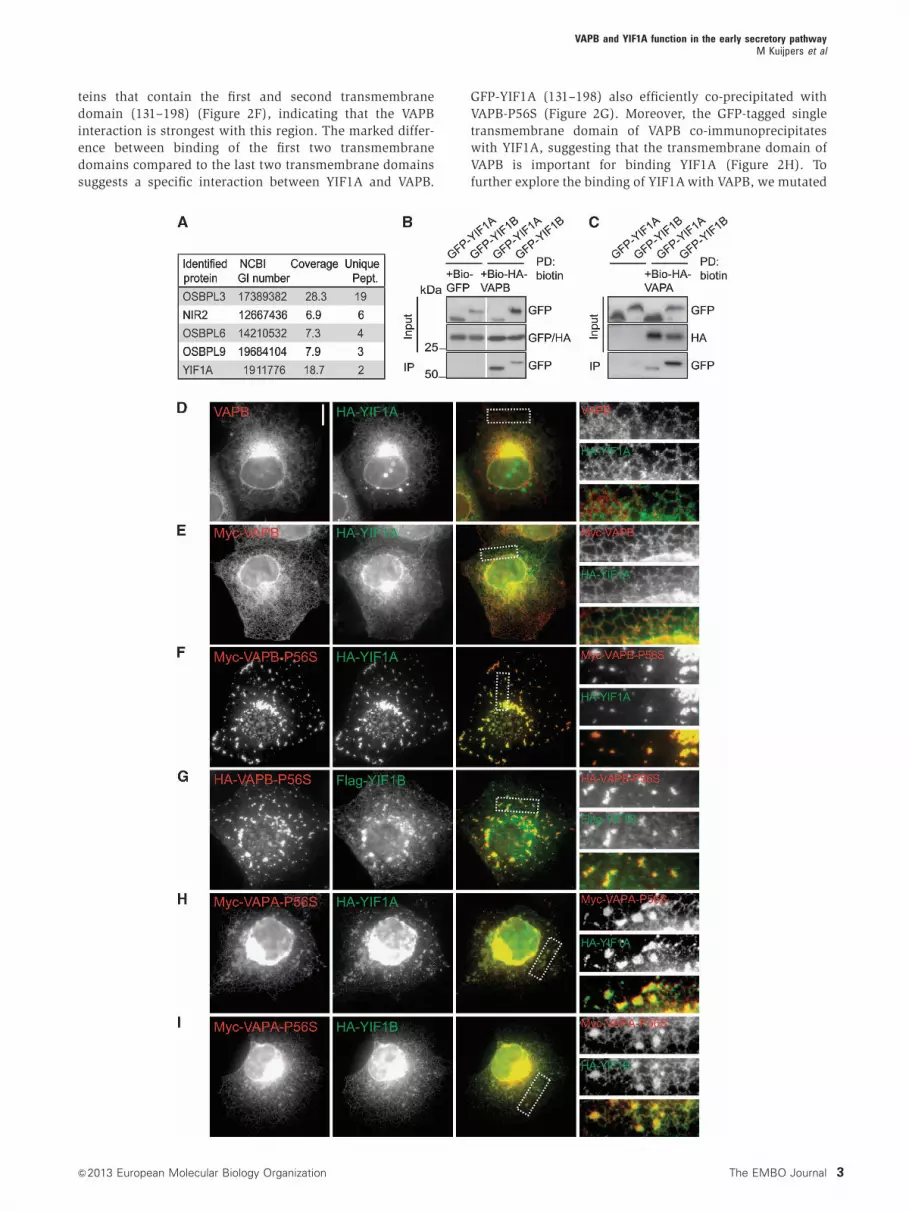
teins that contain the first and second transmembrane
domain (131–198) (Figure 2F), indicating that the VAPB
interaction is strongest with this region. The marked differ-
ence between binding of the first two transmembrane
domains compared to the last two transmembrane domains
suggests a specific interaction between YIF1A and VAPB.
GFP-YIF1A (131–198) also efficiently co-precipitated with
VAPB-P56S (Figure 2G). Moreover, the GFP-tagged single
transmembrane domain of VAPB co-immunoprecipitates
with YIF1A, suggesting that the transmembrane domain of
VAPB is important for binding YIF1A (Figure 2H). To
further explore the binding of YIF1A with VAPB, we mutated
VAPB and YIF1A function in the early secretory pathwayM Kuijpers et al
3&2013 European Molecular Biology Organization The EMBO Journal

two GxxxG motifs that are present in the first and
third transmembrane domain of YIF1A (Figure 2A). GxxxG
motifs are known to facilitate interactions between trans-
membrane helices and mediate the assembly of the trans-
membrane helices in VAPB (Kim et al, 2010). However,
disruption of these motifs in YIF1A did not interfere with
VAPB binding (Figure 2I). These data were confirmed by
immunofluorescence in COS-7 cells (Figure 2J–L;
Supplementary Figure S1). The results show that the first
two transmembrane domains of YIF1A are important for
Figure 2 The transmembrane domain of YIF1A interacts with VAPB. (A) YIF1A deletion constructs were made containing amino acids 1–131 ofYIF1A, amino acids 131–293, 198–293, 1–198 and amino acids 131–198. GxxxG motifs in transmembrane domain one and three were mutatedby replacing the glycine residues with isoleucine. The predicted transmembrane domains are labelled with TM. (B) Biotin pull-down todetermine the topology of YIF1A using HEK293T extracts transfected with bio-GFP-YIF1A and BirA (cytoplasm) or SP-BirA (ER lumen). Bio-GFP-YIF1A binds to streptavidin beads in the presence of cytoplasmic BirA but not in the presence of luminal BirA. Samples wereimmunoblotted using anti-GFP antibodies. (C, D) Analysis of YIF1A binding domain by co-immunoprecipitation. HEK293T cells co-transfectedwith (C) GFP-YIF1A(1–131) or (D) GFP-YIF1A(131-293) and HA-VAPB were immunoprecipitated with anti-GFP or IgG (control) antibodies. (E)Binding domain analysis by GST pull-down assay using lysates of HEK293T cells expressing HA-YIF1A truncated constructs and GST-VAPB orGST-VAPB-P56S. Samples were immunoblotted using anti-HA antibodies. (F) Biotin pull-downs (PD) from HEK293T extracts transfected withGFP-YIF1A truncated constructs and bio-HA-VAPB. Probed for GFP and HA. The asterisk denotes a band corresponding to the YIF1A(1–198) protein. (G) Biotin pull-down (PD) from HEK293T extracts transfected with GFP-YIF1A truncated constructs and bio-HA-VAPB-P56Sand probed for GFP and HA. (H) Immunoprecipitation from extract of HEK293T cells co-expressing GFP-VAPB-TMD and HA-YIF1A.Immunoblot is probed for HA. (I) Biotin pull-down from HEK293T extracts transfected with GFP-YIF1A IxxxI and bio-HA-VAPB and probedfor GFP and HA. The ratio input/pellet is 2–5% for all pull-down and immunoprecipitation experiments. (J–L) COS-7 cells double transfected withmyc-VAPB (J, K) or myc-VAPB-P56S (L) and GFP-YIF1A truncation constructs, fixed and stained with anti-myc (red) antibodies. Scale bar, 10mm.
VAPB and YIF1A function in the early secretory pathwayM Kuijpers et al
4 The EMBO Journal &2013 European Molecular Biology Organization

VAPB binding but that this binding does not depend on
GxxxG motifs. Together, these data indicate that the trans-
membrane domains of both VAPB and YIF1A are important
for their interaction.
YIF1A recycles between the Golgi and ER and localizes
to the ERGIC in hippocampal neurons
Next, we investigated the function of the VAPB–YIF1A inter-
action using primary cultured rat hippocampal neurons as a
model system. VAPB is mainly localized to the ER in neuronal
cells (Teuling et al, 2007), whereas the distribution of YIF1A
in neurons is not clear. Immunofluorescent stainings of HA-
YIF1A expressing neurons at 16 days in vitro (DIV16) showed
that YIF1A is present in a reticular network throughout the
neuron and localizes to discrete puncta in the cell body
(Figure 3A). The reticular YIF1A staining partially co-localizes
with endogenous VAPB and the ER marker protein disulphide
isomerase (PDI) (Figure 3B and C). Double labelling with the
ERGIC marker ERGIC53/p58 showed partial co-localization
in the cell body, indicating that the YIF1A-positive puncta
coincide with the ERGIC (Figure 3D). In contrast, the endo-
some marker early endosomal autoantigen 1 (EEA1) showed
no co-localization with HA-YIF1A (Figure 3E).
To further verify the co-localization of YIF1A with the
ERGIC, we blocked vesicular trafficking between ER and
Golgi using brefeldin A (BFA). Co-stainings with cis-Golgi
marker GM130 and ERGIC marker ERGIC53/p58 show
respectively little and moderate co-localization with YIF1A
in control neurons (Figure 4A, C and E). However, after
blocking vesicular trafficking using BFA, YIF1A accumulates
in the perinuclear region of the cell body where it strongly
co-localizes with Golgi and ERGIC markers (Figure 4B and D).
Analysis of the Pearson’s correlation coefficients of fluores-
cent signals confirmed that BFA treatment significantly
increases the co-localization of YIF1A with Golgi and ERGIC
(Figure 4E). In contrast, VAPB is present in the ER, not
localized to Golgi and ERGIC structures and its localization
is unaffected by BFA treatment (Figure 4F, G and E). These
results indicate that while VAPB is restricted to the ER, YIF1A
recycles between the Golgi and ER and is predominantly
present in the ERGIC.
VAPB retains YIF1A in the ER and inhibits its recycling
into ERGIC and Golgi
We next determined whether the specific distribution of
YIF1A and VAPB depends on each other’s subcellular locali-
zation. We generated YIF1A, YIF1B, VAPA and VAPB shRNAs
based on the previously published siRNA sequences to per-
form knockdown experiments in neurons (Teuling et al, 2007;
Carrel et al, 2008). Both VAPA and VAPB shRNAs showed a
significant reduction in immunostaining for the respective
VAP, indicating an effective knockdown for all VAP-shRNA
constructs (Supplementary Figure S2). First, we tested if the
VAPB localization in the ER depends on YIF1. In DIV16
neurons, absence of YIF1A/B did not affect the localization
of VAPB. Second, we tested if YIF localization depends on
VAP using VAPA and VAPB shRNAs. The VAPA/B-shRNA
expressing neurons at DIV16 showed a consistent change in
YIF1A localization. Whereas control cells have a widespread
reticular and punctate YIF1A staining (Figure 5A), VAPA/B
knockdown neurons displayed a strong accumulation of
YIF1A in the perinuclear region of the cell body (Figure 5B)
Figure 3 YIF1A localization in cultured hippocampal neurons.(A) Representative images of rat hippocampal neurons (DIV16)co-transfected with HA-YIF1A and GFP to visualize morphologyand labelled with anti-HA (red) and anti-MAP2 (blue) antibodies.Scale bar, 20mm. In the right panel, a dendritic segment is enlargedto show the presence of HA-YIF1A in proximal dendrites. (B–E)Representative images of rat hippocampal neurons transfected withHA-YIF1A and labelled with anti-HA (green) and anti-VAPB (red inB), anti-PDI (red in C), anti-ERGIC53/p58 (red in D) or anti-EEA1(red in E). Solid lines indicate the cell edge and arrows showco-localization. Scale bars represent 20mm in (A) and 5mm in (B).(B’–E’) Enlargement of dendritic segments to show localization ofendogenous proteins and overexpressed YIF1A.
VAPB and YIF1A function in the early secretory pathwayM Kuijpers et al
5&2013 European Molecular Biology Organization The EMBO Journal

and a marked co-distribution of YIF1A with cis-Golgi marker
GM130 and ERGIC marker ERGIC53/p58. Quantification re-
vealed that the co-localization of YIF1A with GM130 and
ERGIC53/p58 is strongly increased in the absence of VAP
(Figure 5C–G). These data indicate that knockdown of
VAP in neurons leads to a translocation of YIF1A in post-ER
structures.
The above data imply that VAPB is important to retain
YIF1A in the ER and might control the recycling of YIF1A
from the ER to the ERGIC and Golgi. To test this hypothesis,
we overexpressed myc-VAPB together with HA-YIF1A.
Indeed, YIF1A shows a strong reticular distribution through-
out the neuron and loses its characteristic ERGIC localization
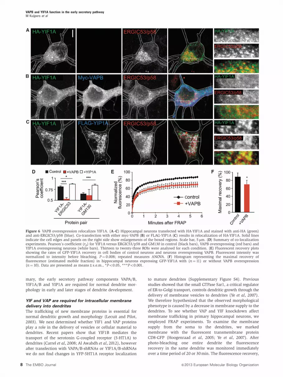
(Figure 6A, B and D). Next, we tested whether a previously
characterized interacting partner of YIF1A, YIP1, also retains
YIF1A in the ER. YIP1 has been shown to localize to ER and
Golgi membranes as well as coat protein complex II (COPII)
transport vesicles in yeast (Yang et al, 1998; Matern et al,
2000; Heidtman et al, 2005; Jin et al, 2005). In hippocampal
neurons, Flag-YIP1A has moderate Golgi and a pronounced
ERGIC localization (Figure 6C). In contrast to VAPB, Flag-
YIP1A overexpression increased YIF1A in the Golgi and
ERGIC, while reducing its ER localization, thus having the
opposite effect of VAPB (Figure 6C and D). Next, we used
fluorescence recovery after photobleaching (FRAP) to exam-
ine the mobility of GFP-YIF1A molecules under the influence
of increased VAPB levels. A 3-by-3 mm region of the cell soma
was bleached by high laser power and fluorescence intensity
was measured over a period of B5 min (Figure 6E). GFP-
YIF1A fluorescence recovered rapidly in control neurons and
reached a maximal recovery of B90% within this time frame.
In neurons co-expressing myc-VAPB the maximal recovery of
GFP-YIF1A is decreased to B70% (Figure 6E and F), indicat-
ing an increase in the immobile fraction of YIF1A molecules
in the ER. Taken together, these data suggest that the ER-
resident protein VAPB indeed binds YIF1A in the ER and
thereby inhibits its recycling to the ERGIC and Golgi.
YIF and VAP are required for normal dendrite
morphology
Studies in Drosophila neurons and recently in zebrafish
suggest that VAPB has an important role in the transport of
proteins to the axon (Yang et al, 2012; Forrest et al, 2013) and
that loss of VAP proteins affects neuron morphology
(Pennetta et al, 2002; Tsuda et al, 2008; Forrest et al, 2013).
To assess the morphological effects of depleting YIF1, YIP1
and VAP proteins, we analysed the length of axons and
dendrites in hippocampal neurons transfected at DIV1 with
VAPA/B-shRNAs, YIF1A/B-shRNAs or YIP1A-shRNA,
together with b-galactosidase to visualize neuron
morphology (Figure 7A). Neurons were fixed at DIV5 and
immunofluorescent staining with antibody against tau was
used to distinguish the axon from the dendrites (Figure 7B).
Depletion of YIF1 and VAP, but not YIP1, caused a significant
decrease in axonal length (Figure 7C). Previous data showed
that ER-to-Golgi transport is particularly important for den-
drite morphology (Ye et al, 2007). Since YIF1, YIP1 and VAP
proteins localize to the early secretory pathway in
hippocampal neurons we wondered whether depletion of
these proteins had any effect on dendrite morphology.
Figure 4 YIF1A localizes to the ER–Golgi intermediate compartment (ERGIC). (A) Image of the cell body of a hippocampal neuron transfectedwith HA-YIF1A and stained with anti-HA (red) and anti-GM130 (green) antibodies. (B) Redistribution of HA-YIF1A by BFA treatment. Neuronswere transfected with HA-YIF1A, treated with BFA (5 mg/ml) for 15 min fixed and labelled with anti-HA (red) and anti-GM130 (green).(C) Representative image of the cell body of a hippocampal neuron transfected with HA-YIF1A and labelled with anti-HA (red) and anti-ERGIC(green). Redistribution of HA-YIF1A after BFA treatment is shown in (D). (E) Summary of co-localization experiments. Pearson’s coefficient (rp)for YIF1A versus ERGIC53/p58, YIF1A versus GM130 and VAPB versus ERGIC53/p58 in control (black bars) and BFA-treated cells (white bars).Twenty-three to twenty-five ROIs were analysed for each condition. Error bars indicate s.e.m., ***Po0.001. (F, G) Images of cell bodies ofhippocampal neurons transfected with myc-VAPB and labelled with anti-myc (red) and anti-ERGIC53/p58 (green). BFA treatment has no effecton myc-VAPB distribution (G). Solid lines indicate the cell edges; the insets show magnifications of boxed areas and arrows indicateco-localization. Scale bar, 5mm.
VAPB and YIF1A function in the early secretory pathwayM Kuijpers et al
6 The EMBO Journal &2013 European Molecular Biology Organization

Indeed, DIV5 neurons showed a significant decrease in
dendritic length when transfected with VAPA/B-shRNAs,
YIF1A/B-shRNAs and YIP1A-shRNA (Figure 7D). Because in
DIV5 neurons dendrites are short and hardly branched we
used mature neurons to examine more closely the effect of
VAP, YIF1 and YIP1 depletion on dendrite morphology. At
DIV19, VAPA/B-shRNAs, YIF1A/B-shRNAs and YIP1A-
shRNA transfected neurons showed a striking dendritic phe-
notype (Figure 7E). Quantitative analysis of the pattern of
dendritic branching by Sholl analysis (Sholl, 1953) revealed a
simplified dendritic tree with reduced branching in proximal
dendrites in neurons transfected with VAPA/B-shRNAs,
YIF1A/B-shRNAs and YIP1A-shRNA (Figure 7F). The reduced
dendritic complexity in the VAPA/B, YIF1A/B and YIP1A
knock-down neurons was also revealed by measuring the
total dendritic length, counting the number of primary den-
drites directly emanating from the soma, and analysing the
total number of dendritic tips (Figure 7G–I). Similar results
were obtained with independent VAPA, VAPB, YIF1A and
YIF1B shRNA sequences (Supplementary Figure S3). In sum-
Figure 5 Effect of VAP knockdown on YIF1A localization in cultured hippocampal neurons. (A, B) Representative images of cell bodies ofhippocampal neurons co-transfected at DIV16 for 4 days with HA-YIF1A (red), GFP and pSuper control vector (A) or pSuper-VAPA and pSuper-VAPB shRNAs (B). VAP knockdown results in relocalization of HA-YIF1A. (C–F) Cell bodies of neurons co-transfected with HA-YIF1A (red) andpSuper control vector (C, E) or pSuper-VAPA and pSuper-VAPB shRNAs (D, F). Neurons were stained for either GM130 (C, D) or ERGIC53/p58(E, F). Solid lines indicate the cell edges. Scale bar, 5mm. (G) Summary of co-localization experiments. Pearson’s coefficient (rp) for YIF1Aversus ERGIC53/p58 and GM130 in control (black bars) and VAP knockdown neurons (white bars). Seventeen to twenty-three ROIs wereanalysed for each condition. Error bars indicate s.e.m., ***Po0.001.
VAPB and YIF1A function in the early secretory pathwayM Kuijpers et al
7&2013 European Molecular Biology Organization The EMBO Journal
mary, the early secretory pathway components VAPA/B,
YIF1A/B and YIP1A are required for normal dendrite mor-
phology in early and later stages of dendrite development.
YIF and VAP are required for intracellular membrane
delivery into dendrites
The trafficking of new membrane proteins is essential for
normal dendritic growth and morphology (Lecuit and Pilot,
2003). We next determined whether YIF1 and VAP proteins
play a role in the delivery of vesicles or cellular material to
dendrites. Recent papers show that YIF1B mediates the
transport of the serotonin G-coupled receptor (5-HT1A) to
dendrites (Carrel et al, 2008; Al Awabdh et al, 2012), however
after transfection with VAPA/B-shRNAs or YIF1A/B-shRNAs
we do not find changes in YFP-5HT1A receptor localization
to mature dendrites (Supplementary Figure S4). Previous
studies showed that the small GTPase Sar1, a critical regulator
of ER-to-Golgi transport, controls dendritic growth through the
delivery of membrane vesicles to dendrites (Ye et al, 2007).
We therefore hypothesized that the observed morphological
phenotype is caused by a decrease in membrane supply to the
dendrites. To see whether VAP and YIF knockdown affect
membrane trafficking in primary hippocampal neurons, we
employed FRAP experiments. To examine the membrane
supply from the soma to the dendrites, we marked
membrane with the fluorescent transmembrane protein
CD8-GFP (Hoogenraad et al, 2005; Ye et al, 2007). After
photo-bleaching one entire dendrite the fluorescence
recovery in the same dendrite was monitored immediately
over a time period of 20 or 30 min. The fluorescence recovery,
Figure 6 VAPB overexpression relocalizes YIF1A. (A–C) Hippocampal neurons transfected with HA-YIF1A and stained with anti-HA (green)and anti-ERGIC53/p58 (blue). Co-transfection with either myc-VAPB (B) or FLAG-YIP1A (C) results in relocalization of HA-YIF1A. Solid linesindicate the cell edges and panels on the right side show enlargements of the boxed regions. Scale bar, 5 mm. (D) Summary of co-localizationexperiments. Pearson’s coefficient (rp) for YIF1A versus ERGIC53/p58 and GM130 in control (black bars), VAPB overexpressing (red bars) andYIP1A overexpressing neurons (white bars). Thirteen to twenty-three ROIs were analysed for each condition. (E) Fluorescent recovery plotsshowing the rates of GFP-YIF1A recovery in cell bodies of control neurons and neurons overexpressing VAPB. Fluorescent intensity wasnormalized to intensity before bleaching. P¼ 0.008; repeated measures ANOVA. (F) Histogram representing the maximal recovery offluorescence (estimated mobile fraction) in hippocampal neurons expressing GFP-YIF1A with (n¼ 11) or without VAPB overexpression(n¼ 10). Data are presented as means±s.e.m., *Po0.05, ***Po0.001.
VAPB and YIF1A function in the early secretory pathwayM Kuijpers et al
8 The EMBO Journal &2013 European Molecular Biology Organization
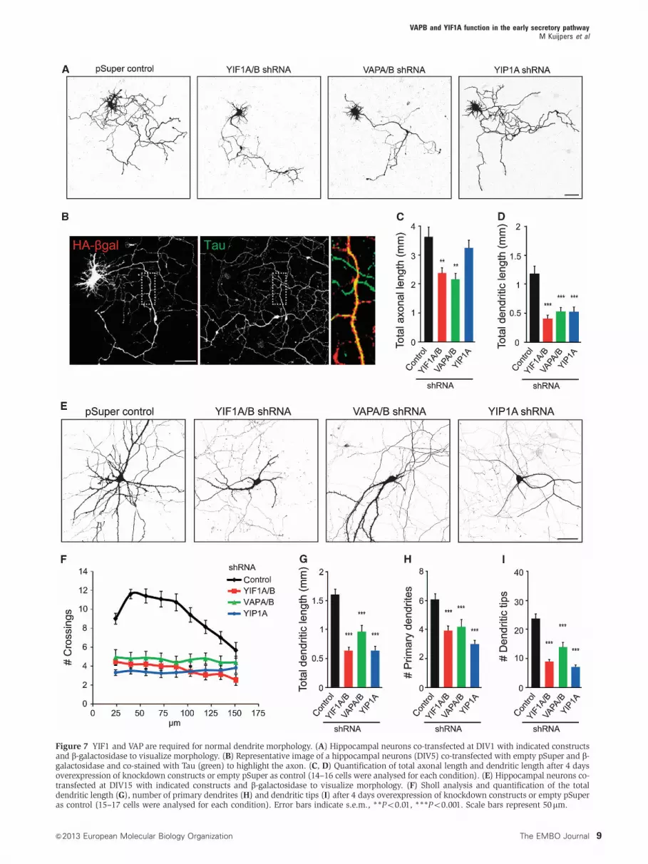
Figure 7 YIF1 and VAP are required for normal dendrite morphology. (A) Hippocampal neurons co-transfected at DIV1 with indicated constructsand b-galactosidase to visualize morphology. (B) Representative image of a hippocampal neurons (DIV5) co-transfected with empty pSuper and b-galactosidase and co-stained with Tau (green) to highlight the axon. (C, D) Quantification of total axonal length and dendritic length after 4 daysoverexpression of knockdown constructs or empty pSuper as control (14–16 cells were analysed for each condition). (E) Hippocampal neurons co-transfected at DIV15 with indicated constructs and b-galactosidase to visualize morphology. (F) Sholl analysis and quantification of the totaldendritic length (G), number of primary dendrites (H) and dendritic tips (I) after 4 days overexpression of knockdown constructs or empty pSuperas control (15–17 cells were analysed for each condition). Error bars indicate s.e.m., **Po0.01, ***Po0.001. Scale bars represent 50mm.
VAPB and YIF1A function in the early secretory pathwayM Kuijpers et al
9&2013 European Molecular Biology Organization The EMBO Journal
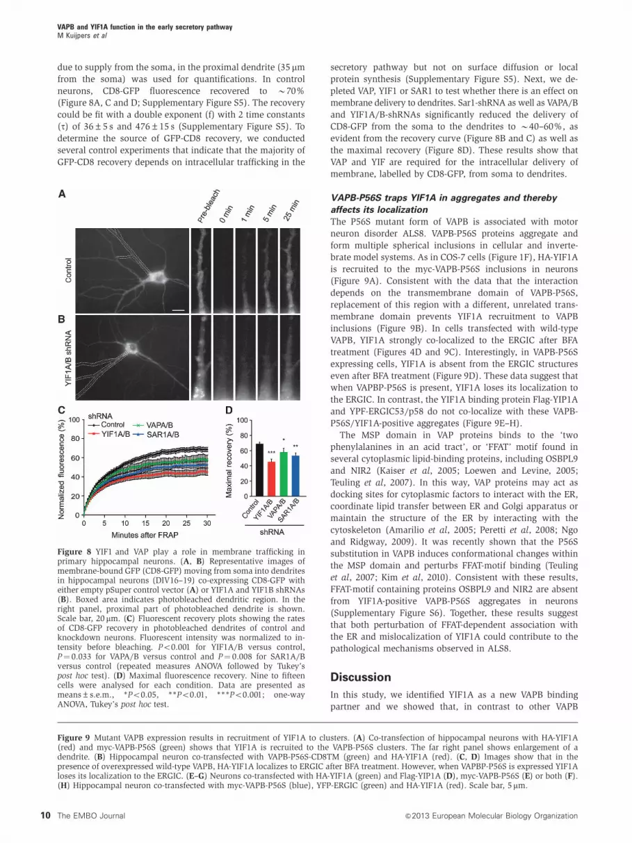
due to supply from the soma, in the proximal dendrite (35 mm
from the soma) was used for quantifications. In control
neurons, CD8-GFP fluorescence recovered to B70%
(Figure 8A, C and D; Supplementary Figure S5). The recovery
could be fit with a double exponent (f) with 2 time constants
(t) of 36±5 s and 476±15 s (Supplementary Figure S5). To
determine the source of GFP-CD8 recovery, we conducted
several control experiments that indicate that the majority of
GFP-CD8 recovery depends on intracellular trafficking in the
secretory pathway but not on surface diffusion or local
protein synthesis (Supplementary Figure S5). Next, we de-
pleted VAP, YIF1 or SAR1 to test whether there is an effect on
membrane delivery to dendrites. Sar1-shRNA as well as VAPA/B
and YIF1A/B-shRNAs significantly reduced the delivery of
CD8-GFP from the soma to the dendrites to B40–60%, as
evident from the recovery curve (Figure 8B and C) as well as
the maximal recovery (Figure 8D). These results show that
VAP and YIF are required for the intracellular delivery of
membrane, labelled by CD8-GFP, from soma to dendrites.
VAPB-P56S traps YIF1A in aggregates and thereby
affects its localization
The P56S mutant form of VAPB is associated with motor
neuron disorder ALS8. VAPB-P56S proteins aggregate and
form multiple spherical inclusions in cellular and inverte-
brate model systems. As in COS-7 cells (Figure 1F), HA-YIF1A
is recruited to the myc-VAPB-P56S inclusions in neurons
(Figure 9A). Consistent with the data that the interaction
depends on the transmembrane domain of VAPB-P56S,
replacement of this region with a different, unrelated trans-
membrane domain prevents YIF1A recruitment to VAPB
inclusions (Figure 9B). In cells transfected with wild-type
VAPB, YIF1A strongly co-localized to the ERGIC after BFA
treatment (Figures 4D and 9C). Interestingly, in VAPB-P56S
expressing cells, YIF1A is absent from the ERGIC structures
even after BFA treatment (Figure 9D). These data suggest that
when VAPBP-P56S is present, YIF1A loses its localization to
the ERGIC. In contrast, the YIF1A binding protein Flag-YIP1A
and YPF-ERGIC53/p58 do not co-localize with these VAPB-
P56S/YIF1A-positive aggregates (Figure 9E–H).
The MSP domain in VAP proteins binds to the ‘two
phenylalanines in an acid tract’, or ‘FFAT’ motif found in
several cytoplasmic lipid-binding proteins, including OSBPL9
and NIR2 (Kaiser et al, 2005; Loewen and Levine, 2005;
Teuling et al, 2007). In this way, VAP proteins may act as
docking sites for cytoplasmic factors to interact with the ER,
coordinate lipid transfer between ER and Golgi apparatus or
maintain the structure of the ER by interacting with the
cytoskeleton (Amarilio et al, 2005; Peretti et al, 2008; Ngo
and Ridgway, 2009). It was recently shown that the P56S
substitution in VAPB induces conformational changes within
the MSP domain and perturbs FFAT-motif binding (Teuling
et al, 2007; Kim et al, 2010). Consistent with these results,
FFAT-motif containing proteins OSBPL9 and NIR2 are absent
from YIF1A-positive VAPB-P56S aggregates in neurons
(Supplementary Figure S6). Together, these results suggest
that both perturbation of FFAT-dependent association with
the ER and mislocalization of YIF1A could contribute to the
pathological mechanisms observed in ALS8.
Discussion
In this study, we identified YIF1A as a new VAPB binding
partner and we showed that, in contrast to other VAPB
Figure 8 YIF1 and VAP play a role in membrane trafficking inprimary hippocampal neurons. (A, B) Representative images ofmembrane-bound GFP (CD8-GFP) moving from soma into dendritesin hippocampal neurons (DIV16–19) co-expressing CD8-GFP witheither empty pSuper control vector (A) or YIF1A and YIF1B shRNAs(B). Boxed area indicates photobleached dendritic region. In theright panel, proximal part of photobleached dendrite is shown.Scale bar, 20 mm. (C) Fluorescent recovery plots showing the ratesof CD8-GFP recovery in photobleached dendrites of control andknockdown neurons. Fluorescent intensity was normalized to in-tensity before bleaching. Po0.001 for YIF1A/B versus control,P¼ 0.033 for VAPA/B versus control and P¼ 0.008 for SAR1A/Bversus control (repeated measures ANOVA followed by Tukey’spost hoc test). (D) Maximal fluorescence recovery. Nine to fifteencells were analysed for each condition. Data are presented asmeans±s.e.m., *Po0.05, **Po0.01, ***Po0.001; one-wayANOVA, Tukey’s post hoc test.
Figure 9 Mutant VAPB expression results in recruitment of YIF1A to clusters. (A) Co-transfection of hippocampal neurons with HA-YIF1A(red) and myc-VAPB-P56S (green) shows that YIF1A is recruited to the VAPB-P56S clusters. The far right panel shows enlargement of adendrite. (B) Hippocampal neuron co-transfected with VAPB-P56S-CD8TM (green) and HA-YIF1A (red). (C, D) Images show that in thepresence of overexpressed wild-type VAPB, HA-YIF1A localizes to ERGIC after BFA treatment. However, when VAPBP-P56S is expressed YIF1Aloses its localization to the ERGIC. (E–G) Neurons co-transfected with HA-YIF1A (green) and Flag-YIP1A (D), myc-VAPB-P56S (E) or both (F).(H) Hippocampal neuron co-transfected with myc-VAPB-P56S (blue), YFP-ERGIC (green) and HA-YIF1A (red). Scale bar, 5mm.
VAPB and YIF1A function in the early secretory pathwayM Kuijpers et al
10 The EMBO Journal &2013 European Molecular Biology Organization

VAPB and YIF1A function in the early secretory pathwayM Kuijpers et al
11&2013 European Molecular Biology Organization The EMBO Journal
interaction partners, YIFIA does not bind to the MSP domain
of VAPB, but via its transmembrane domain interacts with the
transmembrane domain of VAPB. In hippocampal neurons,
we showed that VAPB–YIF1A interaction controls the shut-
tling of YIF1A between the ERGIC and the ER and plays an
important role in early secretory events in neurons, promot-
ing membrane trafficking and normal dendritic growth. In
addition, we show that YIF1A in contrast to other VAPB
interaction partners also binds ALS8-mutant VAPB and
co-accumulates in abnormal ER-derived structures. We propose
that the mislocalization of YIF1A into VAPB-P56S aggregates
could contribute to pathological mechanisms observed in
VAPB-associated motor neuron diseases.
YIF1A interacts with VAPB in the early secretory
pathway
VAPB has been implicated in multiple cellular functions,
either as a conserved ER anchoring protein or as a secreted
signalling molecule in invertebrates (Lev et al, 2008; Han
et al, 2012). Proteins with an FFAT or an FFAT-like motif that
can bind the FFAT-motif binding site in the MSP domain of
VAPA and VAPB represent a major group of VAPB interacting
proteins. FFAT-motif proteins include oxysterol binding
proteins (OSBPs), OSBP-related proteins (ORPs) and NIR
proteins (Wyles and Ridgway, 2004; Amarilio et al, 2005;
Loewen and Levine, 2005; Kawano et al, 2006) and have been
shown to play a role in ER structure (Wyles et al, 2002;
Amarilio et al, 2005; Lehto et al, 2005) and the transport of
membrane lipids (Kawano et al, 2006; Peretti et al, 2008).
Other proteins with FFAT-like motifs include for instance PKA
anchoring proteins that could play a role in cAMP signalling
on the ER (Mikitova and Levine, 2012). In addition, an outer
mitochondrial membrane protein (PTPIP51) was shown to
interact with VAPB (De Vos et al, 2012) putatively via FFAT-
like motifs, although the basis for this interaction was not
studied (Mikitova and Levine, 2012). In contrast to the
previously described VAP binding partners, the interaction
with YIF1 occurs via the transmembrane domain, although
we cannot fully exclude a role for the cytosolic VAPB domain.
During submission of this paper, another study reported a
similar transmembrane mediated interaction between
Drosophila VAP and Sac1, a phosphoinositide phosphatase
(Forrest et al, 2013). YIF1A and YIF1B in mammals are
homologous to Yif1p in yeast. Yif1p is located to the Golgi
membrane and COPII vesicles, forms a tight complex with its
family member Yip1p and plays an important role in early
secretory transport in yeast (Entian et al, 1999; Matern et al,
2000; Otte et al, 2001). Cell-free assays and thermosensitive
yeast strains demonstrated that Yif1p and Yip1p play a critical
role in the biogenesis of ER-derived COPII transport vesicles
(Barrowman et al, 2003; Heidtman et al, 2005). Recent
genome-wide RNA interference screens in Drosophila S2
and HeLa cells found that depletion of YIF1 or YIP1 inhibits
secretion, indicating that YIF1/YIP1 are functionally
conserved components of the secretory pathway (Wendler
et al, 2010; Simpson et al, 2012). We find that YIF1A is
predominantly localized to the ERGIC and recycles between
the ER and Golgi in hippocampal neurons. In agreement with
these results, proteomics analysis identified YIF1A in ERGIC-
enriched membranes (Breuza et al, 2004) and BFA treatment
of HeLa cells results in YIF1A accumulations in the ERGIC
(Yoshida et al, 2008). Our data indicate that both VAPB and
YIF1A are needed for an efficient transition of membrane
cargo through the early secretory pathway in neurons.
Furthermore, we show that, while YIF1A is cycling between
ER and Golgi, VAPB is restricted to the ER and is not present
in ERGIC or Golgi, indicating that YIF1A-VAPB binding is
most likely to occur in the ER membrane (Supplementary
Figure S7). We hypothesize that YIF1A is retained in the ER
membrane by its interaction with VAPB which subsequently
allows specific membrane lipids and/or cargo proteins to
concentrate at ERES before budding off in carriers destined
for further transport towards the ERGIC and cis-Golgi mem-
brane (Gurkan et al, 2006; Jensen and Schekman, 2011).
Moreover, interactome analyses in yeast revealed that the
Yip1p/Yif1p family proteins associate with a large range of
gene products that function in the vesicular transport
pathway, including SNAREs, Rab GTPases, ARFGAP and
sorting nexins (Ito et al, 2000; Uetz et al, 2000). Future
studies using cell-free assays driven by purified proteins
that recapitulate protein transport between the ER and the
Golgi complex will help to elucidate the mechanisms
regulating the interplay between VAPB, YIF1A and the other
components of the ER-to-Golgi trafficking pathway and
advance our understanding of the biogenesis of transport
carriers in the early secretory transport.
YIF and VAP are required for intracellular membrane
delivery into dendrites
The early secretory pathway is fundamentally important for
neuronal development and function (Hanus and Ehlers, 2008;
Tang, 2008; Aridor and Fish, 2009; Jan and Jan, 2010), and
some lines of evidence suggest that the organization of
secretory trafficking in neurons differs in several ways from
the pathways in non-neuronal cells (Nelson and Yeaman,
2001; Stephens and Pepperkok, 2001; Tang, 2008). In
particular components of the ER, ERGIC and Golgi have
been identified in dendrites, specifically localizing at branch
points and close to dendritic spines (Krijnse-Locker et al,
1995; Pierce et al, 2001; Horton et al, 2005; Appenzeller-
Herzog and Hauri, 2006). Here, we identify VAPB and YIF1A
as early secretory trafficking components that are required for
membrane transport and important for normal dendrite
morphology. First, we showed that VAPB is able to retain
YIF1A in the ER thereby regulating its recycling to ERGIC and
Golgi. Depletion of VAP proteins results in a relocalization of
ERGIC protein YIF1A to the Golgi, indicating that VAPB
regulates trafficking of YIF1A through the ERGIC. Recent
studies showed that disruption of VAPB affects ER-to-Golgi
transport and anterograde VSVG trafficking in HeLa cells
(Peretti et al, 2008; Rao et al, 2012). Second, depletion of
VAP, YIF1 as well as YIP1 affects dendrite morphology. This is
consistent with the observed dendritic phenotype in secretory
trafficking deficient Drosophila mutants (Ye et al, 2007).
Third, we show that both VAPA/B and YIF are required for
a normal membrane supply to dendrites, indicating that the
observed effect on dendrite morphology is caused by a defect
in early secretory trafficking. The fact that other proteins of
the ER-to-Golgi trafficking pathway, such as Sar1, also affect
dendritic growth and membrane trafficking (Ye et al, 2007)
strengthens the model that VAPB and YIF1A have an
important role in the secretory transport. Interestingly, YIF1B
has been shown to play a role in the neuronal ER–Golgi
trafficking machinery by specific targeting the serotonin
VAPB and YIF1A function in the early secretory pathwayM Kuijpers et al
12 The EMBO Journal &2013 European Molecular Biology Organization
G-coupled receptor (5-HT1A) to dendrites (Carrel et al, 2008).
A recent follow-up study proposed that YIF1B acts as a
scaffolding complex that recruits 5-HT1A together with
Yip1A and Rab6 in dendritic transport vesicles (Al Awabdh
et al, 2012). These results imply that at least YIF1B and
possibly other Yip1p/Yif1p family proteins play a role in
the anterograde transport of a limited subset of cargo
molecules from the ER to the Golgi. Although in our study
we do not find evidence that YIF1 or VAP plays a role in the
transport of the serotonin receptor we believe that defects in
the transport of specific proteins under the influence of VAP
or YIF impairments are difficult to measure as other,
unconventionally, transport routes can be used as default
(Grieve and Rabouille, 2011).
YIF1A and ALS8
The P56S mutation in the gene encoding VAPB causes ALS8
and some other related forms of motor neuron disease
(Nishimura et al, 2004). Several studies already showed
that VAPB-P56S induces the formation of abnormal ER-
derived inclusions, perturbs ER–Golgi trafficking and triggers
ER stress (Nishimura et al, 2004; Teuling et al, 2007; Tsuda
et al, 2008; Suzuki et al, 2009; Chen et al, 2010; Fasana et al,
2010; Moumen et al, 2011). Ubiquitin (Moumen et al, 2011)
and BAP31 (Fasana et al, 2010) have been described to
accumulate in the VAPB-P56S clusters, suggesting that
mutant VAPB-induced aggregation is part of the ER-
associated degradation pathway. Mutant VAPB-P56S also
induces the co-aggregation of wild-type VAPB, suggesting a
dominant-negative mode of pathogenesis (Teuling et al,
2007). Disruption of ER and Golgi structure and function
has previously been suggested as a possible pathological
mechanism for neurodegenerative diseases (Mourelatos
et al, 1996; Lehotsky et al, 2003; Paschen and Mengesdorf,
2005; Vlug et al, 2005; Yoshida, 2007). How VAPB-P56S
disrupts protein trafficking between the ER and Golgi and
how this may lead to the pathogenesis of ALS8 is not yet
understood. The identification of the VAPB–YIF1A interaction
and its role in early secretory transport raises the possibility
that YIF1A might be involved in the development of neuro-
degenerative diseases. Both YIF1A and YIF1B are ubiqui-
tously expressed in the CNS (Carrel et al, 2008) and a
microarray analysis showed downregulation of YIF1A in
motor neurons isolated from the spinal cord of SOD1-G93A
transgenic mice, a model for familial ALS (Ferraiuolo et al,
2007). This suggests that YIF1A could be a potential factor
involved in the multi-factorial causes leading to ALS.
Although the relevance of our observations to heterozygous
patients remains uncertain (as our observations are under
overexpression conditions), our model suggests that
mislocalization of YIF1A to aggregates leads to a defect in
secretory trafficking and a subsequent inhibition of dendritic
growth and maintenance. Defects in neuronal secretory
trafficking already have been shown to affect dendritic
growth (Horton et al, 2005; Ye et al, 2007). Dendritic
alterations have been documented in mouse models for
motor neuron dysfunction (Wiggins et al, 2012) and in ALS-
affected patients (Liu et al, 2011). In patients, dendrites in
anterior horn cells are shorter and thinner, or are lost (Kato
et al, 1987; Sasaki and Iwata, 1996). Moreover, loss and
atrophy of dendrites was found in motor neurons in the
spinal cord (Karpati et al, 1988). Similarly, in pre-
symptomatic SOD1-G93A transgenic mice dendrites in the
motor cortex show signs of degeneration and in the prefrontal
cortex a reduction in the length of basal dendrites and branch
points was observed (Sgobio et al, 2008; Jara et al, 2012).
Changes in dendritic architecture are indicative of defects
in neuronal function and connectivity, and could be
contributing to the development of sites of degeneration.
Though there is overwhelming evidence for axonal
transport dysfunctions in the pathogenesis of ALS
(Chevalier-Larsen and Holzbaur, 2006; Sau et al, 2011), our
study indicates that alterations in dendritic structure and
trafficking may also play an important role in
neurodegeneration. A recent study has reported mild late
onset motor deficits in VAPB-deficient mice in the absence of
axonal and neuromuscular junction abnormalities (Forrest
et al, 2013). Whether these mice develop dendritic
abnormalities and the degree of compensation of VAPB
deficiency by VAPA in these mice requires further
investigation.
In conclusion, we have identified YIF1A as a novel binding
partner of VAPB and established a key role for these proteins
in the neuronal early secretory pathway. We found that both
VAP and YIF1 proteins are important for proper membrane
trafficking and normal dendrite growth. Moreover, the ALS-
linked mutant VAPB-P56S recruits YIF1A to aggregates and
disrupts its normal localization to ERGICs. Understanding the
cellular function of VAPB may indicate what molecular and
cellular events are associated with the disease process of
ALS8. Our current findings provide new molecular targets to
investigate VAPB-linked neurodegeneration. It is likely that
this information will be of relevance to both the inherited
condition and the more common sporadic forms of disease.
Materials and methods
Expression constructs and shRNAThe following mammalian expression plasmids have beendescribed previously: bio-HA-VAPB, GFP-VAPB-TMD, GFP-VAPB,HA- and myc-tagged VAPA, VAPB and VAPA/B-P56S constructs(Teuling et al, 2007), protein-biotin ligase BirA (Lansbergen et al,2006), flag-YIF1B (Carrel et al, 2008), flag-YIP1A (Dykstra et al,2010), ERGIC/p58-YFP (Ward et al, 2001) and CD8-GFP(Hoogenraad et al, 2005). Bio-GFP-VAPB, Bio-HA-VAPB-P56S andBio-HA-VAPA were generated by incorporating a biotinylation-tag(MSGLNDIFEAQKIEWHE) before the GFP- or HA-tagged mutant orwild-type VAPA/B construct. VAPB-P56S-CD8TM was made byremoving the transmembrane domain of VAPB-P56S and addingthe transmembrane domain of CD8 with a PCR-based strategy usingHA-VAPB-P56S and GFP-CD8 as a template and subcloned into apbactin expression vector. Full-length and truncated human YIF1Aconstructs were generated by PCR using IMAGE clone 3451489 astemplate and cloned into HA- and GFP-tagged pGW1-expressionvectors. For bio-GFP-YIF1A, a biotinylation tag was inserted in frontof the pEGFP-C2 (Clontech) and the YIF1A openreading framesubsequently subcloned in the biotin-tag-GFP vector. GFP-YIF1AIxxxI was generated from GFP-YIF1A by introducing G154I, G158I,G222I and G226I mutations using a PCR-based strategy. GFP-YIF1Bwas generated by a PCR-based strategy using flag-YIF1B andsubcloned into a GFP-tagged pGW1-expression vector. Sp-BirAwas made by incorporating the Ig kappa light chain leadersequence (METDTLLLWLLLLWVPGSTG) at the N-terminus ofBirA and myc tag at its C-terminus in a pSCT expression vector.The following shRNA sequences were used in this study:VAPA#1 and #2 and VAPB#1 shRNA constructs (Teuling et al,2007) and rat VAPB#2 (50-GTTTATGGTTCAGTCTATG-30), YIF1A#1(50-CCATGGCCTTCATCACATA-30), rat YIF1B#1 (50-gtactcatgtactggctca-30), rat YIF1B#2 (50-GCCATGGCTTTCATAACCT-30), ratSAR1A (50-AACCACTCTTCTTCACAT-30) and rat SAR1B (50-AACTA
VAPB and YIF1A function in the early secretory pathwayM Kuijpers et al
13&2013 European Molecular Biology Organization The EMBO Journal

CCTTCCTGCTATCA-30) sequences were designed based on pre-viously published sequences (Kanekura et al, 2006; Ye et al,2007; Carrel et al, 2008). The targeting sequences for rat YIP1A(50-GCAGTATGCTGGCTGTGAG-30) and YIF1A#2 (50-gaagctagggctattggtc-30) were designed by using the siRNA selection programat the Whitehead Institute for Biomedical Research (http://jura.wi.mit.edu/bioc/siRNAext/home.php) (Yuan et al, 2004). Thecomplementary oligonucleotides were annealed and inserted into apSuper vector (Brummelkamp et al, 2002). Unless otherwise specified,all VAP and YIF shRNAs presented in the paper refer to shRNAs#1.
Antibodies and reagentsThe following antibodies were used for immunocytochemistry:rabbit-anti-VAPB and rabbit-anti-VAPA (1:500) (Teuling et al,2007); NIR2 (1:500) (Litvak et al, 2004), OSBPL9 (1:600) (Ngoand Ridgway, 2009); human-anti-EEA1 (1:500) (Selak et al, 2000).The following antibodies were obtained from commercial sources:mouse-anti-tau (1:500, Chemicon); mouse-anti-disulphideisomerase (PDI; 1:300, Affinity BioReagents); mouse-anti-GM130(1:1000, BD Biosciences); mouse-anti-HA (1:500, Roche); mouse-anti-MAP2 (1:2000, Sigma); mouse-anti-myc (1:200, Santa CruzBiotechnology); mouse-anti-flag (1:2000, Sigma); rabbit-anti-ERGIC53/p58 (1:200, Sigma); rabbit-anti-HA (1:500; Santa CruzBiotechnology); rabbit-anti-flag (1:2000, Sigma); rabbit-anti-myc(1:200; Cell Signaling Technology); rabbit-anti-b-galactosidase(1:2000, MP Biomedicals); rat-anti-HA (1:200, Roche); mouse-anti-CD8 (1:20, Mabtech); Alexa Fluor 488-, Alexa Fluor 598-, andAlexa Fluor 633-conjugated secondary antibodies (1:400,Invitrogen) and Cy5-conjugated secondary antibody (1:400,Jackson ImmunoResearch Labs). The following antibodies wereused for western blot analysis: rabbit anti-GFP (1:1000, Abcam);rabbit anti-HA (1:500, Santa Cruz); mouse anti-HA (1:500,Covance) and HRP-conjugated secondary antibodies (1:5000,Dako). BFA and cycloheximide were obtained from Sigma.
Transfection and immunofluorescence of cultured COS-7 cellsCOS-7 cells were cultured in DMEM/Ham’s F10 (50/50%) mediumcontaining 10% FCS and 1% penicillin/streptomycin. Two daysbefore transfection, cells were plated at 1:30 in Lab-tek chamberslides (Nunc). Cells were transfected with Fugene6 (Roche) accord-ing to manufacturer’s protocol and incubated overnight. Cells werefixed in 4% paraformaldehyde for 10 min at room temperaturefollowed by 5 min in 0.1% Triton X-100 in PBS. Slides were blockedin 0.5% BSA/0.02% glycine in PBS and labelled with primaryantibody for 2 h at room temperature. Slides were washed threetimes with 0.05% Tween-20 in PBS, labelled with secondary anti-bodies for 1 h at room temperature, washed three times with 0.05%Tween-20 in PBS and mounted using Vectashield mounting medium(Vector laboratories). Images were acquired using a Leica DMRBEmicroscope equipped with � 40 and � 100 oil objectives.
AnimalsAll animal experiments were performed in compliance with theguidelines for the welfare of experimental animals issued by theFederal Government of The Netherlands. All animal experimentswere approved by the Animal Ethical Review Committee (DEC) ofthe Erasmus Medical Center and Utrecht University.
Hippocampal neuron cultures, transfection andimmunohistochemistryPrimary hippocampal cultures were prepared from embryonic day18 (E18) rat brains (Banker and Goslin, 1988; Kapitein et al, 2010).Cells were plated on coverslips coated with poly-L-lysine (35mg/ml)and laminin (5mg/ml) at a density of 75 000/well. Hippocampalcultures were grown in Neurobasal medium (NB) supplementedwith B27, 0.5 mM glutamine, 12.5mM glutamate and penicillin/streptomycin. Hippocampal neurons were transfected usingLipofectamine 2000 (Invitrogen). Briefly, DNA (3.6mg/well) wasmixed with 3ml of Lipofectamine 2000 in 200ml of NB, incubated for30 min, and then added to the neurons in NB at 371C in 5% CO2 for45 min. Next, neurons were washed with NB and transferred in theoriginal medium at 371C in 5% CO2. After 2–4 days of transfection,neurons were fixed with 4% paraformaldehyde/4% sucrose inPBS, washed three times in PBS for 10 min and incubatedwith the indicated primary antibodies in GDB buffer (0.2% BSA,0.8 M NaCl, 0.5% Triton X-100, 30 mM phosphate buffer, pH 7.4)overnight at 41C. Neurons were then washed three times in PBS
for 30 min, incubated with secondary antibodies in GDB for 1 hat room temperature and washed three times in PBS for 30 min.Slides were mounted using Vectashield mounting medium (VectorLaboratories). Images for co-localization measurements were ac-quired using a Nikon microscope equipped with a � 100 oilobjective. Confocal images were acquired using a LSM510 confocalmicroscope (Zeiss) with a � 40 or � 63 oil objective.
ImmunoprecipitationHEK293T cells were cultured in DMEM/Hams-F10 (50/50%) med-ium containing 10% FCS and 1% penicillin/streptomycin and weretransfected using Lipofectamine2000 (Invitrogen). Cells were har-vested 24 h after transfection, by scraping the cells in ice-cold PBSand lysing cell pellets in lysis buffer (50 mM Tris–HCl, 100 mMNaCl, 1.0% Triton X-100 and protease inhibitors (Roche).Supernatant and pellet fractions were separated by centrifugationat 13 200 r.p.m. for 5 min. Supernatants were mixed with an equalamount of lysis buffer, protein-A-agarose beads (GE Healthcare),and 3mg of rabbit anti-GFP, mouse anti-HA or control IgG (Sigma).Samples were incubated 4 h while rotating at 41C, centrifuged at2000 r.p.m. and pellets were washed three times with lysis buffer.Samples were mixed with 4� Sample Buffer (8% SDS, 25%Glycerol, 0.05 M Tris pH 6.8, 200 mM DTT, 40 mg/l BromophenolBlue) and boiled. Equal amounts of protein were loaded onto SDS–PAGE gels and subjected to western blotting on polyvinylidenedifluoride membrane. Blots were blocked with 2% bovine serumalbumin/0.05% Tween-20 in PBS and incubated with primaryantibodies at 41C overnight. Blots were washed with 0.05%Tween-20 in PBS three times for 10 min at room temperature andincubated with secondary antibodies conjugated to horseradishperoxidase (Dako). Blots were developed with enhanced chemilu-minescent Western blotting substrate (Pierce).
GST pull-downFull-length wild-type and mutant VAPB GST fusion proteins wereobtained as described earlier (Teuling et al, 2007). HEK293T cellswere transfected as described before with HA-YIF1A, HA-YIF1A(1–131), HA-YIF1A (131–193) and HA-YIF1A (198–293) and lysedin 50 mm Tris–HCl, 100 mm NaCl, and 1% Triton X-100 containingprotease inhibitors (Roche). Lysates were incubated with GST beads(GE Healthcare Bio-Sciences) for 2 h at 41C, washed four times withlysis buffer, and analysed by SDS–PAGE and western blotting asdescribed before.
Biotin-streptavidin pull-down and mass spectrometryFor biotin-streptavidin pull-down assays, HeLa or HEK293Tcells weretransfected with biotin-tagged VAPB using Lipofectamine-2000(Invitrogen) transfection reagent according to manufacturer’s instruc-tions. Cells were lysed 16h later in 20 mm Tris–HCl, pH 8.0, 150 mmKCl, 1% Triton X-100, and protease inhibitors (Roche). To increasethe solubility of VAPB-P56S, transfected cells were incubated for 2 hat 201C before lysis. Cell lysates were centrifuged at 13 000 r.p.m. for15 min and the supernatants were incubated with Dynabeads M-280streptavidin (Dynal; Invitrogen) for 45 min. Beads were separated byusing a magnet (Dynal; Invitrogen) and washed five times in lysisbuffer. For protein elution, the beads were boiled in NuPAGE LDS 4sample buffer (Invitrogen), separated, and supernatants were run ona 10% NuPAGE Bis-Tris gel (Invitrogen). The gel was stained with theColloidal Blue staining kit (Invitrogen) and analysed by westernblotting. Mass spectrometry was performed as described previously(Lansbergen et al, 2006). The Mascot score cutoff value for a positiveprotein hit was set to 100. Individual peptide tandem massspectrometry spectra with Mascot scores below 100 were checkedmanually and either interpreted as valid identifications or discarded.Proteins present in the negative controls (pull-down assays withbio-GFP alone) were regarded as background.
Photobleaching experimentsFor quantitative FRAP experiments, neurons were transfected asdescribed before, and imaged on a Nikon Eclipse TE2000E (Nikon)equipped with an incubation chamber (INUG2-ZILCS-H2; Tokai Hit)mounted on a motorized stage (Prior) (Jaworski et al, 2009).Coverslips (24 mm) were mounted in metal rings and maintainedat 371C and 5% CO2. A dendrite or a 3� 3mm regions of interest(ROI) in the cell body was photobleached with high laser power.Immediately after photobleaching, images of GFP fluorescence wereacquired using a � 40 objective (Nikon) and a Coolsnap HQ camera
VAPB and YIF1A function in the early secretory pathwayM Kuijpers et al
14 The EMBO Journal &2013 European Molecular Biology Organization

(Photometrics). Intensity of GFP signal in dendrites was measuredover a length of 30mm from the soma with MetaMorph imageanalysis software (Universal Imaging). Background intensity wassubtracted and fluorescence intensity after photobleaching wascalculated relative to the intensity before bleaching. Sigmaplot12.3 software was used to perform curve fitting.
Image analysis and quantification
Measurement of neurite outgrowth. To measure neurite length, weused b-galactosidase as an unbiased cell-fill. Hippocampal neuronswere transfected with the indicated constructs, fixed at the appro-priate time point and subjected to immunofluorescent staining.Confocal images were obtained at 1024�1024 pixel resolutionusing a LSM510 confocal microscope (Zeiss) with a � 40 oilobjective (0.7 digital zoom). Each image was a z-series of images;the obtained stack was ‘flattened’ into a single image using max-imum projection. Morphometric analysis and quantification wereperformed using MetaMorph image analysis software (UniversalImaging). For measurement of total dendrite or axonal length, alldendrites or axons of individual neurons were traced. All non-axonal protrusions initiating from the cell soma longer than 10mmwere defined as primary dendrites. For dendrite tip number, tips ofall non-axonal protrusions longer than 10 mm were counted. ForSholl analysis, concentric circles with 16 mm differences in diameterwere automatically drawn around the cell body, and the number ofdendrites crossing each circle was counted.
Co-localization of two fluorescent signals. Co-localization of twofluorescent signals was indicated by the Pearson’s coefficient (rp),determined using the JACoP plugin (Bolte and Cordelieres, 2006)for ImageJ. For each cell, three ROIs were selected; n was defined asthe number of ROIs.
Statistical analysesStatistical analyses were performed with MS Excel or SPSS software.Data were averaged over multiple cells and statistical analysis wasperformed with Student’s t-test, one-way ANOVA or repeated mea-sures ANOVA and Tukey’s post hoc test. Po0.05 was considered assignificant.
Supplementary dataSupplementary data are available at The EMBO Journal Online(http://www.embojournal.org).
Acknowledgements
We thank Dr N Ridgway for the OSBP9 antibody, Dr S Lev for theNIR2 antibody, Dr T Lee for the Flag-YIP1A construct, Dr M Darmonfor the Flag-YIF1B and YFP-5HT1A plasmid, Dr. JF Presley for thep58-YFP construct and Jeroen Demmers and Karel Bezstarosti forhelp with mass spectrometry analyses. This work was supported byHet Prinses Beatrix Spierfonds grant (DJ and CCH), NetherlandsOrganization for Scientific Research (NWO-ALW-VICI, AA andCCH), the Netherlands Organization for Health Research andDevelopment (ZonMW-TOP, CCH), the European ScienceFoundation (EURYI, CCH), EMBO Young Investigators Program(YIP, CCH) and the Human Frontier Science Program (HFSP-CDA,CCH).
Author contributions: MK, DJ, AA and CCH designed research;MK, KLYand ET performed research; MK analysed the data; MK andCCH wrote the paper; CCH supervised the project.
Conflict of interest
The authors declare that they have no conflict of interest.
ReferencesAl Awabdh S, Miserey-Lenkei S, Bouceba T, Masson J, Kano F,
Marinach-Patrice C, Hamon M, Emerit MB, Darmon M (2012) Anew vesicular scaffolding complex mediates the G-protein-coupled 5-HT1A receptor targeting to neuronal dendrites. JNeurosci 32: 14227–14241
Altschul SF, Madden TL, Schaffer AA, Zhang J, Zhang Z, Miller W,Lipman DJ (1997) Gapped BLAST and PSI-BLAST: a new genera-tion of protein database search programs. Nucleic Acids Res 25:3389–3402
Amarilio R, Ramachandran S, Sabanay H, Lev S (2005) Differentialregulation of endoplasmic reticulum structure through VAP-Nirprotein interaction. J Biol Chem 280: 5934–5944
Anagnostou G, Akbar MT, Paul P, Angelinetta C, Steiner TJ, deBelleroche J (2010) Vesicle associated membrane protein B(VAPB) is decreased in ALS spinal cord. Neurobiol Aging 31:969–985
Appenzeller-Herzog C, Hauri HP (2006) The ER-Golgi intermediatecompartment (ERGIC): in search of its identity and function.J Cell Sci 119: 2173–2183
Aridor M, Fish KN (2009) Selective targeting of ER exit sitessupports axon development. Traffic 10: 1669–1684
Banker G, Goslin K (1988) Developments in neuronal cell culture.Nature 336: 185–186
Barrowman J, Wang W, Zhang Y, Ferro-Novick S (2003) TheYip1p.Yif1p complex is required for the fusion competence ofendoplasmic reticulum-derived vesicles. J Biol Chem 278:19878–19884
Bolte S, Cordelieres FP (2006) A guided tour into subcellularcolocalization analysis in light microscopy. J Microsc 224:213–232
Breuza L, Halbeisen R, Jeno P, Otte S, Barlowe C, Hong W, Hauri HP(2004) Proteomics of endoplasmic reticulum-Golgi intermediatecompartment (ERGIC) membranes from brefeldin A-treatedHepG2 cells identifies ERGIC-32, a new cycling protein thatinteracts with human Erv46. J Biol Chem 279: 47242–47253
Brummelkamp TR, Bernards R, Agami R (2002) A system for stableexpression of short interfering RNAs in mammalian cells. Science296: 550–553
Carrel D, Masson J, Al Awabdh S, Capra CB, Lenkei Z, Hamon M,Emerit MB, Darmon M (2008) Targeting of the 5-HT1A serotoninreceptor to neuronal dendrites is mediated by Yif1B. J Neurosci28: 8063–8073
Chai A, Withers J, Koh YH, Parry K, Bao H, Zhang B, Budnik V,Pennetta G (2008) hVAPB, the causative gene of a heterogeneousgroup of motor neuron diseases in humans, is functionallyinterchangeable with its Drosophila homologue DVAP-33A atthe neuromuscular junction. Hum Mol Genet 17: 266–280
Chen HJ, Anagnostou G, Chai A, Withers J, Morris A, Adhikaree J,Pennetta G, de Belleroche JS (2010) Characterization of theproperties of a novel mutation in VAPB in familial amyotrophiclateral sclerosis. J Biol Chem 285: 40266–40281
Chevalier-Larsen E, Holzbaur EL (2006) Axonal transport andneurodegenerative disease. Biochim Biophys Acta 1762:1094–1108
Dancourt J, Barlowe C (2010) Protein sorting receptors in the earlysecretory pathway. Annu Rev Biochem 79: 777–802
De Vos KJ, Morotz GM, Stoica R, Tudor EL, Lau KF, Ackerley S,Warley A, Shaw CE, Miller CC (2012) VAPB interacts with themitochondrial protein PTPIP51 to regulate calcium homeostasis.Hum Mol Genet 21: 1299–1311
Dykstra KM, Pokusa JE, Suhan J, Lee TH (2010) Yip1A structuresthe mammalian endoplasmic reticulum. Mol Biol Cell 21:1556–1568
Entian KD, Schuster T, Hegemann JH, Becher D, Feldmann H,Guldener U, Gotz R, Hansen M, Hollenberg CP, Jansen G,Kramer W, Klein S, Kotter P, Kricke J, Launhardt H, MannhauptG, Maierl A, Meyer P, Mewes W, Munder Tet al. (1999) Functionalanalysis of 150 deletion mutants in Saccharomyces cerevisiae bya systematic approach. Mol Gen Genet 262: 683–702
Fasana E, Fossati M, Ruggiano A, Brambillasca S, Hoogenraad CC,Navone F, Francolini M, Borgese N (2010) A VAPB mutant linkedto amyotrophic lateral sclerosis generates a novel form of orga-nized smooth endoplasmic reticulum. FASEB J 24: 1419–1430
Ferraiuolo L, Heath PR, Holden H, Kasher P, Kirby J, Shaw PJ (2007)Microarray analysis of the cellular pathways involved in theadaptation to and progression of motor neuron injury in the
VAPB and YIF1A function in the early secretory pathwayM Kuijpers et al
15&2013 European Molecular Biology Organization The EMBO Journal
SOD1 G93A mouse model of familial ALS. J Neurosci 27:9201–9219
Forrest S, Chai A, Sanhueza M, Marescotti M, Parry K, Georgiev A,Sahota V, Mendez-Castro R, Pennetta G (2013) Increased levels ofphosphoinositides cause neurodegeneration in a Drosophilamodel of amyotrophic lateral sclerosis. Hum Mol Genet (advanceonline publication, 29 March 2013; doi:10.1093/hmg/ddt118)
Grieve AG, Rabouille C (2011) Golgi bypass: skirting around theheart of classical secretion. Cold Spring Harb Perspect Biol 3:a005298
Gurkan C, Stagg SM, Lapointe P, Balch WE (2006) The COPII cage:unifying principles of vesicle coat assembly. Nat Rev Mol Cell Biol7: 727–738
Han SM, Tsuda H, Yang Y, Vibbert J, Cottee P, Lee SJ, Winek J,Haueter C, Bellen HJ, Miller MA (2012) Secreted VAPB/ALS8major sperm protein domains modulate mitochondrial localiza-tion and morphology via growth cone guidance receptors. DevCell 22: 348–362
Hanus C, Ehlers MD (2008) Secretory outposts for the local processingof membrane cargo in neuronal dendrites. Traffic 9: 1437–1445
Heidtman M, Chen CZ, Collins RN, Barlowe C (2005) Yos1p is anovel subunit of the Yip1p-Yif1p complex and is required fortransport between the endoplasmic reticulum and the Golgicomplex. Mol Biol Cell 16: 1673–1683
Hirokawa T, Boon-Chieng S, Mitaku S (1998) SOSUI: classificationand secondary structure prediction system for membrane pro-teins. Bioinformatics 14: 378–379
Hoogenraad CC, Milstein AD, Ethell IM, Henkemeyer M, Sheng M(2005) GRIP1 controls dendrite morphogenesis by regulatingEphB receptor trafficking. Nat Neurosci 8: 906–915
Horton AC, Ehlers MD (2003) Dual modes of endoplasmic reticu-lum-to-Golgi transport in dendrites revealed by live-cell imaging.J Neurosci 23: 6188–6199
Horton AC, Racz B, Monson EE, Lin AL, Weinberg RJ, Ehlers MD(2005) Polarized secretory trafficking directs cargo for asym-metric dendrite growth and morphogenesis. Neuron 48: 757–771
Ito T, Tashiro K, Muta S, Ozawa R, Chiba T, Nishizawa M,Yamamoto K, Kuhara S, Sakaki Y (2000) Toward a protein-proteininteraction map of the budding yeast: A comprehensive system toexamine two-hybrid interactions in all possible combinationsbetween the yeast proteins. Proc Natl Acad Sci USA 97: 1143–1147
Jan YN, Jan LY (2010) Branching out: mechanisms of dendriticarborization. Nat Rev Neurosci 11: 316–328
Jara JH, Villa SR, Khan NA, Bohn MC, Ozdinler PH (2012) AAV2mediated retrograde transduction of corticospinal motor neuronsreveals initial and selective apical dendrite degeneration in ALS.Neurobiol Dis 47: 174–183
Jaworski J, Kapitein LC, Gouveia SM, Dortland BR, Wulf PS,Grigoriev I, Camera P, Spangler SA, Di Stefano P, Demmers J,Krugers H, Defilippi P, Akhmanova A, Hoogenraad CC (2009)Dynamic microtubules regulate dendritic spine morphology andsynaptic plasticity. Neuron 61: 85–100
Jensen D, Schekman R (2011) COPII-mediated vesicle formation at aglance. J Cell Sci 124: 1–4
Jin C, Zhang Y, Zhu H, Ahmed K, Fu C, Yao X (2005) Human Yip1Aspecifies the localization of Yif1 to the Golgi apparatus. BiochemBiophys Res Commun 334: 16–22
Kaiser SE, Brickner JH, Reilein AR, Fenn TD, Walter P, Brunger AT(2005) Structural basis of FFAT motif-mediated ER targeting.Structure 13: 1035–1045
Kanekura K, Nishimoto I, Aiso S, Matsuoka M (2006)Characterization of amyotrophic lateral sclerosis-linked P56Smutation of vesicle-associated membrane protein-associated pro-tein B (VAPB/ALS8). J Biol Chem 281: 30223–30233
Kapitein LC, Yau KW, Hoogenraad CC (2010) Microtubule dynamicsin dendritic spines. Methods Cell Biol 97: 111–132
Karpati G, Carpenter S, Durham H (1988) A hypothesis for thepathogenesis of amyotrophic lateral sclerosis. Rev Neurol (Paris)144: 672–675
Kato T, Hirano A, Donnenfeld H (1987) A Golgi study of the largeanterior horn cells of the lumbar cords in normal spinal cords andin amyotrophic lateral sclerosis. Acta Neuropathol 75: 34–40
Kawano M, Kumagai K, Nishijima M, Hanada K (2006) Efficienttrafficking of ceramide from the endoplasmic reticulum to theGolgi apparatus requires a VAMP-associated protein-interactingFFAT motif of CERT. J Biol Chem 281: 30279–30288
Kim S, Leal SS, Ben Halevy D, Gomes CM, Lev S (2010) Structuralrequirements for VAP-B oligomerization and their implication inamyotrophic lateral sclerosis-associated VAP-B(P56S) neurotoxi-city. J Biol Chem 285: 13839–13849
Krijnse-Locker J, Parton RG, Fuller SD, Griffiths G, Dotti CG (1995)The organization of the endoplasmic reticulum and the inter-mediate compartment in cultured rat hippocampal neurons. MolBiol Cell 6: 1315–1332
Lansbergen G, Grigoriev I, Mimori-Kiyosue Y, Ohtsuka T, Higa S,Kitajima I, Demmers J, Galjart N, Houtsmuller AB, Grosveld F,Akhmanova A (2006) CLASPs attach microtubule plus ends to thecell cortex through a complex with LL5beta. Dev Cell 11: 21–32
Lecuit T, Pilot F (2003) Developmental control of cell morphogen-esis: a focus on membrane growth. Nat Cell Biol 5: 103–108
Lee MC, Miller EA, Goldberg J, Orci L, Schekman R (2004) Bi-directional protein transport between the ER and Golgi. Annu RevCell Dev Biol 20: 87–123
Lehotsky J, Kaplan P, Babusikova E, Strapkova A, Murin R (2003)Molecular pathways of endoplasmic reticulum dysfunctions:possible cause of cell death in the nervous system. Physiol Res52: 269–274
Lehto M, Hynynen R, Karjalainen K, Kuismanen E, Hyvarinen K,Olkkonen VM (2005) Targeting of OSBP-related protein 3 (ORP3)to endoplasmic reticulum and plasma membrane is controlled bymultiple determinants. Exp Cell Res 310: 445–462
Lev S, Ben Halevy D, Peretti D, Dahan N (2008) The VAP proteinfamily: from cellular functions to motor neuron disease. TrendsCell Biol 18: 282–290
Litvak V, Argov R, Dahan N, Ramachandran S, Amarilio R,Shainskaya A, Lev S (2004) Mitotic phosphorylation of theperipheral Golgi protein Nir2 by Cdk1 provides a docking me-chanism for Plk1 and affects cytokinesis completion. Mol Cell 14:319–330
Liu M, Duggan J, Salt TE, Cordeiro MF (2011) Dendritic changes invisual pathways in glaucoma and other neurodegenerative con-ditions. Exp Eye Res 92: 244–250
Loewen CJ, Levine TP (2005) A highly conserved binding site invesicle-associated membrane protein-associated protein (VAP) forthe FFAT motif of lipid-binding proteins. J Biol Chem 280:14097–14104
Lorente-Rodriguez A, Barlowe C (2011) Entry and exit mechanismsat the cis-face of the Golgi complex. Cold Spring Harb perspectBiol 3: a005207
Matern H, Yang X, Andrulis E, Sternglanz R, Trepte HH, Gallwitz D(2000) A novel Golgi membrane protein is part of a GTPase-binding protein complex involved in vesicle targeting. EMBO J 19:4485–4492
Mikitova V, Levine TP (2012) Analysis of the key elements ofFFAT-like motifs identifies new proteins that potentially bindVAP on the ER, including two AKAPs and FAPP2. PLoS One 7:e30455
Mitne-Neto M, Machado-Costa M, Marchetto MC, Bengtson MH,Joazeiro CA, Tsuda H, Bellen HJ, Silva HC, Oliveira AS, Lazar M,Muotri AR, Zatz M (2011) Downregulation of VAPB expression inmotor neurons derived from induced pluripotent stem cells ofALS8 patients. Hum Mol Genet 20: 3642–3652
Moumen A, Virard I, Raoul C (2011) Accumulation of wildtype andALS-linked mutated VAPB impairs activity of the proteasome.PLoS ONE 6: e26066
Mourelatos Z, Gonatas NK, Stieber A, Gurney ME, Dal Canto MC(1996) The Golgi apparatus of spinal cord motor neurons intransgenic mice expressing mutant Cu,Zn superoxide dismutasebecomes fragmented in early, preclinical stages of the disease.Proc Natl Acad Sci USA 93: 5472–5477
Nelson WJ, Yeaman C (2001) Protein trafficking in the exocyticpathway of polarized epithelial cells. Trends Cell Biol 11: 483–486
Ngo M, Ridgway ND (2009) Oxysterol binding protein-relatedProtein 9 (ORP9) is a cholesterol transfer protein that regulatesGolgi structure and function. Mol Biol Cell 20: 1388–1399
Nishimura AL, Mitne-Neto M, Silva HC, Richieri-Costa A, MiddletonS, Cascio D, Kok F, Oliveira JR, Gillingwater T, Webb J, Skehel P,Zatz M (2004) A mutation in the vesicle-trafficking protein VAPBcauses late-onset spinal muscular atrophy and amyotrophic lat-eral sclerosis. Am J Hum Genet 75: 822–831
Nishimura Y, Hayashi M, Inada H, Tanaka T (1999) Molecularcloning and characterization of mammalian homologues of
VAPB and YIF1A function in the early secretory pathwayM Kuijpers et al
16 The EMBO Journal &2013 European Molecular Biology Organization

vesicle-associated membrane protein-associated (VAMP-asso-ciated) proteins. Biochem Biophys Res Commun 254: 21–26
Otte S, Belden WJ, Heidtman M, Liu J, Jensen ON, Barlowe C (2001)Erv41p and Erv46p: new components of COPII vesicles involvedin transport between the ER and Golgi complex. J Cell Biol 152:503–518
Papiani G, Ruggiano A, Fossati M, Raimondi A, Bertoni G,Francolini M, Benfante R, Navone F, Borgese N (2012)Restructured endoplasmic reticulum generated by mutant amyo-trophic lateral sclerosis-linked VAPB is cleared by the proteasome.J Cell Sci 125: 3601–3611
Paschen W, Mengesdorf T (2005) Cellular abnormalities linked toendoplasmic reticulum dysfunction in cerebrovascular disease–therapeutic potential. Pharmacol Ther 108: 362–375
Pennetta G, Hiesinger PR, Fabian-Fine R, Meinertzhagen IA, BellenHJ (2002) Drosophila VAP-33A directs bouton formation atneuromuscular junctions in a dosage-dependent manner.Neuron 35: 291–306
Peretti D, Dahan N, Shimoni E, Hirschberg K, Lev S (2008)Coordinated lipid transfer between the endoplasmic reticulumand the Golgi complex requires the VAP proteins and is essentialfor Golgi-mediated transport. Mol Biol Cell 19: 3871–3884
Pfeffer S, Aivazian D (2004) Targeting Rab GTPases to distinctmembrane compartments. Nat Rev Mol Cell Biol 5: 886–896
Pierce JP, Mayer T, McCarthy JB (2001) Evidence for a satellitesecretory pathway in neuronal dendritic spines. Curr Biol 11:351–355
Rao M, Song W, Jiang A, Shyr Y, Lev S, Greenstein D, Brantley-Sieders D, Chen J (2012) VAMP-Associated Protein B (VAPB)promotes breast tumor growth by modulation of Akt Activity.PLoS One 7: e46281
Ratnaparkhi A, Lawless GM, Schweizer FE, Golshani P, Jackson GR(2008) A Drosophila model of ALS: human ALS-associated muta-tion in VAP33A suggests a dominant negative mechanism. PloSOne 3: e2334
Saraste J, Dale HA, Bazzocco S, Marie M (2009) Emerging new rolesof the pre-Golgi intermediate compartment in biosynthetic-secre-tory trafficking. FEBS Lett 583: 3804–3810
Sasaki S, Iwata M (1996) Dendritic synapses of anterior hornneurons in amyotrophic lateral sclerosis: an ultrastructuralstudy. Acta Neuropathol 91: 278–283
Sau D, Rusmini P, Crippa V, Onesto E, Bolzoni E, Ratti A, Poletti A(2011) Dysregulation of axonal transport and motorneuron dis-eases. Biol Cell 103: 87–107
Selak S, Woodman RC, Fritzler MJ (2000) Autoantibodies to earlyendosome antigen (EEA1) produce a staining pattern resemblingcytoplasmic anti-neutrophil cytoplasmic antibodies (C-ANCA).Clin Exp Immunol 122: 493–498
Sgobio C, Trabalza A, Spalloni A, Zona C, Carunchio I, Longone P,Ammassari-Teule M (2008) Abnormal medial prefrontal cortexconnectivity and defective fear extinction in the presymptomaticG93A SOD1 mouse model of ALS. Genes Brain Behav 7: 427–434
Shakoori A, Fujii G, Yoshimura S, Kitamura M, Nakayama K, Ito T,Ohno H, Nakamura N (2003) Identification of a five-pass trans-membrane protein family localizing in the Golgi apparatus andthe ER. Biochem Biophys Res Commun 312: 850–857
Sholl DA (1953) Dendritic organization in the neurons of the visualand motor cortices of the cat. J Anat 87: 387–406
Simpson JC, Joggerst B, Laketa V, Verissimo F, Cetin C, Erfle H,Bexiga MG, Singan VR, Heriche JK, Neumann B, Mateos A, BlakeJ, Bechtel S, Benes V, Wiemann S, Ellenberg J, Pepperkok R(2012) Genome-wide RNAi screening identifies human proteinswith a regulatory function in the early secretory pathway. Nat CellBiol 14: 764–774
Skehel PA, Fabian-Fine R, Kandel ER (2000) Mouse VAP33 isassociated with the endoplasmic reticulum and microtubules.Proc Natl Acad Sci USA 97: 1101–1106
Soussan L, Burakov D, Daniels MP, Toister-Achituv M, Porat A,Yarden Y, Elazar Z (1999) ERG30, a VAP-33-related protein,functions in protein transport mediated by COPI vesicles. J CellBiol 146: 301–311
Stephens DJ, Pepperkok R (2001) Illuminating the secretory path-way: when do we need vesicles? J Cell Sci 114: 1053–1059
Suzuki H, Kanekura K, Levine TP, Kohno K, Olkkonen VM, Aiso S,Matsuoka M (2009) ALS-linked P56S-VAPB, an aggregated loss-of-function mutant of VAPB, predisposes motor neurons to ERstress-related death by inducing aggregation of co-expressed wild-type VAPB. J Neurochem 108: 973–985
Tang BL (2008) Emerging aspects of membrane traffic in neuronaldendrite growth. Biochim Biophys Acta 1783: 169–176
Teuling E, Ahmed S, Haasdijk E, Demmers J, Steinmetz MO,Akhmanova A, Jaarsma D, Hoogenraad CC (2007) Motor neurondisease-associated mutant vesicle-associated membrane protein-associated protein (VAP) B recruits wild-type VAPs into endoplas-mic reticulum-derived tubular aggregates. J Neurosci 27:9801–9815
Tsuda H, Han SM, Yang Y, Tong C, Lin YQ, Mohan K, Haueter C,Zoghbi A, Harati Y, Kwan J, Miller MA, Bellen HJ (2008) Theamyotrophic lateral sclerosis 8 protein VAPB is cleaved, secreted,and acts as a ligand for Eph receptors. Cell 133: 963–977
Uetz P, Giot L, Cagney G, Mansfield TA, Judson RS, Knight JR,Lockshon D, Narayan V, Srinivasan M, Pochart P, Qureshi-EmiliA, Li Y, Godwin B, Conover D, Kalbfleisch T, Vijayadamodar G,Yang M, Johnston M, Fields S, Rothberg JM (2000) A compre-hensive analysis of protein-protein interactions in Saccharomycescerevisiae. Nature 403: 623–627
Vlug AS, Teuling E, Haasdijk ED, French P, Hoogenraad CC,Jaarsma D (2005) ATF3 expression precedes death of spinalmotoneurons in amyotrophic lateral sclerosis-SOD1 transgenicmice and correlates with c-Jun phosphorylation, CHOP expres-sion, somato-dendritic ubiquitination and Golgi fragmentation.Eur J Neurosci 22: 1881–1894
Ward TH, Polishchuk RS, Caplan S, Hirschberg K, Lippincott-Schwartz J (2001) Maintenance of Golgi structure andfunction depends on the integrity of ER export. J Cell Biol 155:557–570
Watson P, Stephens DJ (2005) ER-to-Golgi transport: form andformation of vesicular and tubular carriers. Biochim BiophysActa 1744: 304–315
Wendler F, Gillingham AK, Sinka R, Rosa-Ferreira C, Gordon DE,Franch-Marro X, Peden AA, Vincent JP, Munro S (2010) Agenome-wide RNA interference screen identifies two novel com-ponents of the metazoan secretory pathway. EMBO J 29: 304–314
Wiggins LM, Kuta A, Stevens JC, Fisher EM, von Bartheld CS (2012)A novel phenotype for the dynein heavy chain mutation Loa:Altered dendritic morphology, organelle density, and reducednumbers of trigeminal motoneurons. J Comp Neurol 520:2757–2773
Wyles JP, McMaster CR, Ridgway ND (2002) Vesicle-associatedmembrane protein-associated protein-A (VAP-A) interacts withthe oxysterol-binding protein to modify export from the endo-plasmic reticulum. J Biol Chem 277: 29908–29918
Wyles JP, Ridgway ND (2004) VAMP-associated protein-A regulatespartitioning of oxysterol-binding protein-related protein-9 be-tween the endoplasmic reticulum and Golgi apparatus. Exp CellRes 297: 533–547
Yang X, Matern HT, Gallwitz D (1998) Specific binding to a noveland essential Golgi membrane protein (Yip1p) functionally linksthe transport GTPases Ypt1p and Ypt31p. EMBO J 17: 4954–4963
Yang Z, Huh SU, Drennan JM, Kathuria H, Martinez JS, Tsuda H,Hall MC, Clemens JC (2012) Drosophila Vap-33 is required foraxonal localization of Dscam isoforms. J Neurosci 32:17241–17250
Ye B, Zhang Y, Song W, Younger SH, Jan LY, Jan YN (2007) Growingdendrites and axons differ in their reliance on the secretorypathway. Cell 130: 717–729
Yoshida H (2007) ER stress and diseases. FEBS J 274: 630–658Yoshida Y, Suzuki K, Yamamoto A, Sakai N, Bando M, Tanimoto K,
Yamaguchi Y, Sakaguchi T, Akhter H, Fujii G, Yoshimura S, OgataS, Sohda M, Misumi Y, Nakamura N (2008) YIPF5 and YIF1Arecycle between the ER and the Golgi apparatus and are involvedin the maintenance of the Golgi structure. Exp Cell Res 314:3427–3443
Yuan B, Latek R, Hossbach M, Tuschl T, Lewitter F (2004) siRNASelection Server: an automated siRNA oligonucleotide predictionserver. Nucleic Acids Res 32: W130–W134
VAPB and YIF1A function in the early secretory pathwayM Kuijpers et al
17&2013 European Molecular Biology Organization The EMBO Journal