Translocation of the inhibitor of apoptosis protein c-IAP1 from the nucleus to the Golgi in...
10
doi:10.1182/blood-2004-01-0065 Prepublished online June 8, 2004; 2004 104: 2035-2043 Eric Solary Sordet, Tibor Ponnelle, Najet Debili, Thi-Hai Phan, Rose-Ann Padua, Laurence Dubrez-Daloz and Stéphanie Plenchette, Séverine Cathelin, Cédric Rébé, Sophie Launay, Sylvain Ladoire, Olivier nuclear export signal-mediated event nucleus to the Golgi in hematopoietic cells undergoing differentiation: a Translocation of the inhibitor of apoptosis protein c-IAP1 from the http://bloodjournal.hematologylibrary.org/content/104/7/2035.full.html Updated information and services can be found at: (973 articles) Phagocytes (4217 articles) Neoplasia (3131 articles) Hematopoiesis and Stem Cells Articles on similar topics can be found in the following Blood collections http://bloodjournal.hematologylibrary.org/site/misc/rights.xhtml#repub_requests Information about reproducing this article in parts or in its entirety may be found online at: http://bloodjournal.hematologylibrary.org/site/misc/rights.xhtml#reprints Information about ordering reprints may be found online at: http://bloodjournal.hematologylibrary.org/site/subscriptions/index.xhtml Information about subscriptions and ASH membership may be found online at: Copyright 2011 by The American Society of Hematology; all rights reserved. Washington DC 20036. by the American Society of Hematology, 2021 L St, NW, Suite 900, Blood (print ISSN 0006-4971, online ISSN 1528-0020), is published weekly For personal use only. by guest on May 29, 2013. bloodjournal.hematologylibrary.org From
-
Upload
u-bourgogne -
Category
Documents
-
view
1 -
download
0
Transcript of Translocation of the inhibitor of apoptosis protein c-IAP1 from the nucleus to the Golgi in...

doi:10.1182/blood-2004-01-0065Prepublished online June 8, 2004;2004 104: 2035-2043
Eric SolarySordet, Tibor Ponnelle, Najet Debili, Thi-Hai Phan, Rose-Ann Padua, Laurence Dubrez-Daloz and Stéphanie Plenchette, Séverine Cathelin, Cédric Rébé, Sophie Launay, Sylvain Ladoire, Olivier nuclear export signal-mediated eventnucleus to the Golgi in hematopoietic cells undergoing differentiation: a Translocation of the inhibitor of apoptosis protein c-IAP1 from the
http://bloodjournal.hematologylibrary.org/content/104/7/2035.full.htmlUpdated information and services can be found at:
(973 articles)Phagocytes � (4217 articles)Neoplasia �
(3131 articles)Hematopoiesis and Stem Cells �Articles on similar topics can be found in the following Blood collections
http://bloodjournal.hematologylibrary.org/site/misc/rights.xhtml#repub_requestsInformation about reproducing this article in parts or in its entirety may be found online at:
http://bloodjournal.hematologylibrary.org/site/misc/rights.xhtml#reprintsInformation about ordering reprints may be found online at:
http://bloodjournal.hematologylibrary.org/site/subscriptions/index.xhtmlInformation about subscriptions and ASH membership may be found online at:
Copyright 2011 by The American Society of Hematology; all rights reserved.Washington DC 20036.by the American Society of Hematology, 2021 L St, NW, Suite 900, Blood (print ISSN 0006-4971, online ISSN 1528-0020), is published weekly
For personal use only. by guest on May 29, 2013. bloodjournal.hematologylibrary.orgFrom
HEMATOPOIESIS
Translocation of the inhibitor of apoptosis protein c-IAP1 from the nucleusto the Golgi in hematopoietic cells undergoing differentiation:a nuclear export signal–mediated eventStephanie Plenchette, Severine Cathelin, Cedric Rebe, Sophie Launay, Sylvain Ladoire, Olivier Sordet,Tibor Ponnelle, Najet Debili, Thi-Hai Phan, Rose-Ann Padua, Laurence Dubrez-Daloz, and Eric Solary
The caspase inhibitor and RING finger–containing protein cellular inhibitor ofapoptosis protein 1 (c-IAP1) has beenshown to be involved in both apoptosisinhibition and signaling by members ofthe tumor necrosis factor (TNF) receptorfamily. The protein is regulated transcrip-tionally (eg, is a target for nuclear fac-tor–�B [NF-�B]) and can be inhibited bymitochondrial proteins released in thecytoplasm upon apoptotic stimuli. Thepresent study indicates that an additionallevel of regulation of c-IAP1 may be cellcompartmentalization. The protein ispresent in the nucleus of undifferentiated
U937 and THP1 monocytic cell lines. Whenthese cells undergo differentiation underphorbol ester exposure, c-IAP1 translo-cates to the cytoplasmic side of the Golgiapparatus. This redistribution involves anuclear export signal (NES)–mediated,leptomycin B–sensitive mechanism. Us-ing site-directed mutagenesis, we local-ized the functional NES motif in thecaspase recruitment domain (CARD) ofc-IAP1. A nucleocytoplasmic redistribu-tion of the protein was also observed inhuman monocytes as well as in tumorcells from epithelial origin when undergo-ing differentiation. c-IAP1 does not trans-
locate from the nucleus of cells whosedifferentiation is blocked (ie, in cell linesand monocytes from transgenic miceoverexpressing B-cell lymphoma 2 [Bcl-2] and in monocytes from patients withchronic myelomonocytic leukemia). Alto-gether, these observations associatec-IAP1 cellular location with cell differen-tiation, which opens new perspectives onthe functions of the protein. (Blood. 2004;104:2035-2043)
© 2004 by The American Society of Hematology
Introduction
The inhibitors of apoptosis proteins (IAPs) have been initially defined asnatural cellular inhibitors of cell death. These proteins were identified inbaculoviral genome as regulators of host-cell viability during virusinfection,1 and cellular orthologues were subsequently described inyeast, nematodes, drosophila, and mammals. The human genomeencodes at least 8 IAPs (X-linked IAP [XIAP], cellular IAP1 [c-IAP1],c-IAP2, melanoma IAP [ML-IAP], neuronal apoptosis inhibitory pro-tein [NAIP], survivin, IAP-like protein 2 [ILP-2],Apollon).2All of theseproteins have in common the presence of 1 to 3 copies of a baculovirusIAP repeat (BIR) domain.1 These domains are essential for theantiapoptotic properties of the IAPs, which have been attributed to thedirect binding and inhibition of caspases. XIAP binds the small subunitof caspase-9 through its BIR3 domain3 and masks the active site ofcaspase-3 and -7 through a distinct segment, which is immediatelyamino-terminal to its BIR2 domain.4,5c-IAP1 and c-IAP2 bind caspase-3and -7 but their inhibitory effect on caspases is 2- to 3-log lower than thatof XIAP.6 Some of the BIR-containing proteins do not have clear linkswith apoptosis and several members of the family have demonstrateddistinct functions including cell cycle regulation,7 protein degradation,8
and caspase-independent signal transduction.9-12
In addition to the BIR domains, several IAPs, including XIAP,c-IAP1, and c-IAP2, contain a highly conserved carboxy-terminalRING finger domain that confers them an enzyme 3 (E3) function
in the protein ubiquitylation process. Several proteins specificallytargeted for ubiquitylation by IAPs have been identified. At least invitro, XIAP and c-IAP2 direct the ubiquitylation of caspase-3 andcaspase-7,13,14 whereas c-IAP1 and c-IAP2 mediate ubiquitylationof second mitochondria-derived activator of caspase (Smac)/DIABLO, an antagonist of IAPs.15 c-IAP1 and c-IAP2 are alsocomponents of the type 2 tumor necrosis factor (TNF) receptorcomplex through interaction with the signaling intermediates TNFreceptor–associated factor 1 (TRAF1) and TRAF2.9 c-IAP1 couldinduce the ubiquitylation of TRAF2 and participated in theTNF-�–mediated proteasomal degradation of TRAF2,16 and c-IAP2 has been involved in the TNF-�signaling leading to nuclearfactor–�B (NF-�B) activation.17
The expression and activity of IAPs are regulated at several levels.The transcription factor NF-�B enhances the expression of c-IAP1,c-IAP2, and XIAP, which may contribute to the prosurvival effectexerted in many situations by this transcription factor.18,19 XIAPtranslation can be enhanced through the use of an internal ribosomalentry site in the 5�-untranslated region of its messenger RNA.20 IAPscould regulate their own degradationthrough autoubiquitylation,8
whereas the IAP-interacting proteins Smac/DIABLO and Omi/HtrA2 neutralize XIAP and possibly other IAPs when releasedfrom the mitochondria under apoptotic stimuli.21
From the Institut National de la Sante et de la Recherche Medicale (INSERM)U517, INSERM EPI 106, IFR100, Dijon, France; INSERM U362, InstitutGustave Roussy, Villejuif, France; INSERM EMI00-03, Institut Universitaired’Hematologie, Hopital St Louis, Paris, France; The Rayne Institute, King’sCollege Hospital, London, United Kingdom.
Submitted January 8, 2004; accepted May 26, 2004. Prepublished online asBlood First Edition Paper, June 8, 2004; DOI 10.1182/blood-2004-01-0065.
Supported by grants from the Ligue Nationale Contre le Cancer.
Reprints: Eric Solary, INSERM U517, IFR100, 7 boulevard Jeanne d’Arc,21000 Dijon, France; e-mail: [email protected].
The publication costs of this article were defrayed in part by page chargepayment. Therefore, and solely to indicate this fact, this article is herebymarked ‘‘advertisement’’ in accordance with 18 U.S.C. section 1734.
© 2004 by The American Society of Hematology
2035BLOOD, 1 OCTOBER 2004 � VOLUME 104, NUMBER 7
For personal use only. by guest on May 29, 2013. bloodjournal.hematologylibrary.orgFrom
Another level of regulation of IAP functions is the modulationof their subcellular location. Such a regulation has been describedfor XIAP whose interaction with the protein XIAP-associatedfactor 1 (XAF1) induces its sequestration in the nucleus andsuppresses its caspase-inhibitory function.22 The present studydemonstrates that c-IAP1 is located in the nucleus of variousundifferentiated cells and migrates to the cytoplasm, more specifi-cally to the Golgi apparatus, when these cells undergo differentia-tion. This redistribution of c-IAP1 involves a nuclear export signal(NES) located in its caspase recruitment domain (CARD). Overex-pression of c-IAP1 interferes with 12-O-tetradecanoylphorbol13-acetate (TPA)–induced differentiation of leukemic cells, aprocess also inhibited by the nuclear export inhibitor leptomycin B(LMB). Altogether, these observations suggest a role for c-IAP1 incell differentiation.
Patients, materials, and methods
Antibodies and chemicals
We used mouse monoclonal antibodies (mAbs) directed against c-IAP1(PharMingen, La Jolla, CA); Golgin 97 (clone CDF4; Molecular Probes,Eugene, OR); mitochondrial HSP70 (mHSP70; Affinity BioReagent, Golden,CO); HSC70 (Santa Cruz Biotechnology, Santa Cruz, CA); GM130 (GolgiMatrix protein of 130 kDa; fluorescein isothiocyanate [FITC]–conjugatedantibody; Transduction Laboratories, Lexington, KY); rabbit polyclonalAbs targeting c-IAP1 (Santa Cruz Biotechnology and R&D Systems,Abington, United Kingdom); macrophage antigen-1 (Mac-1; phycoerythrin[PE]–conjugated antibody; Pharmingen, Becton Dickinson, Heidelberg,Germany); Bcl-2 (FITC-conjugated antibody; Pharmingen, Becton Dickin-son); CD1a (FITC-conjugated antibody; Pharmingen, Becton Dickinson),CD71 (FITC-conjugated antibody; Pharmingen, Becton Dickinson); poly-(adenosine diphosphate-ribose) polymerase (PARP; Boehringer-Mann-heim, Mannheim, Germany); XIAP (R&D Systems and Stressgen Biotech,San Diego, CA); protein disulfide isomerase (PDI; Calbiochem, La Jolla,CA); green fluorescent protein (GFP; Invitrogen, Cergy Pontoise, France);and survivin (Novus Biologicals, Littleton, CO). Macrophage colony-stimulating factor (M-CSF), granulocyte-macrophage colony-stimulatingfactor (GM-CSF), and interleukin-4 (IL-4) were obtained from R&DSystems; erythropoietin (EPO) was from Amgen (Thousand Oaks, CA);TPA was from Sigma-Aldrich Laboratories (St Quentin Fallavier, France);brefeldin A (BFA) and nocodazole were from Alexis Biochemicals (Lausen,Switzerland); and trypsin-EDTA (ethylenediaminetetraacetic acid) wasfrom Gibco-BRL (Carlsbad, CA). LMB was kindly provided by Dr M.Yoshida (Tokyo, Japan) and thrombopoietin (TPO) was kindly provided byKirin Brewery (Tokyo, Japan).
Cell culture and differentiation
Cell lines were obtained from the American Type Culture Collection(ATCC, Rockville, MD) and cultured as described.23 We also tested thepreviously described Bcl-2–transfected U937 and HT29 cells and HT29-MTX cells.23-25 The TPA-resistant variant of U937 cells were kindlyprovided by Prof P. J. Parker (London, United Kingdom).26 Monocytesfrom human peripheral blood were obtained with informed consent fromhealthy donors and 7 patients with chronic myelomonocytic leukemia(CMML) and purified using an isolation kit (Miltenyi Biotec, Paris, France)following the manufacturer’s instructions. Cells were differentiated intomacrophages or dendritic cells and checked for the expression of differen-tiation marker CD71 and CD1a as described.23 Peripheral blood CD34�
cells were cultured in liquid conditions in the presence of cytokines togenerate megakaryocytes or erythroid cells as described.27,28 The Bcl-2transgenic mice were obtained from Irv Weismann (Stanford, CA).29 Bcl-2overexpression in Mac-1� cells of transgenic mice was verified by flowcytometry using a FACSCalibur cytometer and Cell Quest software(Pharmingen, Becton Dickinson). Femoral bone marrow cells were isolated
from 6- to 8-week-old control and transgenic FVB/N female mice andcultured for 4 hours on plastic plates before culturing adherent cells for 6days in the presence of 10% L929 cell–conditioned medium as source ofCSF-1. Macrophage differentiation was assessed by May-Grunwald-Giemsa staining.
Immunofluorescence studies
Cells were fixed in paraformaldehyde (PFA; 2%) for 10 minutes at roomtemperature, washed twice, saturated in phosphate-buffered saline (PBS)containing 0.1% saponin and 5% nonfat milk, and incubated overnight atroom temperature in the presence of primary Ab diluted in PBS containing0.1% saponin and 0.5% bovine serum albumin (BSA). After washing, cellswere incubated for 30 minutes with 488-alexa goat antirabbit or antimouseAb (Molecular Probes) and washed 3 times with PBS. Nuclei were stainedby Hoechst 33342 (Sigma-Aldrich). To demonstrate colocalization ofc-IAP1 with Golgin 97 or GM130, cells were first incubated withanti–c-IAP1 Ab overnight at 4°C, then with the secondary biotinylated–immunoglobulin (Ig; 1:100; Amersham Biosciences, Orsay, France) for 1hour at room temperature, then with a streptavidin–texas red–conjugatedAb (Molecular Probes; 1:2000) for 1 hour. Cells were subsequentlyincubated for 1 hour at room temperature with anti-GM130–FITC (1:100)or anti–Golgin 97 (1:100), then with FITC-conjugated antimouse Ab.Fluorescence was preserved using the FluorSave mounting medium (Calbio-chem). Analysis was performed using either a fluorescence (Nikon Eclipse80i; Nikon, Champigny, France) or a confocal (Leica TCS SP2; Leica,Bron, France) microscope (objective � 50; original magnification � 500).The images were captured by a 3 CCD (charge-coupled device) color videocamera (Sony, Paris, France), digitally saved using Archimed-Pro software(Microvision Instruments, Evry, France), and further processed usingPhotoshop software (Adobe Systems France, Paris, France).
Preparation of cellular extracts and Western blot analysis
Whole-cell lysates and nuclear-free extracts were prepared as described.23
Nuclear and cytoplasmic fractions were obtained by lysing the cells in lysisbuffer (10 mM Hepes [N-2-hydroxyethylpiperazine-N�-2-ethanesulfonicacid], 10 mM KCl, 0.1 mM EDTA, 0.1 mM EGTA [ethyleneglycoltetraace-tic acid], 1 mM DTT [dithiothreitol], 0.6% NP-40 [nonidet P–40]) in thepresence of the protease inhibitors. Cell lysate was centrifuged at 1200g for10 minutes. The supernatant was carefully collected (cytoplasmic fraction[C]) and the pellet was washed once then resuspended in lysis buffer(nuclear fraction [N]). Further cell fractionation was performed as de-scribed.30 All fractions were stored at �80°C until Western blottinganalysis, and protein concentration was measured using the Bio-Rad DCprotein assay kit (Hercules, CA). Western blot experiments were performedas previously described.23
Trypsin digestion of microsomal proteins
Proteins from reticular/microsomal-enriched fraction were digested by0.05% trypsin in the presence of 0.02% EDTA for 30 minutes at 37°C andanalyzed by Western blotting for c-IAP1 content.31
Plasmid constructs
Plasmid-enhanced GFP (pEGFP)–c-IAP1 was constructed by subcloningfull-length c-IAP1 cDNA (kindly provided by J. C. Reed, La Jolla, CA) intothe BglII/SalI site of pEGFP-C1 (Clontech, Palo Alto, CA). Sense andantisense oligonucleotides corresponding to leucine-rich motif (LRM)putative NES were as follows: LRM1 sense, 5�-GAT CTT TTT TGG AAAATT CTC TAG AAA CTC TGA GGA-3�; LRM1 antisense, 5�-GAT CTCCTC AGA GTT TCT AGA GAA TTT TCC AAA AAA-3�; LRM2 sense,5�-GAT CTC TCT TTC AAC AAT TGA CAT GTG TGC TTC CTA TCCTGG ATA ATC TTT TAA-3�; LRM2 antisense, 5�-GAT CTT AAA AGATTA TCC AGG ATA GGA AGC ACA CAT GTC AAT TGT TGA AAGAGA-3�; LRM3 sense, 5�-GAT CTC TGT CAC TGG AAG AAC AAT TGAGGA GGT TGC AAA-3�; and LRM3 antisense, 5�-GAT CTT TGC AACCTC CTC AAT TGT TCT TCC AGT GAC AGA-3� (Proligo France SAS,Paris, France). Complementary oligonucleotides were annealed and cloned
2036 PLENCHETTE et al BLOOD, 1 OCTOBER 2004 � VOLUME 104, NUMBER 7
For personal use only. by guest on May 29, 2013. bloodjournal.hematologylibrary.orgFrom

in a sense orientation into the BglII site of pEGFP-C1 (Clontech). Allsequences are expressed at the C-terminus of GFP. Full-length c-IAP1mutants (GFP–c-IAP1–LRM1*,–LRM2*, and–LRM3*) were obtained bymutagenesis of LRM1, 2, and 3, separately or in combination (leucine werereplaced by alanine) using the Quick-Change Site-directed Mutagenesis Kit(Stratagene, La Jolla, CA). All constructs were sequenced to ensure theaccuracy of the reading frames and the site-directed mutations.
Cell transfection
HeLa cells were transfected 24 hours after seeding using Superfecttransfection reagent (Qiagen, Valencia, CA) following the manufacturer’sinstructions. Cells were studied 24 hours after transient transfection: nucleiwere stained with Hoechst 33342 and cells were fixed with 2% PFA for 5minutes before studying the subcellular distribution of GFP fusion proteinusing a fluorescence (Nikon) or a confocal (Leica) microscope. THP1 cellswere transiently transfected using the AMAXA nucleofector kit (Amaxa,Koln, Germany) and transfected cells were enriched by a 10-day geneticinselection (0.7 �g/mL) before expansion and treatment.
Results
TPA-induced differentiation of human monocytic cell linesis associated with the redistribution of c-IAP1 and XIAPfrom the nucleus into the cytoplasm
It has been previously shown that exposure of U937 cells to 20 nM TPAinduced their differentiation into macrophage-like cells. Cells becomeadherent to the culture flask and the expression of CD11b at their plasmamembrane increases.23 We used Western blotting to analyze the expres-sion of XIAP, c-IAP1, c-IAP2, and survivin, 4 proteins that belong to theIAP family, in U937 cells undergoing TPA-induced differentiation(Figure 1A). c-IAP2 could not be detected in undifferentiated U937 cellsand remained undetectable at all steps of the differentiation process (notshown). Survivin expression was limited to the nucleus of undifferenti-ated cells and disappeared upon differentiation. This may be related tothe differentiation-associated cell cycle exit since this protein, which hasan evolutionarily conserved role as a mitotic spindle checkpoint protein,is expressed mainly in dividing cells.7 The expression of XIAP andc-IAP1 was poorly influenced by the differentiation process whenstudied in whole-cell lysates (Figure 1Aleft). However, c-IAP1, and to alesser extent XIAP, progressively accumulated in nuclear-free extractsas the cells underwent differentiation (Figure 1A right). The presentstudy focused on c-IAP1 redistribution.
Differentiation-associated redistribution of c-IAP1 from thenucleus to the cytoplasm was further confirmed by Western blottinganalysis of c-IAP1 expression in TPA-treated THP1 cells (Figure1C) and by fluorescent microscopy analysis of the 2 cell lines(Figure 1B,D). c-IAP1 was located mainly in the nucleus of U937and THP1 undifferentiated cells and in the cytoplasm of TPA-differentiated cells. A kinetic analysis identified a transient diffusestaining of the cytoplasm in the first hours of TPA treatment. As thecells progressed toward the differentiation process, a more patchystaining close to the nucleus was observed (see THP1 cells inFigure 1D).
c-IAP1 colocalizes with the Golgi apparatusof differentiated cells
To precisely determine the subcellular localization of c-IAP1 inTPA-differentiated cells, we performed Western blot experimentsin enriched cellular fractions. Figure 2A shows that c-IAP1 islocalized in the nucleus of undifferentiated U937 cells and in thereticular fraction of TPA-differentiated U937 cells. Thus, in
accordance with Figure 1A, the protein migrates from the nucleusto the cytoplasm. A similar observation was made by comparingcellular fractions of undifferentiated and differentiated THP1 cells(not shown). Fluorescence microscopy experiments indicated thatc-IAP1 colocalized with Golgin 97, a Golgi matrix protein, inTPA-differentiated THP1 (Figure 2B) and U937 (not shown) cells.c-IAP1 also colocalized, although less precisely, with GM130, aprotein associated with the cis-Golgi (Figure 2B). Addition ofeither BFA, a fungal metabolite that causes disintegration of Golgistructure through inhibition of adenosine diphosphate (ADP)–ribosylation factor (ARF) guanosine 5�-triphosphate (GTP)–binding proteins,32 or nocodazole, a microtubule-depolarizingagent, suppressed the patchy staining of c-IAP1 in TPA-differentiated THP1 (Figure 2C) and U937 (not shown) cells. In thetested conditions, BFA did not modify calnexin C subcellularlocalization, indicating that the endoplasmic reticulum was notaltered (not shown). Altogether, these observations indicated thatc-IAP1 was redistributed to the Golgi apparatus in cells undergoingdifferentiation.
Figure 1. c-IAP1 redistribution in human leukemia cell lines undergoingTPA-induced differentiation. U937 (A-B) and THP1 (C-D) cells were treatedfor indicated times with 20 nM TPA to induce a macrophage-like differentiation.(A,C) Western blot analysis of indicated proteins in whole-cell, cytoplasmic, andnuclear extracts. HSC70 was used as a loading control. (B,D) Fluorescencemicroscopy analysis of c-IAP1 (green), as observed using an anti–c-IAP1 mAb(Pharmingen). Nuclei, labeled with Hoechst 33342, are stained in blue. Magnification� 300.
DIFFERENTIATION-INDUCED NUCLEAR EXPORT OF c-IAP1 2037BLOOD, 1 OCTOBER 2004 � VOLUME 104, NUMBER 7
For personal use only. by guest on May 29, 2013. bloodjournal.hematologylibrary.orgFrom
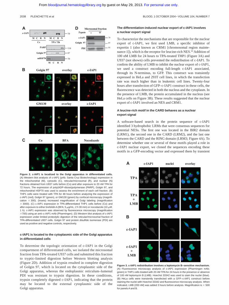
c-IAP1 is located to the cytoplasmic side of the Golgi apparatusin differentiated cells
To determine the topologic orientation of c-IAP1 in the Golgicompartment of differentiated cells, we isolated the microsomalfraction from TPA-treated U937 cells and submitted this fractionto tryptic-limited digestion before Western blotting analysis(Figure 2D). Addition of trypsin resulted in complete digestionof Golgin 97, which is located on the cytoplasmic side of theGolgi apparatus, whereas the endoplasmic reticulum–lumenalPDI was resistant to trypsin digestion. In these conditions,trypsin completely digested c-IAP1, indicating that the proteinmay be located to the external cytoplasmic side of theGolgi apparatus.
The differentiation-induced nuclear export of c-IAP1 involvesa nuclear export signal
To characterize the mechanisms that are responsible for the nuclearexport of c-IAP1, we first used LMB, a specific inhibitor ofexportin 1 (also known as CRM1 [chromosomal region mainte-nance-1]), which is the receptor for leucine-rich NES.33 Addition of100 nM LMB for 24 hours to TPA-treated THP1 (Figure 3A) andU937 (not shown) cells prevented the redistribution of c-IAP1. Toconfirm the ability of LMB to inhibit the nuclear export of c-IAP1,we used a construct encoding full-length c-IAP1 associated,through its N-terminus, to GFP. This construct was transientlyexpressed in HeLa and 293T cell lines, in which the transfectionrate was much higher than in leukemic cell lines. Twenty-fourhours after transfection of GFP–c-IAP1 construct in these cells, thefluorescence was detected in both the nucleus and the cytoplasm. Inthe presence of LMB, the protein accumulated in the nucleus (seeHeLa cells on Figure 3B). These results suggested that the nuclearexport of c-IAP1 involved an NES and CRM1.
A leucine-rich motif in the CARD behaves as a nuclearexport signal
A software-based search in the protein sequence of c-IAP1identified 3 hydrophobic LRMs that were consensus sequences forpotential NESs. The first one was located in the BIR2 domain(LRM1), the second one in the CARD (LRM2), and the last onebetween the CARD and the RING domain (LRM3; Figure 4A). Todetermine whether one or several of these motifs played a role inc-IAP1 nuclear export, we cloned the sequences encoding thesemotifs in a GFP-encoding vector and expressed them by transient
Figure 3. c-IAP1 redistribution involves a leptomycin B–sensitive mechanism.(A) Fluorescence microscopy analysis of c-IAP1 expression (Pharmingen mAb;green) in THP1 cells treated with 20 nM TPA for 24 hours in the presence or absenceof 100 nM leptomycin B (LMB). Hoechst 33342 was used to stain the nuclei (blue).(B) HeLa cells were transiently transfected with a GFP–c-IAP1 construct beforestaining the nuclei with Hoechst 33342 and fluorescence microscopy analysis. Whenindicated, LMB (200 nM) was added 3 hours before analysis. Magnification is � 500for panels A and B.
Figure 2. c-IAP1 is localized to the Golgi apparatus in differentiated cells.(A) Western blot analysis of c-IAP1 (pAb; Santa Cruz Biotechnology) expression inthe mitochondrial (M), cytosolic (C), reticular/microsomal (R), and nuclear (N)fractions obtained from U937 cells before (Co) and after exposure to 20 nM TPA for72 hours. The expression of poly(ADP-ribose)polymerase (PARP), Golgin 97, andmitochondrial HSP70 was used to assess the enrichment of each cell fraction. (B)THP1 cells were treated with TPA for 48 hours before analyzing the expression ofc-IAP1 (red), Golgin 97 (green), or GM130 (green) by confocal microscopy (magnifi-cation � 300). (Insets) Increased magnification of Golgi labeling (magnification� 3000). (C) c-IAP1 expression in TPA-differentiated THP1 cells before (Co) andafter exposure to either brefeldin A (BFA; 5 �g/mL; 2 h 30 min) or nocodazole (10 �M;1 h). c-IAP1 expression was observed by fluorescence microscopy (magnification�700) using an anti–c-IAP1 mAb (Pharmingen). (D) Western blot analysis of c-IAP1expression under limited proteolytic digestion of the reticular/microsomal fraction ofTPA-differentiated U937 cells. Golgin 97 and protein disulfide isomerase (PDI) areused as positive and negative controls, respectively.
2038 PLENCHETTE et al BLOOD, 1 OCTOBER 2004 � VOLUME 104, NUMBER 7
For personal use only. by guest on May 29, 2013. bloodjournal.hematologylibrary.orgFrom
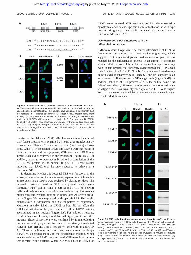
transfection in HeLa and 293T cells. The subcellular location ofGFP fusion proteins was examined 24 hours after transfection byconventional (Figure 4B) and confocal laser (not shown) micros-copy. While GFP-associated LRM1 and LRM3 were expressed inboth the nucleus and the cytoplasm, GFP-associated LRM2 wasalmost exclusively expressed in the cytoplasm (Figure 4B-C). Inaddition, exposure to leptomycin B induced accumulation of theGFP-LRM2 protein in the nucleus (Figure 4C). These resultsindicated that LRM2 was the only sequence to behave as afunctional NES.
To determine whether this potential NES was functional in thewhole protein, a series of mutants were prepared in which leucineamino acids in the LRMs were replaced by alanine residues. Themutated constructs fused to GFP in a plasmid vector weretransiently transfected in HeLa (Figure 5) and THP1 (not shown)cells, and their subcellular location was analyzed by fluorescencemicroscopy and Western blotting 24 hours later. As shown previ-ously (Figure 3B), overexpressed wild-type c-IAP1 in HeLa cellsdemonstrated a cytoplasmic and nuclear pattern of expression.Mutations in either LRM1 or LRM3 or both did not affect thecellular distribution of the protein, whereas all the LRM2 mutantsaccumulated in the nucleus (Figure 5A). For unknown reasons,LRM2 mutant was less expressed than wild-type protein and othermutants. These observations were confirmed by immunoblottingthe nuclear and cytoplasmic fractions of transiently transfectedHeLa (Figure 5B) and THP1 (not shown) cells with an anti-GFPAb. These experiments indicated that overexpressed wild-typec-IAP1 was detected mainly in the cytoplasmic fraction. Whenleucine residues in LRM2 were mutated, GFP-associated proteinwas located in the nucleus. When leucine residues in LRM1 or
LRM3 were mutated, GFP-associated c-IAP1 demonstrated acytoplasmic and nuclear expression similar to that of the wild-typeprotein. Altogether, these results indicated that LRM2 was afunctional NES in c-IAP1.
Overexpressed c-IAP1 interferes with thedifferentiation process
LMB was observed to prevent TPA-induced differentiation of THP1, asdemonstrated by studying the CD11b marker (Figure 6A), whichsuggested that a nucleocytoplasmic redistribution of proteins wasrequired for the differentiation process. In an attempt to determinewhether c-IAP1 was one of the proteins whose nuclear export was a keyevent in this process, we transiently overexpressed the GFP-taggedLRM2 mutant of c-IAP1 in THP1 cells. The protein was located mainlyin the nucleus of transfected cells (Figure 6B) and TPA exposure failedto increase CD11b expression in GFP-tagged cells (Figure 6C-D). Inaddition, adhesion of GFP-positive cells to the culture flasks wasdelayed (not shown). However, similar results were obtained whenwild-type c-IAP1 was transiently overexpressed in THP1 cells (Figure6B-C). These results indicated that c-IAP1 overexpression could inter-fere with cell differentiation.
Figure 5. LRM2 is the functional nuclear export signal in c-IAP1. (A) Fluores-cence microscopy analysis of HeLa cells transfected for 24 hours with constructsencoding wild-type or mutated GFP–c-IAP1 (nuclei were stained with Hoechst33342). Leucine residues in LRMs (LRM1*: Leu250, Leu254, Leu257; LRM2*:Leu468, Leu472, Leu476, Leu483; LRM3*: Leu556, Leu558, Leu562, Leu565) werereplaced by alanine residues using site-directed mutagenesis. *Mutated constructs(magnification �1000). (B) Western blot analysis of GFP expression in nuclear (N)and cytoplasmic (C) extracts from HeLa cells transfected 24 hours before withindicated constructs.
Figure 4. Identification of a potential nuclear export sequence in c-IAP1.(A) (Top) Schematic representation of amino acid motifs in c-IAP1 protein (619 aminoacids). Leucine-rich motifs (LRMs) that could behave as nuclear export signal (NES)are indicated (BIR indicates baculovirus IAP repeat; CARD, caspase recruitmentdomain). (Bottom) Amino acid sequence of regions containing a potential LRM(underlined). (B-C) The cDNA sequences encoding the 3 LRMs were fused to GFP inthe pEGFP-C1 vector. These constructs were transiently transfected into HeLa cellsand microscopy analyses were performed 24 hours later. Nuclei were stained withHoechst 33342 (magnification � 500). When indicated, LMB (200 nM) was added 3hours before analysis.
DIFFERENTIATION-INDUCED NUCLEAR EXPORT OF c-IAP1 2039BLOOD, 1 OCTOBER 2004 � VOLUME 104, NUMBER 7
For personal use only. by guest on May 29, 2013. bloodjournal.hematologylibrary.orgFrom

The nucleocytoplasmic redistribution of c-IAP1 is observedin several differentiation pathways
c-IAP1 was observed to be present mainly in the nucleus of theCD34� progenitor, in both the nucleus and the cytoplasm ofperipheral blood monocytes, and exclusively in the cytoplasm ofmacrophages and dendritic cells obtained by ex vivo differentiationof monocytes (Figure 7A) as well as erythroblasts and megakaryo-cytes obtained by ex vivo differentiation of CD34� cells (Figure7B).27,28 As previously observed in TPA-differentiated cells (Figure1B), c-IAP1 demonstrated a punctuated expression in the peri-nuclear zone and colocalized with Golgin 97 in macrophages(Figure 7C) and dendritic cells (not shown) obtained by differentia-tion of normal peripheral blood monocytes. A differentiation-associated redistribution of c-IAP1 from the nucleus to thecytoplasm was also observed in nonhematopoietic cells (ie, inHT29 human colon carcinoma cells undergoing partial differentia-tion when grown at confluence).34 c-IAP1 also demonstrated acytoplasmic expression in the well-differentiated, mucus-secretingHT29/MTX clone (Figure 7D).25
c-IAP1 does not translocate from the nucleus when celldifferentiation is inhibited
The redistribution of c-IAP1 observed in parental U937 cellswhen undergoing TPA-induced differentiation was not identifiedin a TPA-resistant U937 cell clone treated under the sameconditions (Figure 7E). We have previously shown that Bcl-2overexpression prevented TPA-induced differentiation in U937human leukemia cells.23 The nucleocytoplasmic redistributionof c-IAP1 observed in TPA-treated parental cells transfected
with an empty vector was not identified in Bcl-2–overexpressingU937 cells treated under similar conditions (Figure 7E). Toconfirm this latter observation, we tested the differentiation ofbone marrow monocytes obtained from control and transgenicFVB/N mice overexpressing Bcl-2 in Mac-1� cells. Whereasc-IAP1 translocation to the cytoplasm was observed in controlcells induced to differentiate into macrophages by ex vivoculture in the presence of CSF-1–containing medium, noredistribution of the protein could be detected in cells fromtransgenic FVB/N mice cultured under similar conditions(Figure 7F). We also cultured peripheral blood mononuclearcells from 7 patients with chronic myelomonocytic leukemia(CMML) in the presence of M-CSF or the GM-CSF/IL-4combination for 6 days. Cell differentiation was assessedmorphologically and confirmed by studying cell phenotypeusing CD71 and CD1a to identify macrophages and dendriticcells, respectively. Mononuclear cells from these patients failedto differentiate, and this blockade in cell differentiation wasassociated with a lack of c-IAP1 nucleus export (see Figure 7Gfor example).
Discussion
Two main functions have been assigned to c-IAP1. The protein isinvolved in the signaling induced by engagement of severalmembers of the TNF receptor family including TNF-R2,9 CD40,35
and the lymphotoxin-� receptor.36 c-IAP1 was also described as anendogenous inhibitor of apoptosis through direct binding to theactive sites of caspase-3 and -7.37 The recently described role of the
Figure 6. Overexpressed c-IAP1 interferes with TPA-induced THP1 celldifferentiation. (A) Flow cytometry analysis of CD11b membrane expressionin THP1 cells treated with 20 nM TPA for indicated times (h) in the presence orabsence of 100 nM leptomycin B (LMB). Gray histograms indicate treatedcells; and white histograms, untreated cells. (B) Western blot analysis of GFPexpression in nuclear (N) and cytoplasmic (C) extracts from THP1 cellstransfected with wild-type (wt–c-IAP1) and LRM2-mutated (c-IAP1–LRM2*)GFP–c-IAP1. (C) Flow cytometry analysis of CD11b expression in GFP-positive THP1 cells transfected with pEGFP empty vector (vector) or wild-type(wt–c-IAP1) or LRM2-mutated (c-IAP1–LRM2*) GFP–c-IAP1 constructs. Cellswere treated with 20 nM TPA for indicated times (h). Gray histograms indicatetreated cells; and black lines, control untreated cells. (D) Fluorescencemicroscopy analysis of THP1 cells transfected with pEGFP empty vector(vector) or LRM2-mutated GFP–c-IAP1 construct (c-IAP1–LRM2*). Cells wereincubated with 20 nM TPA for 48 hours and labeled with an anti-CD11b Ab(red). Nuclei were stained with Hoechst 33342 (blue). Magnification � 600.
2040 PLENCHETTE et al BLOOD, 1 OCTOBER 2004 � VOLUME 104, NUMBER 7
For personal use only. by guest on May 29, 2013. bloodjournal.hematologylibrary.orgFrom

protein in ubiquitylation may contribute to both functions (eg, byregulating the cellular level of the adaptor molecule TRAF216 orthe apoptosis inducer Smac/DIABLO).15 These functions imply acytoplasmic localization of the protein. We show here that c-IAP1is present almost exclusively in the nucleus of the studiedundifferentiated cells, translocates to the cytoplasm when thesecells undergo differentiation, and localizes mainly to the Golgiapparatus in differentiated cells.
Several arguments suggest a translocation of the protein ratherthan a degradation followed by a synthesis in a distinct cellularcompartment. First, c-IAP1 mRNA (not shown) and protein levelsremain stable along the differentiation. Secondly, by immunofluo-rescence analysis, the protein was identified in the nucleus ofundifferentiated cells, in the cytosol of cells undergoing differentia-tion, and in the Golgi of differentiated cells, suggesting a migration.Third, we have identified a functional NES in the protein. Thenuclear transport of proteins through nuclear pores requires the
presence of specific signals such as nuclear localization signals(NLSs) and NESs. The size of c-IAP1 prevents it from passivelydiffusing through nuclear pores38 and we did not identify anyclassical NLSs in the protein, which suggests that the protein iseither carried by a cofactor or enters the nucleus using a nonconven-tional mechanism of active import. On the other hand, the proteinexport may involve an NES-mediated, LMB-sensitive mechanism.The retention of c-IAP1 in the nucleus of undifferentiated cellssuggests inhibition of this NES-mediated export. Activation ofsuch an active export mechanism has been shown in other proteinsto require an event that promotes their interaction with CRM1 suchas phosphorylation,39 monoubiquitylation,40 conformationalchanges,41 or proteolytic cleavage.42 So far, we did not detect anyposttranslational modification of the protein in cells undergoingdifferentiation. The location of the functional NES of c-IAP1 in itsCARD, a motif involved in protein-protein interactions, could alsoindicate a role for an unidentified protein partner in the modulation
Figure 7. c-IAP1 redistribution is a differentiation-associatedevent in various cell types. (A) Fluorescence microscopyanalysis of c-IAP1 (mAb; Pharmingen) in peripheral blood CD34�
cells and monocytes (Mo) obtained from healthy donors and inmacrophages (M) and dendritic cells (DC) obtained from mono-cytes cultured for 6 days in the presence of M-CSF or GM-CSF/IL-4, respectively. The top panels show c-IAP1 alone (green); thebottom panels, c-IAP1 (green) � Hoechst 33352–labeled nuclei(blue). (B) Fluorescence microscopy analysis of c-IAP1 (mAb;Pharmingen; green) in CD34� cell–derived erythoblasts (Ery) andmegakaryocytes (Meg). Nuclei were labeled simultaneously withHoechst 33352. (C) Colocalization of Golgin 97 (green) andc-IAP1 (red) in macrophages derived from peripheral bloodmonocytes as described for panel A. (D) Fluorescence micros-copy analysis of c-IAP1 (pAb; Santa Cruz Biotechnology) in HT29cells studied before (control) and after (confluent) reaching conflu-ence in culture and in a methotrexate-resistant, well-differentiatedderivative cell clone (HT29-MTX). (E) Fluorescence microscopyanalysis of c-IAP1 (pAb; Santa Cruz Biotechnology) in control(Co), Bcl-2–overexpressing (Bcl-2), and TPA-resistant U937 cellsexposed for 72 hours to 20 nM TPA. (F) May-Grunwald-Giemsastaining (MGG) and fluorescence microscopy analysis of c-IAP1(pAb; Santa Cruz Biotechnology; Bcl-2) in bone marrow mono-cytes from control (Co) and Bcl-2 transgenic (Bcl-2) mice, culturedfor 3 days in the presence of CSF-1–containing medium.(G) Peripheral blood mononuclear cells obtained from patientswith chronic myelomonocytic leukemia (CMML) were cultured for6 days in the presence of M-CSF or GM-CSF/IL-4. The top panelsshow fluorescence microscopy analysis of c-IAP1; the bottompanels, flow cytometry analysis of CD71 and CD1a membraneexpression. White histograms indicate CMML patient; and grayhistograms, healthy donor. One representative of 7 studied patientsis shown. Magnification: � 700 (A-C, E, G) and � 500 (D, F).
DIFFERENTIATION-INDUCED NUCLEAR EXPORT OF c-IAP1 2041BLOOD, 1 OCTOBER 2004 � VOLUME 104, NUMBER 7
For personal use only. by guest on May 29, 2013. bloodjournal.hematologylibrary.orgFrom
of c-IAP1 translocation. Such a protein partner has been identifiedfor XIAP, which can be retained in the nucleus through interactionwith XAF1.22
We have observed that c-IAP1 was located in the Golgiapparatus of differentiated cells. Another IAP, referred to asBRUCE in mice and Apollon in humans, also localizes to the Golgicompartment and the vesicular system.43 In addition to beingrelated to IAPs through a BIR motif, this giant protein is anubiquitin-conjugating enzyme (E2).44 However, the functions ofboth Apollon and c-IAP1 in the Golgi apparatus remain to beelucidated. Proteins that accumulate in the Golgi structure can besecreted.45 We have observed that c-IAP1 did not accumulate incholesterol- and sphingomyelin-enriched fractions of differentiatedcells (data not shown), suggesting that the protein did not associatewith microdomains described in the secretory pathway.46 c-IAP1was shown to alter the cellular distribution of coexpressed reaperand grim drosophila proteins in mammalian cells, suggesting thatc-IAP1 could modulate signaling pathways by sequestration ofproteins in cell compartments.47
c-IAP1 is not the only IAP to translocate from the nucleus to thecytoplasm. The main isoform of survivin, a single-BIR–containingIAP survivin involved in the control of the mitotic spindlecheckpoint,7 can be exported from the nucleus through an LMB-sensitive mechanism, whereas its alternative isoform survivin-Ex3 is driven to the nucleus by a C-terminal NLS.48 In the presentstudy, we have observed that XIAP was also exported from thenucleus in U937 cells undergoing differentiation, together withc-IAP1. Of importance, survivin is ubiquitylated and degradedwhen cells exit the cell cycle, whereas c-IAP1 and XIAP proteinlevels remain stable along the differentiation process, whichsuggest that cellular redistribution of these latter proteins may beessential in their regulation.
Two IAPs have previously been shown to interfere with celldifferentiation (ie, overexpressed human NAIP prevents neuronaldifferentiation of PC12 cells,49 whereas transgenic mice overexpress-ing XIAP in their lymphocytes demonstrate altered T-cell matura-tion).50 Here, we show that overexpression of either c-IAP1 or itsNES-mutated protein interferes with TPA-induced differentiationof human leukemic cells. How these IAPs interfere with celldifferentiation remains unidentified. The first function assigned toc-IAP1 has been its ability to interact with and to inhibit caspases.38
These enzymes have been involved in cell differentiation pro-cesses, both in humans23,27 and drosophila.51 Obviously, caspaseactivity must be carefully controlled when associated with celldifferentiation to prevent cell death by apoptosis and it is attractiveto speculate that IAPs play a role in this control. In accordance withthis hypothesis, the BIR-containing protein dBRUCE was pro-posed to bind to and degrade caspases involved in drosophilaspermatogenesis.51 We have recently shown that a limited activa-tion of several caspases was required for the differentiation ofhuman peripheral blood monocytes into macrophages, whereastheir differentiation into dendritic cells did not depend on caspaseactivation.23 Since the nucleocytoplasmic translocation of c-IAP1was observed in both caspase-dependent (macrophages) and -inde-
pendent (dendritic cells) pathways of differentiation, the negativecontrol of caspase activity may not be the function of c-IAP1shuttling. In addition, caspases have been observed moving fromthe cytoplasm to other cell compartments in leukemic cellsundergoing differentiation but were not identified to associate withthe Golgi apparatus.52 Finally, we have shown previously that thepostmitochondrial pathway to apoptosis remained functional inTPA-differentiated U937 cells, indicating that redistributed c-IAP1did not prevent caspase activation by cytochrome-c released fromthe mitochondria.
Another function assigned to c-IAP1 and other IAPs containinga RING finger motif is ubiquitylation of proteins. c-IAP1 catalyzesits own ubiquitylation in vitro,8 promotes ubiquitylation of theapoptosis inducer Smac/DIABLO,15 and modulates cell response toTNF-� through the regulation of the intracellular level of TRAF216
and NF-�B essential modulator (NEMO).53 Preliminary studiessuggest that c-IAP1 down-regulation decreases the proliferationrate of THP1 cells (data not shown). Together with the role of theubiquitin/proteasome pathway in the regulation of cell cycle, thisobservation could indicate a connection between c-IAP1 redistribu-tion and growth inhibition in cells undergoing differentiation.
The nucleocytoplasmic traffic of proteins modulates cellularfunctions.54 Accordingly, LMB prevents TPA-induced differentia-tion of U937 and THP1 cells. However, the role of the inhibition ofc-IAP1 translocation in LMB-induced inhibition of cell differentia-tion remains to be determined. On the other hand, inhibition of thedifferentiation process correlates with a lack of nuclear export ofc-IAP1, as observed in Bcl-2–overexpressing cells, in TPA-resistant U937 cells, and in monocytes from patients with CMMLthat do not respond to M-CSF–induced differentiation. Bcl-2 andrelated proteins were involved in the regulation of differentiation(eg, in restricting lineage determination during hematopoieticdifferentiation).55 The ability of overexpressed Bcl-2 to prevent thetranslocation of c-IAP1 could be a nonspecific consequence of itsinfluence on the differentiation process. However, we cannot ruleout that Bcl-2 directly interferes with the translocation of c-IAP1,since the protein is expressed at multiple cellular sites including thenuclear outer membrane56 and the nucleus.57
Altogether, the present study suggests that c-IAP1 functionsmay be regulated in part by the subcellular location of the proteinthat is present mainly in the nucleus of various types of undifferen-tiated cells and translocates to the cytoplasm when these cellsundergo differentiation. Ongoing studies may indicate whether thisredistribution of c-IAP1 plays an active role in the differentiationprocess or confers specific functions to differentiated cells.
Acknowledgments
The author thanks M. Yoshida for kindly providing LMB; J. C.Reed for c-IAP1 cDNA; P. J. Parker, T. Lesuffleur, and J. Breard forcell lines; Irving Weismann and Eric Lagasse for the MRP8BCL-2transgenic mice; and B. Goud, S. Khochbin, O. Hermine, and M.Fontenay for fruitful discussions.
References
1. Uren AG, Coulson EJ, Vaux DL. Conservation ofbaculovirus inhibitor of apoptosis repeat proteins(BIRPs) in viruses, nematodes, vertebrates andyeasts. Trends Biochem Sci. 1998;23:159-162.
2. Salvesen GS, Duckett CS. IAP proteins: blockingthe road to death’s door. Nat Rev Mol Cell Biol.2002;3:401-410.
3. Srinivasula SM, Hegde R, Saleh A, et al. A con-served XIAP-interaction motif in caspase-9 andSmac/DIABLO regulates caspase activity andapoptosis. Nature. 2001;410:112-116.
4. Chai J, Shiozaki E, Srinivasula SM, et al. Struc-tural basis of caspase-7 inhibition by XIAP. Cell.2001;104:769-780.
5. Riedl SJ, Renatus M, Schwarzenbacher R, et al.Structural basis for the inhibition of caspase-3 byXIAP. Cell. 2001;104:791-800.
6. Roy N, Deveraux QL, Takahashi R, Salvesen GS,Reed JC. The c-IAP-1 and c-IAP-2 proteins aredirect inhibitors of specific caspases. EMBO J.1997;16:6914-6925.
2042 PLENCHETTE et al BLOOD, 1 OCTOBER 2004 � VOLUME 104, NUMBER 7
For personal use only. by guest on May 29, 2013. bloodjournal.hematologylibrary.orgFrom

7. Li F, Ambrosini G, Chu EY, et al. Control of apo-ptosis and mitotic spindle checkpoint by survivin.Nature. 1998;396:580-584.
8. Yang Y, Fang S, Jensen JP, Weissman AM,Ashwell JD. Ubiquitin protein ligase activity ofIAPs and their degradation in proteasomes inresponse to apoptotic stimuli. Science. 2000;288:874-877.
9. Rothe M, Pan MG, Henzel WJ, Ayres TM, Goed-del DV. The TNFR2-TRAF signaling complexcontains two novel proteins related to baculoviralinhibitor of apoptosis proteins. Cell. 1995;83:1243-1252.
10. Yamaguchi K, Nagai S, Ninomiya-Tsuji J, et al.XIAP, a cellular member of the inhibitor of apopto-sis protein family, links the receptors to TAB1-TAK1 in the BMP signaling pathway. EMBO J.1999;18:179-187.
11. Birkey Reffey S, Wurthner JU, Parks WT, RobertsAB, Duckett CS. X-linked inhibitor of apoptosisprotein functions as a cofactor in transforminggrowth factor-beta signaling. J Biol Chem. 2001;276:26542-26549.
12. Sanna MG, da Silva Correia J, Ducrey O, et al.IAP suppression of apoptosis involves distinctmechanisms: the TAK1/JNK1 signaling cascadeand caspase inhibition. Mol Cell Biol. 2002;22:1754-1766.
13. Huang H, Joazeiro CA, Bonfoco E, Kamada S,Leverson JD, Hunter T. The inhibitor of apoptosis,cIAP2, functions as a ubiquitin-protein ligase andpromotes in vitro monoubiquitination of caspases3 and 7. J Biol Chem. 2000;275:26661-26664.
14. Suzuki Y, Nakabayashi Y, Takahashi R. Ubiquitin-protein ligase activity of X-linked inhibitor ofapoptosis protein promotes proteasomal degra-dation of caspase-3 and enhances its anti-apo-ptotic effect in Fas-induced cell death. Proc NatlAcad Sci U S A. 2001;98:8662-8667.
15. Hu S, Yang X. Cellular inhibitor of apoptosis 1and 2 are ubiquitin ligases for the apoptosis in-ducer Smac/DIABLO. J Biol Chem. 2003;278:10055-10060.
16. Li X, Yang Y, Ashwell JD. TNF-RII and c-IAP1 me-diate ubiquitination and degradation of TRAF2.Nature. 2002;416:345-347.
17. Chu ZL, McKinsey TA, Liu L, Gentry JJ, MalimMH, Ballard DW. Suppression of tumor necrosisfactor-induced cell death by inhibitor of apoptosisc-IAP2 is under NF-kappaB control. Proc NatlAcad Sci U S A. 1997;94:10057-10062.
18. Wang CY, Mayo MW, Korneluk RG, Goeddel DV,Baldwin AS Jr. NF-kappaB antiapoptosis: induc-tion of TRAF1 and TRAF2 and c-IAP1 and c-IAP2to suppress caspase-8 activation. Science. 1998;281:1680-1683.
19. Stehlik C, de Martin R, Kumabashiri I, Schmid JA,Binder BR, Lipp J. Nuclear factor (NF)-kappaB-regulated X-chromosome-linked iap gene expres-sion protects endothelial cells from tumor necro-sis factor alpha-induced apoptosis. J Exp Med.1998;188:211-216.
20. Holcik M, Lefebvre C, Yeh C, Chow T, KornelukRG. A new internal-ribosome-entry-site motif po-tentiates XIAP-mediated cytoprotection. Nat CellBiol. 1999;1:190-192.
21. Verhagen AM, Ekert PG, Pakusch M, et al. Identi-fication of DIABLO, a mammalian protein thatpromotes apoptosis by binding to and antagoniz-ing IAP proteins. Cell. 2000;102:43-53.
22. Liston P, Fong WG, Kelly NL, et al. Identificationof XAF1 as an antagonist of XIAP anti-Caspaseactivity. Nat Cell Biol. 2001;3:128-133.
23. Sordet O, Rebe C, Plenchette S, et al. Specificinvolvement of caspases in the differentiation ofmonocytes into macrophages. Blood. 2002;100:4446-4453.
24. Lacour S, Micheau O, Hammann A, et al. Chemo-therapy enhances TNF-related apoptosis-induc-ing ligand DISC assembly in HT29 human coloncancer cells. Oncogene. 2003;22:1807-1816.
25. Lesuffleur T, Porchet N, Aubert JP, et al. Differen-tial expression of the human mucin genes MUC1to MUC5 in relation to growth and differentiationof different mucus-secreting HT-29 cell subpopu-lations. J Cell Sci. 1993;106(pt 3):771-783.
26. Kiley SC, Adams PD, Parker PJ. Cloning andcharacterization of phorbol ester differentiation-resistant U937 cell variant. Cell Growth Differ.1997;8:221-230.
27. Zermati Y, Garrido C, Amsellem S, et al. Caspaseactivation is required for terminal erythroid differ-entiation. J Exp Med. 2001;193:247-254.
28. De Botton S, Sabri S, Daugas E, et al. Plateletformation is the consequence of caspase activa-tion within megakaryocytes. Blood. 2002;100:1310-1317.
29. Lagasse E, Weissman IL. bcl-2 inhibits apoptosisof neutrophils but not their engulfment by macro-phages. J Exp Med. 1994;179:1047-1052.
30. Sordet O, Rebe C, Leroy I, et al. Mitochondria-targeting drugs arsenic trioxide and lonidaminebypass the resistance of TPA-differentiated leu-kemic cells to apoptosis. Blood. 2001;97:3931-3940.
31. Rao RV, Hermel E, Castro-Obregon S, et al. Cou-pling endoplasmic reticulum stress to the celldeath program: mechanism of caspase activa-tion. J Biol Chem. 2001;276:33869-33874.
32. Togawa A, Morinaga N, Ogasawara M, Moss J,Vaughan M. Purification and cloning of a brefeldinA-inhibited guanine nucleotide-exchange proteinfor ADP-ribosylation factors. J Biol Chem. 1999;274:12308-12315.
33. Fornerod M, Ohno M, Yoshida M, Mattaj IW.CRM1 is an export receptor for leucine-richnuclear export signals. Cell. 1997;90:1051-1060.
34. Pizao PE, Lyaruu DM, Peters GJ, et al. Growth,morphology and chemosensitivity studies onpostconfluent cells cultured in ‘V’-bottomed mi-crotiter plates. Br J Cancer. 1992;66:660-665.
35. Werneburg BG, Zoog SJ, Dang TT, Kehry MR,Crute JJ. Molecular characterization of CD40 sig-naling intermediates. J Biol Chem. 2001;276:43334-43342.
36. Kuai J, Nickbarg E, Wooters J, Qiu Y, Wang J, LinLL. Endogenous association of TRAF2, TRAF3,cIAP1, and Smac with lymphotoxin beta receptorreveals a novel mechanism of apoptosis. J BiolChem. 2003;278:14363-14369.
37. Deveraux QL, Roy N, Stennicke HR, et al. IAPsblock apoptotic events induced by caspase-8 andcytochrome c by direct inhibition of distinctcaspases. EMBO J. 1998;17:2215-2223.
38. Gorlich D. Transport into and out of the cellnucleus. EMBO J. 1998;17:2721-2727.
39. Kao HY, Verdel A, Tsai CC, Simon C, Juguilon H,Khochbin S. Mechanism for nucleocytoplasmicshuttling of histone deacetylase 7. J Biol Chem.2001;276:47496-47507.
40. Lohrum MA, Woods DB, Ludwig RL, Balint E,Vousden KH. C-terminal ubiquitination of p53contributes to nuclear export. Mol Cell Biol. 2001;21:8521-8532.
41. Perander M, Bjorkoy G, Johansen T. Nuclear im-
port and export signals enable rapid nucleocyto-plasmic shuttling of the atypical protein kinase Clambda. J Biol Chem. 2001;276:13015-13024.
42. Ura S, Masuyama N, Graves JD, Gotoh Y.Caspase cleavage of MST1 promotes nucleartranslocation and chromatin condensation. ProcNatl Acad Sci U S A. 2001;98:10148-10153.
43. Chen Z, Naito M, Hori S, Mashima T, Yamori T,Tsuruo T. A human IAP-family gene, apollon, ex-pressed in human brain cancer cells. BiochemBiophys Res Commun. 1999;264:847-854.
44. Hauser HP, Bardroff M, Pyrowolakis G, JentschS. A giant ubiquitin-conjugating enzyme related toIAP apoptosis inhibitors. J Cell Biol. 1998;141:1415-1422.
45. Tooze SA, Martens GJ, Huttner WB. Secretorygranule biogenesis: rafting to the SNARE. TrendsCell Biol. 2001;11:116-122.
46. Gkantiragas I, Brugger B, Stuven E, et al. Sphin-gomyelin-enriched microdomains at the Golgicomplex. Mol Biol Cell. 2001;12:1819-1833.
47. McCarthy JV, Dixit VM. Apoptosis induced byDrosophila reaper and grim in a human system:attenuation by inhibitor of apoptosis proteins(cIAPs). J Biol Chem. 1998;273:24009-24015.
48. Rodriguez JA, Span SW, Ferreira CG, Kruyt FA,Giaccone G. CRM1-mediated nuclear export de-termines the cytoplasmic localization of the anti-apoptotic protein Survivin. Exp Cell Res. 2002;275:44-53.
49. Gotz R, Karch C, Digby MR, Troppmair J, RappUR, Sendtner M. The neuronal apoptosis inhibi-tory protein suppresses neuronal differentiationand apoptosis in PC12 cells. Hum Mol Genet.2000;9:2479-2489.
50. Conte D, Liston P, Wong JW, Wright KE, KornelukRG. Thymocyte-targeted overexpression of xiaptransgene disrupts T lymphoid apoptosis andmaturation. Proc Natl Acad Sci U S A. 2001;98:5049-5054.
51. Arama E, Agapite J, Steller H. Caspase activityand a specific cytochrome C are required forsperm differentiation in Drosophila. Dev Cell.2003;4:687-697.
52. Sordet O, Rebe C, Dubrez-Daloz L, Boudard D,Solary E. Intracellular redistribution of pro-caspases during TPA-induced differentiation ofU937 human leukemic cells. Leukemia. 2002;16:1569-1570.
53. Tang ED, Wang CY, Xiong Y, Guan KL. A role forNEMO/IKKgamma ubiquitination in the activationof the Ikappa B kinase complex by TNF-alpha.J Biol Chem. 2003;278:37297-37305.
54. Weis K. Regulating access to the genome:nucleocytoplasmic transport throughout the cellcycle. Cell. 2003;112:441-451.
55. Haughn L, Hawley RG, Morrison DK, von Boeh-mer H, Hockenbery DM. BCL-2 and BCL-XL re-strict lineage choice during hematopoietic differ-entiation. J Biol Chem. 2003;278:25158-25165.
56. Krajewski S, Tanaka S, Takayama S, Schibler MJ,Fenton W, Reed JC. Investigation of the subcellu-lar distribution of the bcl-2 oncoprotein: residencein the nuclear envelope, endoplasmic reticulum,and outer mitochondrial membranes. CancerRes. 1993;53:4701-4714.
57. Hoetelmans R, van Slooten HJ, Keijzer R, Erke-land S, van de Velde CJ, Dierendonck JH. Bcl-2and Bax proteins are present in interphase nucleiof mammalian cells. Cell Death Differ. 2000;7:384-392.
DIFFERENTIATION-INDUCED NUCLEAR EXPORT OF c-IAP1 2043BLOOD, 1 OCTOBER 2004 � VOLUME 104, NUMBER 7
For personal use only. by guest on May 29, 2013. bloodjournal.hematologylibrary.orgFrom