Thalamocortical Connections of Functional Zones in Posterior Parietal Cortex and Frontal Cortex...
20
Cerebral Cortex October 2010;20:2391--2410 doi:10.1093/cercor/bhp308 Advance Access publication January 15, 2010 Thalamocortical Connections of Functional Zones in Posterior Parietal Cortex and Frontal Cortex Motor Regions in New World Monkeys Omar A. Gharbawie, Iwona Stepniewska, Mark J. Burish and Jon H. Kaas Psychology Department, Vanderbilt University, Nashville, TN 37203, USA Address correspondence to Omar A. Gharbawie, Psychology Department, Vanderbilt University, 301 Wilson Hall, 111 21st Avenue S., Nashville, TN 37203, USA. Email: [email protected]. Posterior parietal cortex (PPC) links primate visual and motor systems and is central to visually guided action. Relating the anatomical connections of PPC to its neurophysiological functions may elucidate the organization of the parietal--frontal network. In owl and squirrel monkeys, long-duration electrical stimulation distin- guished several functional zones within the PPC and motor/premotor cortex (M1/PM). Multijoint forelimb movements reminiscent of reach, defense, and grasp behaviors characterized each functional zone. In PPC, functional zones were organized parallel to the lateral sulcus. Thalamocortical connections of PPC and M1/PM zones were in- vestigated with retrograde tracers. After several days of tracer transport, brains were processed, and labeled cells in thalamic nuclei were plotted. All PPC zones received dense inputs from the lateral posterior nucleus and the anterior pulvinar. PPC zones received additional projections from ventral lateral (VL) divisions of motor thalamus, which were also the primary source of input to M1/PM. Projections to PPC from rostral motor thalamus were sparse. Dense projections from ventral posterior (VP) nucleus of somatosensory thalamus distinguished the rostrolateral grasp zone from the other PPC zones. PPC connections with VL and VP provide links to cerebellar nuclei and the somatosensory system, respectively, that may integrate PPC functions with M1/PM. Keywords: intracortical microstimulation, motor cortex, premotor cortex, pulvinar, ventral lateral thalamus Introduction The present paper is part of a series of comparative studies on the connections of functionally defined zones of posterior pa- rietal cortex (PPC) and frontal cortex motor regions in non- human primates. The PPC of macaque monkeys in the region of the intraparietal sulcus (IPS) has been subdivided into a number of functionally related areas on the basis of connectional, archi- tectonic, and functional criteria. Frontal motor cortex has long been divided into primary motor (M1), dorsal premotor, ventral premotor, and supplementary motor areas using similar criteria. The functional zones in PPC and frontal cortex motor regions are major components of the ‘‘dorsal stream’’ involved in visually driven motor action (Wise et al. 1997). Recently, Graziano et al. (2002) extended the understanding of the organization of motor/premotor cortex (M1/PM). De- livering longer than commonly used trains of electrical pulses with microelectrodes in macaque monkeys revealed an orga- nization of M1/PM not apparent in studies based on short bursts of electrical pulses (Asanuma and Rosen 1972; Strick and Peterson 1978; Sessle and Wiesendanger 1982; Gould et al. 1986; Donoghue et al. 1992; Nudo et al. 1992; Stepniewska et al. 1993). In brief, electrical stimulation of different cortical sites evoked complex motor behaviors that resembled reaching, defensive, and grasping behaviors. Each behavioral category was evoked from a discrete zone, and the entire map extended across M1/PM. In a less extensive exploration, long trains of electrical stimulation evoked defensive behavior from the ventral intra- parietal area (VIP) of PPC (Cooke et al. 2003). Using similar electrical stimulation mapping parameters, Stepniewska et al. (2005) evoked reaching, defensive, and grasping behaviors from functionally distinct zones of PPC in prosimian primates (galagos). More recent results indicate that such complex move- ments can also be evoked from different zones of M1/PM in galagos (Stepniewska, Cerkevich, et al. 2009; Stepniewska, Fang, and Kaas 2009). Collectively, results in Old World primates and prosimian primates suggest that PPC and M1/PM have similar functional zones specialized in mediating ethologically relevant behaviors. If this is indeed the case, we may expect PPC and M1/ PM to share some connections with other components of the motor subsystems but that the connections also differ in ways that reflect the roles of PPC and M1/PM. Although the thalamocortical connections of PPC have been extensively studied in macaque monkeys (Jones et al. 1979; Yeterian and Pandya 1985; Schmahmann and Pandya 1990; Cappe et al. 2007), the connections of functional zones in PPC as defined by neurophysiological mapping, and how they over- lap or differ across functional zones, are not known. In addition, much less is known in general about the thalamocortical connections of PPC and M1/PM in New World monkeys than in macaque monkeys. Here, we used long trains of electrical stimulation to define functional zones in PPC and regions of M1/PM in New World squirrel and owl monkeys. Anatomical tracers were injected into some of these functional zones to reveal their connections. To some extent, injections were delivered into zones of similar functions in both PPC and M1/ PM to facilitate direct comparisons of their thalamocortical connections. Squirrel and owl monkeys were used because the IPS is absent, and M1/PM is rostral to the central sulcus. Thus, most of the functional zones in PPC and M1/PM are accessible on the cortical surface where they can be readily explored with electrical stimulation and localized with facility for tracer injections. The thalamocortical connections revealed by tracer injections are described in the present report. Subsequent reports will describe the electrical stimulation results in de- tail, as well as patterns of cortical connections. Some of the present results have appeared previously in an abstract format (Gharbawie et al. 2008). Materials and Methods Animals Three squirrel monkeys (Saimiri sciureus) and 5 owl monkeys (Aotus trivirgatus) were studied. Animals from both species were 3--6 years old. Squirrel monkeys weighed 800--900 g, whereas owl monkeys Ó The Author 2010. Published by Oxford University Press. All rights reserved. For permissions, please e-mail: [email protected] by guest on March 17, 2016 http://cercor.oxfordjournals.org/ Downloaded from
-
Upload
vanderbilt -
Category
Documents
-
view
0 -
download
0
Transcript of Thalamocortical Connections of Functional Zones in Posterior Parietal Cortex and Frontal Cortex...

Cerebral Cortex October 2010;20:2391--2410
doi:10.1093/cercor/bhp308
Advance Access publication January 15, 2010
Thalamocortical Connections of Functional Zones in Posterior Parietal Cortexand Frontal Cortex Motor Regions in New World Monkeys
Omar A. Gharbawie, Iwona Stepniewska, Mark J. Burish and Jon H. Kaas
Psychology Department, Vanderbilt University, Nashville, TN 37203, USA
Address correspondence to Omar A. Gharbawie, Psychology Department, Vanderbilt University, 301 Wilson Hall, 111 21st Avenue S., Nashville,
TN 37203, USA. Email: [email protected].
Posterior parietal cortex (PPC) links primate visual and motorsystems and is central to visually guided action. Relating theanatomical connections of PPC to its neurophysiological functionsmay elucidate the organization of the parietal--frontal network. In owland squirrel monkeys, long-duration electrical stimulation distin-guished several functional zones within the PPC and motor/premotorcortex (M1/PM). Multijoint forelimb movements reminiscent of reach,defense, and grasp behaviors characterized each functional zone. InPPC, functional zones were organized parallel to the lateral sulcus.Thalamocortical connections of PPC and M1/PM zones were in-vestigated with retrograde tracers. After several days of tracertransport, brains were processed, and labeled cells in thalamic nucleiwere plotted. All PPC zones received dense inputs from the lateralposterior nucleus and the anterior pulvinar. PPC zones receivedadditional projections from ventral lateral (VL) divisions of motorthalamus, which were also the primary source of input to M1/PM.Projections to PPC from rostral motor thalamus were sparse. Denseprojections from ventral posterior (VP) nucleus of somatosensorythalamus distinguished the rostrolateral grasp zone from the otherPPC zones. PPC connections with VL and VP provide links tocerebellar nuclei and the somatosensory system, respectively, thatmay integrate PPC functions with M1/PM.
Keywords: intracortical microstimulation, motor cortex, premotor cortex,pulvinar, ventral lateral thalamus
Introduction
The present paper is part of a series of comparative studies on
the connections of functionally defined zones of posterior pa-
rietal cortex (PPC) and frontal cortex motor regions in non-
human primates. The PPC of macaque monkeys in the region of
the intraparietal sulcus (IPS) has been subdivided into a number
of functionally related areas on the basis of connectional, archi-
tectonic, and functional criteria. Frontal motor cortex has long
been divided into primary motor (M1), dorsal premotor, ventral
premotor, and supplementary motor areas using similar criteria.
The functional zones in PPC and frontal cortex motor regions are
major components of the ‘‘dorsal stream’’ involved in visually
driven motor action (Wise et al. 1997).
Recently, Graziano et al. (2002) extended the understanding
of the organization of motor/premotor cortex (M1/PM). De-
livering longer than commonly used trains of electrical pulses
with microelectrodes in macaque monkeys revealed an orga-
nization of M1/PM not apparent in studies based on short
bursts of electrical pulses (Asanuma and Rosen 1972; Strick and
Peterson 1978; Sessle and Wiesendanger 1982; Gould et al.
1986; Donoghue et al. 1992; Nudo et al. 1992; Stepniewska et al.
1993). In brief, electrical stimulation of different cortical sites
evoked complex motor behaviors that resembled reaching,
defensive, and grasping behaviors. Each behavioral category was
evoked from a discrete zone, and the entire map extended across
M1/PM. In a less extensive exploration, long trains of electrical
stimulation evoked defensive behavior from the ventral intra-
parietal area (VIP) of PPC (Cooke et al. 2003). Using similar
electrical stimulation mapping parameters, Stepniewska et al.
(2005) evoked reaching, defensive, and grasping behaviors
from functionally distinct zones of PPC in prosimian primates
(galagos). More recent results indicate that such complex move-
ments can also be evoked from different zones of M1/PM in
galagos (Stepniewska, Cerkevich, et al. 2009; Stepniewska, Fang,
and Kaas 2009). Collectively, results in Old World primates and
prosimian primates suggest that PPC and M1/PM have similar
functional zones specialized in mediating ethologically relevant
behaviors. If this is indeed the case, we may expect PPC and M1/
PM to share some connections with other components of the
motor subsystems but that the connections also differ in ways
that reflect the roles of PPC and M1/PM.
Although the thalamocortical connections of PPC have been
extensively studied in macaque monkeys (Jones et al. 1979;
Yeterian and Pandya 1985; Schmahmann and Pandya 1990;
Cappe et al. 2007), the connections of functional zones in PPC
as defined by neurophysiological mapping, and how they over-
lap or differ across functional zones, are not known. In addition,
much less is known in general about the thalamocortical
connections of PPC and M1/PM in New World monkeys than in
macaque monkeys. Here, we used long trains of electrical
stimulation to define functional zones in PPC and regions of
M1/PM in New World squirrel and owl monkeys. Anatomical
tracers were injected into some of these functional zones to
reveal their connections. To some extent, injections were
delivered into zones of similar functions in both PPC and M1/
PM to facilitate direct comparisons of their thalamocortical
connections. Squirrel and owl monkeys were used because the
IPS is absent, and M1/PM is rostral to the central sulcus. Thus,
most of the functional zones in PPC and M1/PM are accessible
on the cortical surface where they can be readily explored with
electrical stimulation and localized with facility for tracer
injections. The thalamocortical connections revealed by tracer
injections are described in the present report. Subsequent
reports will describe the electrical stimulation results in de-
tail, as well as patterns of cortical connections. Some of the
present results have appeared previously in an abstract format
(Gharbawie et al. 2008).
Materials and Methods
AnimalsThree squirrel monkeys (Saimiri sciureus) and 5 owl monkeys (Aotus
trivirgatus) were studied. Animals from both species were 3--6 years
old. Squirrel monkeys weighed 800--900 g, whereas owl monkeys
� The Author 2010. Published by Oxford University Press. All rights reserved.
For permissions, please e-mail: [email protected]
by guest on March 17, 2016
http://cercor.oxfordjournals.org/D
ownloaded from

weighed 800--1300 g. All procedures were approved by Vanderbilt
University Animal Care and Use Committee and followed the guidelines
of the National Institutes of Health guide for the care and use of lab-
oratory animals.
Intracortical Electrical StimulationMonkeys were preanesthetized with ketamine hydrochloride (10--30
mg/kg i.m.) and maintained on 2% isoflurane during surgical pro-
cedures. Animals were placed in a stereotaxic frame for aseptic surgery.
The skull was opened to expose the parietal and frontal lobes of a single
hemisphere. The opening extended from the tip of the lateral sulcus to
approximately 7 mm rostral to the central sulcus. In its caudal extent,
the craniotomy was bound by the midline of the hemisphere and the
lateral sulcus. The opening widened rostrally to ensure that the hand--
forelimb representations of M1/PM were exposed. The more medial
trunk representations of M1/PM were also exposed. The dura was
dissected, and the exposed cortex was covered with inert silicon fluid.
The surface of the cortex was digitally photographed, and a printout
was used to record microelectrode penetrations. Anesthesia was
switched at this point to a continuous perfusion of ketamine mixed
in physiological saline (20--40 mg/kg/h) delivered through the tail vein.
Intracortical electrical stimulation motor mapping was conducted
with the objective of identifying the topography of functional zones in
PPC and in M1/PM according to the forelimb movements evoked.
A tungsten microelectrode (1-MX impedance) was perpendicularly
lowered with a micromanipulator into the cortex to depths 1600--1800
lm beneath the surface. Interpenetration distances were 0.5--1.0 mm,
varying primarily to avoid vascular branches on the cortex. Stimulation
trains consisted of 150 biphasic pulses delivered over 500 ms. The
duration of each phase was 0.2 ms at 350 Hz. Similar stimulation pa-
rameters have been shown to evoke multijoint movements reminiscent
of ethologically relevant behaviors from PPC of macaque monkeys
(Cooke et al. 2003) and galagos (Stepniewska et al. 2005). For M1/PM
electrical stimulation, current intensity was increased from low levels
(20 lA) until a movement was reliably evoked to a maximum of 200 lA,whereas the starting point for PPC was 150 lA to a maximum of 400 lA.
Because the primary objective was to investigate PPC thalamocortical
connections, PPC was mapped to identify as many functional zones as
possible for tracer injections. In the interest of minimizing duration of
anesthesia, M1/PM was less comprehensively mapped. Current thresh-
olds were therefore not consistently determined to identify the borders
between motor and premotor cortex. Thus, the 2 areas are referred to
as M1/PM. In addition, M1/PM was not explored in its entirety.
Nevertheless, several zones were consistently identified in M1/PM and
injected in each animal for comparison to the PPC results.
Anatomical Tracer InjectionOnce electrical stimulation mapping was complete, 2--4 retrograde
tracers were injected into M1/PM and PPC of each animal. A total of 27
injections were delivered into 8 hemispheres of 8 monkeys. Tracers
were pressure injected from a 1- or 2-lL Hamilton syringe fitted with
a glass pipette beveled to a sharp tip. Tracers included cholera toxin b-
subunit (CTB; Molecular Probes, Carlsbad, CA; 10% in distilled water),
and the fluorescent tracers Diamidino Yellow (DY; Sigma, 2% in dis-
tilled water), Fluoro Ruby (FR; Molecular Probes, 10% in distilled
water), and Fast Blue (FB; Polysciences, Warrington, PA; 2% in distilled
water). Two depths beneath the surface of the cortex (800 and 400
lm) were targeted at each injection site. Total volume for each
injection site was 0.4 lL for DY, FB, and CTB and 0.6 lL for FR.
HistologyApproximately 8 days were allowed for tracer transport. Animals were
then injected with a lethal dose of sodium pentobarbital (80 mg/kg)
and perfused intracardially with phosphate-buffered saline (PBS; pH
7.4). For fixation, 2% paraformaldehyde in PBS and 2% paraformalde-
hyde in PBS with 10% sucrose solution were delivered in succession.
The brain was removed from the skull. The thalamus and brainstem
were separated, blocked, and submerged in 2% paraformaldehyde
in PBS with 30% sucrose overnight for additional fixation
and cryoprotection. The thalamus was sectioned in the coronal plane
at 40 lm and saved in 5 series of adjacent sections. One series was
mounted onto glass slides unprocessed for analysis of the distribution
of fluorochrome-labeled cells. A second series was reacted for CTB
immunohistochemistry, and then labeled cells were visualized with
a diaminobenzidine dihydrochloride reaction that was nickel intensi-
fied (Veenman et al. 1992). Three successive series were stained
for cytochrome oxidase (CO, Wong-Riley 1979), acetylcholinesterase
(AChE, Geneser-Jensen and Blackstad 1971), or Nissl substance to
identify architectonic subdivisions of thalamus.
Data AnalysisDistributions of labeled cells in the thalamus were plotted with a Leitz
microscope (Leica Microsystems, Wetzlar, Germany) connected to an X--Y
encoder. An experimenter manually marked the positions of labeled cells
on a computer system running Neurolucida software (V. 5.05.4, Chicago,
IL). Cells labeled with DY and FB were visualized with fluorescence
illumination passed through a 360-nm wavelength filter, whereas a 530- to
560-nm-wavelength filter was used for FR-labeled cells. Cells labeled with
CTB were visualized under bright field illumination.
Architectonic borders of thalamic nuclei in sections stained for CO,
AChE, and Nissl were traced on paper using a projection microscope.
Tracings were digitized with a scanner and architectonic borders were
retraced using Adobe Illustrator software (CS2). Plots generated in
Neurolucida were aligned to digitized tracings of thalamic nuclei using
Adobe Illustrator. Blood vessels and major fiber pathways guided
alignment. Symbols marking the location of labeled cells were adjusted
for shape and color and digitally merged onto the retraced borders.
Results
Electrical stimulation mapping was conducted to identify
functional zones in PPC and M1/PM for tracer injections. Func-
tional zones were identified in PPC and in M1/PM according to
constellations of evoked multijoint movements. Thalamocort-
ical connections of M1/PM were primarily with ventral lateral
(VL) divisions of motor thalamus. PPC connections were pri-
marily with lateral posterior nucleus (LP) and anterior pulvinar
(PA). Additional PPC connections were with VL divisions and
ventral posterior (VP) nuclei.
Thalamic Nuclei Architectonic Borders
Thalamic nuclei relevant to the present study are shown in
sections from an owl monkey (Fig. 1). The same nuclei were
identified in the present squirrel monkeys as well as in previous
squirrel monkeys (Emmers and Akert 1963) and macaque
monkeys (Olszewski 1952; Jones 1985). Although most of the
nomenclature in the present study is consistent with this pre-
vious use, nuclei of motor thalamus follow those described for
owl monkeys (Stepniewska et al. 1994a, 1994b) and the recently
revised nomenclature for macaque monkeys (Jones 2007). Ac-
cordingly, 4 main divisions of VL thalamus are recognized. Those
were most readily identified with AChE staining. The most
anterior is VLa, which has been referred to as VLo (Olszewski
1952) or VLa (Jones 1985). The largest division of VL is VLp,
which has been referred to as VPLo (Olszewski 1952) or VLp
(Jones 1985). The VLx division is medial to VLp and has been
referred to as area X (Olszewski 1952) or recognized only as
part of VLp (Jones 1985). The most dorsal division is VLd, which
has been referred to as VLc (Olszewski 1952) or recognized only
as part of VLp (Jones 1985).
The motor thalamus also includes the ventral anterior
nucleus (VA), which is mostly rostral to VL. The VA subdivision
characterized by large cells is referred to as magnocellular
(VAmc), whereas the subdivision characterized by smaller cells
is coined parvocellular (VApc). This terminology is consistent
2392 PPC Thalamocortical Connections d Gharbawie et al.
by guest on March 17, 2016
http://cercor.oxfordjournals.org/D
ownloaded from

with macaque and squirrel monkey nomenclature (Olszewski
1952; Emmers and Akert 1963; Jones 1985). It is generally
agreed upon that VA subdivisions and VLa receive basal ganglia
projections, whereas the remainder of VL receives cerebellar
projections. The present ventral medial nucleus (VM) has been
considered a subdivision of the VL complex, VLm (Olszewski
1952), or recognized as separate from VL (Jones 1985).
Additional differences in terminology are with reference to
parts of somatosensory thalamus. The lateral division of the VP
nucleus, which receives most of the somatosensory input from
below the face (via the cuneate and gracilis nuclei), is termed
VPL in the present study but has been referred to as VPLc
(Olszewski 1952; Jones 1985). The PA is also known as the oral
division of pulvinar or pulvinar pars oralis (PO) (Olszewski
1952; Jones 1985).
Tracer Injections
Retrograde tracers were injected into functional zones of PPC
and M1/PM and labeled cells in thalamic nuclei. Electrolytic
lesions induced at the completion of mapping confirmed in
postmortem tissue that tracer injections were confined to
target zones (e.g., Fig. 2).
Forelimb Movements Evoked with Electrical Stimulation
Short trains of microstimulation (18 monophasic pulses in 50
ms) evoked muscle twitches from M1/PM but no apparent
movement from PPC. Long trains of electrical stimulation (150
biphasic pulses in 500 ms) evoked multijoint forelimb move-
ments reminiscent of ethologically relevant behaviors from M1/
PM and PPC. Body movements were primarily contralateral to
electrical stimulation, but bilateral trunk and hindlimb move-
ments were evoked from some sites. Although several classes of
movement were evoked with long trains of electrical stimula-
tion from different zones, only those pertinent to tracer in-
jections are described here. Detailed mapping results will be
presented in a future report. The reaching and defensive
movements described below are similar to those illustrated for
galagos (Stepniewska et al. 2005).
Reaching Movements
The shoulder was flexed and the forearm extended. Digits
extension and opening were typically concurrent with forearm
extension. Forelimb trajectories were consistent for repeated
electrical stimulation of a penetration site. Nevertheless, tra-
jectories varied across sites to include end points in upper
space, lower space, or level with the animal’s horizontal body.
Defensive Movements
Two constellations of movements were characterized as
defensive. First, the shoulder extended and the forelimb con-
currently abducted while the digits extended. The end point of
the open palm was either near the face or lateral to the body
presumably for protection. Second, the forelimb was with-
drawn directly toward the body by a shoulder retraction and
a slight elbow adduction. The utility of the actions was pre-
sumed to remove the forelimb from a threatening stimulus.
Aggressive face gestures were evoked either independently or
in conjunction with defensive forelimb movements. The ear
pinna retracted against the neck, the eye blinked, and the
upper lip exposed the teeth in a grimace posture.
Grasping Movements
The digits flexed and the wrist concurrently dorsiflexed. In
some sites, these movements were accompanied with wrist su-
pination toward the mouth.
M1/PM Organization (Squirrel Monkeys)
Functional zones were identified with electrical stimulation in
3 squirrel monkeys. Organization of M1/PM varied across cases
and zones intermingled within each case. In addition, similar
forelimb movements could be evoked from separate zones in
M1/PM. Nevertheless, the general topography included a de-
fense zone caudally. A grasp zone bordered it rostrally and
included sites of grasp and concurrent wrist supination in its
Figure 1. Photomicrographs of coronal sections of owl monkey thalamus cut at 40lm and stained for AChE. Representative sections were selected to show nucleipertinent to cell labeling from tracers injections. Organization is from rostral (top) tocaudal (bottom). Section numbers are in the bottom left corner of each panel. Scalebar 5 1 mm.
Cerebral Cortex October 2010, V 20 N 10 2393
by guest on March 17, 2016
http://cercor.oxfordjournals.org/D
ownloaded from

lateral extension (Fig. 3A). A reach zone was rostral and medial
to the grasp zone. A zone of concurrent forelimb and face
movements was at the approximate rostral extent as the reach
zone but lateral. The 2 movement constellations primarily
evoked from this concurrent response zone included grasp and
mouth open as well as defensive forelimb movements and
aggressive face gestures.
M1/PM Thalamocortical Connections (Squirrel Monkeys)
Eight days were allowed for tracer transport except in case 08-
03, which was terminated approximately 48 h after tracer
injections. Numerous cells were labeled in the thalamus of this
monkey however to merit inclusion. Distributions of labeled
cells in thalamic nuclei from 5 tracer injections in M1/PM are
summarized in Table 1.
Defense Zone
Defense zones were consistently identified in caudal aspects of
M1/PM in all squirrel monkeys and were injected with tracers
in 2 cases. In case 08-03, FB was injected into the caudal extent
of the defense zone (Fig. 4A). The densest concentration (80%)
of labeled cells was in VLx (Table 1). A few labeled cells were in
VP, and they were primarily concentrated in VPL (Fig. 4B).
Even fewer cells were present in LP. In case 07-118, FB was
injected near the center of the defense zone (Fig. 5A) at the
approximate location of the previous injection. Nearly 65% of
labeled cells were in motor thalamus with the densest
concentration in VLp (Table 1). Labeled cells were primarily
concentrated in VLp in rostral thalamic sections and shifted
medially into VLx in more caudal sections (Fig. 5B). Most of the
other cells labeled in the motor thalamus were in VLa. The
number of cells labeled in intralaminar nuclei was minimal and
mostly limited to CM. Only a small number of cells was labeled
in MD. The proportion of cells labeled in VP was comparable
with the previous injection, albeit with most of the labeled cells
distributed between VPL and VPS. Perhaps the most distin-
guishing feature about the present distribution of labeled cells
was the comparatively dense proportion (7%) identified in PA.
The caudal location of the injection and distribution of labeled
cells outside of motor thalamus suggests that the present
injection may have encroached into area 3a.
Grasp Zone
In case 08-09, CTB was injected in the caudal aspect of the
grasp zone, which was located just rostral to the defense zone
(Fig. 3A). This injection was therefore rostral relative to the
previous defense zone injections. Nearly 72% of the labeled
cells were in VLp and VLx, with a higher concentration in VLx
(Table 1). Labeled cells were concentrated in the VLp/VLx
border in rostral sections of the thalamus but shifted medially
into VLx in more caudal sections (Fig. 3B). A small concentra-
tion of labeled cells was in intralaminar nuclei (CL and CM) as
well as in adjacent MD. A few cells were labeled in VPL and PA.
Reach Zone
In case 08-03, FR was injected near the center of the reach zone
(Fig. 4A) and labeled 20 cells with the densest concentration in
VLx and the remainder in VLd and VLa (Table 1; Fig. 4B).
Concurrent Response Zone
In case 08-09, FR was injected near electrical stimulation sites
that evoked concurrent grasping and mouth opening move-
ments (Fig. 3A). This injection was at the approximate rostral
level of the previous injection. Nearly all labeled cells were in
motor thalamus (Fig. 3B) with the densest concentration in
VLx (56%) and to a lesser extent in VLp (Table 1).
M1/PM Organization (Owl Monkeys)
Functional zones of M1/PM were identified with electrical
stimulation in 5 owl monkeys. In most cases, a caudal grasp
zone characterized M1/PM (Fig. 6A). The representations
Figure 2. Photomicrographs of tracer injection sites in the cortex (left panels)and examples of the corresponding cells labeled in the thalamus (right panels).Examples are from squirrel monkey case 08-09 shown in more detail in Figure 3.Sections of cortex were flattened and cut parallel to the surface at 40 lm.Photomicrographs were captured using a light microscope to show (A) CTB or usinga fluorescent microscope to show (B) FB, (C) DY, and (D) FR. The inner outline marksthe core of each tracer injection, whereas the surrounding outline marks a halo ofintense tracer diffusion. Electrolytic lesions—delivered at the end of motor mappingto mark the boundaries of specific zones identified with electrical stimulation—areindicated with asterisks. The medial and lateral borders of the M1/PM grasp zone aremarked with asterisks in (A). The caudomedial border of the PPC reach zone ismarked in (B). The medial and lateral borders of PPC grasp zone are marked in (C).One-millimeter scale bar applies to all panels showing tracer injections. Cells labeledfrom corresponding injections to show (E) CTB-labeled cells in VLx, (F) FB-labeledcells in VLd/LP, (G) DY-labeled nuclei in VLx, (H) FR-labeled cells in VLx. Arrows pointto examples of clearly labeled cells. Contrast was digitally enhanced for FR- and DY-labeled cells to improve illustration. Photomicrographs of labeled cells were capturedfrom thalamic section 59 in Figure 3. Hundred-micrometer scale bar applies to allpanels showing labeled cells.
2394 PPC Thalamocortical Connections d Gharbawie et al.
by guest on March 17, 2016
http://cercor.oxfordjournals.org/D
ownloaded from

Figure 3. (A) Map of multijoint forelimb movements evoked with intracortical electrical stimulation (0.2-ms biphasic current trains delivered at 300 Hz for 500 ms) from squirrelmonkey case 08-09. Microelectrode penetration sites are color coded to reflect evoked movements. Sites that evoked dual movements are represented in 2 colors derived from thecolor code in the figure legend. Major functional zones are highlighted. M1/PM was mostly characterized by reach, defense, grasp/wrist supination zones, organized from caudal torostral. The 3 zones shared borders. Rostral and medial to M1 was another reach zone. At the same rostral extent, but lateral, was another grasp zone that included sites ofconcurrent wrist supination or mouth opening. In PPC, zones of reach, defense, and grasp were organized in a caudomedial to rostrolateral progression. The defense zone involvedforelimb movements, but aggressive face gesturers were evoked from the caudal half of this functional zone. Reach and defense zones were separated by sites that evoked forelimb-lift responses, whereas the defense and grasp zones were separated by sites that evoked forelimb supination, face movements, and unresponsive sites. Four retrograde tracers wereinjected: CTB into M1/PM grasp zone, FR into M1/PM near sites that evoked concurrent grasping and mouth opening, FB into PPC reach zone, and DY into the PPC grasp zone. Majorlandmarks include lateral sulcus (LS) and central sulcus (CS). (B) Distributions of labeled cells in a series of coronal thalamic sections from the same case. Sections were cut at 40lm, and the ascending section numbers are organized from rostral to caudal. Each symbol represents a single cell labeled by 1 of the 4 retrograde tracers. Borders of thalamic nucleiwere identified from architectonic analysis in adjacent sections.
Cerebral Cortex October 2010, V 20 N 10 2395
by guest on March 17, 2016
http://cercor.oxfordjournals.org/D
ownloaded from

rostral to the grasp zone varied among cases and included
defense, reach, forelimb-to-body, or forelimb supination zones.
As in squirrel monkeys, a reach zone was rostral and medial
within M1/PM. Also, as in squirrel monkeys, a zone that evoked
concurrent forelimb and face movements was at the same
rostral extent as the reach zone but lateral.
M1/PM Thalamocortical Connections (Owl Monkeys)
Survival for tracer transport ranged from 7 to 9 days. The dis-
tributions of labeled cells in thalamic nuclei from 7 injections in
M1/PM are summarized in Table 1. The pattern of connections
was similar to squirrel monkeys with cells primarily labeled in VL
divisions.
Defense Zone
In case 07-103, a large defense zone bordered the grasp zone
medially in caudal M1/PM (Fig. 7A). An injection of FR near the
center of this zone labeled a limited number of cells (Table 1) but
all were in motor thalamus (Fig. 7B). The densest concentration
was in VLp followed in decreasing order by VApc, VLx, and VLa.
Grasp Zone
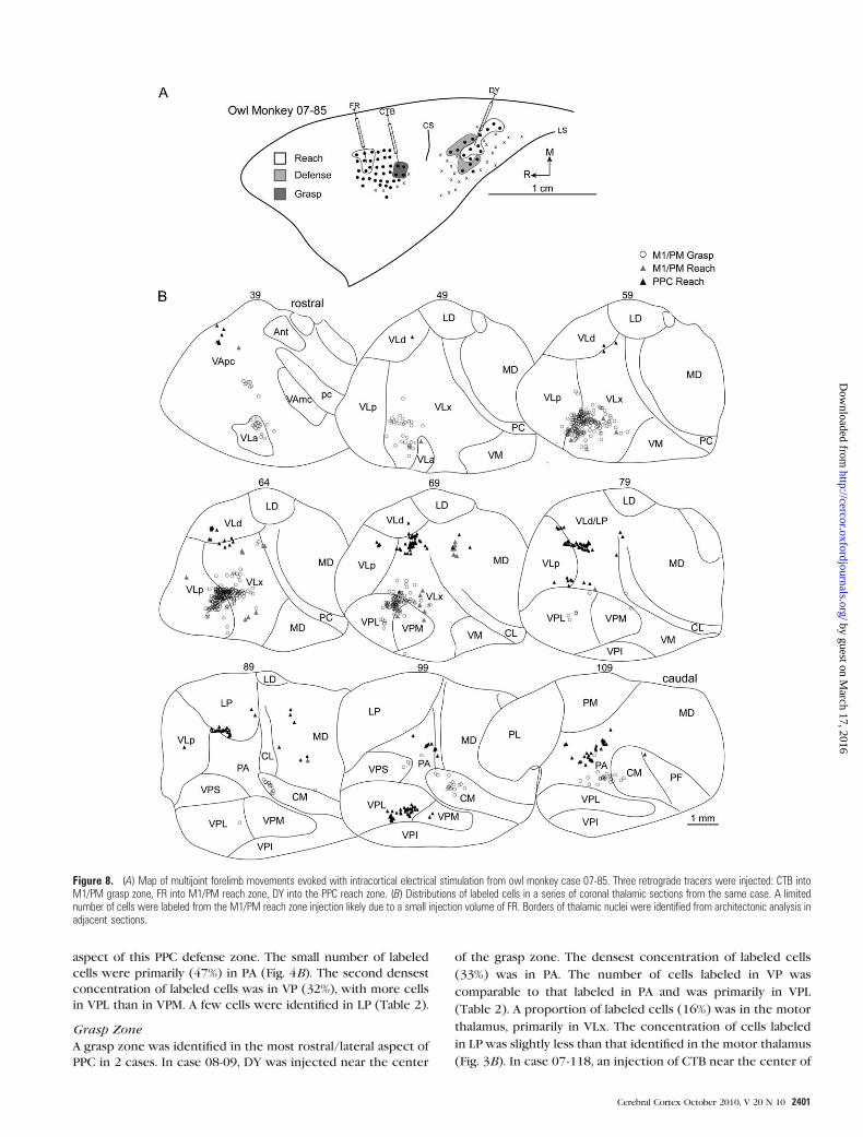
In case 07-85, CTB was injected into the center of the grasp
zone in caudal M1/PM (Fig. 8A). More than 71% of labeled cells
were in the motor thalamus with the densest concentration in
VLx (Table 1). The second densest concentration of labeled
cells was in VLp (Fig. 8B). The number of cells labeled in
rostral motor thalamus was markedly less. A small proportion of
labeled cells were in intralaminar nuclei, primarily in CM. A
small proportion of cells was labeled in VP, largely in VPL. A
small proportion of labeled cells was also identified in PA.
Reach Zone
Injections into the reach zone in rostral/medial M1/PM in 3
cases revealed consistent patterns of cell labeling. In case 08-
41, FB was injected into the center of the reach zone (Fig. 9A).
More than 82% of labeled cells were in motor thalamus with
the densest concentration in VLx (Table 1). The next densest
concentration of labeled cells was in VLa and VApc (Fig. 9B).
The proportion of labeled cells in VLp and VLd was com-
paratively less. A small proportion of cells was labeled in
intralaminar nuclei and was primarily concentrated in CL. A
comparable proportion of cells was labeled in adjacent MD. In
case 07-85, a small volume of FR was injected near the center of
the reach zone (Fig. 8A) at the approximate location of the
previous injection. A modest number of cells was labeled
(Table 1), and the majority (74%) was in the motor thalamus,
with the densest concentration in VLx (Fig. 8B). A few cells
were labeled in MD. In case 07-77, FR was injected near the
center of the reach zone at a more lateral location than the
previous 2 injections (Fig. 10A). Nearly 87% of labeled cells
were in motor thalamus (Table 1). The densest concentration
was in VLx, and fewer cells were labeled in VLp (Fig. 10B). A
few cells were labeled in MD.
Concurrent Response Zone
Tracers were injected into the concurrent response zone in
rostral/lateral M1/PM in 2 cases. In case 08-45, DY injection
was near sites that produced concurrent defensive forelimb
movements and face grimace (Fig. 6A). Labeled cells were
primarily in motor thalamus (70%) with the densest concen-
tration in VLx (34%). Rostral motor thalamus comprised the
next densest concentration of labeled cells (Table 1). The ma-
jority of those cells was in VApc, and the remainder was in
VLa. A small proportion of cells were identified in intralaminar
nuclei PC and CL as well as in adjacent MD. A few cells were
labeled in PA. In case 07-103, DY injection (Fig. 7A) was near
sites that evoked concurrent grasping and mouth opening and
was in the approximate location of the previous injection. The
distribution of labeled cells was similar for the 2 injections.
Nearly 87% of labeled cells were in motor thalamus (Table 1).
The densest concentration was in VLx (39%), whereas only
Table 1Distributions of labeled cells from retrograde tracer injections in M1/PM functional zones in 3 squirrel monkeys and 5 owl monkeys
Defense Grasp Reach Concurrent
Sq Sq Owl Sq Owl Sq Owl Owl Owl Sq Owl Owl07-118 08-03 07-103 08-09 07-85 08-03 08-41 07-85 07-77 08-09 08-45 07-103FB FB FR CTB CTB FR FB FR FR FR DY DY1705.00 50.00 10.00 1423.00 685.00 20.00 804.00 50.00 117.00 117.00 432.00 807.00
Vapc 1.11 — 30.00 — 1.46 — 18.16 4.00 — — 22.45 13.75Vla 11.03 — 10.00 — 6.57 10.00 22.14 4.00 — — 12.73 20.20VLD — — — 0.77 — 30.00 2.99 — — — — 0.25VLx 16.48 80.00 20.00 56.50 44.53 55.00 28.73 64.00 57.98 56.41 34.26 39.16VLp 36.89 — 40.00 15.04 18.69 — 10.32 2.00 28.57 36.75 0.46 13.63VM 3.40 — — — — — — 2.00 — 1.71 — 1.61PC 2.11 — — 0.63 — — 0.62 2.00 0.84 — 6.71 2.11CL 0.76 — — 9.98 0.29 — 7.59 — 0.84 — 11.34 6.07CM 4.63 — — 4.78 10.07 — 1.24 — — 2.56 — —LD — — — 0.07 — — 0.62 — — — — —MD 2.35 — — 7.24 0.44 5.00 7.21 18.00 8.40 — 7.64 2.60VPM 0.65 2.00 — 0.14 0.88 — — 4.00 — — 0.69 0.25VPL 7.51 14.00 — 1.48 11.24 — — — 1.68 1.71 0.46 —VPS 5.22 — — — 1.02 — — — — — — —VPI 0.11 — — — — — — — — — — —LP 0.35 4.00 — 0.56 — — — — 1.68 — 0.23 0.25PL — — — 0.35 — — — — — — — —PA 7.33 — — 2.46 4.82 — 0.37 — — 0.85 3.01 0.12PM — — — — — — — — — — — —
Rows sequentially list M1/PM functional zones, species of monkey investigated (Sq: squirrel monkey and Owl: owl monkey), case number of each monkey, tracer injected, and total number of cells
labeled in the thalamus from each injection. Thalamic nuclei are listed in the first column, and successive columns contain the percentage of cells labeled in each thalamic nucleus. The densest
concentration of labeled cells for each injection is listed in bold.
2396 PPC Thalamocortical Connections d Gharbawie et al.
by guest on March 17, 2016
http://cercor.oxfordjournals.org/D
ownloaded from

half that proportion was in VLa (Fig. 7B). An equal number of
cells were labeled in VLp and VApc. A small proportion of
labeled cells was in intralaminar nuclei and was primarily
limited to CL.
PPC Organization (Squirrel Monkeys)
Functional zones of PPC were identified with intracortical
electrical stimulation in the same squirrel monkeys. The
responsive region was medial to the lateral sulcus and caudal
to the central sulcus. Functional zones were organized approx-
imately parallel to the lateral sulcus (Fig. 3A). A reach zone was
in caudomedial PPC. A defensive zone was lateral and slightly
rostral to the reach zone. A grasp zone was lateral to
the defense zone. The reach and defense zones were often
separated by sites that evoked forelimb lifting as well as
forelimb adduction to the body. The defense and grasp zones
were separated by sites that evoked wrist supination to the
mouth and by unresponsive sites.
Figure 4. (A) Map of multijoint forelimb movements evoked with intracortical electrical stimulation from squirrel monkey case 08-03. Four retrograde tracers were injected: FBinto M1/PM defense zone, FR into M1/PM reach zone, CTB into PPC reach zone, and DY into PPC defense zone. (B) Distributions of labeled cells in a series of coronal thalamicsections from the same case. A brief survival period limited tracer transport for at least 3 injections. The adequate number of labeled cells from tracer injection in the PPC reachzone was likely due to rapid CTB transport. Borders of thalamic nuclei were identified from architectonic analysis in adjacent sections.
Cerebral Cortex October 2010, V 20 N 10 2397
by guest on March 17, 2016
http://cercor.oxfordjournals.org/D
ownloaded from

PPC Thalamocortical Connections (Squirrel Monkeys)
Distributions of labeled cells from 6 injections in PPC are
summarized in Table 2. In general, labeled cells were concen-
trated in dorsal and posterior thalamic nuclei. Concentrations of
labeled cells were densest in LP and PA. Cells were also labeled in
the motor thalamus and to a lesser extent in VP.
Reach Zone
A reach zone was identified in the most caudomedial aspect of
PPC in 2 cases. In case 08-09, FB was injected near the center of
the PPC reach zone (Fig. 3A). The densest concentration of
labeled cells was in LP (45%). A less dense concentration (23%)
was in the motor thalamus, primarily localized in VLd with
Figure 5. (A) Map of multijoint forelimb movements evoked with intracortical electrical stimulation from squirrel monkey case 07-118. Three retrograde tracers were injected: FB into M1/PM defense zone, DY into PPC forelimb-to-body representation, and CTB into PPC grasp zone. (B) Distributions of labeled cells in a series of coronal thalamic sections from the same case.
2398 PPC Thalamocortical Connections d Gharbawie et al.
by guest on March 17, 2016
http://cercor.oxfordjournals.org/D
ownloaded from

fewer cells labeled in VLx (Table 2). In some sections, cells
labeled in VLx were close to the VLd border (Fig. 3B). Cells
labeled in VLd might have been a rostral extension to those
labeled in LP. Similarly, cells labeled in PL might have been a
caudal extension to those labeled in LP. A small proportion of
cells was labeled in CL, but the extent of labeling in the
intralaminar nuclei was otherwise negligible. A slightly higher
concentration of labeled cells was identified in adjacent MD.
The proportion of cells labeled in the pulvinar nuclei (13%) was
primarily concentrated in PA. In case 08-03, an injection of CTB
in the center of the PPC reach zone (Fig. 4A) confirmed the
distribution pattern of cells labeled from the previous injection.
Nearly 65% of all labeled cells were in LP (Table 2). Cells
labeled in the motor thalamus were primarily in VLx and to a
lesser extent in VLd (Fig. 4B). Pulvinar labeling from the pres-
ent injection was limited to PL and PM.
Forelimb-to-Body Zone
In case 07-118, sites that evoked forelimb adduction to the body
were concentrated in a zone in medial PPC (Fig. 5A). A DY in-
jection near the center of this zone labeled a modest number
of cells due to a small injection volume. More than half of the
Figure 6. (A) Map of multijoint forelimb movements evoked with intracortical electrical stimulation from owl monkey case 08-45. Microelectrode penetration sites are colorcoded to reflect evoked movements. Sites that evoked dual movements are represented in 2 colors derived from the color code in the figure legend. Major functional zones arehighlighted. M1/PM was primarily characterized by grasp, wrist supination, and defense zones. A reach zone was rostral and medial in M1/PM, whereas concurrent face andforelimb movements were evoked from the same approximate rostral extent but lateral. In PPC, reach, defense, and grasp zones were organized in a caudomedial to rostrolateralprogression. Reach and defense zones bordered one another, whereas unresponsive sites separated the defense and grasp zones of PPC. Four retrograde tracers were injected:DY into M1/PM near sites that evoked concurrent defensive forelimb movements and aggressive face gestures, CTB into PPC reach zone, FB into PPC defense zone, FR into PPCgrasp zone. (B) Distributions of labeled cells in a series of coronal thalamic sections (40 lm) from the same case. A limited number of cells were labeled from the PPC grasp zonebecause a small volume of FR was purposely injected to minimize tracer spread beyond this limited target zone.
Cerebral Cortex October 2010, V 20 N 10 2399
by guest on March 17, 2016
http://cercor.oxfordjournals.org/D
ownloaded from

labeled cells were in PA (Fig. 5B), and the second densest
concentration of labeled cells was in motor thalamus (39%),
primarily in VLp followed by VLx. A few labeled cells were in LP.
Defense Zone
In case 08-03, the defense zone was lateral and slightly rostral to
the reach zone (Fig. 4A). DY was injected into the rostrolateral
Figure 7. (A) Map of multijoint forelimb movements evoked with intracortical electrical stimulation from owl monkey case 07-103. Four retrograde tracers were injected: FR into theM1/PM defense zone, DY into M1/PM near sites that evoked grasping and mouth opening, CTB into PPC reach zone, and FB into PPC defense zone. (B) Distributions of labeled cells ina series of coronal thalamic sections from the same case. A limited number of cells were labeled from the M1/PM defense zone injection likely because of a small injection volume of FR.
2400 PPC Thalamocortical Connections d Gharbawie et al.
by guest on March 17, 2016
http://cercor.oxfordjournals.org/D
ownloaded from
aspect of this PPC defense zone. The small number of labeled
cells were primarily (47%) in PA (Fig. 4B). The second densest
concentration of labeled cells was in VP (32%), with more cells
in VPL than in VPM. A few cells were identified in LP (Table 2).
Grasp Zone
A grasp zone was identified in the most rostral/lateral aspect of
PPC in 2 cases. In case 08-09, DY was injected near the center
of the grasp zone. The densest concentration of labeled cells
(33%) was in PA. The number of cells labeled in VP was
comparable to that labeled in PA and was primarily in VPL
(Table 2). A proportion of labeled cells (16%) was in the motor
thalamus, primarily in VLx. The concentration of cells labeled
in LP was slightly less than that identified in the motor thalamus
(Fig. 3B). In case 07-118, an injection of CTB near the center of
Figure 8. (A) Map of multijoint forelimb movements evoked with intracortical electrical stimulation from owl monkey case 07-85. Three retrograde tracers were injected: CTB intoM1/PM grasp zone, FR into M1/PM reach zone, DY into the PPC reach zone. (B) Distributions of labeled cells in a series of coronal thalamic sections from the same case. A limitednumber of cells were labeled from the M1/PM reach zone injection likely due to a small injection volume of FR. Borders of thalamic nuclei were identified from architectonic analysis inadjacent sections.
Cerebral Cortex October 2010, V 20 N 10 2401
by guest on March 17, 2016
http://cercor.oxfordjournals.org/D
ownloaded from

the grasp zone (Fig. 5A) confirmed the cell-labeling pattern from
the previous injection. The densest concentration (27%) of
labeled cells was in PA (Table 2). The proportion of labeled cells
in the motor thalamus (40%) was equally distributed between
VLx and VLp (Fig. 5B). The proportion of cells labeled in VP was
approximately the same as in PA with denser concentrations in
VPL. Unlike the previous injection, the number of cells labeled in
LP was negligible.
PPC Organization (Owl Monkeys)
Reach and defense zones were organized approximately parallel
to the lateral sulcus as in squirrel monkeys. A grasp represen-
tation zone was only identified in a single case, and it was
separated laterally from the defense zone by unresponsive sites
(Fig. 6A).
PPC Thalamocortical Connections (Owl Monkey)
The distributions of labeled cells from 9 tracer injections in PPC
are summarized in Table 2. The general pattern of connections
was similar to that observed in squirrel monkeys. Labeled cells
were mostly concentrated in LP and PA. Cells were also labeled
in motor thalamus and to a lesser extent in VP, PM, and PL.
Reach Zone
A reach zone was identified in the most caudomedial aspect
of PPC in all owl monkeys and was injected in 4 cases. In case
Figure 9. (A) Map of multijoint forelimb movements evoked with intracortical electrical stimulation from owl monkey case 08-41. Three retrograde tracers were injected: FB intoM1/PM reach zone, FR into PPC reach zone, and CTB into PPC defense zone. (B) Distributions of labeled cells in a series of coronal thalamic sections from the same case.
2402 PPC Thalamocortical Connections d Gharbawie et al.
by guest on March 17, 2016
http://cercor.oxfordjournals.org/D
ownloaded from

08-45, CTB was injected in the caudal aspect of the reach zone.
The densest concentration of labeled cells was in LP (37%).
Nevertheless, the number of cells labeled in the motor
thalamus was nearly 42% (Table 2). Those cells were primarily
concentrated in VLx near its border with VLd and LP (Fig. 6B).
The remainder of the cells labeled in the motor thalamus was in
VLd and to a lesser extent in VLp and VApc. A small proportion
of labeled cells were in intralaminar nuclei, primarily CL.
Another small proportion of cells was identified in PA and PM.
In case 07-85, DY was injected into the rostral/lateral aspect of
the reach zone (Fig. 8A) and revealed a distribution of labeled
cells similar to the previous injection. The densest concen-
trations of labeled cells were in LP (21%) and PA (22%).
Nevertheless, nearly 34% of labeled cells were in the motor
thalamus with the densest concentration in VLx (Table 2).
Most of the remaining cells labeled in motor thalamus were
Figure 10. (A) Map of multijoint forelimb movements evoked with intracortical electrical stimulation from owl monkey case 07-77. Two retrograde tracers were injected: FR intoM1/PM reach zone and DY into PPC defense zone. (B) Distributions of labeled cells in a series of coronal thalamic sections from the same case.
Cerebral Cortex October 2010, V 20 N 10 2403
by guest on March 17, 2016
http://cercor.oxfordjournals.org/D
ownloaded from

equally distributed between VLd and VLp (Fig. 8B). A small
proportion of labeled cells was present in adjacent MD. A
proportion of labeled cells was in VP (15%) and was primarily
localized to VPL. In case 08-41, FR was injected into the center
of the reach zone (Fig. 9A). The distribution of labeled cells was
comparable with the previous 2 injections despite the modest
number of cells labeled (Table 2). Nearly 50% of labeled cells
were in the motor thalamus (Fig. 9B). The densest concentra-
tion was in VLd (38%), which might have been a rostral
extension of the cells labeled in LP that comprised the second
densest concentration of labeled cells (36%). A few cells were
labeled in PM. In case 07-103, CTB was injected near the center
of this reach zone (Fig. 7A). The densest concentration (27%)
of labeled cells was in PA (Table 1). Nevertheless, nearly 55% of
labeled cells were in the motor thalamus with the densest
concentration in VLp being comparable with that in PA (Fig.
7B). The second densest concentration of labeled cells within
the motor thalamus was equally distributed between VLd and
VLx. A few cells were labeled in VApc. A small number of cells
was labeled in intralaminar nuclei, MD, LP, and PL.
Defense Zone
A defense zone was lateral and slightly rostral to the reach zone
in all owl monkeys and was injected in 4 cases. In case 08-45,
FB was injected near the center of the defense zone (Fig. 6A). A
modest number of cells was labeled likely due to a limited
volume of injected FB (Table 2). The densest concentration
of labeled cells was in LP (35%). Nevertheless, nearly 54% of
labeled cells were in the motor thalamus (Table 2), and they
were primarily concentrated in VLx near its border with VLd
(Fig. 6B). A smaller proportion of labeled cells was approxi-
mately distributed between VLd and VLp. A few cells were
labeled in PA. In case 07-77, DY was injected near the center of
the defense zone (Fig. 10A) and revealed a similar distribution
of labeled cells to the previous injection. The densest con-
centration of labeled cells was in LP (60%). Nearly 30% of
labeled cells were in the motor thalamus (Table 2), and they
were concentrated in VLd as well as VLx near its border with
VLd and LP (Fig. 10B). A few cells were labeled in the in-
tralaminar nuclei, and they were primarily in CL. In case 07-
103, FB was injected into the medial aspect of the defense
zone. Nearly 41% of labeled cells were in the motor thalamus
(Table 2) with the densest concentration in VLp and only
a small proportion in VLx (Fig. 7B). The second densest con-
centration of labeled cells was in PA (30%). A small proportion
of labeled cells was identified in VPS (17%). The border
separating VPS from VLp was not clear in at least one thalamic
section leaving open the possibility that a small proportion of
labeled cells was mischaracterized in VPS or VLp. An even
smaller concentration of cells was labeled in LP (8%). A few
labeled cells were identified in PL. In case 08-41, CTB was
injected into the caudal aspect of the defense zone (Fig. 9A).
Nearly 55% of labeled cells were in the motor thalamus with
the densest concentration in VLd (Table 2). A slightly smaller
proportion of labeled cells was in VLx near its border with VLd
(Fig. 9B). A markedly smaller proportion of labeled cells was in
VLp also near the VLd border. A few cells were labeled in CL.
Comparable concentrations of labeled cells were identified in
LP (14%), PA (12%), and PM (11%).
Grasp Zone
In cases 08-45, a grasp zone was identified in the most lateral
aspect of PPC (Fig. 6A) and was injected with a small volume of
FR, which labeled a limited number of cells (Table 2). Although
cells were primarily labeled in PA (30%), nearly 37% of cells
were in VP, with the densest concentration in VPL (Fig. 6B). A
proportion of labeled cells (22%) was in LP.
Topography of Thalamocortical Connections
Injecting multiple tracers in each monkey revealed the
thalamic nuclei that project to functional zones within PPC
and M1/PM. In addition, the topography of thalamocortical
Table 2Distributions of cells labeled from retrograde tracer injections in PPC functional zones in 3 squirrel monkeys and 5 owl monkeys
Reach Defense Grasp
Sq Sq Owl Owl Owl Owl Sq Owl Owl Owl Owl Sq Sq Owl08-09 08-03 08-45 07-85 08-41 07-103 08-03 08-45 07-77 07-103 08-41 08-09 07-118 08-45FB CTB CTB DY FR CTB DY FB DY FB CTB DY CTB FR1743.00 424.00 425.00 414.00 56.00 171.00 19.00 74.00 252.00 188.00 994.00 344.00 642.00 27.00
Vapc — — 4.00 2.42 3.57 2.92 — — — 0 0.30 — — —Vla — — — — — — — — — — — 0.29 0.31 —VLd 17.27 4.72 9.41 6.28 37.50 13.45 — 14.86 16.27 — 26.26 0.29 2.65 —VLx 5.22 9.19 24.24 18.12 1.79 13.45 5.26 27.03 13.49 8.51 21.23 14.82 18.38 7.41VLp 0.34 — 4.71 6.76 7.14 25.15 — 12.16 — 48.94 7.55 0.29 18.85 —VM — — — — — — — — — — — — — —PC 0.69 0.24 0.24 0.24 — 2.34 — — 1.19 — 0.91 0.29 2.18 —CL 6.94 2.59 4.47 2.17 — 1.75 — 1.35 4.37 — 2.52 1.74 1.09 —CM 0.92 0.47 1.41 0.72 — 1.17 — — — — 1.41 — 0.78 —LD 10.04 1.65 0.24 5.07 — 2.34 — — 0.40 — 0.20 0.29 0.00 —MD 10.04 1.65 0.24 5.07 — 2.34 — — 0.40 — 0.80 0.87 0.16 —VPM — — — 0.48 — — 21.05 — — — 0.00 4.65 4.67 7.41VPL — — — 12.56 — — 10.53 — — 1.06 0.20 29.94 16.36 18.52VPS — — — 1.69 — — — — 0.79 4.26 1.11 — 1.56 11.11VPI — — — — — — — — — — — — 5.76 —LP 45.09 64.62 37.18 20.29 35.70 4.09 15.79 35.14 59.52 7.98 14.10 13.08 0.31 22.22PL 2.35 6.6 — — — 5.85 — — — 3.19 0.80 — — 3.70PA 8.84 — 8.94 21.50 1.79 26.90 47.37 9.46 2.38 26.06 11.97 34.43 26.95 29.63PM 1.20 9.91 5.18 1.45 10.71 — — — 1.59 — 10.66 — — —
Rows sequentially list M1/PM functional zones, species of monkey investigated (Sq: squirrel monkey, Owl: owl monkey), case number of each monkey, tracer injected, and total number of cells labeled in
the thalamus from each injection. Thalamic nuclei are listed in the first column, and successive columns contain the percentage of cells labeled in each thalamic nucleus. The densest concentration of
labeled cells for each injection is listed in bold.
2404 PPC Thalamocortical Connections d Gharbawie et al.
by guest on March 17, 2016
http://cercor.oxfordjournals.org/D
ownloaded from

projections could also be compared across functional zones.
The topographical organization of PPC thalamocortical con-
nections was most apparent in squirrel monkey cases 08-09
and 07-118 (Figs. 3B and 5B, respectively) because PPC in-
jections in those cases were spatially segregated. The general
distribution of labeled cells was such that cells labeled from
injections into medial PPC (reach zone and forelimb-to-body
zone) were dorsal and slightly lateral to cells labeled from
lateral PPC injections (grasp zone). Injections into the PPC
reach zone and PPC defense zone in case 08-03 were spatially
closer (Fig. 4B). Nevertheless, cells labeled from the reach zone
injection were mostly dorsal to those labeled from the defense
zone injection. Three PPC injections in owl monkey case 08-45
confirmed the topographical organization observed in squirrel
monkeys. Cells labeled from the reach zone injection were
dorsal to those labeled from the defense zone injection, which
were in turn dorsal to those labeled from the grasp zone
injection (Fig. 6B). Similarly in owl monkey case 07-103, cells
labeled from the reach zone injection were primarily dorsal to
those labeled from the defense zone injection (Fig. 7B).
Although the topographic organization of labeled cells was
less obvious in case 08-41, some of the cells labeled from the
reach zone injection were lateral and dorsal to most cells
labeled from the defense zone injection. In the main, cells
labeled from PPC injections in both species occupied a more
dorsal and slightly lateral position, than cells labeled from M1/
PM injections. Perhaps the most apparent exception was for
proportions of cells labeled from injections into the PPC grasp
zone partially overlapping in VLx/VLp with cells labeled from
injections into caudal M1/PM (Figs. 3B and 5B).
Summary of Thalamocortical Connections
The densities of thalamocortical connections with PPC and M1/
PM zones are summarized in Figure 11. Connection patterns
were similar for squirrel and owl monkeys and results from the 2
species were pooled for the summary. Twelve injections into
M1/PM showed that it was primarily connected to VL divisions.
Nearly half of the connections were with VLx and the second
densest projection was from VLp. Connections with VLd, VLa,
and VApc were comparatively weaker. Connections with intra-
laminar nuclei and MD were even less dense. The weakest
connections were with VPL and PA. Fifteen injections into PPC
showed that its zones were most densely connected with dorsal
and posterior thalamic nuclei. The caudomedial reach zone was
primarily connected to LP followed by motor thalamus (VLd,
VLx, and VLp). The next densest connections were with PA.
Additional connections were with intralaminar nuclei, VApc, CL,
MD, VPL, PL, and PM. Connections of the more lateral and
slightly rostral defense zone were similar to those of the PPC
reach zone. The densest connections were with LP followed by
PA. Connections with motor thalamus were nearly equally
distributed between VLd, VLx, and VLp. The PPC defense zone
received denser projections from VP than the PPC reach zone.
Connections with CL, PL, and PM were minimal. The most lateral
PPC grasp zone was most densely connected with PA.
Connections with LP were less dense than for the PPC reach
and defense zones. Projections from the motor thalamus were
from VLx and VLp and only minimally from VLd. Connections
with VP were denser for the PPC grasp zone than for the reach
and defense zones, particularly for VPL. Minimal connections
were with CL, MD, and PL.
Discussion
Thalamocortical connections of PPC were investigated in squirrel
and owl monkeys. PPC zones were identified according to
forelimb movements evoked with long-train intracortical elec-
trical stimulation. Retrograde tracers were injected into PPC
zones and in M1/PM for comparison. M1/PM was primarily con-
nected to VL divisions of the motor thalamus, whereas PPC zones
were primarily connected to LP and PA. The most interesting
finding of the present study was the additional input from motor
and somatosensory thalamus that differentially projected to PPC
zones. In addition, the caudomedial to rostrolateral topographical
organization of PPC zones was reflected in a dorsoventral pattern
of thalamocortical connections.
Figure 11. Histogram summaries of the distribution of cells labeled in thalamicnuclei from 3 squirrel monkeys and 5 owl monkeys. The mean percentage of labeledcells and corresponding standard errors of the mean were calculated from thedistributions presented in Tables 1 and 2. The number of injections into eachfunctional zone is listed in the same tables. Thalamic nuclei included in the histogramscontained at least a mean of 2% of the labeled cells for any given functional zone.
Cerebral Cortex October 2010, V 20 N 10 2405
by guest on March 17, 2016
http://cercor.oxfordjournals.org/D
ownloaded from

PPC Organization
Electrical stimulation evoked multijoint forelimb movements
from PPC zones in similar locations in squirrel and owl
monkeys. Functional zones were approximately parallel to
the lateral sulcus and included a caudomedial reach zone and
a more lateral and slightly rostral defense zone. A grasp zone
was less consistently identified lateral to the defense zone. Sites
that evoked other forelimb movements bordered those 3 zones,
but their numbers were comparatively lower and their dis-
tributions were less consistent. Both the reach and the defense
zones were too caudal to the central sulcus to be part of
somatosensory cortex. The same cannot be ascertained for the
grasp zone because its proximity to the central sulcus leaves
open the possibility that it partially overlapped the anterior pa-
rietal cortex, for example, areas 1 and 2 (Merzenich et al. 1978;
Pons and Kaas 1985).
Long-train electrical stimulation of PPC has only been
reported in galagos and macaque monkeys. The absence of an
IPS in the present New World monkeys complicates comparison
of the present results to those published reports. Furthermore,
PPC microstimulation in macaques has been limited to the
lateral bank (Thier and Andersen 1996) and fundus (Cooke et al.
2003) of the IPS. Thus, we will refer to relevant results from
single unit recordings in PPC of behaving macaque monkeys to
compensate for the dearth of PPC stimulation studies.
A reach zone was identified in caudomedial PPC in squirrel and
owl monkeys. This relative location is similar to the reach zone
evoked from caudomedial PPC in galagos (Stepniewska et al.
2005; Stepniewska, Cerkevich, et al. 2009; Stepniewska, Fang, and
Kaas 2009). Single-unit recordings in macaque monkeys showed
that neural activity in segments of the medial bank of the IPS,
known as the medial intraparietal area (MIP), is predictive of
forelimb extension during a reach (Johnson et al. 1996). Unit
activity in the dorsal aspect of the parietal--occipital area adjacent
to MIP is also sensitive to reaching (Battaglia-Mayer et al. 2000).
The 2 regions have been collectively coined the parietal reach
region (Cohen and Andersen 2002), and the present PPC reach
zone may be partly or wholly homologous to it. The caveat for this
interpretation is the assumption of faithful overlap between the
actions encoded by neural activity and actions evoked with
electrical stimulation from the same group of neurons, that is,
neurons active during reaching also evoke reaching movements
during electrical stimulation under anesthesia.
A defense zone was adjacent and lateral to the PPC reach
zone. Defensive forelimb movements were evoked from a
relatively similar location in galagos (Stepniewska et al. 2005;
Stepniewska, Cerkevich, et al. 2009; Stepniewska, Fang, and
Kaas 2009). Aggressive face movements in the present study
also paralleled the organization in galagos. Defensive forelimb
movements and aggressive face gestures have also been evoked
from the fundus of the IPS in macaque monkeys (Cooke et al.
2003), a region coined the VIP. Given the relative position of
the present defense zone and its constellations of movements,
it is likely homologous to the defense zones in galagos and ma-
caque monkeys.
A grasp/wrist supination zone was lateral to the defense
zone, albeit less consistently identified than the reach and
defense zones, in squirrel and owl monkeys. Although grasping
movements were not reported from PPC electrical stimulation
in galagos, a small zone of hand-to-mouth movements was
identified lateral to the defense zone (Stepniewska et al. 2005).
Thus, there is spatial overlap between the present grasp zone
and the hand-to-mouth zone in galagos. Moreover, recent
results from galagos have identified a small grasp zone slightly
rostral to the hand-to-mouth zone (Stepniewska, Cerkevich,
et al. 2009; Stepniewska, Fang, and Kaas 2009). There have been
no reports of grasping movements evoked with electrical
stimulation from PPC in macaque monkeys. Nevertheless,
single-unit activity within rostral aspects of the lateral bank of
the IPS overlapped hand shaping for grasping (Sakata et al.
1995). Temporary deactivation of the same region impaired
digit shaping to the contours of target objects (Gallese et al.
1994). This grasp sensitive zone has been coined the anterior
intraparietal area (AIP), and the grasp zone identified in the
present monkeys may be its homolog. Again, this interpretation
assumes faithful correspondence between actions encoded
by single units in behaving monkeys and those evoked with
electrical stimulation under anesthesia.
Forelimb movements evoked from PPC with electrical
stimulation were likely driven indirectly through activation of
parietal--frontal networks. Short-train microstimulation (10--18
pulses in 50 ms), effective in evoking muscle twitches from M1/
PM, did not evoke movements from PPC. This is not surprising
considering corticospinal projections arising in PPC are sparse by
comparison to projections from M1/PM and limited to area 5 as
well as rostral aspects of the medial bank of the IPS (Nudo and
Masterton 1990; Galea and Darian-Smith 1994; Matelli et al.
1998). The effectiveness of long-train electrical stimulation (150
pulses in 500 ms) in evoking forelimb movements from PPC
suggests the recruitment of distant targets. It is unlikely that
movements were evoked due to indiscriminate current spread
for at least 2 reasons. First, PPC zones were separated from M1/
PM by somatosensory cortex, which was mostly unresponsive to
microstimulation. Second, there were subtle variations in fore-
limb movements evoked within a PPC zone and more substantial
shifts in forelimb movements across functional zones. Both
reasons diminish the possibility that indiscriminate current
spread prompted movements from PPC, in which case a contin-
uous map joining PPC and M1/PM as well as homogenous
forelimb responses from electrical stimulation sites, would have
been expected. The most parsimonious explanation for move-
ments evoked from PPC zones is that electrical stimulation
activated specific neuroanatomical channels linking PPC with
M1/PM. Supporting anatomical evidence in owl monkeys
(Stepniewska et al. 1993, 2006) and galagos (Stepniewska,
Cerkevich, et al. 2009; Stepniewska, Fang, and Kaas 2009) shows
strong connections between M1/PM and the approximate
location of PPC zones identified here. Parietal--frontal networks
have been shown in macaque monkeys with tracer injections in
M1/PM as well as with PPC injections (Strick and Kim 1978;
Petrides and Pandya 1984; Matelli et al. 1998; Luppino et al. 1999;
Lewis and Van Essen 2000; Tanne-Gariepy et al. 2002).
M1/PM Thalamocortical Connections
Thalamocortical connection patterns of M1/PM were similar
for squirrel and owl monkeys. Connections were primarily with
VL and to a much lesser extent rostral motor thalamus. The
results are consistent with previous studies in New World
monkeys (Stepniewska et al. 1994a, 1994b, 2007), Old World
monkeys (Strick 1975; Matelli et al. 1989; Darian-Smith et al.
1990), and prosimian galagos (Fang et al. 2006). This con-
nection pattern is likely conserved in other phylogenetic
orders considering it has been documented for motor cortex in
2406 PPC Thalamocortical Connections d Gharbawie et al.
by guest on March 17, 2016
http://cercor.oxfordjournals.org/D
ownloaded from

rats (Donoghue and Parham 1983; Alder 1988) and cats (Strick
1970; Larsen and Asanuma 1979).
Electrical stimulation mapping and tracer injections into M1/
PM were included in the present study for 2 reasons: first, to
confirm that the electrical stimulation parameters reliably
identified zones within M1/PM and can therefore be used
to map PPC—indeed, the present thalamocortical connections
demonstrate that tracers were injected into traditional M1/PM
regions identified with cytoarchitecture and short-train micro-
stimulation (Asanuma and Rosen 1972; Strick and Peterson
1978; Sessle and Wiesendanger 1982; Gould et al. 1986;
Donoghue et al. 1992; Nudo et al. 1992; Stepniewska et al.
1993); second, for comparing the thalamocortical connections
of PPC zones with matching counterparts in M1/PM.
PPC Thalamocortical Connections
Relating the present PPC thalamocortical connections to pre-
vious work is constrained by the same limitations of comparing
the present neurophysiological results to existing reports. With
the exception of one report on titi monkeys (Padberg and
Krubitzer 2006), the thalamocortical connections of PPC have
not been studied in New World monkeys. Comparisons to the
better-documented PPC connections of macaque monkeys are
complicated because cortical landmarks (e.g., IPS) that guided
tracer injections in those studies are absent in the present
species. Thus, the locations of the present tracer injections are
described according to the forelimb movements evoked from
each PPC zone to facilitate comparisons to other studies.
In general, the present results showed that the densest
thalamic projections to PPC zones were from LP and PA.
Additional connections were with motor thalamus, somatosen-
sory thalamus, and other pulvinar nuclei. This is in agreement
with reports showing that the densest thalamocortical con-
nections to area 5 and the medial bank of the IPS (also known,
respectively, as PE and PEa) are from LP and pars oralis division
(PO) of pulvinar (Jones et al. 1979; Schmahmann and Pandya
1990; Cappe et al. 2007).
The most caudomedial PPC zone (reach) received its
densest connections from LP. Dense input from LP has been
consistently reported for PPC, especially area 5 in macaque
monkeys (Jones et al. 1979; Schmahmann and Pandya 1990;
Cappe et al. 2007; Padberg et al. 2009). However, dense LP
projections were not reported for area 5 in titi monkeys
(Padberg and Krubitzer 2006). The discrepancy should be
interpreted with caution because the dearth of LP connections
was reported from only 2 tracer injections into the hand
representation of area 5 identified by multiunit recordings.
Those injections might have been somatotopically incongruent
with injections into the present reach zone identified with
electrical stimulation.
The PPC reach zone received additional inputs from PA and
in one injection VPL, which imply direct somatosensory inputs
from the thalamus. Although PA projections are consistent with
the thalamic input of area 5 in Old and New World monkeys,
they also characterize somatosensory areas of the anterior
parietal cortex. For example, PA input to area 2, in macaque
monkeys (Pons and Kaas 1985; Schmahmann and Pandya 1990).
Nevertheless, the distance separating injections into the PPC
reach zone from the central sulcus precludes the possibility of
tracer spread into anterior parietal cortex and suggests that the
present projections likely parallel those reported for area 5.
Additional inputs to PPC reach zone, albeit minimal by
comparison, were from the lateral (PL) and medial (PM)
pulvinar. Reports from Old and New World monkeys have
shown PL projections to be related to vision. Nevertheless, cells
labeled in PL from the present injections were likely rostral to
those labeled in PL by injections into the dorsolateral area,
which is also known as V4 (Weller et al. 2002). In addition, the
dorsomedial division of PL is known to have connections with
PPC (Stepniewska 2004). Projections from PM are considered
multisensory. Diversity of input modality to the PPC reach zone
may be central to accurate forelimb trajectory during reaching.
The convergence of thalamic input from multiple nuclei
further supports the assertion that injections were caudal to
somatosensory cortex.
The PPC reach zone received projections from the motor
thalamus, primarily VLd, VLx, and VLp. The collective density of
this input rivaled that from LP in owl monkeys but was less
intense in squirrel monkeys. It is possible that the density of
these connections may have been overestimated due to
difficulties in identifying thalamic borders. For example,
discerning the border between VLd and LP in coronal sections
was challenging in instances of insufficient staining contrast
and was inferred from surrounding nuclei. Identifying the
ventral border of LP was occasionally complicated for the same
reasons. Thus, the possibility exists that labeled cells were
mischaracterized in VLd, VLx, or VLp, although they belonged
to LP. Nevertheless, this only raises concerns about a small
proportion of cells labeled in the motor thalamus. Although
cells labeled in VL from PPC reach zone injections were
separate from those labeled by M1/PM injections, the close
proximity of the 2 populations of cells suggests similarities
between the connections of PPC with motor thalamus and the
M1/PM counterpart. VL divisions receive dense projections
from deep cerebellar nuclei and are generally considered the
node that provides cortical motor regions with cerebellar
feedback (Alexander et al. 1986; Sakai et al. 2000).
A defense zone was lateral and slightly rostral to the PPC
reach zone. Thalamocortical connections of the PPC defense
zone were more thoroughly investigated in owl monkeys and
were similar to those of the PPC reach zone. The densest
connections were with LP, and a slightly weaker projection was
from PA. Direct somatosensory input was also apparent from VP
projections, which were relatively denser than those that
reached the PPC reach zone. Nevertheless, injections were too
distant from the central sulcus to have spread to the anterior
parietal somatosensory cortex. Cases that received injections in
the reach and defense zones showed that input to the defense
zone arose from neurons that were generally ventral to those
that projected to the reach zone. The slight spatial offsetting of
thalamic cells projecting to the reach and dense zones suggests
that they may represent different sequences of forelimb move-
ments that can be evoked from the same cortical area, likely area
5. The PPC defense zone received minimal input from PL and PM
reflecting various sensory modalities. The importance of such
input to a cortical zone involved in protecting the body can be
easily envisaged. Dense connections with VL divisions rivaled
those with LP and highlight the association of this PPC zone with
the motor system.
A grasp zone was identified lateral and slightly rostral to the
defense zone. Although the grasp zone was more thoroughly
investigated in squirrel monkeys, a comparable pattern of
thalamocortical connections was revealed for the 2 species,
which were also distinct from the pattern of connections of the
Cerebral Cortex October 2010, V 20 N 10 2407
by guest on March 17, 2016
http://cercor.oxfordjournals.org/D
ownloaded from

other PPC zones. Perhaps the most distinguishing feature of the
grasp zone was the limited number of cells labeled in LP.
Pulvinar projections were primarily from PA and characterized
the bulk of the grasp zone input. In addition, VP connections
with the PPC grasp zone were denser than with the other PPC
zones. The distribution of cells labeled in VLx/VLp from PPC
grasp zone injections partially overlapped cells labeled from
injections into caudal M1/PM (squirrel monkey case 08-09 and
07-118), whereas they were spatially segregated from cells
labeled from more rostral M1/PM injections (squirrel monkey
case 08-09 and owl monkey case 08-45). This overlap may be
a feature of the relative proximity of the corresponding
injections. It may also reflect a closer thalamocortical relation-
ship between PPC grasp and caudal M1/PM than is the case for
other functional zones.
The spatial location of the PPC grasp zone and its pattern of
thalamocortical connections complicate assignment to a con-
ventional cortical area. The limited number of labeled cells in
LP is inconsistent with the dense thalamocortical connections
of area 5 in macaque monkeys (Jones et al. 1979; Schmahmann
and Pandya 1990; Cappe et al. 2007). Limited LP connections
are consistent however with the results from a small number of
injections in area 5 of titi monkeys (Padberg and Krubitzer
2006) as well as connection patterns of the inferior parietal
lobe in macaque monkeys (Schmahmann and Pandya 1990).
Even though the pattern of connections of the PPC grasp zone
is similar to that reported for area 2 in macaque monkeys (Pons
and Kaas 1985), electrophysiological and neuroanatomical
investigations of the same region in titi monkeys have been
interpreted as evidence for area 5 and not area 2 (Padberg et al.
2005; Padberg and Krubitzer 2006). Tentatively, repealing
those 2 areas as equivalents of the PPC grasp zone limits the
possibilities to area 1 and area 7b. Dense inputs from VP, and
particularly VPS from the present injections, are consistent
with cell-labeling patterns after area 1 injections in titi
monkeys (Coq et al. 2004; Padberg and Krubitzer 2006). Dense
inputs from PA to the present grasp zone is also consistent with
area 1 connections. However, projections from VL divisions
may reflect injections into area 1, area 7b, or both. As explained
earlier, if the sequence of forelimb movements evoked with
electrical stimulation is a faithful representation of the actions
that drive neural activity, then the present PPC grasp zone
likely corresponds to 7b or AIP in macaque monkeys. In this
case, the present projections from VL divisions may be the link
revealed with transynaptic tracing methods that connects deep
cerebellar nuclei with AIP via VL (Clower et al. 2005).
Nevertheless, the present PPC grasp zone appears to share
thalamocortical connection patterns with both anterior (areas
1 and 2) and posterior (areas 5 and 7b) parietal cortex.
The density of the present VL projections to PPC zones
rivaled those from LP. Reports in macaque monkeys have
generally shown that LP projections to PPC are much denser
than the VL counterpart. Nevertheless, the relative density of
VL projections to PPC varies between reports. For example,
Schmahmann and Pandya (1990) estimate substantial projec-
tions from VL to PE and PEa. That those connections arise from
dorsal VL (VLps and VLc) further supports the present results
showing that cells labeled in motor thalamus were primarily
concentrated in VLd. Nevertheless, a separate investigation of
PE and PEa showed only weak connections with VL (Cappe
et al. 2007). The similarities and discrepancies in the findings
may reflect differences in somatotopic locations of the
injections. Indeed, the present results show that caudomedial
PPC (reach zone) is less densely connected to VL than
rostrolateral PPC (grasp zone). Another source of discrepancy
between the present results and macaque monkeys (Cappe
et al. 2007) may be related to species differences.
Convergence of PPC Thalamocortical Connections
The rostrocaudal extent of the thalamocortical connection
origins was markedly different for M1/PM and PPC. Thalamic
input to M1/PM was primarily from VL divisions. Only a marginal
number of cells were labeled more caudally in VP or PA. In
contrast, thalamic inputs to PPC zones were distributed across
a longer rostrocaudal axis. The near even distribution of labeled
cells in several thalamic nuclei (LP, VL, VP, and pulvinar) from
PPC injections suggests a convergence of thalamic input onto
PPC. The wide range of modalities from this converging input
likely reflects higher order processing in PPC as compared with
M1/PM. A similar contrast has been demonstrated between the
concentrated input from VP to anterior parietal cortex (areas 3b,
1, and 2) and the wide convergence of thalamic inputs to area 5
(Padberg et al. 2009).
Conclusions
Electrical stimulation with long trains of current proved reliable
for identifying PPC zones involved in action. The pattern of
thalamocortical connections distinguished the individual PPC
zones (Fig. 11). In addition to thalamocortical connections with
dorsal and posterior nuclei of the thalamus, PPC zones received
projections from motor and sensory thalamus. The caudomedial
to rostrolateral arrangement of the functional zones was
paralleled by a reduction in projections from LP and increases
in the density of projections from PA and VP. Dense inputs from
VL reached all PPC functional zones. Motor and sensory inputs
are consistent with the proposed role of this region of PPC in
generating actions.
Funding
National Institutes of Health (NS16446 to J.H.K and NS055843
to I.S.); Natural Sciences and Engineering Research Council of
Canada (postdoctoral fellowship to O.A.G).
Notes
We are grateful to Laura Trice for assistance with histology and Mary
Feurtado for pre- and postoperative animal care. Conflict of Interest :
None declared.
References
Alder LD. 1988. Thalamic connectivity of rat somatic motor cortex.
Brain Res Bull. 20:333--348.
Alexander GE, DeLong MR, Strick PL. 1986. Parallel organization of
functionally segregated circuits linking basal ganglia and cortex.
Annu Rev Neurosci. 9:357--381.
Asanuma H, Rosen I. 1972. Topographical organization of cortical
efferent zones projecting to distal forelimb muscles in the monkey.
Exp Brain Res. 14:243--256.
Battaglia-Mayer A, Ferraina S, Mitsuda T, Marconi B, Genovesio A,
Onorati P, Lacquaniti F, Caminiti R. 2000. Early coding of reaching in
the parietooccipital cortex. J Neurophys. 83:2374--2391.
2408 PPC Thalamocortical Connections d Gharbawie et al.
by guest on March 17, 2016
http://cercor.oxfordjournals.org/D
ownloaded from

Cappe C, Morel A, Rouiller EM. 2007. Thalamocortical and the dual
pattern of corticothalamic projections of the posterior parietal
cortex in macaque monkeys. Neuroscience. 146:1371--1387.
Clower DM, Dum RP, Strick PL. 2005. Basal ganglia and cerebellar
inputs to ‘AIP’. Cereb Cortex. 15:913--920.
Cohen YE, Andersen RA. 2002. A common reference frame for movement
plans in the posterior parietal cortex. Nat Rev Neurosci. 3:553--562.
Cooke DF, Taylor CS, Moore T, Graziano MS. 2003. Complex
movements evoked by microstimulation of the ventral intraparietal
area. Proc Natl Acad Sci U S A. 100:6163--6168.
Coq JQ, Qi H, Collins CE, Kaas JH. 2004. Anatomical and functional
organization of somatosensory areas of the lateral fissure of the New
World titi monkey (Callicebus moloch). J Comp Neurol.
476:368--387.
Darian-Smith C, Darian-Smith I, Cheema SS. 1990. Thalamic projections
to sensorimotor cortex in the macaque monkey: use of multiple
retrograde fluorescent tracers. J Comp Neurol. 299:17--46.
Donoghue JP, Leibovic S, Sanes JN. 1992. Organization of the forelimb
area in squirrel monkey motor cortex: representation of digit, wrist,
and elbow muscles. Exp Brain Res. 89:1--19.
Donoghue JP, Parham C. 1983. Afferent connections of the lateral
agranular field of the rat motor cortex. J Comp Neurol. 217:390--404.
Emmers R, Akert K. 1963. A stereotaxic atlas of the brain of the squirrel
monkey. Madison (WI): The University of Wisconsin Press.
Fang PC, Stepniewska I, Kaas JH. 2006. The thalamic connections of
motor, premotor, and prefrontal areas of cortex in a prosimian
primate (Otolemur garnetti). J Comp Neurol. 143:987--1020.
Galea MP, Darian-Smith I. 1994. Multiple corticospinal neuron populations
in the macaque monkey are specified by their unique cortical origins,
spinal terminations, and connections. Cereb Cortex. 4:166--194.
Gallese V, Murata A, Kaseda M, Niki N, Sakata H. 1994. Deficit of hand
preshaping after muscimol injection in monkey parietal cortex.
Neuroreport. 5:1525--1529.
Geneser-Jensen FA, Blackstad TW. 1971. Distribution of acetyl
cholinesterase in the hippocampal region of the guinea pig. I.
Entorhinal area, parasubiculum, and presubiculum. Z Zellforsch
Mikrosk Anat. 114:460--481.
Gharbawie OA, Stepniewska I, Burish BJ, Kaas JH. 2008. Comparison of
thalamocortical connections of frontal and posterior parietal cortex in
New World primates. Abstract 78.5. Society for Neuroscience 38th
annual meeting. Washington, DC.
Gould H, Jr., Cusick CG, Pons TP, Kaas JH. 1986. The relationship of
corpus callosum connections to electrical stimulation maps of
motor, supplementary motor, and the frontal eye fields in owl
monkeys. J Comp Neurol. 247:297--325.
Graziano MS, Taylor CS, Moore T. 2002. Complex movements evoked
by microstimulation of precentral cortex. Neuron. 34:841--851.
Johnson PB, Ferraina S, Bianchi L, Caminiti R. 1996. Cortical networks
for visual reaching: physiological and anatomical organization of
frontal and parietal lobe arm regions. Cereb Cortex. 6:102--119.
Jones EG. 1985. The thalamus. New York: Plenum Press.
Jones EG. 2007. The thalamus. Cambridge (MA): Cambridge University
Press.
Jones EG, Wise SP, Coulter JD. 1979. Differential thalamic relationships
of sensory-motor and parietal cortical fields in monkeys. J Comp
Neurol. 183:833--881.
Larsen KD, Asanuma H. 1979. Thalamic projections to the feline motor
cortex studied with horseradish peroxidase. Brain Res. 172:209--215.
Lewis JW, Van Essen DC. 2000. Corticocortical connections of visual,
sensorimotor, and multimodal processing areas in the parietal lobe
of the macaque monkey. J Comp Neurol. 428:112--137.
Luppino G, Murata A, Govoni P, Matelli M. 1999. Largely segregated
parietofrontal connections linking rostral intraparietal cortex (areas
AIP and VIP) and the ventral premotor cortex (areas F5 and F4). Exp
Brain Res. 128:181--187.
Matelli M, Govoni P, Galletti C, Kutz DF, Luppino G. 1998. Superior area
6 afferents from the superior parietal lobule in the macaque
monkey. J Comp Neurol. 402:327--352.
Matelli M, Luppino G, Fogassi L, Rizzolatti G. 1989. Thalamic input to
inferior area 6 and area 4 in the macaque monkey. J Comp Neurol.
280:468--488.
Merzenich MM, Kaas JH, Sur M, Chia-Sheng L. 1978. Double
representation of the body surface within cytoarchitectonic areas
3b and 1 in ‘‘S1’’ in the owl monkey (Aotus trivirgatus). J Comp
Neurol. 181:41--74.
Nudo RJ, Masterton RB. 1990. Descending pathways to the spinal cord,
III: sites of origin of the corticospinal tract. J Comp Neurol.
296:559--583.
Nudo RJ, Jenkins WM, Merzenich MM, Prejean T, Grenda R. 1992.
Neurophysiological correlates of hand preference in primary motor
cortex of adult squirrel monkeys. J Neurosci. 12:2918--2947.
Olszewski J. 1952. The thalamus of Macaca Mulatta. Basel (Switzer-
land): Krager.
Padberg J, Cerkevich C, Engle J, Rajan AT, Recanzone G, Kaas JH,
Krubitzer L. 2009. Thalamocortical connections of parietal somato-
sensory cortical fields in macaque monkeys are highly divergent and
convergent. Cereb Cortex. 9:2038--2064.
Padberg J, Disbrow E, Krubitzer L. 2005. The organization and
connections of anterior and posterior parietal cortex in titi monkeys:
do NewWorld monkeys have an area 2? Cereb Cortex. 12:1938--1963.
Padberg J, Krubitzer L. 2006. Thalamocortical connections of anterior
and posterior parietal cortical areas in New World titi monkeys. J
Comp Neurol. 487:416--435.
Petrides M, Pandya DN. 1984. Projections to the frontal cortex from the
posterior parietal region in the rhesus monkey. J Comp Neurol.
228:105--116.
Pons TP, Kaas JH. 1985. Connections of area 2 of somatosensory cortex
with the anterior pulvinar and subdivisions of the ventroposterior
complex in macaque monkeys. J Comp Neurol. 240:16--36.
Sakai ST, Stepniewska I, Kaas JH. 2000. Pallidal and cerebellar afferents
to pre-supplementary motor area thalamocortical neurons in the
owl monkey: a multiple labeling study. J Comp Neurol. 417:164--180.
Sakata H, Taira M, Murata A, Mine S. 1995. Neural mechanisms of visual
guidance of hand action in the parietal cortex of the monkey. Cereb
Cortex. 5:429--438.
Schmahmann JD, Pandya DN. 1990. Anatomical investigation of
projections from thalamus to posterior parietal cortex in the rhesus
monkey: a WGA-HRP and fluorescent tracer study. J Comp Neurol.
295:299--326.
Sessle BJ, Wiesendanger M. 1982. Structural and functional definition of
the motor cortex the monkey (Macaca fascicularis). J Physiol.
323:245--265.
Stepniewska I. 2004. The pulvinar complex. In: Kaas JH, Collins CE, editors.
The primate visual system. Boca Raton (FL): CRC Press. p. 53--80.
Stepniewska I, Cerkevich C, Fang PC, Kaas JH. 2009. Organization of the
posterior parietal cortex in galagos: iI. Ipsilateral cortical connec-
tions of physiologically identified zones within anterior sensorimo-
tor region. J Comp Neurol. 517:783--807.
Stepniewska I, Fang PC, Kaas JH. 2005. Microstimulation reveals
specialized subregions for different complex movements in
posterior parietal cortex of prosimian galagos. Proc Natl Acad Sci
U S A. 102:4878--4883.
Stepniewska I, Fang PC, Kaas JH. 2009. Organization of the posterior
parietal cortex in galagos: i. Functional zones identified by micro-
stimulation. J Comp Neurol. 517:765--782.
Stepniewska I, Preuss TM, Kaas JH. 1993. Architectonics, somatotopic
organization, and ipsilateral cortical connections of the primary
motor area (M1) of owl monkeys. J Comp Neurol. 330:238--271.
Stepniewska I, Preuss TM, Kaas JH. 1994a. Architectonic subdivisions of
the motor thalamus of owl monkeys: Nissl, acetylcholinesterase, and
cytochrome oxidase patterns. J Comp Neurol. 349:536--557.
Stepniewska I, Preuss TM, Kaas JH. 1994b. Thalamic connections of the
primary motor cortex (M1) of owl monkeys. J Comp Neurol.
349:558--582.
Stepniewska I, Preuss TM, Kaas JH. 2006. Ipsilateral cortical connec-
tions of dorsal and ventral premotor areas in New World owl
monkeys. J Comp Neurol. 495:691--708.
Stepniewska I, Preuss TM, Kaas JH. 2007. Thalamic connections of the
dorsal and ventral premotor areas in New World owl monkeys.
Neuroscience. 147:727--745.
Strick PL. 1970. Cortical projections of the feline thalamic nucleus
ventralis lateralis. Brain Res. 20:130--134.
Cerebral Cortex October 2010, V 20 N 10 2409
by guest on March 17, 2016
http://cercor.oxfordjournals.org/D
ownloaded from

Strick PL. 1975. Multiple source of thalamic input to the primate motor
cortex. Brain Res. 88:372--377.
Strick PL, Kim CC. 1978. Input to primate motor cortex from posterior
parietal cortex (area 5). I. Demonstration by retrograde transport.
Brain Res. 157:325--330.
Strick PL, Peterson JB. 1978. Multiple representations in the primate
motor cortex. Brain Res. 154:366--370.
Tanne-Gariepy J, Rouiller EM, Boussaoud D. 2002. Parietal inputs to
dorsal versus ventral premotor areas in the macaque monkey:
evidence for largely segregated visuomotor pathways. Exp Brain
Res. 145:91--103.
Thier P, Andersen RA. 1996. Electrical microstimulation suggests two
different forms of representation of head-centered space in the
intraparietal sulcus of rhesus monkeys. Proc Natl Acad Sci U S A.
93:4962--4967.
Veenman CL, Reiner A, Honing MG. 1992. Biotinylated dextran amine as
an anterograde tracer for single- and double-labeling studies. J
Neurosci Methods. 41:239--254.
Weller RE, Steele GE, Kaas JH. 2002. Pulvinar and subcortical
connection of dorsal lateral visual cortex in monkeys. J Comp
Neurol. 450:215--240.
Wise SP, Boussaoud D, Johnson PB, Caminiti R. 1997. Premotor and
parietal cortex: corticocortical connectivity and combinatorial
computations. Annu Rev Neurosci. 20:25--42.
Wong-Riley M. 1979. Changes in the visual system of monocularly
sutured or enucleated cats demonstrable with cytochrome oxidase
histochemistry. Brain Res. 171:11--28.
Yeterian EH, Pandya DN. 1985. Corticothalamic connections of the
posterior parietal cortex in the rhesus monkey. J Comp Neurol.
237:408--426.
2410 PPC Thalamocortical Connections d Gharbawie et al.
by guest on March 17, 2016
http://cercor.oxfordjournals.org/D
ownloaded from