Neural correlates of learning and working memory in the primate posterior parietal cortex
21
Neural correlates of learning and working memory in the primate posterior parietal cortex Justin B. Rawley and Christos Constantinidis * Department of Neurobiology and Anatomy, Wake Forest University School of Medicine Abstract The posterior parietal cortex has been traditionally associated with coordinate transformations necessary for interaction with the environment and with visual-spatial attention. More recently, involvement of posterior parietal cortex in other cognitive functions such as working memory and task learning has become evident. Neurophysiological experiments in non-human primates and human imaging studies have revealed neural correlates of memory and learning at the single neuron and at the brain network level. During working memory, posterior parietal neurons continue to discharge and to represent stimuli that are no longer present. This activation resembles the responses of prefrontal neurons, although important differences have been identified in terms of the ability to resist stimulation by distracting stimuli, which is more evident in the prefrontal than the posterior parietal cortex. Posterior parietal neurons also become active during tasks that require the organization of information into larger structured elements and their activity is modulated according to learned context-dependent rules. Neural correlates of learning can be observed in the mean discharge rate and spectral power of neuronal spike trains after training to perform new task sets or rules. These findings demonstrate the importance of posterior parietal cortex in brain networks mediating working memory and learning. Keywords Monkey; area 7a; intraparietal sulcus; attention; neurophysiology 1. Introduction Experiencing the world is dependent upon an observer’s ability to amalgamate disjunct waves of sensation into a cohesive internalized model that depicts the surrounding environment. From this internalized representation, the viewer can extract the necessary information to effect meaningful interaction with the environment and its component objects. The brains of higher animals adapted to handle the increasing sensory load by diversifying and distributing processing. In the primate visual system, segregated streams emerged to convey different aspects of each stimulus to be processed in parallel. The ventral visual pathway encodes physical attributes of a stimulus important for recognition and identification such as its color and shape, whereas the dorsal visual pathway tracks its location and movement (Ungerleider *Address correspondence to: Christos Constantinidis, Ph.D. Department of Neurobiology and Anatomy, Wake Forest University School of Medicine, Medical Center Blvd., Winston-Salem, NC 27157-1010, Tel: +1 336-716-7424, Fax: +1 336-716-4534, E-mail: [email protected]. Publisher's Disclaimer: This is a PDF file of an unedited manuscript that has been accepted for publication. As a service to our customers we are providing this early version of the manuscript. The manuscript will undergo copyediting, typesetting, and review of the resulting proof before it is published in its final citable form. Please note that during the production process errors may be discovered which could affect the content, and all legal disclaimers that apply to the journal pertain. NIH Public Access Author Manuscript Neurobiol Learn Mem. Author manuscript; available in PMC 2010 February 1. Published in final edited form as: Neurobiol Learn Mem. 2009 February ; 91(2): 129–138. doi:10.1016/j.nlm.2008.12.006. NIH-PA Author Manuscript NIH-PA Author Manuscript NIH-PA Author Manuscript
-
Upload
independent -
Category
Documents
-
view
0 -
download
0
Transcript of Neural correlates of learning and working memory in the primate posterior parietal cortex
Neural correlates of learning and working memory in the primateposterior parietal cortex
Justin B. Rawley and Christos Constantinidis*Department of Neurobiology and Anatomy, Wake Forest University School of Medicine
AbstractThe posterior parietal cortex has been traditionally associated with coordinate transformationsnecessary for interaction with the environment and with visual-spatial attention. More recently,involvement of posterior parietal cortex in other cognitive functions such as working memory andtask learning has become evident. Neurophysiological experiments in non-human primates andhuman imaging studies have revealed neural correlates of memory and learning at the single neuronand at the brain network level. During working memory, posterior parietal neurons continue todischarge and to represent stimuli that are no longer present. This activation resembles the responsesof prefrontal neurons, although important differences have been identified in terms of the ability toresist stimulation by distracting stimuli, which is more evident in the prefrontal than the posteriorparietal cortex. Posterior parietal neurons also become active during tasks that require theorganization of information into larger structured elements and their activity is modulated accordingto learned context-dependent rules. Neural correlates of learning can be observed in the meandischarge rate and spectral power of neuronal spike trains after training to perform new task sets orrules. These findings demonstrate the importance of posterior parietal cortex in brain networksmediating working memory and learning.
KeywordsMonkey; area 7a; intraparietal sulcus; attention; neurophysiology
1. IntroductionExperiencing the world is dependent upon an observer’s ability to amalgamate disjunct wavesof sensation into a cohesive internalized model that depicts the surrounding environment. Fromthis internalized representation, the viewer can extract the necessary information to effectmeaningful interaction with the environment and its component objects. The brains of higheranimals adapted to handle the increasing sensory load by diversifying and distributingprocessing. In the primate visual system, segregated streams emerged to convey differentaspects of each stimulus to be processed in parallel. The ventral visual pathway encodesphysical attributes of a stimulus important for recognition and identification such as its colorand shape, whereas the dorsal visual pathway tracks its location and movement (Ungerleider
*Address correspondence to: Christos Constantinidis, Ph.D. Department of Neurobiology and Anatomy, Wake Forest University Schoolof Medicine, Medical Center Blvd., Winston-Salem, NC 27157-1010, Tel: +1 336-716-7424, Fax: +1 336-716-4534, E-mail:[email protected]'s Disclaimer: This is a PDF file of an unedited manuscript that has been accepted for publication. As a service to our customerswe are providing this early version of the manuscript. The manuscript will undergo copyediting, typesetting, and review of the resultingproof before it is published in its final citable form. Please note that during the production process errors may be discovered which couldaffect the content, and all legal disclaimers that apply to the journal pertain.
NIH Public AccessAuthor ManuscriptNeurobiol Learn Mem. Author manuscript; available in PMC 2010 February 1.
Published in final edited form as:Neurobiol Learn Mem. 2009 February ; 91(2): 129–138. doi:10.1016/j.nlm.2008.12.006.
NIH
-PA Author Manuscript
NIH
-PA Author Manuscript
NIH
-PA Author Manuscript
& Mishkin, 1982). The posterior parietal cortex (PPC) sits at the apex of the dorsal pathwayand it is recognized as an important association area that acts as an interface between theexternal world and the actions of an animal. Functions of the PPC can be grouped in fourdomains: sensorimotor transformations, selective attention, working memory, and learning.We will provide a brief overview of these functions next, and focus on the latter two for theremainder of this review.
An important role of the PPC, recognized very early, is its function in receiving and integratinginformation across multiple sensory areas, and transforming it into a common reference systemfrom which goal-directed motor movements can be computed (Batista, Buneo, Snyder, &Andersen, 1999; Seal, 1989; Stricanne, Andersen, & Mazzoni, 1996). To do this, it must takeinto account the changing alignment of body parts during movement. Eyes, head, trunk, andlimbs move independently, each with its own intrinsic coordinate reference system that placesitems relative to itself. For example, an object tracked on a region of the observer’s retina willbe in a different location with respect to the head when the eyes are deviated within the orbitsthan when the eyes are oriented straight ahead. A turn of the head will produce a different frameof reference with respect to the trunk, limbs and so on. An ongoing series of computations mustcontinuously update the body’s internal model in order to maintain the fluid dynamic ofinteraction with the environment. These coordinate subsystems are encompassed by two largerpoints of view within the world schema: that of the egocentric observer, who understands thedisposition of surrounding space and its contents in relationship to itself, and that of theallocentric point of view in which spatial maps are constructed based upon landmarks orrelationships of objects relative to one another; the latter has emerged only recently in thephylogenetic lineage and also develops fairly late in adolescence in humans (Mathews, 1992;Pine, Grun, Maguire, Burgess, Zarahn, Koda, Fyer, Szeszko, & Bilder, 2002). Posterior parietalcortex has been implicated in mediating sensory motor behavior with reference to bothviewpoints.
In spite of its level of development, the primate brain cannot process all sensory stimulisimultaneously. Instead, it must allocate resources to a particular stimulus or region in spacein which a relevant event is occurring, or expected to occur. A second important function ofthe posterior parietal cortex is to act as a mediator of this process, known as selective attention.PPC has been shown under different experimental paradigms to be activated when an organismdirects its attention towards or away from a specific location, as well as when the organism isactively maintaining attention on a locale (Constantinidis, 2006a).
Closely tied with the function of selective attention is working memory, or the ability to keeppieces of pertinent information temporarily available during ongoing execution of a mentaloperation. Working memory has traditionally been ascribed to the prefrontal cortex (PFC), buta wealth of recent research demonstrates that posterior parietal cortex is not only activatedduring working memory, but that inactivation of, or damage to posterior parietal regions cancause specific working memory deficits. This newfound role of PPC is not entirely surprisingin view of the fact that this region has dense reciprocal connections with frontal cortex andlimbic areas, thus placing it within a distributed network with other areas known to participatein the working memory circuit (Constantinidis & Procyk, 2004). The role of PPC in workingmemory will be the first focus of this review.
Selective attention and working memory are subservient to learning and long-term memory.Some prior experience must govern where an animal attends, what stimulus is to beremembered, and ultimately how to act with respect to that stimulus in order to achieve a result.Learning imparts the ability to react to a stimulus differently depending on the behavioralcontext and according to a desired goal. The effects of learning on PPC have received relativelyless attention until recently, and will make up the second focus of this review.
Rawley and Constantinidis Page 2
Neurobiol Learn Mem. Author manuscript; available in PMC 2010 February 1.
NIH
-PA Author Manuscript
NIH
-PA Author Manuscript
NIH
-PA Author Manuscript
2. Anatomical OrganizationA gross overview of anatomical connections reveals that PPC receives inputs from primaryand secondary sensory modalities and has extensive reciprocal connections with prefrontalassociation areas, frontal regions, limbic structures, as well as superior colliculus and basalganglia. The triad of network modules comprised by the parietal association, the prefrontalassociation cortices and limbic regions defines one of the mainstays of higher cognitivebehavior. Within PPC, there exists differential connectivity within these larger networks anddiverse interconnectivity among distinct sub-regions, each with its own specialization.
The human posterior parietal region occupies the largest percentage of cortical area comparedto any other primate (Hyvarinen, 1982). It lies within and along either side of the intraparietalsulcus (IPS), bounded anteriorly by the postcentral sulcus. It is comprised of Brodmann’s areas(BA) 5, 7, 40 and 39 (Figure 1). Functional and anatomical studies that map posterior parietalfunction from the macaque onto human cortical analogues confirm that the PPC is relativelywell aligned across species (Astafiev, 2003). Two general functional gradients can beidentified, one running anterior to posterior and the other medial to lateral (Grefkes, 2005).Anterior structures, closest to somatosensory and motor brain regions, are involved insensorimotor processing, whereas posterior structures in PPC, located next to extrastriate visualareas, are visually related. Areas situated within the medial bank of intraparietal sulcus aremore closely associated with arm movement whereas regions along the lateral bank are mostlyeye-movement related (Grefkes, 2005).
Though the human PPC is considerably more specialized than that of other primates, themonkey PPC is phylogenetically similar and can account for a wide range of functions observedin the human. As in the human brain, macaque PPC centers around the IPS and occupies areas5 and 7 on either side of it (Figure 2). One can loosely define the boundary delineated by theSylvian Sulcus (SS), the Superior Temporal Sulcus (STS) and the bend in the Lunate Sulcus,as the posterior-lateral edge of the PPC (Brodmann, 1909). Various subregions of PPC havebeen proposed (Pandya, 1982;Vogt & Vogt, 1919;von Bonin & Bailey, 1947;von Economo &Koskinas, 1925). Areas 5 and 7 have been further subdivided based on cytoarchitectonics andfunctional specialization. Within IPS, anterior, ventral, lateral, medial, and caudal intraparietaldivisions have been identified, each also handling a different aspect of neural processing(Cavada & Goldman-Rakic, 1989a;Cavada & Goldman-Rakic, 1989b;Colby, 1999;Grefkes,2005).
A source of confusion regarding PPC organization and function involves the correspondencebetween human and monkey PPC subdivisions. The first part of this dilemma arises from thediscrepancy of area assignments from one species to another and the various naming schemesthat have been applied by the pioneering anatomists. The gross level differences in areaplacements between species are immediately obvious in the anatomical atlases. In the human,Brodmann’s areas 5 and 7 reside within the superior temporal lobule while areas 40 and 39occupy most of the inferior parietal lobule. In monkeys, area 5 is found in the superior parietallobule whereas areas 7a and 7b fall in the inferior parietal lobule. Brodmann’s Areas 39 and40 have not been identified in the monkey. An equivalent arrangement has been proposed byVon Economo and Koskinas for the human brain. In this nomenclature human area PG isroughly equivalent to Brodmann’s human area 39, PF is in register with area 40, PE correspondsto area 7, and PA to area 5. Von Bonin and Baily adapted Von Economo’s and Koskinas’terminology to the macaque brain. In their scheme, monkey area PG is best aligned with(monkey) area 7a, PF with 7b, and PE with area 5.
This anatomical ambiguity has impeded the establishment of a functional correspondencebetween human and monkey parietal areas, though some insights have been gained recently.
Rawley and Constantinidis Page 3
Neurobiol Learn Mem. Author manuscript; available in PMC 2010 February 1.
NIH
-PA Author Manuscript
NIH
-PA Author Manuscript
NIH
-PA Author Manuscript
Activation of the human superior parietal gyrus has been observed during spatial attentionshifts and visually guided saccades (Vandenberghe, Gitelman, Parrish, & Mesulam, 2001).Based on observations of activation to selectivity for saccade direction, others have ascribedmacaque LIP as homolog to this area, (Koyama, Hasegawa, Osada, Adachi, Nakahara, &Miyashita, 2004; Nakahara, Adachi, Osada, & Miyashita, 2007; Sereno & Maunsell, 1998).Corbetta and colleagues have characterized activation of the temporoparietal junction (TPJ) inredirecting attention to behaviorally relevant stimuli in novel or unexpected locations(Corbetta, Kincade, Ollinger, McAvoy, & Shulman, 2000; Corbetta & Shulman, 2002), inanalogy to area 7a activation in the monkey (Constantinidis & Steinmetz, 2001; Steinmetz &Constantinidis, 1995). However, few monkey neurophysiological studies have directlycompared neuronal responses in multiple cortical areas in order to establish clear functionaldifferences between them. Therefore, the functional correspondence between monkey andhuman areas remains unclear.
Despite this uncertainty, cytoarchitectonic and physiological studies in the macaque brain havedetermined a number of well-defined cortical areas. A brief overview of these areas is presentednext, along with their major cortical patterns of connectivity and physiological properties thatoffer insights on their main functions.
2.1 Area 5 and MIP (PRR)Area 5 (Figure 2) lies anterior to the intraparietal cortex and is interconnected with the primaryand secondary somatosensory cortex, other parietal areas, as well as motor, premotor andsupplemental motor cortices (Jones, 1978;Jones, 1969;Jones, 1970;Kunzle, 1978;Lynch,1980;Petrides M, 1984;Strick & Kim, 1978). It additionally sends efferents to the corticospinaltract (Peele, 1942;Toyoshima K, 1982;Weisendanger, 1979). Adjacent to area 5, lining themedial bank of the Intra-Parietal Suclus is the Medial Intraparietal area (MIP). This anatomicalarea corresponds to the functionally-defined Parietal Reach Region (PRR), although the lattermay extend into area V6a as well (Cohen & Andersen, 2002). Both area 5 and PRR have beenidentified as part of a fronto-parietal network involved in generating reaching movements(Caminiti, Ferraina, & Mayer, 1998;Wise, Boussaoud, Johnson, & Caminiti, 1997). TheParietal Reach Region coordinates hand movements to visual targets within reaching distance,encoding impending arm movements in eye-centered coordinates (Batista et al., 1999;Snyder,Batista, & Andersen, 2000) and is activated during delay periods before memory guided armmovements (Snyder, Batista, & Andersen, 1997;Snyder et al., 2000). A gradient of neuronswith strictly visual to strictly sensorimotor response properties runs from the fundus of theintraparietal sulcus to the outer edge within MIP, with bimodal neurons bridging the two pools(Colby & Duhamel, 1991). Activity during saccades is proposed to facilitate hand-eyecoordination or maintain eye-centered object representation during reaching movements as eyemovements are being made (Snyder, 2000). Neurons in the area 5 encode target location beforeand during a movement in both hand and the eye-centered reference frame (Andersen & Buneo,2002). Within the network, the transformation from eye to hand coordinates arisessimultaneously without intermediary steps relative to the head or body (Buneo, Batista, Jarvis,& Andersen, 2008).
2.2 VIPThe Ventral Intra-Parietal area has been implicated in motion detection across sensorymodalities including tactile, visual and vestibular (Bremmer, Duhamel, Ben Hamed, & Graf,2002; Colby, Duhamel, & Goldberg, 1993; Duhamel, Colby, & Goldberg, 1998). Neurons inthis area are sensitive to stimulus speed and direction, as can be hinted by their anatomicalconnection with areas MT and MST (Boussaoud, Ungerleider, & Desimone, 1990; Rosa,Soares, Fiorani, & Gattass, 1993; Ungerleider & Desimone, 1986). Multisensory neurons thatrespond to both visual and tactile stimuli are head-centered and demonstrate alignment of the
Rawley and Constantinidis Page 4
Neurobiol Learn Mem. Author manuscript; available in PMC 2010 February 1.
NIH
-PA Author Manuscript
NIH
-PA Author Manuscript
NIH
-PA Author Manuscript
receptive fields such that a correspondence exits between regions of space with analogouslyplaced body parts; a neuron that is activated by a visual stimulus presented in the upper leftquadrant of a screen will also react to a tactile stimulus on the upper left part of the head(Duhamel et al., 1998). Another distinguishing feature of VIP is sensitivity for the distance ofstimuli, with “ultranear” neurons encoding visual stimuli only when they come within 5 cm ofthe face (Colby et al., 1993). This area is interconnected with parietal areas including LIP, MIPand 7b, somatorsensory areas including 5 and S2, and projects to premotor area F4, which hasbeen implicated in the encoding of head and mouth movements (Gentilucci M, 1988; Lewis,2000; Luppino, 1999; Rizzolatti G, 1988).
2.3 Area 7bArea 7b is mainly connected with the primary and secondary somatosensory cortex, parietalarea 5, dorsal premotor, and orbital prefrontal cortex (Andersen, Asanuma, Essick, & Siegel,1990a; Cavada & Goldman-Rakic, 1989a; Neal, Pearson, & Powell, 1990). Area 7b has a roughmotor somatotopic organization, and neurons are activated both by a motor act and duringobservation of the act performed by a different individual, in analogy to “mirror neurons” firstdescribed in the premotor cortex (Fogassi, Ferrari, Gesierich, Rozzi, Chersi, & Rizzolatti,2005).
2.4 AIP and CIPThe Anterior Intraparietal (AIP) and Caudal Intraparietal (CIP) areas, both in the lateral bankof the IPS, are involved in the transformation of visual 3D information into appropriate handmovement for grasping objects (Fogassi, Gallese, Buccino, Craighero, Fadiga, & Rizzolatti,2001; Jeannerod, Arbib, Rizzolatti, & Sakata, 1995; Murata, Gallese, Luppino, Kaseda, &Sakata, 2000; Rizzolatti, 1998; Sakata, 2003). Different populations of neurons in area AIPrespond to either object manipulation alone, simultaneous visual and tactile stimulation, or 3-D visual stimulation (Sakata, Taira, Murata, & Mine, 1995). Some neurons also demonstratesustained discharges to 3-D objects after viewing before handling, interpreted to encodememory for shape or motor preparation to grasp (Murata, Gallese, Kaseda, & Sakata, 1996).The anterior intraparietal area (AIP) has connections with the inferior parietal lobule, area LIP,and secondary somatorsensory area (SII), as well as the lower bank of STS, ventral premotorarea F5, and prefrontal areas 12 and 46 (Borra, 2008; Matelli, Camarda, Glickstein, &Rizzolatti, 1986; Murata et al., 2000; Nakamura, Kuroda, Wakita, Kusunoki, Kato, Mikami,Sakata, & Itoh, 2001; Rizzolatti, 1998). Like area AIP, CIP is involved with processing 3Dattributes. It responds to 3D visual cues and surface orientation and is involved in stereopsis,or the computation 3D surfaces from binocular disparity (Tsutsui, Jiang, Yara, Sakata, & Taira,2001). It is thought to combine feature and location information relevant to guiding handmovement (Tsutsui, Jiang, Sakata, & Taira, 2003). Neurons here also show sustaineddischarges after stimulus presentation when a surface orientation feature must be rememberedin order to complete a task (Tsutsui et al., 2003). The Caudal Intraparietal area receives afferentsfrom visual areas V3, V3A and projects to AIP (Adams, 1997; Nakamura et al., 2001).
2.5 LIPArea LIP is located between AIP and CIP within the lateral bank of the intraparietal sulcus andhas been shown to guide saccades to both visual and auditory targets (Barash, Bracewell,Fogassi, Gnadt, & Andersen, 1991; Linden, Grunewald, & Andersen, 1999). An extensiveliterature has implicated area LIP in visual attention (reviewed by Gottlieb in this issue). LIPneurons continue firing after the offset of a behaviorally relevant stimulus, implying a functionin working memory (Gnadt & Andersen, 1988). LIP is connected with visual extrastriateregions, parietal areas AIP, VIP, 7a, and the Frontal Eye Fields (Andersen et al., 1990a; Blatt,Andersen, & Stoner, 1990; Nakamura et al., 2001; Stanton, Bruce, & Goldberg, 1995).
Rawley and Constantinidis Page 5
Neurobiol Learn Mem. Author manuscript; available in PMC 2010 February 1.
NIH
-PA Author Manuscript
NIH
-PA Author Manuscript
NIH
-PA Author Manuscript
2.6 Area 7a and DPArea 7a is connected with visual cortical areas including area MST and PO, other parietal areas,including areas LIP and 7m, and the dorsolateral prefrontal cortex, with connections mostheavily concentrated in the posterior part of the principal suclus (Cavada & Goldman-Rakic,1989a; Cavada & Goldman-Rakic, 1989b). Its limbic connections encompass theparahippocampal gyrus, the presubiculum, ventral posterior cingulate cortex, agranularretrosplenial cortex, and the caudomedial lobule, while it sends direct connections to CA1 ofhippocampus (Rockland, 1999). Neurons in area 7a have large and often bilateral visualreceptive fields (Lynch, Mountcastle, Talbot, & Yin, 1977; Motter & Mountcastle, 1981). Alarge literature also exists regarding the modulation of area 7a responses by attention(Constantinidis, 2006a). The dorsal prelunate (DP) is continuous with area 7a, and has similarvisual properties (Andersen et al., 1990a; Siegel, Raffi, Phinney, Turner, & Jando, 2003). Bothareas exhibit gain fields such that the amplitude of the neural response is modulated as afunction of eye position of the orbit. Furthermore neurons in both areas are responsive to opticflow, which provide motion cues for navigation (Motter & Mountcastle, 1981; Read & Siegel,1997; Siegel & Read, 1997).
2.7 Area 7mArea 7m lies in the medial surface of the parietal lobe; the human homologue is often referredto as the precuneus area (Liu, Slotnick, Serences, & Yantis, 2003; Serences, Schwarzbach,Courtney, Golay, & Yantis, 2004). Area 7m is interconnected mainly with visual cortical areasPO and MST, parietal area 7a, dorsal prefrontal cortex, and the Frontal Eye Fields (Cavada &Goldman-Rakic, 1989a; Leichnetz, 2001). Cingulate sulcus and granular retrosplenial cortexcomprise the major limbic constituents of the network connected with area 7m (Cavada &Goldman-Rakic, 1989a).
3. Clinical Relevance and PathologyAn understanding of the importance of posterior parietal cortex was initially derived from thedeficits observed in human lesion patients. Perhaps the signature effect of PPC damage is theneglect syndrome, typically occurring after a lesion in the right hemisphere. Disturbancesinclude the inability to detect an object in the contralateral (left) visual hemifield (egocentricneglect) or to perceive the left side of objects, regardless of where they appear within the visualfield (allocentric neglect). Related conditions involve constructional apraxia and extinction,which can occur after lesions on either side of PPC. These conditions are characterized byinability to reconstruct a visual scene (apraxia) and to attend to stimulus in the hemispherecontralateral to the lesion when a stimulus in the ipsilateral hemifield appears at the same time(extinction). The most severe effects of bilateral parietal lesions produce Balint’s syndrome,characterized by the inability to perceive more than one visual stimulus at a time, to directvisual attention outside the fovea, to scan visual space, and to coordinate hand movements tovisual targets (Constantinidis, 2006b).
Although the study of parietal lesions has emphasized the attentional character of the deficits,spatial working memory performance and capacity can also be affected. An interaction betweenworking memory deficits and neglect may exacerbate the effect of neglect, possibly byinterfering with the ability to keep track of what spatial locations have already been searched(Husain, Mannan, Hodgson, Wojciulik, Driver, & Kennard, 2001; Wojciulik, Husain, Clarke,& Driver, 2001). Additionally, damage to the right Temporal-Parietal Junction (TPJ), whichhas been implicated in neglect (Heilman KM, 1983; Leibovitch FS, 1998; Vallar, 2001; Vallar& Perani, 1986) has also been shown to produce spatial working memory deficits (Malhotra,2005). In the latter study, modified versions of the Corsi blocks task were used to dissociatethe working memory aspect of the pathology from neglect; viewers had to remember the order
Rawley and Constantinidis Page 6
Neurobiol Learn Mem. Author manuscript; available in PMC 2010 February 1.
NIH
-PA Author Manuscript
NIH
-PA Author Manuscript
NIH
-PA Author Manuscript
of a series of highlighted discs whose locations were restricted to the vertical midline, thuseliminating the attentional bias towards the ipsilesional visual field. Damage to the Temporal-Parietal Junction was associated with poor performance in the task. Interestingly, poorperformance was also observed in two patients with right lateral frontal lesions who showedno neglect (Malhotra, 2005), confirming that a dissociation can exist between spatial workingmemory and neglect in some cases. While the symptoms of neglect often attenuate over time,sustained attention and visual memory problems have been found in long-term neglect patientsas well (Maguire & Ogden, 2002). These studies support the idea that when important nodesin the spatial working memory network are compromised or deafferented, an inability to keeptrack of previously searched locations in the ipsilateral hemifield may contribute to the failureto attend to the contralateral one (Driver, 2002; Malhotra, 2005).
Recent neuropsychological research provides more details about the nature of memory deficitsproduced by parietal damage (Berryhill, 2008a; Berryhill, 2008b; Berryhill, Phuong, Picasso,Cabeza, & Olson, 2007; Davidson, 2008; Finke, 2006; Haramati, 2008; Pisella, 2004). For acomprehensive review, see Olson and Berryhill (Olson, 2008). Visual working memoryimpairment in both location and object working memory has been shown in patients with rightand bilateral parietal lesions (Berryhill, 2008a; Berryhill, 2008b), though these deficits maydepend on the particular demands of the task (Finke, 2006; Pisella, 2004). Work by Berryhilland Olson has pointed specifically to a deficit in retrieval of information from WorkingMemory (Berryhill, 2008a; Berryhill, 2008b). Quite unexpectedly, recent work has also pointedto a deficit in long-term, episodic memory after parietal lesions. For example, one studyreported that bilateral damage decreases the amount of detail with which patients describeautobiographical events during free recall, but not when prompted by specific questions(Berryhill et al, 2007). In another study employing a source memory task, recalling the correctanswer when cued was unimpeded, whereas recognition that an item was encountered beforewas impaired and remembering subjective details was found to be significantly diminished(Davidson et al, 2008). Episodic memory deficits described in free recall tasks may be attributedto a failure of bottom up processes to capture attention, a phenomenon termed “memoryneglect” (Cabeza, 2008; Cabeza, Ciaramelli, Olson, & Moscovitch, 2008). Specific probequestions may provide top-down cues that induce patients to recall details that they wouldotherwise miss. A related possibility is that this disruption in internal attention is caused by“memory simultanagnosia”, an inability to pay attention to more than one memory at once(Cabeza, 2008; Cabeza et al., 2008).
4. Neural correlates of working memoryNeurophysiological recordings in monkeys have been instrumental for the investigation ofcognitive functions given the monkey brain’s close structural homology with the human brainand the monkeys’ ability to perform complex behavioral tasks. The role of the PPC in workingmemory has been investigated in this fashion. The bulk of physiological studies related toworking memory have been conducted in areas LIP and 7a and these will be the basis of ourreview. It is instructive to discuss the concept of working memory in the psychologicalliterature and its correlates in posterior parietal neuronal activity.
4.1 Characteristics of working memoryShort-term memory, lasting for a period of a few seconds, was first addressed in primates bytesting them in delayed-response tasks. These types of tasks require subjects to remember acued location or object in the absence of its continued physical presence for short periods oftime in order to receive a reward. Lesions to the prefrontal cortex produced deficits inperformance (Jacobsen, 1936). Failure to perform the task in lesioned animals was attributedto a disruption in the ability to maintain an internalized representation of the cue and earlyinvestigators deduced that the anatomical correlate of this internalized representation must be
Rawley and Constantinidis Page 7
Neurobiol Learn Mem. Author manuscript; available in PMC 2010 February 1.
NIH
-PA Author Manuscript
NIH
-PA Author Manuscript
NIH
-PA Author Manuscript
localized in prefrontal cortex. Early neurophysiological studies provided neural correlates forsuch an internalized representation in the form of sustained discharges for neurons in themacaque prefrontal cortex and mediodorsal nucleus of the thalamus (Fuster & Alexander,1971; Kubota & Niki, 1971). Fuster further described the attributes of this sustained activityas being dependent on a goal-directed need to hold information in memory, not induced simplyby reward expectancy, being correlated with ability to remember the information, and beingsusceptible to distracting influences (Fuster, 1973; Fuster, 2003). Cells exhibiting workingmemory characteristics have subsequently been found distributed throughout many other areasincluding, inferotemporal cortex, somatosensory and posterior parietal cortices (Andersen,Bracewell, Barash, Gnadt, & Fogassi, 1990b; Constantinidis & Steinmetz, 1996; Fuster &Jervey, 1981; Miller & Desimone, 1993).
The concept of working memory also emerged in the psychological literature (Baddeley,1986; Baddeley & Hitch, 2000; Baddeley, 1974; Pascual-Leone, 1970). Working memory doesnot refer to a memory system that differs from that of short-term memory; the term ratheremphasizes that this type of memory does not function simply as a passive storage ofinformation but it allows integration of remembered information with current contingenciesand future goals. Recent models propose four subsystems of working memory, a “centralexecutive,” responsible for controlling and coordinating information through “slave” systemscalled the visual-spatial sketchpad for manipulation of visual objects, the phonological loopfor verbal rehearsal, and the latest addition, an episodic buffer, that allows for temporal storageand binding of information within the other subsidiary subsystems (Baddeley & Hitch,2000). It should be made clear however that these working memory subsystems refer tofunctional rather than anatomical units (Constantinidis & Procyk, 2004; Goldman-Rakic,1995). For example, the prefrontal cortex seems a logical functional neural analogue to thecentral executive. However, physiological evidence indicates that prefrontal neurons in area46 represent spatial and object attributes of visual memoranda in correspondence to the visual-spatial sketchpad (Rao, Rainer, & Miller, 1997) while at the same time they provide neuralcorrelates of executive functions such as rule execution and category classification (Freedman,Riesenhuber, Poggio, & Miller, 2001; Wallis, Anderson, & Miller, 2001). Additionally, eachof the working memory “subsystems” activates multiple brain areas: Smith and Jonides identifyselective attention and task management as two of the main components of executive controland cite examples of related activity in both prefrontal and anterior cingulate cortices (Smith,1999). Contrary to the buffer concept, Miller and Cohen propose that activity sustained in PFCmay represent a goal along with an associated plan of action, rather than a memory trace for aparticular item (Miller & Cohen, 2001). It has also been posited that instead of being maintainedin dedicated modality-specific buffers, information held in working memory is represented bythe activity of those processes within the current focus of attention (Cowan, 1988). Alongsimilar lines, the idea has evolved that working memory is an emergent property of cognitionand the network of rain regions that mediate it (D’Esposito, 2007; Hazy, 2006; Postle, 2006).
4.2 Neural mechanisms mediating working memoryOne obvious question is how might the neural activity seen in working memory networks bemaintained during the representation of the memoranda. Computational models have beeninstructive in that respect and spiking neural network with recurrent connections have beenshown to achieve a remarkably close approximation to actual neurons recorded in monkeycortex (Zipser, 1993). Reverberating activity within recurrent circuits is a characteristic of thistype of architecture: pyramidal neurons form reciprocal, excitatory connections between themand transmit action potentials to each other so that, even when the original stimulus is no longerpresent, discharges continue to reverberate in the network. A critical property revealed bymodels is that discharges can be sustained if the postsynaptic neuron continues to be in anexcited state at the time presynaptic inputs accumulate. Given the fairly low firing rate of
Rawley and Constantinidis Page 8
Neurobiol Learn Mem. Author manuscript; available in PMC 2010 February 1.
NIH
-PA Author Manuscript
NIH
-PA Author Manuscript
NIH
-PA Author Manuscript
cortical pyramidal neurons, postsynaptic receptors with fairly slow time constants are essentialfor this function. The NMDA receptor has therefore been proposed as a critical element ofcortical circuits involved in working memory (Lisman, Fellous, & Wang, 1998). Reverberatorynetworks, when left unperturbed, will settle into an attractor state defined by a stereotypedpattern of activity within the network (Hopfield, 1982). Attractor models may undergospontaneous oscillations, although network instantiations have also been shown to be capableof mediating sustained responses without oscillatory discharges (Compte, Brunel, Goldman-Rakic, & Wang, 2000). These models emphasize the importance of spiking mechanisms as ameans for the maintenance of working memory, however it is possible that intracellular andsynaptic mechanisms may also play a role (Egorov, Hamam, Fransen, Hasselmo, & Alonso,2002; Mongillo, Barak, & Tsodyks, 2008).
4.3 Posterior parietal contributions to working memoryAlthough working memory has been traditionally associated with the prefrontal cortex andneural correlates of working memory where first described there, the posterior parietal cortexis closely associated with the prefrontal cortex via reciprocal connections and shares many ofits functional properties. In fact, prefrontal and parietal regions are almost invariably activatedsimultaneously in human imaging studies of working memory (Bunge, Ochsner, Desmond,Glover, & Gabrieli, 2001; Courtney, Ungerleider, Keil, & Haxby, 1997; Jonides, Smith,Koeppe, Awh, Minoshima, & Mintun, 1993; Marshuetz, Smith, Jonides, DeGutis, &Chenevert, 2000; Munk, Linden, Muckli, Lanfermann, Zanella, Singer, & Goebel, 2002;Owen, Stern, Look, Tracey, Rosen, & Petrides, 1998; Raye, Johnson, Mitchell, Reeder, &Greene, 2002; Stern, Sherman, Kirchhoff, & Hasselmo, 2001; Ungerleider, Courtney, &Haxby, 1998). A meta-analysis of imaging studies involving working memory tasks revealedthat area 7 was reliably activated by all aspects of executive function examined (ordering,updating, and manipulating items held in working memory), as well as the spatial content ofworking memory (Wager & Smith, 2003).
Initial reports of working memory activity in the posterior parietal cortex described dischargesin monkey area LIP that followed the offset of a visual stimulus which was to be a target fora planned eye movement (Gnadt & Andersen, 1988). Later studies in area 7a demonstratedthat posterior parietal neurons represent the remembered locations of visual stimuli,independent of a planned motor response (Constantinidis & Steinmetz, 1996). Activity of area7a neurons was directionally tuned but independent of a directional motor movement,suggesting that the activity is indicative of a memory signal rather than a motor preparationsignal. Two types of neural activity were revealed: The first was a sustained response duringthe delay period following a stimulus presentation that evoked a strong response. This activityremained elevated or gradually decreased until a stimulus at a new location was presented. Thesecond type was an anticipatory response that was observed in some neurons as a ramping upof activity before a following stimulus, a type of activity also observed during the planning ofa motor response (Quintana & Fuster, 1992).
The similarity in patterns of activity between prefrontal and parietal neurons was alsoconfirmed by neurophysiological studies in both areas. Virtually identical percentages ofneurons exhibiting working memory responses were observed in parietal areas 7a and LIP andprefrontal areas 46 and FEF (Chafee & Goldman-Rakic, 1998). The importance in theinteraction between the two areas was revealed by deactivation of either PPC or PFC by corticalcooling which was shown to produce a profound effect on the output of neurons in the otherarea (Chafee & Goldman-Rakic, 2000).
Despite these similarities between PPC and PFC, important differences have also emerged(Constantinidis & Procyk, 2004). Memory-related activity in the parietal cortex differs fromthe memory trace held in frontal cortical neurons in that it is disrupted by intervening,
Rawley and Constantinidis Page 9
Neurobiol Learn Mem. Author manuscript; available in PMC 2010 February 1.
NIH
-PA Author Manuscript
NIH
-PA Author Manuscript
NIH
-PA Author Manuscript
distracting stimuli; the activity of parietal neurons represents the most recent stimulus to appear,whether it was behaviorally relevant or not (Constantinidis & Steinmetz, 1996). In contrast,frontal neurons continue to discharge even after the presentation of intervening stimuli,representing the behaviorally relevant stimulus held in memory (di Pellegrino & Wise, 1993;Miller, Erickson, & Desimone, 1996). These results suggest that posterior parietal cortexencodes the memory of the last stimulus location whereas prefrontal areas are critical for mentaloperations that span multiple items of a sequence of stimuli. This idea has been supported byhuman fMRI studies which revealed that while parietal and prefrontal areas are activated duringspatial working memory, prefrontal activation is critical for memory of sequences of items andpredictive of error trials (Sakai, Rowe, & Passingham, 2002). Similarly, right TranscranialMagnetic Stimulation (TMS) applied over both prefrontal and posterior parietal cortex duringthe delay phase of a spatial working memory task increased reaction times whereas TMSapplied during the decision phase of the task disrupted performance only when applied overthe prefrontal, but not PPC (Koch, Oliveri, Torriero, Carlesimo, Turriziani, & Caltagirone,2005). These results clearly demonstrate a specialization in working memory function forposterior parietal compared to prefrontal cortex. It should be noted however that thisspecialization does not correspond to distinct components or subsystems of the Baddeleymodel, as both visual-spatial and executive functions have been described in both parietal andprefrontal areas.
A series of recent imaging studies has indicated posterior parietal activation during recall ofinformation from long-term memory (Cabeza et al., 2008; Wagner, 2005). Wagner and hiscoworkers put forward three hypotheses about episodic memory retrieval (Wagner, 2005): first,the parietal cortex may subserve attention to an internal representation, shifting, or maintainingattention on internally derived mnemonic representations; second, parietal cortex may functionas a mnemonic accumulator such that memory strength signals are accrued until they reach adecision criterion; or third, the parietal cortex may provide a working memory buffer, holdinga readily-accessible transient memory trace available to decision making processes analogousto the working memory buffers of Baddeley’s model (Baddeley, 1998). The first of thesetheories directly links attention to the memory retrieval process. An earlier model of attentionprocessing, the dual attention process (DAP) hypothesis, proposes that there is an interactionin top-down attention mediated by dorsal fronto-parietal networks with bottom-up attentionmediated by ventral frontal-temperoparietal networks (Corbetta & Shulman, 2002). Similarprocesses may occur not just for attention to external events, but also to internally derivedmemory of events (Cabeza, 2008; Cabeza et al., 2008).
5. Neural correlates of learningLearning encompasses cognitive phenomena that traditionally have also not been associatedwith the posterior parietal cortex. However, recent evidence suggests parietal involvement ina number of cognitive tasks that require, or depend on learning. Even if it is not the sole brainarea mediating these effects, the activity of parietal cortical neurons reflects learned tasks andassociations.
5.1 Human imaging studiesAs already discussed, working memory does not involve simple buffering of prior information,but relies on executive processes to organize, monitor, and use it. Information can be re-codedin a structured way to make it easier to remember and utilize in the course of ongoing operations–or for retrieval and use at any given time later on. A strategy known as “chunking” (Miller,1956) takes small pieces of information and combines them into meaningful groups thatconstitute larger, single units of information. This type of manipulation is used in reading,language acquisition and many types of expert learning e.g. chess (Gobet, Lane, Croker, Cheng,Jones, Oliver, & Pine, 2001). In humans, the use of structured grouping strategies in working
Rawley and Constantinidis Page 10
Neurobiol Learn Mem. Author manuscript; available in PMC 2010 February 1.
NIH
-PA Author Manuscript
NIH
-PA Author Manuscript
NIH
-PA Author Manuscript
memory can help reduce the working memory load and allow far greater amounts ofinformation to be retained simultaneously (Bor, 2004; Bor, Duncan, Wiseman, & Owen,2003). When structured grouping is utilized during learning, both short- and long-term memorysystems are engaged to add new information along previously learned patterns or paradigms.Organizational strategies that encode information, including chunking as well as moregeneralized grouping of information into organizational and spatial relational structuresrevealed involvement of both the prefrontal and posterior parietal cortex (Bor, 2004; Bor etal., 2003; Bor & Owen, 2007; Wendelken, Bunge, & Carter, 2008).
In addition to mediating some of the more complex forms of working memory utilized duringgeneralized learning, posterior parietal cortex is also recognized for its role in motor learningand retrieval of acquired motor skills including highly developed, complex motor skills(Gallese V, 1994; Goodale & Milner, 1992; Haaland, Harrington, & Knight, 2000; Rushworth,Nixon, Wade, Renowden, & Passingham, 1998). A compelling demonstration of the latter wasprovided in an fMRI experiment where human subjects were presented with pictures of handtools. Common or familiar tools, in comparison to ones with which the observer was unfamiliaror had little experience, elicited supramarginal and superior parietal lobule activation,consistent with the recall of motor programs for the use of the tool (Vingerhoets, 2008). Anotherrecent study demonstrated that training and execution of a visual motor task in humans engagedseparate areas of posterior parietal areas for different components of motor learning (Grafton,Schmitt, Van Horn, & Diedrichsen, 2008). These investigators showed that acquisition of theability to produce predictive feedforward commands in response to external input activatedareas within the left supramarginal gyrus and intraparietal sulcus while response andmodification of feedback control elements of the task produced bilateral activation in thesuperior parietal lobule and anterior intraparietal sulcus (Grafton et al., 2008).
Expert learning and the development of specific motor and tool-use skills rely upon buildingon knowledge gained from previous experience. These are not simply the result of transientorganization of neural activity for the completion of a short-term goal nor are they limited tosequences of learned procedures to be applied in a stereotypic manner. Skills and knowledgecan be built on over the course of a lifetime, and new approaches to old problems can emergeby the integration of previous experience into new behaviors.
5.2 Neurophysiological studies of learning in posterior parietal cortexIn recent years, neuronal activity in the monkey posterior parietal cortex has also been shownto depend on prior experience and training in a behavioral task. These studies provide insightson the neural correlates underlying learning. PPC neurons typically have low selectivity forcolor; only a small percentage of area 7a neurons demonstrate significantly different responsesto green and red stimuli of equal luminance (Constantinidis & Steinmetz, 2001). Howeverwhen animals execute a behavioral task for which color is important because it signifies eyemovements, a larger percentage of PPC neurons exhibit selectivity for the color of the stimuli(Toth & Assad, 2002).
Similarly, responses to visual stimuli can differ based on the sensorimotor association or rulethat the monkey is trained to perform (Stoet & Snyder, 2004). In its simplest form, a rule canbe explicitly stated about how a task is to be executed by a cue at the beginning of the trial orit can be implicitly deduced by the presence or lack of reinforcement in consequence to a givenaction. Behavior involving rule updating under changing task demands is known as set shiftingand one of the most striking deficits observed is in patients suffering from frontal lobe lesionsis the inability to do this successfully. Human studies indicate that set shifting involves posteriorparietal regions as well (Asari, 2005; Lie, 2006; Ng, 2007). These results suggest that learningthe contingencies of a behavioral task can modify the responses of PPC neurons to identicalstimuli.
Rawley and Constantinidis Page 11
Neurobiol Learn Mem. Author manuscript; available in PMC 2010 February 1.
NIH
-PA Author Manuscript
NIH
-PA Author Manuscript
NIH
-PA Author Manuscript
A more fundamental effect of learning was revealed by comparing responses of PPC neuronsin animals trained to perform a behavioral task (Constantinidis & Steinmetz, 2001) withresponses in animals naïve to behavioral training (Constantinidis & Steinmetz, 2005). Thesestudies revealed that although the overall pattern of responses was similar, neuronal activityin monkeys trained to detect a salient stimulus in a visual display reflected the target faster,and produced a more discriminable level of activation. In an attempt to understand theunderlying neural mechanisms mediating learning, a later study investigated neural responsepatterns in naïve and trained monkeys (Joelving, Compte, & Constantinidis, 2007). The resultsdemonstrated that active attention and maintenance of a stimulus in working memory wascharacterized by distinct firing patterns, most notably a decrease in spectral power in the 5–10Hz frequency range. These studies suggest that learning to perform a behavioral task inducesprofound and enduring changes in neuronal processing.
6. ConclusionsThe research reviewed here demonstrates that the primate PPC plays a central role in themediation of cognitive functions, in agreement with the cognitive deficits following PPClesions. More specifically, the activity of PPC neurons plays a role in the maintenance ofworking memory, at least for the most recent stimulus. Posterior parietal activity could providea physiological correlate to the “visual-spatial sketchpad” in Allan Baddeley’s model ofworking memory by allowing for the manipulation of items held in working memory into largerorganized structures. At the same time, PPC activity is modulated by experience and learning,contributing to the acquisition of new skills and rules. Neural correlates of learning are reflectedin the activity of parietal neurons and performance of different tasks is critically dependant onPPC.
AcknowledgementsSupported in part by NIH grant EY16773 and the Whitehall Foundation.
ReferencesAdams, DL. Functional organization of the monkey visual cortex for stereoscopic depth. London:
University College; 1997.Andersen RA, Asanuma C, Essick G, Siegel RM. Corticocortical connections of anatomically and
physiologically defined subdivisions within the inferior parietal lobule. Journal of ComparativeNeurology 1990a;296:65–113. [PubMed: 2358530]
Andersen RA, Bracewell RM, Barash S, Gnadt JW, Fogassi L. Eye position effects on visual, memory,and saccade-related activity in areas LIP and 7a of macaque. Journal of Neuroscience 1990b;10:1176–96. [PubMed: 2329374]
Andersen RA, Buneo CA. Intentional maps in posterior parietal cortex. Annu Rev Neurosci 2002;25:189–220. [PubMed: 12052908]
Asari T, Konishi S, Jimura K, Miyashita Y. Multiple components of lateral posterior parietal activationassociated with cognitive set shifting. Neuroimage 2005;26:694–702. [PubMed: 15955479]
Astafiev SV, Shulman GL, Stanley CM, Snyder AZ, Van Essen DC, Corbetta M. Functional Organizationof Human Intraparietal and Frontal Cortex for Attending, Looking and Pointing. J Neurosci2003;23:4689–4699. [PubMed: 12805308]
Baddeley A. Recent developments in working memory. Current Opinion in Neurobiology 1998;8:234–8. [PubMed: 9635207]
Baddeley, AD. Working Memory. Oxford: Oxford University Press; 1986.Baddeley AD, Hitch GJ. Development of working memory: should the Pascual-Leone and the Baddeley
and Hitch models be merged? J Exp Child Psychol 2000;77:128–37. [PubMed: 11017721]
Rawley and Constantinidis Page 12
Neurobiol Learn Mem. Author manuscript; available in PMC 2010 February 1.
NIH
-PA Author Manuscript
NIH
-PA Author Manuscript
NIH
-PA Author Manuscript
Baddeley, ADaHGJ. Working memory. In: Bower, GA., editor. Psychology of Learning and Motivation.Academinc Press; 1974. p. 47-89.
Barash S, Bracewell RM, Fogassi L, Gnadt JW, Andersen RA. Saccade-related activity in the lateralintraparietal area. I. Temporal properties; comparison with area 7a. Journal of Neurophysiology1991;66:1095–108. [PubMed: 1753276]
Batista AP, Buneo CA, Snyder LH, Andersen RA. Reach plans in eye-centered coordinates. Science1999;285:257–60. [PubMed: 10398603]
Berryhill ME, Olson IR. Is the posterior parietal lobe involved in working memory retrieval? Evidencefrom patients with bilateral parietal lobe damage. Neuropsychologia 2008a;46:1775–1786. [PubMed:18439630]
Berryhill ME, Olson IR. The right parietal lobe is critical for visual working memory. Neuropsychologia2008b;46:1767–1774. [PubMed: 18308348]
Berryhill ME, Phuong L, Picasso L, Cabeza R, Olson IR. Parietal Lobe and Episodic Memory: BilateralDamage Causes Impaired Free Recall of Autobiographical Memory 2007:14415–14423.
Blatt GJ, Andersen RA, Stoner GR. Visual receptive field organization and cortico-cortical connectionsof the lateral intraparietal area (area LIP) in the macaque. Journal of Comparative Neurology1990;299:421–45. [PubMed: 2243159]
Bor D, Cumming Nick, Scott Catherine EL, Owen Adrian M. Prefrontal cortical involvement in verbalencoding strategies. European Journal of Neuroscience 2004;19:3365–3370. [PubMed: 15217392]
Bor D, Duncan J, Wiseman RJ, Owen AM. Encoding Strategies Dissociate Prefrontal Activity fromWorking Memory Demand. Neuron 2003;37:361–367. [PubMed: 12546829]
Bor D, Owen AM. A Common Prefrontal-Parietal Network for Mnemonic and Mathematical RecodingStrategies within Working Memory. 2007:778–786.
Borra E, Abdelouahed B, Calzavara R, Gerbella M, Murata A, Rozzi S, Luppino G. Cortical Connectionsof the Macaque Anterior Intraparietal (AIP) Area. Cerebral Cortex 2008;18:1094–1111. [PubMed:17720686]
Boussaoud D, Ungerleider LG, Desimone R. Pathways for motion analysis: cortical connections of themedial superior temporal and fundus of the superior temporal visual areas in the macaque. Journalof Comparative Neurology 1990;296:462–95. [PubMed: 2358548]
Bremmer F, Duhamel JR, Ben Hamed S, Graf W. Heading encoding in the macaque ventral intraparietalarea (VIP). Eur J Neurosci 2002;16:1554–68. [PubMed: 12405970]
Brodmann, K. Vergleichende Lokalisationslehre der Grosshirrnrinde in ihren Prinzipien dargestellt aufGrund des Zellenbaues. Leipzig: Barth-Verlag; 1909.
Buneo C, Batista A, Jarvis M, Andersen R. Time-invariant reference frames for parietal reach activity.Experimental Brain Research 2008;188:77–89.
Bunge SA, Ochsner KN, Desmond JE, Glover GH, Gabrieli JD. Prefrontal regions involved in keepinginformation in and out of mind. Brain 2001;124:2074–86. [PubMed: 11571223]
Cabeza R. Role of parietal regions in episodic memory retrieval: The dual attentional processeshypothesis. Neuropsychologia 2008;46:1813–1827. [PubMed: 18439631]
Cabeza R, Ciaramelli E, Olson IR, Moscovitch M. The parietal cortex and episodic memory: an attentionalaccount. Nat Rev Neurosci 2008;9:613–625. [PubMed: 18641668]
Caminiti R, Ferraina S, Mayer AB. Visuomotor transformations: early cortical mechanisms of reaching.Current Opinion in Neurobiology 1998;8:753–761. [PubMed: 9914239]
Cavada C, Goldman-Rakic PS. Posterior parietal cortex in rhesus monkey: I. Parcellation of areas basedon distinctive limbic and sensory corticocortical connections. Journal of Comparative Neurology1989a;287:393–421. [PubMed: 2477405]
Cavada C, Goldman-Rakic PS. Posterior parietal cortex in rhesus monkey: II. Evidence for segregatedcorticocortical networks linking sensory and limbic areas with the frontal lobe. Journal ofComparative Neurology 1989b;287:422–45. [PubMed: 2477406]
Chafee MV, Goldman-Rakic PS. Matching patterns of activity in primate prefrontal area 8a and parietalarea 7ip neurons during a spatial working memory task. Journal of Neurophysiology 1998;79:2919–40. [PubMed: 9636098]
Rawley and Constantinidis Page 13
Neurobiol Learn Mem. Author manuscript; available in PMC 2010 February 1.
NIH
-PA Author Manuscript
NIH
-PA Author Manuscript
NIH
-PA Author Manuscript
Chafee MV, Goldman-Rakic PS. Inactivation of Parietal and Prefrontal Cortex Reveals Interdependenceof Neural Activity During Memory Guided-Saccades. Journal of Neurophysiology 2000;83:1550–1566. [PubMed: 10712479]
Cohen YE, Andersen RA. A common reference frame for movement plans in the posterior parietal cortex.Nat Rev Neurosci 2002;3:553–562. [PubMed: 12094211]
Colby CL, Duhamel JR. Heterogeneity of extrastriate visual areas and multiple parietal areas in theMacaque monkey. Neuropsychologia 1991;29:517–537. [PubMed: 1944859]
Colby CL, Duhamel JR, Goldberg ME. Ventral intraparietal area of the macaque: anatomic location andvisual response properties. J Neurophysiol 1993;69:902–14. [PubMed: 8385201]
Colby CL, Goldberg Michael E. Space and Attention in Parietal Cortex. Annu Rev Neurosci1999;22:319–349. [PubMed: 10202542]
Compte A, Brunel N, Goldman-Rakic PS, Wang XJ. Synaptic mechanisms and network dynamicsunderlying spatial working memory in a cortical network model. Cerebral Cortex 2000;10:910–23.[PubMed: 10982751]
Constantinidis C. Posterior parietal mechanisms of visual attention. Rev Neurosci 2006a;17:415–427.[PubMed: 17139842]
Constantinidis C. Posterior parietal mechanisms of visual attention. Rev Neurosci 2006b;17:415–27.[PubMed: 17139842]
Constantinidis C, Procyk E. The primate working memory networks. Cogn Affect Behav Neurosci2004;4:444–465. [PubMed: 15849890]
Constantinidis C, Steinmetz MA. Neuronal activity in posterior parietal area 7a during the delay periodsof a spatial memory task. Journal of Neurophysiology 1996;76:1352–1355. [PubMed: 8871242]
Constantinidis C, Steinmetz MA. Neuronal responses in area 7a to multiple stimulus displays: I. Neuronsencode the location of the salient stimulus. Cerebral Cortex 2001;11:581–591. [PubMed: 11415960]
Constantinidis C, Steinmetz MA. Posterior parietal cortex automatically encodes the location of salientstimuli. J Neurosci 2005;25:233–8. [PubMed: 15634786]
Corbetta M, Kincade JM, Ollinger JM, McAvoy MP, Shulman GL. Voluntary orienting is dissociatedfrom target detection in human posterior parietal cortex. Nat Neurosci 2000;3:292–7. [PubMed:10700263]
Corbetta M, Shulman GL. Control of goal-directed and stimulus-driven attention in the brain. Nat RevNeurosci 2002;3:201–15. [PubMed: 11994752]
Courtney SM, Ungerleider LG, Keil K, Haxby JV. Transient and sustained activity in a distributed neuralsystem for human working memory. Nature 1997;386:608–611. [PubMed: 9121584]
Cowan N. Evolving conceptions of memory storage, selective attention, and their mutual constraintswithin the human information processing system. Psychol Bull 1988;104:163–191. [PubMed:3054993]
D’Esposito M. From cognitive to neural models of working memory. Philos Trans R Soc Lond B BiolSci 2007;362:761–772. [PubMed: 17400538]
Davidson PSR, Anaki David, Ciaramelli Elisa, Cohn Melanie, Kim Alice SM, Murphy Kelly J, TroyerAngela K, Moscovitch Morris, Levine Brian. Does lateral parietal cortex support episodic memory?Evidence from focal lesion patients. Neuropsychologia 2008;46:1743–1755. [PubMed: 18313699]
di Pellegrino G, Wise SP. Effects of attention on visuomotor activity in the premotor and prefrontal cortexof a primate. Somatosensory & Motor Research 1993;10:245–62. [PubMed: 8237213]
Driver, J.; Husain, M. The role of spatial working memory deficits in pathological search by neglectpatients. In: Karnath, H.; Miller, AD.; Vallar, G., editors. The cognitive and neural bases of spatialneglect. Oxford: Oxford University Press; 2002.
Duhamel JR, Colby CL, Goldberg ME. Ventral intraparietal area of the macaque: congruent visual andsomatic response properties. J Neurophysiol 1998;79:126–36. [PubMed: 9425183]
Egorov AV, Hamam BN, Fransen E, Hasselmo ME, Alonso AA. Graded persistent activity in entorhinalcortex neurons. Nature 2002;420:173–8. [PubMed: 12432392]
Finke K, Bublak P, Zihl J. Visual spatial and visual pattern working memory: Neuropsysiologicalevidence for a differential role of left and right dorsal visual brain. Neuropsychologia 2006;44:649–661. [PubMed: 16111725]
Rawley and Constantinidis Page 14
Neurobiol Learn Mem. Author manuscript; available in PMC 2010 February 1.
NIH
-PA Author Manuscript
NIH
-PA Author Manuscript
NIH
-PA Author Manuscript
Fogassi L, Ferrari PF, Gesierich B, Rozzi S, Chersi F, Rizzolatti G. Parietal Lobe: From ActionOrganization to Intention Understanding. Science 2005;308:662–667. [PubMed: 15860620]
Fogassi L, Gallese V, Buccino G, Craighero L, Fadiga L, Rizzolatti G. Cortical mechanism for the visualguidance of hand grasping movements in the monkey: A reversible inactivation study. Brain2001;124:571–586. [PubMed: 11222457]
Freedman DJ, Riesenhuber M, Poggio T, Miller EK. Categorical representation of visual stimuli in theprimate prefrontal cortex. Science 2001;291:312–6. [PubMed: 11209083]
Fuster JM. Unit activity in prefrontal cortex during delayed-response performance: neuronal correlatesof transient memory. Journal of Neurophysiology 1973;36:61–78. [PubMed: 4196203]
Fuster, JM. Cortex and Mind: Unifying Cognition. Oxford: Oxford University Press; 2003.Fuster JM, Alexander GE. Neuron activity related to short-term memory. Science 1971;173:652–654.
[PubMed: 4998337]Fuster JM, Jervey JP. Inferotemporal neurons distinguish and retain behaviorally relevant features of
visual stimuli. Science 1981;212:952–5. [PubMed: 7233192]Gallese VMA, Kaseda M, Niki N, Sakata H. Deficit of hand preshaping after muscimol injection in
monkey parietal cortex. Neuroreport 1994;5:1525–1529. [PubMed: 7948854]Gentilucci MFL, Luppino G, Matelli M, Camarda R, Rizzolatti G. Functional organization of inferior
area 6 in the macaque monkey. I. Somatotopy and the control of proximal movements. Exp BrainRes 1988;71:475–490. [PubMed: 3416964]
Gnadt JW, Andersen RA. Memory related motor planning activity in posterior parietal cortex of macaque.Experimental Brain Research 1988;70:216–20.
Gobet F, Lane PCR, Croker S, Cheng PCH, Jones G, Oliver I, Pine JM. Chunking mechanisms in humanlearning. Trends in Cognitive Sciences 2001;5:236–243. [PubMed: 11390294]
Goldman-Rakic PS. Cellular basis of working memory. Neuron 1995;14:477–85. [PubMed: 7695894]Goodale MA, Milner AD. Separate visual pathways for perception and action. Trends Neurosci
1992;15:20–5. [PubMed: 1374953]Grafton ST, Schmitt P, Van Horn J, Diedrichsen J. Neural substrates of visuomotor learning based on
improved feedback control and prediction. NeuroImage 2008;39:1383–1395. [PubMed: 18032069]Grefkes C, Fink Gereon R. The functional organization of the intraparietal sulcus in humans and monkeys.
J Anat 2005;207:3–17. [PubMed: 16011542]Haaland KY, Harrington DL, Knight RT. Neural representations of skilled movement. 2000:2306–2313.Haramati S, Soroker N, Dudai Y, Levy DA. The posterior parietal cortex in recognition memory: A
neuropsychological study. Neuropsychologia 2008;46:1756–1766. [PubMed: 18178228]Hazy TE, Frank MJ, O’Reilly RC. Banishing the homunculus: making working memory work.
Neuroscience 2006;139:105–118. [PubMed: 16343792]Heilman KMBD, Watson RT. Performance on hemispatial pointing task by patients with neglect
syndrome. Neurology 1983;33:661–664. [PubMed: 6682508]Hopfield J. Neural networks and physical systems with emergent collective computational abilities. Proc
Natl Acad Sci U S A 1982;79:2554–2558. [PubMed: 6953413]Husain M, Mannan S, Hodgson T, Wojciulik E, Driver J, Kennard C. Impaired spatial working memory
across saccades contributes to abnormal search in parietal neglect. Brain 2001;124:941–952.[PubMed: 11335696]
Hyvarinen J. Posterior parietal lobe of the primate brain. Physiology Review 1982;62:1060–1129.Jacobsen CF. Studies of cerebral function in primates. Comp Psychol Monogr 1936;13:1–68.Jeannerod M, Arbib MA, Rizzolatti G, Sakata H. Grasping objects: the cortical mechanisms of visuomotor
transformation. Trends in Neurosciences 1995;18:314–320. [PubMed: 7571012]Joelving FC, Compte A, Constantinidis C. Temporal properties of posterior parietal neuron discharges
during working memory and passive viewing. J Neurophysiol 2007;97:2254–66. [PubMed:17202244]
Jones EG, Coulter JD, Hendry SH. Intracortical connectivity of architectonic fields in the somatic sensory,motor, and parietal cortex of monkeys. J comp Neurol 1978;181:291–347. [PubMed: 99458]
Jones EG, Powell TPS. Connections of the somatic sensory cortex of the rhesus monkey. I. Ipsilateralcortical connections. Brain 1969;92:447–502.
Rawley and Constantinidis Page 15
Neurobiol Learn Mem. Author manuscript; available in PMC 2010 February 1.
NIH
-PA Author Manuscript
NIH
-PA Author Manuscript
NIH
-PA Author Manuscript
Jones EG, Powell TPS. An anatomical study of converging sensory pathways within the cerebral cortexof the monkey. Brain 1970;93:793–820. [PubMed: 4992433]
Jonides J, Smith EE, Koeppe RA, Awh E, Minoshima S, Mintun MA. Spatial working memory in humansas revealed by PET. Nature 1993;363:623–625. [PubMed: 8510752]
Koch G, Oliveri M, Torriero S, Carlesimo GA, Turriziani P, Caltagirone C. rTMS evidence of differentdelay and decision processes in a fronto-parietal neuronal network activated during spatial workingmemory. NeuroImage 2005;24:34–39. [PubMed: 15588594]
Koyama M, Hasegawa I, Osada T, Adachi Y, Nakahara K, Miyashita Y. Functional Magnetic ResonanceImaging of Macaque Monkeys Performing Visually Guided Saccade Tasks: Comparison of CorticalEye Fields with Humans. Neuron 2004;41:795–807. [PubMed: 15003178]
Kubota K, Niki H. Prefrontal cortical unit activity and delayed alternation performance in monkeys.Journal of Neurophysiology 1971;34:337–347. [PubMed: 4997822]
Kunzle H. Cortico-cortical efferents of primary motor cortex and somatosensory regions of the cerebralcortex in Macaca fasicularis. Neuroscience 1978;3:25–39.
Leibovitch FSBS, Caldwell CB, Ebert PL, Ehrlich LE, Szalai JP. Brain-behavior correlations inhemispatial neglect using CT and SPECT: the Sunnybrook Stroke Study. Neurology 1998;50:901–908. [PubMed: 9566370]
Leichnetz GR. Connections of the medial posterior parietal cortex (area 7m) in the monkey. Anat Rec2001;263:215–36. [PubMed: 11360237]
Lewis JW, Van Essen David C. Corticocortical connections of visual, sensorimotor, and multimodalprocessing areas in the parietal lobe of the macaque monkey. 2000:112–137.
Lie CH, Specht K, Marshall JC, Fink GR. Using fMRI to decompose the neural processes underlying theWisconsin Card Sorting Test.’. Neuroimage 2006;30:1038–1049. [PubMed: 16414280]
Linden JF, Grunewald A, Andersen RA. Responses to auditory stimuli in macaque lateral intraparietalarea. II. Behavioral modulation. Journal of Neurophysiology 1999;82:343–58. [PubMed: 10400963]
Lisman JE, Fellous JM, Wang XJ. A role for NMDA-receptor channels in working memory. NatureNeuroscience 1998;1:273–5.
Liu T, Slotnick SD, Serences JT, Yantis S. Cortical Mechanisms of Feature-based Attentional Control.Cereb Cortex 2003;13:1334–1343. [PubMed: 14615298]
Luppino G, Muratu Akira, Govoni Paolo, Matelli Massimo. Largely segregated parietofrontalconnections linking rostral intraparietal cortex (areas AIP and VIP) and the ventral premotor cortex(areas F5 and F4). Exp Brain Res 1999;128:181–187. [PubMed: 10473756]
Lynch JC. The functional organization of posterior parietal association cortex. Behav Brain Sci1980;3:485–499.
Lynch JC, Mountcastle VB, Talbot WH, Yin TC. Parietal lobe mechanisms for directed visual attention.Journal of Neurophysiology 1977;40:362–89. [PubMed: 403251]
Maguire AM, Ogden JA. MRI brain scan analyses and neuropsychological profiles of nine patients withpersisting unilateral neglect. Neuropsychologia 2002;40:879–887. [PubMed: 11900739]
Malhotra P, Jager, Rolf H, Parton, Andrew, Greenwood, Richardo Playford, Diane E, Brown, Martin M,Driver, Jon, Husain, Masud. Spatial working memory capacity in unilateral neglect. Brain2005;128:424–435. [PubMed: 15644422]
Marshuetz C, Smith EE, Jonides J, DeGutis J, Chenevert TL. Order information in working memory:fMRI evidence for parietal and prefrontal mechanisms. J Cogn Neurosci 2000;12:130–44.[PubMed: 11506653]
Matelli M, Camarda R, Glickstein M, Rizzolatti G. Afferent and efferent projections of the inferior area6 in the macaque monkey. Journal of Comparative Neurology 1986;251:281–98. [PubMed:3021823]
Mathews, MH. Making Sense of Place: Children’s Understanding of Large-Scale Environments.Hertfordsire, England: Harvester Wheatsheaf; 1992.
Miller EK, Cohen JD. An integrative theory of prefrontal cortex function. Annu Rev Neurosci2001;24:167–202. [PubMed: 11283309]
Miller EK, Desimone R. Scopolamine affects short-term memory but not inferior temporal neurons.Neuroreport 1993;4:81–4. [PubMed: 8453043]
Rawley and Constantinidis Page 16
Neurobiol Learn Mem. Author manuscript; available in PMC 2010 February 1.
NIH
-PA Author Manuscript
NIH
-PA Author Manuscript
NIH
-PA Author Manuscript
Miller EK, Erickson CA, Desimone R. Neural mechanisms of visual working memory in prefrontal cortexof the macaque. Journal of Neuroscience 1996;16:5154–5167. [PubMed: 8756444]
Miller GA. The magical number seven, plus or minus two: Some limits on our capacity for processinginformation. Psychological Review 1956;63:81–97. [PubMed: 13310704]
Mongillo G, Barak O, Tsodyks M. Synaptic theory of working memory. Science 2008;319:1543–6.[PubMed: 18339943]
Motter BC, Mountcastle VB. The functional properties of the light-sensitive neurons of the posteriorparietal cortex studied in waking monkeys: foveal sparing and opponent vector organization. Journalof Neuroscience 1981;1:3–26. [PubMed: 7346556]
Munk MH, Linden DE, Muckli L, Lanfermann H, Zanella FE, Singer W, Goebel R. Distributed corticalsystems in visual short-term memory revealed by event-related functional magnetic resonanceimaging. Cereb Cortex 2002;12:866–76. [PubMed: 12122035]
Murata A, Gallese V, Kaseda M, Sakata H. Parietal neurons related to memory-guided hand manipulation.J Neurophysiol 1996;75:2180–2186. [PubMed: 8734616]
Murata A, Gallese V, Luppino G, Kaseda M, Sakata H. Selectivity for the Shape, Size, and Orientationof Objects for Grasping in Neurons of Monkey Parietal Area AIP. J Neurophysiol 2000;83:2580–2601. [PubMed: 10805659]
Nakahara K, Adachi Y, Osada T, Miyashita Y. Exploring the neural basis of cognition: multi-modal linksbetween human fMRI and macaque neurophysiology. Trends in Cognitive Sciences 2007;11:84–92. [PubMed: 17188927]
Nakamura H, Kuroda T, Wakita M, Kusunoki M, Kato A, Mikami A, Sakata H, Itoh K. From Three-Dimensional Space Vision to Prehensile Hand Movements: The Lateral Intraparietal Area Linksthe Area V3A and the Anterior Intraparietal Area in Macaques. Journal of Neuroscience2001;21:8174–8187. [PubMed: 11588190]
Neal JW, Pearson RC, Powell TP. The ipsilateral corticocortical connections of area 7 with the frontallobe in the monkey. Brain Research 1990;509:31–40. [PubMed: 1689604]
Ng CW, Noblejas MI, Rodefer JS, Smith CB, Poremba A. Double dissociation of attential resources:prefrontal versus cingulate cortices. J Neurosci 2007;27:12123–12131. [PubMed: 17989278]
Olson IR, Berryhill M. Some suprising findings on the involvement of the parietal lobe in human memory.Neurobiology of Learning and Memory. 2008
Owen AM, Stern CE, Look RB, Tracey I, Rosen BR, Petrides M. Functional organization of spatial andnonspatial working memory processing within the human lateral frontal cortex. Proceedings of theNational Academy of Sciences of the United States of America 1998;95:7721–7726. [PubMed:9636217]
Pandya DN, Seltzer Benjamin. Intrinsic connections and architectonics of posterior parietal cortex in therhesus monkey. J comp Neurol 1982;204:196–210. [PubMed: 6276450]
Pascual-Leone J. A mathematical model for the transition rule in Piaget’s developmental stages. ActaPsychologia 1970;63:301–345.
Peele LT. Cytoarchitecture of individual parietal areas in the monkey (Macaca mulatta) and thedistribution of the efferent fibres. J Comp Neurol 1942;77:693–738.
Petrides MPD. Projections to the frontal cortex from the posterior parietal region in the rhesus monkey.J comp Neurol 1984;228:105–116. [PubMed: 6480903]
Pine DS, Grun J, Maguire EA, Burgess N, Zarahn E, Koda V, Fyer A, Szeszko PR, Bilder RM.Neurodevelopmental Aspects of Spatial Navigation: A Virtual Reality fMRI Study. NeuroImage2002;15:396–406. [PubMed: 11798274]
Pisella L, Berberovic N, Mattingley JB. Impaired working memory for location but not for colour orshape in visual neglect: a comparison of parietal and non-parietal lesions. Cortex 2004;40:379–390.[PubMed: 15156795]
Postle BR. Working memory as an emergent property of the mind and brain. Neuroscience 2006;139:23–38. [PubMed: 16324795]
Quintana J, Fuster JM. Mnemonic and predictive functions of cortical neurons in a memory task.Neuroreport 1992;3:721–724. [PubMed: 1520863]
Rao SC, Rainer G, Miller EK. Integration of what and where in the primate prefrontal cortex. Science1997;276:821–4. [PubMed: 9115211]
Rawley and Constantinidis Page 17
Neurobiol Learn Mem. Author manuscript; available in PMC 2010 February 1.
NIH
-PA Author Manuscript
NIH
-PA Author Manuscript
NIH
-PA Author Manuscript
Raye CL, Johnson MK, Mitchell KJ, Reeder JA, Greene EJ. Neuroimaging a single thought: dorsolateralPFC activity associated with refreshing just-activated information. Neuroimage 2002;15:447–53.[PubMed: 11798278]
Read HL, Siegel RM. Modulation of responses to optic flow in area 7a by retinotopic and oculomotorcues in monkey. Cerebral Cortex 1997;7:647–661. [PubMed: 9373020]
Rizzolatti GCR, Fogassi L, Gentilucci M, Luppino G, Matelli M. Functional organization of inferior area6 in the macaque monkey. II. Area F5 and the control of distal movements. Exp Brain Res1988;71:491–507. [PubMed: 3416965]
Rizzolatti G, Luppino G, Matelli M. The organization of the cortical motor system: new concepts.Electroencephalogr Clin Neurophysiol 1998;106:283–296. [PubMed: 9741757]
Rockland KS, Van Hoesen GW. Some Temporal and Parietal Cortical Connections Converge in CA1 ofthe Primate Hippocampus. Cereb Cortex 1999;9:232–237. [PubMed: 10355903]
Rosa MG, Soares JG, Fiorani M Jr, Gattass R. Cortical afferents of visual area MT in the Cebus monkey:possible homologies between New and Old World monkeys. Visual Neuroscience 1993;10:827–55. [PubMed: 8217935]
Rushworth MFS, Nixon PD, Wade DT, Renowden S, Passingham RE. The left hemisphere and theselection of learned actions. Neuropsychologia 1998;36:11–24. [PubMed: 9533383]
Sakai K, Rowe JB, Passingham RE. Active maintenance in prefrontal area 46 creates distractor-resistantmemory. Nat Neurosci 2002;5:479–84. [PubMed: 11953754]
Sakata H. The role of the parietal cortex in grasping. Adv Neurol 2003:121–139. [PubMed: 12894405]Sakata H, Taira M, Murata A, Mine S. Neural Mechanisms of Visual Guidance of Hand Action in the
Parietal Cortex of the Monkey. Cerebral Cortex 1995;5:429–438. [PubMed: 8547789]Seal J. Sensory and Motor Functions of the Superior Parietal Cortex of the Monkey as Revealed by Single-
Neuron Recordings. Brain Behav Evol 1989;33:113–117. [PubMed: 2758287]Serences JT, Schwarzbach J, Courtney SM, Golay X, Yantis S. Control of object-based attention in human
cortex. Cereb Cortex 2004;14:1346–57. [PubMed: 15166105]Sereno AB, Maunsell JHR. Shape selectivity in primate lateral intraparietal cortex. Nature 1998;395:500–
503. [PubMed: 9774105]Siegel RM, Raffi M, Phinney RE, Turner JA, Jando G. Functional Architecture of Eye Position Gain
Fields in Visual Association Cortex of Behaving Monkey. J Neurophysiol 2003;90:1279–1294.[PubMed: 12672786]
Siegel RM, Read HL. Analysis of optic flow in the monkey parietal area 7a. Cerebral Cortex 1997;7:327–346. [PubMed: 9177764]
Snyder LH. Coordinate transformations for eye and arm movements in the brain. Curr Opin Neurobiol2000;10:747–54. [PubMed: 11240284]
Snyder LH, Batista AP, Andersen RA. Coding of intention in the posterior parietal cortex. Nature1997;386:167–170. [PubMed: 9062187]
Snyder LH, Batista AP, Andersen RA. Saccade-related activity in the parietal reach region. JNeurophysiol 2000;83:1099–102. [PubMed: 10669521]
Stanton GB, Bruce CJ, Goldberg ME. Topography of projections to posterior cortical areas from themacaque frontal eye fields. Journal of Comparative Neurology 1995;353:291–305. [PubMed:7745137]
Steinmetz MA, Constantinidis C. Neurophysiological evidence for a role of posterior parietal cortex inredirecting visual attention. Cerebral Cortex 1995;5:448–56. [PubMed: 8547791]
Stern CE, Sherman SJ, Kirchhoff BA, Hasselmo ME. Medial temporal and prefrontal contributions toworking memory tasks with novel and familiar stimuli. Hippocampus 2001;11:337–46. [PubMed:11530838]
Stoet G, Snyder LH. Single neurons in posterior parietal cortex of monkeys encode cognitive set. Neuron2004;42:1003–12. [PubMed: 15207244]
Stricanne B, Andersen RA, Mazzoni P. Eye-centered, head-centered, and intermediate coding ofremembered sound locations in area LIP. Journal of Neurophysiology 1996;76:2071–6. [PubMed:8890315]
Rawley and Constantinidis Page 18
Neurobiol Learn Mem. Author manuscript; available in PMC 2010 February 1.
NIH
-PA Author Manuscript
NIH
-PA Author Manuscript
NIH
-PA Author Manuscript

Strick PL, Kim CC. Input to primate motor cortex from posterior parietal cortex (area 5). I. Demonstrationby retrograde transport. Brain Research 1978;157:325–330. [PubMed: 102409]
Toth LJ, Assad JA. Dynamic coding of behaviourally relevant stimuli in parietal cortex. Nature2002;415:165–8. [PubMed: 11805833]
Toyoshima KSH. Exact cortical extent of the origin of the corticospinal tract (CST) and the quantitativecontribution to the CST in different cytoarchitectonic areas. A study with horseradish peroxidasein the monkey. J Hirnforsch 1982;23:257–269. [PubMed: 7130676]
Tsutsui K-I, Jiang M, Sakata H, Taira M. Short-Term Memory and Perceptual Decision for Three-Dimensional Visual Features in the Caudal Intraparietal Sulcus (Area CIP). 2003:5486–5495.
Tsutsui K-I, Jiang M, Yara K, Sakata H, Taira M. Integration of Perspective and Disparity Cues inSurface-Orientation-Selective Neurons of Area CIP. 2001:2856–2867.
Ungerleider LG, Courtney SM, Haxby JV. A neural system for human visual working memory.Proceedings of the National Academy of Sciences of the United States of America 1998;95:883–890. [PubMed: 9448255]
Ungerleider LG, Desimone R. Cortical connections of visual area MT in the macaque. Journal ofComparative Neurology 1986;248:190–222. [PubMed: 3722458]
Ungerleider, LG.; Mishkin, M. Two cortical visual systems. In: Ingle, DJ.; Goodale, MA.; Mansfield,RJW., editors. Analysis of Visual Behavior. Cambridge, MA: MIT Press; 1982. p. 549-586.
Vallar G. Extrapersonal Visual Unilateral Spatial Neglect and Its Neuroanatomy. NeuroImage2001;14:S52–S58. [PubMed: 11373133]
Vallar G, Perani D. The anatomy of unilateral neglect after right-hemisphere stroke lesions. A clinical/CT-scan correlation study in man. Neuropsychologia 1986;24:609–622. [PubMed: 3785649]
Vandenberghe R, Gitelman DR, Parrish TB, Mesulam MM. Functional Specificity of Superior ParietalMediation of Spatial Shifting. NeuroImage 2001;14:661–673. [PubMed: 11506539]
Vingerhoets G. Knowing about tools: Neural correlates of tool familiarity and experience. NeuroImage2008;40:1380–1391. [PubMed: 18280753]
Vogt C, Vogt O. Allgemeinere Ergebnisse unserer Hirnforschung. J Psychol Neurol Leipzig1919;25:279–462.
von Bonin, G.; Bailey, P. The Neocortex of Macaca Mulatta. Urbana, IL: University of Illinois Press;1947.
von Economo, C.; Koskinas, GN. Die Cytoarchitektonik der Hirnrinde des erwachsenen Menschen.Vienna: Springer; 1925.
Wager TD, Smith EE. Neuroimaging studies of working memory: A meta-analysis. Cognitive, Affective,& Behavioral Neuroscience 2003;3:255–274.
Wagner AD, Shannon BJ, Kahn I, Buckner RL. Parietal lobe contributions to episodic memory retrieval.Trends Cong Sci 2005;9:445–453.
Wallis JD, Anderson KC, Miller EK. Single neurons in prefrontal cortex encode abstract rules. Nature2001;411:953–6. [PubMed: 11418860]
Weisendanger R, Weisendanger M, Ruegg DG. An anatomical investigation of the corticopontineprojection in the primate (Macacca fascicularis and Saimiri sciureus) II: The prokection from frontaland parietal association areas. Neuroscience 1979;4:747–765. [PubMed: 113692]
Wendelken C, Bunge SA, Carter CS. Maintaining structured information: An investigation into functionsof parietal and lateral prefrontal cortices. Neuropsychologia 2008;46:665–678. [PubMed:18022652]
Wise SP, Boussaoud D, Johnson PB, Caminiti R. Premotor and parietal cortex: corticocorticalconnectivity and combinatorial computations. Annu Rev Neurosci 1997;20:25–42. [PubMed:9056706]
Wojciulik E, Husain M, Clarke K, Driver J. Spatial working memory deficit in unilateral neglect.Neuropsychologia 2001;39:390–396. [PubMed: 11164877]
Zipser D, Brandt K, Littlewort G, Fuster J. A Spiking Network Model of Short-Term Active Memory. JNeurosci 1993;13:3406–3420. [PubMed: 8340815]
Rawley and Constantinidis Page 19
Neurobiol Learn Mem. Author manuscript; available in PMC 2010 February 1.
NIH
-PA Author Manuscript
NIH
-PA Author Manuscript
NIH
-PA Author Manuscript
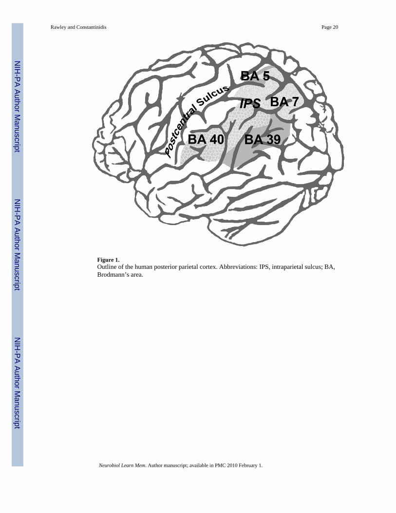
Figure 1.Outline of the human posterior parietal cortex. Abbreviations: IPS, intraparietal sulcus; BA,Brodmann’s area.
Rawley and Constantinidis Page 20
Neurobiol Learn Mem. Author manuscript; available in PMC 2010 February 1.
NIH
-PA Author Manuscript
NIH
-PA Author Manuscript
NIH
-PA Author Manuscript
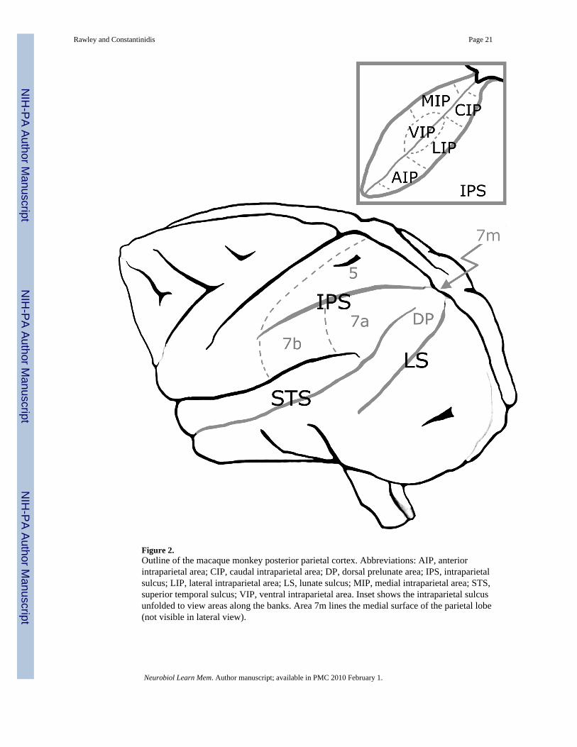
Figure 2.Outline of the macaque monkey posterior parietal cortex. Abbreviations: AIP, anteriorintraparietal area; CIP, caudal intraparietal area; DP, dorsal prelunate area; IPS, intraparietalsulcus; LIP, lateral intraparietal area; LS, lunate sulcus; MIP, medial intraparietal area; STS,superior temporal sulcus; VIP, ventral intraparietal area. Inset shows the intraparietal sulcusunfolded to view areas along the banks. Area 7m lines the medial surface of the parietal lobe(not visible in lateral view).
Rawley and Constantinidis Page 21
Neurobiol Learn Mem. Author manuscript; available in PMC 2010 February 1.
NIH
-PA Author Manuscript
NIH
-PA Author Manuscript
NIH
-PA Author Manuscript