
A Mirror Representation of Others' Actions in the Human Anterior Parietal Cortex
10
Chapter 2 A Mirror Representation of Others’ Actions in the Human Anterior Parietal Cortex Lior Shmuelof 1 and Ehud Zohary 1,2 1 Neurobiology Department, Life Science Institute and 2 Interdisciplinary Center for Neural Computation, Hebrew University, Jerusalem 91904, Israel. The Journal of Neuroscience 26, 9736-9742 (2006).
Transcript of A Mirror Representation of Others' Actions in the Human Anterior Parietal Cortex
Chapter 2
A Mirror Representation of Others’ Actions in the Human Anterior Parietal Cortex
Lior Shmuelof 1and Ehud Zohary1,2
1Neurobiology Department, Life Science Institute and 2Interdisciplinary Center for Neural Computation, Hebrew University, Jerusalem 91904, Israel.
The Journal of Neuroscience 26, 9736-9742 (2006).
Behavioral/Systems/Cognitive
A Mirror Representation of Others’ Actions in the HumanAnterior Parietal Cortex
Lior Shmuelof1 and Ehud Zohary1,2
1Department of Neurobiology and 2Interdisciplinary Center for Neural Computation, Hebrew University, Jerusalem, Israel 91904
The anterior part of the human intraparietal sulcus is known to be involved in visually guided grasping. This region is also active duringthe observation of object manipulation by others. Here, we explore the nature of action representation using functional magneticresonance imaging (fMRI). Fourteen subjects observed video clips showing object manipulation by the right or left hand . The clips werepresented in either the right or left peripheral visual field. The fMRI activation in the occipital cortex and in the caudal sections of theparietal cortex was specific to the visual-field location of the clips. In contrast, the principal factor determining the response in anteriorintraparietal cortex was the identity of the observed hand. Furthermore, these “hand-specific” parietal areas also showed contralateralhand specificity during self action (i.e., object manipulation) without visual feedback. A similar selectivity for the identity of the observedhand was seen when using a region of interest analysis, focusing on individually defined visuomotor voxels within the parietal cortex. Thisdual visuomotor grasping representation lends further evidence for the existence of a mirror system in humans and suggests that theanterior intraparietal cortex is involved in the specific motor simulation of hand actions.
Key words: fMRI; vision; motor; object manipulation; action observation; mirror system
IntroductionEvidence from recent years suggests that some cortical motorareas (i.e., engaged in motor planning and execution), are alsoactive during mere viewing of others’ actions (Gangitano et al.,2001; Buccino et al., 2004; Nelissen et al., 2005). The first clearevidence for this was the discovery of mirror neurons in the ma-caque monkey’s frontal lobe (area F5) and inferior parietal lobule(Gallese et al., 1996; Rizzolatti et al., 1996; Fogassi et al., 2005).These neurons discharge when the monkey executes goal-directed actions, such as grasping, even when the action is exe-cuted in the dark. Importantly, they are also active during obser-vation of another monkey, or the experimenter, performing asimilar action.
According to the direct matching hypothesis (DMH) raised byRizzolatti and colleagues (Iacoboni et al., 1999; Rizzolatti et al.,2001), observation of actions made by others should be mani-fested in a change in the excitability of the observer’s motor ele-ments that match the ones that are seen. One way to assess this isby measuring the magnitude of motor-evoked potential (MEP),as a result of applying transcranial magnetic stimulation (TMS)to the motor cortex. Indeed, Gangitano et al. (2004) have shownthat passive observation of object-grasping video clips amplifiesthe magnitude of the MEP resulting from the TMS pulse, inconcordance with the kinematic profile of the observed action. Ina functional magnetic resonance imaging (fMRI) study, Buccino
et al. (2001) have studied patterns of activation in the posteriorparietal cortex (and the premotor cortex) during observation ofobject manipulation by the foot, hand, or mouth. They foundthat the evoked fMRI activation in these areas was according tothe somatotopic mapping principles of the motor cortex (i.e., thehuman homunculus).
The anterior intraparietal cortex is known to be active duringgrasping movements in both monkeys (Murata et al., 2000) andhumans (Culham et al., 2003; Frey et al., 2005). Imaging studiesin humans have shown that this cortex is also active during visu-ally elicited motor imagery (Parsons et al., 1995; Bonda et al.,1996; de Lange et al., 2005). We have recently shown (Shmuelofand Zohary, 2005) that viewing video clips of object manipula-tion results in a robust activation along the intraparietal sulcus(IPS). Interestingly, the anterior parietal activation showed con-tralateral preference for the location of the hand (rather than theobject) and was specific to the grasping component of the action.This dual grasping-specific activity during both action executionand action observation suggests that this region may be part ofthe putative human mirror system. However, because the righthand was displayed mainly in the right visual field (and the lefthand in the left field), we could not determine whether the ante-rior parietal preference was attributable to the fact that the handwas seen in the contralateral field (visual-field representation) orthat the viewed acting hand was the contralateral one (suggestingan internal motor representation of the viewed action). We dem-onstrate here that in anterior intraparietal cortex, external actionsare represented according to the encoding principles of the motorsystem (Penfield and Rasmussen, 1952), regardless of the loca-tion of the actions in the visual field. Second, we show that theanterior intraparietal cortex is active both during action observa-tion and action execution (without visual feedback) using a spe-cific body part.
Received May 1, 2006; revised Aug. 9, 2006; accepted Aug. 10, 2006.This work was supported by the Israel Science Foundation of the Israel Academy of Sciences Grant 80009. We
thank Tanya Orlov for the help with the 3D cortex reconstructions. We thank Tamar Makin and Alit Stark for helpfuldiscussions.
Correspondence should be addressed to Ehud Zohary, Department of Neurobiology and Interdisciplinary Centerfor Neural Computation, Hebrew University, Jerusalem, Israel 91904. E-mail: [email protected].
DOI:10.1523/JNEUROSCI.1836-06.2006Copyright © 2006 Society for Neuroscience 0270-6474/06/269736-07$15.00/0
9736 • The Journal of Neuroscience, September 20, 2006 • 26(38):9736 –9742

Materials and MethodsSubjects. Fourteen volunteers without a history of neurological, psychi-atric, or visual deficits (seven women and seven men, 25–35 years of age)participated in the fMRI experiments. The Tel-Aviv Sourasky MedicalCenter Ethics Committee approved the experimental procedure. A writ-ten informed consent was obtained from each subject.
MRI acquisition. The blood oxygenation level-dependent (BOLD)fMRI measurements were performed in a whole-body 1.5 T, Signa Ho-rizon, LX8.25 General Electric (Fairfield, CT) scanner. The functionalMRI protocols were based on a multislice gradient echoplanar imagingand a standard head or surface coil. The functional data were obtainedunder the optimal timing parameters. For experiment 1 (visual experi-ment): repetition time (TR), 3 s; echo time (TE), 55 ms; flip angle, 90°;imaging matrix, 64 � 64; field of view (FOV), 21 cm. The 29 slices withslice thickness of 4 mm (with no gap) were oriented in the axial position.The scan covered the whole brain.
The somatomotor mapping was conducted using the following pa-rameters: TR, 2 s; TE, 55 ms; flip angle, 90°; imaging matrix, 64 � 64;FOV, 21 cm. The 19 slices with slice thickness of 4 mm (with 1 mm gap)were oriented in an oblique axis. The scan covered most of the cortexexcept for the most anterior parts of the frontal lobe. The fMRI imageswere superimposed on T1-weighted three-dimensional (3D) SPGR im-ages (spatial resolution, 1 � 1 � 1 mm).
Experimental setup. The video clips were taken using a digital camera(TRV60E; Sony, Tokyo, Japan), edited on a personal computer (using theprogram Windows Movie Maker), and projected via a liquid crystal dis-play projector (MP 7200; Epson, Nagano, Japan) onto a tangent screenlocated inside the scanner in front of the subject. The subjects viewed thescreen through a tilted mirror.
Main experimentA set of 16 object-manipulation epochs, 12 s long, and 2 visual controlepochs were shown in this experiment. Each epoch of right-hand footage(in the left and right periphery) was composed of 10 clips (900 –1500 mseach), of a right hand approaching from the right, grasping and releasingan object (total time, 12 s). The clips were taken using a set of 15 man-made graspable objects, such as different jars, cups, scissors, etc. Six to 10clips were taken with each object, showing different grasping move-ments. Each epoch was composed of 10 clips of different objects. Left-hand clips were generated by a “flip horizontal” operation on the “right-hand” clips. In all clips, the objects remained stationary and did not movethroughout the grasping movements. In the control “scramble” condi-tions, a 12 s object-manipulation epoch was decomposed to frames (30frames a second), every frame was spatially scrambled (36 � 24 frag-ments in each frame, ensuring that the hand and the objects could nolonger be identified), and the frames were then recomposed to a 12 sepoch. A fixation point appeared in the middle of the screen throughoutthe experiment. The clips were centered 16.5° to the right or the left of thefixation point and subtended 20 � 15°.
Experimental paradigmThe experiment was performed using a block design format. Six blockswere interleaved and were repeated four times, with different stimuli, ina counterbalanced manner. Each block lasted 12 s, followed by a blankperiod of 9 s. The first and last blank periods were longer (21 and 15 s,respectively). Subjects were trained before the experiment to make surethat they maintained fixation and understood the instructions.
The conditions (Fig. 1) involved viewing the following scenes:(1) object manipulations by the right hand, shown in the left visualfield; (2) object manipulation by the right hand, shown in the rightvisual field; (3) object manipulation by the left hand, shown in the leftvisual field; (4) object manipulation by the left hand, shown in theright visual field; (5) spatially scrambled version of the object-manipulation clips, shown in the left visual field; and (6) spatiallyscrambled version of the object-manipulation clips, shown in theright visual field. The “scramble” control conditions (numbers 5 and6 above) were introduced to allow selection of voxels showing differ-ential activation during the observation of the object-manipulationclips (compared with the scrambled versions).
Somatomotor mappingWe also conducted a somatomotor localizer experiment in which thesubjects were required to move their left or right hand, foot, or mouth,according to oral instructions. The words were recorded by a digitalrecording device using GoldWave Shareware software (GoldWave, St.John’s, Newfoundland, Canada). The instructions were 1 s long andconsisted of the name of the body part to be moved (right/left hand,right/left foot, mouth) or a “stop” instruction. The execution instruc-tions were presented in the last 1 s of the rest block, and the “stop” signalwas given in the last second of the specific epoch.
The five conditions included (1) left-hand movements, (2) right-handmovements (in both cases subjects manipulated a cube with their hand),(3) flexion and extension of the left ankle, (4) flexion and extension of theright ankle, and (5) movement of the tongue along the teeth. All condi-tions were interleaved and repeated 4 times. Each block lasted 12 s fol-lowed by a blank period of 10 s. The first and last blank periods werelonger (20 and 14 s, respectively). Subjects were trained before the exper-iment to make sure that they understood the commands and maintainedfixation. The subjects focused on a fixation point at the center of thescreen throughout the experiment. Because of the supine posture of thesubjects, they could not see their actions.
Data analysisData analysis was performed using the BrainVoyager 4.96 and BrainVoy-ager Qx software packages (Brain Innovation, Maastricht, The Nether-lands). For each subject, the two-dimensional (2D) functional data werealigned to 2D anatomical slices of the same subject. Before statisticalanalysis, raw data were examined for motion and signal artifacts. Headmotion correction and high-pass temporal smoothing in the frequencydomain (3 cycles/total scan time) were applied to remove drifts and toimprove the signal-to-noise ratio. The complete data set was transformedinto Talairach space, Z-normalized and concatenated. A general linearmodel (GLM) approach was used to generate statistical parametric maps.The hemodynamic response function was modeled using parameterssuch as those used by Boynton et al. (1996). Significance levels were
Figure 1. Experimental conditions. Six conditions were interleaved in a block design experiment.In four of them, the subjects observed a single hand approaching and grasping different objects. Twovariables were manipulated in the experiment: hand identity (left or right; top and middle rows) andvisual field [the location of the clips on the screen (left or right periphery, columns)]. In addition, weincluded two visual control conditions, in which a spatially scrambled version of the object-manipulation clips was presented in the left or right periphery (bottom row). The subjects were re-quired to maintain fixation on a central dot throughout the experiment.
Shmuelof and Zohary • Mirror Representation in the Parietal Cortex J. Neurosci., September 20, 2006 • 26(38):9736 –9742 • 9737

calculated, taking into account the probability of afalse detection for any given cluster, by requiringthat statistically significant activation is seen incontiguous groups of voxels. The implementedmethod is based on the approach of Forman et al.(1995) and accomplished by Monte Carlo simu-lations (using a BV QX plug-in).
Across-subjects statistical parametric maps(Figs. 2, 6) were calculated using a hierarchicalrandom-effects model analysis (Friston et al.,1999). The cortical surface was reconstructedfrom the 3D SPGR scan. The procedure includedsegmentation of the white matter using a grow-region function, the smooth covering of a spherearound the segmented region, and the expansionof the reconstructed white matter into the graymatter. The surface was then unfolded, cut alongthe calcarine sulcus and flattened.
Region-of-interest analysisThe single-subject visuomotor regions of inter-est (ROIs) were identified in the parietal lobesbased on conjunction analysis between visualobject-manipulation areas and motor hand ar-eas (Fig. 3a). Visual object-manipulation areaswere identified as the voxels showing signifi-cantly higher activation during observation ofthe hand action clips than during observationof a spatially scrambled version of those clips( p � 0.01, cluster-size correction). Motor handareas were identified as the voxels showing sig-nificantly higher activation ( p � 0.001, cluster-size correction) during object manipulation bythe contralateral hand than during contralat-eral foot movements.
The activation time course of individual sub-jects was obtained from each ROI, using a GLMapproach. The time course was pooled acrosssubjects to yield the average activation timecourse (Fig. 4a). Similarly, the average percent-age signal change (from 9 to 15 s after the onsetof the condition) was calculated for each subjectand then averaged across subjects to yield thebar histograms (Figs. 4b, 5). A paired two-tailedt test was applied to test for significant differ-ences between conditions.
ResultsIn experiment 1, we investigate the patternsof brain activation during observation of ob-ject-manipulation clips, presented in the pe-riphery. The goal of this experiment was todifferentiate between brain areas that showpreference to stimuli presented in the con-tralateral visual field [a pattern of represen-tation that is characteristic throughout thevisual system (Engel et al., 1996)] and thosethat showed preference for the contralateralacting hand [a feature that characterizes mo-tor system brain areas (Penfield and Ras-mussen, 1952)]. Note that in all conditions,the subjects viewed the clips without makingany grasping movements. Therefore, areassensitive to the identity of the viewed actinghand are likely to be candidates for having aninternal motor representation of externalobserved actions.
Figure 3. Identifying visuomotor areas: single-subject analysis. a, Inflated cortical maps of seven of the nine subjects thatparticipated in the motor mapping experiment. Visual hand action areas (purple) were identified as voxels showing significantlyhigher activation during observation of the hand action clips than during observation of a spatially scrambled version of thoseclips. Motor hand areas (green) were identified as the voxels showing significantly greater activation during object manipulationusing the contralateral hand compared with contralateral ankle movement. Voxels showing both visual and motor preference forhand actions are depicted in cyan. b, Superposition of the 3D structure of the visuomotor ROIs of each of the nine subjects overlaidon a representative horizontal slice of the cortical sheet. Each subject’s ROI is depicted in a different color. Note the spatialcongruency between the subjects’ visuomotor ROIs in the anterior intraparietal cortex.
Figure 2. Visual-field versus hand-identity effects. A statistical parametric map of the group results (14 subjects), using arandom-effect GLM analysis and cluster-size correction (corr.) for multiple comparisons. Red-to-yellow colors depict hand-identity areas, i.e., voxels demonstrating increasingly significant preference for clips showing the contralateral hand actioncompared with the ipsilateral (Ipsi.) hand action. Blue-to-green colors indicate areas showing a visual-field effect, i.e., voxelsshowing increasingly significant preference for object-manipulation clips presented in the contralateral field than in the ipsilateralfield. Significant hand-identity effects can be seen bilaterally in the superior bank of the aIPS and the superior parietal lobule, aswell as in the precentral sulcus of the right hemisphere. Significant visual-field effects can be seen throughout the object-selectiveoccipital cortex bilaterally, in the caudal section of the right parietal cortex, and in the right precentral sulcus.
9738 • J. Neurosci., September 20, 2006 • 26(38):9736 –9742 Shmuelof and Zohary • Mirror Representation in the Parietal Cortex

To differentiate between a visual-oriented and a motor-oriented representa-tion, we used a two-by-two factorial design;the first variable was the identity of the seenacting hand (left or right), and the secondvariable was the location of the viewed ob-jects (and grasping hand) in the peripheralvisual field (right or left of the fixationpoint). We expected that the dominant fac-tor in the visual areas of occipitotemporalcortex would be the visual-field position.Our main focus, however, was in areasshowing a hand-identity effect: having agreater BOLD signal when viewing the con-tralateral acting hand than when viewing theipsilateral hand, regardless of the location ofthe stimuli in the visual field (for example,viewing the right-hand manipulating objectsshould generate greater activation in the lefthemisphere than in the right one). Becausecontralateral hand preference is a typicalcharacteristic of motor areas, such mirroractivity, in the absence of motor behavior bythe subject, is consistent with an internalmotor representation of the observedactions.
Group analysisFigure 2 depicts the brain areas that displaysignificant visual-field and/or hand-identity effects, using a random-effectanalysis and correction for multiple com-parisons. Cortical areas showing signifi-cantly higher BOLD activation during ob-servation of clips in the contralateral visualfield than in the ipsilateral field are de-picted in a green-to-blue scale. Contralat-eral visual-field preference can be seen inboth hemispheres, throughout the occipi-totemporal cortex, as well as in dorsal ar-eas, in the transverse occipital sulcus. Inaddition, in the right hemisphere, we
found a significant preference for left-field clips in the parietallobe, in the caudal intraparietal cortex and in the superior parietallobule. Cortical areas that displayed a significant hand-identity ef-fect, (i.e., preference to video clips showing the contralateral hand)are depicted in a yellow-to-red scale. These are mainly found be-tween the superior bank of the anterior intraparietal sulcus (aIPS)and the postcentral gyrus (the anterior intraparietal cortex).
To summarize, we replicated the well known contralateralvisual-field representation in the occipitotemporal cortex. More im-portantly, we found evidence for a different type of representation, inwhich the dominant factor determining the level of activity is theidentity of the viewed acting hand (in the absence of the subject’sown hand action), in the anterior intraparietal cortex.
Identifying visuomotor areas: ROI analysisThe results of the group analysis are consistent with the idea of aninternal motor representation of others’ actions in the anteriorintraparietal cortex. However, if this activation is indeed an in-ternal motor representation of someone else’s action (i.e., part ofthe mirror system), the same areas ought to be selectively activeduring motor action of the same body part. We therefore per-
Figure 4. Hand-identity and visual-field effects: ROI analysis. a, Averaged hemodynamic response curves for the four experi-mental conditions in the visuomotor ROIs of the anterior intraparietal cortex (n�9). Error bars denote the SEM. The conditions arespecified separately to each of the two hemispheres, according to its contralateral (cont.) or ipsilateral (ipsi.) viewed hand, as wellas visual field. b, Bar histograms of the two effects, hand identity and visual field, plotted separately for each hemisphere, showingthe averaged activation (across all subjects) for each of the factors (cont./ipsi. viewed hand, cont./ipsi. visual field, icons belowindicate the visual condition). Error bars denote the SEM. Asterisks denote significance (**p � 0.01). Significant visual-field effectcan be seen in the right anterior intraparietal ROI, whereas significant hand-identity effects can be seen bilaterally, in both ROIs.
Figure5. Analysisof fMRIactivationintheparietal“hand-identity”areasduringthesomatomotormapping. An analysis of the fMRI signal in the areas that show a clear dependence on the viewed handidentity in the group analysis (shown in the bottom and in Fig. 2) during the somatomotor mapping.Averaged activation (n � 9) during the somatomotor mapping experiment in which the subjectsmovedtheirrightor lefthand,foot,ormouthinthedark.ErrorbarsdenoteSEM.Significantpreferencecan be seen for actions of the contralateral hand compared with the other body parts and the ipsilat-eral hand (paired, two-tailed t test; left ROI: right hand � left hand, p � 0.0002; right ROI: righthand � left hand, p � 0.005). Thus, the parietal regions that are sensitive to the identity of theviewed hand show a similar contralateral specificity for the acting hand.
Shmuelof and Zohary • Mirror Representation in the Parietal Cortex J. Neurosci., September 20, 2006 • 26(38):9736 –9742 • 9739

formed another experiment, mapping the patterns of activationelicited by action using various body parts without visual feed-back (see Materials and Methods). Next, we identified (in each ofthe 9 subjects that participated in this experiment) the visuomo-tor areas as those voxels that show both (1) selective activationduring object-manipulation observation (all object-manipulation epochs � scramble epochs, p � 0.01, cluster-sizecorrection) and (2) hand-specific motor activation ( p � 0.001,cluster-size correction). Hand-specific motor activation was de-fined by contrasting the activation during object manipulationusing the contralateral hand with the activation during contralat-eral ankle movement. Figure 3a presents the motor hand areas(green), the object-manipulation observation areas (purple), andthe conjunction between the areas (visuomotor areas, cyan) inseven of the nine subjects that participated in both experiments(the conjunction between the visual and motor areas of the re-maining two subjects was evident in the 2D slice views, but couldnot be seen clearly on their reconstructed surface maps). Figure3b shows the superposition of the 3D structure of the ROIs of allnine subjects overlaid on a representative horizontal slice. Eachsubject’s ROI (selected on the basis of their combined visual &motor selectivity) is depicted in a different color. One can appre-ciate that the ROIs in the parietal cortex are located roughly in thesame region, within the anterior section of the intraparietal cor-tex (left anterior intraparietal cortex: average size, 2792 � 685mm 3; right anterior intraparietal cortex: average size, 1711 � 454mm 3).
We further investigated whether the visual properties of theseparately defined visuomotor ROIs are consistent with the directmatching hypothesis, i.e., that external actions are represented onthe basis of the motor properties of the viewed action (in our case,according to the identity of the acting hand). Therefore, we com-pared the fMRI activation in the four different object-manipulation clips. Note that the ROIs were selected on the basis oftheir visual preference for the object-manipulation clips (as well astheir motor hand selectivity), but no preference was given for oneobject-manipulation viewing condition over the other. Thus, wecould compare the fMRI activation elicited during the four differentobject-manipulation viewing conditions to examine the effect of thetwo factors (visual field and hand identity) in the two visuomotorROIs.
We find that in the left anterior intraparietal cortex, the great-est fMRI activation occurred during viewing of the contralateral(right) hand grasping objects, regardless of the grasping scene’slocation in the visual field. This suggests that hand identity wasthe main factor in determining the fMRI activation in this ROI. Inthe right anterior intraparietal cortex, the greatest fMRI activa-tion was for viewing the contralateral (left) hand in the contralat-eral (left) visual field, whereas the weakest BOLD signal was elic-ited when viewing the ipsilateral hand in the ipsilateral visualfield. This suggests that both hand identity and visual field play arole in the level of activation in this ROI. Indeed, a two-factorANOVA showed a significant hand-identity effect in the left an-terior intraparietal cortex (F(1,8) � 6.7, p � 0.032). In the rightanterior intraparietal cortex, both hand-identity and visual-fieldeffects were significant, although the effect of hand identity wasmore pronounced (hand identity: F(1,8) � 23, p � 0.001; visualfield: F(1,8) � 11.3, p � 0.01). No interaction between the twoeffects was found in the two ROIs. Note that this analysis matchesthe hand-identity effect in the anterior intraparietal cortex, pre-sented in the group analysis (Fig. 2). In fact, close inspection ofthe group analysis map in Figure 2 shows the same qualitativedifference between the two hemispheres (i.e., preference for the
contralateral hand in the left anterior intraparietal cortex and toboth the contralateral hand and visual field in the right anteriorintraparietal cortex).
Motor properties in “hand-identity areas”The results from the ROI analysis indicate that regions within theanterior intraparietal cortex have both visual and motor proper-ties. Furthermore, in these regions, the fMRI activation evoked bythe observed hand actions depends on the identity of the actinghand. This result is in accordance with the direct-matching hy-pothesis, stating that seeing action performed by others invokesan internal motor representation of the same action.
To test this issue further, from another angle, we looked at thespecific motor properties of cortical areas that showed a prefer-ence for the contralateral hand viewed actions [in the group anal-ysis (Fig. 5, inset, same as orange clusters in Fig. 2)]. If seeingaction performed by others evokes an internal motor representa-tion of the same hand, we would expect to find activation in theseareas during motor behavior, using the same hand, even withoutany visual feedback. Indeed, the fMRI activation (in the areas thatshowed visual hand-identity specificity in the anterior intrapari-etal cortex) is clearly greater for hand movement compared withthe other body parts (i.e., foot or mouth movements). Further-more, in both hemispheres, there is an obvious and statisticallysignificant preference for motor actions using the contralateralhand over the ipsilateral one [paired two-tailed t test; p � 0.005 inthe right hemisphere; p � 0.001 in the left hemisphere (Fig. 5a)].
Grasp-viewing-based adaptationWe have previously demonstrated (Shmuelof and Zohary,2005) that certain areas in the anterior intraparietal cortexshow grasp-viewing-based adaptation: repetitive observationof the same object grasping movement leads to a reduction ofthe BOLD signal (compared with observation of differentgrasping movements). To give further evidence that the acti-vation of the “hand-identity” ROIs may be viewed as classical“mirror activity,” we tested whether these regions also showgrasp-viewing-based adaptation. The fMRI signal in thesegroup-based ROIs (selected on the basis of their preference forthe contralateral hand viewed actions) clearly shows grasp-viewing-based adaptation (paired two-tailed t test; p � 0.002 in theright anterior intraparietal and p � 0.001 in the left anterior intrapa-rietal, n � 14; object-based adaptation not significant in the twoROIs), suggesting that they are also sensitive to the fine details of theviewed action.
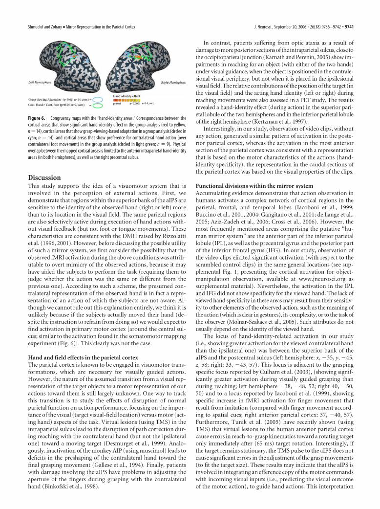
Finally, we studied the degree of overlap between the(group-based) cortical areas showing viewed-hand prefer-ence, hand motor properties (in the current study) and thecortical areas that show grasp-viewing-based adaptation. Notethat although the video clips were projected in different loca-tions in the two studies (central visual field in the previousadaptation study, peripheral in the current one), there is astriking congruency between the two loci (Fig. 6).
Interestingly, a large proportion of the areas that show con-gruency between the two visual experiments, showing a hand-identity effect and a grasping-based adaptation, are also selec-tively active during movement of the contralateral hand. Thisonce again suggests that observed hand actions are represented inthe anterior intraparietal cortex according to implicit motorprinciples, which are very different from the known rules of rep-resentation in the visual cortex.
9740 • J. Neurosci., September 20, 2006 • 26(38):9736 –9742 Shmuelof and Zohary • Mirror Representation in the Parietal Cortex
DiscussionThis study supports the idea of a visuomotor system that isinvolved in the perception of external actions. First, wedemonstrate that regions within the superior bank of the aIPS aresensitive to the identity of the observed hand (right or left) morethan to its location in the visual field. The same parietal regionsare also selectively active during execution of hand actions with-out visual feedback (but not foot or tongue movements). Thesecharacteristics are consistent with the DMH raised by Rizzolattiet al. (1996, 2001). However, before discussing the possible utilityof such a mirror system, we first consider the possibility that theobserved fMRI activation during the above conditions was attrib-utable to overt mimicry of the observed actions, because it mayhave aided the subjects to perform the task (requiring them tojudge whether the action was the same or different from theprevious one). According to such a scheme, the presumed con-tralateral representation of the observed hand is in fact a repre-sentation of an action of which the subjects are not aware. Al-though we cannot rule out this explanation entirely, we think it isunlikely because if the subjects actually moved their hand (de-spite the instruction to refrain from doing so) we would expect tofind activation in primary motor cortex [around the central sul-cus; similar to the activation found in the somatomotor mappingexperiment (Fig. 6)]. This clearly was not the case.
Hand and field effects in the parietal cortexThe parietal cortex is known to be engaged in visuomotor trans-formations, which are necessary for visually guided actions.However, the nature of the assumed transition from a visual rep-resentation of the target objects to a motor representation of ouractions toward them is still largely unknown. One way to trackthis transition is to study the effects of disruption of normalparietal function on action performance, focusing on the impor-tance of the visual (target visual-field location) versus motor (act-ing hand) aspects of the task. Virtual lesions (using TMS) in theintraparietal sulcus lead to the disruption of path correction dur-ing reaching with the contralateral hand (but not the ipsilateralone) toward a moving target (Desmurget et al., 1999). Analo-gously, inactivation of the monkey AIP (using muscimol) leads todeficits in the preshaping of the contralateral hand toward thefinal grasping movement (Gallese et al., 1994). Finally, patientswith damage involving the aIPS have problems in adjusting theaperture of the fingers during grasping with the contralateralhand (Binkofski et al., 1998).
In contrast, patients suffering from optic ataxia as a result ofdamage to more posterior sections of the intraparietal sulcus, close tothe occipitoparietal junction (Karnath and Perenin, 2005) show im-pairments in reaching for an object (with either of the two hands)under visual guidance, when the object is positioned in the contrale-sional visual periphery, but not when it is placed in the ipsilesionalvisual field. The relative contributions of the position of the target (inthe visual field) and the acting hand identity (left or right) duringreaching movements were also assessed in a PET study. The resultsrevealed a hand-identity effect (during action) in the superior pari-etal lobule of the two hemispheres and in the inferior parietal lobuleof the right hemisphere (Kertzman et al., 1997).
Interestingly, in our study, observation of video clips, withoutany action, generated a similar pattern of activation in the poste-rior parietal cortex, whereas the activation in the most anteriorsection of the parietal cortex was consistent with a representationthat is based on the motor characteristics of the actions (hand-identity specificity), the representation in the caudal sections ofthe parietal cortex was based on the visual properties of the clips.
Functional divisions within the mirror systemAccumulating evidence demonstrates that action observation inhumans activates a complex network of cortical regions in theparietal, frontal, and temporal lobes (Iacoboni et al., 1999;Buccino et al., 2001, 2004; Gangitano et al., 2001; de Lange et al.,2005; Aziz-Zadeh et al., 2006; Cross et al., 2006). However, themost frequently mentioned areas comprising the putative “hu-man mirror system” are the anterior part of the inferior parietallobule (IPL), as well as the precentral gyrus and the posterior partof the inferior frontal gyrus (IFG). In our study, observation ofthe video clips elicited significant activation (with respect to thescrambled control clips) in the same general locations (see sup-plemental Fig. 1, presenting the cortical activation for object-manipulation observation, available at www.jneurosci.org assupplemental material). Nevertheless, the activation in the IPLand IFG did not show specificity for the viewed hand. The lack ofviewed hand specificity in these areas may result from their sensitiv-ity to other elements of the observed action, such as the meaning ofthe action (which is clear in gestures), its complexity, or to the task ofthe observer (Molnar-Szakacs et al., 2005). Such attributes do notusually depend on the identity of the viewed hand.
The locus of hand-identity-related activation in our study(i.e., showing greater activation for the viewed contralateral handthan the ipsilateral one) was between the superior bank of theaIPS and the postcentral sulcus (left hemisphere: x, �35, y, �45,z, 58; right: 33, �43, 57). This locus is adjacent to the graspingspecific focus reported by Culham et al. (2003), (showing signif-icantly greater activation during visually guided grasping thanduring reaching; left hemisphere �38, �48, 52; right 40, �50,50) and to a locus reported by Iacoboni et al. (1999), showingspecific increase in fMRI activation for finger movement thatresult from imitation (compared with finger movement accord-ing to spatial cues; right anterior parietal cortex: 37, �40, 57).Furthermore, Tunik et al. (2005) have recently shown (usingTMS) that virtual lesions to the human anterior parietal cortexcause errors in reach-to-grasp kinematics toward a rotating targetonly immediately after (65 ms) target rotation. Interestingly, ifthe target remains stationary, the TMS pulse to the aIPS does notcause significant errors in the adjustment of the grasp movements(to fit the target size). These results may indicate that the aIPS isinvolved in integrating an efference copy of the motor commandswith incoming visual inputs (i.e., predicting the visual outcomeof the motor action), to guide hand actions. This interpretation
Figure 6. Congruency maps with the “hand-identity areas.” Correspondence between thecortical areas that show significant hand-identity effect in the group analysis (red to yellow;n �14), cortical areas that show grasp-viewing-based adaptation in a group analysis (circled incyan; n � 14), and cortical areas that show preference for contralateral hand action (overcontralateral foot movement) in the group analysis (circled in light green; n � 9). Physicaloverlap between the mapped cortical areas is limited to the anterior intraparietal hand-identityareas (in both hemispheres), as well as the right precentral sulcus.
Shmuelof and Zohary • Mirror Representation in the Parietal Cortex J. Neurosci., September 20, 2006 • 26(38):9736 –9742 • 9741

may also explain the unique visuomotor representation in theanterior intraparietal cortex, showing clear hand specificity, be-cause the motor commands and expected outcome are obviouslydependent on the acting hand. Additional research is obviouslyrequired to elucidate the division of labor within areas compris-ing the putative human mirror system.
Laterality effectsAlthough we demonstrate a symmetric representation of handidentity in the right and left anterior parietal cortex, both thegroup results and the ROI analysis reveal a convergence of thehand-identity and visual-field effects only in the right anteriorintraparietal cortex. Interestingly, we found a similar conver-gence of “ventral” object-oriented representation and “dor-sal” grasping-oriented representation in the right anterior pa-rietal cortex, but not in the left hemisphere, in a recent studythat mapped cortical areas showing grasp viewing-based ad-aptation and object viewing-based adaptation (Shmuelof andZohary, 2005). This asymmetry is also consistent with behav-ioral experiments, showing that precision grasping using theleft hand (but not the right hand) is affected by pictorial illu-sions that distort the perceived size of the object (Gonzalez etal., 2006). It may also explain why performance in simplevisuospatial tasks, such as line bisection, are typically ham-pered after lesions in the right parietal cortex but not in the leftside (Halligan et al., 2003).
ConclusionsIn this study, we demonstrate that external actions are repre-sented in the human anterior intraparietal cortex according totheir motor attributes (i.e., the identity of the acting hand).Furthermore, we show that this motor oriented representationis a property of specific visuo-motor areas, which are activeduring both object-manipulation (without visual feedback)and observation of actions made by others. This unique dualrepresentation, demonstrated on a subject by subject basis,forms a solid support for the mirror system hypothesis. Inparticular, the specificity of the anterior intraparietal cortex tothe identity of the seen hand, as well as to the nature of theviewed grasping, suggests that it is involved in the specificsimulation of hand actions.
ReferencesAziz-Zadeh L, Koski L, Zaidel E, Mazziotta J, Iacoboni M (2006) Lateraliza-
tion of the human mirror neuron system. J Neurosci 26:2964 –2970.Binkofski F, Dohle C, Posse S, Stephan KM, Hefter H, Seitz RJ, Freund HJ
(1998) Human anterior intraparietal area subserves prehension: a com-bined lesion and functional MRI activation study. Neurology50:1253–1259.
Bonda E, Frey S, Petrides M (1996) Evidence for a dorso-medial parietalsystem involved in mental transformations of the body. J Neurophysiol76:2042–2048.
Boynton GM, Engel SA, Glover GH, Heeger DJ (1996) Linear systems anal-ysis of functional magnetic resonance imaging in human V1. J Neurosci16:4207– 4221.
Buccino G, Binkofski F, Fink GR, Fadiga L, Fogassi L, Gallese V, Seitz RJ, ZillesK, Rizzolatti G, Freund HJ (2001) Action observation activates premo-tor and parietal areas in a somatotopic manner: an fMRI study. Eur J Neu-rosci 13:400 – 404.
Buccino G, Vogt S, Ritzl A, Fink GR, Zilles K, Freund HJ, Rizzolatti G (2004)Neural circuits underlying imitation learning of hand actions: an event-related fMRI study. Neuron 42:323–334.
Cross ES, Hamilton AF, Grafton ST (2006) Building a motor simulation denovo: observation of dance by dancers. Neuroimage 31:1257–1267.
Culham JC, Danckert SL, DeSouza JF, Gati JS, Menon RS, Goodale MA(2003) Visually guided grasping produces fMRI activation in dorsal butnot ventral stream brain areas. Exp Brain Res 153:180 –189.
Desmurget M, Epstein CM, Turner RS, Prablanc C, Alexander GE, GraftonST (1999) Role of the posterior parietal cortex in updating reachingmovements to a visual target. Nat Neurosci 2:563–567.
Engel SA, Glover GH, Wandell BA (1996) Retinotopic organization in hu-man visual cortex and the spatial precision of functional MRI. CerebCortex 7:181–192.
Fogassi L, Ferrari PF, Gesierich B, Rozzi S, Chersi F, Rizzolatti G (2005)Parietal lobe: from action organization to intention understanding. Sci-ence 308:662– 667.
Forman SD, Cohen JD, Fitzgerald M, Eddy WF, Mintun MA, Noll DC (1995)Improved assessment of significant activation in functional magnetic res-onance imaging (fMRI): use of a cluster-size threshold. Magn Reson Med33:636 – 647.
Frey SH, Vinton D, Norlund R, Grafton ST (2005) Cortical topography ofhuman anterior intraparietal cortex active during visually guided grasp-ing. Brain Res Cogn Brain Res 23:397– 405.
Friston KJ, Holmes AP, Worsley KJ (1999) How many subjects constitute astudy? Neuroimage 10:1–5.
Gallese V, Murata A, Kaseda M, Niki N, Sakata H (1994) Deficit of handpreshaping after muscimol injection in monkey parietal cortex. Neurore-port 5:1525–1529.
Gallese V, Fadiga L, Fogassi L, Rizzolatti G (1996) Action recognition in thepremotor cortex. Brain 119:593– 609.
Gangitano M, Mottaghy FM, Pascual-Leone A (2001) Phase-specific mod-ulation of cortical motor output during movement observation. Neuro-report 12:1489 –1492.
Gangitano M, Mottaghy FM, Pascual-Leone A (2004) Modulation of pre-motor mirror neuron activity during observation of unpredictable grasp-ing movements. Eur J Neurosci 20:2193–2202.
Gonzalez CL, Ganel T, Goodale MA (2006) Hemispheric specialization forthe visual control of action is independent of handedness. J Neurophysiol95:3496 –3501.
Halligan PW, Fink GR, Marshall JC, Vallar G (2003) Spatial cognition: evi-dence from visual neglect. Trends Cogn Sci 7:125–133.
Iacoboni M, Woods RP, Brass M, Bekkering H, Mazziotta JC, Rizzolatti G(1999) Cortical mechanisms of human imitation. Science 286:2526–2528.
Karnath HO, Perenin MT (2005) Cortical control of visually guided reach-ing: evidence from patients with optic ataxia. Cereb Cortex 15:1561–1569.
Kertzman C, Schwarz U, Zeffiro TA, Hallett M (1997) The role of posteriorparietal cortex in visually guided reaching movements in humans. ExpBrain Res 114:170 –183.
de Lange FP, Hagoort P, Toni I (2005) Neural topography and content ofmovement representations. J Cogn Neurosci 17:97–112.
Molnar-Szakacs I, Iacoboni M, Koski L, Mazziotta JC (2005) Functional segre-gation within pars opercularis of the inferior frontal gyrus: evidence fromfMRI studies of imitation and action observation. Cereb Cortex 15:986–994.
Murata A, Gallese V, Luppino G, Kaseda M, Sakata H (2000) Selectivity forthe shape, size, and orientation of objects for grasping in neurons ofmonkey parietal area AIP. J Neurophysiol 83:2580 –2601.
Nelissen K, Luppino G, Vanduffel W, Rizzolatti G, Orban GA (2005) Ob-serving others: multiple action representation in the frontal lobe. Science310:332–336.
Parsons LM, Fox PT, Downs JH, Glass T, Hirsch TB, Martin CC, Jerabek PA,Lancaster JL (1995) Use of implicit motor imagery for visual shape dis-crimination as revealed by PET. Nature 375:54 –58.
Penfield W, Rasmussen T (1952) The cerebral cortex of man. New York:MacMillan.
Rizzolatti G, Fadiga L, Gallese V, Fogassi L (1996) Premotor cortex and therecognition of motor actions. Brain Res Cogn Brain Res 3:131–141.
Rizzolatti G, Fogassi L, Gallese V (2001) Neurophysiological mechanismsunderlying the understanding and imitation of action. Nat Rev Neurosci2:661– 670.
Shmuelof L, Zohary E (2005) Dissociation between ventral and dorsal fMRIactivation during object and action recognition. Neuron 47:457–470.
Tunik E, Frey SH, Grafton ST (2005) Virtual lesions of the anterior intrapa-rietal area disrupt goal-dependent on-line adjustments of grasp. Nat Neu-rosci 8:505–511.
9742 • J. Neurosci., September 20, 2006 • 26(38):9736 –9742 Shmuelof and Zohary • Mirror Representation in the Parietal Cortex

Supplementary figure 1 - Visual object manipulation areas A statistical parametric maps of the group results (n=14), using a random effect GLM analysis, and cluster-size correction for multiple comparisons. Red to yellow colors depict visual object manipulation areas, i.e., voxels demonstrating increasingly significant preference for clips showing object manipulation by a hand (right or left) over a spatially scrambled version of the clips.
Table 1 – Visual object manipulation areas
Cortical regions showing significantly greater activation during observation of object
manipulation clips than during observation of spatially scrambled versions of the clips
(p<0.01, random effect analysis, cluster-size correction). The areas correspond to the
areas shown in supplementary Figure 1.
Talairach coordinates X Y Z
Cluster size (mm³)
BA Area name
-44 12 6 846 44 LH, Precentral Gyrus -48 -4 45 2736 4 Lh, Precentral Gyrus -33 -8 53 2795 6 Lh, Precentral Gyrus 33 20 11 1876 13 Rh, Insula 44 1 45 3372 6 Rh, Middle Frontal Gyrus 1 2 57 3831 6 Rh, Middle Frontal Gyrus 35 -5 55 5555 6 Rh, Middle Frontal Gyrus -49 -31 45 3932 40 Lh, Inferior Parietal Lobule -32 -50 46 6825 40 Lh, Inferior Parietal Lobule

-47 -32 46 4337 40 Lh, Inferior Parietal Lobule 56 -30 24 2692 40 Rh, Inferior Parietal Lobule 28 -50 56 7757 7 Rh, Superior Parietal Lobule -45 -67 3 8517 37 Lh, Middle Occipital Gyrus 45 -56 5 7629 37 Rh, Middle Occipital Gyrus 40 -52 -11 4193 37 Rh, Fusiform Gyrus