Sensory Experience Restructures Thalamocortical Axons during Adulthood
8
Neuron Report Sensory Experience Restructures Thalamocortical Axons during Adulthood Marcel Oberlaender, 1,3 Alejandro Ramirez, 2,3 and Randy M. Bruno 2, * 1 Digital Neuroanatomy, Max Planck Florida Institute, Jupiter, FL 33458, USA 2 Department of Neuroscience and the Kavli Institute for Brain Science, Columbia University, New York, NY 11032, USA 3 These authors contributed equally to this work *Correspondence: [email protected] DOI 10.1016/j.neuron.2012.03.022 SUMMARY The brain’s capacity to rewire is thought to diminish with age. It is widely believed that development stabilizes the synapses from thalamus to cortex and that adult experience alters only synaptic connections between cortical neurons. Here we show that thalamocortical (TC) inputs themselves undergo massive plasticity in adults. We combined whole-cell recording from individual thalamocortical neurons in adult rats with a recently developed auto- matic tracing technique to reconstruct individual axonal trees. Whisker trimming substantially re- duced thalamocortical axon length in barrel cortex but not the density of TC synapses along a fiber. Thus, sensory experience alters the total number of TC synapses. After trimming, sensory stimulation evoked more tightly time-locked responses among thalamorecipient layer 4 cortical neurons. These find- ings indicate that thalamocortical input itself remains plastic in adulthood, raising the possibility that the axons of other subcortical structures might also remain in flux throughout life. INTRODUCTION Numerous studies have concluded that the thalamocortical (TC) projection, along which sensory information flows into the cere- bral cortex, is fixed by the end of development. During a critical period in early postnatal life, monocular vision loss can trigger robust anatomical plasticity of TC axons in the mouse and cat visual systems (Antonini et al., 1999; Antonini and Stryker, 1993). Such anatomical changes are thought to be driven, at least in part, by the strengthening and weakening of existing TC synapses, which in slices of somatosensory cortex cannot be induced beyond the first few postnatal weeks, probably due to the known developmental downregulation of NMDA receptors (Feldman et al., 1999). In both the visual and somatosensory systems, sensory experience during adulthood has little or no effect on the receptive fields of neurons in cortical layer 4 (L4) but has robust effects on other layers (reviewed in Feldman and Brecht, 2005; Fox, 2002; Karmarkar and Dan, 2006). From such physiological studies, it has been inferred that adult plasticity, learning, and memory is mediated by functional and/or anatomical changes among corticocortical connections (De Paola et al., 2006; Feldman and Brecht, 2005; Fox, 2002; Karmarkar and Dan, 2006). A purely cortical locus for adult plasticity has, however, recently become controversial. Brief periods of monocular deprivation can alter the size of pharmacologically isolated TC- evoked field potentials in adult mouse visual cortex (Khibnik et al., 2010). Whisker trimming for as few as 3 days similarly reduces the overall density of TC synapses in adult rat barrel cortex (Wimmer et al., 2010). These recent findings prompted us to investigate the effect of sensory experience during adult- hood anatomically on individual TC axons and functionally on the magnitude and synchrony of cortical activity. We manipulated sensory experience in adult (3-month-old) rats using a painless, nondestructive approach. Trimming the large facial whiskers alters sensory experience without engaging potential trophic mechanisms that might be triggered by pluck- ing whiskers or lesioning whisker follicles. Individual thalamo- cortical neurons were filled in vivo by whole-cell recording and reconstructed in three dimensions via a recently developed semiautomatic method. We discovered that TC axonal arbors remain plastic in adulthood, with whisker trimming causing axons originating from the deprived representation to lose on average a quarter of their length across layers. Within L4, axonal branch reduction was higher and topographically specific. Dual cell-attached recordings in vivo revealed that sensory stimuli evoked greater levels of synchrony among L4 neurons but the same number of action potentials from individual cells. Our findings demonstrate that adult plasticity is not limited to cortico- cortical connections and potentially explain why previous functional studies of L4 could not infer such massive anatomical changes. RESULTS Conventional bulk tracers label the axons of many neurons, whose overlap confounds their reconstruction and quantifica- tion. Whole-cell recording, while challenging to obtain from a cell 5 mm deep within the brain, robustly labels a single axon when successful, facilitating unambiguous tracing (Bruno et al., 2009). We therefore patched a single thalamocortical neuron in the ventral posterior medial thalamic nucleus in each of 28 adult rats. All the large facial whiskers except two had 648 Neuron 74, 648–655, May 24, 2012 ª2012 Elsevier Inc.
-
Upload
independent -
Category
Documents
-
view
2 -
download
0
Transcript of Sensory Experience Restructures Thalamocortical Axons during Adulthood

Neuron
Report
Sensory Experience RestructuresThalamocortical Axons during AdulthoodMarcel Oberlaender,1,3 Alejandro Ramirez,2,3 and Randy M. Bruno2,*1Digital Neuroanatomy, Max Planck Florida Institute, Jupiter, FL 33458, USA2Department of Neuroscience and the Kavli Institute for Brain Science, Columbia University, New York, NY 11032, USA3These authors contributed equally to this work*Correspondence: [email protected]
DOI 10.1016/j.neuron.2012.03.022
SUMMARY
The brain’s capacity to rewire is thought to diminishwith age. It is widely believed that developmentstabilizes the synapses from thalamus to cortexand that adult experience alters only synapticconnections between cortical neurons. Here weshow that thalamocortical (TC) inputs themselvesundergo massive plasticity in adults. We combinedwhole-cell recording from individual thalamocorticalneurons in adult rats with a recently developed auto-matic tracing technique to reconstruct individualaxonal trees. Whisker trimming substantially re-duced thalamocortical axon length in barrel cortexbut not the density of TC synapses along a fiber.Thus, sensory experience alters the total number ofTC synapses. After trimming, sensory stimulationevoked more tightly time-locked responses amongthalamorecipient layer 4 cortical neurons. These find-ings indicate that thalamocortical input itself remainsplastic in adulthood, raising the possibility that theaxons of other subcortical structures might alsoremain in flux throughout life.
INTRODUCTION
Numerous studies have concluded that the thalamocortical (TC)
projection, along which sensory information flows into the cere-
bral cortex, is fixed by the end of development. During a critical
period in early postnatal life, monocular vision loss can trigger
robust anatomical plasticity of TC axons in the mouse and cat
visual systems (Antonini et al., 1999; Antonini and Stryker,
1993). Such anatomical changes are thought to be driven, at
least in part, by the strengthening and weakening of existing
TC synapses, which in slices of somatosensory cortex cannot
be induced beyond the first few postnatal weeks, probably due
to the known developmental downregulation of NMDA receptors
(Feldman et al., 1999). In both the visual and somatosensory
systems, sensory experience during adulthood has little or no
effect on the receptive fields of neurons in cortical layer 4 (L4)
but has robust effects on other layers (reviewed in Feldman
and Brecht, 2005; Fox, 2002; Karmarkar and Dan, 2006). From
648 Neuron 74, 648–655, May 24, 2012 ª2012 Elsevier Inc.
such physiological studies, it has been inferred that adult
plasticity, learning, and memory is mediated by functional
and/or anatomical changes among corticocortical connections
(De Paola et al., 2006; Feldman and Brecht, 2005; Fox, 2002;
Karmarkar and Dan, 2006).
A purely cortical locus for adult plasticity has, however,
recently become controversial. Brief periods of monocular
deprivation can alter the size of pharmacologically isolated TC-
evoked field potentials in adult mouse visual cortex (Khibnik
et al., 2010). Whisker trimming for as few as 3 days similarly
reduces the overall density of TC synapses in adult rat barrel
cortex (Wimmer et al., 2010). These recent findings prompted
us to investigate the effect of sensory experience during adult-
hood anatomically on individual TC axons and functionally on
the magnitude and synchrony of cortical activity.
We manipulated sensory experience in adult (3-month-old)
rats using a painless, nondestructive approach. Trimming the
large facial whiskers alters sensory experience without engaging
potential trophic mechanisms that might be triggered by pluck-
ing whiskers or lesioning whisker follicles. Individual thalamo-
cortical neurons were filled in vivo by whole-cell recording and
reconstructed in three dimensions via a recently developed
semiautomatic method. We discovered that TC axonal arbors
remain plastic in adulthood, with whisker trimming causing
axons originating from the deprived representation to lose on
average a quarter of their length across layers. Within L4, axonal
branch reduction was higher and topographically specific. Dual
cell-attached recordings in vivo revealed that sensory stimuli
evoked greater levels of synchrony among L4 neurons but the
same number of action potentials from individual cells. Our
findings demonstrate that adult plasticity is not limited to cortico-
cortical connections and potentially explain why previous
functional studies of L4 could not infer such massive anatomical
changes.
RESULTS
Conventional bulk tracers label the axons of many neurons,
whose overlap confounds their reconstruction and quantifica-
tion. Whole-cell recording, while challenging to obtain from
a cell �5 mm deep within the brain, robustly labels a single
axon when successful, facilitating unambiguous tracing (Bruno
et al., 2009). We therefore patched a single thalamocortical
neuron in the ventral posterior medial thalamic nucleus in each
of 28 adult rats. All the large facial whiskers except two had

Figure 1. Whisker Trimming during Adulthood Substantially Reduces the Length of Thalamocortical Axons
(A and B) Reconstructions of thalamocortical axons in control (A) and deprived (B) animals, respectively. Axons in deprived animals correspond to trimmed
whiskers. Left, tangential view; right, radial view; lines, barrel borders; dots, branch points and end points. Insets: adult rats were sham trimmed (A) or had all
whiskers but two trimmed off (B). (C) Columns targeted by control (black) and deprived (gray) axons. The position of symbols within any given column is arbitrary.
(D) Distributions of total axon lengths within cortex. Lines represent means ± SD. (E) Distributions of branch points within cortex. Lines represent means ± SD.
Neuron
Thalamocortical Plasticity in Adulthood
been trimmed daily (deprived group) or sham trimmed
(control group) for the preceding 13–27 days. Cages were not
left empty but instead were enriched with cardboard boxes
and tubes to encourage whisker-based exploration of the envi-
ronment, which has been suggested to enhance thalamocortical
plasticity (Wimmer et al., 2010). In deprived rats, we filled
neurons belonging to the trimmed whisker representation.
Axons were three-dimensionally reconstructed with submicron
resolution (Figures 1A and 1B; see Figure S1 available online)
using a recent semiautomatic tracing system, which rivals
human experts in both accuracy and completeness (Ober-
laender et al., 2007, 2009, 2011). The combination of these filling
and reconstruction approaches recovered total lengths of
normal TC axons far greater than previously observed. Control
axons ranged from 32.5 to 72.5 mm, with even the smallest TC
arbor longer than those previously reported for rat barrel cortex
or kitten visual cortex (Antonini and Stryker, 1993; Arnold et al.,
2001).
Axons in control and deprived animals targeted barrels with
similar spatial distributions, with no obvious spatial bias in our
samples (Figure 1C). Average total length of individual axonal
arbors within barrel cortex was 54.1 ± 3.7 mm for control animals
(Figure 1D, black circles; mean ± SEM, n = 12). Simply trimming
the whiskers significantly decreased the average length of
axons corresponding to deprived whiskers by 25%, down to
an average of 40.6 ± 4.7 mm (gray circles; n = 11, p = 0.017).
Trimming similarly decreased the number of branch points by
32% (Figure 1E; control 232 ± 27 versus deprived 158 ± 17;
p = 0.016). Due to the extreme depth of the thalamus, its
complicated three-dimensional geometry, and the small size of
individual thalamic ‘‘barreloids,’’ we recovered only two axons
corresponding to spared whiskers. Given that their lengths
(46.5 and 37.7mm) are in the ranges of both control and deprived
distributions, no inferences can be made regarding this small
spared sample. Nevertheless, our results clearly demonstrate
that innocuous sensory experience can substantially alter the
structure of inputs to cortex in adulthood.
Barrel size iswell known to dependon locationwithin the barrel
subfield. There was, however, no significant relationship of the
length of thalamocortical axon to the size of the innervated barrel,
regardless of whether control and deprived groups were
analyzed separately or pooled (Figure 2A; all p values > 0.5).
This surprising result suggests that the size of a barrel reflects
the number of neurons in the corresponding thalamic barreloid,
rather than the lengths of individual innervating axons.
Consistent with this finding, the trimming-induced decrease in
innervation is still significant even after normalizing the length
of each axon by the area of its respective barrel (Figure 2B).
Indeed, trimming appeared to impact axonal length relatively
consistently across whisker arcs and rows (Figure 2C).
Other morphological features remained unaffected. Trimming
produced no concomitant change in the area or height
of the barrels innervated by these axons (Figures 2D–2F;
p values > 0.1) consistent with previous studies (Fox, 1992).
The areas innervated (field spans; Figures 2G and 2H) by both
the superficial L3-L4 collaterals (Figure 2I) and the deeper L5/6
collaterals (Figure 2J; p values > 0.1) were stable. The distances
between boutons along axons, putative synapses, were similarly
unaffected by trimming (Figures 2K and 2L; p > 0.1). The experi-
ence-induced decrease in TC axonal length therefore appears to
reflect an absolute reduction in afferent synapses, perhaps via
pruning of specific branches.
Individual axons span multiple functionally distinct zones,
such as different cortical layers (Figure 3A) and somatotopic
columns (Figure 3C). We therefore considered how the effects
of experience on synaptic connectivity might depend on cortical
location. As in the visual and possibly auditory systems (Ferster
and LeVay, 1978; Smith et al., 2012), TC axons were mainly
‘‘bistratified,’’ with two distinct sets of collaterals at depths of
600–1,000 and 1,300–1,500 mm from the pia, corresponding to
L4 and L5B-L6A, respectively (Figure 3B; Figure S1). Sensory
deprivation impacted the total length of axon most noticeably
at these depths, with both decreases being statistically signifi-
cant (p values = 0.024).
Neuron 74, 648–655, May 24, 2012 ª2012 Elsevier Inc. 649
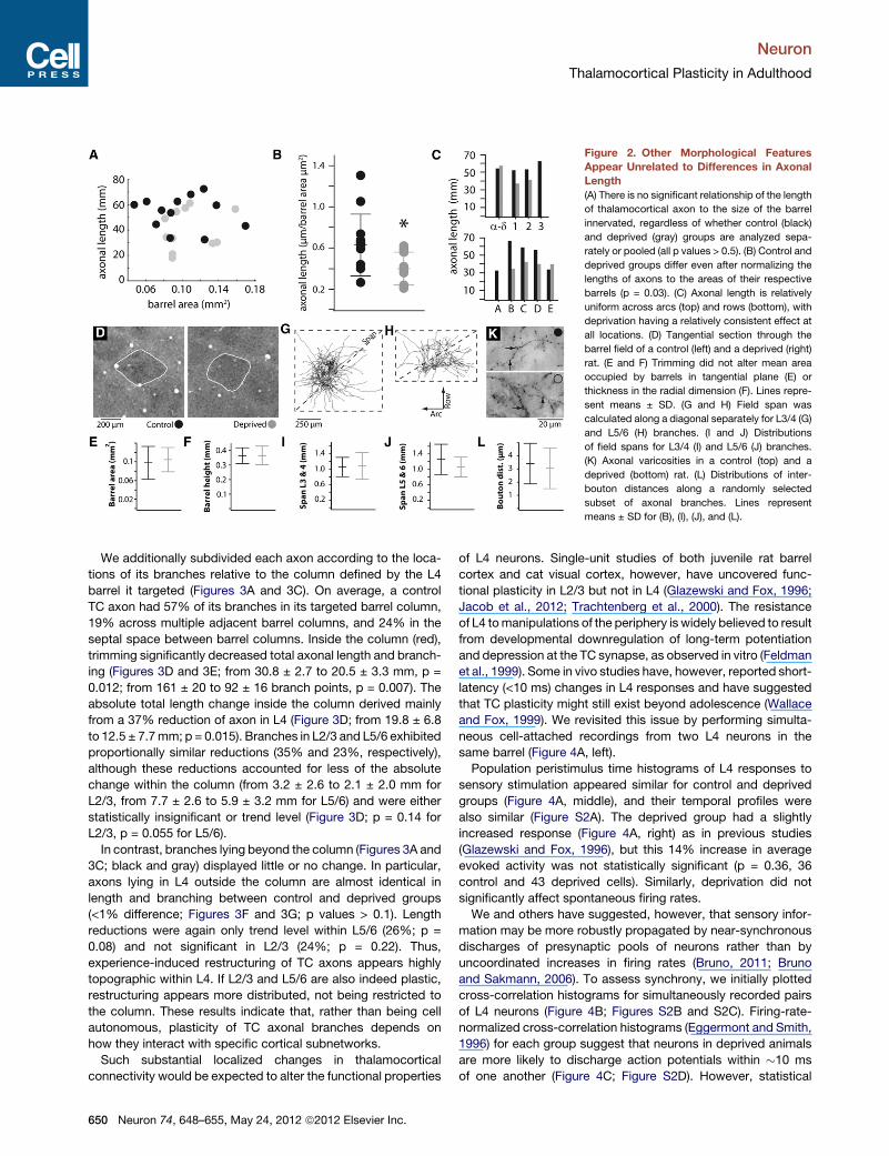
Figure 2. Other Morphological Features
Appear Unrelated to Differences in Axonal
Length
(A) There is no significant relationship of the length
of thalamocortical axon to the size of the barrel
innervated, regardless of whether control (black)
and deprived (gray) groups are analyzed sepa-
rately or pooled (all p values > 0.5). (B) Control and
deprived groups differ even after normalizing the
lengths of axons to the areas of their respective
barrels (p = 0.03). (C) Axonal length is relatively
uniform across arcs (top) and rows (bottom), with
deprivation having a relatively consistent effect at
all locations. (D) Tangential section through the
barrel field of a control (left) and a deprived (right)
rat. (E and F) Trimming did not alter mean area
occupied by barrels in tangential plane (E) or
thickness in the radial dimension (F). Lines repre-
sent means ± SD. (G and H) Field span was
calculated along a diagonal separately for L3/4 (G)
and L5/6 (H) branches. (I and J) Distributions
of field spans for L3/4 (I) and L5/6 (J) branches.
(K) Axonal varicosities in a control (top) and a
deprived (bottom) rat. (L) Distributions of inter-
bouton distances along a randomly selected
subset of axonal branches. Lines represent
means ± SD for (B), (I), (J), and (L).
Neuron
Thalamocortical Plasticity in Adulthood
We additionally subdivided each axon according to the loca-
tions of its branches relative to the column defined by the L4
barrel it targeted (Figures 3A and 3C). On average, a control
TC axon had 57% of its branches in its targeted barrel column,
19% across multiple adjacent barrel columns, and 24% in the
septal space between barrel columns. Inside the column (red),
trimming significantly decreased total axonal length and branch-
ing (Figures 3D and 3E; from 30.8 ± 2.7 to 20.5 ± 3.3 mm, p =
0.012; from 161 ± 20 to 92 ± 16 branch points, p = 0.007). The
absolute total length change inside the column derived mainly
from a 37% reduction of axon in L4 (Figure 3D; from 19.8 ± 6.8
to 12.5 ± 7.7mm; p = 0.015). Branches in L2/3 and L5/6 exhibited
proportionally similar reductions (35% and 23%, respectively),
although these reductions accounted for less of the absolute
change within the column (from 3.2 ± 2.6 to 2.1 ± 2.0 mm for
L2/3, from 7.7 ± 2.6 to 5.9 ± 3.2 mm for L5/6) and were either
statistically insignificant or trend level (Figure 3D; p = 0.14 for
L2/3, p = 0.055 for L5/6).
In contrast, branches lying beyond the column (Figures 3A and
3C; black and gray) displayed little or no change. In particular,
axons lying in L4 outside the column are almost identical in
length and branching between control and deprived groups
(<1% difference; Figures 3F and 3G; p values > 0.1). Length
reductions were again only trend level within L5/6 (26%; p =
0.08) and not significant in L2/3 (24%; p = 0.22). Thus,
experience-induced restructuring of TC axons appears highly
topographic within L4. If L2/3 and L5/6 are also indeed plastic,
restructuring appears more distributed, not being restricted to
the column. These results indicate that, rather than being cell
autonomous, plasticity of TC axonal branches depends on
how they interact with specific cortical subnetworks.
Such substantial localized changes in thalamocortical
connectivity would be expected to alter the functional properties
650 Neuron 74, 648–655, May 24, 2012 ª2012 Elsevier Inc.
of L4 neurons. Single-unit studies of both juvenile rat barrel
cortex and cat visual cortex, however, have uncovered func-
tional plasticity in L2/3 but not in L4 (Glazewski and Fox, 1996;
Jacob et al., 2012; Trachtenberg et al., 2000). The resistance
of L4 tomanipulations of the periphery is widely believed to result
from developmental downregulation of long-term potentiation
and depression at the TC synapse, as observed in vitro (Feldman
et al., 1999). Some in vivo studies have, however, reported short-
latency (<10 ms) changes in L4 responses and have suggested
that TC plasticity might still exist beyond adolescence (Wallace
and Fox, 1999). We revisited this issue by performing simulta-
neous cell-attached recordings from two L4 neurons in the
same barrel (Figure 4A, left).
Population peristimulus time histograms of L4 responses to
sensory stimulation appeared similar for control and deprived
groups (Figure 4A, middle), and their temporal profiles were
also similar (Figure S2A). The deprived group had a slightly
increased response (Figure 4A, right) as in previous studies
(Glazewski and Fox, 1996), but this 14% increase in average
evoked activity was not statistically significant (p = 0.36, 36
control and 43 deprived cells). Similarly, deprivation did not
significantly affect spontaneous firing rates.
We and others have suggested, however, that sensory infor-
mation may be more robustly propagated by near-synchronous
discharges of presynaptic pools of neurons rather than by
uncoordinated increases in firing rates (Bruno, 2011; Bruno
and Sakmann, 2006). To assess synchrony, we initially plotted
cross-correlation histograms for simultaneously recorded pairs
of L4 neurons (Figure 4B; Figures S2B and S2C). Firing-rate-
normalized cross-correlation histograms (Eggermont and Smith,
1996) for each group suggest that neurons in deprived animals
are more likely to discharge action potentials within �10 ms
of one another (Figure 4C; Figure S2D). However, statistical
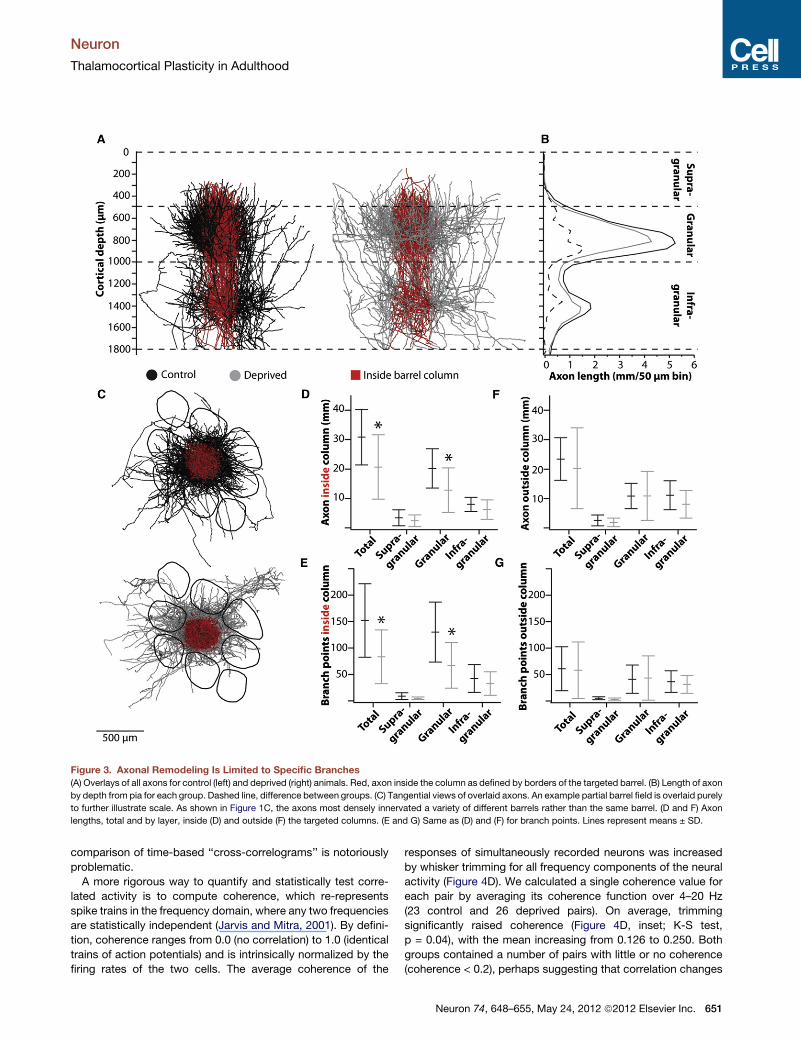
Figure 3. Axonal Remodeling Is Limited to Specific Branches
(A) Overlays of all axons for control (left) and deprived (right) animals. Red, axon inside the column as defined by borders of the targeted barrel. (B) Length of axon
by depth from pia for each group. Dashed line, difference between groups. (C) Tangential views of overlaid axons. An example partial barrel field is overlaid purely
to further illustrate scale. As shown in Figure 1C, the axons most densely innervated a variety of different barrels rather than the same barrel. (D and F) Axon
lengths, total and by layer, inside (D) and outside (F) the targeted columns. (E and G) Same as (D) and (F) for branch points. Lines represent means ± SD.
Neuron
Thalamocortical Plasticity in Adulthood
comparison of time-based ‘‘cross-correlograms’’ is notoriously
problematic.
A more rigorous way to quantify and statistically test corre-
lated activity is to compute coherence, which re-represents
spike trains in the frequency domain, where any two frequencies
are statistically independent (Jarvis and Mitra, 2001). By defini-
tion, coherence ranges from 0.0 (no correlation) to 1.0 (identical
trains of action potentials) and is intrinsically normalized by the
firing rates of the two cells. The average coherence of the
responses of simultaneously recorded neurons was increased
by whisker trimming for all frequency components of the neural
activity (Figure 4D). We calculated a single coherence value for
each pair by averaging its coherence function over 4–20 Hz
(23 control and 26 deprived pairs). On average, trimming
significantly raised coherence (Figure 4D, inset; K-S test,
p = 0.04), with the mean increasing from 0.126 to 0.250. Both
groups contained a number of pairs with little or no coherence
(coherence < 0.2), perhaps suggesting that correlation changes
Neuron 74, 648–655, May 24, 2012 ª2012 Elsevier Inc. 651

Figure 4. Experience Alters the Synchrony, but Not Magnitude, of Sensory-Evoked Activity among Cortical Layer 4 Neurons
(A) Dual cell-attached recordings were made from the same barrel in control and deprived animals (left), and peristimulus time histograms for each population
were plotted (middle). Evoked and spontaneous firing rates are plotted for individual cells (right). Box plots show medians and interquartile ranges. (B) Example
rasters and cross-correlograms for a control pair (left) and deprived pair (right). (C) Mean firing rate normalized cross-correlograms for each group. The cross-
correlogram of each pair was normalized by the geometric mean of the two cells’ firing rates. (D) Coherence functions averaged over all pairs. Inset: individual
coherence values for each pair (mean over 4–20 Hz). (E) Examples of pairs with (left) and without (right) signs of shared synaptic inputs. Both pairs were recorded
in trimmed rats. The raw cross-correlogram (CCG) measures total correlated activity of a pair of neurons (top). The shift corrector (middle), the recalculation of the
correlogram after shifting one of the spike trains by a trial, measures stimulus-induced correlation. The corrected cross-correlogram (bottom) is their difference. A
peak exceeding 3.3 standard deviations (a = 0.001) of the shift corrector (red lines) is taken as evidence of significant shared synaptic input (arrow). (F) Distri-
butions of the strength of significant shared synaptic inputs for individual pairs (circles). Box plots show medians and interquartile ranges.
Neuron
Thalamocortical Plasticity in Adulthood
may be limited to only certain combinations of cell types. These
time-domain (cross-correlation) and frequency-domain (coher-
ence) analyses together indicate that sensory experience alters
the synchrony of neuronal groups more than it detectably alters
the absolute firing rates of individual cells.
All other things being equal, the reduced TC synaptic connec-
tivity we found should decrease rather than increase L4
synchrony. Enhanced L4 synchrony suggests that experience
alters an additional element of the circuit. One possibility is
that the pruning of TC-L4 synapses triggers homeostatic rescal-
ing of the strength of synapses—afferent and/or intracortical—
onto an excitatory L4 neuron to maintain its normal firing rate.
To check this, we removed the stimulus-induced correlation to
reveal millisecond-scale neural interactions. Near-synchronous
events in a ‘‘raw’’ cross-correlogram (Figure 4B, bottom; Fig-
ure 4E, top) result from a pair of cells receiving shared common
input(s) and/or being embedded in independent circuits whose
activity is transiently modulated by the same stimulus. The stim-
ulus-induced correlation can be estimated by shifting one of the
652 Neuron 74, 648–655, May 24, 2012 ª2012 Elsevier Inc.
spike trains by a stimulus trial and calculating a ‘‘shift corrector’’
(Figure 4E, middle). The difference of the raw correlogram and
corrector is an estimate of shared input, synapses that derive
from the same divergent axons. The millisecond-scale locking
of such synapses produces a sharp peak in the correlogram (Fig-
ure 4E, arrow), which represents some unknown number of
diverging fibers that contact both cells.
Significant shared inputs occurred in 13 out of 23 (57%)
control pairs and 12 out of 26 (46%) trimmed pairs. For each
of these significant pairs, we measured the strengths of shared
inputs (Figure 4F). Trimming significantly increased the strengths
of shared inputs (t test, p = 0.017). Enhancement of shared
inputs is also visible in normalized population cross-correlo-
grams, in which the relative sizes of the fast millisecond-scale
component and slower stimulus-induced component differ
between groups (Figure S2D). These results suggest that
homeostatic strengthening of corticocortical synapses and/or
unpruned thalamocortical synapses may parallel or follow TC
synapse loss, thereby enhancing correlated activity in L4.

Neuron
Thalamocortical Plasticity in Adulthood
Because the synchrony of a neuronal population can impact the
response magnitude of its downstream targets (Bruno, 2011),
experience-induced changes in L4 synchrony may constitute
a previously unconsidered contributor to functional plasticity in
layer 2/3 (Feldman and Brecht, 2005; Fox, 2002; Karmarkar
and Dan, 2006).
DISCUSSION
Changes in corticocortical connectivity have long been thought
to mediate adult plasticity. Our study reveals that thalamocorti-
cal axons also remain plastic in adulthood. Simply trimming
whiskers, a nondestructive alteration in sensory experience,
brought about a 25% decrease in total thalamocortical arboriza-
tion. Furthermore, innervation of L4 was decreased by 37% in
the targeted column with virtually no change of branches lying
in L4 beyond the column borders. These findings, taken together
with our previous bulk tracing results (Wimmer et al., 2010),
indicate that such experience-dependent rewiring of the thala-
mocortical projection may occur in as little as 3 days. Rapid
receptive field changes in any TC-innervated layer, as recently
observed for L5 (Jacob et al., 2012), may partially derive from
rapid rewiring of TC anatomy.
Given that interbouton distances along axons were unper-
turbed by trimming, our results indicate a striking reduction in
the number of thalamocortical synapses. This reduction was
highly unexpected because the sensory responses of single units
in L4 are largely regarded as stable, whereas other layers seem
robustly plastic (Feldman and Brecht, 2005; Fox, 2002; Karmar-
kar and Dan, 2006). We too observed that L4 response magni-
tudes are relatively stable. Our results demonstrate that single-
unit recordings from a neuronal population do not necessarily
allow the inference of anatomical changes among its inputs.
One possible explanation is that feedforward inhibition in the
thalamocortical circuit maintains L4 responsiveness in the face
of TC pruning. Trimming would simultaneously decrease both
feedforward excitation and inhibition, possibly leaving L4
response magnitudes unchanged. In this scenario, other func-
tional aspects of cortical activity, beyond the magnitude of
sensory-evoked responses, might be plastic. Sensory informa-
tion may be robustly encoded by near-synchronous discharges
of neurons rather than by uncoordinated increases in their firing
rates (reviewed in Bruno, 2011). For example, the degree of milli-
second-timescale synchrony among TC neurons and conse-
quent L4 discharges varies depending on features of whisker
stimuli (Bruno and Sakmann, 2006; Temereanca et al., 2008;
Wang et al., 2010). Experience-induced reduction in TC axonal
arborization in and of itself would reduce the common input
shared by cortical neurons, which in the simplest case would
decrease correlated discharges among L4 neurons during
sensory stimulation.
Our data show, however, that reduced TC innervation does
not guarantee reduced L4 synchrony, indicating that additional
elements of the thalamocortical circuit are plastic. The loss of
afferent input might additionally trigger homeostatic rescaling
of the strength of synapses—afferent and/or intracortical—
onto an excitatory L4 neuron to maintain its normal firing rate.
Consistent with this possibility, we observed that trimming
enhances the strengths of common inputs shared by L4 neurons.
Synaptic rescaling of intracortical connections within layer 4 is
thought to switch off during development but has not yet been
studied for thalamocortical connections (Turrigiano, 2011).
Reduced TC innervation may directly or indirectly lead to poten-
tiation of unpruned TC synapses.
An additional possibility, not mutually exclusive, is that
inhibitory synapses are homeostatically weakened or removed,
producing the strengthening of shared excitatory inputs we
observed. Consistent with this, sensory experience in adults
alters the density of inhibitory corticocortical connections, which
is increased by overstimulation as seen ultrastructurally (Knott
et al., 2002) and decreased after deprivation as observed via
glutamic acid decarboxylase staining orGABA receptor radiolab-
eling (Akhtar and Land, 1991; Fuchs and Salazar, 1998). Future
studies, such as minimal stimulation of TC axons and paired
recordings from connected cortical cells in vitro, are needed to
assess the relative contributions of thalamocortical strength-
ening, inhibitory synapse weakening or pruning, and their induc-
tion times to L4 synchrony. Changes in L4 synchrony may
partially explain why trimming suppresses L2/3 responses during
adolescence but not adulthood (Glazewski and Fox, 1996).
Our results clearly show that innocuous, nondestructive sen-
sory experience during adulthood induces large-scale changes
in thalamocortical axons. This contradicts the idea that adult
plasticity has a purely cortical locus and raises the possibility
that the structure of other subcortical regions might remain in
flux throughout life. Subcortical connections, such as primary
afferents traversing the spinal cord or brainstemfibers ascending
to thalamus, may be more plastic than currently thought. While
largely stable in their branching patterns and size, axons from
superficial and deep cortical layers as well as nonprimary
thalamic nuclei continuously elongate and retract short branches
in wild-type animals (De Paola et al., 2006). Our study indicates
that axons from primary thalamic nuclei exhibit similar ongoing
structural dynamics. Changes in sensory experience, whether
by experimental manipulation (e.g., trimming) or in the natural
environment, probably stabilize and destabilize axonal bouton/
branch turnover, slowly sculpting out new axonal morphology
and patterns of connectivity. Rapid spine turnover is known to
exist on dendritic trees with otherwise stable morphology in
motor, somatosensory, and visual cortices (Grutzendler et al.,
2002; Trachtenberg et al., 2002; Xu et al., 2009). Our study indi-
cates that experience-induced plasticity involves not only
synaptic strengthening/weakening and fine-scale formation/
pruning of synapses but also gross axonal remodeling.
We conclude that thalamocortical input to cortex remains
plastic in adulthood, raising the possibility that the axons of other
subcortical structures might also remain in flux throughout life.
EXPERIMENTAL PROCEDURES
All procedures were approved by the Columbia University Institutional Animal
Care and Use Committee. Twenty-eight adult (weight 200–500 g, mean 290 g)
Wistar rats (Hilltop Laboratories) were used for anatomy experiments. All whis-
kers except two (D2 and D3) on the right side of the face were trimmed to
a length of <1 mm every second day, without anesthesia, for 13–27 days prior
to cell filling. All whiskers on the left side of the face were trimmed. Two whis-
kers were spared, as opposed to trimming off all whiskers bilaterally, to ensure
Neuron 74, 648–655, May 24, 2012 ª2012 Elsevier Inc. 653

Neuron
Thalamocortical Plasticity in Adulthood
that animals continued to explore the environment via the whisker system.
Deprived rats continued to whisk over large arcs and actively palpate objects
and surfaces with their spared whiskers. Deprived rats were housed in the
same cages as control littermates, which were handled similarly. Control
rats were sham trimmed by gently brushing the whiskers with scissors. Cages
were enriched with cardboard boxes or tubes to encourage whisker use.
Cell Filling
Rats were initially anesthetizedwith isoflurane and then transferred to urethane
(1.6 g/kg intraperitoneally; 10% supplements as needed). Body temperature
was maintained at 37�C by a heating blanket. The parietal and occipital bones
were exposed, and a metal post for positioning the head was attached to the
skull using dental acrylic. The skull overlying the ventral posterior medial
thalamic nucleus of the left hemisphere was thinned with a dental drill until
transparent, and a craniotomy was opened (�2 mm2, centered 3.0 mm poste-
rior to bregma and 3.5 mm lateral of the midline).
Thalamus was mapped extracellularly by conventional means. Glass
pipettes with tips of �5 mm inside diameter (ID) were filled with artificial cere-
brospinal fluid (aCSF; 135 mM NaCl, 5.4 mM KCl, 1.8 mM CaCl2, 1.0 mM
MgCl2, and 5.0 mM HEPES [pH 7.2]) and inserted vertically to a microdrive
depth of 4,700–5,700 mm. Signals were amplified, band-pass filtered at 0.3–
9 kHz, and played over an audio monitor. Whiskers were deflected manually
using hand-held probes to determine the principal whisker corresponding to
any given location.
Cells were filled by whole-cell recording. Patch pipettes were pulled from
2 mm unfilamented borosilicate glass. Tip ID was �0.5 mm. Pipettes were tip
filled with 135 mM K-gluconate, 10 mM HEPES, 10 mM phosphocreatine-
Na2, 4 mM KCl, 4 mM ATP-Mg, 0.3 mM GTP, and 1% biocytin (pH 7.2). Cells
were searched for in voltage-clampmode using pulses. Whole-cell recordings
were made in bridge mode for 15–40 min. We subsequently allowed 12–19 hr
to elapse to permit adequate tracer diffusion. To ensure accurate axonal
reconstruction, we usually filled only one neuron per rat. Occasionally, addi-
tional neurons were filled but, in these cases, they would be targeted 2–3
barreloids away from previous penetrations.
Histology
The rat was deeply anesthetized and perfused transcardially with cold 0.1 M
sodium phosphate buffer followed by 4% paraformaldehyde (in 0.1 M buffer).
Barrel cortex was cut tangentially in 50 mm sections on a freezing microtome,
and thalamus was cut coronally in 100 mm sections. Sections were stained for
cytochrome oxidase (CO) and subsequently biocytin. Twenty-five cells out of
a total of 37 filled ones were recovered.
3D Semiautomated Reconstruction of Neuron Morphology
Approximately 40 sections, spanning from the pia to the white matter, were re-
constructed per neuron. Axonal branches were detected and traced using
a previously described automated method (Oberlaender et al., 2007). In
each section, stacks of �1.5 mm 3 1.5 mm 3 0.05 mm were imaged using
optimized mosaic optical-sectioning microscopy (Oberlaender et al., 2009)
and an oil-immersion objective (Olympus 1003 UPLAN S APO, NA 1.4),
yielding a voxel size of 0.184 mm3 0.184 mm3 0.5 mm.Manual postprocessing
of individual sections (Dercksen et al., 2012), as well as automated alignment
of reconstructed branches across sections (Dercksen et al., 2009), was per-
formed using a custom-designed three-dimensional (3D) editing environment
based on ZIBamira visualization software v2010.06 (Zuse Institute Berlin). Pia
and barrel outlines were manually traced in each section at low resolution
(Olympus 43 UPLAN S APO, NA 0.16) and added to the tracings in Neurolu-
cida software (MicroBrightfield).
Morphological Analysis
Reconstructions were placed into a standardized coordinate system. The
origin was defined as the center of the L4 barrel that contained the majority
of the neuron’s axon (‘‘the principal barrel’’). The z axis was set to point
dorsally, parallel to the vertical axis of the principal barrel. The x axis was
defined as the line joining the centers of the principal barrel and the first rostrally
neighboring barrel within the same row. Measurements were performed in
ZIBamira and double checked in NeuroExplorer v9.03 (MicroBrightfield).
654 Neuron 74, 648–655, May 24, 2012 ª2012 Elsevier Inc.
Axon length per individual column was determined by extrapolating the
respective L4 barrel contours, rather than idealized barrels, along the vertical
axis toward the pia and white matter. Supragranular, granular, and infragranu-
lar projections (i.e., above, within, and below the principal barrel) were
measured for each column individually because barrel height varied between
columns.
Average interbouton distances were obtained from high-resolution image
stacks (1003 objective, 0.2 mm optical sections). Horizontally projecting
axonal segments were randomly selected for analysis because varicosities
are difficult to unambiguously identify when an axon travels along the optical
axis (vertically). Interbouton distances were determined by manually marking
the 3D location of each bouton along the reconstructed axons. Custom-written
ZIBamira routines were used to measure distance along the axons between
these markers. Measurements were performed for 1,835 boutons from axonal
segments in ten different rats (six control and four deprived). IgorPro (Wave-
Metrics) was used for statistical analysis of morphological data.
All reconstructions, analyses, and bouton counting were performed double
blind (i.e., control and deprived groups were only known after reconstructions
and analyses were finalized).
Physiology
An additional 11 adult rats were used for physiology experiments. Prior
to surgery, whiskers were trimmed (n = 6) or sham trimmed (n = 5) for
8–25 days. Rats were initially anesthetized with isoflurane. A single craniotomy
was made over a thin region of skull overlying the left barrel cortex (0.2 3
1.0 mm; centered 2.5 mm posterior to bregma and 5.5 mm lateral of the
midline). Screws were inserted in the right frontal and occipital bones for elec-
trocorticogram recording. Head posts were attached and body temperature
maintained as above. The right jugular was cannulated for intravenous drug
infusion, the left femoral artery was cannulated for blood pressure monitoring,
and a tracheotomy performed. The animal was then transferred to a respirator
and continuously infused with intravenous fentanyl (�10 mg/kg/hr). To prevent
spontaneous whisker movement, we induced neuromuscular blockade with
pancuronium bromide (1.6 mg/kg/hr). A computer monitored electrocortico-
gram, mean arterial pressure, arterial pulse rate, and tracheal airway pressure.
Experiments were terminated if any of these indicators could not be main-
tained within normal physiological range.
Extracellular recordings were made from pairs of neurons located in the
same barrel using two pipettes. Initially, the barrel field was mapped as above.
A mapping pipette normal to the pial surface was used to locate the barrel
corresponding to the principal whisker (PW). The correct spatial location was
triangulated for another pipette 45� from normal that would place its tip
50 mm away from the first pipette. Proper placement was confirmed by
mapping. Once barrel locations relative to the vasculature were determined,
recording pipettes (ID < 1 mm; filled with aCSF containing 2% biocytin) were
advanced slowly into the barrel cortex to obtain loose-seal cell-attached
recordings from pairs. At the end of the experiment, recording sites were con-
firmed by juxtasomal filling or ejection of biocytin into the extracellular space.
A multidirectional piezoelectric stimulator was used to move the PW in the
eight cardinal directions randomized across trials. For both control and
deprived animals, a stimulator was placed �2 mm from the base of the hair
and deflected 14� (500 mm amplitude) using high-velocity (measured average
velocity �1,400�/s, measured peak velocity �3,200�/s) ramp-and-hold move-
ments. The whisker was held for a 200 ms period between stimulus onset
and offset.
Physiological data was analyzed using custom-written routines in MATLAB.
Coherence analysis was performed using theChronux toolbox (http://chronux.
org). The strength of near-synchronous correlations was assessed using a
simple common measure that normalized for the firing rates of the two
neurons:
strength=Ncffiffiffiffiffiffiffiffiffiffiffiffiffiffiffiffiffiffiffiffiffiffiffiffiffiffiffiffi�
N21 +N2
2
��2�q
whereNc is the sum of shift-corrected events over -5 to +5ms andN1 andN2
are the numbers of cell 1 and cell 2 spikes used to compute the raw correlo-
gram (Bruno and Sakmann, 2006).

Neuron
Thalamocortical Plasticity in Adulthood
Statistical Methods
Control and deprived data were compared nonparametrically using the Wil-
coxon rank-sum test, except where otherwise noted. Substituting nonpara-
metric tests with parametric ones did not change the qualitative significance
of any of the results.
SUPPLEMENTAL INFORMATION
Supplemental Information includes two figures and can be found with this
article online at doi:10.1016/j.neuron.2012.03.022.
ACKNOWLEDGMENTS
We thank Philip Broser, Bert Sakmann, and Verena Wimmer for advice and
assistance with pilot experiments; Kate Hong, Ken Miller, Nate Sawtell, and
Verena Wimmer for comments on the manuscript; Drew Baughman for
histology; and Tatjana Kleele and Kevin Pels for semiautomated reconstruc-
tions. Funding was provided by NINDS R01 NS069679, the Klingenstein
Fund, the Rita Allen Foundation (R.M.B.), NIGMS T32 GM07367-35 (A.R.),
and the Max Planck Society (M.O.).
Accepted: March 6, 2012
Published: May 9, 2012
REFERENCES
Akhtar, N.D., and Land, P.W. (1991). Activity-dependent regulation of glutamic
acid decarboxylase in the rat barrel cortex: effects of neonatal versus adult
sensory deprivation. J. Comp. Neurol. 307, 200–213.
Antonini, A., and Stryker, M.P. (1993). Rapid remodeling of axonal arbors in the
visual cortex. Science 260, 1819–1821.
Antonini, A., Fagiolini, M., and Stryker, M.P. (1999). Anatomical correlates of
functional plasticity in mouse visual cortex. J. Neurosci. 19, 4388–4406.
Arnold, P.B., Li, C.X., and Waters, R.S. (2001). Thalamocortical arbors extend
beyond single cortical barrels: an in vivo intracellular tracing study in rat. Exp.
Brain Res. 136, 152–168.
Bruno,R.M. (2011).Synchrony in sensation.Curr.Opin.Neurobiol.21, 701–708.
Bruno, R.M., and Sakmann, B. (2006). Cortex is driven by weak but synchro-
nously active thalamocortical synapses. Science 312, 1622–1627.
Bruno, R.M., Hahn, T.T., Wallace, D.J., de Kock, C.P., and Sakmann, B. (2009).
Sensory experience alters specific branches of individual corticocortical axons
during development. J. Neurosci. 29, 3172–3181.
De Paola, V., Holtmaat, A., Knott, G., Song, S., Wilbrecht, L., Caroni, P., and
Svoboda, K. (2006). Cell type-specific structural plasticity of axonal branches
and boutons in the adult neocortex. Neuron 49, 861–875.
Dercksen, V.J., Oberlaender, M., Sakmann, B., and Hege, H.-C. (2012).
Interactive visualization—a key prerequisite for reconstruction of anatomically
realistic neural networks. In Visualization in Medical and Life Sciences II, L.
Linsen, H. Hagen, B. Hamann, and H.-C. Hege, eds. (Berlin: Spinger-Verlag),
pp. 27–44.
Dercksen, V.J., Weber, B., Gunther, D., Oberlaender, M., Prohaska, S., and
Hege, H.-C. (2009). Automatic alignment of stacks of filament data.
Proceedings of the IEEE International Symposium on Biomedical Imaging,
971–974.
Eggermont, J.J., and Smith, G.M. (1996). Neural connectivity only accounts
for a small part of neural correlation in auditory cortex. Exp. Brain Res. 110,
379–391.
Feldman, D.E., and Brecht, M. (2005). Map plasticity in somatosensory cortex.
Science 310, 810–815.
Feldman, D.E., Nicoll, R.A., and Malenka, R.C. (1999). Synaptic plasticity at
thalamocortical synapses in developing rat somatosensory cortex: LTP,
LTD, and silent synapses. J. Neurobiol. 41, 92–101.
Ferster, D., and LeVay, S. (1978). The axonal arborizations of lateral geniculate
neurons in the striate cortex of the cat. J. Comp. Neurol. 182, 923–944.
Fox, K. (1992). A critical period for experience-dependent synaptic plasticity in
rat barrel cortex. J. Neurosci. 12, 1826–1838.
Fox, K. (2002). Anatomical pathways and molecular mechanisms for plasticity
in the barrel cortex. Neuroscience 111, 799–814.
Fuchs, J.L., and Salazar, E. (1998). Effects of whisker trimming on GABA(A)
receptor binding in the barrel cortex of developing and adult rats. J. Comp.
Neurol. 395, 209–216.
Glazewski, S., and Fox, K. (1996). Time course of experience-dependent
synaptic potentiation and depression in barrel cortex of adolescent rats.
J. Neurophysiol. 75, 1714–1729.
Grutzendler, J., Kasthuri, N., and Gan, W.B. (2002). Long-term dendritic spine
stability in the adult cortex. Nature 420, 812–816.
Jacob, V., Petreanu, L., Wright, N., Svoboda, K., and Fox, K. (2012). Regular
spiking and intrinsic bursting pyramidal cells show orthogonal forms of experi-
ence-dependent plasticity in layer V of barrel cortex. Neuron 73, 391–404.
Jarvis, M.R., and Mitra, P.P. (2001). Sampling properties of the spectrum and
coherency of sequences of action potentials. Neural Comput. 13, 717–749.
Karmarkar, U.R., and Dan, Y. (2006). Experience-dependent plasticity in adult
visual cortex. Neuron 52, 577–585.
Khibnik, L.A., Cho, K.K., and Bear, M.F. (2010). Relative contribution of feed-
forward excitatory connections to expression of ocular dominance plasticity
in layer 4 of visual cortex. Neuron 66, 493–500.
Knott, G.W., Quairiaux, C., Genoud, C., and Welker, E. (2002). Formation of
dendritic spines with GABAergic synapses induced by whisker stimulation in
adult mice. Neuron 34, 265–273.
Oberlaender, M., Bruno, R.M., Sakmann, B., and Broser, P.J. (2007).
Transmitted light brightfield mosaic microscopy for three-dimensional tracing
of single neuron morphology. J. Biomed. Opt. 12, 064029.
Oberlaender, M., Broser, P.J., Sakmann, B., and Hippler, S. (2009). Shack-
Hartmann wave front measurements in cortical tissue for deconvolution of
large three-dimensional mosaic transmitted light brightfield micrographs.
J. Microsc. 233, 275–289.
Oberlaender, M., Boudewijns, Z.S., Kleele, T., Mansvelder, H.D., Sakmann, B.,
and de Kock, C.P. (2011). Three-dimensional axon morphologies of individual
layer 5 neurons indicate cell type-specific intracortical pathways for whisker
motion and touch. Proc. Natl. Acad. Sci. USA 108, 4188–4193.
Smith, P.H., Uhlrich, D.J., Manning, K.A., and Banks, M.I. (2012).
Thalamocortical projections to rat auditory cortex from the ventral and dorsal
divisions of the medial geniculate nucleus. J. Comp. Neurol. 520, 34–51.
Temereanca, S., Brown, E.N., and Simons, D.J. (2008). Rapid changes in
thalamic firing synchrony during repetitive whisker stimulation. J. Neurosci.
28, 11153–11164.
Trachtenberg, J.T., Trepel, C., and Stryker, M.P. (2000). Rapid extragranular
plasticity in the absence of thalamocortical plasticity in the developing primary
visual cortex. Science 287, 2029–2032.
Trachtenberg, J.T., Chen, B.E., Knott, G.W., Feng, G., Sanes, J.R., Welker, E.,
and Svoboda, K. (2002). Long-term in vivo imaging of experience-dependent
synaptic plasticity in adult cortex. Nature 420, 788–794.
Turrigiano, G. (2011). Too many cooks? Intrinsic and synaptic homeostatic
mechanisms in cortical circuit refinement. Annu. Rev. Neurosci. 34, 89–103.
Wallace, H., and Fox, K. (1999). The effect of vibrissa deprivation pattern on the
form of plasticity induced in rat barrel cortex. Somatosens. Mot. Res. 16,
122–138.
Wang, Q., Webber, R.M., and Stanley, G.B. (2010). Thalamic synchrony
and the adaptive gating of information flow to cortex. Nat. Neurosci. 13,
1534–1541.
Wimmer, V.C., Broser, P.J., Kuner, T., and Bruno, R.M. (2010). Experience-
induced plasticity of thalamocortical axons in both juveniles and adults.
J. Comp. Neurol. 518, 4629–4648.
Xu, T., Yu, X., Perlik, A.J., Tobin, W.F., Zweig, J.A., Tennant, K., Jones, T., and
Zuo, Y. (2009). Rapid formation and selective stabilization of synapses for
enduring motor memories. Nature 462, 915–919.
Neuron 74, 648–655, May 24, 2012 ª2012 Elsevier Inc. 655