Tendon and Ligament Genetics: How Do They Contribute to ...
36
Citation: Ribbans, W.J.; September, A.V.; Collins, M. Tendon and Ligament Genetics: How Do They Contribute to Disease and Injury? A Narrative Review. Life 2022, 12, 663. https://doi.org/10.3390/life12050663 Academic Editor: Nicola Maffulli Received: 27 March 2022 Accepted: 26 April 2022 Published: 29 April 2022 Publisher’s Note: MDPI stays neutral with regard to jurisdictional claims in published maps and institutional affil- iations. Copyright: © 2022 by the authors. Licensee MDPI, Basel, Switzerland. This article is an open access article distributed under the terms and conditions of the Creative Commons Attribution (CC BY) license (https:// creativecommons.org/licenses/by/ 4.0/). life Review Tendon and Ligament Genetics: How Do They Contribute to Disease and Injury? A Narrative Review William J. Ribbans 1,2, * , Alison V. September 3,4,5 and Malcolm Collins 3,4,5 1 School of Health, The University of Northampton, Northampton NN1 5PH, UK 2 The County Clinic, Northampton NN1 5DB, UK 3 Division of Physiological Sciences, Department of Human Biology, Health Sciences Faculty, University of Cape Town, Cape Town 7700, South Africa; [email protected] (A.V.S.); [email protected] (M.C.) 4 Health Through Physical Activity, Lifestyle and Sport Research Centre (HPALS), Department of Human Biology, Health Sciences Faculty, University of Cape Town, Cape Town 7700, South Africa 5 International Federation of Sports Medicine (FIMS), Collaborative Centre of Sports Medicine, Department of Human Biology, University of Cape Town, Cape Town 7700, South Africa * Correspondence: [email protected]; Tel.: +44-1604-795414 Abstract: A significant proportion of patients requiring musculoskeletal management present with tendon and ligament pathology. Our understanding of the intrinsic and extrinsic mechanisms that lead to such disabilities is increasing. However, the complexity underpinning these interactive multifactorial elements is still not fully characterised. Evidence highlighting the genetic components, either reducing or increasing susceptibility to injury, is increasing. This review examines the present understanding of the role genetic variations contribute to tendon and ligament injury risk. It examines the different elements of tendon and ligament structure and considers our knowledge of genetic influence on form, function, ability to withstand load, and undertake repair or regeneration. The role of epigenetic factors in modifying gene expression in these structures is also explored. It considers the challenges to interpreting present knowledge, the requirements, and likely pathways for future research, and whether such information has reached the point of clinical utility. Keywords: genetics; epigenetics; tendon injury; ligament injury; tendinopathy 1. Introduction It has been estimated that tendon and ligament injuries account for 30–50% of all sporting injuries [1]. The impact on people’s overall well-being, ability to work, and partici- pate in exercise is significant. The health economic burden of managing such conditions is huge [2,3]. Is the prevalence of such injuries simply related to increased societal involve- ment in sport increasing exposure to extrinsic risk factors, such as running or other high impact activities? Alternatively, has the more sedentary lifestyle of many, combined with increasing levels of obesity, simply led to deconditioning and increased loading causing failure of these critical structures? How much does our inherited genotype influence our risk profile? This review considers the impact of genetic factors on the development of tendon and ligament injury and disease. What is our present state of knowledge of tendon and ligament structure and function? What do we understand about the influence of genetic factors and how might this knowledge be used in the future to reduce risk? 2. The Injury Causation Model and Jar Model Tendon and ligament pathologies usually follow the injury causation model described by Meeuwisse in 1994 [4] and refined by Bittencourt in 2016 [5]. A complex interaction of intrinsic (including genetic factors) and extrinsic factors influences an individual’s specific profile along the ‘reduced to increased risk (predisposed) spectrum’. Following Life 2022, 12, 663. https://doi.org/10.3390/life12050663 https://www.mdpi.com/journal/life
-
Upload
khangminh22 -
Category
Documents
-
view
0 -
download
0
Transcript of Tendon and Ligament Genetics: How Do They Contribute to ...

Citation: Ribbans, W.J.; September,
A.V.; Collins, M. Tendon and
Ligament Genetics: How Do They
Contribute to Disease and Injury? A
Narrative Review. Life 2022, 12, 663.
https://doi.org/10.3390/life12050663
Academic Editor: Nicola Maffulli
Received: 27 March 2022
Accepted: 26 April 2022
Published: 29 April 2022
Publisher’s Note: MDPI stays neutral
with regard to jurisdictional claims in
published maps and institutional affil-
iations.
Copyright: © 2022 by the authors.
Licensee MDPI, Basel, Switzerland.
This article is an open access article
distributed under the terms and
conditions of the Creative Commons
Attribution (CC BY) license (https://
creativecommons.org/licenses/by/
4.0/).
life
Review
Tendon and Ligament Genetics: How Do They Contribute toDisease and Injury? A Narrative ReviewWilliam J. Ribbans 1,2,* , Alison V. September 3,4,5 and Malcolm Collins 3,4,5
1 School of Health, The University of Northampton, Northampton NN1 5PH, UK2 The County Clinic, Northampton NN1 5DB, UK3 Division of Physiological Sciences, Department of Human Biology, Health Sciences Faculty,
University of Cape Town, Cape Town 7700, South Africa; [email protected] (A.V.S.);[email protected] (M.C.)
4 Health Through Physical Activity, Lifestyle and Sport Research Centre (HPALS), Department of Human Biology,Health Sciences Faculty, University of Cape Town, Cape Town 7700, South Africa
5 International Federation of Sports Medicine (FIMS), Collaborative Centre of Sports Medicine,Department of Human Biology, University of Cape Town, Cape Town 7700, South Africa
* Correspondence: [email protected]; Tel.: +44-1604-795414
Abstract: A significant proportion of patients requiring musculoskeletal management present withtendon and ligament pathology. Our understanding of the intrinsic and extrinsic mechanisms thatlead to such disabilities is increasing. However, the complexity underpinning these interactivemultifactorial elements is still not fully characterised. Evidence highlighting the genetic components,either reducing or increasing susceptibility to injury, is increasing. This review examines the presentunderstanding of the role genetic variations contribute to tendon and ligament injury risk. It examinesthe different elements of tendon and ligament structure and considers our knowledge of geneticinfluence on form, function, ability to withstand load, and undertake repair or regeneration. The roleof epigenetic factors in modifying gene expression in these structures is also explored. It considersthe challenges to interpreting present knowledge, the requirements, and likely pathways for futureresearch, and whether such information has reached the point of clinical utility.
Keywords: genetics; epigenetics; tendon injury; ligament injury; tendinopathy
1. Introduction
It has been estimated that tendon and ligament injuries account for 30–50% of allsporting injuries [1]. The impact on people’s overall well-being, ability to work, and partici-pate in exercise is significant. The health economic burden of managing such conditions ishuge [2,3]. Is the prevalence of such injuries simply related to increased societal involve-ment in sport increasing exposure to extrinsic risk factors, such as running or other highimpact activities? Alternatively, has the more sedentary lifestyle of many, combined withincreasing levels of obesity, simply led to deconditioning and increased loading causingfailure of these critical structures? How much does our inherited genotype influence ourrisk profile? This review considers the impact of genetic factors on the development oftendon and ligament injury and disease. What is our present state of knowledge of tendonand ligament structure and function? What do we understand about the influence ofgenetic factors and how might this knowledge be used in the future to reduce risk?
2. The Injury Causation Model and Jar Model
Tendon and ligament pathologies usually follow the injury causation model describedby Meeuwisse in 1994 [4] and refined by Bittencourt in 2016 [5]. A complex interactionof intrinsic (including genetic factors) and extrinsic factors influences an individual’sspecific profile along the ‘reduced to increased risk (predisposed) spectrum’. Following
Life 2022, 12, 663. https://doi.org/10.3390/life12050663 https://www.mdpi.com/journal/life

Life 2022, 12, 663 2 of 36
an inciting episode, such as an injurious event, the problem becomes symptomatic to thepatient (Figure 1). An injury may manifest itself as an acute episode or chronic condition.For the latter, there is often initial damage that is asymptomatic. Further subsequentinciting episodes, which can be identified by the patient, causes the damage to becomesymptomatic [6].
Life 2022, 12, x FOR PEER REVIEW 2 of 50
action of intrinsic (including genetic factors) and extrinsic factors influences an individ-
ual’s specific profile along the ‘reduced to increased risk (predisposed) spectrum’. Follow-
ing an inciting episode, such as an injurious event, the problem becomes symptomatic to
the patient (Figure 1). An injury may manifest itself as an acute episode or chronic condi-
tion. For the latter, there is often initial damage that is asymptomatic. Further subsequent
inciting episodes, which can be identified by the patient, causes the damage to become
symptomatic [6].
Figure 1. A hypothetical diagram showing the complex interaction of numerous genetic factors and
extrinsic factors in determining an individual’s specific profile along the ‘reduced to increased risk
(predisposed) spectrum’.
People inherit genetic factors which can result in an increased or reduced risk for
many potential tendon and ligament injuries. However, the possession of such factors
does not necessarily lead to the clinical development of such multifactorial conditions.
Austin’s Jar Model for genetic risk was developed for use in psychiatric counselling [7]
and applied to other areas of medicine, including cancers. It can be used in discussing risk
for developing musculoskeletal damage (Figure 2).
Figure 1. A hypothetical diagram showing the complex interaction of numerous genetic factors andextrinsic factors in determining an individual’s specific profile along the ‘reduced to increased risk(predisposed) spectrum’.
People inherit genetic factors which can result in an increased or reduced risk for manypotential tendon and ligament injuries. However, the possession of such factors does notnecessarily lead to the clinical development of such multifactorial conditions. Austin’s JarModel for genetic risk was developed for use in psychiatric counselling [7] and applied toother areas of medicine, including cancers. It can be used in discussing risk for developingmusculoskeletal damage (Figure 2).

Life 2022, 12, 663 3 of 36Life 2022, 12, x FOR PEER REVIEW 3 of 50
Figure 2. The Jar Model. Accumulative risk factors for developing chronic tendon and ligament
disease. Adapted from the ‘Jar Model’ of Jehannine Austin used in psychiatric genetic counselling
(Austin, 2008) [7].
Extrinsic factors can be overwhelming, leading to injury such as in a tackle-playing
sport causing ligament injury. However, for many instances of damage (usually sub-acute
or chronic), both genetic and non-genetic intrinsic elements combine with extrinsic or en-
vironmental factors. Once the jar has ‘overflowed’, the damage becomes apparent. Careful
review of modifiable risk factors (such as flexibility and body composition variables) com-
bined with attempts to reduce their influence and the adoption of protective factors (such
as improved nutrition and rest intervals) can improve tendon and ligament health. Med-
ications such as steroids and certain fluoroquinolone antibiotics (examples include ciprof-
loxacin), can initiate tendon damage that can be ameliorated following their cessation
[8,9].
3. Genetic Involvement in Musculoskeletal Condition
Most intrinsic risk factors for common tendon and ligament injuries have a genetic
contribution [10]. For instance, flexibility has a heritability component estimated to be-
tween 64 and 70% [11,12]. Familial studies have reported a 40% heritability between twins
for lateral epicondylitis [13], a five times increased risk for rotator cuff issues in siblings
[14], and twin studies describing a knee anterior cruciate ligament (ACL) tear risk herita-
bility of 69% [15].
Some rare orthopaedic conditions, such as pseudoachondroplasia and osteogenesis
imperfecta, are caused by gene mutations [16,17]. However, most sports injuries are
caused by extrinsic factors (such as training load) interacting with an individual’s genetic
background and other intrinsic factors [18]. Additionally, some medical conditions with
Figure 2. The Jar Model. Accumulative risk factors for developing chronic tendon and ligamentdisease. Adapted from the ‘Jar Model’ of Jehannine Austin used in psychiatric genetic counselling(Austin, 2008) [7].
Extrinsic factors can be overwhelming, leading to injury such as in a tackle-playingsport causing ligament injury. However, for many instances of damage (usually sub-acuteor chronic), both genetic and non-genetic intrinsic elements combine with extrinsic orenvironmental factors. Once the jar has ‘overflowed’, the damage becomes apparent.Careful review of modifiable risk factors (such as flexibility and body composition vari-ables) combined with attempts to reduce their influence and the adoption of protectivefactors (such as improved nutrition and rest intervals) can improve tendon and ligamenthealth. Medications such as steroids and certain fluoroquinolone antibiotics (examplesinclude ciprofloxacin), can initiate tendon damage that can be ameliorated following theircessation [8,9].
3. Genetic Involvement in Musculoskeletal Condition
Most intrinsic risk factors for common tendon and ligament injuries have a geneticcontribution [10]. For instance, flexibility has a heritability component estimated to between64 and 70% [11,12]. Familial studies have reported a 40% heritability between twins forlateral epicondylitis [13], a five times increased risk for rotator cuff issues in siblings [14],and twin studies describing a knee anterior cruciate ligament (ACL) tear risk heritability of69% [15].
Some rare orthopaedic conditions, such as pseudoachondroplasia and osteogenesisimperfecta, are caused by gene mutations [16,17]. However, most sports injuries arecaused by extrinsic factors (such as training load) interacting with an individual’s geneticbackground and other intrinsic factors [18]. Additionally, some medical conditions with a

Life 2022, 12, 663 4 of 36
genetic background increase the risk of tendon pathology, including various seropositiveand seronegative rheumatological conditions (such as gout and ankylosing spondylitis),Ehlers–Danlos syndrome, and other endocrine and metabolic disorders [19].
We note a spectrum of genetic-related connective tissue disorders. At one extreme areconditions associated with a classical Mendelian inheritance pattern—such as osteogenesisimperfecta [20], Ehlers–Danlos syndrome [21] and Marfan’s syndrome [22]. Establishingthe genetic pattern of these conditions usually involves techniques such as linkage analysisor direct gene sequencing [23]. At the other end of the spectrum are conditions with amultifactorial aetiology often involving multiple and complex interactions between variousgenes with non-genetic intrinsic and extrinsic factors. These include gene–gene and genewith non-genetic factor interactions, making the investigation of the genetic contribution tosuch conditions more complicated.
4. Types of Genetic Studies
Depending on the prevalence of the phenotype and, therefore, the sample size, severalstudy designs can be applied to investigate genetic influences on the development ofmultifactorial phenotypes such as tendon and ligament injuries.
4.1. Family Studies
The most basic of these formats are family studies. Disease inheritance patterns be-tween family members indicate the degree of hereditability of such conditions. Examplesof autosomal dominant (e.g., Huntington’s disease), autosomal recessive (e.g., phenylke-tonuria), and sex-linked related (e.g., Vitamin D resistant rickets with hypophosphatemia)are readily identified. However, the interactions of many factors (including genetic andnon-genetic factors), such as a shared environmental load, influence the susceptibility tocomplex multifactorial clinical conditions such as tendon and ligament injuries. Therefore,identifying several members of a family with an identical injury is not common and, forthis reason, a classical inheritance pattern is usually not identifiable. Twin studies representa unique opportunity to identify the shared heritability component between twins togetherwith a potential shared environmental exposure.
4.2. Case-Control Studies
Case-control genetic association studies allow interrogation and comparison of largedata sets both within a population and between populations of different geographicallocations and ancestry. This has been a popular method of identifying genetic risk factorsof common multifactorial phenotypes including tendon and ligament pathologies [24].However, it requires rigour in the phenotyping of cases and controls. Cases should be welldefined, and diagnoses confirmed using preferably ‘gold standard’ methodology, such asimaging or surgical confirmation. Additionally, the multifactorial nature of the suscep-tibility, a comprehensive medical history, sporting history (including training regimes),medicines’ use, and familial injury history should be recorded for both cases and controls.
Controls’ selection is equally important, and individuals should be matched for sex,age, body mass index (BMI), and sports participation exposure and level, and any otherpotential confounders. Both the cases and controls may harbour genetic and non-geneticelements, which may confer an increased or decreased risk to sustaining a potential tendonor ligament injury. It is the balance of these multifactorial risk factors which will determineif the injury presents.
4.3. Hypothesis-Free Approach
Almost all past and current research has focused on the candidate gene approachwhere knowledge of a given gene and the injury is assumed (Section 4.2 above). Therehas been progress towards a hypothesis-free approach using the application of next gen-eration sequencing technologies, such as genome wide association studies (GWAS). MostGWAS studies have used canine models [25–27] although one human study highlighted

Life 2022, 12, 663 5 of 36
three independent DNA sequence variants associated with ACL rupture—albeit of bor-derline significance [28]. A whole exome sequencing (WES) approach was also recentlyundertaken in a twin family study, where 11 novel variants were highlighted for furtherexploration in ACL injury susceptibility [29]. Recently, Gibbons used a hybrid approach ofWES on targeted participants and applied a tiered filtering strategy to identify potentiallybiologically relevant new candidate-variants within previously implicated genes. Thisallowed further prioritisation in larger independent cohorts [30]. Currently, there are nowhole genome sequencing datasets specific to exercise-related injury phenotypes, such astendon and ligament injuries. In the future, genetic susceptibility would gain from researchcharacterising the genome in relation to tendon and ligament injury.
5. Tendon: Structure, Function and Genetic Research
Tendons transfer the forces generated within muscles to their bone insertions. Ten-dons are composed of a heterogeneous population of tendon cells embedded within anextracellular matrix (ECM) consisting of collagen fibres, elastin fibres, proteoglycans, gly-cosaminoglycans, and glycoproteins.
5.1. Tendon Cells
Ninety to 95% of the cellular population within the mature tendon, which synthesiseand regulate the components of the ECM, consist predominately of tenocytes and imma-ture tenoblasts [31]. Tenocytes are predominately found within the fascicles between thecollagen fibres and have an intricate network of connections using processes producingintercellular links via gap junctions [32]. The more rounded and metabolically activetenoblasts are primarily situated between fascicle units within the inter-fascicular matrix(IFM) [33,34]. Tendon cell activity alters with exposure to normal stresses, injury, andageing. One to 4% of the cells within tendons are tendon stem/progenitor cells (TSPCs)which have similar characteristics to mesenchymal stem cells [35].
5.2. Collagen
Within the ECM, collagen is the major structural protein and constitutes 60–85% of thedry tendon weight. The collagen is arranged in a hierarchical manner [36] (Figure 3).

Life 2022, 12, 663 6 of 36Life 2022, 12, x FOR PEER REVIEW 6 of 50
Figure 3. Hierarchical structure of the tendon.
5.2.1. Type I Collagen
Type I collagen accounts for up to 90% of total collagen content. Type I collagen fibrils
are the tendon’s primary structural elements. They provide tensile strength enhanced by
cross-linking. Fibrils aggregate to fibres and, once again, fibres combine to form fascicles.
Each fascicle is surrounded by an endotenon or IFM. Fascicles combine to form the tendon
entity, which is bound together by a surrounding epitenon. A paratenon surrounds many
tendons, such as the Achilles (Table 1).
Table 1. Principal tendon collagen types.
Collagen
Type Functions Associated Diseases
Type I • Major tendon in collagen—90%
• Primary structural elements
• Rare mutations within COL1A1 and COL1A2
genes, which encode for the α1 and α2 chains
of type I collagen, respectively, cause osteo-
genesis imperfecta (OGI) [17]
Type III
• Regulates type I collagen size (fibrillogene-
sis) [37]
• Increases in ageing tendons [38]
• Important role in flexibility and tissue
strength
• COL3A1 mutations have been found to cause
vascular type of Ehlers–Danlos syndrome
(EDS) [39]
Figure 3. Hierarchical structure of the tendon.
5.2.1. Type I Collagen
Type I collagen accounts for up to 90% of total collagen content. Type I collagen fibrilsare the tendon’s primary structural elements. They provide tensile strength enhanced bycross-linking. Fibrils aggregate to fibres and, once again, fibres combine to form fascicles.Each fascicle is surrounded by an endotenon or IFM. Fascicles combine to form the tendonentity, which is bound together by a surrounding epitenon. A paratenon surrounds manytendons, such as the Achilles (Table 1).
Table 1. Principal tendon collagen types.
Collagen Type Functions Associated Diseases
Type I • Major tendon in collagen—90%• Primary structural elements
• Rare mutations within COL1A1 and COL1A2genes, which encode for the α1 and α2 chainsof type I collagen, respectively, causeosteogenesis imperfecta (OGI) [17]
Type III
• Regulates type I collagen size (fibrillogenesis) [37]• Increases in ageing tendons [38]• Important role in flexibility and tissue strength• Active in early wound healing and, later, gradually
replaced by type I collagen as the wound matures
• COL3A1 mutations have been found to causevascular type of Ehlers–Danlos syndrome(EDS) [39]
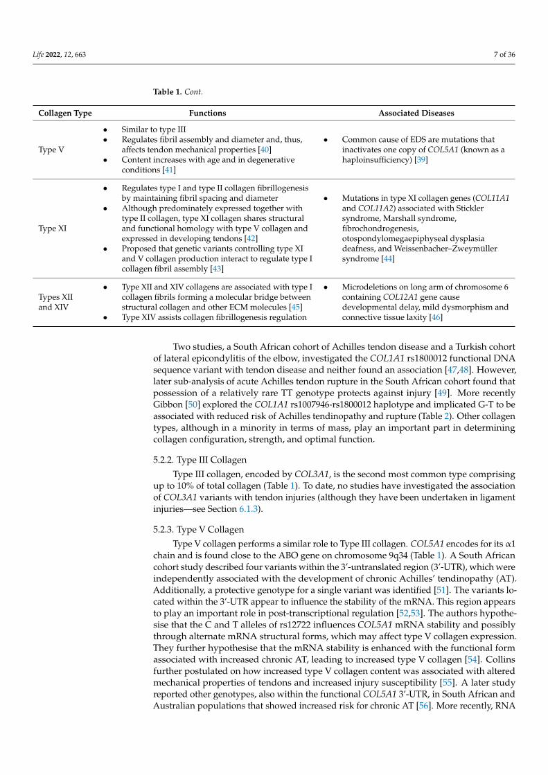
Life 2022, 12, 663 7 of 36
Table 1. Cont.
Collagen Type Functions Associated Diseases
Type V
• Similar to type III• Regulates fibril assembly and diameter and, thus,
affects tendon mechanical properties [40]• Content increases with age and in degenerative
conditions [41]
• Common cause of EDS are mutations thatinactivates one copy of COL5A1 (known as ahaploinsufficiency) [39]
Type XI
• Regulates type I and type II collagen fibrillogenesisby maintaining fibril spacing and diameter
• Although predominately expressed together withtype II collagen, type XI collagen shares structuraland functional homology with type V collagen andexpressed in developing tendons [42]
• Proposed that genetic variants controlling type XIand V collagen production interact to regulate type Icollagen fibril assembly [43]
• Mutations in type XI collagen genes (COL11A1and COL11A2) associated with Sticklersyndrome, Marshall syndrome,fibrochondrogenesis,otospondylomegaepiphyseal dysplasiadeafness, and Weissenbacher–Zweymüllersyndrome [44]
Types XIIand XIV
• Type XII and XIV collagens are associated with type Icollagen fibrils forming a molecular bridge betweenstructural collagen and other ECM molecules [45]
• Type XIV assists collagen fibrillogenesis regulation
• Microdeletions on long arm of chromosome 6containing COL12A1 gene causedevelopmental delay, mild dysmorphism andconnective tissue laxity [46]
Two studies, a South African cohort of Achilles tendon disease and a Turkish cohortof lateral epicondylitis of the elbow, investigated the COL1A1 rs1800012 functional DNAsequence variant with tendon disease and neither found an association [47,48]. However,later sub-analysis of acute Achilles tendon rupture in the South African cohort found thatpossession of a relatively rare TT genotype protects against injury [49]. More recentlyGibbon [50] explored the COL1A1 rs1007946-rs1800012 haplotype and implicated G-T to beassociated with reduced risk of Achilles tendinopathy and rupture (Table 2). Other collagentypes, although in a minority in terms of mass, play an important part in determiningcollagen configuration, strength, and optimal function.
5.2.2. Type III Collagen
Type III collagen, encoded by COL3A1, is the second most common type comprisingup to 10% of total collagen (Table 1). To date, no studies have investigated the associationof COL3A1 variants with tendon injuries (although they have been undertaken in ligamentinjuries—see Section 6.1.3).
5.2.3. Type V Collagen
Type V collagen performs a similar role to Type III collagen. COL5A1 encodes for its α1chain and is found close to the ABO gene on chromosome 9q34 (Table 1). A South Africancohort study described four variants within the 3’-untranslated region (3’-UTR), which wereindependently associated with the development of chronic Achilles’ tendinopathy (AT).Additionally, a protective genotype for a single variant was identified [51]. The variants lo-cated within the 3’-UTR appear to influence the stability of the mRNA. This region appearsto play an important role in post-transcriptional regulation [52,53]. The authors hypothe-sise that the C and T alleles of rs12722 influences COL5A1 mRNA stability and possiblythrough alternate mRNA structural forms, which may affect type V collagen expression.They further hypothesise that the mRNA stability is enhanced with the functional formassociated with increased chronic AT, leading to increased type V collagen [54]. Collinsfurther postulated on how increased type V collagen content was associated with alteredmechanical properties of tendons and increased injury susceptibility [55]. A later studyreported other genotypes, also within the functional COL5A1 3’-UTR, in South African andAustralian populations that showed increased risk for chronic AT [56]. More recently, RNA

Life 2022, 12, 663 8 of 36
sequencing analyses in torn rotator cuff tissue vs. control tissue, showed that COL5A1gene expression was markedly (3.01×) increased in tears, demonstrating its role in healingand/or remodelling [57].
The effect of altered genotypes in the MIR608 gene, which encodes a small micro-RNA(miRNA), was undertaken in Australian and South African groups [58]. An increasedrisk of AT was demonstrated with the rs4919510 CC genotype. This miRNA can bindto a recognition sequence within the COL5A1 and other genes 3’-UTR and inhibits trans-lation. It was the first non-coding gene to be associated with soft-tissue injuries andincreased understanding of the complex mechanisms involved in the regulation of typeV collagen production. A GWAS study investigating a large cohort, including samples ofdifferent ancestry, suggested that MIR608 rs4919510 showed moderate evidence for ATsusceptibility—although not at the level of significance required for a GWAS study [29](Table 2).
Table 2. Summary of genetic research and tendon injuries: Collagen types I and V.
CollagenType Reference Country Variant Pathology Subjects Controls Comments
Type I
[46] South Africa COL1A1rs1800012
Achilles’tendinopathyand rupture
126 125 No significant association
[47] Turkey COL1A1rs1800012
Lateralepicondylitis(commonextensor origin)
183 123 No significant association
[58] Spain COL1A1rs1800012
Patellar tendoninjury 15 0
No difference between severity ofinjury and gene variants. Nocontrols were included
[48]South Africaand Sweden(controls only)
COL1A1rs1800012
Achilles’tendon rupture 41 581
Significant association of the TTgenotype protecting against acuteAchilles’ tendon rupture
[49] South Africaand UK
COL1A1rs1107946rs1800012
Achilles’tendinopathyand rupture
216 193Significant association of the (G-T)haplotype protecting againstAchilles tendinopathy and rupture
Type V
[50] South Africa COL5A1rs12722
Achilles’tendinopathyand rupture
111 129 Significant protective associationwith variant in tendon disease
[59] Italy COL5A1rs12722
Bilateralquadricepsrupture
9 0 No controls but association with apolymorphism and tendon rupture
[60] Italy COL5A1rs12722
Bilateralquadricepsrupture
1 0No controls but association with apolymorphism and tendon rupture(same finding as Galasso)
[58] Spain COL5A1rs12722
Patellar tendoninjury 15 0
No difference between severity ofinjury and gene variants. Nocontrols were included
[55] South Africa,Australia
COL5A1rs12722COL5A1rs3196378
Achilles’tendinopathyand rupture
178 342
Significant protective associationwith variant of COL5A1 rs12722 intendon diseaseIn Australian cohort only, rs31996378associated with tendinopathy
[57] South Africa,Australia
COL5A13’-UTRrs71746744,rs1134170,rs16399MIR608rs4919510
Achilles’tendinopathyand rupture
160 342
All variants within the untranslatedregion of the COL5A1 gene and MIRvariant were associated withAchilles tendon disease
Green indicates an association and red indicates no association found.

Life 2022, 12, 663 9 of 36
5.2.4. Type XI Collagen
Type XI collagen is found in many structures including articular cartilage, bone,and muscle (Table 1). Variants in COL11A1 and COL11A2 interact with one another andwith a COL5A1 3’-UTR variant to modulate the AT risk in South African and Australiancohorts [43] (Table 3).
5.2.5. Types XII, XIV, and XXVII Collagens
None of the investigated functional variants of COL12A1 or COL14A1 which encodethe α-chains of types XII and XIV collagen have been associated with AT [59]. However,COL12A1 may be associated with acute Achilles tendon ruptures [59]. Saunders inves-tigated several COL27A1 gene variants that encode Type XXVII collagen but could notidentify any independent association with AT [60] (Tables 1 and 3).
5.3. Proteoglycans
Proteoglycans (PGs) represent up to 5% of tendon dry weight. They consist of a coreprotein attached to one or more glycosaminoglycans (GAGs). The GAG’s negative chargebinds water, which makes up 55–70% of the total tendon weight. PGs are found betweenthe ECM’s collagenous structural components (Table 4). There are no reported geneticstudies associated with tendon PG. However, ligament PG genetic variants have beeninvestigated (see Section 6.2.2).
5.4. Glycoproteins
Glycoproteins are a large family of structurally and functionally diverse proteins towhich a carbohydrate group(s) is covalently attached. Two important tendinous glycopro-teins are tenascin-C and cartilage oligomeric matrix protein (COMP) (Table 4).
Table 3. Summary of genetic research and tendon injuries: Collagen types XI, XII, XIV, and XXVII.
CollagenTypes Reference Country Variant Pathology Subjects Controls Comments
Type XI [43] South Africa,Australia
COL11A1rs1676486,rs3753841COL11A2rs1799907
Achilles’tendinopathyand rupture
184 338
No significant independentassociations foundGenes encoding structural andfunctionally related type XI (COL11A1and COL11A2) and type V (COL5A1)collagens interact with one another tocollectively modulate Achilles’tendinopathy risk
Type XII [59] South Africa,Australia
COL12A1rs240736,rs970547
Achilles’tendinopathyand rupture
137 131No significant associations foundThere may be an association withAchilles tendon rupture
Type XIV [59] South Africa,Australia
COL14A1rs4870723,rs1563392
Achilles’tendinopathyand rupture
137 131 No significant associations found
TypeXXVII [60] South Africa,
Australia
COL27A1(severalpolymor-phisms)
Achilles’tendinopathyand rupture
178 340 No significant associations foundamong variants
Green indicates an association and red indicates no association found.

Life 2022, 12, 663 10 of 36
Table 4. Regulatory and structural components of tendons.
Component Form and Types Functions Associated Diseases
Proteoglycans(PGs)
PGs• Found between extra-cellular matrix’s (ECM) collagenous structural
components• PGs include Decorin, Lubricin, and Versican• Consist of a core protein attached to one or more glycosaminoglycans
(GAGs)
• Decorin (80%): roles include collagen fibrillogenesisand potentially enhancing strength by facilitating loadtransfer between discontinuous collagen fibrils. Mostabundant in tendinous areas most subject to tension
• Lubricin: Found close to tendon periphery and aidstendon gliding with lubrication
• Versican: Found within the interfascicular matrix (IFM)
Glycoproteins
Tenascin-C (TNC)• Protein is a hexamere, which binds to both ECM components and
tenocyte surface receptors• Produced by TNC gene located close to the ABO gene on chromosome 9q34• TNC gene expression appears influenced by tension loading [61]
• Important role in regulating the tenocytes need tointeract with ECM components
• Involved in cell adhesion and signalling, affecting cellproliferation and migration [62–64]
• Appears to increase in the presence of tendonpathology [65]
• Mutations within TNX gene (a tenascin family member)causes an autosomal recessive form of EDS fromtenascin-X deficiency [66]
Thrombospondins (THBS)
• Family of glycoproteins performing dynamic role within ECM
Cartilage oligomeric matrix protein (COMP)
• Also known as thrombospondin V• Most abundant tendinous glycoprotein• Links to type I collagen and has five subunits allowing linkage between
multiple fibrils• Found within the inter-fibrillar matrix [67] and tissues subject to
significant loading, such as tendons, ligaments, cartilage, menisci, andintervertebral discs
Thrombospondin 2 (THBS2)
• Mediates cell-to-cell and cell-to-matrix interactions• Involved in cell-to-cell adhesion and ECM communication
Thrombospondin type 1 domain–containing protein 7A
• This protein regulates focal adhesions during angiogenic cell migration
• Role debated• COMP-null knock out mice did not demonstrate any
tendon abnormality [68]• Others reported in horses that COMP levels during
growth corelate with mechanical strength at skeletalmaturity [66]
• Pseudo-achondroplasia is caused by a heterozygousmutation in the gene encoding COMP [16]
• A COMP gene mutation can cause multiple epiphysealdysplasia (MED)
Elastin andMicrofibrils
Elastin (ELN)• Elastin provides elasticity to tendons, allowing them to
stretch and return to their original state. Playsimportant load-bearing role in tendons and ligaments.
ELN rs2071307 variant has been associated with othermultifactorial ECM conditions, such as aortic stenosis [69] andaortic aneurysm [70]ELN rs2071307 and FBN2 rs331079 variants associate with aorticand intracranial aneurysms, respectively, causing ECM disruptionFibrillin-1 abnormalities have been associated with Marfan’ssyndrome and Fibrillin-2 have with Beal’s syndrome orcongenital contractural arachnoldactyly [71,72]
Microfibrils, such as the glycoprotein, Fibrillin:
• Found in ECM and become incorporated into insoluble microfibrils• Plays a role in early elastogenesis acting as a scaffold
for elastin deposition [73]

Life 2022, 12, 663 11 of 36
5.4.1. Tenascin-C
Tenascin-C (TNC) plays an important role in regulating tenocytes that need to inter-act with ECM components (Table 4). The first research establishing a genetic associationfor a predisposition to Achilles tendon pathology was published in 2005 and implicatedTNC [74]. A South African case-control association study reported that a Guanine-ThymineDinucleotide repeat variant was associated with Achilles tendon injuries. A second study in-vestigating different TNC variants (rs13321; rs2104772; rs1330363) found altered frequenciesbetween cases and controls in Australian and South African populations—but did not reachstatistical significance [75] (Table 5). However, the study did implicate a genetic regionspanning both the TNC and the COL27A1 genes using haplotype analysis. More recently,the application of WES analyses assisted the identification of potential functional TNCvariants implicating the TNC gene, specifically rs1061494 and the T-T haplotype (rs1061494-rs2104772) with increased risk of Achilles tendinopathy in a South African cohort [30]. Thisstudy therefore provided evidence that the risk susceptibility to Achilles’ tendinopathy ismost likely within the TNC gene rather than within the COL27A1 gene locus.
Several TNC variants have been associated with rotator cuff injury [76,77]. Previouslyimplicated TNC variants (rs1138545, rs72758637, rs7021589) have been replicated in aGWAS study using the UK biobank to be associated with an increased risk of rotator cuffinjury [57,76]. Tashjian reported increased gene expression, for TNC (2.2×) in tears vs.controls [57] (Table 5).
5.4.2. COMP and Other Thrombospondins
COMP, also known as thrombospondin 5, is the most abundant tendinous glycoprotein.Thrombospondin 2 (THBS2) mediates cell-to-cell and cell-to-matrix interactions and isinvolved in cell-to-cell adhesion and ECM communication (Table 4). COMP (rs730079;rs28494505) and THBS2 (rs9505888; rs6422747) variants failed to show significant differencein Achilles tendon studies in Australian and South African cohorts [75]. No human geneticstudies exist relating to the potential role of Thrombospondin 2 in tendon pathology,although its absence has been associated with connective tissue abnormalities in mice [76].
A GWAS study identified rs575224171 within the gene THSD7A encoding the endothe-lia protein thrombospondin type 1 domain, containing protein 7A, to be associated withincreased risk of rotator cuff injury [57]. These authors hypothesised that gene variantsmay lead to poor rotator cuff angiogenesis, predisposing individuals towards increasedtear risk. RNA sequencing analyses established a 2.6× decreased expression of this genewithin rotator cuff tears vs. control tissues [57].
5.5. Elastin and Microfibrils
Elastin represents 1–10% of a tendon’s dry weight and is found in both the IFM andwithin the fascicles—especially around tenocytes. Microfibrils, such as the glycoproteinFibrillin, are found in the ECM and become incorporated into insoluble microfibrils (Table 4).South African, Australian, and British cohorts did not find an association between the ELNrs2071307 variant and risk of developing Achilles’ tendon pathology [78,79]. However,the FBN2 rs331079 variant was associated with risk for Achilles’ tendon disease and ACLruptures [78].

Life 2022, 12, 663 12 of 36
Table 5. Summary of genetic research and tendon injuries: Glycoproteins in the ECM and elastinand fibrillin.
Type Protein Reference Country Variant Pathology Subjects Controls Comments
Glycoproteinsin the ECM
Tenascin-C(TNC)
[74]SouthAfrica,Australia
TNC—GTdinucleotiderepeats
Achilles’tendinopathyand rupture
144 127Association with the number ofGT repeat polymorphism and therisk of tendinopathy and rupture
[60]SouthAfrica,Australia
TNC rs13321,rs2104772,rs1330363
Achilles’tendinopathyand rupture
179 339 No significant associationsfound among variants
[80] Spain TNCrs2104772
Patellartendon injury 15 0
No difference between severityof injury and gene variants. Nocontrols were included
Cartilageoligomericmatrix protein(COMP)
[75]SouthAfrica,Australia
COMPrs730079,rs2849450THBS2rs9505888
Achilles’tendinopathyand rupture
178 340 No significant associationsfound among variants
Elastin andFibrillin
Elastin (ELN)and Fibrillin(FBN)
[78]SouthAfrica,Australia
ELNrs2071307FBN2rs331079
Achilles’tendinopathyand rupture
135 239
No association with the ELNvariant and tendon pathologySignificant association betweenthe FBN2 gene variant andtendon disease
[81] UK ELNrs2071307
Achilles’tendinopathyand rupture
108 131 No association with the ELNvariant and tendon pathology
[80] Spain ELNrs2289630
Patellartendon injury 15 0
No difference between severityof injury and gene variants. Nocontrols were included
Green indicates an association and red indicates no association found.
5.6. Tendon Development, Homeostasis, and Remodelling
Tendon ECM homeostasis and remodelling are maintained by complex enzyme sys-tems. These include matrix metalloproteases (MMPs), ADAMTSs (a disintegrin and met-alloproteinase with thrombospondin motifs) and ADAMs (a disintegrin and metallopro-teinase), tissue inhibitors of MMPs (TIMPs), and growth factors like the transforminggrowth factor-ß (TGF-ß) families [82] (Table 6). Genetic research undertaken in this area oftendon development, homeostasis, and remodelling is summarised in Table 7.
5.6.1. MMPs, TIMPs, ADAMTSs, and ADAMs
The balance between MMPs and TIMPs is necessary to maintain tendon homeostasisand remodelling [82]. If intrinsic control of these systems is compromised by extraneousfactors, the tendon’s ability to respond appropriately to loading will be affected, risking ten-don disease (Table 6). No association was reported of ADAMTS2, ADAMTS5, ADAMTS14,and ADAM12 variants with Achilles’ tendon pathology in South African and Australian co-horts [83]. However, Raleigh found significant associations for MMP3 variants and AT (butnot rupture) in a South African population [84]. In a British population, El Khoury foundno associations in Achilles’ pathology groups overall—although subgroups did show somecorrelations [83]. Furthermore, El Khoury found a significant association with a variantwithin the TIMP2 gene rs478932 for Achilles’ tendon pathology in both a South Africanand Australian cohort. Different genotypes were overrepresented in the subject groupsin each population [83]. Gibbon explored the MMP3 locus in an Australian cohort withAT and identified a 6A-G-C-G haplotype (rs3025058, rs679620, rs591058, rs650108) withreduced risk [85].

Life 2022, 12, 663 13 of 36
Table 6. Tendon development, homeostasis and remodelling.
Families Functions Key Regulators Associated Diseases
Matrix metalloproteases (MMPs)(23 family members)
Four subgroups:• Collagenases• Gelatinases• Stromelysins• Membrane-type• Broad proteolytic activity against
collagen and other ECM compounds
• MMP3: degrades different collagen types, proteoglycans, andfibronectin, and laminin
Fluoroquinolone antibiotics, whichare associated with increased Achillesrupture risk [86], can affect MMPexpression in human tendons [9,87]
A disintegrin and metalloproteinasewith thrombospondinmotifs (ADAMTSs)(20 family members)
• Procollagen processing• ECM remodelling
• ADAMTS2 and 14: regulate conversion of procollagen to collagen• ADAMTS5: cleaves proteoglycans, e.g., aggrecan
ADAMTS2 gene mutation causesEDS dermatosparaxis type
A disintegrin andmetalloproteinase (ADAMs)(19 family members)
• Proteases• ADAM12: a marker of skeletal muscle regeneration and binds insulin
growth factor binding protein-3 (IGFBP-3)
Tissue inhibitors ofmetalloproteases (TIMPs)
• Inhibit MMPs activities• Balance between MMPs and TIMPs
is necessary to maintain tendonhomeostasis and remodelling
Transforming growth factor beta(TGF-ß) superfamily • ECM homeostasis and remodelling
• TGF-ß1 and growth differentiation factor 5 (GDF-5), can increaseAchilles tendon strength in animals [88].
• Mechanical loading releases TGFß-1 and aids cell growth,proliferation, differentiation, and migration, as well as cell death(apoptosis). Involved in collagen and proteoglycan synthesis.
• GDF-5 (also called CDMP-1 (cartilage-derived morphogenetic protein1) or BMP-14 (bone morphogenic protein 14). Expressed in developingCNS and maintenance, development and repair of bones, cartilage,and various other musculoskeletal soft tissues, including tendons.
GDF-5 gene mutations (CDMP1)implicated in Hunter–Thompsontype dwarfism and in GrebeSyndrome (characterized by shortstature, extra digits, and short anddeformed extremities) [89–92].
Bone morphogenetic proteins(BMPs) (20 family members)
• Grouped into subfamilies of theTGF-ß superfamily
• Growth factors and cytokines inmany tissues in the body
Fibroblast growth factors (FGFs)(22 family members)
• Influence cell development andmaturation by binding to receptors(FGFR) triggering intracellular events
Point mutation in FGFR3 can leadto achondroplasia.

Life 2022, 12, 663 14 of 36
5.6.2. Transforming Growth Factor-ß (TGF-ß) Superfamily
The TGF-ß superfamily has a similar role as the MMP/TIMP system in ECM homeosta-sis and remodelling (Table 6). Mechanotransduction is the process of converting mechanicalforces into a cellular response. Tendon exposure to increased loading (within safe physi-ological limits) causes tenocytes to increase collagen synthesis and enhance tendon loadresistance. It is the end goal of sensible incremental training [92]. The reverse occurswith inactivity. If a tendon’s tensile load is temporarily decreased, there is a reduction insecreted ECM structures including Type I collagen and COMP [93]. TGF-ß appears to be amajor regulator of tendon development secondary to mechanical loading [94]. The exactmechanism of how loading activates the TGF-ß signalling pathway appears to involve aninduction of scleraxis (Scx) and other markers, such as tenomodulin [95]. This promotesthe synthesis and secretion into the ECM of collagen and other ECM components. TGF-ßand GDF-5 genes functional variants have been studied with an association established forGDF-5 rs143383, but not TGF-ß rs1800469, with risk of Achilles’ tendinopathy [96].
5.6.3. Bone Morphogenic Glycoproteins (BMP)
BMPs are grouped into subfamilies of the TGF-ß superfamily and function as growthfactors or cytokines. Originally studied for their effect upon bone and cartilage formation,they are recognised to have a widespread function as signallers and regulators of manyorgans systems’ development [35]. A significant association was reported in a Brazilianmixed-injury cohort for the BMP4 variant (rs2761884) in tendinopathies [97].
5.6.4. Fibroblast Growth Factors (FGFs)
FGFs are required for normal development and cell maturation. They bind to recep-tors (FGFR), triggering intracellular events. Salles studied a group of Brazilian volleyballplayers with variously located tendinopathies. However, none of the investigated FGF3,FGF10, and FGFR1 variants were associated with altered risk for tendinopathy [97]. Simi-larly, no associations of the same genes with rotator cuff tears in American patients werereported [98]. Conversely, Motta found significant associations of the FGF3, FGF10, andFGFR1 variants with rotator cuff disease in large Brazilian case-control study [99].
Table 7. Summary of genetic research and tendon injuries: Development, homeostasis and remodelling.
Families Reference Country Variant Pathology Subjects Controls Comments
A disintegrin andmetalloproteinase withthrombospondin motifs(ADAMTSs)A disintegrin and metal-loproteinase (ADAMs)
[83]SouthAfrica,Australia
ADAMTS2 rs1054480ADAMTS5 rs226794ADAMTS14rs4747096ADAM12 rs4747096
Achilles’tendinopathy andrupture
173 248
No significant association withAchilles’ diseaseADAMTS14 associated with a lateronset of disease
Matrix metalloproteases(MMPs)and Tissue inhibitors ofmetalloproteases (TIMPs)
[84] SouthAfrican
MMP3 rs591058,rs650108, rs591058
Achilles’tendinopathy andrupture
114 98
Variants significantly associatedwith chronic tendinopathyNo increased risk of rupturewith variantsLowest risk of tendinopathyassociated with combination of aMMP3 rs679620 and COL5A1rs12722 variants
[81] UK MMP3 rs679620TIMP2 rs4789932
Achilles’tendinopathy andrupture
118 131
MMP3 variant significantlyassociated with tendinopathy andrupture groups in males but notfemales or overall.TIMP2 variants overrepresented intendinopathy group
[83]SouthAfrica,Australia
TIMP2 rs4789932Achilles’tendinopathy andrupture
173 248 SNP significantly overrepresented intendinopathy group
[85] AustraliaMMP3 rs679620,rs591058, rs650108,rs3025058
Achilles’tendinopathy 79 195
An association was found with a6A-G-C-G haplotype (rs3025058,rs679620, rs591058, rs650108) withreduced risk for Achilles tendinopathy

Life 2022, 12, 663 15 of 36
Table 7. Cont.
Families Reference Country Variant Pathology Subjects Controls Comments
Transforming GrowthFactor-ß Superfamily(TGFß)Growth/differentiationfactors (GDF)
[96]SouthAfrica,Australia
TGFB1 rs1800469GDF5 rs143383
Achilles’tendinopathy andrupture
171 238
No associations with TGFB1genotype variantGDF5 variant associated withAchilles’ disease.
Bone morphogeneticproteins (BMPs) [97] Brazil BMP4 rs2761884
Mixed—Achilles;Patellar; Rotatorcuff; Hip abductors
52 86 Significant association with BMP4variant for tendinopathy
Fibroblast GrowthFactors
[97] Brazil
FGF3 rs7932320,rs1893047, rs12574452,rs4631909, rs4980700FGF10 rs1448037rs900379, rs1011814,rs593307FGFR1 rs13317
Mixed—Achilles;Patellar; Rotatorcuff; Hip abductors
52 86 No significant associations withtendinopathy
[98] USAFGF3FGF10FGFR1
Rotator Cuff tears 175 2595 No significant associations
[99] BrazilFGF3 rs12574452FGF10 rs11750845,rs1011814FGFR1 rs13317
Rotator Cuff tears 203 207 All showed significant associationswith rotator cuff tears
Green indicates an association and red indicates no association found.
5.7. Cell Death (Apoptosis) and Inflammation in Tendons
Apoptosis is a natural phenomenon in many living tissues. In tendons, damagedtenocytes’ removal is facilitated by cytokine activity. Excessive tendon loading can in-crease apoptosis and affects the cell population’s abilities to respond effectively to exercisewith secondary effects upon the ECM leading to tendon disease [100]. The role of in-flammation in chronic tendinopathy has long been debated [101]. Early research utilisingmicroscopic examination of tendinopathic tissue and biochemical analysis reported noevidence of the normal elements associated with ‘classical’ inflammation in chronic tendoninjuries [102–105]. Animal work indicated that early tendinosis was associated with teno-cyte stimulation rather than apoptosis and modulated by growth factors such as insulin-likegrowth factor 1 (IGF-1) [106]. However, the authors were unable to comment on the chroniceffect of prolonged loading on cell survival. (Table 8).

Life 2022, 12, 663 16 of 36
Table 8. Tendon disease: Inflammatory cascade, apoptosis, and other elements.
Type Elements or Activity Functions Key Elements
Inflammatory Cascadeand Apoptosis
Interleukins (ILs)• Intimately involved in the
inflammatory pathway
• Interleukin-1ß (IL1B) influences cellular death, proliferation, and differentiation• Interleukin-1 receptor antagonist (ILRN) inhibits activities of both IL1B and
interleukin-1α (IL1A)• Interleukin-6 (IL6) is found in both acute and chronic inflammation
Caspases (CASP) • Family of protease enzymes intimatelyinvolved in apoptosis
• CASP8 is a pro-enzyme central to cell death regulation
Nitric OxideSynthases (NOS)
• Nitric oxide (NO), produced by NOSenzymes, is a free radical and importantcellular signalling messenger with manyfunctions including increased productionof IL6 and Il8 [107]
• Three NOS genes, NOS1, NOS2, and NOS3, encode for nNOS, iNOS and eNOSenzymes, respectively
• These genes are designed to code for differing elements of the inflammatorycascade and control over cell death
• Gene variations associated with alterations in susceptibility to tendinopathy [108]• NOS2 expression elevated 23-fold higher than controls within days of Achilles’
tendon injury• NO catalysed by the iNOS isoform of NOS induces apoptosis in inflammatory cells
to eradicate cells from damaged area, preventing chronic inflammation andallowing remodelling to occur [109]
Angiogenesis• Marked increase in new blood vessels in
chronic tendon disease [110]
• Vascular endothelial growth factor A (VEGFA)• Kinase domain receptor (KDR)• Hypoxia inducible factor 1 subunit alpha (HIFIA)• Encoded by VEGFA, KDR, and HIF1A genes, respectively [111–113]
Others
Estrogen-relatedreceptor beta (ERR-β)
• Function unknown• May influence the expression of PPARGC1
and ESRR-inducing regulator muscle 1(PERM1) in skeletal muscle
Defensins • Microbicidal and cytotoxic peptides madeby neutrophils

Life 2022, 12, 663 17 of 36
5.7.1. Interleukins
Inflammatory pathways throughout the body involve numerous elements, interactingin a complex manner and resulting in gene expression alterations, apoptosis, and detrimen-tal changes to the ECM. The protein family of interleukins are intimately involved in theinflammatory pathway. Interleukins are upregulated in early tendinopathy and involved inthe inflammatory cascade and remodelling activities [114]. September reported interleukingene-gene interactions with COL5A1 rs12772, suggesting that type V collagen may beregulated by certain inflammatory mediator proteins in the IL-1β-signalling pathway [115].Altering the amount of type V collagen expression could impact α1(V) collagen chains and,thereby, the collagen tendon fibril diameter and ultimately tendon capacity (Table 9).
5.7.2. Caspases
Caspases are a family of protease enzymes that are integral to programmed cell death(apoptosis). The South African research group reported that two CASP8 genotypes hadsignificant associations with AT [116] (Table 9).
5.7.3. Nitric Oxide Synthase (NOS) Enzymes
Nell found no association with gene variants for NOS2 and NOS3 and AT [115].However, Brookes reported a reduced risk for AT with the NOS2 rs2779249 heterozygotevariant, but no association with NOS2 rs2248814 [117] (Table 9).
5.8. Angiogenesis
Angiogenesis is the formation of new blood vessels from the existing vasculature.Tendons and ligaments have a poor blood supply and low metabolic rate. Consequently,their healing capacity is low [118]. Histopathological examination of chronic AT specimensshows marked increases in angiogenesis [110]. It is hypothesized that this is triggeredby mechanical loading and designed to promote tendon remodelling. Further studieshave identified increased levels of pro-angiogenic expression profiles and, specifically,vascular endothelial growth factor A after tenocyte mechanical loading [119–121]. Poorlyregulated angiogenesis may lead to distortion of the neat parallel collagen fibril arrayin the tendon ECM. Increased levels of angiogenic associated proteins have been notedin both ruptured tendons and ligaments and including degenerative tendons [121–123].Angiogenesis elements are placed centrally within the network of partners regulating keyECM components within tendon and ligament.
Several functional variants within the VEGFA (rs699947, rs1570360, and rs2010963)gene have been explored and a risk haplotype was implicated both in a (i) South Africancohort and a (ii) combined South African and British cohort of mid portion chronic AT [124].Specifically, the VEGFA A-G-G (rs699947 C/A–rs1570360 G/A–rs2010963 G/C) inferredhaplotype was associated with increased risk of AT. This haplotype includes the collectivealleles associated with decreased VEGFA gene transcription and a corresponding lowerVEGFA plasma level [125]. Therefore, it is reasonable to hypothesise that these allelecombinations would potentially contribute to limiting the capacity of the structure toregulate ECM remodelling within a hypovascular tendon [122]. The authors did not reportan association with Achilles’ tendon risk for any of the variants explored in KDR (rs2071559and rs1870377) [123] (Table 8).
Like other gene loci, differences in associations at the VEGFA and KDR loci have beennoted in populations of different ancestry. One study reported no associations in two KDRpolymorphisms (rs1870377; rs2071559) with AT in South African and UK cohorts [124](Table 9).

Life 2022, 12, 663 18 of 36
5.9. Other Areas of Genetic Study in Tendons5.9.1. ESRRB
Estrogen-related receptor beta (ERR-β) is a nuclear receptor encoded by ESRRB (Estro-gen Related Receptor Beta) gene. Its function is unknown; however, a similar protein inmice plays an essential role in placental development. It appears to influence the expressionof PPARGC1 and ESRR-inducing regulator muscle 1 (PERM1) in skeletal muscle. Mottaidentified two 2 SNPs in the ESRRB gene that were associated with rotator cuff disease [99].Teerlink found a significant association for rotator cuff injury with an ESRRP rs17583842variant [98] (Table 9).
5.9.2. Defensin ß1
Defensins form a family of microbicidal and cytotoxic peptides made by neutrophils.Defensin ß1 is encoded by the DFNB gene. It resists microbial organisms from attachingto epithelial surfaces. Motta found the DEFB-1 rs1800972 SNP to be associated witha preventive effect for rotator cuff tears [99] while Teerlink found no association withDEFB-1 [98] (Table 9).
Table 9. Summary of genetic research and tendon injuries: Inflammatory cascade, apoptosis, and others.
Type Protein Reference Country Variant Pathology Subjects Controls Comments
InflammatoryCascadeandApoptosis
Interleukins (ILs) [116]SouthAfrica,Australia
IL1B rs1143627,rs16944IL1RN rs2234663IL6 rs1800795
Achillestendinopathyand rupture
175 369
No independentassociations.In combination withCOL5A1 rs12722 thesealleles had significantassociation withAchilles’ tendinopathy
Caspases (CASP) [115]SouthAfrica,Australia
CASP8 rs3834129,rs1045485
Achillestendinopathyand rupture
166 358 Significant associationwith both polymorphisms
Nitric OxideSynthases (NOS)
[115]SouthAfrica,Australia
NOS2 rs2779249NOS3 rs1799983
Achillestendinopathyand rupture
166 358 No significant associations
[117] UK iNOS rs2779249iNOS rs2248814
Achillestendinopathyand rupture
132 145
Significant association(protective effect)with rs2779249No associationwith rs2248814
Angiogenesis AngiogenicFactors [124]
SouthAfrica andUK
VEGFA rs699947,rs1570360,rs2010963KDR rs1870377,rs2071559
Achillestendinopathy
120130
10887
Significant associationwith all 3 VEGFApolymorphismsNo association with theKDR polymorphisms
Others
Defensins (DEF)[98] USA DEFB1 Rotator Cuff
tears 175 2595 No association
[99] Brazil DEFB1 rs1800972 Rotator Cufftears 203 207 Significant association
(protective effect)
Estrogen-relatedreceptor ß(ESRRB)
[98] USA ESRRB rs17583842 Rotator Cufftears 175 2595 Significantly associated
with rotator cuff disease
[99] Brazil ESRRB rs1676303,rs4903399
Rotator Cufftears 203 207 Significantly associated
with rotator cuff disease
Green indicates an association and red indicates no association found.
5.10. Ageing in Tendons
Diseased and damaged tendons increase with age. For instance, 11–37% developtears in an ankle peroneal tendon during their lifetime [126]. Long exposure to mechanicalstresses and the increasing inefficiency of tendon repair are implicated. It is a classicexample of the Injury Causation model (Figure 1). As a result, performance and functionare impaired [127]. Many age-related changes occur to tendons during their lifetimeand differences are apparent in the way men and women respond to tendon loading [128].Additional knowledge has been derived from animal research, especially equine studies andit has been proposed that the ability of pluripotent mesenchymal stem cells to differentiateinto tenocytes reduces with time. Tendon stem cells become less numerous with age

Life 2022, 12, 663 19 of 36
and their ability to differentiate and produce competent mature tenocytes reduces [129].The senescence-inhibited gene within the tenocyte is downregulated [130]. The ageingtenocyte’s complement of produced proteins (proteome) is restricted [131]. The proteinsaffected include those with roles involved in cell survival and death, cytoskeletal changes,and antioxidant response.
The ability to repair damaged tendons is also affected by non-collagenous ECM proteinturnover, including cytokines and various growth factors, and a disruption to the fine home-ostatic mechanisms outlined in Section 5.6 [132]. Equine research has revealed that with age,the tendons’ protein turnover diminishes [133], glycosaminoglycans increase [134], the typeIII collagen increases proportionately [38], and collagen fibril diameter diminishes [135].Changes occur to the IFM with reduced protein turnover and elasticity, increasing therisk of injury [136,137]. MMP activity reduces tendon strength [127]. There is additionalevidence that age carries with it a reduced capacity to resolve inflammation [138]. In addi-tion, degenerative human tendons can have altered responses to reactive oxygen specieswith age, and therefore oxidative stress may be an important pathway in tendinopathydevelopment [139].
The progressive alteration in tendon function involves a complex interplay betweenmany influences. This includes not only our inherited genetic material but temporalchanges to gene expression regulation, including an array of epigenetic mechanisms [140].The potential role of epigenetics will be elaborated upon in Section 9.
6. Ligament: Structure, Function, and Genetic Research
Ligaments span joints attaching at either end to bones. Like tendons, they are fibrous,dense connective tissues. They are designed to resist excessive load, control joint motion,prevent instability, and have a vital proprioceptive role on account of their rich innervation.They have a similar hierarchical structure to tendons. However, the degree of packing ofthe collagen is slightly different. Whereas tendons organise collagen fibres in an orderly,parallel orientation, ligament organisation is more random and less parallel. This allowsligaments to respond to tensile loads in different directions. The cellular elements tendto be more randomly distributed and rounder in shape than tendons. There is a higherpercentage of proteoglycans and water and reduced percentage of collagen. The elastincontent in ligament is higher than tendons.
6.1. Collagen6.1.1. Type I Collagen
There has been considerable interest in the genetic architecture of the COL1A geneand susceptibility to tendon and ligament injuries. Differences in the genetic susceptibilityto acute and chronic injuries have been linked to rs1107946 (−1997 G/T) and rs1800012(+1245 G/T), within the COL1A1 gene. The rare TT genotype of the Sp1 binding site variant(rs1800012) was associated with decreased risk for acute injuries such as shoulder disloca-tions [141], ACL ruptures [47], and acute soft tissue ruptures [49]. A meta-analysis reportedthe association of the rs1800012 TT genotype with reduced risk for sports-related tendonand ligament injuries [142]. The same genotype has been associated with increased risk forintervertebral disc degeneration in the elderly and increased risk for lumbar disc disease inyoung military recruits [143,144]. The alternate rs1800012 GG genotype was reported toreduce risk for ACL ruptures sustained while skiing [145]. The rs1107946 variant, which isin linkage disequilibrium with rs1800012, has been independently associated with the riskof skiing-associated ACL ruptures [146]. Haplotype analyses with these two functionalvariants have been associated with ACL rupture risk in a Polish cohort [147]. The cur-rent theory proposes that these functional promotor variants work in concert to regulateCOL1A1 expression [148].
However, several studies have failed to reflect an association between these COL1A1variants and susceptibility to several musculoskeletal soft tissue injury phenotypes. Thismay result from insufficient power of the studies to detect the rare rs1800012 TT genotype

Life 2022, 12, 663 20 of 36
in and may explain conflicting results when comparing data from larger combined analysesto that of smaller independent cohorts [50]. No associations were noted in the ChineseYunnan Han ACL samples for the COL1A1 or COL5A1 locus [149]. However, the TT geno-type of the COL1A1 Sp1 binding site polymorphism has been reported to be significantlyunderrepresented in South African participants with ACL ruptures [150] (Table 10).
Table 10. Summary of genetic research and ligament injuries: Collagen types I and III.
Collagen Types Reference Country Variant Pathology Subjects Controls Comments
Type I
[150] South Africa COL1A1rs1800012 ACL rupture 117 130 Significant
association
[147] PolandCOL1A1rs1107946,rs1800012
ACL rupture 91 143 Significantassociation
[146] PolandCOL1A1rs1107946,rs1800012
ACL rupture 138 183 Significantassociation
[142] Meta analyses COL1A1rs1800012
Tendon andligament injuries 933 1381 Significant
association
[149] Chinese Yunnan Han COL1A1rs1800012 ACL rupture 101 110
Nosignificantassociation
[50]Combined populationfrom SA, Sweden,Poland, Finland
COL1A1rs1800012
ACL rupture andCruciateligament rupture
1425 407 Significantassociation
Type III[151] Poland COL3A1
rs1800255 ACL rupture 138 183 Significantassociation
[152] South Africa, Poland COL3A1rs1800255 ACL rupture 333 378 Significant
association
Green indicates an association and red indicates no association found.
6.1.2. Type V Collagen
As in AT research, variants within the 3’-UTR of the COL5A1 gene have been associatedwith ACL rupture susceptibility, specifically in females [152–154], and more recently withligament injuries [155]. Laguette explored the intron 4-exon 5 region of COL5A1, which waspreviously implicated with ligament injuries in a canine model but found no significantassociations in a South African ACL cohort [156]. The COL5A1 rs12722 C/T and COL5A1rs13945 C/T polymorphisms were also associated with reduced ACL injury risk in maleskiers [157]. Furthermore, Suijkerbuijk reported an association with ACL ruptures andCOL5A1 rs12722 in a combined Swedish and South African cohort [158] (Table 11).

Life 2022, 12, 663 21 of 36
Table 11. Summary of genetic research and ligament injuries: Collagen types V and XII.
Collagen Types Reference Country Variant Pathology Subjects Controls Comments
Type V
[153] South Africa COL5A1 rs12722,rs13946 ACL rupture 129 216 Significant
association
[157] Poland COL5A1 rs12722,rs13946 ACL rupture 138 183
Associatedwith reducedinjury risk
[152] South Africaand Poland COL5A1 rs12722 ACL rupture 333 378
Significantgene-geneassociation
[154] Poland COL5A1 rs12722,rs13946 ACL rupture 134 211 Significant
association
[158] South Africaand Sweden COL5A1 rs12722 ACL rupture 98
79 116 Significantassociation
[156] South Africa COL5A1 rs3922912,rs4841926 ACL rupture 249 210 No significant
association
[149] ChineseYunnan Han
COL5A1 rs12722,rs13946 ACL rupture 101 110 No significant
association
[155] South Africa,Australia, Japan
COL5A1 rs12722,rs10628678
ACL rupture andLigament injury 311 592 Significant
association
Type XII
[159] South Africa COL12A1 rs970547 ACL rupture 129 216 Significantassociation
[160] Poland COL12A1 rs970547 ACL rupture 91 143 No significantassociation
[152] South Africaand Poland COL12A1 rs970547 ACL rupture 333 378 Significant
association
[149] ChineseYunnan Han
COL12A1 rs970547,rs240736 ACL rupture 101 110 Significant
association
Green indicates an association and red indicates no association found.
6.1.3. Types III and XII Collagen
Associations have been noted for variants in COL3A1 [151,152], and COL12A1 [149,159]with ACL rupture risk, while others reported no significant associations [147,161].
6.2. Regulatory and Structural Components of the Extracellular Matrix6.2.1. Tenascin-C
Historically, there has been much interest in the associations between the TNC gene andits neighbouring genes with tendon and ligament injury susceptibility. Gibbon explored thisregion for ACL susceptibility. Variants within the TNC gene: rs2104772 and a TT haplotype(rs1061494 and rs2104772) were associated with ACL susceptibility using a tailored WESand bioinformatics approach [30]. However, no associations for the TNC locus were notedin a Polish cohort [162]. More functional research is required to understand the biologicalsignificance underpinning this genetic locus and tendon and ligament injury susceptibility.
6.2.2. Proteoglycans
Recent studies have investigated proteoglycans. Variants within their controllinggenes have been implicated with susceptibility to ACL ruptures in independent cohortsfrom South Africa [163,164] and Poland [165] (Table 12).

Life 2022, 12, 663 22 of 36
Table 12. Summary of genetic research and ligament injuries: Glycoproteins in the ECM and proteoglycans.
Type Protein Reference Country Variant Pathology Subjects Controls Comments
Glycoproteinsin the ECM
Tenascin C(TNC)
[30] SouthAfrica
TNC rs1061494,rs1138545, rs2104772,rs1061495
ACL rupture 234 232 Significantassociation
[162] Poland TNC rs1330363,rs2104772, rs13321 ACL rupture 229 192
Nosignificantassociation
Proteoglycans
Aggrecan(ACAN)
[163] SouthAfrica
ACAN rs2351491,rs1042631, rs1516797 ACL rupture 227 234 Significant
association
[165] Poland ACAN rs2351491 ACL rupture 143 229 Significantassociation
Biglycan (BGN)
[163] SouthAfrica
BGN rs11264797,rs1126499, rs1042103 ACL rupture 227 234 Significant
association
[165] Poland BGN rs11264797,rs1042103 ACL rupture 143 229 Significant
association
[166] SouthAfrica
BGN rs11264797,rs1042103 ACL rupture 227 234
Significantgene-geneinteractions
Decorin (DCN)Lumican (LUM)Fibromodulin(FMOD)
[163] SouthAfrica
DCN rs13312816,rs516115LUM rs2268578FMOD rs7543148
ACL rupture 227 234 Significantassociation
Green indicates an association and red indicates no association found.
6.2.3. MMPs
As in tendinopathy, the MMP locus (chr11q22) has been association with susceptibilityto ACL ruptures. Posthumus demonstrated that MMP3 rs679620 variant may interactwith several other MMP loci, MMP10 rs485055, MMP1 rs1799750, and MMP12 rs2276109,to collectively contribute to ACL rupture susceptibility in a South African cohort [167].The MMP3 rs3025058 variant, which is tagged by rs679620, was independently associatedwith ACL ruptures in a Thai population [168]. No associations were noted when MMP1rs1799750, MMP10 rs486055, and MMP12 rs2276109 variants were explored with ACLrupture susceptibility in a Polish cohort [169]. The MMP genes have been investigated withseveral different exercise-related phenotypes and conflicting associations have been noted.This suggest that there may be specific genetic signatures which are inherited together andunderpin specific exercise-related phenotypes, which still require functional unravelling(Table 13).
6.2.4. Transforming Growth Factor Superfamily
Variants in several such genes controlling the TGF superfamily have been exploredwith an association to ACL rupture risk. These include variants within the TGF-β receptorIII (TGFβR3) and the TGF-β induced (TGFβI) genes. An independent association of TGFBR3rs1805113 G allele with a decreased risk of ACL injury has been described. Additionally, agenetic interval between TGFBR3 rs1805113-rs1805117 was associated with ACL injury riskin a South African cohort [158].
GDF5 plays a critical role in tendon and ligament repair. Variant analyses withinGDF5 gene have shown conflicting risk associations with ACL injury and larger studies arerequired to understand the significance of this locus with ACL injury risk [170,171].

Life 2022, 12, 663 23 of 36
6.3. Signalling Factors6.3.1. Interleukins
Investigation of interleukins have shown similar findings to tendinopathy. An in-ferred allele combination (IL1B, IL6, IL6R, and COL5A1) was associated with ACL rupturerisk [158,172,173]. Differences were noted at the alleles implicated for the IL1RN rs2234663and IL6 rs1800795 loci. The functional consequence of these genetic loci was subsequentlyexplored [157]. Cells treated with either hrIL-β or hrTNF-α expressed altered levels ofBGN mRNA (which encodes for the biglycan PG) and COL5A1 mRNA depending on theirIL1B-high risk or IL1B-low genotype profiles. Evidence suggests that the inflammatorymicro-environment together with an individual’s genetic profile can modulate ECM expres-sion of tendon and ligament components and thereby potentially impact these structures’functional capacity.
6.3.2. Caspases
Both Rahim and Seale have reported associations between caspase functional genevariants and ACL ruptures [174,175].
Table 13. Summary of genetic research and ligament injuries: Development, homeostasis andremodelling and inflammatory cascade and apoptosis.
Type Protein Reference Country Variant Pathology Subjects Controls Comments
Development,Homeostasis,andRemodelling
TGFβGrowthFactorSuperfamily
[156] SouthAfrica
TGFB1 rs1442,TGFBR3 rs1805113,rs1805117
ACL rupture 249 210 Significantassociation
[170] SouthAfrica GDF5 rs1413383 ACL rupture 126 214
Nosignificantassociation
[171] China GDF5 rs1413383 ACL rupture 286 500 Significantassociation
Matrixmetallopro-teases(MMPs)
[167] SouthAfrica
MMP1 rs1799750MMP3 rs679620MMP10 rs486055MMP12 rs2276109
ACL rupture 129 216 Significantassociation
[168] Thailand MMP3 rs3025058,rs679620 ACL rupture 86 100 Significant
association
[169] PolandMMP1 rs1799750MMP10 rs486055MMP12 rs2276109
ACL rupture 228 202 Significantassociation
InflammatoryCascade, andApoptosis
Interleukins(ILs)
[173] Poland
IL1B rs1143627,rs16944IL6R rs2228145,IL6 rs1800795
ACL rupture 229 194 Significantassociation
[158]SouthAfrica andSweden
IL1B rs16944, IL6rs1800795IL6R rs2228145
ACL rupture 79 116 Significantassociation
[176] SouthAfrica
IL1B rs16944, IL6rs1800795IL6R rs2228145
ACL rupture 234 232 Significantassociation
Caspases(CASP)
[172] SouthAfrica CASP8 rs3834129 ACL rupture 234 232 Significant
association
[175] SouthAfrica
CASP8 rs383412,rs1045485, rs13113 ACL rupture 102 116 Significant
association
Green indicates an association and red indicates no association found.

Life 2022, 12, 663 24 of 36
6.3.3. Angiogenesis
Independent and haplotype associations were noted for VEGFA functional variants(rs699947, rs1570360, and rs2010963) with ACL rupture susceptibility [172,174,177,178]including contrasting associations between ACL rupture and AT susceptibility. For ex-ample, the VEGFA rs699947 CC was associated with increased risk of non-contact ACLruptures [174] but associated with a reduced risk of AT [124]. Similarly, the inferred hap-lotype, associated with increased VEGF production [125], was more often observed withan increased risk of ACL rupture whereas the low-VEGF producing haplotype was associ-ated with a reduced risk of injury [174]. In contrast, the low-VEGF producing haplotypewas associated with increased risk of tendinopathy [124]. Following a pathway-basedapproach, including DNA variants within the interleukin and the angiogenesis encodinggenes, Rahim highlighted that VEGFA rs699947 CC, VEGFA rs2010963 GC, BMI, and ageremain significant biological components in ACL rupture susceptibility [176].
Evidence suggests that a lower-level blood flow increase after running is associatedwith higher risk for developing AT in an age and sex-dependent manner [179]. Whereas,in the ACL model, overexpression of VEGFA may reduce the biomechanical strength ofthe tendon graft in the early stages of an ACL ligament reconstruction, whilst in the laterstages of graft incorporation increased expression is essential [180].
Therefore, it does seem, that the “Goldilocks affect” is still plausible [115] suggestingthat a finely tuned homeostatic feedback regulation of ECM components is required tomaintain both tendon and ligament tissue integrity. Willard and Suijkerbuijk have shownfunctional evidence linking a genetic contribution, at key proteoglycan, interleukin, andcollagen genes, to the expression of ECM components in a susceptibility model [158,164].
Further exploring the angiogenesis pathway and knowing the VEGF biological effectsare mediated via its receptor kinase insert-domain receptor (KDR), Rahim showed that theinferred G-A KDR haplotype (rs2071559 A/G, rs1870377 A/T) was significantly associatedwith increased susceptibility to ACL ruptures [174]. It was suggested that the rs2071559G allele alters a potential transcription factor binding site in the promoter region, therebyreducing KDR transcription [181] and the A allele of rs1870377 T/A was associated withreduced VEGF-binding efficiency [181]. Lulinska-Kuklik identified an association betweenVEGFA rs2010963 and ACL injuries in a Polish population, but not with VEGFA rs699947or VEGFA rs1570360 [178]. These associations need to be repeated in larger ACL datasets. More recently, Feldmann investigated ACL injury risk in a combined cohort fromSweden, Poland, Australia, and South Africa, and further implicated the VEGFA rs201093CC genotype and the VEGFA (rs699947 C/A, rs57036 G/A and rs2010963 G/C) A-A-Ghaplotype to be associated with reduced risk of an ACL injury [182]. In addition, the authorssuggest that possibly variants in KDR are not associated with ACL risk susceptibility. Thedifferences noted with KDR in the various populations may represent Type I statisticalerrors (Table 14).

Life 2022, 12, 663 25 of 36
Table 14. Summary of genetic research and tendon injuries: Angiogenesis and fibrinogens.
Type Protein Reference Country Variant Pathology Subjects Controls Comments
AngiogenicFactors
Vascular endothelialgrowth factorreceptor (KDR)Vascular endothelialgrowth factor (VEGF)Beta-nerve growthfactor (NGF)Hypoxia-induciblefactor (HIF)
[174] SouthAfrica
KDR rs2071559,rs1870377VEGFA rs699947,rs1570360, rs2010963NGFB rs6678788HIF1A rs11549465
ACL rupture 227 126 Significantassociation
[177] SouthAfrica
KDR rs2071559,rs1870377VEGFA rs699947,rs1570360, rs2010963
ACL rupture 98 100 Significantassociation
[176] SouthAfrica
KDR rs2071559,rs1870377VEGFA rs699947,rs1570360, rs2010963
ACL rupture 234 232 Significantassociation
[178] Poland VEGFA rs699947,rs1570360, rs2010963 ACL rupture 222 190
Significantassociation forrs2010963
[182]
Sweden,Poland,Australiaand SouthAfrica
VEGFA rs699947,rs1570360, rs2010963KDR rs2071559,rs1870377
ACL rupture 912 765
Significantassociation forrs201093 and thers699947, rs57036rs2010963haplotype
Fibrinogens Fibrinogen betachain (FGB) [149]
ChineseYunnanHan
FGB rs1800789,rs1800790,rs1800791, rs2227389
ACL rupture 101 110 No significantassociation
Green indicates an association and red indicates no association found.
More informative population specific genetic variants are required to be tested [183–186].Observing the differences and similarities between these genetic association findings withtendon and ligament injury susceptibility may highlight different biological mechanismsunderpinning the two injury models (acute vs. chronic) or it may be indicative of thedifferences between ligament and tendon molecular-functional correlations. Moreover, itmay also be reflective that larger data sets are required to comprehensively screen a gene ofinterest across populations. It remains essential that the genetic associations are explored ata functional level to assess the biological significance of these variants in both acute andchronic injuries.
7. Epigenetics
Despite the growing number of genetic loci implicated in tendon and ligament injurysusceptibility, a large unknown heritability component remains unresolved [24]. The impactof epigenetic regulation of the genome is an emerging area of research in understandingand deciphering the heritability of common complex phenotypes. Epigenetics was firstdescribed in 1968 [187]. It refers to heritable changes (from parent to child) which do notdepend upon DNA sequence changes. DNA within every one of our cells is identicalin content and sequencing with a few exceptions. What determines an individual cell’sfunction is the gene expression within it. Differential expression will determine, for example,if a cell acts as a tenocyte or osteoblast.
DNA must be accessible for transcription by RNA polymerase to be active. Geneticmaterial transcription ‘visibility’ can be controlled by several mechanisms, which areimportant during development and differentiation of cellular components and tissues.Equally, they are important in influencing the way that mature tendons and ligamentsrespond to normal adaptation to loading and pathological disease evolution.
The main epigenetic mechanisms facilitating protein expression regulation, withoutaltering the DNA sequence, involve chemical modifications to the genome and DNA, andthe associated proteins. This occurs through either DNA methylation, histone modificationsand the actions of non-coding RNAs (ncRNA). The latter is an RNA molecule producedby DNA, but not translated into protein. They regulate gene expression at both the tran-scriptional and post-transcriptional stages of protein synthesis. This type of RNA includes

Life 2022, 12, 663 26 of 36
micro-RNAs. They influence and act alongside other epigenetic mechanisms to create‘gene silencing’. Parts of epigenetic regulation can be inherited via the germline. Othersresult from a response to an individual’s environmental exposure, which can include butis not limited to lifestyle choices. Epigenetic mechanisms may play an important part inexplaining the variation of phenotype in complex disease processes. This includes the lackof reproducibility in some linkage and association studies, observed twin discordance, anddifferent ages of disease-onset when subjects possess the same genotype [188].
7.1. DNA Methylation
Some of the genes/gene families implicated in either tendon or ligament injury risksusceptibility have been associated with altered methylation status [189]. For example, ahypermethylation status, typically associated with reduced gene expression, of severalCpG sites within the promoter regions of the MMP11 and ADAMTS4 genes but not TIMP2was noted in pathological human patellar tendon samples compared to control tendontissue samples [190,191]. The MMP11 and ADAMTS4 enzymes play an important role inECM regulation and in particular PGs such as aggrecan. Enzyme reduction could result insubstrate accumulation. An increased expression profile of large proteoglycans, such asaggrecan, was reported in pathological human patellar tendons [192,193].
The methylation status, together with the corresponding mRNA expression profilesof several MMPs (MMP1, −2, −3, −9, −13, and −14) and TIMP (TIMP1, 2 and 3) genes,was investigated using ruptured shoulder supraspinatus tendon tissue [194]. Significantlyaltered DNA methylation patterns for MMP1, MMP9, MMP13, TIMP2, and TIMP3 weredescribed at the torn tendon edge compared to uninjured control tendon samples. Aninverse correlation was noted between the overall methylation status of promotor siteswithin MMP1, −9, −13, and TIMP3, including the 5’-UTR of TIMP3, and the respectivemRNA expression. Changes in the methylation pattern of some of these genes were partlyinfluenced by age at surgery, sex, smoking habit, tear size, and/or duration of symptoms,which are some of the confounders previously implicated in injury susceptibility and/orimpaired tendon healing [158].
7.2. RNA Interference
There has been increased interest in miRNAs and miRNA binding sites and their rolein protein expression regulation at the post-translational level. In particular, the associationof the putative hsa-miR-608 binding site within the 3’UTR region of COL5A1 gene withboth AT and ACL rupture susceptibility [54,58].
The miRNA-29 family, for which direct/indirect targets include collagens, MMPs,integrins and some DNA methylases have been examined [195]. No significant differencesin expression profile were noted in torn supraspinatus tendons versus uninjured tendonsamples. However, there was an inverse correlation between the expression profiles ofhsa-miR-29a-3p, hsa-miR-29b-3p and -5p and MMP2, MMP9 and MMP14 expression. hsa-miR-29a-3p and miR-29b-5p were inversely correlated with MMP1 expression [194]. Thissuggests that the miRNA-29 family contributes to regulating specific ECM components ofrotator cuff tendons.
Several potential epigenetic candidate loci and regulators have been highlighted inboth tendon and ligament biology. However, little has been explored in the context oftendon and ligament injury and susceptibility [196].
8. Discussion
This review has highlighted both overlaps and distinctions in the genetic loci impli-cated between tendon and ligament injuries, as well as for acute and chronic injuries of both.This underlines the subtleties within musculoskeletal soft tissue pathology. Underlyingfactors behind acute, subacute, and chronic pathology are likely to be very different. Foracute injuries, extrinsic factors will probably dominate—irrespective of the genotype of theindividual involved, e.g., an impact injury. More chronic problems (and those who fail to

Life 2022, 12, 663 27 of 36
recover from acute injuries) are more likely to identify intrinsic factors, including genetic,as part of the underlying disease pathway.
It is important for researchers to avoid categorising all tendon and ligament issues ashaving a common pathology. Structural changes can affect different parts of the tendon andligament. For example, the Achilles tendon can have problems arising from within boththe main tendon and its calcaneal insertion. The anatomy, physiology, and biomechanicswill vary according to site. Within the main tendon, changes can affect the paratenon ormain tendon. The medially placed plantaris tendon can be the primary source of ‘Achilles’pathology’. The tendon may suffer small splits and partial tears or, more commonly, tendonenlargement. An acute rupture needs to be categorised differently to a decade old swollentendon. These different subsets of potential pathology highlight the importance of the carerequired when assembling cohorts for genetic research analysis.
It is becoming evident that tendon and ligament matrix remodelling is complexand, therefore, not surprising that genetic loci controlling both structural pathways andregulators of ECM homeostasis have been implicated in injury susceptibility. There appearsto be a lag in progression in understanding the functional significance of most of these loci.Preliminary functional theories have been presented for COL1A1, COL5A1, and for theVEGFA loci. It is critical that (i) genetic loci are explored in large data sets, (ii) explored inmultiple populations, (iii) and that the functional significances of these loci are exploredat the cellular and tissue levels for us to (iv) start determining the clinical relevance ofthese genetic contributions to normal adaptation and injury susceptibility, recovery, andtissue capacity.
One reason for a lag in expression and functional analyses, is access to pathologicaltissue to explore gene-phenotype correlations and impact on these tissues of time on the“remodelling curve”. Tendons and ligaments are dynamic tissues capable of responding tochanging environments and the capacity to be influenced by both environmental, geneticand non-genetic factors supporting and guiding ECM remodelling. The contributions todate of genetic research on tendon and ligament injury and disease have represented nomore than the identification of small pieces in a huge jigsaw puzzle. We are still to fullyunderstand the exact position, importance, and relationships of these pieces.
Most research to date has followed a case-control genetic association approach andhave been relatively restricted in location and subject and control ethnicity. The need forlarger, more highly powered studies is essential. The ability to replicate the findings of theearly studies in more geographical locations and representing more diverse ethnic group-ings is essential. The era of omics and high throughput technologies is well-establishedand there is a growing trend for its application in tendon and ligament injury susceptibility.These approaches are highlighting a few of the susceptibility loci identified in the case-control studies such as TNC and COL5A1. However, the large majority of these have notbeen identified in GWAS, whole genome sequencing (WGS), and WES analyses.
Poorly defined cases and controls have been a limitation of the hypothesis-free ap-proaches to date. Moving forward, cases and controls recruited to genetic studies shouldbe classified as carefully as possible based on the most appropriate clinical and imagingassessment tools. Exposure to other confounding risk factors, an injury profile of the fullspectrum of tendon and ligament or any other connective tissue injuries, medication history,pain symptoms, flexibility and other relative measurements need careful documentation.Failure to do so will reduce the probability of identifying important genetic factors atvarious stages of the underlying pathology, as well as improving our understanding of thecapacity of the tissues to resist, respond, and recover from load.
Future work should attempt to determine the relative contribution of different geneticfactors on overall risk and the potential influence of epigenetic factors affecting geneexpression. Once we have more puzzle pieces fitting, we should be in an improvedposition towards a golden opportunity of improving patients’ care, towards reducingand preventing musculoskeletal conditions [197]. The ‘holy grail’ of genetic research inthis field is a better understanding and management of soft-tissue pathology utilising

Life 2022, 12, 663 28 of 36
laboratory-based findings during clinical work. This may arise as part of a preventativeprogramme recognising certain individuals increased susceptibility towards developingcertain disorders. Equally, it might identify which patients should respond better todifferent treatment strategies or improve understanding of why certain managementavenues have failed.
The ethical considerations of using such data in a clinical or sporting context must beconsidered. Within clinical professions, understanding the subtleties of such research resultsis more readily appreciated when interpreted in a medical context. However, the samedoes not necessarily apply within a sporting context—particularly in elite sport. Ideally,polygenic profiling of identified gene candidates and scientifically assigned weighting totheir importance would allow athletes to be scored to determine relative risk of developingtendon or ligament injury. Preventative programmes could be instituted to minimise risk.However, at this stage, such profiling only identifies relative risk. It carries no certaintyof freedom from, or the development of, certain problems. Ethical issues will occur ifathletes are selected for, or more importantly denied, sporting opportunities based on suchresearch. Already companies exist globally providing genetic profiling and major sportsorganisations have screened their athletes [198]. Our level of understanding as cliniciansand biologists has not yet reached the point where such developments should be condoned.
9. Conclusions
The understanding of tendon and ligament structure, function and pathology andpotential genetic influences has increased dramatically in the last decade. This reviewhighlights overlaps and distinctions in the genetic loci implicated in tendon and ligamentinjuries. It also highlights the need for improved study designs, including well phenotypedparticipants, larger samples sets, increased utilisation of next generation sequencing tech-nologies, and functional studies of implicated loci towards improved understanding ofthe molecular mechanisms of these genetic loci in injury biology and their clinical signifi-cance. This would facilitate steps towards improved management of the large numbers ofproblems presenting in health care settings.
Our present knowledge levels are still imprecise and subject to contradictions as newemerging research and technologies appear. Consequently, our ability to translate thisinformation into meaningful patient interventions remains limited. However, the authorsare confident that, with the present level of advances in research, the ability to fuse scientificwork and clinical applications will emerge in the short- to medium-term.
Author Contributions: W.J.R. conceptualized and wrote the manuscript. A.V.S. and M.C. wrote partsof the manuscript and critically revised the manuscript. All authors have read and agreed to thepublished version of the manuscript.
Funding: This review article did not require any funding.
Institutional Review Board Statement: Not applicable.
Informed Consent Statement: Not applicable.
Data Availability Statement: Not applicable.
Acknowledgments: The authors would like to thank their many colleagues at The Universities ofCape Town and Northampton for their support over many years of research in this area. Additionally,we are indebted to our many colleagues throughout the world who have collaborated, encouraged,and advised in our research.
Conflicts of Interest: The authors declare that no conflict of interest exists.

Life 2022, 12, 663 29 of 36
References1. Jarvinen, T.A.; Kannus, P.; Maffulli, N.; Khan, K.M. Achilles tendon disorders: Etiology and epidemiology. Foot Ankle Clin. 2005,
10, 255–266. [CrossRef] [PubMed]2. Herzog, M.M.; Marshall, S.W.; Lund, J.L.; Pate, V.; Spang, J.T. Cost of Outpatient Arthroscopic Anterior Cruciate Ligament Recon-
struction Among Commercially Insured Patients in the United States, 2005–2013. Orthop. J. Sports Med. 2017, 5, 2325967116684776.[CrossRef] [PubMed]
3. Carmont, M.R.; Heaver, C.; Pradhan, A.; Mei-Dan, O.; Gravare Silbernagel, K. Surgical repair of the ruptured Achilles tendon:The cost-effectiveness of open versus percutaneous repair. Knee Surg. Sports Traumatol. Arthrosc. 2013, 21, 1361–1368. [CrossRef][PubMed]
4. Meeuwisse, W.H. Assessing causation in sport injury: A multifactorial model. Clin. J. Sport Med. 1994, 4, 166–170. [CrossRef]5. Bittencourt, N.F.N.; Meeuwisse, W.H.; Mendonca, L.D.; Nettel-Aguirre, A.; Ocarino, J.M.; Fonseca, S.T. Complex systems approach
for sports injuries: Moving from risk factor identification to injury pattern recognition-narrative review and new concept. Br. J.Sports Med. 2016, 50, 1309–1314. [CrossRef]
6. Gibbon, W.W.; Cooper, J.R.; Radcliffe, G.S. Sonographic incidence of tendon microtears in athletes with chronic Achilles tendinosis.Br. J. Sports Med. 1999, 33, 129–130. [CrossRef]
7. Austin, J.C.; Honer, W.G. Psychiatric genetic counselling for parents of individuals affected with psychotic disorders: A pilotstudy. Early Interv. Psychiatry 2008, 2, 80–89. [CrossRef]
8. Khaliq, Y.; Zhanel, G.G. Fluoroquinolone-associated tendinopathy: A critical review of the literature. Clin. Infect. Dis. 2003, 36,1404–1410. [CrossRef]
9. Tsai, W.C.; Hsu, C.C.; Chen, C.P.; Chang, H.N.; Wong, A.M.; Lin, M.S.; Pang, J.H. Ciprofloxacin up-regulates tendon cells toexpress matrix metalloproteinase-2 with degradation of type I collagen. J. Orthop. Res. 2011, 29, 67–73. [CrossRef]
10. Tucker, R.; Collins, M. Athletic performance and risk of injury: Can genes explain all? Dialog. Cardiovasc. Med. 2012, 17, 31–39.11. Maes, H.H.M.; Beunen, G.P.; Vlietinck, R.F.; Neale, M.C.; Thomis, M.; VandenEynde, B.; Lysens, R.; Simons, J.; Derom, C.; Derom,
R. Inheritance of physical fitness in 10-yr-old twins and their parents. Med. Sci. Sport Exer. 1996, 28, 1479–1491. [CrossRef][PubMed]
12. Battie, M.C.; Levalahti, E.; Videman, T.; Burton, K.; Kaprio, J. Heritability of lumbar flexibility and the role of disc degenerationand body weight. J. Appl. Physiol. 2008, 104, 379–385. [CrossRef] [PubMed]
13. Hakim, A.J.; Cherkas, L.F.; Spector, T.D.; MacGregor, A.J. Genetic associations between frozen shoulder and tennis elbow: Afemale twin study. Rheumatology 2003, 42, 739–742. [CrossRef] [PubMed]
14. Harvie, P.; Ostlere, S.J.; Teh, J.; McNally, E.G.; Clipsham, K.; Burston, B.J.; Pollard, T.C.; Carr, A.J. Genetic influences in theaetiology of tears of the rotator cuff. Sibling risk of a full-thickness tear. J. Bone Jt. Surg. Br. 2004, 86, 696–700. [CrossRef]
15. Magnusson, K.; Turkiewicz, A.; Hughes, V.; Frobell, R.; Englund, M. High genetic contribution to anterior cruciate ligamentrupture: Heritability ~69. Br. J. Sports Med. 2020, 55, 385–389. [CrossRef]
16. Hecht, J.T.; Nelson, L.D.; Crowder, E.; Wang, Y.; Elder, F.F.; Harrison, W.R.; Francomano, C.A.; Prange, C.K.; Lennon, G.G.; Deere,M.; et al. Mutations in exon 17B of cartilage oligomeric matrix protein (COMP) cause pseudoachondroplasia. Nat. Genet. 1995, 10,325–329. [CrossRef]
17. Gajko-Galicka, A. Mutations in type I collagen genes resulting in osteogenesis imperfecta in humans. Acta Biochim. Pol. 2002, 49,433–441. [CrossRef]
18. Collins, M.; Raleigh, S.M. Genetic Risk Factors for Musculoskeletal Soft Tissue Injuries. In Genetics and Sports, 1st ed.; Collins, M.,Ed.; Karger: Basel, Switzerland, 2009; pp. 136–149.
19. Magnan, B.; Bondi, M.; Pierantoni, S.; Samaila, E. The pathogenesis of Achilles tendinopathy: A systematic review. Foot AnkleSurg. 2014, 20, 154–159. [CrossRef]
20. Prockop, D.J.; Chu, M.L.; de Wet, W.; Myers, J.C.; Pihlajaniemi, T.; Ramirez, F.; Sippola, M. Mutations in osteogenesis imperfectaleading to the synthesis of abnormal type I procollagens. Ann. N. Y. Acad. Sci. 1985, 460, 289–297. [CrossRef]
21. De Paepe, A.; Nuytinck, L.; Hausser, I.; Anton-Lamprecht, I.; Naeyaert, J.M. Mutations in the COL5A1 gene are causal in theEhlers-Danlos syndromes I and II. Am. J. Hum. Genet. 1997, 60, 547–554.
22. Judge, D.P.; Dietz, H.C. Marfan’s syndrome. Lancet 2005, 366, 1965–1976. [CrossRef]23. Dawn Teare, M.; Barrett, J.H. Genetic linkage studies. Lancet 2005, 366, 1036–1044. [CrossRef]24. Rahim, M.; Gibbon, A.; Collins, M.; September, A.V. Genetics of musculoskeletal soft tissue injuries: Current status, challenges,
and future directions. In Sports, Exercise, and Nutritional Genomics; Academic Press: Cambridge, MA, USA, 2019; pp. 317–339.[CrossRef]
25. Baird, A.E.; Carter, S.D.; Innes, J.F.; Ollier, W.; Short, A. Genome-wide association study identifies genomic regions of associationfor cruciate ligament rupture in Newfoundland dogs. Anim. Genet. 2014, 45, 542–549. [CrossRef] [PubMed]
26. Baker, L.A.; Rosa, G.J.M.; Hao, Z.; Piazza, A.; Hoffman, C.; Binversie, E.E.; Sample, S.J.; Muir, P. Multivariate genome-wideassociation analysis identifies novel and relevant variants associated with anterior cruciate ligament rupture risk in the dogmodel. BMC Genet. 2018, 19, 39. [CrossRef] [PubMed]
27. Baker, L.A.; Kirkpatrick, B.; Rosa, G.J.; Gianola, D.; Valente, B.; Sumner, J.P.; Baltzer, W.; Hao, Z.; Binversie, E.E.; Volstad, N.; et al.Genome-wide association analysis in dogs implicates 99 loci as risk variants for anterior cruciate ligament rupture. PLoS ONE2017, 12, e0173810. [CrossRef] [PubMed]

Life 2022, 12, 663 30 of 36
28. Kim, S.K.; Roos, T.R.; Roos, A.K.; Kleimeyer, J.P.; Ahmed, M.A.; Goodlin, G.T.; Fredericson, M.; Ioannidis, J.P.; Avins, A.L.;Dragoo, J.L. Genome-wide association screens for Achilles tendon and ACL tears and tendinopathy. PLoS ONE 2017, 12, e0170422.[CrossRef] [PubMed]
29. Caso, E.; Maestro, A.; Sabiers, C.C.; Godino, M.; Caracuel, Z.; Pons, J.; Gonzalez, F.J.; Bautista, R.; Claros, M.G.; Caso-Onzain, J.;et al. Whole-exome sequencing analysis in twin sibling males with an anterior cruciate ligament rupture. Injury 2016, 47, S41–S50.[CrossRef]
30. Gibbon, A.; Saunders, C.J.; Collins, M.; Gamieldien, J.; September, A.V. Defining the molecular signatures of Achilles tendinopathyand anterior cruciate ligament ruptures: A whole-exome sequencing approach. PLoS ONE 2018, 13, e0205860. [CrossRef]
31. Sykaras, N.; Opperman, L.A. Bone morphogenetic proteins (BMPs): How do they function and what can they offer the clinician?J. Oral Sci. 2003, 45, 57–73. [CrossRef]
32. Thorpe, C.T.; Screen, H.R. Tendon Structure and Composition. Adv. Exp. Med. Biol. 2016, 920, 3–10. [CrossRef]33. Spiesz, E.M.; Thorpe, C.T.; Chaudhry, S.; Riley, G.P.; Birch, H.L.; Clegg, P.D.; Screen, H.R. Tendon extracellular matrix damage,
degradation and inflammation in response to in vitro overload exercise. J. Orthop. Res. 2015, 33, 889–897. [CrossRef] [PubMed]34. Thorpe, C.T.; Chaudhry, S.; Lei, I.I.; Varone, A.; Riley, G.P.; Birch, H.L.; Clegg, P.D.; Screen, H.R. Tendon overload results in
alterations in cell shape and increased markers of inflammation and matrix degradation. Scand. J. Med. Sci. Sports 2015, 25,e381–e391. [CrossRef] [PubMed]
35. Russo, V.; El Khatib, M.; Prencipe, G.; Citeroni, M.R.; Faydaver, M.; Mauro, A.; Berardinelli, P.; Cervero-Varona, A.; Haidar-Montes,A.A.; Turriani, M.; et al. Tendon Immune Regeneration: Insights on the Synergetic Role of Stem and Immune Cells during TendonRegeneration. Cells 2022, 11, 434. [CrossRef] [PubMed]
36. Snedeker, J.G.; Foolen, J. Tendon injury and repair—A perspective on the basic mechanisms of tendon disease and future clinicaltherapy. Acta Biomater. 2017, 63, 18–36. [CrossRef] [PubMed]
37. Kadler, K.E.; Hojima, Y.; Prockop, D.J. Collagen fibrils in vitro grow from pointed tips in the C- to N-terminal direction. Biochem. J.1990, 268, 339–343. [CrossRef]
38. Birch, H.L.; Bailey, J.V.; Bailey, A.J.; Goodship, A.E. Age-related changes to the molecular and cellular components of equineflexor tendons. Equine Vet. J. 1999, 31, 391–396. [CrossRef]
39. Malfait, F.; Wenstrup, R.J.; De Paepe, A. Clinical and genetic aspects of Ehlers-Danlos syndrome, classic type. Genet. Med. 2010,12, 597–605. [CrossRef]
40. Riley, G. The pathogenesis of tendinopathy. A molecular perspective. Rheumatology 2004, 43, 131–142. [CrossRef]41. September, A.V.; Schwellnus, M.P.; Collins, M. Tendon and ligament injuries: The genetic component. Br. J. Sports Med. 2007, 41,
241–246; discussion 246. [CrossRef]42. Mayne, R. Cartilage collagens. What is their function, and are they involved in articular disease? Arthritis Rheum. 1989, 32,
241–246. [CrossRef]43. Hay, M.; Patricios, J.; Collins, R.; Branfield, A.; Cook, J.; Handley, C.J.; September, A.V.; Posthumus, M.; Collins, M. Association of
type XI collagen genes with chronic Achilles tendinopathy in independent populations from South Africa and Australia. Br. J.Sports Med. 2013, 47, 569–574. [CrossRef] [PubMed]
44. Majava, M.; Hoornaert, K.P.; Bartholdi, D.; Bouma, M.C.; Bouman, K.; Carrera, M.; Devriendt, K.; Hurst, J.; Kitsos, G.; Niedrist,D.; et al. A report on 10 new patients with heterozygous mutations in the COL11A1 gene and a review of genotype-phenotypecorrelations in type XI collagenopathies. Am. J. Med. Genet. A 2007, 143, 258–264. [CrossRef] [PubMed]
45. Banos, C.C.; Thomas, A.H.; Kuo, C.K. Collagen fibrillogenesis in tendon development: Current models and regulation of fibrilassembly. Birth Defects Res. C Embryo Today 2008, 84, 228–244. [CrossRef] [PubMed]
46. Van Esch, H.; Rosser, E.M.; Janssens, S.; Van Ingelghem, I.; Loeys, B.; Menten, B. Developmental delay and connective tissuedisorder in four patients sharing a common microdeletion at 6q13-14. J. Med. Genet. 2010, 47, 717–720. [CrossRef] [PubMed]
47. Posthumus, M.; September, A.V.; Schwellnus, M.P.; Collins, M. Investigation of the Sp1-binding site polymorphism within theCOL1A1 gene in participants with Achilles tendon injuries and controls. J. Sci. Med. Sport 2009, 12, 184–189. [CrossRef] [PubMed]
48. Erduran, M.; Altinisik, J.; Meric, G.; Ates, O.; Ulusal, A.E.; Akseki, D. Is Sp1 binding site polymorphism within COL1A1 geneassociated with tennis elbow? Gene 2014, 537, 308–311. [CrossRef]
49. Collins, M.; Posthumus, M.; Schwellnus, M.P. The COL1A1 gene and acute soft tissue ruptures. Br. J. Sports Med. 2010, 44,1063–1064. [CrossRef]
50. Gibbon, A.; Raleigh, S.M.; Ribbans, W.J.; Posthumus, M.; Collins, M.; September, A.V. Functional COL1A1 variants are associatedwith the risk of acute musculoskeletal soft tissue injuries. J. Orthop. Res. 2020, 38, 2290–2298. [CrossRef]
51. Mokone, G.G.; Schwellnus, M.P.; Noakes, T.D.; Collins, M. The COL5A1 gene and Achilles tendon pathology. Scand. J. Med. Sci.Sports 2006, 16, 19–26. [CrossRef]
52. Mazumder, B.; Seshadri, V.; Fox, P.L. Translational control by the 3′-UTR: The ends specify the means. Trends Biochem. Sci. 2003,28, 91–98. [CrossRef]
53. Xie, X.; Lu, J.; Kulbokas, E.J.; Golub, T.R.; Mootha, V.; Lindblad-Toh, K.; Lander, E.S.; Kellis, M. Systematic discovery of regulatorymotifs in human promoters and 3′ UTRs by comparison of several mammals. Nature 2005, 434, 338–345. [CrossRef] [PubMed]
54. Laguette, M.J.; Abrahams, Y.; Prince, S.; Collins, M. Sequence variants within the 3′-UTR of the COL5A1 gene alters mRNAstability: Implications for musculoskeletal soft tissue injuries. Matrix Biol. 2011, 30, 338–345. [CrossRef] [PubMed]

Life 2022, 12, 663 31 of 36
55. Collins, M.; Posthumus, M. Type V collagen genotype and exercise-related phenotype relationships: A novel hypothesis. Exerc.Sport Sci. Rev. 2011, 39, 191–198. [CrossRef] [PubMed]
56. September, A.V.; Cook, J.; Handley, C.J.; van der Merwe, L.; Schwellnus, M.P.; Collins, M. Variants within the COL5A1 gene areassociated with Achilles tendinopathy in two populations. Br. J. Sports Med. 2009, 43, 357–365. [CrossRef]
57. Tashjian, R.Z.; Kim, S.K.; Roche, M.D.; Jones, K.B.; Teerlink, C.C. Genetic variants associated with rotator cuff tearing utilizingmultiple population-based genetic resources. J. Shoulder Elb. Surg. 2021, 30, 520–531. [CrossRef]
58. Abrahams, Y.; Laguette, M.J.; Prince, S.; Collins, M. Polymorphisms within the COL5A1 3’-UTR that alters mRNA structure andthe MIR608 gene are associated with Achilles tendinopathy. Ann. Hum. Genet. 2013, 77, 204–214. [CrossRef]
59. September, A.V.; Posthumus, M.; van der Merwe, L.; Schwellnus, M.; Noakes, T.D.; Collins, M. The COL12A1 and COL14A1genes and Achilles tendon injuries. Int. J. Sports Med. 2008, 29, 257–263. [CrossRef]
60. Saunders, C.J.; van der Merwe, L.; Posthumus, M.; Cook, J.; Handley, C.J.; Collins, M.; September, A.V. Investigation of variantswithin the COL27A1 and TNC genes and Achilles tendinopathy in two populations. J. Orthop. Res. 2013, 31, 632–637. [CrossRef]
61. Jarvinen, T.A.; Jozsa, L.; Kannus, P.; Jarvinen, T.L.; Hurme, T.; Kvist, M.; Pelto-Huikko, M.; Kalimo, H.; Jarvinen, M. Mechanicalloading regulates the expression of tenascin-C in the myotendinous junction and tendon but does not induce de novo synthesis inthe skeletal muscle. J. Cell Sci. 2003, 116, 857–866. [CrossRef]
62. Erickson, H.P. Tenascin-C, tenascin-R and tenascin-X: A family of talented proteins in search of functions. Curr. Opin. Cell Biol.1993, 5, 869–876. [CrossRef]
63. Riley, G.P.; Harrall, R.L.; Cawston, T.E.; Hazleman, B.L.; Mackie, E.J. Tenascin-C and human tendon degeneration. Am. J. Pathol.1996, 149, 933–943. [PubMed]
64. Jones, P.L.; Crack, J.; Rabinovitch, M. Regulation of tenascin-C, a vascular smooth muscle cell survival factor that interacts withthe alpha v beta 3 integrin to promote epidermal growth factor receptor phosphorylation and growth. J. Cell Biol. 1997, 139,279–293. [CrossRef]
65. Jarvinen, T.A.; Jozsa, L.; Kannus, P.; Jarvinen, T.L.; Kvist, M.; Hurme, T.; Isola, J.; Kalimo, H.; Jarvinen, M. Mechanical loadingregulates tenascin-C expression in the osteotendinous junction. J. Cell Sci. 1999, 112 Pt 18, 3157–3166. [CrossRef] [PubMed]
66. Smith, R.K.; Gerard, M.; Dowling, B.; Dart, A.J.; Birch, H.L.; Goodship, A.E. Correlation of cartilage oligomeric matrix protein(COMP) levels in equine tendon with mechanical properties: A proposed role for COMP in determining function-specificmechanical characteristics of locomotor tendons. Equine Vet. J. Suppl. 2002, 34, 241–244. [CrossRef] [PubMed]
67. Sodersten, F.; Hultenby, K.; Heinegard, D.; Johnston, C.; Ekman, S. Immunolocalization of collagens (I and III) and cartilageoligomeric matrix protein in the normal and injured equine superficial digital flexor tendon. Connect. Tissue Res. 2013, 54, 62–69.[CrossRef]
68. Svensson, L.; Aszodi, A.; Heinegard, D.; Hunziker, E.B.; Reinholt, F.P.; Fassler, R.; Oldberg, A. Cartilage oligomeric matrixprotein-deficient mice have normal skeletal development. Mol. Cell. Biol. 2002, 22, 4366–4371. [CrossRef] [PubMed]
69. Ellis, S.G.; Dushman-Ellis, S.; Luke, M.M.; Murugesan, G.; Kottke-Marchant, K.; Ellis, G.M.; Griffin, B.; Tuzcu, E.M.; Hazen, S.Pilot candidate gene analysis of patients >/= 60 years old with aortic stenosis involving a tricuspid aortic valve. Am. J. Cardiol.2012, 110, 88–92. [CrossRef]
70. Saracini, C.; Bolli, P.; Sticchi, E.; Pratesi, G.; Pulli, R.; Sofi, F.; Pratesi, C.; Gensini, G.F.; Abbate, R.; Giusti, B. Polymorphismsof genes involved in extracellular matrix remodeling and abdominal aortic aneurysm. J. Vasc. Surg. 2012, 55, 171–179.e172.[CrossRef]
71. Gupta, P.A.; Putnam, E.A.; Carmical, S.G.; Kaitila, I.; Steinmann, B.; Child, A.; Danesino, C.; Metcalfe, K.; Berry, S.A.; Chen,E.; et al. Ten novel FBN2 mutations in congenital contractural arachnodactyly: Delineation of the molecular pathogenesis andclinical phenotype. Hum. Mutat. 2002, 19, 39–48. [CrossRef]
72. Mellody, K.T.; Freeman, L.J.; Baldock, C.; Jowitt, T.A.; Siegler, V.; Raynal, B.D.; Cain, S.A.; Wess, T.J.; Shuttleworth, C.A.; Kielty,C.M. Marfan syndrome-causing mutations in fibrillin-1 result in gross morphological alterations and highlight the structuralimportance of the second hybrid domain. J. Biol. Chem. 2006, 281, 31854–31862. [CrossRef]
73. Kielty, C.M.; Baldock, C.; Lee, D.; Rock, M.J.; Ashworth, J.L.; Shuttleworth, C.A. Fibrillin: From microfibril assembly tobiomechanical function. Philos. Trans. R. Soc. Lond. B Biol. Sci. 2002, 357, 207–217. [CrossRef] [PubMed]
74. Mokone, G.G.; Gajjar, M.; September, A.V.; Schwellnus, M.P.; Greenberg, J.; Noakes, T.D.; Collins, M. The guanine-thyminedinucleotide repeat polymorphism within the tenascin-C gene is associated with achilles tendon injuries. Am. J. Sports Med. 2005,33, 1016–1021. [CrossRef] [PubMed]
75. Saunders, C.J.; Van Der Merwe, L.; Cook, J.; Handley, C.J.; Collins, M.; September, A.V. Variants within the COMP and THBS2genes are not associated with Achilles tendinopathy in a case-control study of South African and Australian populations. J. SportsSci. 2014, 32, 92–100. [CrossRef] [PubMed]
76. Kluger, R.; Burgstaller, J.; Vogl, C.; Brem, G.; Skultety, M.; Mueller, S. Candidate gene approach identifies six SNPs in tenascin-C(TNC) associated with degenerative rotator cuff tears. J. Orthop. Res. 2017, 35, 894–901. [CrossRef] [PubMed]
77. Figueiredo, E.A.; Loyola, L.C.; Belangero, P.S.; Campos Ribeiro-Dos-Santos, A.K.; Emanuel Batista Santos, S.; Cohen, C.;Wajnsztejn, A.; Martins de Oliveira, A.; Smith, M.C.; Pochini, A.C.; et al. Rotator Cuff Tear Susceptibility Is Associated WithVariants in Genes Involved in Tendon Extracellular Matrix Homeostasis. J. Orthop. Res. 2020, 38, 192–201. [CrossRef] [PubMed]
78. El Khoury, L.; Posthumus, M.; Collins, M.; van der Merwe, W.; Handley, C.; Cook, J.; Raleigh, S.M. ELN and FBN2 gene variantsas risk factors for two sports-related musculoskeletal injuries. Int. J. Sports Med. 2015, 36, 333–337. [CrossRef]

Life 2022, 12, 663 32 of 36
79. El Khoury, L.; Ribbans, W.J.; Raleigh, S.M. Investigating the role of ELN rs2071307 gene variant as a risk factor for Achilles TendonPathologies in a British Cohort. S. Afr. J. Sports Med. 2016, 28, 4–5.
80. Pruna, R.; Artells, R.; Ribas, J.; Montoro, B.; Cos, F.; Munoz, C.; Rodas, G.; Maffulli, N. Single nucleotide polymorphismsassociated with non-contact soft tissue injuries in elite professional soccer players: Influence on degree of injury and recoverytime. BMC Musculoskelet. Disord. 2013, 14, 221. [CrossRef]
81. El Khoury, L.; Ribbans, W.J.; Raleigh, S.M. MMP3 and TIMP2 gene variants as predisposing factors for Achilles tendon pathologies:Attempted replication study in a British case-control cohort. Meta Gene 2016, 9, 52–55. [CrossRef]
82. Pasternak, B.; Aspenberg, P. Metalloproteinases and their inhibitors-diagnostic and therapeutic opportunities in orthopedics.Acta Orthop. 2009, 80, 693–703. [CrossRef]
83. El Khoury, L.; Posthumus, M.; Collins, M.; Handley, C.J.; Cook, J.; Raleigh, S.M. Polymorphic variation within the ADAMTS2,ADAMTS14, ADAMTS5, ADAM12 and TIMP2 genes and the risk of Achilles tendon pathology: A genetic association study.J. Sci. Med. Sport 2013, 16, 493–498. [CrossRef] [PubMed]
84. Raleigh, S.M.; van der Merwe, L.; Ribbans, W.J.; Smith, R.K.; Schwellnus, M.P.; Collins, M. Variants within the MMP3 gene areassociated with Achilles tendinopathy: Possible interaction with the COL5A1 gene. Br. J. Sports Med. 2009, 43, 514–520. [CrossRef][PubMed]
85. Gibbon, A.; Hobbs, H.; van der Merwe, W.; Raleigh, S.M.; Cook, J.; Handley, C.J.; Posthumus, M.; Collins, M.; September, A.V.The MMP3 gene in musculoskeletal soft tissue injury risk profiling: A study in two independent sample groups. J. Sports Sci.2017, 35, 655–662. [CrossRef] [PubMed]
86. Kim, G.K. The Risk of Fluoroquinolone-induced Tendinopathy and Tendon Rupture: What Does The Clinician Need To Know?J. Clin. Aesthet. Derm. 2010, 3, 49–54.
87. Corps, A.N.; Harrall, R.L.; Curry, V.A.; Hazleman, B.L.; Riley, G.P. Contrasting effects of fluoroquinolone antibiotics on theexpression of the collagenases, matrix metalloproteinases (MMP)-1 and -13, in human tendon-derived cells. Rheumatology 2005,44, 1514–1517. [CrossRef]
88. Rickert, M.; Wang, H.; Wieloch, P.; Lorenz, H.; Steck, E.; Sabo, D.; Richter, W. Adenovirus-mediated gene transfer of growth anddifferentiation factor-5 into tenocytes and the healing rat Achilles tendon. Connect. Tissue Res. 2005, 46, 175–183. [CrossRef]
89. Schwabe, G.C.; Turkmen, S.; Leschik, G.; Palanduz, S.; Stover, B.; Goecke, T.O.; Mundlos, S. Brachydactyly type C caused by ahomozygous missense mutation in the prodomain of CDMP1. Am. J. Med. Genet. A 2004, 124, 356–363. [CrossRef]
90. Szczaluba, K.; Hilbert, K.; Obersztyn, E.; Zabel, B.; Mazurczak, T.; Kozlowski, K. Du Pan syndrome phenotype caused byheterozygous pathogenic mutations in CDMP1 gene. Am. J. Med. Genet. A 2005, 138, 379–383. [CrossRef]
91. Basit, S.; Naqvi, S.K.; Wasif, N.; Ali, G.; Ansar, M.; Ahmad, W. A novel insertion mutation in the cartilage-derived morphogeneticprotein-1 (CDMP1) gene underlies Grebe-type chondrodysplasia in a consanguineous Pakistani family. BMC Med. Genet. 2008,9, 102. [CrossRef]
92. Buchanan, C.I.; Marsh, R.L. Effects of long-term exercise on the biomechanical properties of the Achilles tendon of guinea fowl.J. Appl. Physiol. 2001, 90, 164–171. [CrossRef]
93. Sharir, A.; Zelzer, E. Tendon homeostasis: The right pull. Curr. Biol. 2011, 21, R472–R474. [CrossRef] [PubMed]94. Pryce, B.A.; Watson, S.S.; Murchison, N.D.; Staverosky, J.A.; Dunker, N.; Schweitzer, R. Recruitment and maintenance of tendon
progenitors by TGFbeta signaling are essential for tendon formation. Development 2009, 136, 1351–1361. [CrossRef] [PubMed]95. Mendias, C.L.; Gumucio, J.P.; Bakhurin, K.I.; Lynch, E.B.; Brooks, S.V. Physiological loading of tendons induces scleraxis
expression in epitenon fibroblasts. J. Orthop. Res. 2012, 30, 606–612. [CrossRef] [PubMed]96. Posthumus, M.; Collins, M.; Cook, J.; Handley, C.J.; Ribbans, W.J.; Smith, R.K.; Schwellnus, M.P.; Raleigh, S.M. Components of the
transforming growth factor-beta family and the pathogenesis of human Achilles tendon pathology—A genetic association study.Rheumatology 2010, 49, 2090–2097. [CrossRef]
97. Salles, J.I.; Amaral, M.V.; Aguiar, D.P.; Lira, D.A.; Quinelato, V.; Bonato, L.L.; Duarte, M.E.; Vieira, A.R.; Casado, P.L. BMP4 andFGF3 haplotypes increase the risk of tendinopathy in volleyball athletes. J. Sci. Med. Sport 2015, 18, 150–155. [CrossRef]
98. Teerlink, C.C.; Cannon-Albright, L.A.; Tashjian, R.Z. Significant association of full-thickness rotator cuff tears and estrogen-relatedreceptor-beta (ESRRB). J. Shoulder Elb. Surg. 2015, 24, e31–e35. [CrossRef]
99. Motta Gda, R.; Amaral, M.V.; Rezende, E.; Pitta, R.; Vieira, T.C.; Duarte, M.E.; Vieira, A.R.; Casado, P.L. Evidence of geneticvariations associated with rotator cuff disease. J. Shoulder Elb. Surg. 2014, 23, 227–235. [CrossRef]
100. Pearce, C.J.; Ismail, M.; Calder, J.D. Is apoptosis the cause of noninsertional achilles tendinopathy? Am. J. Sports Med. 2009, 37,2440–2444. [CrossRef]
101. Khan, K.M.; Cook, J.L.; Kannus, P.; Maffulli, N.; Bonar, S.F. Time to abandon the “tendinitis” myth. BMJ 2002, 324, 626–627.[CrossRef]
102. Alfredson, H.; Thorsen, K.; Lorentzon, R. In situ microdialysis in tendon tissue: High levels of glutamate, but not prostaglandinE2 in chronic Achilles tendon pain. Knee Surg. Sports Traumatol. Arthrosc. 1999, 7, 378–381. [CrossRef]
103. Khan, K.M.; Cook, J.L.; Bonar, F.; Harcourt, P.; Astrom, M. Histopathology of common tendinopathies. Update and implicationsfor clinical management. Sports Med. 1999, 27, 393–408. [CrossRef] [PubMed]
104. Khan, K.M.; Cook, J. Overuse tendon injuries: Where does the pain come from. Sports Med. Arthrosc. Rev. 2000, 8, 17–31.[CrossRef]

Life 2022, 12, 663 33 of 36
105. Khan, K.M.; Cook, J.L.; Maffulli, N.; Kannus, P. Where is the pain coming from in tendinopathy? It may be biochemical, not onlystructural, in origin. Br. J. Sports Med. 2000, 34, 81–83. [CrossRef] [PubMed]
106. Scott, A.; Cook, J.L.; Hart, D.A.; Walker, D.C.; Duronio, V.; Khan, K.M. Tenocyte responses to mechanical loading in vivo: A rolefor local insulin-like growth factor 1 signaling in early tendinosis in rats. Arthritis Rheum. 2007, 56, 871–881. [CrossRef] [PubMed]
107. Vaughn, N.H.; Stepanyan, H.; Gallo, R.A.; Dhawan, A. Genetic Factors in Tendon Injury: A Systematic Review of the Literature.Orthop. J. Sports Med. 2017, 5, 2325967117724416. [CrossRef]
108. Bokhari, A.R.; Murrell, G.A. The role of nitric oxide in tendon healing. J. Shoulder Elb. Surg. 2012, 21, 238–244. [CrossRef]109. Lin, J.H.; Wang, M.X.; Wei, A.; Zhu, W.; Diwan, A.D.; Murrell, G.A. Temporal expression of nitric oxide synthase isoforms in
healing Achilles tendon. J. Orthop. Res. 2001, 19, 136–142. [CrossRef]110. Pufe, T.; Petersen, W.J.; Mentlein, R.; Tillmann, B.N. The role of vasculature and angiogenesis for the pathogenesis of degenerative
tendons disease. Scand. J. Med. Sci. Sports 2005, 15, 211–222. [CrossRef]111. Evans, I. An Overview of VEGF-Mediated Signal Transduction. Methods Mol. Biol. 2015, 1332, 91–120. [CrossRef]112. Bao, P.; Kodra, A.; Tomic-Canic, M.; Golinko, M.S.; Ehrlich, H.P.; Brem, H. The role of vascular endothelial growth factor in
wound healing. J. Surg. Res. 2009, 153, 347–358. [CrossRef]113. Ferrara, N.; Gerber, H.P.; LeCouter, J. The biology of VEGF and its receptors. Nat. Med. 2003, 9, 669–676. [CrossRef] [PubMed]114. Millar, N.L.; Silbernagel, K.G.; Thorborg, K.; Kirwan, P.D.; Galatz, L.M.; Abrams, G.D.; Murrell, G.A.C.; McInnes, I.B.; Rodeo, S.A.
Tendinopathy. Nat. Rev. Dis. Primers 2021, 7, 1. [CrossRef] [PubMed]115. Nell, E.M.; van der Merwe, L.; Cook, J.; Handley, C.J.; Collins, M.; September, A.V. The apoptosis pathway and the genetic
predisposition to Achilles tendinopathy. J. Orthop. Res. 2012, 30, 1719–1724. [CrossRef] [PubMed]116. September, A.V.; Nell, E.M.; O’Connell, K.; Cook, J.; Handley, C.J.; van der Merwe, L.; Schwellnus, M.; Collins, M. A pathway-
based approach investigating the genes encoding interleukin-1beta, interleukin-6 and the interleukin-1 receptor antagonistprovides new insight into the genetic susceptibility of Achilles tendinopathy. Br. J. Sports Med. 2011, 45, 1040–1047. [CrossRef][PubMed]
117. Brookes, C.; Ribbans, W.J.; El Khoury, L.Y.; Raleigh, S.M. Variability within the human iNOS gene and Achilles tendon injuries:Evidence for a heterozygous advantage effect. J. Sci. Med. Sport 2020, 23, 342–346. [CrossRef] [PubMed]
118. O’Brien, M. Structure and metabolism of tendons. Scand. J. Med. Sci. Sports 1997, 7, 55–61. [CrossRef]119. Mousavizadeh, R.; Khosravi, S.; Behzad, H.; McCormack, R.G.; Duronio, V.; Scott, A. Cyclic strain alters the expression and
release of angiogenic factors by human tendon cells. PLoS ONE 2014, 9, e97356. [CrossRef]120. Nakama, L.H.; King, K.B.; Abrahamsson, S.; Rempel, D.M. VEGF, VEGFR-1, and CTGF cell densities in tendon are increased with
cyclical loading: An in vivo tendinopathy model. J. Orthop. Res. 2006, 24, 393–400. [CrossRef]121. Petersen, W.; Varoga, D.; Zantop, T.; Hassenpflug, J.; Mentlein, R.; Pufe, T. Cyclic strain influences the expression of the vascular
endothelial growth factor (VEGF) and the hypoxia inducible factor 1 alpha (HIF-1alpha) in tendon fibroblasts. J. Orthop. Res.2004, 22, 847–853. [CrossRef]
122. Bray, R.C.; Leonard, C.A.; Salo, P.T. Correlation of healing capacity with vascular response in the anterior cruciate and medialcollateral ligaments of the rabbit. J. Orthop. Res. 2003, 21, 1118–1123. [CrossRef]
123. Beye, J.A.; Hart, D.A.; Bray, R.C.; McDougall, J.J.; Salo, P.T. Injury-induced changes in mRNA levels differ widely between anteriorcruciate ligament and medial collateral ligament. Am. J. Sports Med. 2008, 36, 1337–1346. [CrossRef] [PubMed]
124. Rahim, M.; El Khoury, L.Y.; Raleigh, S.M.; Ribbans, W.J.; Posthumus, M.; Collins, M.; September, A.V. Human Genetic Variation,Sport and Exercise Medicine, and Achilles Tendinopathy: Role for Angiogenesis-Associated Genes. OMICS 2016, 20, 520–527.[CrossRef] [PubMed]
125. Lambrechts, D.; Storkebaum, E.; Morimoto, M.; Del-Favero, J.; Desmet, F.; Marklund, S.L.; Wyns, S.; Thijs, V.; Andersson, J.; vanMarion, I.; et al. VEGF is a modifier of amyotrophic lateral sclerosis in mice and humans and protects motoneurons againstischemic death. Nat. Genet. 2003, 34, 383–394. [CrossRef] [PubMed]
126. Sobel, M.; DiCarlo, E.F.; Bohne, W.H.; Collins, L. Longitudinal splitting of the peroneus brevis tendon: An anatomic and histologicstudy of cadaveric material. Foot Ankle 1991, 12, 165–170. [CrossRef] [PubMed]
127. Dudhia, J.; Scott, C.M.; Draper, E.R.; Heinegard, D.; Pitsillides, A.A.; Smith, R.K. Aging enhances a mechanically-inducedreduction in tendon strength by an active process involving matrix metalloproteinase activity. Aging Cell 2007, 6, 547–556.[CrossRef]
128. Magnusson, S.P.; Hansen, M.; Langberg, H.; Miller, B.; Haraldsson, B.; Westh, E.K.; Koskinen, S.; Aagaard, P.; Kjaer, M. Theadaptability of tendon to loading differs in men and women. Int. J. Exp. Pathol. 2007, 88, 237–240. [CrossRef]
129. Smith, R.K.; Birch, H.L.; Goodman, S.; Heinegard, D.; Goodship, A.E. The influence of ageing and exercise on tendon growthand degeneration–hypotheses for the initiation and prevention of strain-induced tendinopathies. Comp. Biochem. Physiol. A Mol.Integr. Physiol. 2002, 133, 1039–1050. [CrossRef]
130. Tsai, W.C.; Chang, H.N.; Yu, T.Y.; Chien, C.H.; Fu, L.F.; Liang, F.C.; Pang, J.H. Decreased proliferation of aging tenocytesis associated with down-regulation of cellular senescence-inhibited gene and up-regulation of p27. J. Orthop. Res. 2011, 29,1598–1603. [CrossRef]
131. Peffers, M.J.; Collins, J.; Loughlin, J.; Proctor, C.; Clegg, P.D. A proteomic analysis of chondrogenic, osteogenic and tenogenicconstructs from ageing mesenchymal stem cells. Stem. Cell Res. 2016, 7, 133. [CrossRef]
132. Lopez-Otin, C.; Blasco, M.A.; Partridge, L.; Serrano, M.; Kroemer, G. The hallmarks of aging. Cell 2013, 153, 1194–1217. [CrossRef]

Life 2022, 12, 663 34 of 36
133. Thorpe, C.T.; Clegg, P.D.; Birch, H.L. A review of tendon injury: Why is the equine superficial digital flexor tendon most at risk?Equine Vet. J. 2010, 42, 174–180. [CrossRef] [PubMed]
134. Thorpe, C.T.; Udeze, C.P.; Birch, H.L.; Clegg, P.D.; Screen, H.R. Capacity for sliding between tendon fascicles decreases withageing in injury prone equine tendons: A possible mechanism for age-related tendinopathy? Eur. Cell Mater. 2013, 25, 48–60.[CrossRef] [PubMed]
135. Parry, D.A.; Craig, A.S.; Barnes, G.R. Tendon and ligament from the horse: An ultrastructural study of collagen fibrils and elasticfibres as a function of age. Proc. R. Soc. Lond. B Biol. Sci. 1978, 203, 293–303. [CrossRef] [PubMed]
136. Thorpe, C.T.; Udeze, C.P.; Birch, H.L.; Clegg, P.D.; Screen, H.R. Specialization of tendon mechanical properties results frominterfascicular differences. J. R. Soc. Interface 2012, 9, 3108–3117. [CrossRef] [PubMed]
137. Thorpe, C.T.; Riley, G.P.; Birch, H.L.; Clegg, P.D.; Screen, H.R.C. Fascicles and the interfascicular matrix show decreased fatiguelife with ageing in energy storing tendons. Acta Biomater. 2017, 56, 58–64. [CrossRef]
138. Dakin, S.G.; Dudhia, J.; Werling, N.J.; Werling, D.; Abayasekara, D.R.; Smith, R.K. Inflamm-aging and arachadonic acid metabolitedifferences with stage of tendon disease. PLoS ONE 2012, 7, e48978. [CrossRef]
139. Wang, M.X.; Wei, A.; Yuan, J.; Clippe, A.; Bernard, A.; Knoops, B.; Murrell, G.A. Antioxidant enzyme peroxiredoxin 5 isupregulated in degenerative human tendon. Biochem. Biophys. Res. Commun. 2001, 284, 667–673. [CrossRef]
140. Riasat, K.; Bardell, D.; Goljanek-Whysall, K.; Clegg, P.D.; Peffers, M.J. Epigenetic mechanisms in Tendon Ageing. Br. Med. Bull.2020, 135, 90–107. [CrossRef]
141. Khoschnau, S.; Melhus, H.; Jacobson, A.; Rahme, H.; Bengtsson, H.; Ribom, E.; Grundberg, E.; Mallmin, H.; Michaelsson, K. TypeI collagen alpha1 Sp1 polymorphism and the risk of cruciate ligament ruptures or shoulder dislocations. Am. J. Sports Med. 2008,36, 2432–2436. [CrossRef]
142. Wang, C.; Li, H.; Chen, K.; Wu, B.; Liu, H. Association of polymorphisms rs1800012 in COL1A1 with sports-related tendon andligament injuries: A meta-analysis. Oncotarget 2017, 8, 27627–27634. [CrossRef]
143. Pluijm, S.M.; van Essen, H.W.; Bravenboer, N.; Uitterlinden, A.G.; Smit, J.H.; Pols, H.A.; Lips, P. Collagen type I alpha1 Sp1polymorphism, osteoporosis, and intervertebral disc degeneration in older men and women. Ann. Rheum. Dis. 2004, 63, 71–77.[CrossRef] [PubMed]
144. Tilkeridis, C.; Bei, T.; Garantziotis, S.; Stratakis, C.A. Association of a COL1A1 polymorphism with lumbar disc disease in youngmilitary recruits. J. Med. Genet. 2005, 42, e44. [CrossRef] [PubMed]
145. Stepien-Slodkowska, M.; Ficek, K.; Eider, J.; Leonska-Duniec, A.; Maciejewska-Karlowska, A.; Sawczuk, M.; Zarebska, A.;Jastrzebski, Z.; Grenda, A.; Kotarska, K.; et al. The +1245g/t polymorphisms in the collagen type I alpha 1 (col1a1) gene in polishskiers with anterior cruciate ligament injury. Biol. Sport 2013, 30, 57–60. [CrossRef] [PubMed]
146. Stepien-Slodkowska, M.; Ficek, K.; Zietek, P.; Kaczmarczyk, M.; Lubkowska, W.; Szark-Eckardt, M.; Cieszczyk, P. Is theCombination of COL1A1 Gene Polymorphisms a Marker of Injury Risk? J. Sport Rehabil. 2017, 26, 234–238. [CrossRef]
147. Ficek, K.; Cieszczyk, P.; Kaczmarczyk, M.; Maciejewska-Karlowska, A.; Sawczuk, M.; Cholewinski, J.; Leonska-Duniec, A.;Stepien-Slodkowska, M.; Zarebska, A.; Stepto, N.K.; et al. Gene variants within the COL1A1 gene are associated with reducedanterior cruciate ligament injury in professional soccer players. J. Sci. Med. Sport 2013, 16, 396–400. [CrossRef] [PubMed]
148. Jin, H.; Evangelou, E.; Ioannidis, J.P.; Ralston, S.H. Polymorphisms in the 5′ flank of COL1A1 gene and osteoporosis: Meta-analysisof published studies. Osteoporos. Int. 2011, 22, 911–921. [CrossRef] [PubMed]
149. Zhao, D.; Zhang, Q.; Lu, Q.; Hong, C.; Luo, T.; Duan, Q.; Shu, S.; Lv, J.; Zhao, W. Correlations Between the Genetic Variationsin the COL1A1, COL5A1, COL12A1, and beta-fibrinogen Genes and Anterior Cruciate Ligament Injury in Chinese Patients(a).J. Athl. Train. 2020, 55, 515–521. [CrossRef]
150. Posthumus, M.; September, A.V.; Keegan, M.; O’Cuinneagain, D.; Van der Merwe, W.; Schwellnus, M.P.; Collins, M. Genetic riskfactors for anterior cruciate ligament ruptures: COL1A1 gene variant. Br. J. Sports Med. 2009, 43, 352–356. [CrossRef]
151. Stepien-Slodkowska, M.; Ficek, K.; Maciejewska-Karlowska, A.; Sawczuk, M.; Zietek, P.; Krol, P.; Zmijewski, P.; Pokrywka, A.;Cieszczyk, P. Overrepresentation of the COL3A1 AA genotype in Polish skiers with anterior cruciate ligament injury. Biol. Sport2015, 32, 143–147. [CrossRef]
152. O’Connell, K.; Knight, H.; Ficek, K.; Leonska-Duniec, A.; Maciejewska-Karlowska, A.; Sawczuk, M.; Stepien-Slodkowska, M.;O’Cuinneagain, D.; van der Merwe, W.; Posthumus, M.; et al. Interactions between collagen gene variants and risk of anteriorcruciate ligament rupture. Eur. J. Sport Sci. 2015, 15, 341–350. [CrossRef]
153. Posthumus, M.; September, A.V.; O’Cuinneagain, D.; van der Merwe, W.; Schwellnus, M.P.; Collins, M. The COL5A1 gene isassociated with increased risk of anterior cruciate ligament ruptures in female participants. Am. J. Sports Med. 2009, 37, 2234–2240.[CrossRef] [PubMed]
154. Lulinska-Kuklik, E.; Rahim, M.; Domanska-Senderowska, D.; Ficek, K.; Michalowska-Sawczyn, M.; Moska, W.; Kaczmarczyk, M.;Brzezianski, M.; Brzezianska-Lasota, E.; Cieszczyk, P.; et al. Interactions between COL5A1 Gene and Risk of the Anterior CruciateLigament Rupture. J. Hum. Kinet. 2018, 62, 65–71. [CrossRef] [PubMed]
155. Alvarez-Romero, J.; Laguette, M.N.; Seale, K.; Jacques, M.; Voisin, S.; Hiam, D.; Feller, J.A.; Tirosh, O.; Miyamoto-Mikami,E.; Kumagai, H.; et al. Genetic variants within the COL5A1 gene are associated with ligament injuries in physically activepopulations from Australia, South Africa, and Japan. Eur. J. Sport Sci. 2021, 1–10. [CrossRef] [PubMed]

Life 2022, 12, 663 35 of 36
156. Laguette, M.N.; Barrow, K.; Firfirey, F.; Dlamini, S.; Saunders, C.J.; Dandara, C.; Gamieldien, J.; Collins, M.; September, A.V.Exploring new genetic variants within COL5A1 intron 4-exon 5 region and TGF-beta family with risk of anterior cruciate ligamentruptures. J. Orthop. Res. 2020, 38, 1856–1865. [CrossRef] [PubMed]
157. Stepien-Slodkowska, M.; Ficek, K.; Kaczmarczyk, M.; Maciejewska-Karlowska, A.; Sawczuk, M.; Leonska-Duniec, A.; Stepinski,M.; Zietek, P.; Krol, P.; Chudecka, M.; et al. The Variants Within the COL5A1 Gene are Associated with Reduced Risk of AnteriorCruciate Ligament Injury in Skiers. J. Hum. Kinet. 2015, 45, 103–111. [CrossRef] [PubMed]
158. Suijkerbuijk, M.A.M.; Ponzetti, M.; Rahim, M.; Posthumus, M.; Hager, C.K.; Stattin, E.; Nilsson, K.G.; Teti, A.; Meuffels, D.E.;van der Eerden, B.J.C.; et al. Functional polymorphisms within the inflammatory pathway regulate expression of extracellularmatrix components in a genetic risk dependent model for anterior cruciate ligament injuries. J. Sci. Med. Sport 2019, 22, 1219–1225.[CrossRef] [PubMed]
159. Posthumus, M.; September, A.V.; O’Cuinneagain, D.; van der Merwe, W.; Schwellnus, M.P.; Collins, M. The association betweenthe COL12A1 gene and anterior cruciate ligament ruptures. Br. J. Sports Med. 2010, 44, 1160–1165. [CrossRef] [PubMed]
160. Ficek, K.; Stepien-Slodkowska, M.; Kaczmarczyk, M.; Maciejewska-Karlowska, A.; Sawczuk, M.; Cholewinski, J.; Leonska-Duniec,A.; Zarebska, A.; Cieszczyk, P.; Zmijewski, P. Does the A9285G Polymorphism in Collagen Type XII alpha1 Gene Associate withthe Risk of Anterior Cruciate Ligament Ruptures? Balk. J. Med. Genet. 2014, 17, 41–46. [CrossRef]
161. Lv, Z.T.; Wang, W.; Zhao, D.M.; Huang, J.M. COL12A1 rs970547 Polymorphism Does Not Alter Susceptibility to Anterior CruciateLigament Rupture: A Meta-Analysis. Front. Genet. 2021, 12, 665861. [CrossRef]
162. Lulinska-Kuklik, E.; Laguette, M.N.; Moska, W.; Weber-Rajek, M.; Ficek, K.; Puchala, R.; Cieszczyk, P.; Sawczuk, M.; September,A.V.; Maciejewska-Skrendo, A. Are TNC gene variants associated with anterior cruciate ligament rupture susceptibility? J. Sci.Med. Sport 2019, 22, 408–412. [CrossRef]
163. Mannion, S.; Mtintsilana, A.; Posthumus, M.; van der Merwe, W.; Hobbs, H.; Collins, M.; September, A.V. Genes encodingproteoglycans are associated with the risk of anterior cruciate ligament ruptures. Br. J. Sports Med. 2014, 48, 1640–1646. [CrossRef][PubMed]
164. Willard, K.; Laguette, M.N.; Alves de Souza Rios, L.; D’Alton, C.; Nel, M.; Prince, S.; Collins, M.; September, A.V. Alteredexpression of proteoglycan, collagen and growth factor genes in a TGF-beta1 stimulated genetic risk model for musculoskeletalsoft tissue injuries. J. Sci. Med. Sport 2020, 23, 695–700. [CrossRef] [PubMed]
165. Cieszczyk, P.; Willard, K.; Gronek, P.; Zmijewski, P.; Trybek, G.; Gronek, J.; Weber-Rajek, M.; Stastny, P.; Petr, M.; Lulinska-Kuklik,E.; et al. Are genes encoding proteoglycans really associated with the risk of anterior cruciate ligament rupture? Biol. Sport 2017,34, 97–103. [CrossRef] [PubMed]
166. Willard, K.; Mannion, S.; Saunders, C.J.; Collins, M.; September, A.V. The interaction of polymorphisms in extracellular matrixgenes and underlying miRNA motifs that modulate susceptibility to anterior cruciate ligament rupture. J. Sci. Med. Sport 2018, 21,22–28. [CrossRef] [PubMed]
167. Posthumus, M.; Collins, M.; van der Merwe, L.; O’Cuinneagain, D.; van der Merwe, W.; Ribbans, W.J.; Schwellnus, M.P.; Raleigh,S.M. Matrix metalloproteinase genes on chromosome 11q22 and the risk of anterior cruciate ligament (ACL) rupture. Scand. J.Med. Sci. Sports 2012, 22, 523–533. [CrossRef]
168. Malila, S.; Yuktanandana, P.; Saowaprut, S.; Jiamjarasrangsi, W.; Honsawek, S. Association between matrix metalloproteinase-3polymorphism and anterior cruciate ligament ruptures. Genet. Mol. Res. 2011, 10, 4158–4165. [CrossRef]
169. Lulinska, E.; Gibbon, A.; Kaczmarczyk, M.; Maciejewska-Skrendo, A.; Ficek, K.; Leonska-Duniec, A.; Wilk, M.; Leznicka, K.;Michalowska-Sawczyn, M.; Huminska-Lisowska, K.; et al. Matrix Metalloproteinase Genes (MMP1, MMP10, MMP12) onChromosome 11q22 and the Risk of Non-Contact Anterior Cruciate Ligament Ruptures. Genes 2020, 11, 766. [CrossRef]
170. Raleigh, S.M.; Posthumus, M.; O’Cuinneagain, D.; van der Merwe, W.; Collins, M. The GDF5 gene and anterior cruciate ligamentrupture. Int. J. Sports Med. 2013, 34, 364–367. [CrossRef]
171. Chen, B.; Li, B.; Qi, Y.J.; Tie, K.; Chen, L.B. Association study between growth differentiation factor 5 polymorphism andnon-contact anterior cruciate ligament rupture in Chinese Han population. Int. J. Clin. Exp. Med. 2015, 8, 22484–22490.
172. Rahim, M.; Mannion, S.; Klug, B.; Hobbs, H.; van der Merwe, W.; Posthumus, M.; Collins, M.; September, A.V. Modulators of theextracellular matrix and risk of anterior cruciate ligament ruptures. J. Sci. Med. Sport 2017, 20, 152–158. [CrossRef]
173. Lulinska-Kuklik, E.; Maculewicz, E.; Moska, W.; Ficek, K.; Kaczmarczyk, M.; Michalowska-Sawczyn, M.; Huminska-Lisowska, K.;Buryta, M.; Chycki, J.; Cieszczyk, P.; et al. Are IL1B, IL6 and IL6R Gene Variants Associated with Anterior Cruciate LigamentRupture Susceptibility? J. Sports Sci. Med. 2019, 18, 137–145. [PubMed]
174. Rahim, M.; Gibbon, A.; Hobbs, H.; van der Merwe, W.; Posthumus, M.; Collins, M.; September, A.V. The association of genesinvolved in the angiogenesis-associated signaling pathway with risk of anterior cruciate ligament rupture. J. Orthop. Res. 2014, 32,1612–1618. [CrossRef] [PubMed]
175. Seale, K.; Burger, M.; Posthumus, M.; Hager, C.K.; Stattin, E.; Nilsson, K.G.; Collins, M.; September, A.V. The Apoptosis Pathwayand CASP8 Variants Conferring Risk for Acute and Overuse Musculoskeletal Injuries. J. Orthop. Res. 2020, 38, 680–688. [CrossRef][PubMed]
176. Rahim, M.; Lacerda, M.; Collins, M.; Posthumus, M.; September, A.V. Risk modelling further implicates the angiogenesis pathwayin anterior cruciate ligament ruptures. Eur. J. Sport Sci. 2022, 22, 650–657. [CrossRef] [PubMed]
177. Rahim, M.; Hobbs, H.; van der Merwe, W.; Posthumus, M.; Collins, M.; September, A.V. Investigation of angiogenesis genes withanterior cruciate ligament rupture risk in a South African population. J. Sports Sci. 2018, 36, 551–557. [CrossRef]

Life 2022, 12, 663 36 of 36
178. Lulinska-Kuklik, E.; Leznicka, K.; Huminska-Lisowska, K.; Moska, W.; Michalowska-Sawczyn, M.; Ossowski, Z.; Maculewicz,E.; Cieszczyk, P.; Kaczmarczyk, M.; Ratkowski, W.; et al. The VEGFA gene and anterior cruciate ligament rupture risk in theCaucasian population. Biol. Sport 2019, 36, 3–8. [CrossRef]
179. Wezenbeek, E.; Mahieu, N.; Willems, T.M.; Van Tiggelen, D.; De Muynck, M.; De Clercq, D.; Witvrouw, E. What does normaltendon structure look like? New insights into tissue characterization in the Achilles tendon. Scand. J. Med. Sci. Sports 2017, 27,746–753. [CrossRef]
180. Yoshikawa, T.; Tohyama, H.; Katsura, T.; Kondo, E.; Kotani, Y.; Matsumoto, H.; Toyama, Y.; Yasuda, K. Effects of local administra-tion of vascular endothelial growth factor on mechanical characteristics of the semitendinosus tendon graft after anterior cruciateligament reconstruction in sheep. Am. J. Sports Med. 2006, 34, 1918–1925. [CrossRef]
181. Wang, Y.; Zheng, Y.; Zhang, W.; Yu, H.; Lou, K.; Zhang, Y.; Qin, Q.; Zhao, B.; Yang, Y.; Hui, R. Polymorphisms of KDR gene areassociated with coronary heart disease. J. Am. Coll. Cardiol. 2007, 50, 760–767. [CrossRef]
182. Feldmann, D.C.; Rahim, M.; Suijkerbuijk, M.A.M.; Laguette, M.N.; Cieszczyk, P.; Ficek, K.; Huminska-Lisowska, K.; Hager, C.K.;Stattin, E.; Nilsson, K.G.; et al. Investigation of multiple populations highlight VEGFA polymorphisms to modulate anteriorcruciate ligament injury. J. Orthop. Res. 2021. [CrossRef]
183. Patterson, N.; Petersen, D.C.; van der Ross, R.E.; Sudoyo, H.; Glashoff, R.H.; Marzuki, S.; Reich, D.; Hayes, V.M. Genetic structureof a unique admixed population: Implications for medical research. Hum. Mol. Genet. 2010, 19, 411–419. [CrossRef] [PubMed]
184. de Wit, E.; Delport, W.; Rugamika, C.E.; Meintjes, A.; Moller, M.; van Helden, P.D.; Seoighe, C.; Hoal, E.G. Genome-wide analysisof the structure of the South African Coloured Population in the Western Cape. Hum. Genet. 2010, 128, 145–153. [CrossRef][PubMed]
185. Greeff, J.M. Deconstructing Jaco: Genetic heritage of an Afrikaner. Ann. Hum. Genet. 2007, 71, 674–688. [CrossRef] [PubMed]186. Geldenhuys, G.; Glanzmann, B.; Lombard, D.; Boolay, S.; Carr, J.; Bardien, S. Identification of a common founder couple for 40
South African Afrikaner families with Parkinson’s disease. S. Afr. Med. J. 2014, 104, 413–419. [CrossRef]187. Waddington, C.H. Towards a theoretical biology. Nature 1968, 218, 525–527. [CrossRef]188. Petronis, A. Epigenetics as a unifying principle in the aetiology of complex traits and diseases. Nature 2010, 465, 721–727.
[CrossRef]189. Thankam, F.G.; Boosani, C.S.; Dilisio, M.F.; Agrawal, D.K. Epigenetic mechanisms and implications in tendon inflammation
(Review). Int. J. Mol. Med. 2019, 43, 3–14. [CrossRef]190. Rickaby, R.; El Khoury, L.Y.; Samiric, T.; Raleigh, S.M. Epigenetic Status of The Human MMP11 Gene Promoter is Altered in
Patellar Tendinopathy. J. Sports Sci. Med. 2019, 18, 155–159.191. El Khoury, L.Y.; Rickaby, R.; Samiric, T.; Raleigh, S.M. Promoter methylation status of the TIMP2 and ADAMTS4 genes and
patellar tendinopathy. J. Sci. Med. Sport 2018, 21, 378–382. [CrossRef]192. Parkinson, J.; Samiric, T.; Ilic, M.Z.; Cook, J.; Feller, J.A.; Handley, C.J. Change in proteoglycan metabolism is a characteristic of
human patellar tendinopathy. Arthritis Rheum. 2010, 62, 3028–3035. [CrossRef]193. Attia, M.; Scott, A.; Carpentier, G.; Lian, O.; Van Kuppevelt, T.; Gossard, C.; Papy-Garcia, D.; Tassoni, M.C.; Martelly, I. Greater
glycosaminoglycan content in human patellar tendon biopsies is associated with more pain and a lower VISA score. Br. J. SportsMed. 2014, 48, 469–475. [CrossRef] [PubMed]
194. Leal, M.F.; Caires Dos Santos, L.; Martins de Oliveira, A.; Santoro Belangero, P.; Antonio Figueiredo, E.; Cohen, C.; de SeixasAlves, F.; Hiromi Yanaguizawa, W.; Vicente Andreoli, C.; de Castro Pochini, A.; et al. Epigenetic regulation of metalloproteinasesand their inhibitors in rotator cuff tears. PLoS. ONE. 2017, 12, e0184141. [CrossRef] [PubMed]
195. Slusarz, A.; Pulakat, L. The two faces of miR-29. J. Cardiovasc. Med. 2015, 16, 480–490. [CrossRef]196. van Wijnen, A.J.; Westendorf, J.J. Epigenetics as a New Frontier in Orthopedic Regenerative Medicine and Oncology. J. Orthop.
Res. 2019, 37, 1465–1474. [CrossRef] [PubMed]197. September, A.; Rahim, M.; Collins, M. Towards an Understanding of the Genetics of Tendinopathy. Adv. Exp. Med. Biol. 2016, 920,
109–116. [CrossRef]198. Marsh, B. DNA clue to football injuries. The Sunday Times, 16 October 2011.