Specific Ways Brain SPECT Imaging Enhances Clinical Psychiatric Practice

Ž .Psychiatry Research: Neuroimaging Section 107 2001 51�56
Tc-99m HMPAO brain perfusion SPECT in drug-freeobsessive-compulsive patients without depression
Koksal Alptekina,�, Berna Degirmencib, Berna Kivircika, Hatice Durakb,¨Beyazit Yemeza, Erkan Derebekb, Zeliha Tuncaa
aDepartment of Psychiatry, School of Medicine, Uni�ersity of Dokuz Eylul, 35340 Balco�a-Izmir, Turkey¨b ˙Nuclear Medicine, School of Medicine, Uni�ersity of Dokuz Eylul, 35340 Balco�a-Izmir, Turkey¨
Received 13 April 1999; received in revised form 20 December 2000; accepted 9 March 2001
Abstract
Ž .The aim of this study was to confirm prior results of brain-imaging studies on obsessive-compulsive disorder OCDin a sample of Turkish patients, as a cross-cultural study. Tc-99m HMPAO brain perfusion SPECT imaging wasperformed in nine drug-free OCD patients without depression and six controls. The patients’ Hamilton DepressionRating Scale scores were �16. The severity of obsessive-compulsive symptoms was rated with the Yale-Brown
Ž .Obsessive-Compulsive Rating Scale YBOCS . Quantitative evaluation of regional cerebral blood flow revealed thatright thalamus, left frontotemporal cortex and bilateral orbitofrontal cortex showed significant hyperperfusion inpatients with OCD compared with controls. YBOCS scores did not show any correlation with hyperperfusion inregional cerebral blood flow in these areas. Results of this cross-cultural study may support orbitofrontal andthalamic dysfunction in OCD in a sample of Turkish patients. � 2001 Elsevier Science Ireland Ltd. All rightsreserved.
Keywords: Yale-Brown Obsessive-Compulsive Rating Scale; Thalamus; Orbitofrontal cortex; Cross-cultural
1. Introduction
Brain-imaging studies have suggested that or-bitofrontal cortex and striatum are dysfunctional
Ž . Žin obsessive-compulsive disorder OCD Baxter,
� Corresponding author. 505 Sok. No: 10�25, 35360Bahcelievler-Izmir, Turkey; Tel.: �90-232-2595959, ext. 4157;fax: �90-232-2590541.
Ž .E-mail address: [email protected] K. Alptekin .
1990; Baxter et al., 1990; Hoehn-Saric and Green-. Žberg, 1997 . Decreased volume Luxenberg et al.,
.1988; Robinson et al., 1995 and higher signalŽintensity values Garber et al., 1989; Calabrese et
.al., 1993 of the caudate nucleus have been foundin structural brain-imaging studies in patients withOCD. In some of these studies, the volume of thecaudate nucleus did not show any differences
Žbetween the OCD group and controls Aylward etal., 1996; Jenike et al., 1996; Stein et al., 1993,
0925-4927�01�$ - see front matter � 2001 Elsevier Science Ireland Ltd. All rights reserved.Ž .PII: S 0 9 2 5 - 4 9 2 7 0 1 0 0 0 8 6 - 5

( )K. Alptekin et al. � Psychiatry Research: Neuroimaging 107 2001 51�5652
.1997 . Abnormal resting glucose metabolic ratesand regional cerebral blood flow in OCD mea-
Ž .sured by positron emission tomography PETand single photon emission computed tomogra-
Ž .phy SPECT have been reported in brain regionsinvolving the caudate nucleus, orbitofrontal cor-tex and thalamus. PET studies of patients withOCD revealed that they had significantly higherglucose metabolic rates for the cerebral hemi-spheres, heads of the caudate nuclei and orbitalgyri than those found in normal control subjectsŽBaxter et al., 1987, 1988, 1990; Swedo et al.,
.1989; Horwitz et al., 1991 . SPECT studies pro-vided further evidence of hyperperfusion, andpresumably hypermetabolism, in the frontal lobesof patients with OCD, but they also indicated
Žhypoperfusion in basal ganglia Rubin et al., 1992;.Edmonstone et al., 1994 .
The aim of this study was to confirm priorresults of brain-imaging studies on OCD in asample of Turkish patients.
2. Materials and methods
2.1. Patients
Ž .Nine patients 6 female and 3 male with OCDwere included in the study. All patients metDSM-III-R criteria for OCD. Their mean age was30.8�9.7. Five patients were never medicatedand four patients were drug-free for at least 6months. Depressive symptoms in the patients wereassessed with the Hamilton Depression Rating
Ž .Scale HDRS and patients with HDRS ratings�16 were included in the study. The severity ofobsessive-compulsive symptoms was rated with the‘Yale-Brown Obsessive-Compulsive Rating Scale’Ž .YBOCS . Magnetic resonance imaging of thebrain of the patients with OCD did not revealabnormalities, and the patients did not have anyprevious history of a neurological illness.
ŽSix volunteers 4 female, 2 male, mean age:.37.5�6.4 who did not have any neuropsychiatric
illness were included in the study as a control
Fig. 1. Template used to draw regions of interest. A: Cerebellum, B: right inferior frontal, C: left inferior frontal, D: right inferiortemporal, E: left inferior temporal, F: right orbitofrontal, G: left orbitofrontal, H: right frontotemporal, I: left frontotemporal, J:right midtemporal, K: left midtemporal, L: right posterior temporal, M: left posterior temporal, N: right occipital, O: left occipital,R: right caudate nucleus, S: left caudate nucleus, T: right thalamus, U: left thalamus, X: right midfrontal, V: left midfrontal, Y: rightsuperior frontal, Z: left superior frontal, AA: right parietal, BB: left parietal, CC: right superior parietal, DD: left superior parietal,EE: right lateral temporal, HH: left lateral temporal, FF: right medial temporal, GG: left medial temporal and P: cingulate cortex.

( )K. Alptekin et al. � Psychiatry Research: Neuroimaging 107 2001 51�56 53
group. All of the patients and controls gave writ-ten informed consent.
2.2. Imaging procedures
Brain SPECT imaging was performed concur-rently with neuropsychological examinations.
ŽWithin the 5 min after labeling, 925 MBq 25. ŽmCi of Tc-99m HMPAO Ceretec, Amersham
.Medical Limited, Buckinghamshire, England wasinjected. Radiochemical purity exceeded 90%when tested by instant thin-layer chromatographyŽ .Gelman Instrument Co., Ann Arbor, MI . Theradiopharmaceutical was injected in a quiet and
Table 1Relative regional perfusion: region of interest�cerebellaraverage ratios of OCD group and controls
Ž .Region of interest ROI OCD group Control group
Right inferior frontal 1.02�0.06 1.01�0.03Left inferior frontal 1.03�0.08 1.00�0.01Right inferior temporal 0.84�0.25 0.86�0.38Left inferior temporal 0.87�0.36 0.88�0.42
aRight orbitofrontal 1.20�0.59 0.82�0.22aLeft orbitofrontal 1.30�0.71 0.82�0.06
Right frontotemporal 1.02�0.59 0.87�0.37aLeft frontotemporal 1.43�0.72 0.88�0.43
Right midtemporal 0.92�0.38 0.97�0.32Left midtemporal 0.97�0.44 0.98�0.35Right posterior temporal 0.89�0.43 0.85�0.37Left posterior temporal 0.87�0.45 0.84�0.43Right occipital 0.93�0.37 0.95�0.42Left occipital 0.95�0.42 0.98�0.33Right midfrontal 1.07�0.31 0.99�0.42Left midfrontal 1.01�0.27 1.02�0.38Right parietal 0.99�0.38 0.98�0.41Left parietal 0.87�0.25 0.89�0.42Right superior frontal 0.92�0.33 0.95�0.37Left superior frontal 0.89�0.44 0.82�0.41Right superior parietal 0.87�0.32 0.85�0.37Left superior parietal 0.85�0.38 0.88�0.38Right thalamus 1.57�0.72a 0.99�0.15Left thalamus 1.02�0.42 0.99�0.43Right caudate nucleus 1.05�0.05 1.02�0.03Left caudate nucleus 1.07�0.36 1.05�0.04Right medial temporal 0.98�0.02 0.97�0.08Left medial temporal 0.97�0.07 0.87�0.06Right lateral temporal 0.98�0.08 0.95�0.12Left lateral temporal 0.95�0.07 0.92�0.11Cingulate 1.08�0.07 1.01�0.08
a Ž .Statistically significant. P�0.05 Mann�Whitney U test
darkened room, while the patient’s eyes wereclosed and ears were covered. Within 30�60 minafter injection, scans were performed with a mul-
Žtidetector SPECT system Neurocam, GE Medi-.cal Systems, Milwaukee, WI using high-resolu-
tion collimation. Data were acquired over 30 min,as 128 projections into 64�64 digital matricesŽ .each pixel 4�4 mm . These data were pre-processed using a modified Metz filter and werereconstructed by filtered backprojection with aramp filter. Attenuation correction was applied tothe data using the commercially supplied Soren-
Žson method GE Medical Systems, Star 4000 com-.puter, Milwaukee, WI . Oblique reorientation was
Ž .performed to yield 2-pixel-thick 8 mm transaxialimages parallel to the orbitomeatal line. Quanti-tative evaluations were performed by drawing ir-
Ž .regular regions of interest ROIs in five stan-dardized 8-mm-thick oblique slices correspondingto anatomical levels. Raters were blind to subjectidentity. ROIs were drawn first on the right hemi-
Ž .sphere 15 ROIs and then mirrored ROIs wereplaced on the left hemisphere. ROIs were drawnfor the cerebellum and cingulate cortex, one for
Ž . Ž .each total: 32 ROIs Fig. 1 . The mean countsper pixel were obtained for each ROI and theregion�cerebellum ratios were obtained from thesingle slice that best depicted these anatomicregions.
2.3. Statistics
All results were expressed as mean�1 S.D.The differences in regional cerebral blood flowbetween patients with OCD and controls weretested with the Mann�Whitney U test. The corre-lations between YBOCS scores and the ratiosderived from the ROI analysis in abnormal re-gions were tested using the Spearman-rank corre-lation coefficient. P�0.05 was accepted as sig-nificant.
3. Results
Quantitative and statistical evaluations indi-cated statistically significant hyperfusion in theright thalamus, left fronto-temporal cortex and
( )K. Alptekin et al. � Psychiatry Research: Neuroimaging 107 2001 51�5654
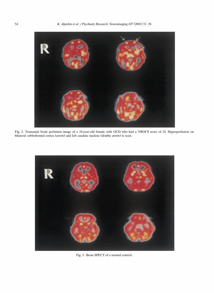
Fig. 2. Transaxial brain perfusion image of a 24-year-old female with OCD who had a YBOCS score of 24. Hyperperfusion onŽ . Ž .bilateral orbitofrontal cortex arrow and left caudate nucleus double arrow is seen.
Fig. 3. Brain SPECT of a normal control.

( )K. Alptekin et al. � Psychiatry Research: Neuroimaging 107 2001 51�56 55
Ž .bilateral orbitofrontal cortex Table 1 . The meanYBOCS score of the patients with OCD was24 � 8. There was no significant correlationbetween YBOCS scores and regional brain perfu-sion alterations in abnormal brain regions. Nosignificant correlation was found between age, sexand either YBOCS values or regional perfusionindexes.
Fig. 2 shows transaxial Tc-99m HMPAO brainSPECT slices of a 24-year-old female with aYBOCS score of 24 and hyperperfusion of bilat-eral orbitofrontal cortex and left caudate nucleus.Fig. 3 shows a SPECT image of a normal control.
4. Discussion
In the present cross-cultural study, altered re-gional brain perfusion compared with perfusionin controls was found in drug-free Turkish patientswith OCD without depression. Quantitative eval-uations revealed that the OCD group had signifi-cantly increased regional brain perfusion in theright thalamus, left frontotemporal cortex andbilateral orbitofrontal cortex in comparison withcontrols. Previously published PET and SPECTfindings on OCD performed in American andEuropean patients have indicated that many brainregions, especially orbitofrontal and striatal re-gions, were dysfunctional in drug-free, non-
Ždepressed patients with OCD Brody and Saxena,. Ž .1996 . The study of Baxter et al. 1988 showed
significantly higher glucose metabolic rates forthe heads of the caudate nuclei and orbital gyri.The OCD group had increased glucosemetabolism in the left orbitofrontal, bilateral pre-frontal, right sensorimotor and cingulate regionsŽ . Ž .Swedo et al., 1989 . Machlin et al. 1991 foundelevated medial�frontal ratios. Edmonstone et al.Ž .1994 observed altered basal ganglia function.
Ž .Rubin et al. 1992 showed that there was apositive correlation between regional blood flowand the severity of obsessive-compulsive symp-toms.
There are cross-cultural differences in obses-Žsive-compulsive symptoms and traits Horwath
.and Weissman, 2000 , but whether there is acultural effect on brain pathology in patients with
OCD is not known. In this study the brain SPECTresults of Turkish patients with OCD were similarto previous ones which had been found in OCDpatients living in the USA and European coun-tries. Orbitofrontal cortex and striatum seem tobe dysfunctional in OCD. Orbitofrontal cortexplays an important role in mediating the sexualand aggressive instincts that originate in the lim-
Žbic system Brody and Saxena, 1996; Saxena et al.,.1998 . Hyperperfusion in orbitofrontal cortex and
thalamic regions implicates a dysfunction of theseregions in OCD. In this cross-cultural study or-bitofrontal and thalamic dysfunction has beenfound in a sample of Turkish patients with OCD.
References
Aylward, E.H., Harris, G.J., Hoehn-Saric, R., Barta, P.E.,Machlin, S.R., Pearlson, G.D., 1996. Normal caudate nu-cleus in obsessive-compulsive disorder assessed by quanti-tative neuroimaging. Archives of General Psychiatry 53,577�584.
Baxter, L.R., Phelps, M.E., Mazziotta, J.C., Guze, B.H.,Schwartz, J.M., Selin, C.E., 1987. Local cerebral glucosemetabolic rates in obsessive-compulsive disorder. Archivesof General Psychiatry 44, 211�218.
Baxter, L.R., Schwartz, J.M., Mazziotta, J.C., Phelps, M.E.,Pahl, J.J., Guze, B.H., Fairbanks, L., 1988. Cerebral glucosemetabolic rates in nondepressed patients with obsessive-compulsive disorder. American Journal of Psychiatry 145,1560�1563.
Baxter, L.R., 1990. Brain imaging as a tool in establishing atheory of brain pathology in obsessive-compulsive disorder.
Ž .Journal of Clinical Psychiatry 51 Suppl , 22�25.Baxter, L.R., Schwartz, J.M., Guze, B.H., Bergman, K., Szuba,
M.P., 1990. PET imaging in obsessive-compulsive disorderwith and without depression. Journal of Clinical PsychiatryŽ .51 Suppl , 61�69.
Brody, A.L., Saxena, S., 1996. Brain imaging in obsessive-com-pulsive disorder: evidence for the involvement of frontal-subcortical circuitry in the mediation of symptomatology.CNS Spectrums 2, 27�41.
Calabrese, G., Colombo, C., Bonfanti, A., Scotti, G., Scarone,S., 1993. Caudate nucleus abnormalities in obsessive-com-pulsive disorder: measurements of MRI signal intensity.Psychiatry Research: Neuroimaging 50, 89�92.
Edmonstone, Y., Austin, M.P., Prentice, N., Dougall, N., Free-man, C.P.L., Ebmeier, K.P., Goodwin, G.M., 1994. Uptakeof 99m Tc-exametazime shown by single photon emissioncomputerized tomography in obsessive-compulsive disordercompared with major depression and normal controls. ActaPsychiatrica Scandinavica 90, 28�303.
Garber, J., Ananth, J.V., Chiu, L.C., Griswold, V.J., Olden-dorf, W.H., 1989. Nuclear magnetic resonance study of

( )K. Alptekin et al. � Psychiatry Research: Neuroimaging 107 2001 51�5656
obsessive-compulsive disorder. American Journal of Psychi-atry 146, 1001�1005.
Hoehn-Saric, R., Greenberg, B.D., 1997. Psychobiology ofobsessive-compulsive disorder: anatomical and physiologi-cal considerations. International Review of Psychiatry 9,15�29.
Horwath, E., Weissman, M.M., 2000. The epidemiology andcross-national presentations of obsessive-compulsive dis-order. Psychiatry Clinics of North America 23, 493�507.
Horwitz, B., Swedo, S.E., Grady, C.L., Pietrini, P., Schapiro,M.B., Rapoport, J.L., Rapoport, S.I., 1991. Cerebralmetabolic pattern in obsessive-compulsive disorder: alteredintercorrelations between regional rates of glucose utiliza-tion. Psychiatry Research: Neuroimaging 40, 221�237.
Jenike, M.A., Breiter, H.C., Baer, L., Kennedy, D.N., Savage,C.R., Olivares, M.J., O’Sullivan, R.L., Shera, D.M., Rauch,S.L., Keuthen, N., Rosen, B.R., Caviness, V.S., Filipek,P.A., 1996. Cerebral structural abnormalities in obsessive-compulsive disorder. Archives of General Psychiatry 53,625�632.
Luxenberg, J.S., Swedo, S.E., Flament, M.F, Friedland, R.P.,Rapoport, J.L, Rapoport, S.I., 1988. Neuroanatomicalabnormalities in obsessive-compulsive disorder detectedwith quantitative X-ray computed tomography. AmericanJournal of Psychiatry 145, 1089�1093.
Machlin, S.R., Harris, G.J., Pearlson, G.D., Hoehn-Saric, R.,Jeffery, P., Camargo, E.E., 1991. Elevated medial-frontalcerebral blood flow in obsessive-compulsive patients: aSPECT study. American Journal of Psychiatry 148,1240�1242.
Robinson, D., Wu, H., Munne, R.A, Ashtari, M., Alvir, J.M.J.,Lerner, G., Koreen, A., Cole, K., Bogerts, B., 1995. Re-duced caudate nucleus volume in obsessive-compulsive dis-order. Archives of General Psychiatry 52, 393�398.
Rubin, R.T., Villanueva-Meyer, J., Ananth, J., Trajmar, P.G,Mena, I., 1992. Regional Xenon 133 cerebral blood flowand cerebral technetium 99m HMPAO uptake in unmedi-cated patients with obsessive-compulsive disorder andmatched normal control subjects. Archives of General Psy-chiatry 49, 695�702.
Saxena, S., Brody, A.L., Schwartz, J.M., Baxter, L.R., 1998.Neuroimaging and frontal-subcortical circuitry inobsessive-compulsive disorder. British Journal of Psychiatry
Ž .173 suppl 35 , 26�37.Stein, D.J., Hollander, E., Chan, S., De Caria, C.M., Hilal, S.,
Liebowitz, M.R., Klein, D.F., 1993. Computed tomographyand neurological soft signs in obsessive-compulsive dis-order. Psychiatry Research: Neuroimaging 50, 143�150.
Stein, D.J., Coetzer, R., Lee, M., Davids, B., Bauwer, C., 1997.Magnetic resonance brain imaging in women with obses-sive-compulsive disorder and trichotillomania. PsychiatryResearch: Neuroimaging 74, 177�182.
Swedo, S.E., Schapiro, M.B., Grady, C.L., Cheslow, D.L., Leo-nard, H.L., Kumar, A., Friedland, R., Rapoport, S.I.,Rapoport, J.L., 1989. Cerebral glucose metabolism in child-hood-onset obsessive-compulsive disorder. Archives ofGeneral Psychiatry 46, 58�523.
Copyright © 2022 FDOKUMEN