Targeting apoptosis in solid tumors: the role of bortezomib from preclinical to clinical evidence
16
Review 10.1517/14728222.11.12.1571 © 2007 Informa UK Ltd ISSN 1472-8222 1571 1. Introduction 2. The ubiquitin–proteosome pathway 3. Proteasome and cancer 4. Bortezomib 5. Preclinical and clinical studies 6. Expert opinion Oncologic, Endocrine & Metabolic Targeting apoptosis in solid tumors: the role of bortezomib from preclinical to clinical evidence Antonio Russo, Maria E Fratto, Viviana Bazan, Valentina Schirò, Valentina Agnese, Giuseppe Cicero, Bruno Vincenzi, Giuseppe Tonini & Daniele Santini † † University Campus Bio-Medico, Department of Oncology, 00155 Rome, Italy The ubiquitin–proteasome pathway is the main proteolytic system present in the nucleus and cytoplasm of all eukaryotic cells. Apoptosis activation induced by ubiquitin–proteasome pathway inhibition makes the proteasome a new target of anticancer therapy. Bortezomib is the first proteasome inhibitor to be approved by the US FDA; in 2003 as a third line and in 2005 as a second line therapy for the treatment of multiple myeloma only. This review focuses on the use of bortezomib, not only in its therapeutic role but also, more specifically, in its biologic role and discusses the most recent applications of the drug in solid tumors, both at a preclinical and clinical level. Keywords: bortezomib, clinical studies, preclinical, ubiquitin–proteasome pathway Expert Opin. Ther. Targets (2007) 11(12):1571-1586 1. Introduction The ubiquitin–proteasome pathway (UPP) is the main proteolytic system present in the nucleus and cytoplasm of all eukaryotic cells. Although this ATP-dependent pathway was discovered in the 1979 [1], proteasome involvement was only demonstrated at the end of the 1980s [2]. Proteasome 26S is a multienzymatic complex found in all eukaryotic cells. The use of specific inhibitors has shown that the UPP is responsible for the cytoplasmatic turnover of a great variety of different cell proteins, thus, assuming an extremely important role in the regulation of not only a large number of physiologic processes, but also in the development of a great many human diseases. In fact, cyclins (A, B, C and D), cyclin-dependent kinase (CDK) inhibitors (p21 and p27), protein inhibitors (I κB), tumor suppressors (p53), regulatory proteins (MDM2), oncogenes (c-fos, c-jun, c-myc, N-myc and β-catenine), together with anti and proapoptotic factors (Bcl-2 and Bax) are all proteasome targets [3-5]. Preclinical studies have shown that the use of proteasome inhibitors in the treatment of several tumors might represent a new possibility of improving anticancer therapy because of their ability to induce the process of programmed cell death [6,7]. Therefore, whether natural or synthetic, they should be considered as a new class of potentially useful anticancer drugs [8]. Bortezomib (PS-341) is a dipeptide boronic acid able to inhibit, selectively and reversibly, the proteasome 26S [8], thus, leading to antiproliferative, proapoptotic and antiangiogenetic activity [9]. This induced inhibition leads to the stabilization of the CDK inhibitors p21 and p27, the tumor suppressor p53,
-
Upload
independent -
Category
Documents
-
view
2 -
download
0
Transcript of Targeting apoptosis in solid tumors: the role of bortezomib from preclinical to clinical evidence

Review
10.1517/14728222.11.12.1571 © 2007 Informa UK Ltd ISSN 1472-8222 1571
1. Introduction
2. The ubiquitin – proteosome
pathway
3. Proteasome and cancer
4. Bortezomib
5. Preclinical and clinical studies
6. Expert opinion
Oncologic, Endocrine & Metabolic
Targeting apoptosis in solid tumors: the role of bortezomib from preclinical to clinical evidence Antonio Russo , Maria E Fratto , Viviana Bazan , Valentina Schir ò , Valentina Agnese , Giuseppe Cicero , Bruno Vincenzi , Giuseppe Tonini & Daniele Santini † † University Campus Bio-Medico , Department of Oncology , 00155 Rome , Italy
The ubiquitin–proteasome pathway is the main proteolytic system present in the nucleus and cytoplasm of all eukaryotic cells. Apoptosis activation induced by ubiquitin–proteasome pathway inhibition makes the proteasome a new target of anticancer therapy. Bortezomib is the first proteasome inhibitor to be approved by the US FDA; in 2003 as a third line and in 2005 as a second line therapy for the treatment of multiple myeloma only. This review focuses on the use of bortezomib, not only in its therapeutic role but also, more specifically, in its biologic role and discusses the most recent applications of the drug in solid tumors, both at a preclinical and clinical level.
Keywords: bortezomib , clinical studies , preclinical , ubiquitin–proteasome pathway
Expert Opin. Ther. Targets (2007) 11(12):1571-1586
1. Introduction
The ubiquitin–proteasome pathway (UPP) is the main proteolytic system present in the nucleus and cytoplasm of all eukaryotic cells. Although this ATP-dependent pathway was discovered in the 1979 [1] , proteasome involvement was only demonstrated at the end of the 1980s [2] .
Proteasome 26S is a multienzymatic complex found in all eukaryotic cells. The use of specific inhibitors has shown that the UPP is responsible for the cytoplasmatic turnover of a great variety of different cell proteins, thus, assuming an extremely important role in the regulation of not only a large number of physiologic processes, but also in the development of a great many human diseases. In fact, cyclins (A, B, C and D), cyclin-dependent kinase (CDK) inhibitors (p21 and p27), protein inhibitors (I κ B), tumor suppressors (p53), regulatory proteins (MDM2), oncogenes (c-fos, c-jun, c-myc, N-myc and β-catenine), together with anti and proapoptotic factors (Bcl-2 and Bax) are all proteasome targets [3-5] .
Preclinical studies have shown that the use of proteasome inhibitors in the treatment of several tumors might represent a new possibility of improving anticancer therapy because of their ability to induce the process of programmed cell death [6,7] . Therefore, whether natural or synthetic, they should be considered as a new class of potentially useful anticancer drugs [8] .
Bortezomib (PS-341) is a dipeptide boronic acid able to inhibit, selectively and reversibly, the proteasome 26S [8] , thus, leading to antiproliferative, proapoptotic and antiangiogenetic activity [9] . This induced inhibition leads to the stabilization of the CDK inhibitors p21 and p27, the tumor suppressor p53,

Targeting apoptosis in solid tumors: the role of bortezomib from preclinical to clinical evidence
1572 Expert Opin. Ther. Targets (2007) 11(12)
the proapoptotic proteins Bid and Bax and of the transcription factor NF- κ B.
Bortezomib is generally well-tolerated by patients and, what is more, is able to increase the antitumoral effects of chemotherapy, radiotherapy and immunotherapy, without adding to their toxic effects [10,11] .
Due to its high level of specificity, bortezomib was the first proteasome inhibitor to be approved by the US FDA; in 2003 as a third line and in 2005 as a second line therapy for the treatment of multiple myeloma (MM) only.
This review focuses on the use of bortezomib, not only in its therapeutic role but also, more specifically, in its biologic role and discusses the most recent applications of the drug in solid tumors, both at a preclinical and clinical level.
2. The ubiquitin–proteosome pathway
The UPP involves two events: first, ubiquitylation and then degradation brought about by the proteasome. The former process requires intervention by the enzymes E1, E2 and E3, which, as soon as they are activated, bind the ubiquitin chain step-by-step to the target protein, thus causing its degradation ( Figure 1 ). All eukaryotes have many E2 and E3 enzymes, but most eukaryotes have only one or a small number of distinct E1 enzymes [12] . The ubiquitin-activating enzyme (E1), which binds ubiquitin by means of an ATP-dependent step, transfers a ubiquitin molecule to the ubiquitin-conjugating carrier protein (E2), which then interacts with the ubiquitin-protein ligase (E3) in order to bind the carrier protein covalently to a protein-target lysine. Several ubiquitin molecules are bound in this way, thus permitting the formation of a chain that is needed for recognition by the proteasome 26S. Ubiquitylation is a reversible process, due to the intervention of the deubiquitylation enzymes. The minimum length of an ubiquitin chain required for protein recognition is at least four molecules [13] .
2.1 Structure of the proteasome The proteasome 26S is a multienzymatic cylindrical complex made up of 2.5 kDa, which belongs to the N-terminal nucleophilic hydrolase family. It is present in the nucleus and in the cytoplasm of several eukaryotic cells [14] . This organelle consists of a 20S tubular core (CP), able to trigger off catalytic activity, with the 19S regulating subunits (RPs) linked to each end; these are responsible for the recognition of the ubiquitinized proteins and for the regula-tion of entry to the 20S core ( Figure 2 ). More specifically, each subunit is made up of a base and a lid. The latter is made up of at least nine polypeptides and is responsible for binding and removing the polyubiquitin chains from the assigned proteins so that the chains can then be recycled. The base consists of eight polypeptides, six of which have ATPase activity, which reduce the polypeptide and catalyze their translocation to the 20S core [15,16] .
The 20S (72 kDa) is made up of four stacked rings; two outer rings consisting of seven α -subunits ( α 1 – α 7) and two inner ones of seven β -subunits ( β 1 – β 7) [17] . Each inside ring presents three sites with chymotrypsin-like, trypsin-like and postglutamyl peptide hydrolase-like catalytic activity, arranged along the inside edge of the channel [17-19] . The β 5 subunit, which possesses a chymotrypsin-like activity, cuts the polypeptides after hydrophobic residues; the β 2 subunit, with trypsin-like activity, cleaves after basic amino acids and, finally, the β 1 subunit, with its post-glutamyl peptide hydrolase-like, preferentially splits peptide bonds after acidic residues. Protein degradation occurs progressively by generating peptides of 3 – 25 amino acids, which are then dispersed on the outside and subsequently recycled by the cells [20,21] .
The specific and reversible inhibition of the chymotrypsin-like activity of the proteasome is sufficient for the induction of cell cycle arrest and apoptosis, attributed to the inhibition of NF-κB. In particular, the inhibition of the chymotrypsin-like site, observed in patients treated with bortezomib, is sufficient to cause apoptosis of MM cells [22] .
3. Proteasome and cancer
In normal conditions, UPP is involved in maintaining the stability of several proteins implicated in the regulation of cell cycle and division, DNA repair and transcription, the immune response and inflammation, antigen processing, differentiation, cell development and in apoptosis [23,24] . Among these, the proteins p21 and p27, belonging to the Cip/Kip family of the CDK inhibitors, prevent the forma-tion of several different CDK–cyclin complexes and inhibit progression of the cell cycle. Low levels of p21 and p27, brought about by proteasome hyperactivity, represent a neg-ative prognostic factor in several types of cancer. As a result, inhibiting action by the proteasome leads to upregulation of these proteins, which may lead to apoptosis in in vitro tumoral cells [25-27] .
Another important proteasome substrate is the p53 tumor-suppressor protein, which play an important role in apoptosis induction. Proteasome inhibition gives rise to an increase in p53 expression and stability, thus, promoting the programmed death process [28] .
The regulation of the NF- κ B by the proteasome is particularly interesting ( Figure 3 ). This consists of a hetero dimeric transcription complex (p50/p65), involved not only in the regulation of immune and inflammatory response but also in tumorigenesis, due to cell-growth stimulation, angiogenesis induction and apoptosis blockage [29] . In normal conditions, NF-κB is kept inactive in the cytoplasm by the I κ B, whereas in conditions of cell stress, the I κ B becomes phosphorylated by the I κ B kinase complex and subsequently ubiquitinized and degraded by the proteasome. I κ B degradation leads to the release of NF- κ B

Russo, Fratto, Bazan, Schirò, Agnese, Cicero, Vincenzi, Tonini & Santini
Expert Opin. Ther. Targets (2007) 11(12) 1573
and its translocation into the nucleus. The interaction of NF- κ B with the DNA gives rise to the expression of target genes such as cytokines (IL-6), survival factors (IAPa, Bcl-XI9), intracellular adhesion molecules, vascular adhesion molecules and E-selectins ( Figure 4 ) [30] . The mechanism of action of NF- κ B leads to cell resistance to chemotherapy [31,32] ; proteasome inhibition strong enough to prevent I κ B degra-dation, might, therefore, be useful in avoiding chemotherapy resistance because it would induce apoptosis. In fact, in vitro studies have shown that the presence of the super repressor I κ B, which is a mutated form able to resist degradation by the proteasome, leads to the death of myeloma cells that have proven resistant to melphalan. Furthermore, the transfection of I κ B leads to an increased response of colon cancer cells to 7-Ethyl-10-Hydroxycamptothecin (SN-38) compared with that of non-transfected cells [32,33] .
Then the apoptosis activation, induced by UPP inhibition, makes the proteasome a new target of anticancer therapy [18] .
4. Bortezomib
Bortezomib (PS-341) is a dipeptidyl boric acid approved by the US FDA in 2003 for the treatment of patients with relapsing MM. This compound has been tested by the National Cancer Institute (NCI) on 60 tumoral cell lines, demonstrating that it has a greater possibility of action in several of these compared with 60,000 other compounds. More specifically, it has been seen that low concentrations of bortezomib (7 nM) are able to reduce tumor growth by ∼ 50%; the injection of bortezomib directly into human tumors implanted into mice has shown that in 40% of the
ATPUb
AMPPPi Ub
Ub
Ub
UbE1
E1
E2
E2
E3
E3
Proteinsubstrate
Figure 1 . Ubiquitination of a target protein for its degradation by the proteasome. A ubiquitin molecule is transfered in an ATP-dependent manner to the ubiquitin-activating enzyme (E1) and then to the ubiquitin-conjugating carrier protein (E2), which then interacts with the ubiquitin-protein ligase (E3) in order to covalently bind the carrier protein to a protein-target lysine. Several ubiquitin molecules are bound in this way, thus permitting the formation of a chain, needed for recognition by the proteosome 26S.
Proteasome
20S
19S
αβ
Protein Ub Ub Ub Ub Ub Ub
Oligopeptides Ub
Ub
Ub
Ub
Ub
Ub
β1
β2β3
β4
β5β5
Free ubiquitin
Figure 2 . Protein degradation by the proteasome pathway. β 1: Post-glutamyl site; β 2: Trypsin-like site; β 5: Chymotrypsin-like site.

Targeting apoptosis in solid tumors: the role of bortezomib from preclinical to clinical evidence
1574 Expert Opin. Ther. Targets (2007) 11(12)
cases there is a reduction of 70% in the volume of the tumoral mass [34] .
Bortezomib inhibits the proteasome by interacting with a threonine residue present in each of the catalytic sites, but with an affinity that gradually decreases for the sites β 5, β 1 and β 2 ( β 5 < β 1 << β 2) respectively [35] .
4.1 Bortezomib and apoptosis The proteasome inhibition brought about by bortezomib contributes to the accumulation of a series of proteic factors
IKBP
P
NF-κB
NF-κB
NF-κB
IKB
P P
UbUbUbUb Ub IKB
Nuclear translocationand target genetranscription
ProteasomeP
IKB
P
UbUbUb
PP
IKB
P
UbUbUb
P
UbUbUb Ub
NF-κB
Ub
Figure 3 . Proteasome action on the IKB-NF-kB pathway. IKB: Inhibitor of NF- κ B.
NF-κBCytokines(IL-6)
Anti-apoptoticfactors (BcL-2,BcLx)
Adhesion molecules(ICAM, VCAM)
Cell cycleregulators
Figure 4 . Principal NF- k B target genes. ICAM: Intracellular adhesion molecules; VCAM: Vascular adhesion molecule.
that play an important role in cell cycle regulation, such as the proapoptotic factors c-jun and p53, thereby inducing or increasing sensitivity to apoptosis ( Figure 5 ). p53 is able to induce apoptosis through both the intrinsic, or mitochon-drial, pathway and through the extrinsic death receptor pathway. In the former, p53 directly or indirectly modulates the expression of the protein targets, which include Bax, Bid, Puma and Noxa, involved in permeability regulation of the mitochondrial membrane. The subsequent emission of mitochondrial proteins causes the release of cytochrome C, caspase activation and subsequent cell death. The extrinsic pathway is needed in order to accentuate apoptotic response and, furthermore, is mediated by activation of the death receptors DR4, DR5, CD95, located on the plasma mem-brane, which inhibit the production of inhibitor of apoptosis proteins [36,37] . Moreover, among the proteins accumulated inside the cell subsequent to proteasome inhibition, there are the TNF-related apoptosis-inducing ligands (TRAIL), which are type II transmembrane proteins able to induce apoptosis in various tumor cell lines ( Figure 6 ) [38] . The TRAIL trigger off the programmed cell death process by means of interaction with two receptors, death receptor 4 (DR4) and death receptor 5 (DR5). In more specific terms, the interaction of TRAIL-R1 with DR4 and

Russo, Fratto, Bazan, Schirò, Agnese, Cicero, Vincenzi, Tonini & Santini
Expert Opin. Ther. Targets (2007) 11(12) 1575
of TRAIL-R2 with DR5 leads to the formation of the death-inducing signaling complex, which is able to recruit the Fas-associated death domain adaptor molecule. The latter then recruits and activates caspases-8 and -10 in order to activate the proteic cascade, which is able to induce programmed cell death [39] . However, caspase-8 also acti-vates Bid, a protein of the Bcl-2 family, which translocates to the mitochondria and, by giving rise to its permeation, induces apoptosis [40] .
Proteasome inhibition also causes the oxidative stress responsible for the activation of Jun N-terminal kinase (JNK), which precedes the phosphorylation of c-jun. Once it is activated, c-jun will form, with c-Fos, the heterodimer AP-1; a transcription factor involved in the regulation of several cell processes, such as proliferation, differentiation, cell death and survival, which then induce the expression of various genes, including c-jun, Fasl and Bim. The latter two will be responsible for the activation of cell death, through both the intrinsic and extrinsic pathways [41] .
Bortezomib also activates programmed cell death by means of other mechanisms, such as the activation of the heat-shock proteins (Hsp). The combined action of geldanamycin plus bortezomib disrupts Hsp90 and proteasome function, promotes the accumulation of aggregated, ubiquitinated proteins and results in enhanced antitumor activity [42-44] .
In a sperimental model of C-26 colon carcinoma in mice, the combined action of bortezomib with TNF induced a dysregulated response to endoplasmic reticulum (ER) stress leading to apoptosis of cancer cells, evidenced by caspase-3
cleavage, p53 accumulation, increased stress-activated protein kinase/c-Jun N-terminal kinase (SAPK/JNK) phosphorylation. Therefore, the combined treatment not only inhibited tumor growth, but also prolonged survival of animals bearing the C-26 tumors [45] .
In breast cancer cell lines, bortezomib reduced the activity of 20S tubular core in a concentration-, time- and cell line-dependent manner. Proteasome inhibition causes nucleus-to-cytoplasm relocalization of the ER and plasma membrane to peri nuclear lysosomes, relocalization of ErbB2, increased degradation and loss of ER and ErbB2 function, and induction of cellular apoptosis [46] .
Proteasome inhibition with bortezomib may be used to prevent graft-versus-host disease (GVHD) and preserving, or possibly even promoting, graft-versus-tumor responses in cancer. Inflammatory cytokines produced by T cells and other immune cells have been shown to be critical for GVHD generation. Bortezomib is capable of inhibiting proliferation of alloreactive T cells in a dose-dependent manner. The suppression of GVHD may be due to a reduction of cytokine production concurrent with the induction of apoptosis of the alloreactive T cells [47] .
Finally, in non-small cell lung cancer (NSCLC) cells, leukemia cells and head and neck squamous cell carcinoma (HNSCC) cells, bortezomib brings about the formation of species are reactive to oxygen, which, by determining the alteration of the mitochondrial membrane potential and the release of cytochrome C, are the primary cause of programmed cell death [48-50] .
Proteasome
Bortezomib
Stabilization
Caveolin-1
Stabilization
Apoptosis
Apoptosis
Migration
p21
Stabilization
p53
NF-κB
JNK
Proliferation
Angiogenesis
Apoptosis
p27
Proliferation
Figure 5 . Effects of proteasome inhibition by bortezomib. Alteration of several proteins leads to apoptosis, reduction of angiogenesis, migration and cellular proliferation.
Targeting apoptosis in solid tumors: the role of bortezomib from preclinical to clinical evidence
1576 Expert Opin. Ther. Targets (2007) 11(12)
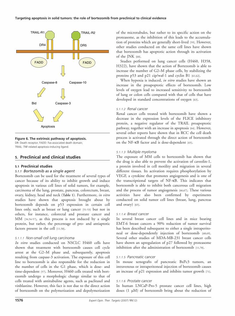
5. Preclinical and clinical studies
5.1 Preclinical studies 5.1.1 Bortezomib as a single agent Bortezomib can be used for the treatment of several types of cancer because of its ability to inhibit growth and induce apoptosis in various cell lines of solid tumors, for example, carcinoma of the lung, prostate, pancreas, colorectum, breast, ovary, kidney, head and neck ( Table 1 ). Furthermore, in vitro studies have shown that apoptosis brought about by bortezomib depends on p53 expression in certain cell lines only, such as breast or lung cancer [51-53] but not in others, for instance, colorectal and prostate cancer and MM [34,54-57] , as this process is not induced by a single protein, but rather, the percentage of pro- and antiaptotic factors present in the cell [11,58] .
5.1.1.1 Non-small cell lung carcinoma In vitro studies conducted on NSCLC H460 cells have shown that treatment with bortezomib causes cell cycle arrest at the G2–M phase and, subsequently, apoptosis resulting from caspase-3 activation. The exposure of this cell line to bortezomib is also responsible for the reduction in the number of cells in the G1 phase, which is dose- and time-dependent [57] . Moreover, H460 cells treated with bort-ezomib undergo a morphologic change similar to that of cells treated with antitubuline agents, such as paclitaxel and vinblastine. However, this fact is not due to the direct action of bortezomib on the polymerization and depolymerization
DR4
FADD
Caspase-10Caspase-8
Caspase-3Bid
FADD
TRAIL-R1 TRAIL-R2
DR5
Apoptosis
Figure 6 . The extrinsic pathway of apoptosis. DR: Death receptor; FADD: Fas-associated death domain; TRAIL: TNF-related apoptosis-inducing ligand.
of the microtubules, but rather to its specific action on the proteasome, as the inhibition of this leads to the accumula-tion of proteins which are generally short-lived [59] . However, other studies conducted on the same cell lines have shown that bortezomib has apoptotic action through its activation of the JNK [60] .
Studies performed on lung cancer cells (H460, H358, H322), have shown that the action of Bortezomib is able to increase the number of G2–M phase cells, by stabilizing the proteins p53 and p21 cip/waf-1 and cyclin B1 [61,62] .
When hypoxia is induced, in vitro studies have shown an increase in the proapoptotic effects of bortezomib. Low levels of oxygen lead to increased sensitivity to bortezomib of lung or colon cells compared with that of cells that have developed in standard concentrations of oxygen [63] .
5.1.1.2 Renal cancer Renal cancer cells treated with bortezomib have shown a decrease in the expression levels of the FLICE inhibitory protein, a negative regulator of the TRAIL proapoptotic pathway, together with an increase in apoptosis [64] . However, several other reports have shown that in RCC the cell death process is activated through the direct action of bortezomib on the NF- κ B factor and is dose-dependent [65] .
5.1.1.3 Multiple myeloma The exposure of MM cells to bortezomib has shown that the drug is also able to prevent the activation of caveolin-1, a protein involved in cell motility and migration in several different tissues. Its activation requires phosphorylation by VEGF, a cytokine that promotes angiogenesis and is one of the transcriptional targets of NF- κ B. This indicates that bortezomib is able to inhibit both cancerous cell migration and the process of tumor angiogenesis [66,67] . These various activities have also been confirmed by experiments conducted on solid tumor cell lines (breast, lung, pancreas and ovary) [65] .
5.1.1.4 Breast cancer In several breast cancer cell lines and in mice bearing EMT-6 breast cancers a 90% reduction of tumor survival has been described subsequent to either a single intraperito-neal or dose-dependently injection of bortezomib [68,69] . Several other studies of MDA-MB-231 breast cancer cells have shown an upregulation of p27 followed by proteasome inhibition after the administration of bortezomib [11,70] .
5.1.1.5 Pancreatic cancer In mouse xenografts of pancreatic BxPc3 tumors, an intravenous or intraperitoneal injection of bortezomib causes an increase of p21 expression and inhibits tumor growth [71] .
5.1.1.6 Prostate cancer In human LNCaP-Pro-5 prostate cancer cell lines, high doses (1 µM) of bortezomib bring about the reduction of

Russo, Fratto, Bazan, Schirò, Agnese, Cicero, Vincenzi, Tonini & Santini
Expert Opin. Ther. Targets (2007) 11(12) 1577
the microcirculation density and of the number of growth factors of the vascular endothelium together with apoptosis activation. A reduction in microcirculation density induced by bortezomib has also been observed in prostate tumors [72,73] . While in human PC3M-Pro4 prostate cancer cell lines, whereas the drug does not cause any particular change in microcirculation density, it does, however, induce an increase in tumor cell death [73] .
5.1.1.7 Head and neck squamous cell carcinomas In HNSCC, bortezomib plays an antitumoral role due to its inhibition of the prosurvival NF- κ B pathway; furthermore, the proteasome inhibition induced by bortezomib causes the release of the reactive oxygen species. These are responsible for the activation of apoptosis. In bortezomib-resistant HNSCC cells, the drug does not induce any destruction of cell adhesion in treated cells [74,75] .
5.1.2 Bortezomib and combined treatments Bortezomib brings about an increase in chemosensitivity to other chemotherapy drugs, thus permitting the administration
Table 1 . Effect of bortezomib in solid tumors in vitro and in vivo .
Cancer Effect
NSCLC Cell cycle arrest at G2–M, activation of caspase-3, activation of JNK, increase in p53, p21 cip/waf-1, cyclin B1, MDM2, tumor growth delay
Renal Decreased c-FLIP, Inhibits NF- κ B pathway, increased tumor apoptosis
Multiple myeloma
Inhibits caveolin-1, Inhibits of VEGF secretion, decreased angiogenesis and tumor growth
Breast Decreased tumor growth, up-regulation of p27
Colorectal Up-regulation of p27, decreased tumor growth, increased tumor apoptosis, enhanced cytotoxic effects of irinotecan, camptothecins, CPT-11 and radiation treatment
Pancreatic Up-regulation of p21, increased tumor apoptosis, enhanced cytotoxic effects of gemcitabine, CPT-11
Prostate Decreased angiogenesis, decreased VEGF, increased tumor apoptosis
Head and neck
Decreased angiogenesis, inhibits NF- κ B pathway, production of ER and ROS, activation of caspase-9, apoptosis
Ovarian Cell–cell interaction destruction, Inhibits NF- κ B pathway, increased tumor apoptosis, enhanced cytotoxic effects of docetaxel
c-FLIP: FLICE inhibitory protein; ER: Endoplasmic reticulum; JNK: c-Jun N-terminal kinase; MDM: Murine double minute; NSCLC: Non-small cell lung cancer; ROS: Reactive oxygen species; VEGF: Vascular endothelial growth factor.
of lower doses of these in combined treatments. Cells which have proven resistant to doxorubicin, mitoxantrone or melphalan, become more treatment-sensitive in the presence of bortezomib [76,77] . The administration of the drug when combined with irinotecan, a topoisomerase inhibitor, has antiproliferative and proapoptotic activity in murine xeno-graft models of colon cancer. This combined therapy causes a reduction in tumor size by 94% (69) and 89% (70) compared with controls. Furthermore, such tumors are smaller when compared with tumors treated with bortezo-mib or irinotecan as single agents. In xenograft models of pancreatic cancer, the use of bortezomib together with gemcytabine and with docetaxel in ovarian cancer brings about an increase in antiproliferative, proapoptotic, anti-tumoral and antiangiogenetic activity. In vitro studies per-formed on melanoma cells have shown that the combined action of bortezomib and temozolomide has cytotoxic and antiproliferative effects; moreover, in in vivo murine xeno-grafts, a complete remission of tumors has been observed for > 200 days [78] . Preclinical studies have shown that bortezo-mib is able to sensitize tumor cells to radiotherapy. The combination of bortezomib with radiotherapy gives rise to a reduction in the growth of colon, prostate tumors and NSCLC [79-81] .
5.2 Clinical studies in solid tumors Considering the action of bortezomib in several tumor cell lines, many Phase I and Phase II clinical trials have explored its toxicity and activity in patients with different solid tumors. The most important results of these studies are summarized in Table 2 and Table 3 .
5.2.1 Phase I clinical trials The first clinical trial exploring the role of bortezomib as a single agent in patients with advanced solid tumors was reported by Aghajanian et al. [82] . In this study, 43 patients were treated with bortezomib doses ranging from 0.13 – 1.56 mg/m 2 administered intravenously twice a week for 2 weeks, with a week rest period. The authors reported one partial response (PR) of 3 months duration in a patient with refractory NSCLC, whereas three patients had stable disease with a median response duration of 4 months. Grade 3 dose-limiting toxic effects were diarrhea and sensory neuro-toxicity but the last one was observed only in patients with pre-existing neuropathy. The authors concluded that the treatment warranted further testing in a Phase II trial, but they cautioned that particular care should be taken when treating patients with pre-existing neuropathy.
Phase I studies evaluated the combination of bortezomib with antineoplastic agents. In the first study, Appleman et al. [83] administered bortezomib in combination with gemcitabine to patients with advanced solid tumors. Thrombocytopenia was the dose-limiting toxicity and the maximum tolerated dose regimen was determined to be 1.0 mg/m 2 for bortezomib and 1000 mg/m 2 for gemcitabine.

Targeting apoptosis in solid tumors: the role of bortezomib from preclinical to clinical evidence
1578 Expert Opin. Ther. Targets (2007) 11(12)
Tab
le 2
. Ph
ase
I clin
ical
tri
als
of
bo
rtez
om
ib in
pat
ien
ts w
ith
so
lid t
um
ors
.
Ref
eren
ceTu
mo
r ty
pe
Nu
mb
er o
f p
atie
nts
Trea
tmen
t sc
hed
ule
Res
ult
sA
dve
rse
even
ts
Agh
ajan
ian
et a
l. (2
002)
[82]
A
dvan
ced
solid
tum
ors
43Bo
rtez
omib
(0.1
3 –
1.56
mg/
m2 )
tw
ice
wee
kly
for
2 w
eeks
and
1 w
eek
rest
per
iod
1 PR
in N
SCLC
2
SDD
LT: d
iarr
hea,
neu
ropa
thy
AE:
fev
er, f
atig
ue, n
ause
a, v
omiti
ng, r
ash,
pr
uritu
s, h
eada
che
App
lem
an e
t al
. (2
003)
[83]
Adv
ance
d so
lid t
umor
s31
Bort
ezom
ib (1
– 1
3 m
g/m
2 ) d
ays
1, 4
, 8
and
11 o
f a
3-w
eek
cycl
e pl
us
gem
cita
bine
on
days
1 a
nd 8
1 PR
in N
SCLC
DLT
: thr
ombo
cyto
peni
a
Alb
anel
l et
al.
2003
[84]
Ant
hrac
yclin
e-pr
etre
ated
br
east
can
cer
17Bo
rtez
omib
(1 –
1.3
mg/
m2 )
day
s 1,
4, 8
an
d 11
of
a 3-
wee
k cy
cle
+ d
ocet
axel
on
day
1
6 PR
DLT
: neu
trop
enia
, neu
ropa
thy,
muc
ositi
s, e
mes
is
Ryan
et
al.
(200
3) [8
5]A
dvan
ced
solid
tum
ors
51Bo
rtez
omib
(1 –
1.3
mg/
m2 )
day
s 1,
4, 8
an
d 11
of
a 3
wee
k cy
cle
plus
Irin
otec
an10
SD
DLT
: nau
sea,
em
esis
, dia
rrhe
a A
E: f
atig
ue, t
hrom
bocy
tope
nia,
neu
trop
enia
Papa
ndre
ou
et a
l. (2
004)
[86]
Adv
ance
d so
lid t
umor
s (A
IPC
)53
Bort
ezom
ib (0
.3 –
2 m
g/m
2 ) d
ays
1, 8
, 15
and
22 e
very
35
days
and
1 w
eek
rest
per
iod
2/47
(4%
) ↓ >
50%
PSA
9/
47 (4
%) s
tabl
e PS
AD
LT: d
iarr
hea,
hyp
oten
sion
, syn
cope
Blan
ey e
t al
. (2
004)
[87]
Refr
acto
ry s
olid
tum
ors
(ped
iatr
ics)
15Bo
rtez
omib
(1.2
– 1
.6 m
g/m
2 ) f
or
2 w
eeks
and
1 w
eek
rest
per
iod
Non
eD
LT: t
hrom
bocy
tope
nia
Dav
ies
et a
l. (2
004)
[88]
Adv
ance
d N
SCLC
16Bo
rtez
omib
(1 –
1.3
mg/
m2 )
day
s 1,
4, 8
an
d 11
of
a 3-
wee
k cy
cle
plus
ge
mci
tabi
ne a
nd c
arbo
plat
in o
n da
y 1
4 PR
5
SDD
LT: t
hrom
bocy
tope
nia
Agh
ajan
ian
et a
l. (2
005)
[89]
Ova
rian
or p
rimar
y pe
riton
eal c
ance
r15
Bort
ezom
ib (0
.75
– 1.
5 m
g/m
2 )
twic
e w
eekl
y fo
r 2
wee
ks a
nd 1
wee
k re
st p
erio
d (c
ycle
s 2
– 6)
plu
s ca
rbop
latin
AU
C 5
day
1 (c
ycle
s 1
– 6)
RR 4
7%
2 C
R 5
PR
DLT
: Dia
rrhe
a, n
euro
path
y, r
ash,
con
stip
atio
n
Voo
rtm
an e
t al
. (2
005)
[90]
Solid
tum
ors
(fi rs
t lin
e)13
Esca
latin
g do
ses
of b
orte
zom
ib
(fro
m 0
.7 t
o 1.
6 m
g/m
2 ) p
lus
cisp
latin
an
d ge
mci
tabi
ne
4 PR
in 1
0 ev
alua
ted
patie
nts
(2 w
ith b
ladd
er,
2 w
ith N
SCLC
)
DLT
: neu
trop
enia
Ham
ilton
et
al.
(200
5) [9
1]A
dvan
ced
solid
tum
ors
and
one
cuta
neou
s T
cell
lym
phom
a
40Bo
rtez
omib
(0.2
5 –
1.9
mg/
m2 )
da
ys 1
and
4 o
f a
2 w
eek
cycl
eN
one
DLT
: neu
ropa
thy,
dia
rrhe
a, f
atig
ue
AE:
Adv
erse
eve
nt; A
IPC
: And
roge
n-in
depe
nden
t pr
osta
te c
ance
r; A
UC
: Are
a un
der
the
curv
e; C
R: C
ompl
ete
resp
onse
; DLT
: Dos
e-lim
iting
tox
icity
; NSC
LC: N
on-s
mal
l-cel
l lun
g ca
ncer
; PR:
Par
tial r
espo
nse;
PS
A: P
rost
ate-
spec
ifi c
antig
en; R
R: R
espo
nse
rate
; SD
: Sta
ble
dise
ase.

Russo, Fratto, Bazan, Schirò, Agnese, Cicero, Vincenzi, Tonini & Santini
Expert Opin. Ther. Targets (2007) 11(12) 1579
Tab
le 3
. Ph
ase-
II cl
inic
al t
rial
s o
f b
ort
ezo
mib
in p
atie
nts
wit
h s
olid
tu
mo
rs.
Ref
eren
ceTu
mo
r ty
pe
Nu
mb
er o
f p
atie
nts
Trea
tmen
t sc
hed
ule
Res
ult
sA
dve
rse
even
ts
Pric
e et
al.
(200
4) [9
5]A
IPC
45Bo
rtez
omib
1.3
(A) o
r 1.
6 (B
) mg/
m2
plus
do
ceta
xel 4
0 m
g/m
2 on
ce w
eekl
y fo
r 2
wee
ks a
nd 1
wee
k re
st p
erio
d
A) 5
/25
(24%
) ↓ >
50%
PS
A B
) 3/1
0 (3
0%) ↓
> 5
0%
PSA
4/1
0 ↓
< 5
0% P
SA
Neu
trop
enia
, neu
ropa
thy,
muc
ositi
s, e
mes
is
Shah
et
al.
(200
4) [9
6]M
etas
tatic
ne
uroe
ndoc
rine
tum
ors
16Bo
rtez
omib
(1.5
mg/
m2 )
tw
ice
wee
kly
and
10 d
ays
rest
per
iod
11 (6
9%) S
DN
euro
path
y, d
iarr
hea,
em
esis
, ile
us
Dav
is e
t al
. (2
004)
[97]
Adv
ance
d re
nal
cell
carc
inom
a23
Bort
ezom
ib (1
.5 m
g/m
2 ) t
wic
e w
eekl
y fo
r 2
wee
ks e
very
3 w
eeks
PR 5
%Th
rom
bocy
tope
nia,
neu
trop
enia
, ane
mia
, ne
urop
athy
, art
hral
gia,
dia
rrhe
a, e
mes
is
Kon
dagu
nta
et a
l. (2
004)
[98]
Adv
ance
d re
nal c
ell
carc
inom
a37
Bort
ezom
ib (1
.5 m
g/m
2 re
duce
d to
1.
3 m
g/m
2 ) t
wic
e w
eekl
y fo
r 2
wee
ks a
nd
1 w
eek
rest
per
iod
4 (1
1%) P
R 14
(38%
) SD
Neu
ropa
thy
Fanu
cchi
et
al.
(200
4) [9
9]A
dvan
ced
NSC
LC60
Bort
ezom
ib (1
.5 m
g/m
2 ) d
ays
1, 4
, 8 a
nd
11 o
f a
3 w
eek
cycl
e pl
us/m
inus
doc
etax
el
on d
ay 1
Bort
ezom
ib: P
R: 1
0.3%
, SD
: 17.
2%
Bort
ezom
ib +
doc
etax
el:
PR: 1
6.1%
, SD
: 45.
2%
(inte
rim a
naly
sis)
Neu
trop
enia
, gas
troi
ntes
tinal
tox
icity
, na
usea
, fat
igue
Stev
enso
n et
al.
(200
4) [1
00]
Adv
ance
d N
SCLC
22Bo
rtez
omib
(1.3
– 1
.5 m
g/m
2 ) d
ays
1, 4
, 8
and
11 o
f a
3 w
eek
cycl
e1
(4%
) PR
9 (4
0%) S
D
(Ong
oing
tria
l)
Nau
sea,
em
esis
, neu
ropa
thy,
con
stip
atio
n,
rash
, thr
ombo
cyto
peni
a.
Mac
kai e
t al
. (2
005)
[101
]M
etas
tatic
co
lore
ctal
can
cer
19Bo
rtez
omib
(1.3
mg/
m2 )
tw
ice
wee
kly
for
2 w
eeks
and
1 w
eek
rest
per
iod
RR 0
%
3 (1
8%) S
DLy
mph
open
ia, d
yspn
ea, p
ain,
ras
h,
hypo
natr
emia
, dia
rrhe
a, e
mes
is, n
euro
path
y,
mya
lgia
Mar
kovi
c et
al.
(200
5) [1
02]
Met
asta
tic
mal
igna
nt m
elan
oma
27Bo
rtez
omib
(1.5
mg/
m2 )
tw
ice
wee
kly
for
2 w
eeks
and
1 w
eek
rest
per
iod
6 (2
2%) S
DN
euro
path
y, c
onst
ipat
ion,
fat
igue
, th
rom
bocy
tope
nia,
ileu
s, a
bdom
inal
pai
n,
infe
ctio
ns
Alb
erts
et
al.
(200
5) [1
03]
Met
asta
tic p
ancr
eatic
ad
enoc
arci
nom
a42
(Arm
A)
39 (A
rmB)
Bort
ezom
ib (1
.5 m
g/m
2 ) t
wic
e w
eekl
y fo
r 2
wee
ks a
nd 1
wee
k re
st p
erio
d Bo
rtez
omib
(1.5
mg/
m2 )
tw
ice
wee
kly
for
2 w
eeks
and
1 w
eek
rest
per
iod
+
gem
cita
bine
on
day
1 –
8
Arm
A :
RR 0
%
Arm
B: R
R 10
% (P
R)N
ause
a, e
mes
is, n
euro
path
y,
thro
mbo
cyto
peni
a, f
atig
ue, a
bdom
inal
pai
n
Mak
i et
al.
(200
5) [1
04]
Recu
rren
t or
m
etas
tatic
sar
com
a23
Bort
ezom
ib (1
.5 m
g/m
2 es
cala
ted
to
1.7
mg/
m2 )
tw
ice
wee
kly
1 PR
Con
stip
atio
n, a
bdom
inal
pai
n, m
yalg
ia,
neur
opat
hy, f
atig
ue
Yan
g et
al.
(200
6) [1
06]
Met
asta
tic b
reas
t ca
ncer
12Bo
rtez
omib
(1.5
mg/
m2 )
tw
ice
wee
kly
for
2 w
eeks
and
1 w
eek
rest
per
iod
1 SD
Fatig
ue, r
ash
Oce
an e
t al
. (2
007)
[107
]A
dvan
ced
gast
ric
aden
ocar
cino
mas
44Bo
rtez
omib
(1.3
mg/
m2 )
tw
ice
wee
kly
for
2 w
eeks
and
1 w
eek
rest
per
iod
plus
/min
us ir
inot
ecan
on
days
1 –
8
RR: 9
ver
sus
44%
Car
diac
arr
est,
leuk
open
ia, d
iarr
hea,
nau
sea,
em
esis
, thr
ombo
cyto
peni
a, a
nem
ia, d
eath
AIP
C: A
ndro
gen-
inde
pend
ent
pros
tate
can
cer;
NSC
LC: N
on-s
mal
l-cel
l lun
g ca
ncer
; PR:
Par
tial r
espo
nse;
PSA
: Pro
stat
e-sp
ecifi
c an
tigen
; RR:
Res
pons
e ra
te; S
D: S
tabl
e di
seas
e.

Targeting apoptosis in solid tumors: the role of bortezomib from preclinical to clinical evidence
1580 Expert Opin. Ther. Targets (2007) 11(12)
In the study of Albanell et al. [84] , bortezomib was administered with docetaxel to anthracycline-treated patients who had advanced breast carcinoma. The maximum tolerated dose had not been reached; however, dose-limiting toxicities of febrile neutropenia and neuropathy occurred in 14% of patients. In the irinotecan–bortezomib trial by Ryan et al. [85] , the maximum tolerated dose was bortezomib 1.3 mg/m 2 and irinotecan 125 mg/m 2 . Ten patients achieved stable disease. The most common grade ≥ 3 bortezomib-related nonhematologic adverse events were fatigue, diarrhea and nausea, whereas grade ≥ 3 bortezomib-related hema-tologic adverse events included neutropenia and thrombo-cytopenia but they were rarely dose-limiting. Although these studies were primarily designed as dose-finding studies to assess toxicities, antitumor activity was also demonstrated.
Papandreou et al. [86] reported the results of a trial exploring the role of single agent bortezomib in advanced solid tumors. Of the 53 patients assessed, 48 had androgen-independent prostate cancer and were treated with bortezo-mib at doses ranging from 0.13 to 2 mg/m 2 , administered weekly for 4 weeks with a week rest period. Two patients had a prostate-specific antigen response and two had a PR in lymph nodes. The authors suggested further exploration of this compound in combination with chemotherapeutic agents as treatment for patients with advanced prostate cancer.
The role of bortezomib in the treatment of various pediatric solid malignancies has also been investigated by Blaney et al. [87] . Thrombocytopenia was the only dose-limiting toxicity but no objective responses were found.
Activity of bortezomib in combination with gemcitabine and carboplatin was reported in NSCLC by Davies et al. [88] . The maximum tolerated dose in this trial was bortezomib 1 mg/m 2 , gemcitabine 1.000 mg/m 2 and carboplatin at an area under the concentration-time curve of five. Four patients experienced a PR and seven patients experienced stable disease.
Aghajanian et al. recently published the results of a study of bortezomib and carboplatin in recurrent ovarian or primary peritoneal cancer [89] . 15 patients were treated with six cycles of a fixed dose of carboplatin and increasing dose of bortezomib (from 0.75 to 1.5 mg/m 2 ), administered twice-weekly for 2 week cycles every 3 weeks. There was no hematologic dose-limiting toxicity at a dose of 1.5 mg/m 2 . The overall response rate was 47%, with two complete responses (CRs) and five PRs, including one CR in a patient with platinum-resistant disease. The recommended dose of bortezomib administered in combination with carboplatin (AUC 5) was 1.3 mg/m 2 .
At present, an on-going trial by Voortman et al. [90] is examining the efficacy of combination therapy of gemcitabine, cisplatin and bortezomib in advanced solid tumor patients in first-line treatment. In the first 10 patients evaluated, there were four PRs (two bladder,
two NSCLC). The most common grade 3 and 4 toxicity was neutropenia.
Moreover, Hamilton et al. [91] treated 40 patients with solid tumors or lymphomas using an intermittent schedule with bortezomib doses ranging from 0.25 to 1.9 mg/m 2 on day 1 and 4, every 2 weeks. Dose-limiting toxicity was evident at the 1.75 and 1.9 mg/m 2 dose levels and was more frequent in patients receiving individual doses > 3 mg/m 2 . Peripheral neuropathy represented the principal dose-limiting toxicity and occurred in patients who received higher dosages and in those previously treated with neurotoxic agents. Other dose-limiting toxic effects included diarrhea, fatigue and thrombocytopenia. Reversible inhibi-tion of 20S proteasome activity was dose-dependent, and treatment was more beneficial when administered as total dose (mg) per fraction rather than in mg/m 2 . Although the authors did not state whether CRs or PRs were obtained, they observed antitumor activity in patients with melanoma, NSCLC and RCC.
At the American Society of Clinical Oncology 2007 meeting, the results of a Phase I study combining vorinostat (a histone deacetylase inhibitor) with bortezomib were presented. Subjective and objective evidence of clinical activity has been observed in patients with refractory solid tumors [92] . The association of bortezomib with topotecan has been explored by GQ Chen et al. in a Phase I study including refractory cancer patients. This association has demonstrated to be feasible and well-tolerated. An expanded cohort at this dose will examine efficacy of this combination in patients with small cell cancers [93] . A Phase I study of bortezo mib in combination with 5FU/LV plus oxaliplatin in patients with advanced colorectal cancer has been recently presented at last American Society of Clinical Oncology meeting. This combination showed a predictable toxicity profile and early evidence of clinical activity. The expanded Phase II study is ongoing [94] .
5.2.2 Phase II clinical trials Price et al. published a Phase I/II trial of bortezomib plus docetaxel in advanced androgen-independent prostate cancer [95] . In the Phase II part of the trial, patients were treated with docetaxel 40 mg/m 2 and two doses of bortezomib (1.3 or 1.6 mg/m 2 ) given on weeks 1 and 2 every 3 weeks. No dose-limiting toxicity was observed with the 1.6 mg/m 2 bortezomib dose. The preliminary results suggested a promising activity of this combination in some patients previously treated with chemotherapy.
Contrasting results were obtained in two Phase II trials at single institutions conducted in patients with advanced RCC [97,98] . Bortezomib was administered twice-weekly for 2 weeks of every 3 weeks in both the studies. In the first trial by Davis et al. [97] , bortezomib was administered at the dose of 1.5 mg/m 2 escalated to 1.7 mg/m 2 because there were not observed grade 3 or 4 toxicity, but the study was closed because a planned analysis on 21 – 23

Russo, Fratto, Bazan, Schirò, Agnese, Cicero, Vincenzi, Tonini & Santini
Expert Opin. Ther. Targets (2007) 11(12) 1581
evaluable patients showed only one objective response. In the other study by Kondagunta et al. [98] , the first 25 of the 37 patients enrolled were treated with a dose of bortezomib of 1.5 mg/m 2 , subsequently decreased to 1.3 mg/m 2 because of toxicity. Of the 37 assessable patients, the best response was a PR in four patients (11%), lasting 8 – 20 months, whereas stable disease was achieved in 14 patients (38%).
Shah et al. [96] evaluated the use of single agent bortez-omib in patients with metastatic neuroendocrine tumors but no objective responses were reached and the median period of stable disease observed was very short. Mackay et al. [101] also did not find any objective response in patients with metastatic colorectal cancer treated with bortezomib alone. Markovic et al. [102] evaluated patients with metastatic malig-nant melanoma treated with bortezomib alone. Response rate in this study was also 0% and only six patients achieved stable disease. Therefore, combination therapy was strongly recommended in all these studies.
Multiple clinical studies in patients with NSCLC were initiated based on promising preclinical and Phase I clinical data. Fanucchi et al. [99] investigated the safety and efficacy of bortezomib monotherapy compared with the combina-tion of bortezomib and docetaxel in patients with advanced NSCLC. At interim analysis, bortezomib demonstrated activity both alone and combined with docetaxel. In fact 10.3% of patients treated with bortezomib alone reached PRs and stable disease was achieved in 17.2% of them. Combination therapy was also more effective with 16.1% of PRs and 45.2% of stable disease. Promising findings of bortezomib alone in patients with advanced NSCLC were also reported in an ongoing trial by Stevenson et al. [100] . Numerous other clinical trials are being conducted at present to investigate the potential of bortezomib in the treatment of NSCLC. Alberts et al. [103] investigated the efficacy of bortezomib monotherapy compared with the combination of bortezomib and gemcitabine in patients with metastatic pancreatic adenocarcinoma. The response rate for patients receiving combination therapy was 10%, the same as that expected for gemcitabine alone. For this reason, the authors concluded that there is not a role for bortezomib in metastatic pancreatic adenocarcinoma. In a multi-center Phase II study, Maki et al. [104] evaluated efficacy of bortezomib in 23 patients with recurrent or metastatic sarcoma. Bortezomib had minimal activity as a single agent in this setting of patients with only a PR. These researchers concluded that bortezomib should be investi-gated in combination with agents with demonstrated preclinical synergy. In a Phase II study, bortezomib has been studied in combination with gemcitabine and carboplatin in 114 chemonaive stage IV and selected stage IIIB NSCLC patients. A total of 20% had a PR and 66% a stable disease. At a median follow-up of 13 months, progression free and median survival times were 5 and 11 months respectively. These results are encouraging and deserve further randomized
clinical trials [105] . Yang et al. [106] evaluated bortezomib monotherapy in patients with metastatic breast cancer. Also in this case, no objective responses were reached. For this reason, the future development of this agent for the treatment of breast cancer should be considered in combination with other antitumor agents.
Recently, Ocean et al. [107] presented a study enrolling patients with advanced gastric adenocarcinomas, randomized to bortezomib monotherapy or to the combination of bortezo mib and irinotecan. Monotherapy with bortezomib reached a 9% response rate, whereas the combination reached a 44% of response rate. Considering that the response rate for irinotecan monotherapy is 14 – 20%, researchers concluded that further studies are warranted in gastric cancer patients. A Phase I/II trial was initiated to evaluate the combination of capecitabine and bortezomib in heavily pretreated patients with metastatic breast cancer (taxane and/or anthracycline resistant). Patients were treated with bortezomib (1.0 – 1.3 mg/m 2 ; days 1, 4, 8 and 11) and capecitabine (1.500 – 2.500 mg/m 2 , days 1 – 14) in 3 week intervals. The maximum tolerated doses were bortezomib 1.3 mg/m 2 and capecitabine 2500 mg/m 2 . Dose-limiting toxicities were Grade 3 stomatitis in 1 out of 6 patients and Grade 3 diarrhea in 1 out of 6 patients. In the 21 patients treated at the maximum tolerated doses, risk ratio was 17.6% and 47% of patients had stable diease. These activity results are promising [108] .
Preclinical studies demonstrated that bortezomib exerts activity against several B cell malignancies by inducing apoptosis and sensitizing tumor cells to radiation or chemotherapy. Based on these results, clinical trials have been conducted with bortezomib in B cell non-Hodgkin’s lymphoma. In these studies, bortezomib presented a good safety profile and showed promising clinical activity. Mantle cell lymphoma appeared significantly more sensitive to bortezo-mib than other non-Hodgkin’s lymphomas. Bortezomib could, in the near future, play a promising role in the treatment of B cell non-Hodgkin’s lymphoma [109-111] . On the other hand, bortezomib has no single agent activity in relapsed/refractory classical Hodgkin lymphoma [112] .
6. Expert opinion
The proteasome plays a central role in regulation of the cell cycle, proliferation, cell death, angiogenesis, metastasis and resistance to chemotherapy and radiation therapy. Results from cell culture systems, animal tumor models and early clinical studies, suggest that proteasome inhibition repre-sents a new therapeutic target for human cancers. Bortezomib is the first agent of this novel class to enter clinical trials and its antitumor activity has been reported from preclinical investigations in a variety of tumor models. Preclinical studies suggest that bortezomib has a unique mechanism of action and may display selective targeting of malignant cells over normal cells. Furthermore, these studies suggest

Targeting apoptosis in solid tumors: the role of bortezomib from preclinical to clinical evidence
1582 Expert Opin. Ther. Targets (2007) 11(12)
that bortezomib may overcome resistance of cancer cells to standard chemotherapy agents and radiation therapy in patients with relapsed or refractory disease, acting synergi-cally with them. In some diseases, like MM, antitumor activity is significant even as a single agent, so bortezomib has been approved for the treatment of some patients with MM [113] . Results of clinical studies in solid tumors are mixed. The response rates of single agent use are very low and appropriate combination chemotherapy should be evaluated in the future. In conclusion, these data collectively suggest that proteasome inhibition is a novel approach to cancer therapy that might offer a response in patients where more conventional chemotherapeutics have failed. At present, several Phase II and Phase III trials in hematologic
malignancies and solid tumors are ongoing. Several open issues are yet unexplored, such as the late toxicity profile, the right dosage when associated with cytotoxic and/or other targeted therapies, the synergistic action with biologic drugs and the real impact on natural history of a variety of solid tumors. In the future, the results from clinical trials and the integration with pharmacogenetic data will offer the possi-bility to better targeting bortezomib therapy, optimizing the combination with cytotoxic agents, radiotherapy and other novel anticancer therapies.
Declaration of interest
The authors have nothing of significance to declare.
Bibliography Papers of special note have been highlighted as either of interest (•) or of considerable interest (••) to readers.
1. KRESGE N, SIMONI RD, HILL RL: The discovery of ubiquitin-mediated proteolysis by Aaron Ciechanover, Avram Hershko, and Irwin Rose. J. Biol. Chem. (2006) 281 (40): 32 .
2. WILKINSON KD: Ubiquitin: a nobel protein. Cell ( 2004 ) 119 : 741 -745.
3. GLICKMAN MH, CIECHANOVER A: The ubiquitin–proteasome proteolytic pathway: destruction for the sake of construction. Physiol. Rev. ( 2002 ) 82 : 373 -428.
4. NAUJOKAT C, HOFFMANN S: Role and function of the 26S proteasome in poliferation and apoptosis. Lab. Invest. ( 2002 ) 82 : 965 -980.
5. VOGES D, ZWICKL P, BAUMEISTER W: The 26S proteasome: a molecular machine designed for controlled proteolysis. Annu. Rev. Biochem. ( 1999 ) 68 : 1015 -1068.
6. DREXLER HC: Activation of the cell death program by inhibition of proteasome fuction. Proc. Natl. Acad. Sci. USA ( 1997 ) 94 : 855 -860.
•• An important and interesting review on the role of the proteasome in cellular functions.
7. ADAMS J: Development of the proteasome inhibitor PS-341. Oncologist ( 2002 ) 7 : 9 -16.
8. ADAMS J, PALOMBAELLA VJ, SAUSVILLE EA et al. : Proteasome inhibitors: a novel class of potent and effective antitumor agents. Cancer Res. ( 1999 ) 59 : 2615 -2622.
9. BOCCADORO M, MORGAN G, CAVENAGH J: Preclinical evaluation of the proteasome inhibitor bortezomib in cancer therapy. Cancer Cell Int. ( 2005 ) 5 : 18 .
10. HIDESHIMA T, RICHARDSON P, CHAUHAN D et al. : The proteasome inhibitor PS-341 inhibits growth, induces apoptosis, and overcomes drug resistance in human multiple myeloma cells. Cancer Res. ( 2001 ) 61 : 3071 -3076.
11. ADAMS J: Proteasome inhibition: a novel approach to cancer therapy. Trends Mol. Med. (2002) 8 (Suppl. 4): S49 -S54.
12. ANDERSEN PL, ZHOU H, PASTUSHOK L et al. : Distinct regulation of Ubc13 functions by the two ubiquitin-conjugating enzyme variants Mms2 and Uev1A. J. Cell. Biol. ( 2005 ) 170 (5): 745 -755.
13. SUNWOO JB, CHEN Z, DONG G et al. : Novel proteasome inhibitor PS-341 inhibits activation of NF- κ B, cell survival, tumor growth, and angiogenesis in squamous cell carcinoma. Clin. Cancer Res. ( 2001 ) 7 : 1419 -1428.
14. ADAMS J: The proteasome: structure, function, and role in the cell. Cancer Treat. Rev. ( 2003 ) 29 (Suppl. 1): 3 -9.
15. VOGES D, ZWICK P, BAUMEISTER W: The 26S proteasome: a molecular machine designed for controlled proteolysis. Annu. Rev. Biochem. ( 1999 ) 68 : 1015 -1068.
16. GLICKMAN MH, ADIR N: The proteasome and the delicate balance between destruction and rescue. PLoS Biol. ( 2004 ) 2 (1): E13 .
17. ALMOND JB, COHEN GM: The proteasome: a novel target
for cancer chemotherapy. Leukemia ( 2002 ) 16 : 433 -443.
•• An important and interesting review on the role of the proteasome as a molecular target of new drugs.
18. MYUNG J, BEHNKE M, CHEN S et al. : Potent and selective inhibitors of the proteasome: dipeptidyl boronic acids. Med. Res. Rev. ( 2001 ) 21 : 245 -273.
19. BURGER AM, SETH AK: The ubiquitin-mediated protein degradation pathway in cancer: therapeutic implications. Eur. J. Cancer ( 2004 ) 40 : 2217 -2129.
20. KISSELEV AF, AKOPIAN, WOO KM, GOLDBERG AL: The size of peptides generated from protein by mammalian 26S and 20S proteasomes. Implications for understanding the degradative mechanism and antigen presentation. J. Biol. Chem. ( 1999 ) 274 : 3363 -3371.
21. DEMARCHI F, BRANCOLINI C: Altering protein turnover in tumor cells: new opportunities for anti-cancer therapies. Drug Res. Updates ( 2005 ) 8 : 359 -368.
22. KISSELEV AF, CALLARD A, GOLDBERG AL: Importance of the different proteolytic sites of the proteasome and the effi cacy of inhibitors varies with the protein substrate. J. Biol. Chem. ( 2006 ) 281 : 8582 -8590.
23. FINLEY D, CIECHANOVER A, VARSHAVSKY A: Ubiquitin as a central cellular regulator. Cell ( 2004 ) 116 : 29 -32.
24. MELINO G: Discovery of the ubiquitin proteasome system and its involvement in apoptosis. Cell Death Differ. ( 2005 ) 12 : 1155 -1157.
25. CHIARLE R, BUDEL LM, SKOLNIK J et al. : Increased proteasome degradation of

Russo, Fratto, Bazan, Schirò, Agnese, Cicero, Vincenzi, Tonini & Santini
Expert Opin. Ther. Targets (2007) 11(12) 1583
cyclin-dependent kinase inhibitor p27 is associated with a decreased overall survival in mantle cell lymphoma. Blood ( 2000 ) 95 (2): 619 -626.
26. LLOYD RV, ERICKSON LA, JIN L et al. : P27kip1: a multifunctional cyclin-dependent kinase inhibitor with prognostic signifi cance in human cancer. Am. J. Pathol. ( 1999 ) 154 (2): 313 -323.
27. LUDWIG H, KHAYAT D, GIACCONE G et al. : Proteasome inhibition and its clinical prospects in the treatment of hematologic and solid malignancies. Cancer ( 2005 ) 104 (9): 1794 -1807.
28. AA FRIDMAN JS, LOWE SW: Control of apoptosis by p53. Oncogene ( 2003 ) 22 : 9030 -9040.
29. HIDESHIMA T, CHAUHAN D, RICHARDSON P et al. : NF- κ B as a therapeutic target in multiple myeloma. J. Biol. Chem. ( 2002 ) 277 : 16639 -16647.
30. KARIN M, YAMAMOTO Y, WANG QM: The IKK NF- κ B system: a treasure trove for drug development. Nat. Rev. Drug Discov. ( 2004 ) 3 : 17 -24.
31. WANG W, ABBRUZZESE JL, EVANS DB et al. : The NF-κB RelA transcription factor is constitutively activated in human pancreatic adenocarcinoma cells. Clin. Cancer Res. ( 1999 ) 5 (1): 119 -127.
32. ARLT A, VORNDAMM J, MUERKOSTER SD et al. : Autocrine production of interleukin 1 β confers constitutive NF-κB activity and chemoresistance in pancreatic carcinoma cell lines. Cancer Res. ( 2002 ) 62 (3): 910 -916.
33. BERENSON JR, MA HM, VESCIO R: The role of NF- κ B in the biology and treatment of multiple myeloma. Semin. Oncol. ( 2001 ) 28 : 626 -633.
34. ADAMS J, PALOMBELLA VT, SAUSVILLE EA et al. : Proteasome inhibitors: a novel class of potent and effective antitumor agents. Cancer Res. ( 1999 ) 59 : 2615 -2622.
•• A complete review on the role and mechanism of action of the proteasome inhibitors drugs.
35. BERKERS CR, VERDOES M, LICHTMAN E et al. : Activity probe for in vivo profi ling of the specifi city of proteasome inhibitor bortezomib. Nat. Methods ( 2005 ) 2 : 357 -362.
36. CORY S, ADAMS JM: The Bcl2 family:regulators of the cellular life-or-death switch. Nat. Rev. Cancer ( 2002 ): 2 (9) 647 -656.
37. KORSMEYER SJ: BCL-2 gene family and the regulation of programmed cell death. Cancer Res. ( 1999 ) 59 : S1693 -S1700.
38. YAGITA H, TAKEDA K, HAYAKAWA Y et al. : TRAIL and its receptors as target for cancer therapy. Cancer Sci. ( 2004 ) 95 (10): 777 -783.
39. WANG S, EL-DEIRY WS: TRAIL and apoptosis induction by TNF-family death receptors. Oncogene ( 2003 ) 22 (53): 8628 -8633.
40. KABORE AF, SUN J, HU X et al. : The TRAIL apoptotic pathway mediates proteasome inhibitor induced apoptosis in primary chronic lymphocytic leukemia cells. Apoptosis ( 2006 ) 11 : 1175 -1193.
41. LAURICELLA M, EMANUELE S, D’ANNEO A et al. : JNK and AP-1 mediate apoptosis induced by bortezomib in HepG2 cells via FasL/caspase-8 and mitochondria-dependent pathways. Apoptosis ( 2006 ) 11 : 607 -625.
42. CHAUGHAN D, LI G, SHRINGARPURE R et al. : Blockade of Hsp27 overcomes bortezomib/proteasome inhibitor PS-341 resistance in lymphoma cells. Cancer Res. ( 2003 ) 63 : 6174 -6177.
43. MITSIADES CS, MITSIADES NS, MCMULLAN CJ et al. : Antimyeloma activity of heat shock protein-90 inhibition. Blood ( 2006 ) 107 : 1092 -1100.
44. MIMNAUGH EG, XU W, VOS M et al. : Simultaneous inhibition of hsp 90 and the proteasome promotes protein ubiquitination, causes endoplasmic reticulum-derived cytosolic vacuolization, and enhances antitumor activity. Mol. Cancer Ther. ( 2004 ) 3 (5): 551 -566.
45. NOWIS D, MCCONNELL EJ, DIERLAM L: TNF potentiates anticancer activity of bortezomib (Velcade1) through reduced expression of proteasome subunits and dysregulation of unfolded protein response. Int. J. Cancer ( 2007 ) 121 : 431 -441.
46. MARX C, YAU C, BANWAIT S et al. : Proteasome-regulated ERBB2 and estrogen receptor pathways in breast cancer. Mol. Pharmacol. ( 2007 ) 71 : 1525 -1534.
47. SUN K, WELNIAK LA, PANOSKALTSIS-MORTARI A et al. : Inhibition of acute graft-versus-host disease with retention of graft-versus-tumor effects
by the proteasome inhibitor bortezomib. Proc. Natl. Acad. Sci. USA ( 2004 ) 101 (21): 8120 -8125.
48. LING HY, LIEBES L, ZOU Y et al. : Reactive oxygen species generation and mitochondrial dysfunction in the apoptotic response to bortezomib, a novel proteasome inhibitor, in human H460 non-small cell lung cancer cells. J. Biol. Chem. ( 2003 ) 278 : 33714 -33723.
49. FRIBLEY A, ZENG Q, WANG CY et al. : Proteasome inhibitor PS-341 induces apoptosis through induction of endoplasmatic reticulum stess-reactive oxygen species in head and neck squamous cell carcinoma cells. Mol. Cell Biol. ( 2004 ) 24 : 9695 -9704.
50. YU C, RAHNMANI, DENT P et al. : The hierarchical relationship between MAPK signaling and ROS generation in human leukemia cells undergoing apoptosis in response to the proteasome inhibitor bortezomib. Exp. Cell Res. ( 2004 ) 295 : 555 -566.
51. MACLAREN AP, CHAPMAN RS, WYLLIE AH et al. : P53-dependent apoptosis induced by proteasome inhibition in mammary epithelial cell. Cell Death Differ. ( 2001 ) 8 (3): 210 -218.
52. KURLAND JF, MEYN RE: Protease inhibitors restore radiation induced apoptosis to Bcl-2 expressing lymphoma cells. Int. J. Cancer ( 2001 ) 96 (6): 327 -333.
53. LOPES UG, ERHARDT P, YAO R et al. : P53-dependent induction of apoptosis by proteasome inhibitors. J. Biol. Chem. ( 1997 ) 272 : 12893 -12896.
54. FAN XM, WONG BC, WANG WP et al. : Inhibition proteasome function induced apoptosis in gastric cancer. Int. J. Cancer ( 2001 ) 93 (4): 481 -488.
55. LEBLANC R, CATLEY L, HIDESHIMA T et al. : Proteasome inhibitor PS-341 inhibits human multiple myeloma cell growth in a murine model. Blood ( 2001 ) 98 (11): A774 .
56. SHINORA K, TOMIOKA M, NAKAN H et al. : Apoptosis induction resulting from proteasome inhibition. Biochem. J. ( 1996 ) 317 : 385 -388.
57. NAUJOKAT C, SEZER O, ZINKE H et al. : Proteasome inhibitors induced caspase-dependent apoptosis and accumulation of P21WAF1/Cip1 in human immature leukemiac cells. Eur. J. Haematol. ( 2000 ) 65 (4): 221 -236.

Targeting apoptosis in solid tumors: the role of bortezomib from preclinical to clinical evidence
1584 Expert Opin. Ther. Targets (2007) 11(12)
58. PEI XY, DAI Y, GRANT S et al. : The proteasome inhibitor bortezomib promotes mitochondrial injuri and apoptosis induced by the small molecule Bcl-2 inhibitor HA 14-1 in multiple myeloma cells. Leukemia ( 2003 ) 17 : 2036 -2045.
59. JOHNSON TR, STONE K, NIKRAD M et al. : The proteasome inhibitor PS-341 overcomes TRAIL resistance in Bax and caspase 9-negative or Bcl-xL over expressing cells. Oncogene ( 2003 ) 22 (32): 4953 -4963.
60. YANG Y, IKEZOE T, KOBAYASHI M et al. : Proteasome inhibitor PS-341 induces growth arrest and apoptosis of non-small cell lung cancer cells via the JNK/c-Jun/AP-1 signaling. Cancer Sci. ( 2004 ) 95 : 176 -180.
61. ANDO T, KAWABE T, OHARA H et al. : Involvement of the interaction between p21 and proliferating cell nuclear antigen for the maintenance of G2/M arrest after DNA damage. J. Biol. Chem. ( 2001 ) 276 : 42971 -42977.
62. LING YH, LIEBES L, JIANG JD et al. : Mechanisms of proteasome inhibitor PS-341-induced G2-M-phase arrest and apoptosis in human non-small cell lung cancer cell lines. Clin. Cancer Res. ( 2003 ) 9 : 1145 -1154.
63. LENZ H-J: Clinical update: proteasome inhibitors in solid tumors. Cancer Treat. Rev. ( 2003 ) 29 (Suppl. 1): 41 -48.
•• A systematic review on the fi rst evidence in humans of activity of proteasome inhibitors.
64. SAYER TJ, BROOKS AD, KOH CY et al. : The proteasome inhibitor PS-341 sensitize neoplastic cells to TRAIL-mediated apoptosis by reducing levels of c-FLIP. Blood ( 2003 ) 102 (1): 303 -310.
65. AN J, SUN Y, FISHER M et al. : Maximal apoptosis of renal cell carcinoma by the proteasome inhibitor bortezomib is NF-κB dependent. Mol. Cancer Ther. ( 2004 ) 3 : 727 -736.
66. BANCROFT CC, CHEN Z, DONG G et al. : Coexpression and VEGF by human head and neck squamous cell carcinoma involves coactivation by κ B signal pathways. Clin. Cancer Res. ( 2001 ) 7 : 435 -442.
67. BANCROFT CC, CHEN Z, DONG G et al. : Coexpression of proangiogenic factors IL-8 and VEGF by human head and neck squamous cell carcinoma involves in coactivation by MEK-MAPK and IKK-NF- κ B signal pathways. Clin. Cancer Res. ( 2001 ) 7 : 435 -442.
68. TEICHER BA, ARA G, HERBST R et al. : The proteasome inhibitor PS-341 in cancer therapy. Clin. Cancer Res. ( 1999 ) 5 (9): 2638 -2645.
69. CUSACK JC Jr, LIU R, HOUSTON M et al. : Enhanced chemosensitivity to CPT-11 with proteasome inhibitor PS-341: implications for systemic NF-κB inhibition. Cancer Res. ( 2001 ) 61 (9): 3535 -3540.
70. AN B, GOLDFARB RH, SIMAN R et al. : Novel dipeptidyl proteasome inhibitors overcome Bcl-2 protective function and selectively accumulate the cyclin-dependent kinase inhibitor p27 and induce apoptosis in transformated, but not normal, human fi broblast. Cell Death Differ. ( 1998 ) 5 (12): 1062 -1075.
71. SHAH SA, POTTER MW, MCDADE TP et al. : 26S proteasome inhibition induces apoptosis and limits growth of human pancreatic cancer. J. Cell Biochem. ( 2001 ) 82 (1): 110 -122.
72. WILLIAMS S, PETTAWAY C, SONG R et al. : Differential effects of the proteasome inhibitor bortezomib on apoptosis and angiogenesis in human prostate tumor xenografts. Mol. Cancer Ther. ( 2003 ) 2 : 835 -843.
73. SUNWOO JB, CHEN Z, DONG G et al. : Novel proteasome inhibitor PS-341 inhibits activation of NF-κB, cell survival, tumor growth, and angiogenesis in squamous cell carcinoma. Clin. Cancer Res. ( 2001 ) 7 : 1419 -1428.
74. FRIBLEY A, ZENG Q, WANG CY: Proteasome inhibitor PS-341 induces apoptosis through induction of endoplasmic reticulum stress-reactive oxygen species in head and neck squamous cell carcinoma cells. Mol. Cell Biol. ( 2004 ) 24 (22): 9695 -9704.
75. FRANKEL A, MAN S, ELLIOTT P et al. : Lack of multicellular drug resistance observed in human ovarian and prostate carcinoma treated with the proteasome inhibitor PS-341. Clin. Cancer Res. ( 2000 ) 6 : 3719 -3728.
76. MA MH, YANG HH, PARKER K et al. : The proteasome inhibitor PS-341 markedly enhances sensitivity of multiple myeloma tumor cells to chemotherapeutic agents. Clin. Cancer Res. ( 2003 ) 9 : 1136 -1144.
77. MITSIADES N, MITSIADES CS, RICHARDSON PG et al. : The proteasome inhibitor PS-341 potentiates sensitivity of multiple myeloma cells to conventional
chemotherapeutic agents: therapeutic application. Blood ( 2003 ) 101 : 2377 -2380.
78. AMIRI KI, HORTON LW, LAFLEUR et al. : Augmenting chemosensitivity of malignant melanoma tumors via proteasome inhibition: implication for bortezomib (VELCADE, PS-341) as a therapeutic agent for malignant melanoma. Cancer Res. ( 2004 ) 101 : 4912 -4918.
79. RUSSO SM, TEPPER JE, BALDWIN AS et al. : Enhancement of radiosensitivity by proteasome inhibition: implication for a role of NF-κB. Int. J. Radiat. Oncol. Biol. Phys. ( 2001 ) 50 : 183 -189.
80. CUSACK JC Jr, LIU R, BALDWIN AS Jr: Inducile chemoresistance to 7-ethyl-10-[4-(1-piperidino)-1-piperi-dino]-carbonyloxycamptothe cin (CPT-11) in colorectal cancer cells and a xenograft model is overcome by inhibition of NF-κB activation. Cancer Res. ( 2000 ) 60 : 2323 -2330.
81. EDELMAN MJ: The potential role of bortezomib in combination with chemotherapy and radiation in non-small-cell lung cancer. Clin. Lung Cancer ( 2005 ) 7 (Suppl. 2): S64 -S66.
82. AGHAJANIAN C, SOIGNET S, DIZON DS et al. : A Phase I trial of the novel proteasome inhibitor PS341 in advanced solid tumor malignancies. Clin. Cancer Res. ( 2002 ) 8 : 2505 -2511.
•• One of the fi rst Phase I studies on bortezomib in advanced solid tumors.
83. APPLEMAN LJ, RYAN DP, CLARK JW et al. : Phase I dose escalation study with bortezomib and gemcitabine safety and tolerability in patients with advanced solid tumors. Proc. Am. Soc. Clin. Oncol. ( 2003 ) 22 : A209 (Abstract).
84. ALBANELL J, BASELGA J, GUIX M et al. : Phase I study of bortezomib in combination with docetaxel in anthracycline pretreated advanced breast cancer. Proc. Am. Soc. Clin. Oncol. ( 2003 ) 22 : A16 (Abstract 63).
85. RYAN DP, O’NEIL B, LIMA CR et al. : Phase I dose escalation study of the proteasome inhibitor, bortezomib, plus irinotecan in patients with advanced solid tumors. Proc. Am. J. Clin. Oncol. ( 2003 ) 22 : 228 (Abstract).
86. PAPANDREOU CN, DALIANI DD, NIX D et al. : Phase I trial of the proteasome inhibitor bortezomib in patients with advanced solid tumors with

Russo, Fratto, Bazan, Schirò, Agnese, Cicero, Vincenzi, Tonini & Santini
Expert Opin. Ther. Targets (2007) 11(12) 1585
observations in androgen-independent prostate cancer . J. Clin. Oncol. ( 2004 ) 22 : 2108 -2121.
87. BLANEY SM, BERNSTEIN M, NEVILLE K et al. : Phase I study of the proteasome inhibitor bortezomib in pediatric patients with refractory solid tumors: a children’s oncology group study (ADVL0015). J. Clin. Oncol. ( 2004 ) 22 : 4804 -4809.
88. DAVIES AM, LARA PN, LAU D et al. : The proteasome inhibitor, bortezomib, in combination with gemcitabine (gem) and carboplatin (carbo) in advanced non-small cell lung cancer (NSCLC): fi nal results of a Phase I California consortium study. J. Clin. Oncol. ( 2004 ) 22 (Suppl. S14): S642 (Abstract 7106).
89. AGHAJANIAN C, DIZON DS, SABBATICI P et al. : Phase I trial of bortezomib and carboplatin in recurrent ovarian or primary peritoneal cancer. J. Clin. Oncol. ( 2005 ) 23 (25): 5943 -5949.
90. VOORTMAN J, SMIT EF, KUENEN BC et al. : A Phase IB, open-label, dose-escalation study of bortezomib in combination with gemcitabine (gem) and cisplatin (cis) in the fi rst-line treatment of patients with advanced solid tumors: preliminary results of a Phase IB study. J. Clin. Oncol. ( 2005 ) 23 (Suppl. S16): 2103 .
91. HAMILTON AL, EDER JP, PAVLICK AC et al. : Proteasome inhibition with bortezomib (PS-341): a Phase I study with pharmacodynamic end points using a day 1 and day 4 schedule in a 14-day cycle. J. Clin. Oncol. ( 2005 ) 23 (25): 6107 -6116.
92. SCHELMAN WR, KOLESAR J, SCHELL K et al. : A Phase I study of vorinostat in combination with bortezomib in refractory solid tumors. Proc. Am. J. Clin. Oncol. ( 2007 ) S18 : 3573 .
93. CHEN GQ, VIKSTROM B, BECKETT L et al. : Proteasome inhibition with bortezomib (BORT) in combination with topotecan: a Phase I solid tumor trial. Proc. Am. J. Clin. Oncol. ( 2007 ) S18 : 14047 .
94. LACOMBE DA, CAPONIGRO F, ANTHONEY A et al. : A Phase I study of bortezomib in combination with 5FU/LV plus oxaliplatin in patients (pts) with advanced colorectal cancer (CRC): EORTC 16029. Proc. Am. J. Clin. Oncol. ( 2007 ) S18 : 4090 .
95. PRICE N, DREICER R: Phase I/II trial of bortezomib plus docetaxel in patients with advanced androgen-independent prostate cancer. Clin. Prostate Cancer ( 2004 ) 3 (3): 141 -143.
96. SHAH MH, YOUNG D, KINDLER HL et al. : Phase II study of the proteasome inhibitor bortezomib (PS-341) in patients with metastatic neuroendocrine tumors. Clin. Cancer Res. ( 2004 ) 10 (18): 6111 -6118.
• The only Phase II study on bortezomib in neuroendocrine tumors.
97. DAVIS NB, TABER DA, ANSARI RH et al. : Phase II trial of PS-341 in patients with renal cell cancer: a University of Chicago Phase II consortium study. J. Clin. Oncol. ( 2004 ) 22 : 115 -119.
• The fi rst Phase II study on bortezomib in advanced renal cancer.
98. KONDAGUNTA GV, DRUCKER B, SCHWARTZ L et al. : Phase II trial of bortezomib for patients with advanced renal cell carcinoma. J. Clin. Oncol. ( 2004 ) 22 : 3720 -3725.
99. FANUCCHI MP, BELT RJ, FOSSELLA FV et al. : Phase (ph) II study of bortezomib-docetaxel in previously treated patients (pts) with advanced non-small cell lung cancer (NSCLC). Preliminary results. J. Clin. Oncol. ( 2004 ) 22 (Suppl. S14): S643 (Abstract 7107).
100. STEVENSON J, NHO CW, JOHNSON SW et al. : Phase II/pharmacodynamic trial of PS-341 (bortezomib, VELCADE) in advanced non-small cell lung cancer. J. Clin. Oncol. . ( 2004 ) 22 (Suppl. S14): S652 (Abstract).
101. MACKAY H, HEDLEY D, MAJOR P et al. : A Phase II trial with pharmacodynamic endpoints of the proteasome inhibitor bortezomib in patients with metastatic colorectal cancer. Clin. Cancer Res. ( 2005 ) 11 : 5526 -5533.
102. MARKOVIC SN, GEYER SM, DAWKINS F et al. : A Phase II study of bortezomib in the treatment of metastatic malignant. Cancer ( 2005 ) 103 : 2584 -2589.
103. ALBERTS SR, FOSTER NR, MORTON RF et al. : PS-341 and gemcitabine in patients with metastatic pancreatic adenocarcinoma: a North central cancer treatment group (NCCTG) randomized Phase II study. Ann. Oncol. ( 2005 ) 16 (10): 1654 -1661.
104. MAKI RG, KRAFT AS, SCHEU K et al. : A multicenter Phase II study of bortezomib
in recurrent or metastatic sarcomas. Cancer ( 2005 ) 103 (7): 1431 -1418.
•• An intriguing and promising Phase II study on bortezomib in metastatic sarcomas.
105. DAVIES AM, MCCOY J, LARA PN et al. : Bortezomib + gemcitabine (gem)/carboplatin (carbo) results in encouraging survival in advanced non-small cell lung cancer (NSCLC): results of a Phase II Southwest oncology group (SWOG) trial (S0339). Proc. Am. J. Clin. Oncol. ( 2006 ) S18: 7017 .
106. YANG CH, GONZALEZ-ANGULO AM, REUBEN JM et al. : Bortezomib (VELCADE) in metastatic breast cancer: pharmacodynamics, biological effects, and prediction of clinical benefi ts. Ann. Oncol. ( 2006 ) 17 (5): 813 -817.
107. OCEAN AJ, SCHNOLL-SUSSMAN F, CHEN XE et al. : Recent results of Phase II study of PS-341 (bortezomib) with or without irinotecan in patients (pts) with advanced gastric adenocarcinomas (AGA). Proc. Am. Soc. Clin. Oncol. ( 2007 ):(Abstract 45).
108. SCHMID P, REGIERER A, KIEWE P et al. : Bortezomib and capecitabine in patients with metastatic breast cancer previously treated with taxanes and/or anthracyclines: fi nal results of a Phase I/II study. Proc. Am. J. Clin. Oncol. ( 2007 ) S18: 1072 .
109. WANG M, ZHOU Y, ZHANG L et al. : Use of bortezomib in B-cell non-Hodgkin’s lymphoma . Expert Rev. Anticancer Ther. ( 2006 ) 6 (7): 983 -991.
110. BELCH A, KOUROUKIS CT, CRUMP M et al. : A Phase II study of bortezomib in mantle cell lymphoma: the National cancer institute of Canada clinical trials group trial IND.150. Ann. Oncol. ( 2007 ) 18 (1): 116 -121.
111. ARMAND JP, BURNETT AK, DRACH J et al. : The emerging role of targeted therapy for hematologic malignancies: update on bortezomib and tipifarnib. Oncologist ( 2007 ) 12 (3): 281 -290.
112. BLUM KA, JOHNSON JL, NIEDZWIECKI D et al. : Single agent bortezomib in the treatment of relapsed and refractory Hodgkin lymphoma: cancer and leukemia group B protocol 50206 . Leuk. Lymphoma ( 2007 ) 48 (7): 1313 -1319.

Targeting apoptosis in solid tumors: the role of bortezomib from preclinical to clinical evidence
1586 Expert Opin. Ther. Targets (2007) 11(12)
113. KANE RC, FARRELL AT, SRIDHARA R et al. : United States food and drug administration approval summary: bortezomib for the treatment of progressive multiple myeloma after one prior therapy. Clin. Cancer Res. ( 2006 ) 12 (10): 2955 -2960.
Affi liation Antonio Russo 1 , Maria E Fratto2 , Viviana Bazan1 , Valentina Schir ò1 , Valentina Agnese1 , Giuseppe Cicero1 , Bruno Vincenzi2 , Giuseppe Tonini2 & Daniele Santini †2 MD PhD †Author for correspondence 1 Universita’ di Palermo,Section of Medical Oncology, Department of Surgical and Oncological Sciences, Italy 2 University Campus Bio-Medico, Medical Oncology, Via Emilio Longoni, 81, 00155 Rome, Italy Tel: +39 06 22541739 ; Fax: +39 06 22541520 ; E-mail: [email protected]