Immune Response Regulation by Leishmania Secreted and Nonsecreted Antigens
Upload
independentCategory
view
2download
0
INFECTION AND IMMUNITY, June 2003, p. 3076–3087 Vol. 71, No. 60019-9567/03/$08.00�0 DOI: 10.1128/IAI.71.6.3076–3087.2003Copyright © 2003, American Society for Microbiology. All Rights Reserved.
T-Cell Responses to CD1-Presented Lipid Antigens in Humans withMycobacterium tuberculosis Infection
Timo Ulrichs,1,2 D. Branch Moody,3 Ethan Grant,3 Stefan H. E. Kaufmann,2 and Steven A. Porcelli1*Department of Microbiology and Immunology, Albert Einstein College of Medicine, Yeshiva University, Bronx, New York1;
Department of Rheumatology, Immunology, and Allergy, Brigham and Women’s Hospital, Harvard Medical School,Boston, Massachusetts3; and Department of Immunology, Max Planck Institute for Infection Biology,
Berlin, Germany2
Received 20 November 2002/Returned for modification 6 January 2003/Accepted 7 March 2003
CD1-restricted presentation of lipid or glycolipid antigens derived from Mycobacterium tuberculosis has beendemonstrated by in vitro experiments using cultured T-cell lines. In the present work, the frequency of T-cellresponses to natural mycobacterial lipids was analyzed in ex vivo studies of peripheral blood lymphocytes fromhuman patients with pulmonary tuberculosis, from asymptomatic individuals with known contact with M.tuberculosis documented by conversion of their tuberculin skin tests, and from healthy tuberculin skin test-negative individuals or individuals vaccinated with Mycobacterium bovis BCG. Proliferation and gamma inter-feron enzyme-linked immunospot assays using peripheral blood lymphocytes and autologous CD1� immaturedendritic cells revealed that T cells from asymptomatic M. tuberculosis-infected donors responded with signif-icantly greater magnitude and frequency to mycobacterial lipid antigen preparations than lymphocytes fromuninfected healthy donors. By use of these methods, lipid-antigen-specific proliferative responses were mini-mally detectable or absent in blood samples from patients with active tuberculosis prior to chemotherapy butbecame detectable in blood samples drawn 2 weeks after the start of treatment. Lipid antigen-reactive T cellswere detected predominantly in the CD4-enriched T-cell fractions of circulating lymphocytes, and anti-CD1antibody blocking experiments confirmed the CD1 restriction of these T-cell responses. Our results providefurther support for the hypothesis that lipid antigens serve as targets of the recall response to M. tuberculosis,and they indicate that CD1-restricted T cells responding to these antigens comprise a significant portion of thecirculating pool of M. tuberculosis-reactive T cells in healthy individuals with previous exposure to M.tuberculosis.
Mycobacterium tuberculosis is a human pathogen of enor-mous importance to global public health. According to themost recent data published by the World Health Organization,approximately 2.2 million deaths per year are attributable toM. tuberculosis, an additional 8 million people develop symp-toms of tuberculosis (TB) every year, and every third humanbeing on earth is infected with the bacterium (36). In spite ofthis devastating impact on human populations, it is also clearthat the human immune system is capable of providing effec-tive protection against disease due to M. tuberculosis infection,since the majority of immunocompetent people infected by thisbacterium do not develop signs of serious illness. A major goalfor future efforts to control tuberculosis is thus to understandhow the immune system successfully recognizes and suppressesthe growth of M. tuberculosis. Understanding the underlyingmechanisms of the natural adaptive immune response to M.tuberculosis may allow development of novel vaccination strat-egies to control disease caused by this pathogen.
A substantial body of clinical and experimental data indicatethat antigen-specific T cells play a major role in maintainingsolid and long-lived immunity to M. tuberculosis (reviewed inreference 4). It also has been shown for animal models and for
humans that both CD4� and CD8� T cells are involved in theadaptive immune response to the pathogen (4, 24, 25). Thus,both classical pathways of antigen presentation, which dependon the peptide binding and presenting functions of the majorhistocompatibility complex (MHC) class I and class II mole-cules, have been shown to be involved in the protective im-mune response to M. tuberculosis. However, it has also becomeclear in recent years that CD1 molecules, a family of antigen-presenting molecules that bind lipids and present these to Tcells, are also involved in the generation of cell-mediated im-mune responses to mycobacterial pathogens (6, 21, 33). Theprecise role and relative importance of this novel pathway forantigen recognition in generating protective immunity to M.tuberculosis remains poorly understood.
Studies of the human CD1 system have identified this as afamily of antigen-presenting molecules related in structure andevolution to MHC class I and class II molecules (21). CD1 isconserved in most or all mammals, although the size and num-ber of CD1 genes and the variety of different CD1 isoformsvary widely among species. In humans, the CD1 family consistsof five isoforms (CD1a, -b, -c, -d, and -e) encoded by a clusterof minimally polymorphic genes that map outside of the MHC.The current system of classification divides the human CD1proteins into at least two distinct groups (group 1 and group 2)based on differences in structure, expression, and function.Group 1 CD1 proteins, which include CD1a, -b, and -c, areexpressed predominantly on professional antigen-presenting
* Corresponding author. Mailing address: Department of Microbi-ology and Immunology, Albert Einstein College of Medicine, YeshivaUniversity, Room 416 Forchheimer Building, 1300 Morris Park Ave.,Bronx, NY 10461. Phone: (718) 430-3228. Fax: (718) 430-8711. E-mail:[email protected].
3076
cells (APCs) such as myeloid lineage dendritic cells (DC). CD1group 2, which consists only of CD1d, is more widely expressedon hematopoietic lineage cells and on certain epithelia. Isola-tion of T cells specific for antigens presented by CD1 mole-cules has led to the demonstration that the foreign antigenspresented by CD1 molecules to T cells include an array ofmycobacterial lipids and glycolipids (1, 20, 21). Investigation ofT-cell lines derived from healthy individuals has revealed thatT cells recognizing CD1-restricted mycobacterial antigens havea broad range of functional activities, suggesting that they maycontribute to the generation or maintenance of immunityagainst mycobacteria (30).
In support of the hypothesis that lipid antigen recognitionoccurs in vivo during infection, it was previously demonstratedthat fresh lymphocytes from humans with prior infection by M.tuberculosis recognize a synthetic analogue of a CD1c-pre-sented mycobacterial isoprenoid glycolipid antigen (16). In thepresent study, we sought to determine whether T cells againstnatural mycobacterial lipid antigens are expanded in humansas a result of previous infection with M. tuberculosis and also toanalyze the frequency and phenotypic properties of such Tcells. By measuring T-cell proliferation and gamma interferon(IFN-�) production by enzyme-linked immunospot (ELI-SPOT) assay, we show that CD1-restricted T-cell responsesagainst natural mycobacterial lipid antigens in the peripheralblood correlate strongly with prior M. tuberculosis infection.Such responses could be detected in both CD4� and CD8�
fractions of circulating lymphocytes and constituted a substan-tial fraction of the total IFN-�-producing cells responding toM. tuberculosis in some individuals. In addition, CD1-restrictedT-cell responses were absent or significantly reduced duringactive pulmonary tuberculosis but appeared soon after theinstitution of successful antimicrobial chemotherapy, thus in-dicating a significant effect of active M. tuberculosis infectionon the modulation of this component of the immune response.
MATERIALS AND METHODS
Human subjects and clinical samples. . The following populations of humansubjects were recruited as donors.
(i) M. tuberculosis-infected healthy donors. Blood samples from 48 individuals(25 females and 23 males; mean age, 31.25 years) were obtained on a volunteerbasis after informed consent from the ambulatory population of the TB clinic ofSt. Elizabeth’s Hospital in Boston, Mass., and from the outpatient clinic of theLungenklinik Heckeshorn in Berlin, Germany. All donors had previous evidenceof subclinical M. tuberculosis infection, as documented by conversion of thetuberculin skin test. A positive tuberculin skin test was defined as �10 mm ofinduration at the injection site 48 h after intradermal injection of 1 U of purifiedprotein derivative (PPD). All individuals in the M. tuberculosis-infected healthydonor group showed �15 mm of induration on their most recent tuberculin skintest and had documentation of a previous negative test. These individuals werefree of active disease and had normal chest X-rays at the time of recruitment.
(ii) Active TB patients. Blood samples from 24 TB patients (8 females and 16males; mean age, 43.4 years) were obtained from the Lungenklinik Heckeshornin Berlin, Germany, after informed consent. TB patients presented to the hos-pital with symptoms of pulmonary TB and in some cases with extrapulmonarysymptoms, and the diagnosis of TB was confirmed by culture of M. tuberculosis.Blood samples were obtained prior to and at several time points after initiationof anti-TB chemotherapy using standard multidrug regimens.
(iii) Healthy donors without M. tuberculosis infection. Thirty-two healthy do-nors (17 females and 15 males; mean age, 30.6 years) with no history of M.tuberculosis exposure were recruited from laboratory and hospital staff person-nel, and blood samples were collected after informed consent was obtained.None of these individuals had any known history of contact with TB, and all hadnegative PPD skin tests at the time of recruitment. Tuberculin skin tests were
considered negative if an initial injection of 1 U of PPD and a follow-up injectionof 10 U of PPD 2 weeks later were both negative (defined as �10 mm ofinduration). The individuals in this group who had not received Mycobacteriumbovis bacillus Calmette-Guerin (BCG) vaccination showed 0 to 3 mm of indu-ration. Nine of the subjects in this group had a remote history of vaccination withBCG. Most of these BCG-vaccinated subjects had weakly reactive PPD skin tests(between 3 and 10 mm of induration) which were classified as negative based onour criteria. Other inclusion criteria that applied for all three study groups wereabsence of a positive human immunodeficiency virus test, age between 18 and 65years, and the absence of any other known concurrent infection. Because oflimitations in the sample size and the numbers of mononuclear cells obtained,detailed analyses including ELISPOT assays and antibody blocking studies werecarried out only on representative subsets of subjects from the various groups.These subsets were in most cases selected as consecutively recruited subjects.They were not selected on the basis of any criteria other than those describedabove, except that in some cases it was necessary to exclude samples fromanalysis because of insufficient mononuclear cell yields. All procedures involvinghuman subjects were fully reviewed and approved by the appropriate institu-tional review boards.
Preparation of cells. Peripheral blood mononuclear cells (PBMC) from bloodsamples from all three study groups were isolated on the same day as samplecollection (generally within 2 h) by Ficoll-Paque (Pharmacia, Uppsala, Sweden)density centrifugation. Isolated PBMC were suspended in complete medium(RPMI 1640, 10 mM HEPES buffer, 200 mM L-glutamine, 5 U of streptomycin-penicillin/ml [all from Gibco-BRL, Rockville, Md.]) supplemented with 10%human male AB serum (Sigma, St. Louis, Mo.) and were cultured in plastic tissueculture flasks for 1 h to allow firm adherence of monocytes. After adherence,nonadherent cells were removed, suspended in freezing medium (85% completemedium–15% dimethyl sulfoxide), and cryopreserved in liquid nitrogen untilfurther use. Adherent cells were washed and then incubated with completemedium supplemented with 10% fetal calf serum (FCS), 200 U of granulocyte-macrophage colony-stimulating factor (GM-CSF; Immunex, Seattle, Wash.)/ml,and 200 U of interleukin-4 (IL-4; PeproTech, Rocky Hill, N.J.)/ml for 4 days toobtain immature monocyte-derived DC. DC were detached and collected by useof 5 mM EDTA in phosphate-buffered saline (PBS) and a cell scraper, irradiatedwith 5,000 rads, suspended in freezing medium (85% FCS–15% dimethyl sulfox-ide), and cryopreserved in liquid nitrogen until use. Expression of CD1a, CD1b,and CD1c by DC was determined by flow cytometry (see below).
MACS cell sorting. For depletion of CD3� cells, nonadherent PBMC wereincubated with anti-CD3 conjugated magnetic microbeads (Miltenyi, Bergisch-Gladbach, Germany), washed with PBS containing 0.05% EDTA, and passedover a magnetic microbead-associated cell sorting (MACS) magnetic separationcolumn (Miltenyi). Cells in the flowthrough were collected and passed over thecolumn a second time, after which they were found to have �1% residualanti-CD3 staining by fluorescence-activated cell sorter (FACS) analysis. Forenrichment of CD4� and CD8� populations, nonadherent cells were incubatedwith magnetic microbeads conjugated with anti-CD4 or anti-CD8 antibodies(Miltenyi). After a wash with PBS–0.05% EDTA, cells were applied to MACSmagnetic separation columns, and retained cells were collected to obtain en-riched CD4� or CD8� fractions. Passage over MACS columns was repeatedonce to obtain populations that were �95% CD4 positive or CD8 positive byFACS analysis.
Flow cytometry. (i) CD1 expression. DC were resuspended in PBS plus 5%FCS and aliquoted in V-bottom plates. After a wash, cells were stained withunlabeled antibodies to CD1a, -b, -c, or -d or to MHC class I or class II molecules(see below) for 15 min at room temperature. The nonspecific mouse myelomaprotein P3 (produced by myeloma line P3X63Ag8; American Type CultureCollection, Manassas, Va.) was used as an immunoglobulin G1 (IgG1) isotypecontrol. Cells were washed with PBS and then stained with a 1/100 dilution offluorescein isothiocyanate-conjugated goat anti-mouse Ig F(ab�)2 fragments(Becton Dickinson, Mountain View, Calif.). Propidium iodide was used to iden-tify and exclude dead cells. Between 5,000 and 10,000 viable cells were analyzedfor each sample with a FACScalibur cytometer (Becton Dickinson) andCellquest software.
(ii) MACS-sorted cells. Nonadherent cells were analyzed similarly except thatthey were stained with Cy5-conjugated anti-CD4, phycoerythrin-conjugated anti-CD8 (Becton Dickinson), and fluorescein isothiocyanate-conjugated anti-CD3(Pharmingen) antibodies.
Mycobacterial antigen preparations. Mycobacterial antigens were derivedfrom M. tuberculosis strain H37Rv grown in Middlebrook 7H9 medium (Difco)containing albumin-dextrose-catalase supplement and 0.1% Tween 80. Bacterialpellets were resuspended in ethanol and allowed to stand for 24 h. Ethanol wasremoved by evaporation, and bacteria were lyophilized until completely dessic-
VOL. 71, 2003 IMMUNE RESPONSE TO MYCOBACTERIAL LIPIDS 3077
cated. A total sonicate of M. tuberculosis was prepared by suspending 10 mg ofdried bacilli per ml in ice-cold PBS and sonicating with a probe sonicator(Sonifier 450; Branson Ultrasonics Corp., Danbury, Conn.) at 40% maximumpower output for 5 min. This M. tuberculosis sonicate represents a suspension ofall of the antigens of the bacillus. The total extractable lipids of M. tuberculosisstrain H37Rv (M. tuberculosis total-lipid extract) were obtained by a modificationof the Folch extraction method using 4 volumes of chloroform-methanol (2:1) foreach volume of M. tuberculosis total sonicate as previously described (5, 8).Evaluation of the M. tuberculosis lipid extracts by sodium dodecyl sulfate-poly-acrylamide gel electrophoresis and silver staining revealed no detectable proteinbands, indicating a level of contamination of �0.01% by weight for any singleprotein constituent (8; unpublished data). Glucose monomycolate (GMM) waspurified from the lipid extract of Mycobacterium phlei grown in Middlebrook 7H9medium supplemented with 1 mg of glucose/ml by silica column chromatographyas previously described (16). The purity of isolated GMM was assessed bychromatography on silica plates followed by treatment with 3% (wt/vol) cupricacetate and 8% (wt/vol) phosphoric acid and charring at 140°C for 20 min.Computer-assisted quantitative analysis of charred lipid species was carried outby using Scion Image software (Scion Corporation, Frederick, Md.) and dem-onstrated the purity of GMM to be �98%.
Proliferation assays. Irradiated (5,000 rads) immature DC were plated inU-bottom microtiter plate wells at 25,000 per well in 0.2 ml of complete mediumcontaining 10% human male AB serum (Sigma). Lipid antigens were added at afinal concentration of 20 �g/ml. In antibody blocking experiments, monoclonalantibodies (MAbs) used were purified IgG obtained from hybridoma culturesupernatants and were added to a final concentration of 20 �g/ml. The followingantibodies were used: 10H3.9.3 (anti-CD1a) (18), BCD1b5 (anti-CD1b) (13),F10/21A3 (anti-CD1c) (12), CD1d42 (anti-CD1d) (27), and W6/32 (anti-HLA-A,-B, and -C) (12). Responding autologous nonadherent lymphocytes were addedto a density of 50,000 unseparated or MACS-sorted cells per well. Wells werepulsed with 1 �Ci of [3H]thymidine per well during the last 6 h of a 96-hincubation period. Cells were harvested onto glass fiber filters, and tritiumincorporation was measured using a Microbeta Trilux scintillation counter (Wal-lac, Turku, Finland).
IFN-� ELISPOT assay. ELISPOT membrane plates (Millipore, Bedford,Mass.) were coated overnight with the IFN-�-specific antibody AB285 (R&DSystems, Minneapolis, Minn.). After being blocked with 1% bovine serum albu-min in PBS for 2 h and washed three times with PBS, immature DC (25,000 perwell) were plated into the wells and incubated together with autologous nonad-herent cells (50,000 per well) in 0.2 ml of complete medium. Mycobacterialantigens were added to wells in triplicate, and some wells contained mediumwithout antigen for determination of background levels of IFN-� spot-formingcells. After 48 h, cells were removed and wells were washed 10 times withwashing buffer (0.05% Tween 20 in PBS). The biotinylated anti-IFN-� detectionantibody BAF285 (R&D Systems) was added according to standardized proto-cols (31). Spots were stained by using alkaline phosphatase chromogen detectionand were counted both by microscopy and by using a computerized ELISPOTreader (Biosys, Karben, Germany).
RESULTS
Proliferative responses of M. tuberculosis-infected donors toM. tuberculosis lipid antigens. In order to assess the effect ofprevious infection with M. tuberculosis on the development ofT-cell responses against mycobacterial lipids, we first studiedthe ex vivo responses of PBMC isolated from humans with orwithout documented M. tuberculosis infection. Proliferative re-sponses of the whole PBMC to mycobacterial antigens re-vealed that the vast majority of all individuals studied, includ-ing PPD-negative healthy donors, responded strongly to the M.tuberculosis total sonicate containing most or all of the antigensof the bacillus (Fig. 1A). In marked contrast, significant re-sponses to the M. tuberculosis total-lipid extract were seen inmost cases for PPD-positive healthy donors known to have hadcontact with TB but only rarely and at low levels for PPD-negative subjects, BCG-immunized subjects without knowncontact with M. tuberculosis, and patients with active TB (Fig.1B) (P � 0.0006 for responses of PPD-positive healthy donors
to the M. tuberculosis lipid extract compared to medium alone,and P � 0.05 for other groups, by the Mann-Whitney U test).A similar result was obtained by calculation of the stimulationindices (SI) for the responses of each group to the M. tuber-culosis lipid extract [SI � (counts per minute of [3H]thymidineincorporation with M. tuberculosis lipid stimulation)/(countsper minute of [3H]thymidine incorporation with mediumalone)]. This revealed significant elevations (i.e., �3 standarddeviations above the background level determined in the ab-sence of added antigen) of the SI in response to the M. tuber-culosis lipid extract for the PPD-positive healthy donor group(median SI, 11.5) but not for the PPD-negative control group(median SI, 1.5).
We also assessed the proliferative responses of someindividuals to GMM, a purified and structurally defined myco-bacterial glycolipid antigen (Fig. 1C). Previous studies haveidentified GMM as a CD1b-presented antigen, and T-cellcross-reactivity between GMMs isolated from different myco-bacterial species has been well documented (14, 15). Here, weused GMM purified to near-homogeneity from extracts of therapidly growing saprophytic mycobacterium M. phlei. Most ofthe PPD-negative subjects tested (n � 8) failed to respond toGMM (median proliferative response, 1,373 cpm; P � 0.05 forcomparison to medium-only controls). In contrast, the prolif-erative responses of a subset of PPD-positive healthy donors (n� 12) to GMM revealed clear responses in the majority ofcases and a significant increase relative to the background levelfor the group (median proliferative response, 5,416 cpm; P �0.0003 for comparison to medium-only controls). Calculationof SI also revealed proliferative responses to GMM to besignificantly increased (�3 standard deviations above back-ground) for the PPD-positive group (mean SI, 9.15) but not thePPD-negative control group (mean SI, 3.45).
Frequencies of IFN-�-producing cells responding to M. tu-berculosis lipid antigens. By use of IFN-� ELISPOT assays withcells from PPD-positive healthy donors with known TB expo-sure and from PPD-negative healthy donors, the differentialresponses to mycobacterial lipid antigens were confirmed andquantitative estimates of the responding cell populations wereobtained (Fig. 2). Frequencies of IFN-�-producing cells weredetermined after stimulation with either the M. tuberculosistotal-lipid extract or highly purified GMM. Control cultureswere either exposed to medium alone or stimulated with asuspension of total sonicated M. tuberculosis. The median fre-quencies of IFN-�-producing cells after lipid antigen stimula-tion in the PPD-positive group were 46 per 100,000 for the M.tuberculosis total-lipid extract and 43 per 100,000 for purifiedGMM. Both of these values represented statistically significantincreases in levels of IFN-�-producing cells relative to those inthe medium-only control wells (P � 0.0003 for total-lipid an-tigen, and P � 0.0012 for GMM, by the Mann-Whitney U test).The frequency of IFN-�-producing cells in response to bothlipid antigen preparations represented a significant fraction ofthe overall response to the complete M. tuberculosis sonicate,which produced a median value of 113 spot-forming cells per100,000 PBMC. The PPD-negative donor group also showedevidence of IFN-� responses to both lipid antigen prepara-tions, although these were generally lower than the responsesseen in the PPD-positive group (median values, 23 and 24IFN-�-producing cells per 100,000 PBMC for total-lipid anti-
3078 ULRICHS ET AL. INFECT. IMMUN.

gen and purified GMM, respectively; P � 0.0002 for total-lipidantigen, and P � 0.0003 for GMM, compared to the medium-only control). Significant differences between the PPD-positiveand PPD-negative groups could be observed in the frequenciesof IFN-�-producing cells in response to the whole M. tubercu-losis sonicate (P � 0.0069 by the Mann-Whitney U test) and tothe total-lipid antigen (P � 0.027 by the Mann-Whitney Utest).
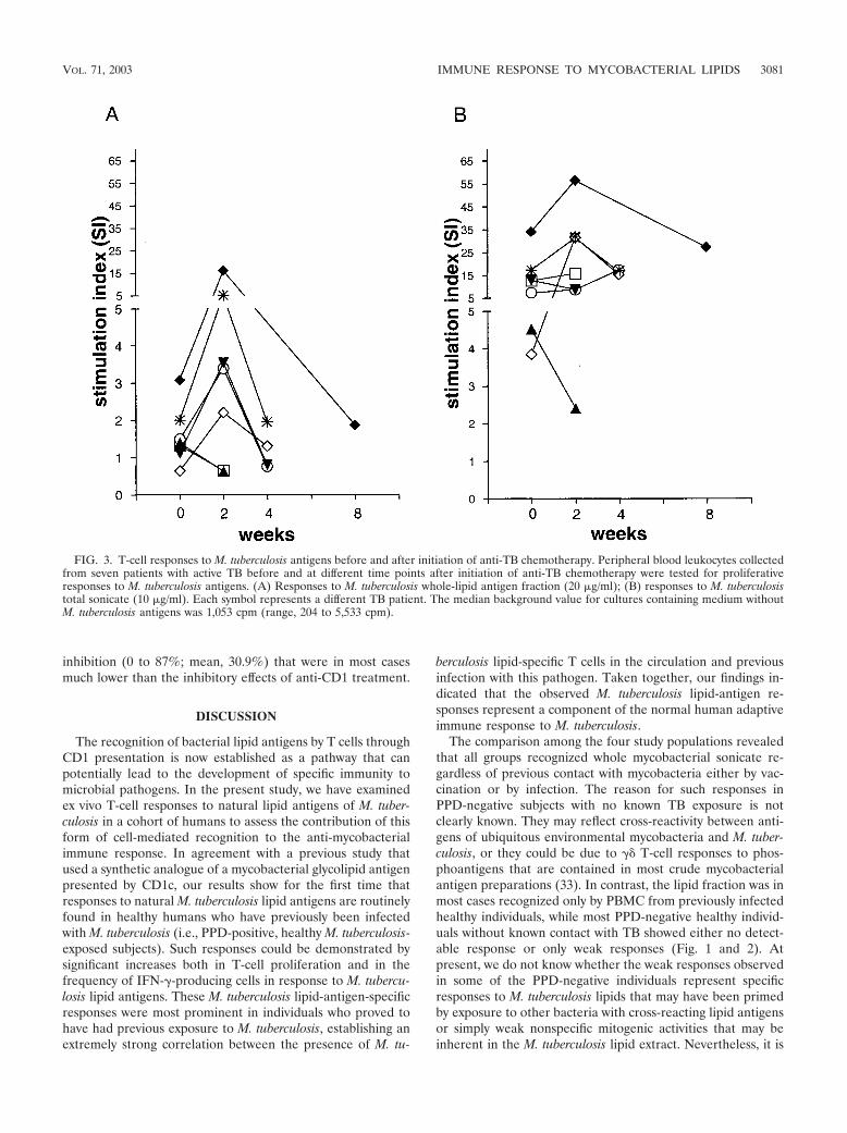
Mycobacterial lipid-antigen-specific T cells in active TB. Inorder to investigate the response to mycobacterial lipid anti-gens in the setting of clinically active infection, additionalblood samples were obtained from patients with active pulmo-
nary TB prior to the start of anti-TB chemotherapy and atseveral points thereafter (i.e., 2 and 4 weeks after initiation oftreatment and at the time of discharge from the hospital).Proliferation assays comparing the reactivities of the PBMCcollected at these time points showed that lipid-antigen-spe-cific T-cell responses were generally detected weakly or not atall prior to initiation of therapy and became detectable inperipheral blood of TB patients approximately 2 weeks afterinitiation of anti-TB chemotherapy (Fig. 3A). The maximumproliferative response to mycobacterial lipid antigen was ob-served at 2 weeks following the start of therapy, and thistended to decline over the next 2 to 4 weeks. The proliferative
FIG. 1. Proliferative responses of the four study groups to M. tuberculosis whole-antigen extract and M. tuberculosis lipid antigens. Proliferationassays were performed by culturing nonadherent PBMC from each individual with irradiated autologous monocyte-derived iDC in completemedium containing M. tuberculosis antigens added to a final concentration of 20 �g/ml. (A) Responses to M. tuberculosis total sonicate;(B) responses to M. tuberculosis total-lipid extract; (C) responses to purified GMM (inset shows structure of M. phlei C80 GMM). Proliferation wasmeasured as counts per minute of [3H]thymidine incorporation, and results are expressed as counts per minute with backgrounds subtracted (seebelow). Each symbol represents the mean of triplicate values for a single individual, and horizontal bars drawn through each group of data pointsshow median proliferation values. Subjects are grouped into the following clinical categories: tuberculin skin test negative with no history of M.tuberculosis exposure or BCG vaccination (PPD-neg), tuberculin skin test negative with a history of previous BCG vaccination (BCG-vac),tuberculin skin test positive without active disease (PPD-pos), and active TB prior to initiation of treatment (active TB). BCG-vac, PPD-pos, andactive TB study groups were compared statistically to the PPD-neg control group (by the Mann-Whitney U test). Significant increases wereobserved only for the proliferative responses of the PPD-pos group relative to those of the PPD-neg group for M. tuberculosis whole-lipid antigen(B) (P � 0.0006) and for purified GMM (C) (P � 0.0223). The number of individuals in each group is given in parentheses below the x-axis labels.The median background value (i.e., proliferation in medium alone without M. tuberculosis antigens) for each group was as follows: 1,930 cpm(range, 445 to 1,629 cpm) for the PPD-neg control group, 630 cpm (range, 401 to 1,667 cpm) for BCG-vac, 641 cpm (range, 134 to 2,823) forPPD-pos, and 1,554 cpm (range, 394 to 2,780 cpm) for active TB.
VOL. 71, 2003 IMMUNE RESPONSE TO MYCOBACTERIAL LIPIDS 3079

responses of these samples to the total M. tuberculosis sonicateshowed less variation over time than the responses to the lipidantigen preparation and tended to remain elevated at a similarlevel throughout the time course of the analysis (Fig. 3B).Similarly, proliferative responses to the mitogen phytohemag-glutinin or to the purified recombinant M. tuberculosis proteinantigen Ag85B (as previously described by Hess et al. [7]) wereelevated in several individuals and showed relatively less vari-ation over time than lipid-antigen-specific responses (data notshown).
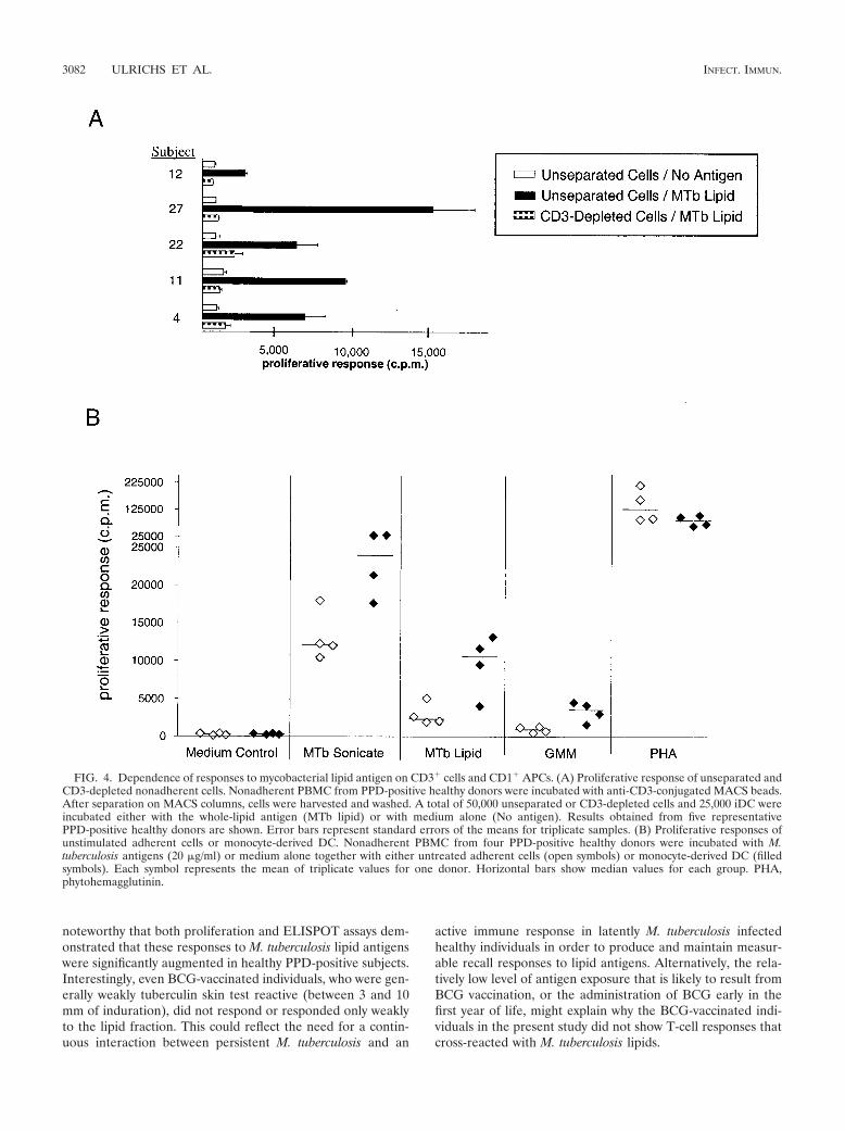
Characterization of M. tuberculosis lipid antigen-reactive Tcells. In initial studies to demonstrate the T-cell dependence ofthe responses observed, proliferation and IFN-� ELISPOTassays using responding nonadherent PBMC that were de-pleted of CD3� cells were performed. These studies showedthat depletion of CD3� cells completely eliminated responsesto the M. tuberculosis total-lipid fraction (Fig. 4A). The ob-served proliferative responses to the M. tuberculosis total-lipidfraction were also dependent on the presence of APCs, as noresponses were observed when nonadherent lymphocytes werecultured with antigens in the absence of autologous adherentcells (data not shown). In addition, proliferation assays usingeither unstimulated APCs (monocytes) or immature DC (iDC)obtained by pretreatment of monocytes with IL-4 and GM-CSF revealed that while both APC preparations supportedresponses to the M. tuberculosis total sonicate, significant re-sponses to lipid antigens were obtained only when iDC wereused as the APCs (Fig. 4B).
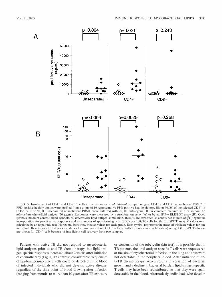
In order to determine if the M. tuberculosis lipid antigenreactivity was predominantly a feature of either CD4� orCD8� T cells, we studied the responses of these populationsseparately after their purification by immunomagnetic sorting.These studies showed that responses to the M. tuberculosiswhole-lipid antigen preparation were much more pronouncedin isolated CD4� T cells and were only weakly detected in mostsamples of CD8� T cells by proliferation assay (Fig. 5A). Thissuggested that for the proliferative responses observed, the T
cells responding to M. tuberculosis lipid antigens were predom-inantly CD4 positive. Very similar findings were obtained byIFN-� ELISPOT assay (Fig. 5B).
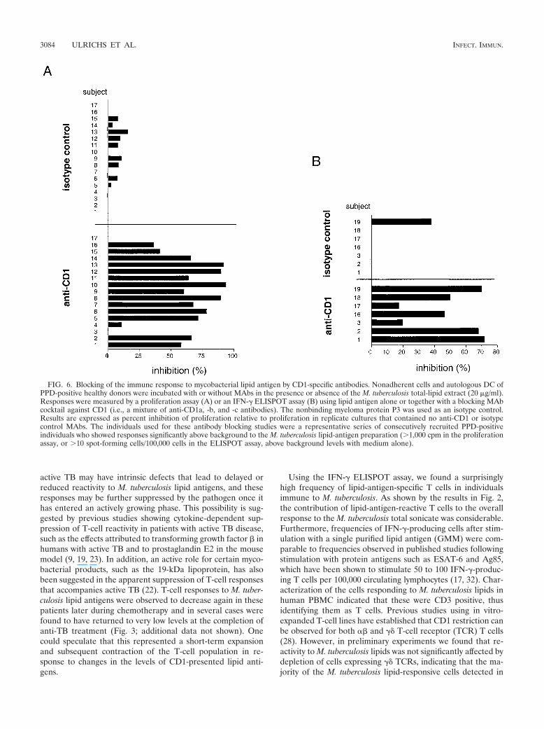
The requirement for GM-CSF and IL-4 pretreatment ofadherent mononuclear cells to generate effective APCs forresponses to M. tuberculosis lipid antigens suggested a directinvolvement of group 1 CD1 molecules, since these are absenton normal monocytes but strongly upregulated by treatmentwith these cytokines (20, 21). To more directly assess the in-volvement of CD1 molecules in triggering the lipid-antigen-specific responses, we determined the effects of adding a mix-ture of MAbs specific for all of the group 1 CD1 isoforms (i.e.,anti-CD1a, -b, and -c) in the proliferation and IFN-� ELI-SPOT assays. In proliferation assays using a series of repre-sentative PPD-positive donors, we observed significant block-ing of the proliferative responses against the M. tuberculosistotal-lipid-antigen preparation for 15 of 17 individuals, withthe level of inhibition ranging from 10.7 to 93.5% (mean,57.8%) (Fig. 6A). Blocking with an isotype-matched controlantibody (P3) was consistently less than 16% (mean, 4.5%).Similar results were obtained in ELISPOT assays using a sam-ple of seven PPD-positive individuals (Fig. 6B). This revealeda 19.6 to 77.6% (mean, 50.9%) inhibition of IFN-� spot-form-ing cells by addition of the anti-CD1 antibody cocktail, whereasthe isotype control antibody revealed virtually no inhibition(mean, 9.5%, maximum, 38.1%).
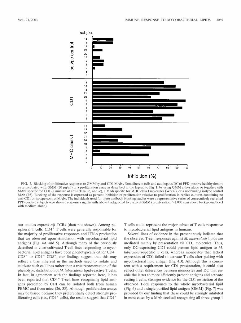
The CD1 restriction of the lipid-antigen response was alsostudied for a single isolated mycobacterial glycolipid antigen,GMM (Fig. 7). A series of 11 PPD-positive subjects who wereresponsive to purified GMM in proliferation assays were ana-lyzed. GMM responses by cells from all of these donors weremarkedly inhibited by the anti-CD1 antibody cocktail (61.9 to98.2% inhibition; mean, 81.2%). In contrast, the nonbindingisotype control antibody showed little or no inhibition (mean,8%). An antibody to MHC class I molecules (W6/32), whichbound to the surfaces of the APCs at levels comparable to thebinding of the anti-CD1 antibodies, showed variable levels of
FIG. 2. Frequency of IFN-�-producing cells responding to M. tuberculosis whole-antigen extract and to M. tuberculosis lipid antigens. IFN-�-producing cells in peripheral blood leukocyte samples were quantitated by ELISPOT assay as described in Materials and Methods. M. tuberculosisantigens were used at a final concentration of 20 �g/ml. Results are presented as numbers of spot-forming cells (SFCs) per 100,000 cells. Opensymbols, PPD-negative healthy donors (n � 14); filled symbols, PPD-positive healthy donors (n � 10). Horizontal bars show median values for eachgroup. P values for the comparisons indicated were determined by using the Mann-Whitney U test.
3080 ULRICHS ET AL. INFECT. IMMUN.
inhibition (0 to 87%; mean, 30.9%) that were in most casesmuch lower than the inhibitory effects of anti-CD1 treatment.
DISCUSSION
The recognition of bacterial lipid antigens by T cells throughCD1 presentation is now established as a pathway that canpotentially lead to the development of specific immunity tomicrobial pathogens. In the present study, we have examinedex vivo T-cell responses to natural lipid antigens of M. tuber-culosis in a cohort of humans to assess the contribution of thisform of cell-mediated recognition to the anti-mycobacterialimmune response. In agreement with a previous study thatused a synthetic analogue of a mycobacterial glycolipid antigenpresented by CD1c, our results show for the first time thatresponses to natural M. tuberculosis lipid antigens are routinelyfound in healthy humans who have previously been infectedwith M. tuberculosis (i.e., PPD-positive, healthy M. tuberculosis-exposed subjects). Such responses could be demonstrated bysignificant increases both in T-cell proliferation and in thefrequency of IFN-�-producing cells in response to M. tubercu-losis lipid antigens. These M. tuberculosis lipid-antigen-specificresponses were most prominent in individuals who proved tohave had previous exposure to M. tuberculosis, establishing anextremely strong correlation between the presence of M. tu-
berculosis lipid-specific T cells in the circulation and previousinfection with this pathogen. Taken together, our findings in-dicated that the observed M. tuberculosis lipid-antigen re-sponses represent a component of the normal human adaptiveimmune response to M. tuberculosis.
The comparison among the four study populations revealedthat all groups recognized whole mycobacterial sonicate re-gardless of previous contact with mycobacteria either by vac-cination or by infection. The reason for such responses inPPD-negative subjects with no known TB exposure is notclearly known. They may reflect cross-reactivity between anti-gens of ubiquitous environmental mycobacteria and M. tuber-culosis, or they could be due to �� T-cell responses to phos-phoantigens that are contained in most crude mycobacterialantigen preparations (33). In contrast, the lipid fraction was inmost cases recognized only by PBMC from previously infectedhealthy individuals, while most PPD-negative healthy individ-uals without known contact with TB showed either no detect-able response or only weak responses (Fig. 1 and 2). Atpresent, we do not know whether the weak responses observedin some of the PPD-negative individuals represent specificresponses to M. tuberculosis lipids that may have been primedby exposure to other bacteria with cross-reacting lipid antigensor simply weak nonspecific mitogenic activities that may beinherent in the M. tuberculosis lipid extract. Nevertheless, it is
FIG. 3. T-cell responses to M. tuberculosis antigens before and after initiation of anti-TB chemotherapy. Peripheral blood leukocytes collectedfrom seven patients with active TB before and at different time points after initiation of anti-TB chemotherapy were tested for proliferativeresponses to M. tuberculosis antigens. (A) Responses to M. tuberculosis whole-lipid antigen fraction (20 �g/ml); (B) responses to M. tuberculosistotal sonicate (10 �g/ml). Each symbol represents a different TB patient. The median background value for cultures containing medium withoutM. tuberculosis antigens was 1,053 cpm (range, 204 to 5,533 cpm).
VOL. 71, 2003 IMMUNE RESPONSE TO MYCOBACTERIAL LIPIDS 3081
noteworthy that both proliferation and ELISPOT assays dem-onstrated that these responses to M. tuberculosis lipid antigenswere significantly augmented in healthy PPD-positive subjects.Interestingly, even BCG-vaccinated individuals, who were gen-erally weakly tuberculin skin test reactive (between 3 and 10mm of induration), did not respond or responded only weaklyto the lipid fraction. This could reflect the need for a contin-uous interaction between persistent M. tuberculosis and an
active immune response in latently M. tuberculosis infectedhealthy individuals in order to produce and maintain measur-able recall responses to lipid antigens. Alternatively, the rela-tively low level of antigen exposure that is likely to result fromBCG vaccination, or the administration of BCG early in thefirst year of life, might explain why the BCG-vaccinated indi-viduals in the present study did not show T-cell responses thatcross-reacted with M. tuberculosis lipids.
FIG. 4. Dependence of responses to mycobacterial lipid antigen on CD3� cells and CD1� APCs. (A) Proliferative response of unseparated andCD3-depleted nonadherent cells. Nonadherent PBMC from PPD-positive healthy donors were incubated with anti-CD3-conjugated MACS beads.After separation on MACS columns, cells were harvested and washed. A total of 50,000 unseparated or CD3-depleted cells and 25,000 iDC wereincubated either with the whole-lipid antigen (MTb lipid) or with medium alone (No antigen). Results obtained from five representativePPD-positive healthy donors are shown. Error bars represent standard errors of the means for triplicate samples. (B) Proliferative responses ofunstimulated adherent cells or monocyte-derived DC. Nonadherent PBMC from four PPD-positive healthy donors were incubated with M.tuberculosis antigens (20 �g/ml) or medium alone together with either untreated adherent cells (open symbols) or monocyte-derived DC (filledsymbols). Each symbol represents the mean of triplicate values for one donor. Horizontal bars show median values for each group. PHA,phytohemagglutinin.
3082 ULRICHS ET AL. INFECT. IMMUN.
Patients with active TB did not respond to mycobacteriallipid antigens prior to anti-TB chemotherapy, but lipid-anti-gen-specific responses increased about 2 weeks after initiationof chemotherapy (Fig. 3). In contrast, considerable frequenciesof lipid-antigen-specific T cells could be detected in the bloodof infected individuals who did not develop active disease,regardless of the time point of blood drawing after infection(ranging from months to more than 10 years after TB exposure
or conversion of the tuberculin skin test). It is possible that inTB patients, the lipid-antigen-specific T cells were sequesteredat the site of mycobacterial infection in the lung and thus werenot detectable in the peripheral blood. After initiation of an-ti-TB chemotherapy, which results in cessation of bacterialgrowth and a decline in bacterial burden, lipid-antigen-specificT cells may have been redistributed so that they were againdetectable in the blood. Alternatively, individuals who develop
FIG. 5. Involvement of CD4� and CD8� T cells in the responses to M. tuberculosis lipid antigen. CD4� and CD8� nonadherent PBMC ofPPD-positive healthy donors were purified from a group of 10 representative PPD-positive healthy donors. Either 50,000 of the selected CD4� orCD8� cells or 50,000 unseparated nonadherent PBMC were cultured with 25,000 autologous DC in complete medium with or without M.tuberculosis whole-lipid antigen (20 �g/ml). Responses were measured by a proliferation assay (A) or by an IFN-� ELISPOT assay (B). Opensymbols, medium control; filled symbols, M. tuberculosis lipid antigen stimulation. Results are expressed as counts per minute of [3H]thymidineincorporation for proliferative responses and as numbers of spot-forming cells (SFC) per 100,000 cells for the ELISPOT assay. P values werecalculated by an unpaired t test. Horizontal bars show median values for each group. Each symbol represents the mean of triplicate values for oneindividual. Results for all 10 donors are shown for unseparated and CD8� cells. Results for only nine (proliferation) or eight (ELISPOT) donorsare shown for CD4� cells because of insufficient cell recovery from two samples.
VOL. 71, 2003 IMMUNE RESPONSE TO MYCOBACTERIAL LIPIDS 3083
active TB may have intrinsic defects that lead to delayed orreduced reactivity to M. tuberculosis lipid antigens, and theseresponses may be further suppressed by the pathogen once ithas entered an actively growing phase. This possibility is sug-gested by previous studies showing cytokine-dependent sup-pression of T-cell reactivity in patients with active TB disease,such as the effects attributed to transforming growth factor inhumans with active TB and to prostaglandin E2 in the mousemodel (9, 19, 23). In addition, an active role for certain myco-bacterial products, such as the 19-kDa lipoprotein, has alsobeen suggested in the apparent suppression of T-cell responsesthat accompanies active TB (22). T-cell responses to M. tuber-culosis lipid antigens were observed to decrease again in thesepatients later during chemotherapy and in several cases werefound to have returned to very low levels at the completion ofanti-TB treatment (Fig. 3; additional data not shown). Onecould speculate that this represented a short-term expansionand subsequent contraction of the T-cell population in re-sponse to changes in the levels of CD1-presented lipid anti-gens.
Using the IFN-� ELISPOT assay, we found a surprisinglyhigh frequency of lipid-antigen-specific T cells in individualsimmune to M. tuberculosis. As shown by the results in Fig. 2,the contribution of lipid-antigen-reactive T cells to the overallresponse to the M. tuberculosis total sonicate was considerable.Furthermore, frequencies of IFN-�-producing cells after stim-ulation with a single purified lipid antigen (GMM) were com-parable to frequencies observed in published studies followingstimulation with protein antigens such as ESAT-6 and Ag85,which have been shown to stimulate 50 to 100 IFN-�-produc-ing T cells per 100,000 circulating lymphocytes (17, 32). Char-acterization of the cells responding to M. tuberculosis lipids inhuman PBMC indicated that these were CD3 positive, thusidentifying them as T cells. Previous studies using in vitro-expanded T-cell lines have established that CD1 restriction canbe observed for both and �� T-cell receptor (TCR) T cells(28). However, in preliminary experiments we found that re-activity to M. tuberculosis lipids was not significantly affected bydepletion of cells expressing �� TCRs, indicating that the ma-jority of the M. tuberculosis lipid-responsive cells detected in
FIG. 6. Blocking of the immune response to mycobacterial lipid antigen by CD1-specific antibodies. Nonadherent cells and autologous DC ofPPD-positive healthy donors were incubated with or without MAbs in the presence or absence of the M. tuberculosis total-lipid extract (20 �g/ml).Responses were measured by a proliferation assay (A) or an IFN-� ELISPOT assay (B) using lipid antigen alone or together with a blocking MAbcocktail against CD1 (i.e., a mixture of anti-CD1a, -b, and -c antibodies). The nonbinding myeloma protein P3 was used as an isotype control.Results are expressed as percent inhibition of proliferation relative to proliferation in replicate cultures that contained no anti-CD1 or isotypecontrol MAbs. The individuals used for these antibody blocking studies were a representative series of consecutively recruited PPD-positiveindividuals who showed responses significantly above background to the M. tuberculosis lipid-antigen preparation (�1,000 cpm in the proliferationassay, or �10 spot-forming cells/100,000 cells in the ELISPOT assay, above background levels with medium alone).
3084 ULRICHS ET AL. INFECT. IMMUN.
our studies express TCRs (data not shown). Among pe-ripheral T cells, CD4� T cells were generally responsible forthe majority of proliferative responses and IFN-� productionthat we observed upon stimulation with mycobacterial lipidantigens (Fig. 4A and 5). Although many of the previouslydescribed in vitro-cultivated T-cell lines responding to myco-bacterial lipid antigens have been phenotypically either CD4�
CD8� or CD4� CD8�, our findings suggest that this mayreflect a bias inherent in the methods used to isolate andcultivate such cell lines rather than a true representation of thephenotypic distribution of M. tuberculosis lipid-reactive T cells.In fact, in agreement with the findings reported here, it hasbeen reported that CD4� T-cell lines recognizing lipid anti-gens presented by CD1 can be isolated both from humanPBMC and from mice (26, 35). Although proliferation assaysmay be biased because they preferentially detect strongly pro-liferating cells (i.e., CD4� cells), the results suggest that CD4�
T cells could represent the major subset of T cells responsiveto mycobacterial lipid antigens in humans.
Several lines of evidence in the present study indicate thatthe observed T-cell responses against M. tuberculosis lipids aremediated mainly by presentation via CD1 molecules. Thus,only DC-expressing CD1 could present lipid antigen to M.tuberculosis-specific T cells, whereas monocytes that lackedexpression of CD1 failed to activate T cells after pulsing withmycobacterial lipid antigen (Fig. 4B). Although this is consis-tent with a requirement for CD1 presentation, it could alsoreflect other differences between monocytes and DC that en-able the latter to more efficiently present antigens and activateresting T cells. Stronger evidence for the CD1 restriction of theobserved T-cell responses to the whole mycobacterial lipid(Fig. 6) and a single purified lipid antigen (GMM) (Fig. 7) wasprovided by our finding that these could be strongly inhibitedin most cases by a MAb cocktail recognizing all three group 1
FIG. 7. Blocking of proliferative responses to GMM by anti-CD1 MAbs. Nonadherent cells and autologous DC of PPD-positive healthy donorswere incubated with GMM (20 �g/ml) in a proliferation assay as described in the legend to Fig. 1, by using GMM either alone or together withMAbs specific for CD1 (a mixture of anti-CD1a, -b, and -c), a MAb specific for MHC class I molecules (W6/32), or a nonbinding isotype controlMAb (P3). Blocking of the response is expressed as percent inhibition of proliferation relative to proliferation in replica cultures containing noanti-CD1 or isotype control MAbs. The individuals used for these antibody blocking studies were a representative series of consecutively recruitedPPD-positive subjects who showed responses significantly above background to purified GMM (proliferation, �1,000 cpm above background levelwith medium alone).
VOL. 71, 2003 IMMUNE RESPONSE TO MYCOBACTERIAL LIPIDS 3085
CD1 proteins, and not by isotype control antibodies that didnot bind to the surfaces of the cells. Moderate variability of theblocking effects of anti-CD1 MAbs on the responses to the M.tuberculosis total-lipid extract was observed in this study, witha few individuals actually showing little or no inhibition of theirresponses by anti-CD1 (Fig. 6). This variability in the level ofinhibition showed no correlation with the strength of the pro-liferative or IFN-� ELISPOT responses of the subjects (datanot shown). Instead, it may be related to the heterogeneity ofthe M. tuberculosis total-lipid-antigen preparation, which couldpotentially evoke qualitatively different responses from differ-ent donors. In support of this possibility was our finding thatthe responses to a homogeneous purified M. tuberculosis gly-colipid antigen (GMM) were consistently and strongly inhib-ited by anti-CD1 MAbs (Fig. 7). Since our present data haveevaluated CD1 restriction of M. tuberculosis lipid-specific re-sponses by using only a mixture of MAbs against all threegroup 1 CD1 isoforms, further studies will be required todetermine whether one or more of these CD1 molecules playa dominant role in these ex vivo responses.
As an additional control for these experiments, we assessedthe effects of MAbs against MHC class I molecules, which areexpressed on both the responding T cells and the APCs. Al-though we did observe partial inhibition of T-cell responses toM. tuberculosis lipid antigens in some cases by using anti-MHCclass I MAbs, these effects were generally much weaker andless consistent than the blocking effects observed with anti-CD1 MAbs (Fig. 7). The mechanism for the partial blockingeffects of anti-MHC class I MAbs in these experiments has notbeen investigated by us. However, reports in the literature haveshown that MHC class I molecules may associate on the T-cellsurface with a variety of molecules involved in T-cell activation,including the CD3–TCR complex and CD2 (11, 34). Thus,anti-MHC class I MAbs might inhibit T-cell activation in anon-antigen-specific manner by interfering with the function ofassociated receptor complexes. Alternatively, it is interesting tospeculate that this finding might reflect a relevant accessoryinteraction between CD1-restricted T cells and APCs that isdependent on MHC class I molecules, or the presence of apopulation of MHC-restricted accessory cells that is requiredin some subjects for the effective stimulation of CD1-restrictedresponses to lipid antigens.
The results provided by the present study show that lipid-antigen-specific T cells are present in the circulation of humansfollowing natural infection with M. tuberculosis and thus mayrepresent one component of the protective immune responseto this organism. Indeed, the presence of these T-cell re-sponses in PPD-positive subjects who developed no clinicalsigns of disease, and the suppression of such responses inactive TB patients, suggests a correlation between circulatinglipid-antigen-reactive T cells and control of the infection. Ourdata indicate that exposure to M. tuberculosis by natural infec-tion can elicit recall responses to M. tuberculosis lipids, whichmost likely represent part of the adaptive immune response tothis pathogen. This function of the group 1 component of theCD1 system appears to contrast with the dominant role of thegroup 2 CD1 molecules, which have been linked to the selec-tion and activation of natural killer T cells that may functionmore as effectors of innate immunity and in immunoregulation(2, 3, 10, 29).
Understanding the basis of the overall acquired immunity toM. tuberculosis infection in humans is crucial for developingnew approaches to therapy and prevention through vaccina-tion. Recent data from studies using guinea pigs as an in vivomodel for the function of group 1 CD1 molecules have shownthat long-term memory against mycobacterial lipid antigenscan be stimulated by specific immunization, further suggestingthat lipids might be useful targets to evaluate further in theongoing search for novel vaccines against M. tuberculosis (8). Inhumans, further investigation of the role of unconventional Tcells such as CD1-restricted and nonclassical HLA-restricted Tcells is required for better understanding of the basis for op-timal and appropriate acquired immunity to M. tuberculosis.
ACKNOWLEDGMENTS
This work was supported by grants from the NIH (AI48933 andAI45889) and by a grant from the Irene Diamond Foundation toS.A.P. D.B.M. was supported by grants from the NIH (AR48632 andAI49313) and by the Research and Education Foundation of theAmerican College of Rheumatology. T.U. received a fellowship fromthe Deutsche Forschungsgemeinschaft (DFG, UL 176/1-1).
We thank Daphney Frederique and Tan-Yun Cheng for excellenttechnical assistance in the preparation of lipid antigens, and we thankSadamu Ishikawa for assistance in identifying human subjects for thisstudy.
REFERENCES
1. Beckman, E. M., S. A. Porcelli, C. T. Morita, S. M. Behar, S. T. Furlong, andM. B. Brenner. 1994. Recognition of a lipid antigen by CD1-restricted �
T cells. Nature 372:691–694.2. Behar, S. M., C. C. Dascher, M. J. Grusby, C. R. Wang, and M. B. Brenner.
1999. Susceptibility of mice deficient in CD1D or TAP1 to infection withMycobacterium tuberculosis. J. Exp. Med. 189:1973–1980.
3. Brossay, L., M. Chioda, N. Burdin, Y. Koezuka, G. Casorati, P. Dellabona,and M. Kronenberg. 1998. CD1d-mediated recognition of an -galactosyl-ceramide by natural killer T cells is highly conserved through mammalianevolution. J. Exp. Med. 188:1521–1528.
4. Flynn, J. L., and J. D. Ernst. 2000. Immune responses in tuberculosis. Curr.Opin. Immunol. 12:432–436.
5. Folch, J., M. Lees, and G. H. Sloane Stanley. 1957. A simple method for theisolation and purification of total lipids from animal tissues. J. Biol. Chem.226:497–509.
6. Gumperz, J. E., and M. B. Brenner. 2001. CD1-specific T cells in microbialimmunity. Curr. Opin. Immunol. 13:471–478.
7. Hess, J., L. Grode, J. Hellwig, P. Conradt, I. Gentschev, W. Goebel, C. Ladel,and S. H. Kaufmann. 2000. Protection against murine tuberculosis by anattenuated recombinant Salmonella typhimurium vaccine strain that secretesthe 30-kDa antigen of Mycobacterium bovis BCG. FEMS Immunol. Med.Microbiol. 27:283–289.
8. Hiromatsu, K., C. C. Dascher, K. P. LeClair, M. Sugita, S. T. Furlong, M. B.Brenner, and S. A. Porcelli. 2002. Induction of CD1-restricted immuneresponses in guinea pigs by immunization with mycobacterial lipid antigens.J. Immunol. 169:330–339.
9. Hirsch, C. S., R. Hussain, Z. Toossi, G. Dawood, F. Shahid, and J. J. Ellner.1996. Cross-modulation by transforming growth factor beta in human tuber-culosis: suppression of antigen-driven blastogenesis and interferon gammaproduction. Proc. Natl. Acad. Sci. USA 93:3193–3198.
10. Kawano, T., Y. Tanaka, E. Shimizu, Y. Kaneko, N. Kamata, H. Sato, H.Osada, S. Sekiya, T. Nakayama, and M. Taniguchi. 1999. A novel recogni-tion motif of human NKT antigen receptor for a glycolipid ligand. Int.Immunol. 11:881–887.
11. Khan, A. A., C. Bose, L. S. Yam, M. J. Soloski, and F. Rupp. 2001. Physio-logical regulation of the immunological synapse by agrin. Science 292:1681–1686.
12. Melian, A., Y. J. Geng, G. K. Sukhova, P. Libby, and S. A. Porcelli. 1999.CD1 expression in human atherosclerosis. A potential mechanism for T cellactivation by foam cells. Am. J. Pathol. 155:775–786.
13. Melian, A., G. F. Watts, A. Shamshiev, G. De Libero, A. Clatworthy, M.Vincent, M. B. Brenner, S. Behar, K. Niazi, R. L. Modlin, S. Almo, D. Ostrov,S. G. Nathenson, and S. A. Porcelli. 2000. Molecular recognition of humanCD1b antigen complexes: evidence for a common pattern of interaction with TCRs. J. Immunol. 165:4494–4504.
14. Moody, D. B., V. Briken, T. Y. Cheng, C. Roura-Mir, M. R. Guy, D. H. Geho,M. L. Tykocinski, G. S. Besra, and S. A. Porcelli. 2002. Lipid length controls
3086 ULRICHS ET AL. INFECT. IMMUN.
antigen entry into endosomal and nonendosomal pathways for CD1b pre-sentation. Nat. Immunol. 3:435–442.
15. Moody, D. B., M. R. Guy, E. Grant, T. Y. Cheng, M. B. Brenner, G. S. Besra,and S. A. Porcelli. 2000. CD1b-mediated T cell recognition of a glycolipidantigen generated from mycobacterial lipid and host carbohydrate duringinfection. J. Exp. Med. 192:965–976.
16. Moody, D. B., T. Ulrichs, W. Muhlecker, D.C. Young, S. S. Gurcha, E. Grant,J. P. Rosat, M. B. Brenner, C. E. Costello, G. S. Besra, and S. A. Porcelli.2000. CD1c-mediated T cell recognition of isoprenoid glycolipids in M.tuberculosis infection. Nature 404:884–888.
17. Mustafa, A. S., F. A. Shaban, A. T. Abal, R. Al Attiyah, H. G. Wiker, K. E.Lundin, F. Oftung, and K. Huygen. 2000. Identification and HLA restrictionof naturally derived Th1-cell epitopes from the secreted Mycobacteriumtuberculosis antigen 85B recognized by antigen-specific human CD4� T-celllines. Infect. Immun. 68:3933–3940.
18. Olive, D., P. Dubreuil, and C. Mawas. 1984. Two distinct TL-like molecularsubsets defined by monoclonal antibodies on the surface of human thymo-cytes with different expression on leukemia lines. Immunogenetics 20:253–264.
19. Pathan, A. A., K. A. Wilkinson, P. Klenerman, H. McShane, R. N. Davidson,G. Pasvol, A. V. Hill, and A. Lalvani. 2001. Direct ex vivo analysis of antigen-specific IFN-�-secreting CD4 T cells in Mycobacterium tuberculosis-infectedindividuals: associations with clinical disease state and effect of treatment.J. Immunol. 167:5217–5225.
20. Porcelli, S., C. T. Morita, and M. B. Brenner. 1992. CD1b restricts theresponse of human CD4�8� T lymphocytes to a microbial antigen. Nature360:593–597.
21. Porcelli, S. A., and R. L. Modlin. 1999. The CD1 system: antigen-presentingmolecules for T cell recognition of lipids and glycolipids. Annu. Rev. Immu-nol. 17:297–329.
22. Post, F. A., C. Manca, O. Neyrolles, B. Ryffel, D. B. Young, and G. Kaplan.2001. Mycobacterium tuberculosis 19-kilodalton lipoprotein inhibits Mycobac-terium smegmatis-induced cytokine production by human macrophages invitro. Infect. Immun. 69:1433–1439.
23. Rangel, M. J., G. Estrada, I. De La Luz Garcia Hernandez, L. D. Aguilar, R.Marquez, and P. R. Hernandez. 2002. The role of prostaglandin E2 in theimmunopathogenesis of experimental pulmonary tuberculosis. Immunology106:257–266.
24. Scanga, C. A., V. P. Mohan, K. Yu, H. Joseph, K. Tanaka, J. Chan, and J. L.Flynn. 2000. Depletion of CD4� T cells causes reactivation of murine per-sistent tuberculosis despite continued expression of interferon gamma andnitric oxide synthase 2. J. Exp. Med. 192:347–358.
25. Serbina, N. V., C. C. Liu, C. A. Scanga, and J. L. Flynn. 2000. CD8� CTLfrom lungs of Mycobacterium tuberculosis-infected mice express perforin invivo and lyse infected macrophages. J. Immunol. 165:353–363.
26. Sieling, P. A., M. T. Ochoa, D. Jullien, D. S. Leslie, S. Sabet, J. P. Rosat,A. E. Burdick, T. H. Rea, M. B. Brenner, S. A. Porcelli, and R. L. Modlin.2000. Evidence for human CD4� T cells in the CD1-restricted repertoire:derivation of mycobacteria-reactive T cells from leprosy lesions. J. Immunol.164:4790–4796.
27. Spada, F. M., F. Borriello, M. Sugita, G. F. Watts, Y. Koezuka, and S. A.Porcelli. 2000. Low expression level but potent antigen presenting functionof CD1d on monocyte lineage cells. Eur. J. Immunol. 30:3468–3477.
28. Spada, F. M., E. P. Grant, P. J. Peters, M. Sugita, A. Melian, D. S. Leslie,H. K. Lee, E. van Donselaar, D. A. Hanson, A. M. Krensky, O. Majdic, S. A.Porcelli, C. T. Morita, and M. B. Brenner. 2000. Self-recognition of CD1 by�� T cells. Implications for innate immunity. J. Exp. Med. 191:937–948.
29. Spada, F. M., Y. Koezuka, and S. A. Porcelli. 1998. CD1d-restricted recog-nition of synthetic glycolipid antigens by human natural killer T cells. J. Exp.Med. 188:1529–1534.
30. Stenger, S., R. J. Mazzaccaro, K. Uyemura, S. Cho, P. F. Barnes, J. P. Rosat,A. Sette, M. B. Brenner, S. A. Porcelli, B. R. Bloom, and R. L. Modlin. 1997.Differential effects of cytolytic T cell subsets on intracellular infection. Sci-ence 276:1684–1687.
31. Ulrichs, T., P. Anding, S. H. Kaufmann, and M. E. Munk. 2000. Numbers ofIFN-�-producing cells against ESAT-6 increase in tuberculosis patients dur-ing chemotherapy. Int. J. Tuberc. Lung Dis. 4:1181–1183.
32. Ulrichs, T., P. Anding, S. Porcelli, S. H. Kaufmann, and M. E. Munk. 2000.Increased numbers of ESAT-6- and purified protein derivative-specificgamma interferon-producing cells in subclinical and active tuberculosis in-fection. Infect. Immun. 68:6073–6076.
33. Ulrichs, T., and S. A. Porcelli. 2000. CD1 proteins: targets of T cell recog-nition in innate and adaptive immunity. Rev. Immunogenet. 2:416–432.
34. Verhagen, A. M., B. Schraven, M. Wild, R. Wallich, and S. C. Meuer. 1996.Differential interaction of the CD2 extracellular and intracellular domainswith the tyrosine phosphatase CD45 and the zeta chain of the TCR/CD3/zetacomplex. Eur. J. Immunol. 26:2841–2849.
35. Wang, B., T. Chun, and C. R. Wang. 2000. Comparative contribution of CD1on the development of CD4� and CD8� T cell compartments. J. Immunol.164:739–745.
36. World Health Organization. 2003. WHO Report 2003. Global tuberculosiscontrol. [Online.] www.who.int/gtb/publications/globrep/index.html.
Editor: J. D. Clements
VOL. 71, 2003 IMMUNE RESPONSE TO MYCOBACTERIAL LIPIDS 3087
Copyright © 2022 FDOKUMEN