T cell recognition of self-antigen presenting cells by protein transfer assay reveals a high...
15
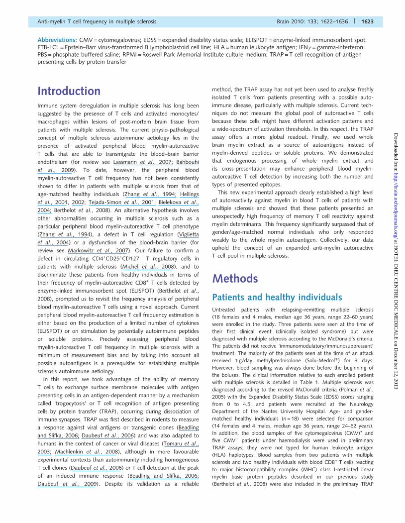
BRAIN A JOURNAL OF NEUROLOGY T cell recognition of self-antigen presenting cells by protein transfer assay reveals a high frequency of anti-myelin T cells in multiple sclerosis Bouchaib Bahbouhi, 1,2,3, * Se ´gole `ne Pettre ´, 1,2,3, * Laureline Berthelot, 1,2,3 Alexandra Garcia, 1,2,3 Annie Elong Ngono, 1,2,3 Nicolas Degauque, 1,2,3 Laure Michel, 4 Sandrine Wiertlewski, 4,5 Fabienne Lefre `re, 4 Claire Meyniel, 5 Catherine Delcroix, 6 Sophie Brouard, 1,2,3 David-Axel Laplaud 1,2,3,4,5, * and Jean-Paul Soulillou 1,2,3, * 1 INSERM, UMR 643, Nantes F44093, France 2 CHU Nantes, Institut de Transplantation et de Recherche en Transplantation (ITERT) Nantes F44000, France 3 Universite ´ de Nantes, Faculte ´ de Me ´ decine, Nantes F44000, France 4 INSERM CIC004, University Hospital, Nantes 44093, France 5 Department of Neurology, CHU Nantes, F-44093 Nantes, France 6 Department of Haemodialysis, CHU Nantes, F-44093 Nantes, France *These authors contributed equally to this work. Correspondence to: Jean-Paul Soulillou, INSERM, UMR 643, Institut de Transplantation Et de Recherches en Transplantation, ITERT, 30 Boulevard Jean Monnet, Nantes, 44093 France E-mail: [email protected] Although peripheral blood myelin-autoreactive T cells are thought to play a key role in multiple sclerosis, they are generally considered to have qualitative differences rather than quantitative ones when compared to those found in healthy individuals. Here, we revisited the assessment of myelin-autoreactive T cells in a new approach based on their combined ability to acquire membrane proteins from autologous antigen presenting cells, and to respond to whole myelin extract as the stimulating autoantigen. Using this approach, the myelin-autoreactive T cell frequency in patients with multiple sclerosis was found to be unexpectedly high (n = 22, subtracted values median 2.08%, range 0–6%; background median 1%, range 0–4%) and to exceed that of age/gender-matched healthy individuals significantly (n = 18, subtracted values median 0.1%, range 0–5.3%, P50.0001; background median 1.45%, range 0.1–4%). Higher anti-myelin autoreactivity was stable in patients with multiple sclerosis after several months. These data correlated with whole myelin-induced gamma interferon-enzyme-linked immunosorb- ent spot assay performed under the same conditions, although the values obtained with enzyme-linked immunosorbent spot assay under all conditions were 58 times lower than with this new method. The myelin-autoreactive T cells were memory T cells expressing CD40L with a CD62 low phenotype, suggesting their ability for homing to tissues. Collectively, these new data show a higher frequency of autoreactive T cells during multiple sclerosis than in age/gender-matched healthy individuals, and support an autoimmune aetiology in multiple sclerosis. Keywords: monocytes; multiple sclerosis; myelin; TRAP; trogocytosis doi:10.1093/brain/awq074 Brain 2010: 133; 1622–1636 | 1622 Received October 30, 2009. Revised February 23, 2010. Accepted February 24, 2010. Advance Access publication April 30, 2010 ß The Author (2010). Published by Oxford University Press on behalf of the Guarantors of Brain. All rights reserved. For Permissions, please email: [email protected] at HOTEL DIEU CENTRE DOC MEDICALE on December 12, 2013 http://brain.oxfordjournals.org/ Downloaded from
Transcript of T cell recognition of self-antigen presenting cells by protein transfer assay reveals a high...
BRAINA JOURNAL OF NEUROLOGY
T cell recognition of self-antigen presenting cellsby protein transfer assay reveals a high frequencyof anti-myelin T cells in multiple sclerosisBouchaib Bahbouhi,1,2,3,* Segolene Pettre,1,2,3,* Laureline Berthelot,1,2,3 Alexandra Garcia,1,2,3
Annie Elong Ngono,1,2,3 Nicolas Degauque,1,2,3 Laure Michel,4 Sandrine Wiertlewski,4,5
Fabienne Lefrere,4 Claire Meyniel,5 Catherine Delcroix,6 Sophie Brouard,1,2,3
David-Axel Laplaud1,2,3,4,5,* and Jean-Paul Soulillou1,2,3,*
1 INSERM, UMR 643, Nantes F44093, France
2 CHU Nantes, Institut de Transplantation et de Recherche en Transplantation (ITERT) Nantes F44000, France
3 Universite de Nantes, Faculte de Medecine, Nantes F44000, France
4 INSERM CIC004, University Hospital, Nantes 44093, France
5 Department of Neurology, CHU Nantes, F-44093 Nantes, France
6 Department of Haemodialysis, CHU Nantes, F-44093 Nantes, France
*These authors contributed equally to this work.
Correspondence to: Jean-Paul Soulillou,
INSERM, UMR 643,
Institut de Transplantation Et de Recherches en Transplantation,
ITERT, 30 Boulevard Jean Monnet,
Nantes, 44093
France
E-mail: [email protected]
Although peripheral blood myelin-autoreactive T cells are thought to play a key role in multiple sclerosis, they are generally
considered to have qualitative differences rather than quantitative ones when compared to those found in healthy individuals.
Here, we revisited the assessment of myelin-autoreactive T cells in a new approach based on their combined ability to acquire
membrane proteins from autologous antigen presenting cells, and to respond to whole myelin extract as the stimulating
autoantigen. Using this approach, the myelin-autoreactive T cell frequency in patients with multiple sclerosis was found to
be unexpectedly high (n = 22, subtracted values median 2.08%, range 0–6%; background median 1%, range 0–4%) and to
exceed that of age/gender-matched healthy individuals significantly (n = 18, subtracted values median 0.1%, range 0–5.3%,
P50.0001; background median 1.45%, range 0.1–4%). Higher anti-myelin autoreactivity was stable in patients with multiple
sclerosis after several months. These data correlated with whole myelin-induced gamma interferon-enzyme-linked immunosorb-
ent spot assay performed under the same conditions, although the values obtained with enzyme-linked immunosorbent spot
assay under all conditions were 58 times lower than with this new method. The myelin-autoreactive T cells were memory T cells
expressing CD40L with a CD62low phenotype, suggesting their ability for homing to tissues. Collectively, these new data show a
higher frequency of autoreactive T cells during multiple sclerosis than in age/gender-matched healthy individuals, and support
an autoimmune aetiology in multiple sclerosis.
Keywords: monocytes; multiple sclerosis; myelin; TRAP; trogocytosis
doi:10.1093/brain/awq074 Brain 2010: 133; 1622–1636 | 1622
Received October 30, 2009. Revised February 23, 2010. Accepted February 24, 2010. Advance Access publication April 30, 2010
� The Author (2010). Published by Oxford University Press on behalf of the Guarantors of Brain. All rights reserved.
For Permissions, please email: [email protected]
at HO
TE
L D
IEU
CE
NT
RE
DO
C M
ED
ICA
LE
on Decem
ber 12, 2013http://brain.oxfordjournals.org/
Dow
nloaded from
Abbreviations: CMV = cytomegalovirus; EDSS = expanded disability status scale; ELISPOT = enzyme-linked immunosorbent spot;ETB-LCL = Epstein–Barr virus-transformed B lymphoblastoid cell line; HLA = human leukocyte antigen; IFN� = gamma-interferon;PBS = phosphate buffered saline; RPMI = Roswell Park Memorial Institute culture medium; TRAP = T cell recognition of antigenpresenting cells by protein transfer
IntroductionImmune system deregulation in multiple sclerosis has long been
suggested by the presence of T cells and activated monocytes/
macrophages within lesions of post-mortem brain tissue from
patients with multiple sclerosis. The current physio-pathological
concept of multiple sclerosis autoimmune aetiology lies in the
presence of activated peripheral blood myelin-autoreactive
T cells that are able to transmigrate the blood–brain barrier
endothelium (for review see Lassmann et al., 2007; Bahbouhi
et al., 2009). To date, however, the peripheral blood
myelin-autoreactive T cell frequency has not been consistently
shown to differ in patients with multiple sclerosis from that of
age-matched healthy individuals (Zhang et al., 1994; Hellings
et al., 2001, 2002; Tejada-Simon et al., 2001; Bielekova et al.,
2004; Berthelot et al., 2008). An alternative hypothesis involves
other abnormalities occurring in multiple sclerosis such as a
particular peripheral blood myelin-autoreactive T cell phenotype
(Zhang et al., 1994), a defect in T cell regulation (Viglietta
et al., 2004) or a dysfunction of the blood–brain barrier (for
review see Markowitz et al., 2007). Our failure to confirm a
defect in circulating CD4+CD25+CD127� T regulatory cells in
patients with multiple sclerosis (Michel et al., 2008), and to
discriminate these patients from healthy individuals in terms of
their frequency of myelin-autoreactive CD8+ T cells detected by
enzyme-linked immunosorbent spot (ELISPOT) (Berthelot et al.,
2008), prompted us to revisit the frequency analysis of peripheral
blood myelin-autoreactive T cells using a novel approach. Current
peripheral blood myelin-autoreactive T cell frequency estimation is
either based on the production of a limited number of cytokines
(ELISPOT) or on stimulation by potentially autoimmune peptides
or soluble proteins. Precisely assessing peripheral blood
myelin-autoreactive T cell frequency in multiple sclerosis with a
minimum of measurement bias and by taking into account all
possible autoantigens is a prerequisite for establishing multiple
sclerosis autoimmune aetiology.
In this report, we took advantage of the ability of memory
T cells to exchange surface membrane molecules with antigen
presenting cells in an antigen-dependent manner by a mechanism
called ‘trogocytosis’ or T cell recognition of antigen presenting
cells by protein transfer (TRAP), occurring during dissociation of
immune synapses. TRAP was first described in rodents to measure
a response against viral antigens or transgenic clones (Beadling
and Slifka, 2006; Daubeuf et al., 2006) and was also adapted to
humans in the context of cancer or viral diseases (Tomaru et al.,
2003; Machlenkin et al., 2008), although in more favourable
experimental contexts than autoimmunity including homogeneous
T cell clones (Daubeuf et al., 2006) or T cell detection at the peak
of an induced immune response (Beadling and Slifka, 2006;
Daubeuf et al., 2009). Despite its validation as a reliable
method, the TRAP assay has not yet been used to analyse freshly
isolated T cells from patients presenting with a possible auto-
immune disease, particularly with multiple sclerosis. Current tech-
niques do not measure the global pool of autoreactive T cells
because these cells might have different activation patterns and
a wide-spectrum of activation thresholds. In this respect, the TRAP
assay offers a more global readout. Finally, we used whole
brain myelin extract as a source of autoantigens instead of
myelin-derived peptides or soluble proteins. We demonstrated
that endogenous processing of whole myelin extract and
its cross-presentation may enhance peripheral blood myelin-
autoreactive T cell detection by increasing both the number and
types of presented epitopes.
This new experimental approach clearly established a high level
of autoreactivity against myelin in blood T cells of patients with
multiple sclerosis and showed that these patients presented an
unexpectedly high frequency of memory T cell reactivity against
myelin determinants. This frequency significantly surpassed that of
gender/age-matched normal individuals who only responded
weakly to the whole myelin autoantigen. Collectively, our data
uphold the concept of an expanded anti-myelin autoreactive
T cell pool in multiple sclerosis.
Methods
Patients and healthy individualsUntreated patients with relapsing–remitting multiple sclerosis
(18 females and 4 males, median age 36 years, range 22–60 years)
were enrolled in the study. Three patients were seen at the time of
their first clinical event (clinically isolated syndrome) but were
diagnosed with multiple sclerosis according to the McDonald’s criteria.
The patients did not receive ‘immunomodulatory/immunosuppressant’
treatment. The majority of the patients seen at the time of an attack
received 1 g/day methylprednisolone (Solu-Medrol�) for 3 days.
However, blood sampling was always done before the beginning of
the boluses. The clinical information relative to each enrolled patient
with multiple sclerosis is detailed in Table 1. Multiple sclerosis was
diagnosed according to the revised McDonald criteria (Polman et al.,
2005) with the Expanded Disability Status Scale (EDSS) scores ranging
from 0 to 4.5, and patients were recruited at the Neurology
Department of the Nantes University Hospital. Age- and gender-
matched healthy individuals (n = 18) were selected for comparison
(14 females and 4 males, median age 36 years, range 24–62 years).
In addition, the blood samples of five cytomegalovirus (CMV)+ and
five CMV� patients under haemodialysis were used in preliminary
TRAP assays; they were not typed for human leukocyte antigen
(HLA) haplotypes. Blood samples from two patients with multiple
sclerosis and two healthy individuals with blood CD8+ T cells reacting
to major histocompatibility complex (MHC) class I-restricted linear
myelin basic protein peptides described in our previous study
(Berthelot et al., 2008) were also included in the preliminary TRAP
Anti-myelin T cell frequency in multiple sclerosis Brain 2010: 133; 1622–1636 | 1623
at HO
TE
L D
IEU
CE
NT
RE
DO
C M
ED
ICA
LE
on Decem
ber 12, 2013http://brain.oxfordjournals.org/
Dow
nloaded from
assays. The study design was approved by the University Hospital
Ethical Committee and all the patients signed an informed consent.
Blood sample processingBlood samples from each enrolled individual were coded, and periph-
eral blood mononuclear cells were isolated from fresh blood samples
on a Ficoll gradient by gently overlaying 20 ml of 2-fold diluted blood
in 1�phosphate buffered saline (PBS) on 10 ml Ficoll (LymphosepTM,
Biowest, Nuaille, France) in a 50 ml tube. The tubes were centrifuged
at 2500 r.p.m./min for 20 min at 20�C, and the lymphocyte layer at
the Ficoll interface collected, washed twice in 1�PBS and counted in
0.2% Eosin (viability at least495%). CD45RO+ memory lymphocytes
and CD14+ monocytes were positively selected with specific
microbead-antibody conjugates in accordance with the manufacturer’s
instructions (Miltenyi Biotech, Paris, France). Briefly, 50–100�106
peripheral blood mononuclear cells were first incubated in 400–
800ml staining buffer (0.5% bovine serum albumin and 2 mM EDTA
in 1�PBS supplemented with 100–200ml undiluted anti-CD45RO or
anti-CD14 microbead-coupled antibodies) (Miltenyi Biotech). The cells
were incubated with gentle agitation for 15 min at +4�C, washed and
resuspended in 5–10 ml staining buffer. Cells were sorted through
manual columns hung on magnets to allow binding of labelled cells
and elimination of non-labelled cells. Labelled cells were finally
recovered by removing the columns from the magnet and flushing
with staining buffer to detach the cells. The purity yield was
routinely 495%.
In the preliminary TRAP validation experiment, frozen peripheral
blood mononuclear cells of patients with multiple sclerosis previously
tested for their myelin autoreactivity were included in the study.
Frozen aliquots were thawed by partly melting them in a water bath
at 37�C and the cells rapidly transferred to pre-warmed culture
medium. The cells were then collected by centrifugation at
1000 r.p.m./min and counted. Roswell Park Memorial Institute culture
medium (RPMI) (Sigma-Aldrich, Saint-Quentin Fallavier, France)
supplemented with 100 U/ml penicillin, 100 mg/ ml streptomycin,
2 mM L-glutamine and 2% foetal calf serum (Biowest), served as a
culture medium throughout the study.
T cell clonesBiotinylated pHLA-A2 monomers, pp65(495–502)/A*0201 were
synthesized as previously described (Bodinier et al., 2000) by the
IFR26 protein core facility (Nantes, France). The HLA-A2 heavy
chain carrying an Ala to Val substitution in the �3 domain at position
245 was used to reduce the affinity for CD8 co-receptor. A tetramer
was generated after incubation of biotinylated pMHC pp65/
A*0201 monomer with phycoerythrin-conjugated streptavidin. The
pp65(495–502)/A*0201-reactive sorted T cell clone and Epstein–Barr
virus-transformed B-lymphoblastoid cell line (ETB-LCL) were kindly
provided by Alexis Morice and Elisabeth Chalmeau (INSERM U892,
Nantes, France). The ETB-LCL was typed by HLA class I DNA
sequencing.
AntigensCytomegalovirus recombinant pp65 protein (Miltenyi Biotech) was
used at the dose recommended by the manufacturer. Brain-derived
myelin basic protein and proteolipid protein from AbD Serotec
(Oxford, UK) were dissolved in RPMI culture medium and used at
30 mg/ml for T cell stimulation. Whole human myelin extract (1 mg
lyophilized product) was from AbD Serotec. Myelin was resuspended
in RPMI at 0.5 mg/ml, roughly vortexed/sonicated and 10mg/ml was
used for T cell stimulation. Delipidated apolipoprotein A-1 (AbD
Serotec) was treated as myelin and served as a control for cell reactiv-
ity to lipid-binding protein. Myelin protein-derived linear peptides
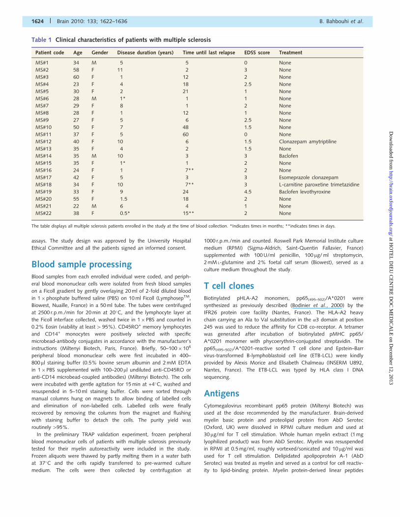
Table 1 Clinical characteristics of patients with multiple sclerosis
Patient code Age Gender Disease duration (years) Time until last relapse EDSS score Treatment
MS#1 34 M 5 5 0 None
MS#2 58 F 11 2 3 None
MS#3 60 F 1 12 2 None
MS#4 23 F 4 18 2.5 None
MS#5 30 F 2 21 1 None
MS#6 28 M 1* 1 1 None
MS#7 29 F 8 1 2 None
MS#8 28 F 1 12 1 None
MS#9 27 F 5 6 2.5 None
MS#10 50 F 7 48 1.5 None
MS#11 37 F 5 60 0 None
MS#12 40 F 10 6 1.5 Clonazepam amytriptiline
MS#13 35 F 4 2 1.5 None
MS#14 35 M 10 3 3 Baclofen
MS#15 35 F 1* 1 2 None
MS#16 24 F 1 7** 2 None
MS#17 42 F 5 3 3 Esomeprazole clonazepam
MS#18 34 F 10 7** 3 L-carnitine paroxetine trimetazidine
MS#19 33 F 9 24 4.5 Baclofen levothyroxine
MS#20 55 F 1.5 18 2 None
MS#21 22 M 6 4 1 None
MS#22 38 F 0.5* 15** 2 None
The table displays all multiple sclerosis patients enrolled in the study at the time of blood collection. *Indicates times in months; **indicates times in days.
1624 | Brain 2010: 133; 1622–1636 B. Bahbouhi et al.
at HO
TE
L D
IEU
CE
NT
RE
DO
C M
ED
ICA
LE
on Decem
ber 12, 2013http://brain.oxfordjournals.org/
Dow
nloaded from
were previously described (Berthelot et al., 2008) and were all
synthesized (Mimotopes, Clayton Australia) and dissolved in 4%
dimethyl sulphoxide. All peptides were used at 10 mg/ml for cell
stimulation.
Myelin labelling and endocytosisWhole myelin extract (1 mg) was washed in 1� PBS by centri-
fugation at 2500 r.p.m. for 5 min, then resuspended in
N-Hydroxysuccinimido-biotin solution (5 mM in 1�PBS) (Perbio
Science France SAS, Brebieres, France), vortexed for 20–30 s and incu-
bated for 30 min at 37�C. Excess N-Hydroxysuccinimido-biotin was
removed by washing. The biotinylated myelin pellet was then labelled
with fluorochrome–streptavidin conjugates (Beckman Coulter,
Miami, FL, USA) for 30 min at 37�C. The tubes were centrifuged to
recover labelled-myelin pellet, washed in 1�PBS by centrifugation
(2500 r.p.m. for 1 min repeated three times) to eliminate free fluoro-
chromes and finally resuspended in 1 ml 1�PBS. Next, 10 mg/ml of
labelled myelin was added to 1�105 enriched CD14+ monocytes in
100ml RPMI overnight in the presence or absence of 10 mM pheny-
larsine oxide (APO) (Sigma-Aldrich). Non-labelled myelin or cells
incubated alone served as negative controls. Myelin-fed monocytes
were washed at least three times in 1� PBS containing 2 mM EDTA
(Sigma-Aldrich) to eliminate free/surface-adsorbed myelin, permeabi-
lized in 1�PERMFIX solution (BD Biosciences, Le-Pont-de-Claix,
France) and analysed on a flow cytometer (LSRII, BD Biosciences).
The background signal was defined by cells incubated alone or with
unlabelled myelin. Monocytes phagocyting myelin were characterized
by the intracellular presence of the marker initially used to label
myelin. In other assays, monocytes were pre-incubated with whole
myelin overnight, myelin excess was eliminated by repeated washing
steps, and then monocytes were analysed for the expression of
HLA-DR.
TRAP assayThe TRAP or trogocytosis protocol was performed as previously
described (Beadling and Slifka, 2006; Daubeuf et al., 2006) but with
modifications to optimize the direct ex vivo detection and quantifica-
tion of myelin-autoreactive T cells in human blood samples. Purified
CD14+ monocytes (3�105 from each patient with multiple sclerosis
and healthy individual) were incubated with antigens overnight (for
peptides) or 48 h (myelin and soluble proteins) in 2% foetal calf
serum-RPMI in 96 round well plates. Meanwhile, the autologous
CD45RO+ T cells purified on the same day were maintained under
resting conditions in culture medium while the monocytes were
‘fed’ with myelin. Antigen-sensitized monocytes were then washed
twice at 2500 r.p.m. for 40 s, biotinylated with 50 ml of 5 mM
N-Hydroxysuccinimido-biotin for 30 min at 37�C, then washed and
labelled with fluorochrome–strepdavidin conjugates for 30 min at
37�C. Labelled monocytes were mixed with 3�105 purified
CD45RO+ lymphocytes in the presence of replenished antigens, cen-
trifuged at 2500 r.p.m. for 40 s to promote cell–cell contacts, and
incubated overnight. At the end of the incubation, co-cultures in the
plates were centrifuged to discard culture medium, and the cell pellets
were resuspended in 2 mM EDTA in 1�PBS containing the labelling
antibodies (200 ml volumes). The plate contents were then transferred
to clean tubes for cytometer reading, vortexed for 20–30 s to disrupt
conjugates and antibodies left to bind for 20 min at +4�C during shak-
ing. The lymphocytes, identified on the basis of their Forward Scatter
and Side Scatter properties and by CD3 staining, were then gated and
analysed for the acquisition of monocyte-fluorescence expressed as
[TRAP (%) = number of fluorescent T cells/total number of gated
T cells�100]. At least 20 000 events were recorded for each sample
tested.
For TRAP assay blocking experiments, fluorochrome-labelled
monocytes were incubated with 500 ng/ml of mouse polyclonal
anti-HLA-II (TU39, BD Biosciences) for 1 h at 37�C or purified
mouse immunoglobulin G. The CD45RO+ enriched cell fraction was
then added and the TRAP assay performed as described above.
In the TRAP experiments using the pp65-tetramer-sorted T cells
(responder) and ETB-LCL (antigen presenting cells), ETB-LCL were
biotinylated with 1 mM N-Hydroxysuccinimido-biotin for 30 min,
washed, labelled with streptavidin–fluorochrome conjugates for
30 min and finally pulsed with 10 mM pp65-derived peptide at 37�C
for 1 h. For the TRAP reaction, labelled and pp65-peptide loaded
ETB-LCL were mixed with pp65(495–502)/A*0201 T cells at a ratio of
1 : 4 for 1 h. An anti-CD3 T cell marker, together with pp65-tetramer,
were used to identify pp65(495–502)/A*0201 T cells and to measure
TRAP frequency.
Flow cytometry immunostainingThe human antibodies were conjugated to phycoerythrin, fluorescein
isothiocyanate, peridinin chlorophyll protein, cy-chrome phycoerythrin
or allophycocyanin (antigen presenting cells) including anti-CD4 (clone
SK3), anti-CD8 (clone 53-1.7) anti-CD45RO (clone UCHL1), anti-CD3
(clone HIT3a), anti-CD8 (clone SK-1), anti-CD25 (clone 2A3),
anti-CD62L (clone DREG-56), anti-CD95 (DX2), anti-HLA-DR [clone
L2-43 (G46-6)] and the unlabelled anti-HLA-II antibody (clone TU39).
All these antibodies were purchased from BD Biosciences.
Anti-CD40L-phycoerythrin conjugate was from Beckman Coulter.
Proliferation assayEquivalent numbers of CD45RO+ T cells from CMV+ and CMV�
normal donors (3� 105) were loaded with carboxy-fluorescein
succinimidyl ester (5 mM) for 15 min at 37�C, washed three times
and co-cultured with 1� 105 syngeneic CD14+ monocytes (1/3
ratio) in the presence or absence of CMV-pp65 recombinant protein
for 3 days. Proliferation in collected co-cultures was defined as the loss
rate of the carboxy-fluorescein succinimidyl ester dye in gated CD3+
T cells by comparison to those cultured without the antigen (used to
set the background quadrants). Cells were stained for the activation
marker CD25 to ensure proliferating cells are activated (by measure-
ment of CD25bright expression).
ELISPOT assayELISPOT assay was performed under TRAP conditions by
co-incubation of myelin-sensitized purified monocytes (1�105) with
3� 105 CD45RO+ lymphocytes in each ELISPOT well plate (Thermo
scientific, Rockford, USA), in the presence or absence of whole myelin
extract, and incubated overnight (longer incubations resulted in
a stronger background signal). As an additional control, CD45RO+
lymphocytes were cultured separately in the presence or absence of
whole myelin extract. The detection of gamma-interferon (IFN�)-
producing cell spots was performed in accordance with the manufac-
turer’s instructions. The spots were counted by the ELISPOT reader
system (AID, Strassberg, Germany) as previously described (Berthelot
Anti-myelin T cell frequency in multiple sclerosis Brain 2010: 133; 1622–1636 | 1625
at HO
TE
L D
IEU
CE
NT
RE
DO
C M
ED
ICA
LE
on Decem
ber 12, 2013http://brain.oxfordjournals.org/
Dow
nloaded from
et al., 2008). The ELISPOT positive reactivity of the co-cultures against
whole myelin extract was defined as a number of spots at least three
times the global backgrounds from co-cultures incubated alone and
from myelin-stimulated CD45RO+ lymphocytes cultured alone. In
these assays, whole myelin extract added alone without the cells or
CD14+ monocytes cultured alone with whole myelin extract did
not form characteristic IFN�-spots after stringent washing of the
ELISPOT filters.
In addition, the ELISPOT assay was also performed by overnight
incubation of 5�105 total peripheral blood mononuclear cells with
or without the same dose of whole myelin extract, followed by spot
revelation performed as described above.
Statistical analysisThe two-tailed non-parametric Mann–Whitney test was used for
comparison between patients with multiple sclerosis and healthy
individuals. Kruskal–Wallis or Friedman tests were used for comparison
of more than two groups. Significant differences were set at *P50.05,
**P50.01, ***P50.001 (GraphPad Prism 4.0).
Results
TRAP detects antigen-specific T cellresponse in selected individualsFirst, we analysed the TRAP response in individuals with
pre-established reactivity against cytomegalovirus or myelin basic
protein-derived MHC class I-restricted peptides.
CMV+ individualsSupplementary Fig. 1 shows T cell proliferation in CMV+ donors
with a median of 5% (range 5–8%) of their CD45RO+CD3+
T cells becoming Carboxy-Fluorescein Succinimidyl Ester
(CSFE)low in the presence of CMV-pp65 protein compared to
only 0.5% in CMV� control individuals (range 0.5–2%).
Furthermore, only CD45RO+ T cells from CMV+ individuals were
IFN�-ELISPOT positive in the presence of CMV-pp65 as shown by
selected examples of CMV� and CMV+ ELISPOT results (represen-
tative inserts, Supplementary Fig. 1A) and by pooled data from all
tested individuals (Supplementary Fig. 2C). Supplementary Fig. 2A
and B shows that only CD45RO+ lymphocytes from CMV+ indi-
viduals (median 3.7%, range 3–5.8%) displayed a higher CD3+
TRAP signal in the presence of pp65 compared to CMV� controls
(median 0.2%, range 0–0.2%). The ELISPOT assay also detected
a greater response in CMV+ individuals (median 86 IFN�-spots,
range 38–385) compared to their CMV� counterparts (median 57
IFN�-spots, range 13–60, P50.008, Supplementary Fig. 2C)
although at a 94-fold lower frequency compared to
the TRAP-based estimations. There was, however, a signifi-
cant correlation between the TRAP and ELISPOT assays
(Supplementary Fig. 2D, r2= 0.82, P50.01). Finally, pp65-
tetramer sorted T cells stimulated by pp65 peptide-pulsed
ETB-LCL (used here as antigen presenting cells) did exhibit a
6.8-fold increase in TRAP signal (68%) compared to stimulation
by non-pulsed ETB-LCL (10%) (Supplementary Fig. 3).
Individuals with pre-establishedCD8+ T cells reactive to MHC classI-restricted myelin basic proteinpeptidesWe first looked whether biotinylated and fluorescently labelled
myelin (myelin-F*) was internalized in the CD14+ monocytes
used for T cell stimulation (Fig. 1A). As a negative control
(designated by no-myelin-F*), non-labelled whole myelin extract
was added to CD14+ monocytes in order to suppress the auto-
fluorescence signals that might result either from cells and/or
whole myelin extract, or a combination of both. Figure 1A
shows that myelin was indeed phagocytosed by monocytes
(median 15%, range 20–25%, P50.01) while myelin internaliza-
tion was nil in the presence of the phagocytosis inhibitor
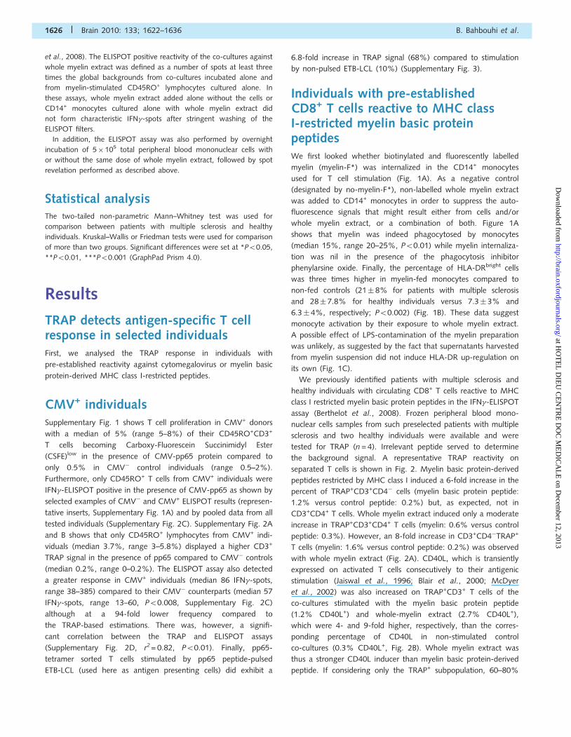
phenylarsine oxide. Finally, the percentage of HLA-DRbright cells
was three times higher in myelin-fed monocytes compared to
non-fed controls (21� 8% for patients with multiple sclerosis
and 28� 7.8% for healthy individuals versus 7.3� 3% and
6.3� 4%, respectively; P50.002) (Fig. 1B). These data suggest
monocyte activation by their exposure to whole myelin extract.
A possible effect of LPS-contamination of the myelin preparation
was unlikely, as suggested by the fact that supernatants harvested
from myelin suspension did not induce HLA-DR up-regulation on
its own (Fig. 1C).
We previously identified patients with multiple sclerosis and
healthy individuals with circulating CD8+ T cells reactive to MHC
class I restricted myelin basic protein peptides in the IFN�-ELISPOT
assay (Berthelot et al., 2008). Frozen peripheral blood mono-
nuclear cells samples from such preselected patients with multiple
sclerosis and two healthy individuals were available and were
tested for TRAP (n = 4). Irrelevant peptide served to determine
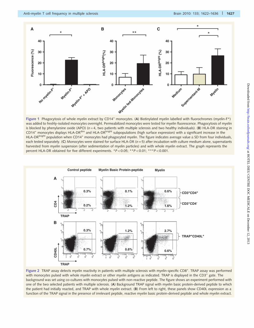
the background signal. A representative TRAP reactivity on
separated T cells is shown in Fig. 2. Myelin basic protein-derived
peptides restricted by MHC class I induced a 6-fold increase in the
percent of TRAP+CD3+CD4� cells (myelin basic protein peptide:
1.2% versus control peptide: 0.2%) but, as expected, not in
CD3+CD4+ T cells. Whole myelin extract induced only a moderate
increase in TRAP+CD3+CD4+ T cells (myelin: 0.6% versus control
peptide: 0.3%). However, an 8-fold increase in CD3+CD4�TRAP+
T cells (myelin: 1.6% versus control peptide: 0.2%) was observed
with whole myelin extract (Fig. 2A). CD40L, which is transiently
expressed on activated T cells consecutively to their antigenic
stimulation (Jaiswal et al., 1996; Blair et al., 2000; McDyer
et al., 2002) was also increased on TRAP+CD3+ T cells of the
co-cultures stimulated with the myelin basic protein peptide
(1.2% CD40L+) and whole-myelin extract (2.7% CD40L+),
which were 4- and 9-fold higher, respectively, than the corres-
ponding percentage of CD40L in non-stimulated control
co-cultures (0.3% CD40L+, Fig. 2B). Whole myelin extract was
thus a stronger CD40L inducer than myelin basic protein-derived
peptide. If considering only the TRAP+ subpopulation, 60–80%
1626 | Brain 2010: 133; 1622–1636 B. Bahbouhi et al.
at HO
TE
L D
IEU
CE
NT
RE
DO
C M
ED
ICA
LE
on Decem
ber 12, 2013http://brain.oxfordjournals.org/
Dow
nloaded from
No mye
lin-F
*
Myelin
-F*
Myelin
-F* +
APO
0
10
20
30
40
Flu
ore
scen
ce (
%)
Monocyte
s
Myelin
fed-M
onocyte
s0
10
20
30
40
HL
A-D
Rb
rig
ht (
%)
A B* **
Mediu
m
Supernan
tant-M
Myelin
0
10
20
30
40
HL
A-D
Rb
rig
ht (
%)
C **
Figure 1 Phagocytosis of whole myelin extract by CD14+ monocytes. (A) Biotinylated myelin labelled with fluorochromes (myelin-F*)
was added to freshly-isolated monocytes overnight. Permeabilized monocytes were tested for myelin fluorescence. Phagocytosis of myelin
is blocked by phenylarsine oxide (APO) (n = 4, two patients with multiple sclerosis and two healthy individuals). (B) HLA-DR staining in
CD14+ monocytes displays HLA-DRdim and HLA-DRbright subpopulations (high surface expression) with a significant increase in the
HLA-DRbright population when CD14+ monocytes had phagocyted myelin. The figure indicates average value� SD from four individuals,
each tested separately. (C) Monocytes were stained for surface HLA-DR (n = 5) after incubation with culture medium alone, supernatants
harvested from myelin suspension (after sedimentation of myelin particles) and with whole myelin extract. The graph represents the
percent HLA-DR obtained for five different experiments. *P50.05; **P50.01; ***P50.001.
Control peptide
0.3%
0.2%
0.1%
1.2%
0.6%
1.6%
TRAP
TRAP
Myelin Basic Protein-peptide Myelin
CD
40L
CD
4
CD3+CD4+
CD3+CD4-
TRAP+CD40L+
0.3%
0.7%
1.2%
0.6%
2.7%
0.6%
A
B
Figure 2 TRAP assay detects myelin reactivity in patients with multiple sclerosis with myelin-specific CD8+. TRAP assay was performed
with monocytes pulsed with whole myelin extract or other myelin antigens as indicated. TRAP is displayed in the CD3+ gate. The
background was set using co-cultures with monocytes pulsed with non-reactive peptide. The figure shows an experiment performed with
one of the two selected patients with multiple sclerosis. (A) Background TRAP signal with myelin basic protein-derived peptide to which
the patient had initially reacted, and TRAP with whole myelin extract. (B) From left to right, these panels show CD40L expression as a
function of the TRAP signal in the presence of irrelevant peptide, reactive myelin basic protein-derived peptide and whole myelin extract.
Anti-myelin T cell frequency in multiple sclerosis Brain 2010: 133; 1622–1636 | 1627
at HO
TE
L D
IEU
CE
NT
RE
DO
C M
ED
ICA
LE
on Decem
ber 12, 2013http://brain.oxfordjournals.org/
Dow
nloaded from
was CD40L+ with myelin basic protein peptide and whole myelin
by comparison to 33% CD40L+ in the absence of myelin stimu-
lation. CD40L induction in this case was in agreement with a
previous report demonstrating the induction of CD40L expression
by antigen-specific T cell interactions with antigen presenting cells
(Frensch et al., 2005).
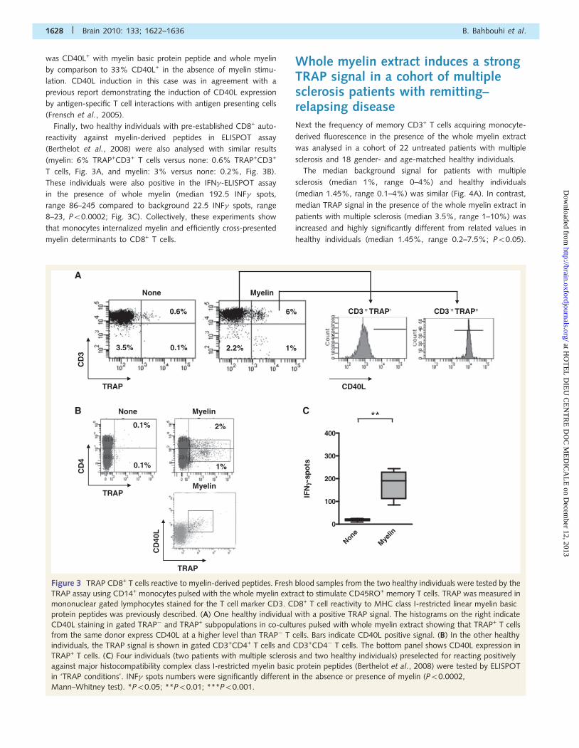
Finally, two healthy individuals with pre-established CD8+ auto-
reactivity against myelin-derived peptides in ELISPOT assay
(Berthelot et al., 2008) were also analysed with similar results
(myelin: 6% TRAP+CD3+ T cells versus none: 0.6% TRAP+CD3+
T cells, Fig. 3A, and myelin: 3% versus none: 0.2%, Fig. 3B).
These individuals were also positive in the IFN�-ELISPOT assay
in the presence of whole myelin (median 192.5 INF� spots,
range 86–245 compared to background 22.5 INF� spots, range
8–23, P50.0002; Fig. 3C). Collectively, these experiments show
that monocytes internalized myelin and efficiently cross-presented
myelin determinants to CD8+ T cells.
Whole myelin extract induces a strongTRAP signal in a cohort of multiplesclerosis patients with remitting–relapsing diseaseNext the frequency of memory CD3+ T cells acquiring monocyte-
derived fluorescence in the presence of the whole myelin extract
was analysed in a cohort of 22 untreated patients with multiple
sclerosis and 18 gender- and age-matched healthy individuals.
The median background signal for patients with multiple
sclerosis (median 1%, range 0–4%) and healthy individuals
(median 1.45%, range 0.1–4%) was similar (Fig. 4A). In contrast,
median TRAP signal in the presence of the whole myelin extract in
patients with multiple sclerosis (median 3.5%, range 1–10%) was
increased and highly significantly different from related values in
healthy individuals (median 1.45%, range 0.2–7.5%; P50.05).
CD
4
0.1%
0.1%
1%
2%
A
CB None Myelin
MyelinTRAP
None
Myelin
0
100
200
300
400
IFNγγ-
spo
ts
**
MyelinNone
0.6% 6%
0.1%3.5% 1%2.2%
CD3 + TRAP- CD3 + TRAP+
CD
3
TRAP
CD
40L
TRAP
CD40L
Figure 3 TRAP CD8+ T cells reactive to myelin-derived peptides. Fresh blood samples from the two healthy individuals were tested by the
TRAP assay using CD14+ monocytes pulsed with the whole myelin extract to stimulate CD45RO+ memory T cells. TRAP was measured in
mononuclear gated lymphocytes stained for the T cell marker CD3. CD8+ T cell reactivity to MHC class I-restricted linear myelin basic
protein peptides was previously described. (A) One healthy individual with a positive TRAP signal. The histograms on the right indicate
CD40L staining in gated TRAP� and TRAP+ subpopulations in co-cultures pulsed with whole myelin extract showing that TRAP+ T cells
from the same donor express CD40L at a higher level than TRAP� T cells. Bars indicate CD40L positive signal. (B) In the other healthy
individuals, the TRAP signal is shown in gated CD3+CD4+ T cells and CD3+CD4� T cells. The bottom panel shows CD40L expression in
TRAP+ T cells. (C) Four individuals (two patients with multiple sclerosis and two healthy individuals) preselected for reacting positively
against major histocompatibility complex class I-restricted myelin basic protein peptides (Berthelot et al., 2008) were tested by ELISPOT
in ‘TRAP conditions’. INF� spots numbers were significantly different in the absence or presence of myelin (P50.0002,
Mann–Whitney test). *P50.05; **P50.01; ***P50.001.
1628 | Brain 2010: 133; 1622–1636 B. Bahbouhi et al.
at HO
TE
L D
IEU
CE
NT
RE
DO
C M
ED
ICA
LE
on Decem
ber 12, 2013http://brain.oxfordjournals.org/
Dow
nloaded from
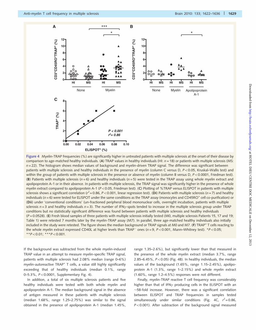
If the background was subtracted from the whole myelin-induced
TRAP value in an attempt to measure myelin-specific TRAP signal,
patients with multiple sclerosis had 2.08% median (range 0–6%)
myelin-autoreactive TRAP+ T cells, a value still highly significantly
exceeding that of healthy individuals (median 0.1%, range
0–5.3%, P50.0001, Supplementary Fig. 4).
In addition, a total of six multiple sclerosis patients and five
healthy individuals were tested with both whole myelin and
apolipoprotein A-1. The median background signal in the absence
of antigen measured in these patients with multiple sclerosis
(median 1.68%, range 1.25–2.75%) was similar to the signal
obtained in the presence of apolipoprotein A-1 (median 1.45%,
range 1.35–2.6%), but significantly lower than that measured in
the presence of the whole myelin extract (median 3.7%, range
2.85–6.45%, P50.05) (Fig. 4B). In healthy individuals, the median
values of the background (1.65%, range 1.15–2.45%), apolipo-
protein A-1 (1.3%, range 1–2.15%) and whole myelin extract
(1.60%, range 1.2–4.5%) responses were not different.
Finally, myelin-TRAP reactive T cell frequency was considerably
higher than that of IFN� producing cells in the ELISPOT with an
�58-fold increase. However, there was a significant correlation
between ELISPOT and TRAP frequencies in samples tested
simultaneously under similar conditions (Fig. 4C, r2 = 0.86,
P50.001). After subtraction of the background signal measured
HI MS HI MS0
2
4
6
8
10
12
CD
3+ CD
45R
O+ T
RA
P+
(%)
None Myelin None Myelin
0.00 0.02 0.04 0.06 0.08 0.100
2
4
6
8
ELISPOT+ (%)
TR
AP
+ (%
)
P < 0.001r2= 0.86
BA
C
****
HI MS HI MS HI MS0
2
4
6
8
10
12
CD
3+ CD
45R
O+ T
RA
P+
(%)
ApolipoproteinA-1
*
Figure 4 Myelin-TRAP frequencies (%) are significantly higher in untreated patients with multiple sclerosis at the onset of their disease by
comparison to age-matched healthy individuals. (A) TRAP values in healthy individuals (HI: n = 18) or patients with multiple sclerosis (MS:
n = 22). The histogram shows median values of background and myelin-driven TRAP signal. The difference was significant between
patients with multiple sclerosis and healthy individuals in the presence of myelin (column C versus D, P50.05, Kruskal–Wallis test) and
within the group of patients with multiple sclerosis in the presence or absence of myelin (column B versus D, P50.0001, Friedman test).
(B) Patients with multiple sclerosis (n = 6) and healthy individuals (n = 5) were tested in the TRAP assay using whole myelin extract and
apolipoprotein A-1 or in their absence. In patients with multiple sclerosis, the TRAP signal was significantly higher in the presence of whole
myelin extract compared to apolipoprotein A-1 (P50.05, Friedman test). (C) Plotting of %TRAP versus ELISPOT in patients with multiple
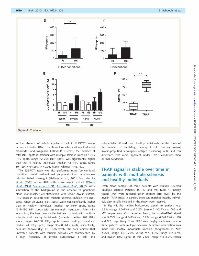
sclerosis shows a significant correlation (r2= 0.86, P50.001, linear regression test). (Di) Patients with multiple sclerosis (n = 7) and healthy
individuals (n = 6) were tested for ELISPOT under the same conditions as the TRAP assay (monocytes and CD45RO+ cell co-purification) or
(Dii) under ‘conventional conditions’ (un-fractioned peripheral blood mononuclear cells, overnight incubation, patients with multiple
sclerosis n = 3 and healthy individuals n = 3). The number of IFN�-spots tended to increase in the multiple sclerosis group under TRAP
conditions but no statistically significant difference was found between patients with multiple sclerosis and healthy individuals
(P = 0.0528). (E) Fresh blood samples of three patients with multiple sclerosis initially tested (M0, multiple sclerosis Patients 15, 17 and 19;
Table 1) were retested 7 months later by the myelin-TRAP assay (M7). In parallel, three age-matched healthy individuals also initially
included in the study were retested. The figure shows the median background or TRAP signals at M0 and M7. (F) TRAP+ T cells reacting to
the whole myelin extract expressed CD40L at higher levels than TRAP� ones (n = 9, P50.001, Mann–Whitney test). *P50.05;
**P50.01; ***P50.001.
Anti-myelin T cell frequency in multiple sclerosis Brain 2010: 133; 1622–1636 | 1629
at HO
TE
L D
IEU
CE
NT
RE
DO
C M
ED
ICA
LE
on Decem
ber 12, 2013http://brain.oxfordjournals.org/
Dow
nloaded from
in the absence of whole myelin extract in ELISPOT assays
performed under TRAP conditions (co-cultures of myelin-loaded
monocytes and syngeneic CD45RO+ T cells), the number of
total INF� spots in patients with multiple sclerosis (median 124.5
INF� spots, range 72–265 INF� spots) was significantly higher
than that in healthy individuals (median 42 INF� spots, range
10–129 INF� spots, P50.05, Mann–Whitney) (Fig. 4D).
The ELISPOT assay was also performed using ‘conventional
conditions’: total un-fractioned peripheral blood mononuclear
cells incubated overnight (Hellings et al., 2001; Van der Aa
et al., 2003) or for 48 h with whole myelin extract (Olsson
et al., 1990; Sun et al., 1991; Andersson et al., 2002). After
subtraction of the background in the absence of peripheral
blood mononuclear cell-stimulation with whole myelin extract,
INF� spots in patients with multiple sclerosis (median 141 INF�
spots, range 75–222.5 INF� spots) were not significantly higher
than in healthy individuals (median 49 INF� spots, range
43.5–130 INF� spots) with an overnight incubation. After 48 h
incubation, the trend was similar between patients with multiple
sclerosis and healthy individuals (patients: median 203 INF�
spots, range 44–258 INF� spots versus healthy individuals:
median 44 INF� spots, range 38–90 INF� spots, respectively,
data not shown) (Fig. 4D). Collectively, the data indicate that
untreated patients with multiple sclerosis are characterized by
a high frequency of myelin autoreactive T cells and
substantially differed from healthy individuals on the basis of
the number of circulating memory T cells reacting against
myelin-prepulsed autologous antigen presenting cells; and this
difference was more apparent under TRAP conditions than
normal conditions.
TRAP signal is stable over time inpatients with multiple sclerosisand healthy individualsFresh blood samples of three patients with multiple sclerosis
(multiple sclerosis Patients 15, 17 and 19; Table 1) initially
tested (M0) were retested seven months later (M7) by the
myelin-TRAP assay. In parallel, three age-matched healthy individ-
uals also initially included in the study were retested.
In Fig. 4E, the median background signals for patients was
1.8% (range 1.5–3%) and 2.2% (range 2.1–2.9%) at M0 and
M7, respectively. On the other hand, the myelin-TRAP signal
was 5.65% (range 4.8–7%) and 6.9% (range 6.6–8.3%) at M0
and M7, respectively. Thus, TRAP was roughly stable over time in
three patients with multiple sclerosis. A similar observation was
made for healthy individuals (median background at M0:
2.95%, range 1.8–3.25% versus M7: 3.6%, range 0.7–3.7%
and myelin TRAP-signal at M0: 2.6%, range 1.8–3.9% versus
TRAP conditions Conventional
None Myelin None Myelin
M0 M7
TRAP- TRAP+
0
25
50
75
100
CD
40L
+ (%
)
D
E F **
HI MS HI MS HI MS HI MS0
2
4
6
8
10
12
CD
3+ CD
45R
O+ T
RA
P+
(%)
HI MS0
100
200
300
400
IFN
g-sp
ots
HI MS0
100
200
300
400
IFN
g-sp
ots
i) ii)*
Figure 4 Continued.
1630 | Brain 2010: 133; 1622–1636 B. Bahbouhi et al.
at HO
TE
L D
IEU
CE
NT
RE
DO
C M
ED
ICA
LE
on Decem
ber 12, 2013http://brain.oxfordjournals.org/
Dow
nloaded from
M7: 3.3%, range 1.1–4.9%). At both time points, the
myelin-TRAP signal seemed to be higher in patients with multiple
sclerosis compared to healthy individuals.
Whole myelin TRAP+ cells are activatedmemory T cells and anti-MHC class IIblocking antibodies prevent TRAPinduction in CD4+ T lymphocytesWe have already mentioned that TRAP+ T cells predominantly
express CD40L in patients with multiple sclerosis and healthy
individuals tested in preliminary experiments (Figs 2 and 3). We
next extended the phenotypic characterization of TRAP+ T cells to
the 22 patients with multiple sclerosis of the study cohort in add-
ition to other activation markers (CD62L, CD95 and HLA-DR).
As suggested by the preliminary data, CD40L discriminated
TRAP+ from TRAP� memory T cells reactive to whole myelin
extract (Fig. 4F; median 82.5%, range 60–96%, CD40L+TRAP+
versus median 12.5%, range 2–27%, CD40L+TRAP�; P50.008).
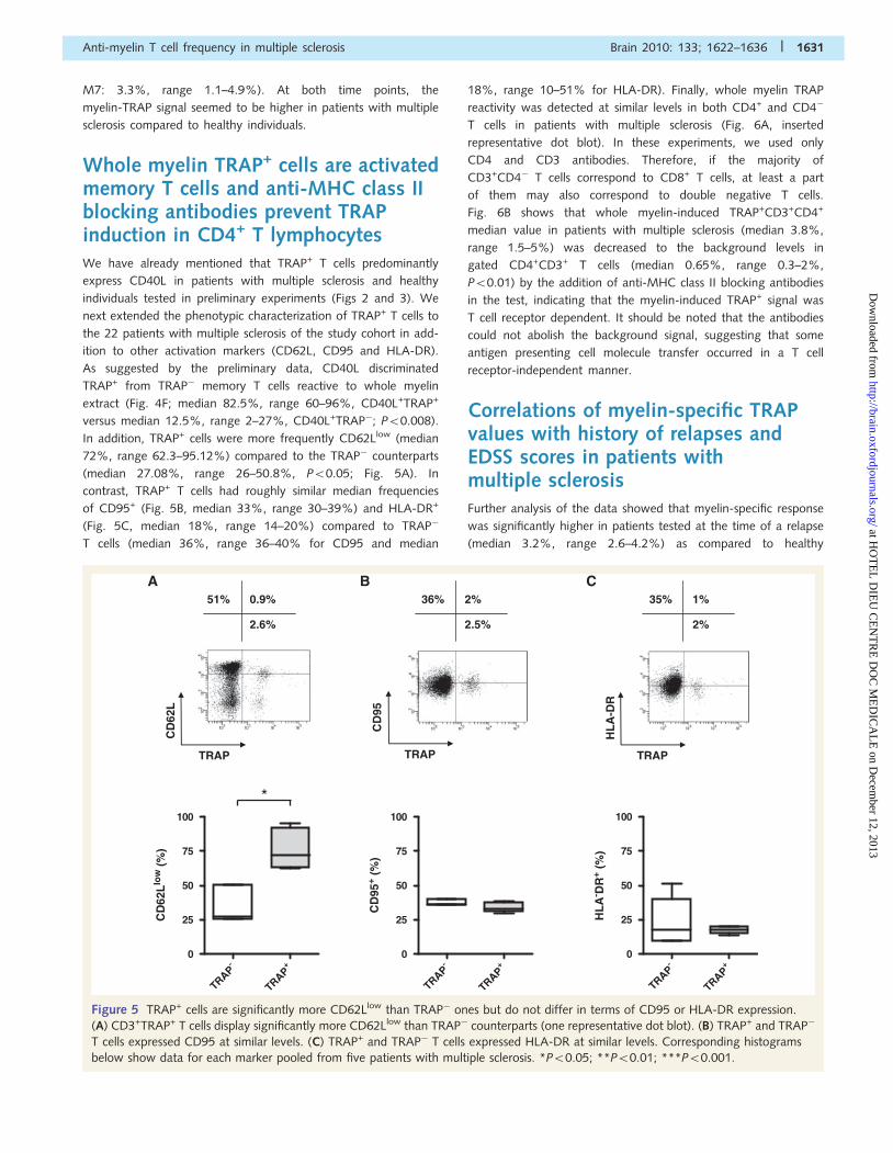
In addition, TRAP+ cells were more frequently CD62Llow (median
72%, range 62.3–95.12%) compared to the TRAP� counterparts
(median 27.08%, range 26–50.8%, P50.05; Fig. 5A). In
contrast, TRAP+ T cells had roughly similar median frequencies
of CD95+ (Fig. 5B, median 33%, range 30–39%) and HLA-DR+
(Fig. 5C, median 18%, range 14–20%) compared to TRAP�
T cells (median 36%, range 36–40% for CD95 and median
18%, range 10–51% for HLA-DR). Finally, whole myelin TRAP
reactivity was detected at similar levels in both CD4+ and CD4�
T cells in patients with multiple sclerosis (Fig. 6A, inserted
representative dot blot). In these experiments, we used only
CD4 and CD3 antibodies. Therefore, if the majority of
CD3+CD4� T cells correspond to CD8+ T cells, at least a part
of them may also correspond to double negative T cells.
Fig. 6B shows that whole myelin-induced TRAP+CD3+CD4+
median value in patients with multiple sclerosis (median 3.8%,
range 1.5–5%) was decreased to the background levels in
gated CD4+CD3+ T cells (median 0.65%, range 0.3–2%,
P50.01) by the addition of anti-MHC class II blocking antibodies
in the test, indicating that the myelin-induced TRAP+ signal was
T cell receptor dependent. It should be noted that the antibodies
could not abolish the background signal, suggesting that some
antigen presenting cell molecule transfer occurred in a T cell
receptor-independent manner.
Correlations of myelin-specific TRAPvalues with history of relapses andEDSS scores in patients withmultiple sclerosisFurther analysis of the data showed that myelin-specific response
was significantly higher in patients tested at the time of a relapse
(median 3.2%, range 2.6–4.2%) as compared to healthy
51% 0.9%
2.6%
36% 2%
2.5%
35% 1%
2%
CBA
CD
62L
TRAP
CD
95
TRAP
HL
A-D
R
TRAP
-
TRAP+
TRAP
0
25
50
75
100
CD
62L
low
(%
)
-
TRAP+
TRAP
0
25
50
75
100
CD
95+
(%)
-
TRAP+
TRAP
0
25
50
75
100
HL
A- D
R+
(%)
*
Figure 5 TRAP+ cells are significantly more CD62Llow than TRAP� ones but do not differ in terms of CD95 or HLA-DR expression.
(A) CD3+TRAP+ T cells display significantly more CD62Llow than TRAP� counterparts (one representative dot blot). (B) TRAP+ and TRAP�
T cells expressed CD95 at similar levels. (C) TRAP+ and TRAP� T cells expressed HLA-DR at similar levels. Corresponding histograms
below show data for each marker pooled from five patients with multiple sclerosis. *P50.05; **P50.01; ***P50.001.
Anti-myelin T cell frequency in multiple sclerosis Brain 2010: 133; 1622–1636 | 1631
at HO
TE
L D
IEU
CE
NT
RE
DO
C M
ED
ICA
LE
on Decem
ber 12, 2013http://brain.oxfordjournals.org/
Dow
nloaded from
individuals (median 0.1%, range 0–5.3%, P50.001, Kruskal–
Wallis test). This response in the remaining patients with multiple
sclerosis seen during a remission period was also significantly
higher (median 1.55%, range 0–6%, P50.01, Kruskal–Wallis
test) than in healthy individuals (Fig. 7A). In addition, if patients
with multiple sclerosis are ranked according to EDSS scores, the
data suggest that those with the highest EDSS scores also had the
highest level of myelin autoreactive T cell frequency (median 3%,
range 2.6–6%) compared to those with relatively lower EDSS
scores (median 1.6%, range 0–4.24%) or to controls (median
0.1%, range 0–5.3%; P50.0001; Fig. 7B). Both multiple sclerosis
subgroups still had a median frequency of memory autoreactive T
cells to myelin significantly higher than healthy individuals. No
statistical significance could be found between age-matched
female and male patients with multiple sclerosis in terms of
myelin-specific TRAP values.
DiscussionThe question of whether or not peripheral blood myelin-
autoreactive T cells are more numerous in peripheral blood of
patients with multiple sclerosis compared to healthy individuals is
an important issue when understanding the disease process. Most
studies have reported similar frequencies of autoreactive T cells in
groups of patients with multiple sclerosis and healthy individuals
(Zhang et al., 1994; Diaz-Villoslada et al., 1999; Hellings et al.,
2001, 2002; Tejada-Simon et al., 2001; Van der Aa et al., 2003;
Bielekova et al., 2004) including our own on CD8+ T cells
(Berthelot et al., 2008). However, some reports have shown
higher frequencies in patients with multiple sclerosis (Sun et al.,
1991; Chou et al., 1992; Pelfrey et al., 2000) or a higher level of
myelin autoreactivity in patients with multiple sclerosis, without a
specific estimate of peripheral blood myelin-autoreactive T cell
frequencies (Kerlero de Rosbo et al., 1997; Zang et al., 2004).
Two important factors might underline these discrepancies: the
type of autoantigens used and the peripheral blood
myelin-autoreactive T cell detection method. In fact, none of
these studies have used whole myelin extract as the autoantigen
but myelin-derived peptides or molecules that probably do not
represent all determinants that could be involved in the disease.
In addition, the ELISPOT or Limiting Dilution Assay readouts only
detect autoreactive T cells secreting a limited number of cytokines
(usually INF-�) or able to proliferate, but not cells secreting other
cytokines or with a high activation threshold. For example,
Bieganowska et al. (1997) reported a frequency of blood T cells
expressing the myelin basic protein p85-99-associated T cell
receptor chain transcripts as high as 1/300, which far exceeded
74% 3%
2%
A
CD
4
TRAP
HI MS HI MS HI MS HI MS0
2
4
6
8
10
12
CD
3+ CD
45R
O+ T
RA
P+
(%)
None Myelin None Myelin
CD4- CD4+
**
B
Isoty
pe
Anti-MHC cl
ass I
I
Isoty
pe
Anti-MHC cl
ass I
I0
2
4
6
8
10
12
CD
3+ CD
4+ CD
45R
O+ T
RA
P+
(%)
None Myelin
**
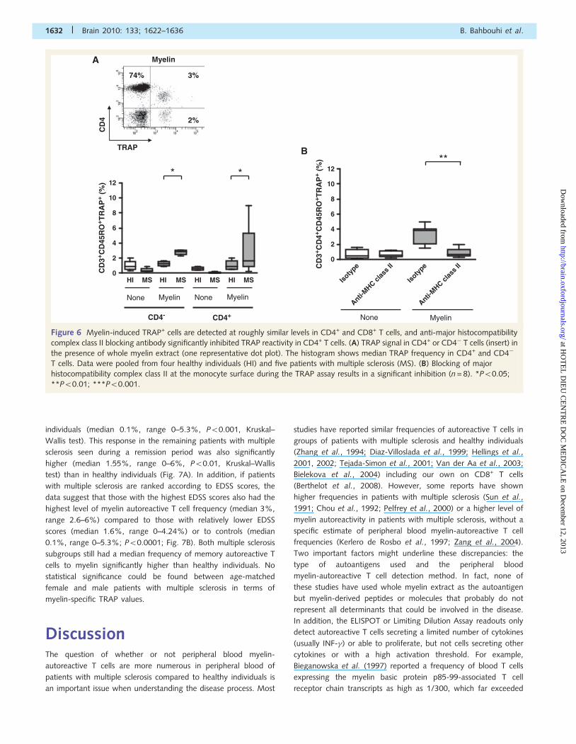
Myelin
Figure 6 Myelin-induced TRAP+ cells are detected at roughly similar levels in CD4+ and CD8+ T cells, and anti-major histocompatibility
complex class II blocking antibody significantly inhibited TRAP reactivity in CD4+ T cells. (A) TRAP signal in CD4+ or CD4� T cells (insert) in
the presence of whole myelin extract (one representative dot plot). The histogram shows median TRAP frequency in CD4+ and CD4�
T cells. Data were pooled from four healthy individuals (HI) and five patients with multiple sclerosis (MS). (B) Blocking of major
histocompatibility complex class II at the monocyte surface during the TRAP assay results in a significant inhibition (n = 8). *P50.05;
**P50.01; ***P50.001.
1632 | Brain 2010: 133; 1622–1636 B. Bahbouhi et al.
at HO
TE
L D
IEU
CE
NT
RE
DO
C M
ED
ICA
LE
on Decem
ber 12, 2013http://brain.oxfordjournals.org/
Dow
nloaded from
the frequencies they calculated by Limiting Dilution Assay
(1� 10�5–10�6) (Bieganowska et al., 1997).
In an attempt to improve the estimates of myelin-autoreactive
T cell frequency, we adopted two novel approaches including the
use of whole myelin extract as the autoantigen and the transfer of
plasma membrane proteins from autologous antigen presenting
cells to T cells upon immune synapse formation, a cytokine and
cell proliferation independent readout which measures T cell
engagement in an immune synapse, an obligatory step for a
T cell immune response (for review see Reichardt et al., 2007)
and allows an estimation of antigen-specific T cell frequency what-
ever the restricting MHC allele (Tomaru et al., 2003; Beadling and
Slifka, 2006; Daubeuf et al., 2006, 2009; Puaux et al., 2006;
Machlenkin et al., 2008). Utilization of whole myelin extract
was suggested by the in vivo observation where monocytes/
macrophages contain myelin determinants in brain tissue during
multiple sclerosis (for review see Lassmann et al., 2007). Boven
et al. (2006) showed that myelin-loaded monocytes express
several anti-inflammatory markers and possibly down-regulate
the immune response in brain tissue.
Because the TRAP assay has been predominantly described in
animal models (Beadling and Slifka, 2006; Daubeuf et al., 2006,
2009), our study conducted in humans included certain validation
steps. We first tested TRAP using CMV-pp65 antigen in individ-
uals with positive CMV serology, and by testing the response
to whole myelin extract in individuals in whom the presence of
circulating CD8+ peripheral blood myelin-autoreactive T cells had
been pre-established by ELISPOT (Berthelot et al., 2008). About
4% blood memory T cells of CMV+ donors were pp65-TRAP
reactive whereas no response was observed in CMV� subjects.
These frequencies in subjects with positive serology at a distance
from the acute disease are within the range of frequencies
reported in human subjects using CMV-specific tetramers
(Gillespie et al., 2000) or by flow cytometry intracellular cytokine
staining (Sylwester et al., 2005). In addition, pp65-TRAP reactivity
correlated to pp65 antigen ability to induce T cell proliferation
(CSFE) or IFN� secretion (ELISPOT). However, pp65-TRAP+
T cell frequencies were 94-fold higher than those measured by
the ELISPOT, suggesting that using IFN� as the unique parameter
to measure the T cell response can result in a gross under-
estimation of the frequency of committed cells, as also reported
recently in animals where the authors have also found comparable
differences in the frequencies estimated by ELISPOT (0.01%)
and TRAP assays (4.77%) (Daubeuf et al., 2009). It is possible
that INF�-negative TRAP+ T cells might express other types
of cytokines (interleukin-4, -17) or might have a regulatory
function. Importantly, TRAP detected a CD8 response both in
the presence of MHC class I restricted peptides and whole
myelin extract in the test, indicating cross-presentation to CD8+
T cells.
Using this experimental strategy in patients with multiple scler-
osis (n = 22) and age/gender-matched healthy individuals (n = 18),
we showed that 3.5% (range 1–10%) of T cells in patients with
multiple sclerosis acquired labelled plasma membrane proteins
from their myelin-pulsed autologous antigen presenting cells,
which strongly exceeded the background signal and that observed
in the group of healthy individuals. When re-tested seven months
after the first analysis, the TRAP signal remained stable over time
in three patients with multiple sclerosis and three healthy individ-
uals, but was still higher in the former. This stability might indicate
continuous stimulation or survival of myelin-specific memory
T cells in patients with multiple sclerosis. The antigen specificity
of the TRAP phenomenon has also been suggested by linear
correlation with tetramer reactivity, dependence upon MHC load-
ing with the specific antigenic peptide (Tomaru et al., 2003;
Beadling and Slifka, 2006; Daubeuf et al., 2006; Machlenkin
HI
No rece
nt rela
pse
Relapse
<1m
onth0
2
4
6
8
10
12
CD
3+ CD
45R
O+ T
RA
P+
(%)
HI
EDSS-Sc <
3
3<EDSS-S
c <5
0
2
4
6
8
10
12
CD
3+ CD
45R
O+ T
RA
P+
(%)
A B
** **
*** ***
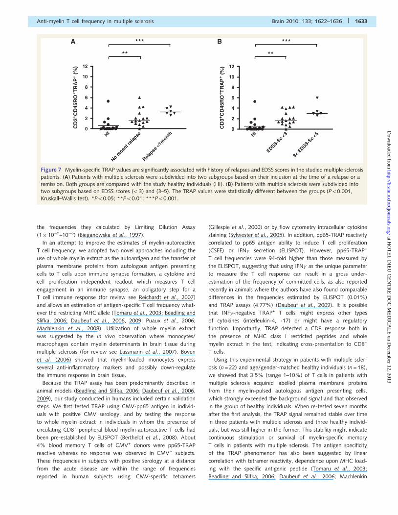
Figure 7 Myelin-specific TRAP values are significantly associated with history of relapses and EDSS scores in the studied multiple sclerosis
patients. (A) Patients with multiple sclerosis were subdivided into two subgroups based on their inclusion at the time of a relapse or a
remission. Both groups are compared with the study healthy individuals (HI). (B) Patients with multiple sclerosis were subdivided into
two subgroups based on EDSS scores (53) and (3–5). The TRAP values were statistically different between the groups (P50.001,
Kruskall–Wallis test). *P50.05; **P50.01; ***P50.001.
Anti-myelin T cell frequency in multiple sclerosis Brain 2010: 133; 1622–1636 | 1633
at HO
TE
L D
IEU
CE
NT
RE
DO
C M
ED
ICA
LE
on Decem
ber 12, 2013http://brain.oxfordjournals.org/
Dow
nloaded from
et al., 2008) and cytotoxic function of isolated TRAP+ T cells
towards target cells pulsed with specific antigenic peptides
(Tomaru et al., 2003; Daubeuf et al., 2006). Despite these studies
suggesting that TRAP is T cell receptor dependent, we found that
MHC blocking did not eradicate the background signal, indicating
that at least some memory T cells interact with monocytes in
a T cell receptor-independent manner. This is also suggested
by the relatively high background signal we observed in the
CMV-tetramer-sorted T cell line. It is thus unlikely that this back-
ground only reflects the physiological size of an endogenous T cell
memory pool against environmental antigens and engaging T cell
receptor-mediated synapses with antigen presenting cells. Such
antigen-independent background signals have also been observed
in other TRAP studies (Kondo et al., 2001; Revy et al., 2001;
Daubeuf et al., 2006, 2009; Hudrisier et al., 2007; LeMaoult
et al., 2007). Because the synapses formed in the absence
of antigen seem to be functional and induce T cell survival
(Kondo et al., 2001; Revy et al., 2001), they should contribute
to the background in both healthy subjects and patients with
multiple sclerosis, and thus cannot explain the myelin-induced
difference between these groups. For these reasons our data
and conclusions were only based on the net increase in TRAP
signal observed in the absence and presence of whole myelin
extract in the test. Interestingly, this antigen-mediated increase
was abolished by blocking MHC class II in CD4+ T cells. In
addition, the background levels were identical in patients with
multiple sclerosis and healthy individuals who thus only differed
after the addition of myelin antigens. TRAP assays were performed
with enriched CD45RO+ T cells rather than un-fractioned
CD14-depleted peripheral blood mononuclear cells to minimize
recruitment of CD45RO� naıve T cells in the test. The importance
of using total myelin extract as antigen was also suggested by the
fact that total myelin extract-induced ELISPOT also discriminated
patients with multiple sclerosis from normal individuals. The
discriminative capacity of the ELISPOT was significant under
TRAP conditions but did not reach significance under ‘conven-
tional conditions’ (despite showing the same trend), probably
because only three patients with multiple sclerosis and three
healthy individuals were tested in this setting. Using a ‘conven-
tional ELISPOT setting’ with 18–20 or 48 h of incubation yielded
roughly similar results.
CD40L was expressed at higher level on TRAP+ T cells. CD40L
has indeed been reported to be involved in immune synapse
formation, antigen presenting cell maturation and their ability to
prime T cell responses (for review see Howard and Miller, 2004;
Schuurhuis et al., 2006) suggesting that the antigen-driven TRAP
signal was mediated by cognate interaction of T cells with autolo-
gous antigen presenting cells. The higher CD40L expression on
TRAP+ T cells is in accordance with the study by Frentsch et al.
(2005), which shows that CD40L can be used as a marker to track
and quantify antigen-specific T-helper cells. In addition, increased
CD62Llow cells of TRAP+ phenotype suggests that these cells could
gain access to the brain by crossing the blood brain barrier. This
possibility is also suggested by the significant correlations between
TRAP values and more recent relapses or EDSS scores in patients
with multiple sclerosis.
Unexpectedly, the expression of other activation markers (CD95
and HLA-DR) did not differ between TRAP� and TRAP+ T cells.
We have no clear explanation for this lack of up-regulation and
can only speculate that acquisition of foreign antigen presenting
cell membranes by T cells might mask some binding sites of the
phenotyping antibodies or that activated cell subpopulation could
be more promoted towards apoptosis.
The median frequency of myelin-driven autoreactivity in
memory T cells of patients with multiple sclerosis 2.07% (range
0–4.24%) belongs to another level of magnitude compared to
previously estimated frequencies in multiple sclerosis, including
Limiting Dilution Assay (0.9–1.2� 10�6) or ELISPOT studies
(1–4�10�5) (Zhang et al., 1994; Hellings et al., 2001, 2002;
Van der Aa et al., 2003; Berthelot et al., 2008). Thus, estimation
of autoreactive T cell frequency is highly dependent on method-
ology and most likely has been underestimated as shown by
Tomaru et al. (2003), when measuring human T-lymphotropic
virus type I and cytomegalovirus-specific CD8+ T cells by a
modified TRAP assay. Lipids (70% of whole myelin) have been
reported possibly to exert a co-stimulatory function (Simon et al.,
1995; Knigge et al., 1996). A lipid-bound native-like preparation
of myelin basic protein has been associated with a higher T cell
proliferation response in patients with multiple sclerosis than in
healthy individuals, compared to purified lipid-free myelin basic
protein (Mazzanti et al., 1998). We tested apolipoprotein A-1 as
an irrelevant antigen and we did not find an increased TRAP
response in patients with multiple sclerosis or in healthy individ-
uals, suggesting that lipid-binding protein motifs were not
sufficient to trigger TRAP. Our data cannot exclude the possibility
that patients with multiple sclerosis and healthy individuals differ in
their response to lipids. However, the TRAP signal was restricted
to gated CD3+ (CD45RO+ memory T cells) known to react only
with MHC–peptide complexes. The difference in TRAP positivity
obtained with apolipoprotein A-1 and whole myelin extract
suggests that CD3+ T cells from patients with multiple sclerosis
recognized specific myelin antigenic determinants regardless of
the lipid-binding motifs.
The myelin-TRAP reactive T cell subpopulation likely contains
multiple antigen specificities, which also partly explains the high
frequency of T cells responding to myelin extract and might be
related in part to a molecular mimicry phenomenon as hypo-
thesized in multiple sclerosis (for reviews see Salvetti et al.,
2009; Stinissen and Hellings, 2008). High peripheral blood
myelin-autoreactive T cell frequencies in patients with multiple
sclerosis could therefore reflect continuous stimulation by environ-
mental antigens expanding the initial anti-myelin memory pool.
Finally, and with the caution required to interpret relatively small
(but also clinically homogeneous) cohorts of patients, our
approach was able to discriminate patients with multiple sclerosis
according to the presence of recent relapses and severity of the
disease (EDSS score), suggesting that the test could be useful in
clinics either as a prognostic or a diagnostic tool.
Collectively, our data show a high frequency of anti-myelin
memory CD3+ T cells in the blood of patients with multiple scler-
osis with an activated phenotype CD40L+CD62Llow. In contrast to
most of the previously published studies, this finding suggests an
abnormal and strongly enlarged autoreactive memory T cell pool
1634 | Brain 2010: 133; 1622–1636 B. Bahbouhi et al.
at HO
TE
L D
IEU
CE
NT
RE
DO
C M
ED
ICA
LE
on Decem
ber 12, 2013http://brain.oxfordjournals.org/
Dow
nloaded from
in multiple sclerosis, therefore strongly supporting the autoimmune
aetiology of the disease. Nevertheless, this study warrants further
confirmation of the difference of T cell frequencies against myelin
between patients with multiple sclerosis and healthy individuals
using a larger cohort and a blinded experimental setting. The
measurement of surface membrane protein exchange in combin-
ation with the use of whole myelin extract as the triggering anti-
gen would thus allow substantial progress in the characterization
of the autoreactive T cells in multiple sclerosis and possibly other
autoimmune diseases.
AcknowledgementsWe would like to thank Dr Joanna Chess-Ashton for assisting with
editing the manuscript and Dr Yohann Foucher for his helpful
advices on statistical analyses.
FundingAssociation pour la Recherche sur la Sclerose En Plaques (ARSEP,
Paris, France).
Supplementary materialSupplementary material is available at Brain online.
ReferencesAndersson M, Yu M, Soderstrom M, Weerth S, Baig S, Solders G, et al.
Multiple MAG peptides are recognized by circulating T and B lympho-
cytes in polyneuropathy and multiple sclerosis. Eur J Neurol 2002; 9:
243–51.
Bahbouhi B, Berthelot L, Pettre S, Michel L, Wiertlewski S, Weksler B,
et al. Peripheral blood CD4+ T-lymphocytes from multiple sclerosis
patients are characterized by higher PSGL-1 expression and
transmigration capacity across a human blood-brain barrier-derived
endothelial cell line. J Leuk Biol 2009; 86: 1049–63.
Beadling C, Slifka MK. Quantifying viable virus-specific T-cells without a
priori knowledge of fine epitope specificity. Nat Med 2006; 12:
1208–12.
Berthelot L, Laplaud DA, Pettre S, Michel L, Hillion S, Braudeau C, et al.
Class-I restricted myelin epitopes eliciting blood CD8+ T-cell response
in multiple sclerosis patients. Eur J Immunol 2008; 38: 1889–99.Bieganowska KD, Ausubel LJ, Modabber Y, Slovik E, Messersmith W,
Hafler DA. Direct ex vivo analysis of activated, Fas-sensitive auto-
reactive T-cells in human autoimmune disease. J Exp Med 1997;
185: 1585–94.
Bielekova B, Sung MH, Kadom N, Simon R, McFarland H, Martin R.
Expansion and functional relevance of high-avidity myelin-specific
CD4+ T-cells in multiple sclerosis. J Immunol 2004; 172: 3893–904.
Blair PJ, Riley JL, Harlan DM, Abe R, Tadaki DK, Hoffmann SC, et al.
CD40 ligand (CD154) triggers a short-term CD4+ T-cell activation
response that results in secretion of immunomodulatory cytokines
and apoptosis. J Exp Med 2000; 191: 651–60.
Bodinier M, Peyrat MA, Tournay C, Davodeau F, Romagne F,
Bonneville M, et al. Efficient detection and immunomagnetic sorting
of specific T-cells using multimers of MHC class I and peptide with
reduced CD8 binding. Nat Med 2000; 6: 707–10.
Boven LA, Van Meurs M, Van Zwam M, Wierenga-Wolf A, Hintzen RQ,
Boot RG, et al. Myelin-laden macrophages are anti-inflammatory,
consistent with foam cells in multiple sclerosis. Brain 2006; 129:
517–26.
Chou YK, Bourdette DN, Offner H, Whitham R, Wang RY, Hashim GA,
et al. Frequency of T-cells specific for myelin basic protein and myelin
proteolipid protein in blood and cerebrospinal fluid in multiple sclerosis.
J Neuroimmunol 1992; 38: 105–13.
Daubeuf S, Preville X, Momot M, Misseri Y, Joly E, Hudrisier D.
Improving administration regimens of CyaA-based vaccines using
TRAP assays to detect antigen-specific CD8+ T cells directly ex vivo.
Vaccine 2009; 27: 5565–73.Daubeuf S, Puaux AL, Joly E, Hudrisier D. A simple trogocytosis-based
method to detect, quantify, characterize and purify antigen-specific
live lymphocytes by flow cytometry, via their capture of membrane
fragments from antigen-presenting cells. Nat Protoc 2006; 1:
2536–42.
Diaz-Villoslada P, Shih A, Shao L, Genain CP, Hauser SL. Autoreactivity
to myelin antigens: myelin/oligodendrocyte glycoprotein is a prevalent
autoantigen. J Neuroimmunol 1999; 99: 36–43.
Frentsch M, Arbach O, Kirchhoff D, Moewes B, Worm M, Rothe M,
et al. Direct access to CD4+ T cells specific for defined antigens
according to CD154 expression. Nat Med 2005; 11: 1118–24.
Gillespie GMA, Wills MR, Appay V, O’Callaghan C, Murphy M, Smith N,
et al. Functional heterogeneity and high frequencies of cytomegalo-
virus-specific CD8+ T lymphocytes in healthy seropositive donors.
J Virol 2000; 74: 8140–50.
Hellings N, Baree M, Verhoeven C, D’hooghe MB, Medaer R,
Bernard CC, et al. T-cell reactivity to multiple myelin antigens in multi-
ple sclerosis patients and healthy controls. J Neurosci Res 2001; 63:
290–302.Hellings N, Gelin G, Medaer R, Bruckers L, Palmers Y, Raus J, et al.
Longitudinal study of antimyelin T-cell reactivity in relapsing-remitting
multiple sclerosis: association with clinical and MRI activity.
J Neuroimmunol 2002; 126: 143–60.Howard LM, Miller SD. Immunotherapy targeting the CD40/CD154
costimulatory pathway for treatment of autoimmune disease.
Autoimmunity 2004; 37: 411–8.
Hudrisier D, Aucher A, Puaux AL, Bordier C, Joly E. Capture of target cell
membrane components via trogocytosis is triggered by a selected
set of surface molecules on T or B cells. J Immunol 2007; 178:
3637–47.
Jaiswal AI, Dubey C, Swain SL, Croft M. Regulation of CD40 ligand
expression on naive CD4 T cells: a role for TCR but not co-stimulatory
signals. Int Immunol 1996; 8: 275–85.
Kerlero de Rosbo N, Hoffman M, Mendel I, Yust I, Kaye J, Bakimer R,
et al. Predominance of the autoimmune response to myelin oligo-
dendrocyte glycoprotein (MOG) in multiple sclerosis: reactivity to the
extracellular domain of MOG is directed against three main regions.
Eur J Immunol 1997; 27: 3059–69.
Knigge H, Simon MM, Meuer SC, Kramer MD, Wallich R. The
outer surface lipoprotein OspA of Borrelia burgdorferi provides
co-stimulatory signals to normal human peripheral CD4+ and CD8+
T lymphocytes. Eur J Immunol 1996; 26: 2299–303.Kondo T, Cortese I, Markovic-Plese S, Wandinger KP, Carter C,
Brown M, et al. Dendritic cells signal T cells in the absence of
exogenous antigen. Nat Immunol 2001; 2: 932–8.Lassmann H, Bruck W, Lucchinetti CF. The immunopathology of multiple
sclerosis: an overview. Brain Pathol 2007; 17: 210–18.LeMaoult J, Caumartin J, Carosella ED. Exchanges of membrane patches
(trogocytosis) split theoretical and actual functions of immune cells.
Hum Immunol 2007; 68: 240–3.Machlenkin A, Uzana R, Frankenburg S, Eisenberg G, Eisenbach L,
Pitcovski J, et al. Capture of tumor cell membranes by trogocytosis
facilitates detection and isolation of tumor-specific functional CTLs.
Cancer Res 2008; 68: 2006–13.
Markowitz CE. Interferon-beta: mechanism of action and dosing issues.
Neurology 2007; 68: 8–11.
Anti-myelin T cell frequency in multiple sclerosis Brain 2010: 133; 1622–1636 | 1635
at HO
TE
L D
IEU
CE
NT
RE
DO
C M
ED
ICA
LE
on Decem
ber 12, 2013http://brain.oxfordjournals.org/
Dow
nloaded from
Mazzanti B, Vergelli M, Riccio P, Martin R, McFarland HF, Liuzzi GM,et al. T-cell response to myelin basic protein and lipid-bound myelin
basic protein in patients with multiple sclerosis and healthy donors. J
Neuroimmunol 1998; 82: 96–100.
McDyer JF, Li Z, John S, Yu X, Wu CY, Ragheb JA. IL-2 receptorblockade inhibits late, but not early, IFN-gamma and CD40 ligand
expression in human T cells: disruption of both IL-12-dependent
and -independent pathways of IFN-gamma production. J Immunol
2002; 169: 2736–46.Michel L, Berthelot L, Pettre S, Wiertlewski S, Lefrere F, Braudeau C,
et al. Patients with relapsing-remitting multiple sclerosis have normal
Treg function when cells expressing IL-7 receptor alpha-chain areexcluded from the analysis. J Clin Invest 2008; 118: 3411–9.
Olsson T, Zhi WW, Hojeberg B, Kostulas V, Jiang YP, Anderson G, et al.
Autoreactive T lymphocytes in multiple sclerosis determined by anti-
gen-induced secretion of interferon-gamma. J Clin Invest 1990; 86:981–5.
Pelfrey CM, Rudick RA, Cotleur AC, Lee JC, Tary-Lehmann M,
Lehmann PV. Quantification of self-recognition in multiple sclerosis
by single-cell analysis of cytokine production. J Immunol 2000; 165:1641–51.
Polman CH, Reingold SC, Edan G, Filippi M, Hartung HP, Kappos L,
et al. Diagnostic criteria for multiple sclerosis: 2005 revisions to the
‘‘McDonald Criteria’’. Ann Neurol 2005; 58: 840–6.Puaux AL, Campanaud J, Salles A, Preville X, Timmerman B, Joly E, et al.
A very rapid and simple assay based on trogocytosis to detect and
measure specific T and B cell reactivity by flow cytometry. Eur JImmunol 2006; 36: 779–88.
Reichardt P, Dornbach B, Gunzer M. The molecular makeup and
function of regulatory and effector synapses. Immunol Rev 2007;
218: 165–77.Revy P, Sospedra M, Barbour B, Trautmann A. Functional antigen-
independent synapses formed between T cells and dendritic cells.
Nat Immunol 2001; 2: 925–31.
Salvetti M, Giovannoni G, Aloisi F. Epstein-Barr virus and multiplesclerosis. Curr Opin Neurol 2009; 22: 201–6.
Schuurhuis DH, Fu N, Ossendorp F, Melief CJ. Ins and outs of dendritic
cells. Int Arch Allergy Immunol 2006; 140: 53–72.
Simon MM, Nerz G, Kramer MD, Hurtenbach U, Schaible UE, Wallich R.
The outer surface lipoprotein A of Borrelia burgdorferi provides direct
and indirect augmenting/co-stimulatory signals for the activation of
CD4+ and CD8+ T cells. Immunol Lett 1995; 46: 137–42.
Stinissen P, Hellings N. Activation of myelin reactive T cells in multiple
sclerosis: a possible role for T cell degeneracy? Eur J Immunol 2008;
38: 1190–3.
Sun J, Link H, Olsson T, Xiao BG, Andersson G, Ekre HP, et al. T and B
cell responses to myelin-oligodendrocyte glycoprotein in multiple
sclerosis. J Immunol 1991; 146: 1490–5.
Sylwester AW, Mitchell BL, Edgar JB, Taormina C, Pelte C, Ruchti F,
et al. Broadly targeted human cytomegalovirus-specific CD4+ and
CD8+ T cells dominate the memory compartments of exposed
subjects. J Exp Med 2005; 202: 673–85.
Tejada-Simon MV, Hong J, Rivera VM, Zhang JZ. Reactivity pattern and
cytokine profile of T cells primed by myelin peptides in multiple sclero-
sis and healthy individuals. Eur J Immunol 2001; 31: 907–17.
Tomaru U, Yamano Y, Nagai M, Maric D, Kaumaya PT, Biddison W,
et al. Detection of virus-specific T cells and CD8+ T-cell epitopes by
acquisition ofpeptide-HLA-GFP complexes: analysis of T-cell pheno-
type and function in chronic viral infections. Nat Med 2003; 9:
469–76.
Van der Aa A, Hellings N, Bernard CC, Raus J, Stinissen P. Functional
properties of myelin oligodendrocyte glycoprotein-reactive T cells in
multiple sclerosis patients and controls. J Neuroimmunol 2003; 137:
164–76.
Viglietta V, Baecher-Allan C, Weiner HL, Hafler DA. Loss of functional
suppression by CD4+CD25+ regulatory T cells in patients with multiple
sclerosis. J Exp Med 2004; 199: 971–9.
Zang YC, Li S, Rivera VM, Hong J, Robinson RR, Breitbach WT, et al.
Increased CD8+ cytotoxic T cell responses to myelin basic protein in
multiple sclerosis. J Immunol 2004; 172: 5120–7.
Zhang J, Markovic-Plese S, Lacet B, Raus J, Weiner HL, Hafler DA.
Increased frequency of interleukin 2-responsive T cells specific for
myelin basic protein and proteolipid protein in peripheral blood and
cerebrospinal fluid of patients with multiple sclerosis. J Exp Med 1994;
179: 973–84.
1636 | Brain 2010: 133; 1622–1636 B. Bahbouhi et al.
at HO
TE
L D
IEU
CE
NT
RE
DO
C M
ED
ICA
LE
on Decem
ber 12, 2013http://brain.oxfordjournals.org/
Dow
nloaded from