Systematic engineering of 3D pluripotent stem cell niches to guide blood development
10
Systematic engineering of 3D pluripotent stem cell niches to guide blood development Kelly A. Purpura a, b , Andrés M. Bratt-Leal e , Katy A. Hammersmith e , Todd C. McDevitt e, f , Peter W. Zandstra a, b, c, d, * a The Department of Chemical Engineering and Applied Chemistry, University of Toronto, Toronto, ON, Canada b The Institute of Biomaterials and Biomedical Engineering, University of Toronto, Toronto, ON, Canada c McEwen Centre for Regenerative Medicine, University Health Network, Toronto, ON, Canada d Heart and Stroke Richard Lewar Centre of Excellence, Toronto, ON, Canada e The Wallace H. Coulter Department of Biomedical Engineering, Georgia Institute of Technology/Emory University, Atlanta, GA, USA f The Parker H. Petit Institute of Bioengineering and Bioscience, Georgia Institute of Technology, Atlanta, GA, USA article info Article history: Received 5 October 2011 Accepted 16 October 2011 Available online 12 November 2011 Keywords: Stem cells Blood progenitor cell Gelatin Heparin Microparticles abstract Pluripotent stem cells (PSC) provide insight into development and may underpin new cell therapies, yet controlling PSC differentiation to generate functional cells remains a significant challenge. In this study we explored the concept that mimicking the local in vivo microenvironment during mesoderm speci- fication could promote the emergence of hematopoietic progenitor cells from embryonic stem cells (ESCs). First, we assessed the expression of early phenotypic markers of mesoderm differentiation (E- cadherin, brachyury (T-GFP), PDGFRa, and Flk1: þ/ETPF) to reveal that ETþPþFþ cells have the highest capacity for hematopoiesis. Second, we determined how initial aggregate size influences the emergence of mesodermal phenotypes (ETþPþFþ,ETPþ/Fþ, and ETPþF) and discovered that colony forming cell (CFC) output was maximal with w100 cells per PSC aggregate. Finally, we introduced these 100-cell PSC aggregates into a low oxygen environment (5%; to upregulate endogenous VEGF secretion) and delivered two potent blood-inductive molecules, BMP4 and TPO (bone morphogenetic protein-4 and thrombopoietin), locally from microparticles to obtain a more robust differentiation response than soluble delivery methods alone. Approximately 1.7-fold more CFCs were generated with localized delivery in comparison to exogenous delivery, while combined growth factor use was reduced w14.2-fold. By systematically engineering the complex and dynamic environmental signals associated with the in vivo blood developmental niche we demonstrate a significant role for inductive endogenous signaling and introduce a tunable platform for enhancing PSC differentiation efficiency to specific lineages. Ó 2011 Elsevier Ltd. All rights reserved. 1. Introduction Many developing cell therapies and tissue engineering approaches seek to mimic aspects of development to produce therapeutic cells or promote healing within specific microenvi- ronmental contexts. Pluripotent stem cells such as embryonic stem cells (ESCs) are a useful resource for elucidating mechanisms of development and offer tremendous potential for regenerative cell therapies. Although progress has been made in generating many cell types from PSC, the challenge to develop appropriate and scalable inductive processes for targeted cell generation still remains. Differentiation of pluripotent stem cells is commonly induced as 3D cell aggregates, termed embryoid bodies (EBs); a multicellular complex capable of recapitulating various morphogenetic cues from gastrulation and responding to exoge- nous factors relevant to lineage specification. EBs reproduce many of the temporal and spatial relationships found during normal embryogenesis [1], however, they lack critical developmental factors including biomechanical regulators [2], paracrine signals and the cellular migration that occurs within the murine yolk sac, embryo proper, and placenta [3,4]. Herein we explore the prospective engineering of mesoderm and blood development inductive signals into differentiating aggregates of pluripotent cells, specifically focusing on environmental control of endogenous signaling and the local delivery of exogenous signaling factors. * Corresponding author. Department of Chemical Engineering and Applied Chemistry, University of Toronto, Terence Donnelly CCBR, 160 College St., Office 1116, Toronto, Ontario, Canada M5S 3E1. E-mail address: [email protected] (P.W. Zandstra). Contents lists available at SciVerse ScienceDirect Biomaterials journal homepage: www.elsevier.com/locate/biomaterials 0142-9612/$ e see front matter Ó 2011 Elsevier Ltd. All rights reserved. doi:10.1016/j.biomaterials.2011.10.051 Biomaterials 33 (2012) 1271e1280
-
Upload
gladstoneinstitutes -
Category
Documents
-
view
0 -
download
0
Transcript of Systematic engineering of 3D pluripotent stem cell niches to guide blood development

at SciVerse ScienceDirect
Biomaterials 33 (2012) 1271e1280
Contents lists available
Biomaterials
journal homepage: www.elsevier .com/locate/biomater ia ls
Systematic engineering of 3D pluripotent stem cell niches to guide blooddevelopment
Kelly A. Purpura a,b, Andrés M. Bratt-Leal e, Katy A. Hammersmith e, Todd C. McDevitt e,f,Peter W. Zandstra a,b,c,d,*
a The Department of Chemical Engineering and Applied Chemistry, University of Toronto, Toronto, ON, Canadab The Institute of Biomaterials and Biomedical Engineering, University of Toronto, Toronto, ON, CanadacMcEwen Centre for Regenerative Medicine, University Health Network, Toronto, ON, CanadadHeart and Stroke Richard Lewar Centre of Excellence, Toronto, ON, Canadae The Wallace H. Coulter Department of Biomedical Engineering, Georgia Institute of Technology/Emory University, Atlanta, GA, USAf The Parker H. Petit Institute of Bioengineering and Bioscience, Georgia Institute of Technology, Atlanta, GA, USA
a r t i c l e i n f o
Article history:Received 5 October 2011Accepted 16 October 2011Available online 12 November 2011
Keywords:Stem cellsBlood progenitor cellGelatinHeparinMicroparticles
* Corresponding author. Department of ChemicaChemistry, University of Toronto, Terence Donnelly1116, Toronto, Ontario, Canada M5S 3E1.
E-mail address: [email protected] (P.W.
0142-9612/$ e see front matter � 2011 Elsevier Ltd.doi:10.1016/j.biomaterials.2011.10.051
a b s t r a c t
Pluripotent stem cells (PSC) provide insight into development and may underpin new cell therapies, yetcontrolling PSC differentiation to generate functional cells remains a significant challenge. In this studywe explored the concept that mimicking the local in vivo microenvironment during mesoderm speci-fication could promote the emergence of hematopoietic progenitor cells from embryonic stem cells(ESCs). First, we assessed the expression of early phenotypic markers of mesoderm differentiation (E-cadherin, brachyury (T-GFP), PDGFRa, and Flk1: þ/�ETPF) to reveal that E�TþPþFþ cells have thehighest capacity for hematopoiesis. Second, we determined how initial aggregate size influences theemergence of mesodermal phenotypes (E�TþPþFþ, E�T�Pþ/�Fþ, and E�T�PþF�) and discovered thatcolony forming cell (CFC) output was maximal with w100 cells per PSC aggregate. Finally, we introducedthese 100-cell PSC aggregates into a low oxygen environment (5%; to upregulate endogenous VEGFsecretion) and delivered two potent blood-inductive molecules, BMP4 and TPO (bone morphogeneticprotein-4 and thrombopoietin), locally from microparticles to obtain a more robust differentiationresponse than soluble delivery methods alone. Approximately 1.7-fold more CFCs were generated withlocalized delivery in comparison to exogenous delivery, while combined growth factor use was reducedw14.2-fold. By systematically engineering the complex and dynamic environmental signals associatedwith the in vivo blood developmental niche we demonstrate a significant role for inductive endogenoussignaling and introduce a tunable platform for enhancing PSC differentiation efficiency to specificlineages.
� 2011 Elsevier Ltd. All rights reserved.
1. Introduction
Many developing cell therapies and tissue engineeringapproaches seek to mimic aspects of development to producetherapeutic cells or promote healing within specific microenvi-ronmental contexts. Pluripotent stem cells such as embryonic stemcells (ESCs) are a useful resource for elucidating mechanisms ofdevelopment and offer tremendous potential for regenerative celltherapies. Although progress has been made in generating manycell types from PSC, the challenge to develop appropriate and
l Engineering and AppliedCCBR, 160 College St., Office
Zandstra).
All rights reserved.
scalable inductive processes for targeted cell generation stillremains. Differentiation of pluripotent stem cells is commonlyinduced as 3D cell aggregates, termed embryoid bodies (EBs);a multicellular complex capable of recapitulating variousmorphogenetic cues from gastrulation and responding to exoge-nous factors relevant to lineage specification. EBs reproduce manyof the temporal and spatial relationships found during normalembryogenesis [1], however, they lack critical developmentalfactors including biomechanical regulators [2], paracrine signalsand the cellular migration that occurs within the murine yolk sac,embryo proper, and placenta [3,4]. Herein we explore theprospective engineering of mesoderm and blood developmentinductive signals into differentiating aggregates of pluripotent cells,specifically focusing on environmental control of endogenoussignaling and the local delivery of exogenous signaling factors.
K.A. Purpura et al. / Biomaterials 33 (2012) 1271e12801272
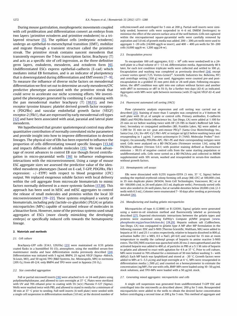
Duringmouse gastrulation, morphogenetic movements coupledwith cell proliferation and differentiation convert an embryo fromtwo layers (primitive ectoderm and primitive endoderm) to a tri-layered structure [5]. The epiblast cells (embryonic ectoderm)undergo an epithelial-to-mesenchymal transition (EMT), mobilizeand migrate through a transient structure called the primitivestreak. The primitive streak contains nascent mesoderm thattransiently expresses the T-box transcription factor, Brachyury (T)and acts as a specific site of cell ingression, as the three definitivegerm layers, endoderm, mesoderm, and ectoderm form [6].Undifferentiated ESCs express epithelial-cadherin (E-cad) whichmediates initial EB formation, and is an indicator of pluripotencythat is downregulated during differentiation and EMTevents [7e9].To measure the influence of diverse niche factors on mesodermaldifferentiationwe first set out to determine an earlymesoderm/CFCpredictive phenotype associated with the primitive streak thatcould serve to accelerate our niche screening efforts. We investi-gated the phenotypes generated by combining E-cad staining withthe pan mesodermal marker brachyury (T) [10,11], and tworeceptor tyrosine kinases: platelet derived growth factor receptor-a (PDGFRa) and vascular endothelial growth factor (VEGF)receptor-2 (Flk1), that are expressed by earlymesodermal cell types[12] and have been associated with axial, paraxial and lateral platemesoderm.
We hypothesized that greater spatiotemporal control may allowquantitative contribution of normally convoluted niche parametersand provide insight into how to improve differentiation to desiredlineages. The physical size of EBs has been reported to influence theproportion of cells differentiating toward specific lineages [13,14]and impacts diffusion of soluble molecules [15]. We took advan-tage of recent advances to control EB size though forced centrifu-gation in micro-pyramidal wells [16] to influence endogenousinteractions with the microenvironment. Using a range of mouseESC aggregate sizes we assessed the predictive value of the iden-tified mesoderm phenotypes (based on E-cad, T-GFP, PDGFRa, Flk1expression: þ/�ETPF) with respect to blood progenitor (CFC)output. We replaced exogenous soluble factors with local deliverywithin the cell aggregate from microscale biomaterials to mimicfactors normally delivered in a more systemic fashion [17,18]. Thisapproach has been used in hESC and mESC aggregates to controlthe release of small molecules and proteins within the local 3Dmicroenvironment [19e22]. These systems employed a variety ofbiomaterials, including poly (lactide-co-glycolide) (PLGA) or gelatinmicroparticles (MPs) capable of sustained release of molecules ina bioactive form. However, none of the previous systems used smallaggregates of ESCs (more closely mimicking the developingembryo) or specifically induced cells towards the hematopoieticlineage.
2. Materials and methods
2.1. Cell culture
Brachyury-GFP cells (E14.1, 129/Ola) [23] were maintained on 0.5% gelatincoated flasks in a humidified 5% CO2 atmosphere, using the modified serum-freemaintenance media and base differentiation media previously described [24].Differentiation was initiated with 5 ng/mL BMP4, 25 ng/mL VEGF (SigmaeAldrich,St.Louis, MO), and 50 ng/mL TPO (R&D Systems, Inc. Minneapolis, MN) in normoxia(20% O2) from d0-2/4; only BMP4 and TPO were used in hypoxia (5% O2).
2.2. Size controlled aggregation
Full or partial microwell inserts [16] were attached to 6- or 24-well plates usingpolydimethylsiloxane, and allowed to cure overnight at 37 �C. Plates were sterilizedwith UV and 70% ethanol prior to coating with 5% (w/v) Pluronic F-127 (Sigma).Wells were washed twice with PBS, and allowed to stand in media for a minimum of30 min at 37 �C prior to seeding. Full-well inserts (6-well plate) were seeded witha single cell suspension in differentiation medium (3.0 mL) at the desired number of
cells/microwell and centrifuged for 5 min at 200 g. Partial-well inserts were simi-larly seeded, however cells were suspended in 4e6 mL DMEM (Invitrogen) tominimize the effect of the uneven surface area of the well bottom. Cells not capturedwithin the micropatterned square-pyramidal wells were carefully removed byaspiration and 3.0 mL of growthmedia was added. 200� 200 mmwells were used toaggregate 1e20 cells (24,000 agg/6-w insert), and 400 � 400 mm wells for 50e200cells (6,000 agg/6-w; 1,200 agg/24-w).
2.3. Encapsulation process
To encapsulate 100 cell aggregates, 0.12 � 106 cells were seeded/well in a 24-well plate in a final volume of 1e1.5 mL differentiation media. Approximately 40 hlater, for each test condition triplicate wells were collected and settled to 100 mL.Encapsulation and washing was completed as previously described [24], excepta lower vortex speed (7.25, Vortex-Genie2�, Scientific Industries Inc. Bohemia, NY)and centrifuge setting (350 g) was used. Aggregates were counted pre-and post-encapsulation in a gridded 35 mm petri dish or 24-well plate. Following encapsu-lation, the sBVT condition was split into one culture without factors and anotherwith sBVT in normoxia or sBT in 5% O2 for a further two days (d2-4) as indicated.Aggregates with MPs were split between normoxia (with 25 ng/mL VEGF d2-4) and5% O2.
2.4. Fluorescent automated cell sorting (FACS)
Flow cytometric analysis expression and cell sorting was carried out asdescribed [25]. Staining of more than 5 samples was completed in a V-bottom 96well plate with 20 mL of sample or control cells. Primary antibodies, E-cadherin(R&D) and PDGFRa-biotin (eBioscience Inc. San Diego, CA) were added at 1:100 for25 min on ice before washing twice with HF (2% FBS in Hank’s Buffered Salt Solu-tion). Secondary or conjugated antibodies (BD, Franklin Lakes, NJ) were added at1:200 for 35 min on ice: goat anti-mouse PECy7 (Santa Cruz Biotechnology Inc.,Santa Cruz, CA), Stv-APC-Cy7, Flk1-APC or isotype rat IgG2 beforewashing twice andresuspending in 1 mg/mL 7-amino-actinomycin D (7AAD, Molecular Probes� Invi-trogen). Occasionally, PDGFRa-APC, Flk1-PE and their corresponding isotypes wereused. Cells were analyzed on a BD FACSCanto (Firmware version 1.14), using BDFACSDiva software (Version 5.0.1) with positive staining defined as fluorescenceemission > 99.1% of negative control cells from the same starting population orundifferentiated cells. Cells were sorted on a BD FACSAria and collected in IMDMsupplemented with 10% serum, washed and resuspended in serum-free mediumwithout growth factors.
2.5. Hematopoietic cell assays
EBs were dissociated with 0.25% trypsin-EDTA (3 min, 37 �C, Sigma) beforeseeding the myeloid-erythroid colony forming cell assay (ME-CFC) at 100,000 c/mLin 35 mm duplicate plates (M3434, Stem Cell Technologies, Vancouver, BC) or at60e100,000 c/mL in 24-well plates (0.5 mL duplicate wells). Previously sorted cellswere also seeded in 24-well plates, but at variable densities below 20,000 c/mL (2-3wells with 0.3 mL). Colonies were enumerated 7e10 days after seeding as previouslydetailed [26].
2.6. Manufacturing and loading gelatin microparticles
Microparticles of type A (G1890) or B (G9391, Sigma) gelatin were generatedusing a water-in-oil emulsion method and fluorescently labeled as previouslydescribed [27]. Expected electrostatic interactions between the gelatin types andproteins were examined using ExPASy’s Compute pI/MW program (www.phosphosite.org/psrSearchAction.do) [28,29]. Heparin sodium salt (CalBiochem,San Diego, CA) was conjugated to gelatin type A MP after MP formulation in thefollowing manner. EDC and S-NHS (Thermo Scientific, Waltham, MA) were added toheparin at 10:1 and 25:1 M ratios respectively, relative to heparin dissolved in 800 mLactivation buffer (0.1 M MES, 0.5 M NaCl, pH 6.0) and reacted for 15 min at roomtemperature to modify the carboxyl groups of heparin to amine reactive S-NHSesters. The EDC/NHS reactionwas quenched with 20mM 2-mercaptoethanol and theactivated heparin was added to 400 mL of particles in PBS at a 5:1 M ratio of heparinto gelatin and allowed to react with agitation for 4 h at 37 �C. Prior to cell culture,MPs were treated in 70% ethanol for a minimum of 30 min before washing 3� withddH2O. Each MP batch was lyophilized and stored at �20 �C. Growth factors wereadded to MPs at 5e5.5 mL/mg and kept overnight at 4 �C, MPs were resuspended indifferentiation media (�500 mL) and counted on a hemocytometer to estimate theconcentration (ng/MP). For usewith cells, BMP-MPswere loaded using 10e50 mg/mLstock solutions, and TPO-MPs were loaded with a 50 mg/mL stock.
2.7. Generating mixed aggregates: microparticles and cells
A single cell suspension was generated from undifferentiated T-GFP ESC andcentrifuged into the microwells as described above; 200 g for 5 min. Resuspendedmicroparticles were added to the wells to obtain the desired factor concentrationbefore centrifuging a second time at 200 g for 5 min. This method of aggregate and

K.A. Purpura et al. / Biomaterials 33 (2012) 1271e1280 1273
MP formation has also been achievedwith D3 ESC [27]. Aggregates were maintainedwith full media exchange in the microwells (daily or daily starting at 48 h) prior toremoval of soluble growth factors and transfer to Petri dishes on day 4. Alternatively,aggregates were removed 38e42 h after formation for encapsulation in agarose and/or dilution into 60e100 mm Petri dishes at approximately 300 aggregates/mL.
2.8. Human BMP4 ELISA
A quantitative sandwich enzyme immunoassay technique was used to deter-mine BMP4 concentration after release from MPs suspended in PBS, based on themanufacturer’s protocol (R & D, DBP400). Briefly, 2e5 mg of MPs were suspended in750 mL of a 0.1% solution of BSA in PBS at 37 �C with rotation; at each sample timepoint 300 mL was removed following centrifugation of the microparticles andreplaced with an equivalent volume.
2.9. Statistical analysis
Unless indicated, data are reported as mean � standard deviation of the mean.Statistical significance was assessed using one-way ANOVA with Tukey’s post hocanalysis, student’s t-test, or nonparametric ManneWhitney test (Minitab 15/16,State College PA and OriginPro7.5 Northampton, MA). P-values of less than 0.05 wereconsidered significant (n � 3).
3. Results
3.1. Cell population phenotypes
We previously demonstrated that in serum-free conditions theaddition of a trio of mesoderm inducing cytokines, BMP4, VEGF,and TPO (BVT) resulted in an induction of myeloid-erythroid colonyforming cells (ME-CFC) [30]. In order to quantitatively measure theimpact of our niche engineering efforts on hemogenic mesodermgeneration we sought to develop a set of predictive phenotypicmarkers. Multiple cell lines respond to this differentiation strategy,however, to trace the dynamic process of mesodermal specificationin greater detail we employed the Brachyury (T)-GFP line [23]. Wepostulated that the dynamic upregulation of brachyury anddownregulation of E-cadherin, that appear to signal the upregula-tion of two mesodermal receptors (Flk1 and PDGFRa), could beused in combination to identify the putative hemogenic populationfor tracking purposes.
Monitoring the expression of E-cadherin, brachyury, PDGFRa,and Flk1 during differentiation distinguishes 16 possible pheno-types (Fig. 1A). Once differentiation was initiated with BVT, E-cad-herin expressing cells (EþT�P�F�) progressively downregulatedthat adhesive molecule while brachyury and both surface receptorswere upregulated (Fig.1B). The presence of either one or both of thetracked receptors in the absence of brachyury was only observedafter the initial peak of E�TþPþFþ/� cells and may correspond tomore differentiated cells (day 5, Fig.1B). Due to the rarity of many ofthe phenotypic populations it is likely that they represent transientexpression states during lineage specification.
We grouped the expression patterns into populations thatcould broadly be classified as having mesendoderm (ME), meso-derm (M), endoderm (E), or unknown potential, and differences intheir gene expression profiles demonstrate this (Suppl. Fig. 1). Wesorted the most abundant day 3.75 phenotypes associated withhemogenic mesoderm and assessed their colony forming capacityafter 3 more days of suspension culture (the standard time toassess CFC). We found that the E�TþPþFþ population had thegreatest hemogenic capacity, and was significantly enrichedcompared to the unsorted population and all other fractions(Fig. 1C). The total number of colonies generated from theunsorted population was equivalent to the sum produced by theindividual sorted fractions once the initial frequency of thesephenotypes was taken into account. This analysis defined thestarting population necessary to further track and optimizeparameters of hemogenic mesoderm differentiation.
3.2. Aggregate size and mesodermal phenotype
Endogenous signals can impact differentiation [31] and it hasbeen established with both 2- and 3-D systems [32,33] that thenumber of neighboring cells impacts autocrine and paracrinefactors within the immediate media surrounding the cells [34,35].Thus, we examined how the initial number of cells per aggregateinfluenced mesoderm differentiation due to the interplay ofendogenous stimulatory or inhibitory signals and exogenousfactors (Fig. 2A) utilizing a centrifugal forced-aggregation strategy[16] and assessing the resultant phenotypes and functional celltypes. Total cell density was controlled by seeding different cellnumbers into 200 or 400micron square-pyramidal well inserts thatcovered an eighth-, quarter-, half-, or full-well within 6-well plates(Fig. 2B) to normalize the levels of nutrients and growth factors inthe bulk media. The conditioning effect that occurs with larger cellaggregates during microwell differentiation was demonstrated byexchanging media between 10- and 100-cell aggregates. Mediaconditioned for two days by 100-cell aggregates boosted the CFCoutput of the smaller aggregates while no striking effect wasobserved with the reverse media exchange (Suppl. Fig. 2).
Mesoderm phenotypes associated with CFC potential weremodestly enhanced with increasing aggregate size (Fig. 2C). Toinsure that similar differentiation kinetics occurred across thedifferent aggregate sizes we evaluated the CFC output of day 7e9EBs (Suppl. Fig. 3); the greatest output occurred on day 7 for eachsize (Fig. 2D). The hemogenic capacity of aggregates mirrored thetrends of the combined phenotypic response and was maximalwith 100-cell aggregates (1 in 145 � 8 d7 cells). Uniformly sizedaggregates initially seeded at 50e200 cells produced significantlymore CFC (p < 0.05) than non-uniform LSC aggregates. Withoutinductive factors (BVT), spontaneous differentiation accounts forless than 0.5% of the CFC produced (data not shown). Thus, theaddition of soluble factors is necessary to initiate hemogenicinduction within the 100-cell aggregates. Based on the results ofthese studies, aggregate size was fixed at 100 cells for furtherinvestigations.
3.3. Microparticle growth factor delivery
Pluripotent populations demonstrate heterogeneity and plas-ticity in vitro and these properties can be modulated by extrinsicsignalling [36], thus we sought to provide exogenous factors locallyusing growth factor delivery vehicles incorporated within the cellaggregates. Local delivery within multicellular aggregates mayenhance the efficiency of differentiation by increasing the effectivegrowth factor concentration and limiting the formation of gradi-ents. We first confirmed that the physical incorporation of gelatinmicroparticles (MPs) within the extracellular space of 100 cellaggregates (Fig. 3Ai) did not adversely influence aggregate forma-tion, brachyury expression, or CD45 output (Suppl. Fig. 4A,B). Byvarying the seed ratio of MP to cells, we noted that 1 MP to 3 ormore cells produced stable aggregates with incorporated particlesdistributed throughout the volume of the 3D spheroids (Fig. 3B,Suppl. Fig. 4C).
The electrostatic affinity between charged biomolecules andgelatin species has been the impetus behind its use as a matrix forthe controlled-release of bioactive molecules (reviewed in [37]).The characteristic isoelectric point (pI) of gelatin depends on themanufacturing conditions and it is expected that protein retentionwould be enhanced in gelatin of the opposite charge (Fig. 3Aii). Wetested the bioactivity of growth factor release from gelatin A or BMP with 100-cell aggregates by seeding different amounts ofBMP4-loaded MPs (B-MP, Fig. 3B). To examine the inductive effectand release kinetics of BMP4, cells were grown in the wells until

Fig. 1. Monitoring mesodermal specification. (A): Four-colour FACS employing a T-GFP cell line sheds light onto pan mesoderm development by tracking the surface expression of E-cadherin, PDGFRa and Flk1. (B): Comparison of undifferentiated cells to cells after 3.75 or 5 days in serum free differentiation media with or without BVT highlights the dynamicprogression of these lineage markers and demonstrates that not all phenotypic combinations occur. (C): Populations were sorted singly or in combination as indicated; the averageefficiency of colony formation from E�TþPþFþ cells (no. of colonies/no. of seeded cells � 100) was significantly (*) higher than all other sorted fractions (n ¼ 4, except E�T�Pþ/�Fþ n ¼ 2; ANOVA with Tukey’s post hoc analysis a ¼ 0.05).
K.A. Purpura et al. / Biomaterials 33 (2012) 1271e12801274

Fig. 2. Controlling initial cell aggregate size influencesmesodermal specification. (A): The overall effect of endogenously produced factorswould depend on the balance of stimulatory orinhibitory regulators that are secreted by themixture of cell types. A higher local cell density would condition themicroenvironment with more endogenous factors than lower densityconditions, as demonstrated in Suppl. Figure 2. (B): Two sizes ofmicropatterned square-pyramidalwells and partial coverage allows similar overall cell densititeswith constant volume tobe compared. Initial 10-or 100-cell aggregates are shown in 200 and 400 mm inserts respectively, immediately after spinning down the cells and following four days of growth. (C): Themesodermal phenotypes associated most closely with CFC, are shown as a stacked percentage of expression for aggregates that were initially 1e200 cells or non-uniform aggregates,formed from liquid suspension culture (LSC). (D): The phenotypic expression increased with larger aggregate sizes, however, CFC are maximal with 100 cell aggregates. The averagenumber of CFC � standard error of the mean are shown. Means that do not share a letter are significantly different (n ¼ 6e8; ANOVAwith Tukey’s post hoc analysis a ¼ 0.05).
K.A. Purpura et al. / Biomaterials 33 (2012) 1271e1280 1275

Fig. 3. Gelatin microparticles do not interfere with cell aggregation and exogenous factors can be delivered locally. (Ai): A schematic of the aggregate formation process with BMP4loaded gelatin MPs. (Aii): Both type A and type B gelatin MPs with characteristic pI were tested within the cell aggregates as it was expected BMP4 would have different inductivecapacities depending on the electrostatic interaction between the gelatin and protein. (B): A range of BMP4 concentrations were tested by calculating the theoretical amount ofBMP4 contained/MP. Brachyury (T-GFP) was assessed on day 4.5 (n ¼ 3; one-way ANOVA with Tukey’s post hoc analysis, a ¼ 0.05). Open symbols: daily media exchange.Abbreviations: MP, microparticle; B-MP, BMP4-MP; U-MP, unloaded MP; sB, soluble BMP4; d2ME, daily media exchange from day two. P < 0.05 for comparison of: *daily exchange(open diamond) to sB (filled square) or d2ME of type A/B (filled diamond) gelatin respectively; **24 h sB (filled grey circle) to continuous BMP4 (filled square/diamond); ydailyexchange type B (open diamond) to both type A MPs with daily exchange and 24 h sBMP4 (filled grey circle). The MP:cell ratio used to deliver 1e20 ng BMP4 are shown below theaxis; representative fluorescent images were taken 24 h after seeding. (C): BMP4 was loaded into type A (square), type B (triangle), and heparinized type A (open square) MPs from5, 10 or 25 mg/mL stock solutions as indicated and its release into PBS was monitored by ELISA (n ¼ 3). (D): Delivery of 5 ng/well BMP4 with heparinized MPs was as effective assoluble delivery for 100 cell aggregates (n ¼ 4, student’s t-test; 1200 aggregates, 24 well plate, 400 mm). (For better interpretation of the fluoresent images in this figure, the reader isreferred to the web version of this article.)
K.A. Purpura et al. / Biomaterials 33 (2012) 1271e12801276

K.A. Purpura et al. / Biomaterials 33 (2012) 1271e1280 1277
T-GFP was measured on day 4.5 with media exchange (ME)occurring daily or daily after an initial 48 h of culture (d2ME).Brachyury expressing cells reached a plateau of w70% with2e15 ng/mL soluble BMP4 (sB) treatment in the presence of eithergelatin A or B unloaded MPs (U-MP, Fig. 3B). Brachyury inductionfrom both type A and B MPs was equivalent to soluble delivery ifmedia was left standing for 2 days, however, with daily mediaexchange T-GFP and hence BMP4 release was significantly reduced(*p < 0.05). However, this robust response was only seen if mediaexchange began 48 h after seeding, suggesting that BMP4 releasekinetics were not optimal. Compared to continuous delivery, 24 hsoluble BMP4 reduced the percent of brachyury positive cells byw10% (**p < 0.05), and a similar proportion of T-GFP cells wasobserved with gelatin A MPs (Fig. 3B). BMP4 release from type Agelatin induced similar levels of T-GFP as 24 h delivery of solubleBMP4, in contrast, type B gelatin was unable to induce Brachyurywith daily media exchange, and T-GFP expression was significantlylower compared to both type A MPs with daily exchange and 24 h(yp < 0.05, Fig. 3B), suggesting that not enough BMP4 was releaseddue to stronger electrostatic interactions with the negativelycharged gelatin. These dose responses indicate that MP deliveryinduced mesoderm differentiation similarly to bulk delivery. Wenext explored the capacity to increase cellular responses by tuningthe release kinetics of our growth factor delivery vehicle throughmolecular engineering.
3.4. Growth factor release
To further investigate the interaction of BMP4with gelatin A or BMPs we varied the stock concentration from 5 to 25 mg/mL anddetermined the characteristic release profiles by ELISA. Very rapidand full release from gelatin A MPs occurred within 24 h whenloaded with a 5 mg/mL BMP4 stock solution (Fig. 3C). In contrast,only 35% BMP4 was released over the same time from gelatin BMPs. Neither of these release kinetic profiles would be capable ofsustained-release on the order of 4 days and allow the cell aggre-gates to be transferred to bulk conditions for extended culture.Doubling the stock concentration of BMP4 doubled its release fromgelatin B, however, using a 25 mg/mL stock resulted in a morevariable and intermediate extent of release (w50%) (Fig. 3C). Theheparin binding domain of BMP4 has recently been identified [38],thus, we conjugated heparin to type A gelatin MPs to take advan-tage of the total release by enhancing the loading capacity via highaffinity binding sites. We found that 40e60% less BMP4 wasdetected in solution with heparinized type A gelatin MPs thanuntreated gelatin A MPs and this was on the same order of releaseas gelatin B MPs (Fig. 3C). We hypothesized that using the hepa-rinized type A MPs loaded with BMP4 from stocks � 10 mg/mLwould enhance the overall delivery and bioactivity of the heparin-bound presentation of the morphogenic factor.
We successfully replaced soluble BMP4 with local delivery orpresentation of affinity-bound BMP4 using the heparinized gelatinA MPs in combination with sVT. The MPs were seeded with 100cells to form aggregates that were transferred to bulk suspensionculture on day two (Fig. 3D). We next sought to integrate localfactor delivery with other cell inductive parameters in a mannerthat would lead to robust blood cell development.
3.5. Low oxygen environment with dual growth factor delivery
We and others have shown that low oxygen tension (5% O2) isbeneficial in generating hemogenic mesoderm [39,40] via a mech-anism that dynamically tunes VEGF signaling. We combined 5%oxygen tension with local factor delivery to enhance blood devel-opment in the absence of exogenous VEGF. Gelatin microparticles
were incorporated in 100-cell aggregates to support mesodermdevelopment either loadedwith BMP4 or TPO (previously shown toenhance CFC production), or as unloaded controls (Fig. 4Ai). Cellswere allowed to form stable aggregates for 32e42 h before theywere removed from the microwells and encapsulated in agarose toallow our engineered niche to be incorporated into suspensionculture processes (Fig. 4Aii). Brachyury induction was apparentafter 3 days (Fig. 4Aiii) and in addition to the soluble growth factorcontrols (BMP4, TPO), 25 ng/mL VEGF was provided for 4 days innormoxia (20% O2) in order to match the CFC induction due to VEGFupregulation in 5% O2.
Although thrombopoietin (pI 9.4) has a slightly higher pI thanBMP4, we hypothesized that it would interact with the heparinizedgelatin MPs in a similar fashion. We found that increasing the doseof BMP4 delivered by the MPs, while holding the dose of TPOrelatively constant (range 9e15 ng), yielded an increasing numberof CFCs (Fig. 4B). The cells were split between normoxic andhypoxic conditions following encapsulation and similar trendswere observed in both the phenotypic induction and CFC output inresponse to the B-MP and TPO-MP delivery regardless of oxygentension, indicating that cells in low oxygen effectively upregulatedendogenous VEGF to replace the exogenous delivery in normoxia.
In normoxic conditions 2e5 ng/well B-MP with TPO-MPdelivery generated an increasing trend in CFC output but did notsignificantly differ from the soluble BVT (d0-2) control. Providing8 ng/well B-MP provided a significant improvement over two daysof soluble factors and was similar to the sBVT (d0-4) control. Thehighest CFC output occured with 12 ng/well of B-MP delivery. Thisindicates that depending on the dose provided the factors wereactive following encapsulation (i.e. 8 ng, d0-4 and 12 ng, > 4 days).The early mesodermal phenotypes observed at day 4(E�TþPþFþand E�T�Pþ/�Fþ) correlated to the CFC producingcells present at day 7 (Fig. 4C). Representative erythroid, myeloidand mixed colonies are shown from the 5 ng/well B-MP normoxiccondition (Fig. 4D). Altering the presentation of the growth factors,either from MP delivery or 5% O2 induction, did not significantlyalter the proportion of colony types produced by themajority of thedifferent treatment conditions (Suppl. Fig. 5AeC; ManneWhitneytest).
Seeding 8 or 12 ng BMP4 and w12 ng TPO within the spatialcontext of 100-cell aggregates (1,200 total) appears to inducecomparable, if not greater, mesoderm differentiation than 5 ng/mLexogenous BMP4 and 50 ng/mL TPO. When the total growth factorloaded into the MPs and the additional media required for thesoluble control following encapsulation (300 agg/mL) are accoun-ted for, roughly 14� less BMP4 and TPO was required for local MPdelivery/presentation than for bulk delivery.
In all, we have demonstrated that tracking the emergence ofE�TþPþFþ, E�T�Pþ/�Fþ, and E�T�PþF� phenotypes can predictthe general capacity of a culture to produce CFC when seeded 3days later, and aid in understanding the various microenviron-mental factors capable of impacting CFC production. Aggregate sizecan be used to enhance CFC with soluble factors and manipulatingthemicroenvironmental niche through controlled aggregationwithmicroparticles that deliver BMP4 and TPO can generate greaternumbers of CFCs than soluble delivery (w1.7�) of factors in bothlow oxygen (sBT) or normoxic (sBVT) environments. These resultsdemonstrate that local delivery by affinity-bound presentation ofgrowth factors from biomaterials can effectively induce mesodermdifferentiation.
4. Discussion
Modulating cellecell interactions and the effects of autocrine,paracrine, and exogenous factors through initial aggregate size,
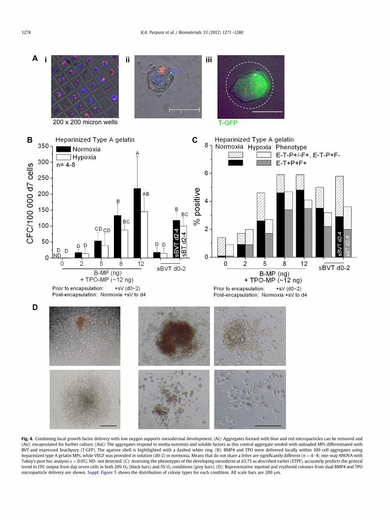
Fig. 4. Combining local growth factor delivery with low oxygen supports mesodermal development. (Ai): Aggregates formed with blue and red microparticles can be removed and(Aii): encapsulated for further culture. (Aiii): The aggregates respond to media nutrients and soluble factors as this control aggregate seeded with unloaded MPs differentiated withBVT and expressed brachyury (T-GFP). The agarose shell is hightlighted with a dashed white ring. (B): BMP4 and TPO were delivered locally within 100 cell aggregates usingheparinized type A gelatin MPs, while VEGF was provided in solution (d0-2) in normoxia. Means that do not share a letter are significantly different (n ¼ 4e8; one-way ANOVAwithTukey’s post hoc analysis a ¼ 0.05). ND- not detected. (C): Assessing the phenotypes of the developing mesoderm at d3.75 as described earlier (ETPF), accurately predicts the generaltrend in CFC output from day seven cells in both 20% O2 (black bars) and 5% O2 conditions (grey bars). (D): Representative myeloid and erythroid colonies from dual BMP4 and TPOmicroparticle delivery are shown. Suppl. Figure 5 shows the distribution of colony types for each condition. All scale bars are 200 mm.
K.A. Purpura et al. / Biomaterials 33 (2012) 1271e12801278

K.A. Purpura et al. / Biomaterials 33 (2012) 1271e1280 1279
oxygen tension, and local growth factor delivery, has providedinsights into directed differentiation by monitoring both cellularphenotypes and functional responses. We first explored thedifferential expression of mesodermal cell phenotypes and thefunctional CFC response to exogenous growth factors in a serum-free media. We used a serum-free culture system that maintainsthe self-renewal of undifferentiated ESCs [30,41] and the embryoidbody system to model blood development as it induces differenti-ation similar to embryonic gastrulation. In an aim to better defineand characterize subsets of nascent mesodermal cells for trackingand culture optimization purposes we monitored E-cadherin, bra-chyury, PDGFRa and Flk1.
Flk1 positive cells have been associated with hematopoiesis andvasculogenesis [23,42,43], as well as other mesodermal cell types[44e46]. Two-marker cell sorting strategies have demonstratedthat PþF� cells have substantial muscle regeneration potential[47], while PþFþ cells demonstrate enhanced cardiac potential[48,49], and P�Fþ cells retain hematopoietic potential [49].Monitoring E-cadherin and T-GFP [23] expression provided anindication that mesodermal differentiation was proceeding in ourcultures and we combined these markers with PDGFRa and Flk1expression to delineate potential hematopoietic progenitors. Forthe first time, we demonstrated that cells co-expressing brachyury,Flk1, and PDGFRa had the highest frequency of CFC formationwhenthey did not express E-cadherin. Presumably the CFC outputrepresents the cell autonomous capacity of the sorted phenotypes,as the cells were allowed to mature for an additional three dayswithout the addition of exogenous growth factors. Furthermore,the close link between the cardiac potential (PþFþ) and hemato-poietic potential (E�TþPþFþ) raises the question whether a bi-potent cell population remains at the time we sorted. Insightfrom these phenotypes can be used to aid future developments incell generation processes.
Focusing on parameters that may easily be incorporated intoengineered systems to enhance hematopoietic specification, wenext investigated the influence of local cell density. Examining theinitial developmental stages leading up to the assessment ofphenotypic expression, we showed an increasing proportion ofhemogenic cells with increasing aggregate size (1e200 cells), witha maximum at 100-cell aggregates (Fig. 2B). Aggregates seededwith 5e200 cells with or without growth factors continued toexpand at similar growth rates, such that there were no significantdifferences in population doublings between the conditions. As therate of aggregate growth was not strongly influenced by initial cellnumbers, controlling both aggregate size and the total aggregatesper well assured similar numbers of the bioactivemolecules (BMP4,VEGF, or TPO) were available on a per cell basis in the bulk media ormacroenvironment.
Taking advantage of the capacity for microenvironmentalcontrol from within the aggregate itself we incorporated gelatinMPs as a delivery vehicle to locally present BMP4 [38] and TPO [50],which have known and presumed heparin binding domains andhemogenic induction capacity. Heparin has previously been usedwith scaffolds to sustain the release/presentation of heparinbinding growth factors (HBGF) [51] and its use with particulatesystems for controlled delivery of HBGF is an emerging area intissue engineering [52]. In addition, although the specific heparinbinding consensus sequence to various hematopoietic cytokineshave not been established, cytokine-immobilization using lowmolecular weight heparin was comparable for TPO, SCF and Flt-3(both basic and acidic proteins) and w32% remained after 6 days[50], suggesting that heparinized gelatin MPs are able to delivera variety of morphogenic factors in a similar manner.
Finally, we established a system incorporating heparinizedmicroparticle delivery of two factors, BMP4 and TPO, into 100-cell
aggregates, which supports the generation of hematopoieticprogenitors equivalently, if not to a greater extent (w1.7-fold moreCFCs) than bulk exogenous delivery. Similar cell numbers weregenerated with or without the provision of growth factors and inthe presence or absence of microparticles, thus differences incolony numbers and enhanced CFC output represent better processyields from the initial ESC input. In all, integration of amicroparticleapproach for bioactive molecule delivery within EBs provides theopportunity to deconvolute the complex biological signals whichcells receive during blood development.
In the future, time-lapse imaging studies may be used toinvestigate the localization of the developing mesoderm withenriched CFC activity or the relation between the hemogenic andnon-hemogenic endothelium produced by the mesodermalprecursors. Furthermore with the establishment of this system,studies may now be designed to explore asymmetrical signalswithin the aggregate (by providing hemispherical delivery) andfurther mimic embryonic development or polar axis definitions.Overall, this approach based on engineered combinations ofphysical and biochemical signaling serves as a model to guidedifferentiation of pluripotent stem cells to specific mesodermphenotypes, such as blood, as well as to quantitatively investigatethe contribution of normally convoluted niche parameters onpluripotent cell developmental fate decisions.
5. Conclusion
We have provided a model system that used mesodermalphenotype characterizationwith a forced-aggregation technique tocontrol aggregate size and to embed MPs to serve as local deliveryvehicles. Under serum-free conditions, heparinized MPs incorpo-rated prior to aggregate encapsulation were able to induce differ-entiation to levels that were similar or exceeded bulk deliverymethods.
Disclosure of potential conflicts of interest
The authors have no conflicts of interest to disclose.
Acknowledgements
The authors would like to thank Dr. G. Clarke for statisticalrecommendations and valuable discussion, Dr. C. Ito for construc-tive discussions, and N. Rahman, Dr. H. Song, Dr. M. Ungrin, C. Yoon,and M. Yu at the University of Toronto for technical assistance. Thiswork is funded by CIHR (MOP-57885), NSERC, and the CanadianStem Cell Network. K.A.P. was supported by an Ontario GraduateScholarship; P.W.Z. is the Canada Research Chair in Stem CellBioengineering. A.M.B.L. was supported by an NIH Training Grant(GM008433), as well as funding from the Goizueta Foundation.T.C.M. is supported by grants from the National Science Foundation(CBET 0651739) and the National Institutes of Health (GM088291).
Appendix. Supplementary data
Supplementary data related to this article can be found online atdoi:10.1016/j.biomaterials.2011.10.051.
References
[1] Leahy A, Xiong JW, Kuhnert F, Stuhlmann H. Use of developmental markergenes to define temporal and spatial patterns of differentiation duringembryoid body formation. J Exp Zool 1999;284(1):67e81.
[2] Adamo L, Naveiras O, Wenzel PL, McKinney-Freeman S, Mack PJ, Gracia-Sancho J, et al. Biomechanical forces promote embryonic haematopoiesis.Nature 2009;459(7250):1131e5.

K.A. Purpura et al. / Biomaterials 33 (2012) 1271e12801280
[3] Ferkowicz MJ, Yoder MC. Whole embryo imaging of hematopoietic cellemergence and migration. Methods Mol Biol 2011;750:143e55.
[4] Yoshimoto M, Montecino-Rodriguez E, Ferkowicz MJ, Porayette P, Shelley WC,Conway SJ, et al. Embryonic day 9 yolk sac and intra-embryonic hemogenicendothelium independently generate a B-1 and marginal zone progenitorlacking B-2 potential. Proc Natl Acad Sci U S A 2011;108(4):1468e73.
[5] Tam PP, Behringer RR. Mouse gastrulation: the formation of a mammalianbody plan. Mech Dev 1997;68(1e2):3e25.
[6] Gadue P, Huber TL, Nostro MC, Kattman S, Keller GM. Germ layer inductionfrom embryonic stem cells. Exp Hematol 2005;33(9):955e64.
[7] Acloque H, Adams MS, Fishwick K, Bronner-Fraser M, Nieto MA. Epithelial-mesenchymal transitions: the importance of changing cell state in develop-ment and disease. J Clin Invest 2009;119(6):1438e49.
[8] Spencer H, Keramari M, Ward CM. Using cadherin expression to assessspontaneous differentiation of embryonic stem cells. Methods Mol Biol 2011;690:81e94.
[9] Spencer HL, Eastham AM, Merry CL, Southgate TD, Perez-Campo F, Soncin F,et al. E-cadherin inhibits cell surface localization of the pro-migratory 5T4oncofetal antigen in mouse embryonic stem cells. Mol Biol Cell 2007;18(8):2838e51.
[10] Wilkinson DG, Bhatt S, Herrmann BG. Expression pattern of the mouse T geneand its role in mesoderm formation. Nature 1990;343(6259):657e9.
[11] Keller G, Kennedy M, Papayannopoulou T, Wiles MV. Hematopoieticcommitment during embryonic stem cell differentiation in culture. Mol CellBiol 1993;13(1):473e86.
[12] Yoshida H, Takakura N, Hirashima M, Kataoka H, Tsuchida K, Nishikawa S,et al. Hematopoietic tissues, as a playground of receptor tyrosine kinases ofthe PDGF-receptor family. Dev Comp Immunol 1998;22(3):321e32.
[13] Ng ES, Davis RP, Azzola L, Stanley EG, Elefanty AG. Forced aggregation of definednumbers of human embryonic stem cells into embryoid bodies fosters robust,reproducible hematopoietic differentiation. Blood 2005;106(5):1601e3.
[14] Hwang YS, Chung BG, Ortmann D, Hattori N, Moeller HC, Khademhosseini A.Microwell-mediated control of embryoid body size regulates embryonic stemcell fate via differential expression of WNT5a and WNT11. Proc Natl Acad SciU S A 2009;106(40):16978e83.
[15] Sachlos E, Auguste DT. Embryoid body morphology influences diffusivetransport of inductive biochemicals: a strategy for stem cell differentiation.Biomaterials 2008;29(34):4471e80.
[16] Ungrin MD, Joshi C, Nica A, Bauwens C, Zandstra PW. Reproducible, ultra high-throughput formation of multicellular organization from single cellsuspension-derived human embryonic stem cell aggregates. PLoS One 2008;3(2):e1565.
[17] Discher DE, Mooney DJ, Zandstra PW. Growth factors, matrices, and forcescombine and control stem cells. Science 2009;324(5935):1673e7.
[18] Bratt-Leal AM, Carpenedo RL, McDevitt TC. Engineering the embryoid bodymicroenvironment to direct embryonic stem cell differentiation. BiotechnolProg 2009;25(1):43e51.
[19] Ferreira L, Squier T, Park H, Choe H, Kohane DS, Langer R. Human embryoidbodies containing nano- and microparticlulate delivery vehicles. Adv Mater2008;20:2285e91.
[20] Carpenedo RL, Bratt-Leal AM, Marklein RA, Seaman SA, Bowen NJ,McDonald JF, et al. Homogeneous and organized differentiation withinembryoid bodies induced by microsphere-mediated delivery of small mole-cules. Biomaterials 2009;30(13):2507e15.
[21] Carpenedo RL, Seaman SA, McDevitt TC. Microsphere size effects on embryoidbody incorporation and embryonic stem cell differentiation. J Biomed MaterRes A 2010;94(2):466e75.
[22] Mahoney MJ, Saltzman WM. Transplantation of brain cells assembled arounda programmable synthetic microenvironment. Nat Biotechnol 2001;19(10):934e9.
[23] Fehling HJ, Lacaud G, Kubo A, Kennedy M, Robertson S, Keller G, et al. Trackingmesoderm induction and its specification to the hemangioblast duringembryonic stem cell differentiation. Development 2003;130(17):4217e27.
[24] Rahman N, Purpura KA, Wylie RG, Zandstra PW, Shoichet MS. The use ofvascular endothelial growth factor functionalized agarose to guide pluripotentstem cell aggregates toward blood progenitor cells. Biomaterials 2010;31(32):8262e70.
[25] Dang SM, Kyba M, Perlingeiro R, Daley GQ, Zandstra PW. Efficiency ofembryoid body formation and hematopoietic development from embryonicstem cells in different culture systems. Biotechnol Bioeng 2002;78(4):442e53.
[26] Eaves C, Fraser C, Udomsakdi C, Sutherland H, Barnett M, Szilvassy S, et al.Manipulation of the hematopoietic stem cell in vitro. Leukemia 1992;6(Suppl.1):27e30.
[27] Bratt-Leal AM, Carpenedo RL, Ungrin MD, Zandstra PW, McDevitt TC. Incor-poration of biomaterials in multicellular aggregates modulates pluripotentstem cell differentiation. Biomaterials 2011;32(1):48e56.
[28] Bjellqvist B, Hughes GJ, Pasquali C, Paquet N, Ravier F, Sanchez JC, et al. Thefocusing positions of polypeptides in immobilized pH gradients can be pre-dicted from their amino acid sequences. Electrophoresis 1993;14(10):1023e31.
[29] Gasteiger E, Hoogland C, Gattiker A, Duvaud S, Wilkins MR, Appel RD, et al.Protein identification and analysis tools on the ExPASy server. Totowa:Humana Press Inc.; 2005.
[30] Purpura KA, Morin J, Zandstra PW. Analysis of the temporal andconcentration-dependent effects of BMP-4, VEGF, and TPO on development ofembryonic stem cell-derived mesoderm and blood progenitors in a defined,serum-free media. Exp Hematol 2008;36(9):1186e98.
[31] Keller GM. In vitro differentiation of embryonic stem cells. Curr Opin Cell Biol1995;7(6):862e9.
[32] Davey RE, Zandstra PW. Spatial organization of embryonic stem cell respon-siveness to autocrine gp130 ligands reveals an autoregulatory stem cell niche.Stem Cells 2006;24(11):2538e48.
[33] Hoelker M, Rings F, Lund Q, Phatsara C, Schellander K, Tesfaye D. Effect ofembryo density on in vitro developmental characteristics of bovine pre-implantative embryos with respect to micro and macroenvironments. ReprodDomest Anim 2010;45(5):e138e45.
[34] Groebe K, Mueller-Klieser W. Distributions of oxygen, nutrient, and metabolicwaste concentrations in multicellular spheroids and their dependence onspheroid parameters. Eur Biophys J 1991;19(4):169e81.
[35] Peerani R, Onishi K, Mahdavi A, Kumacheva E, Zandstra PW. Manipulation ofsignaling thresholds in "engineered stem cell niches" identifies design criteriafor pluripotent stem cell screens. PLoS One 2009;4(7):e6438.
[36] Pera MF, Tam PP. Extrinsic regulation of pluripotent stem cells. Nature 2011;465(7299):713e20.
[37] Young S, Wong M, Tabata Y, Mikos AG. Gelatin as a delivery vehicle for thecontrolled release of bioactive molecules. J Control Release 2005;109(1e3):256e74.
[38] Choi YJ, Lee JY, Park JH, Park JB, Suh JS, Choi YS, et al. The identification ofa heparin binding domain peptide from bone morphogenetic protein-4 and itsrole on osteogenesis. Biomaterials 2010;31(28):7226e38.
[39] Ramirez-Bergeron DL, Runge A, Dahl KD, Fehling HJ, Keller G, Simon MC.Hypoxia affects mesoderm and enhances hemangioblast specification duringearly development. Development 2004;131(18):4623e34.
[40] Purpura KA, George SH, Dang SM, Choi K, Nagy A, Zandstra PW. Soluble Flt-1regulates Flk-1 activation to control hematopoietic and endothelial develop-ment in an oxygen-responsive manner. Stem Cells 2008;26(11):2832e42.
[41] Ying QL, Smith AG. Defined conditions for neural commitment and differen-tiation. Methods Enzymol 2003;365:327e41.
[42] Shalaby F, Ho J, Stanford WL, Fischer KD, Schuh AC, Schwartz L, et al.A requirement for Flk1 in primitive and definitive hematopoiesis and vascu-logenesis. Cell 1997;89(6):981e90.
[43] Niwa A, Umeda K, Chang H, Saito M, Okita K, Takahashi K, et al. Orderlyhematopoietic development of induced pluripotent stem cells via Flk-1(þ)hemoangiogenic progenitors. J Cell Physiol 2009;221(2):367e77.
[44] Yamashita JK, Takano M, Hiraoka-Kanie M, Shimazu C, Peishi Y, Yanagi K, et al.Prospective identification of cardiac progenitors by a novel single cell-basedcardiomyocyte induction. Faseb J 2005;19(11):1534e6.
[45] Motoike T, Markham DW, Rossant J, Sato TN. Evidence for novel fate of Flk1þprogenitor: contribution to muscle lineage. Genesis 2003;35(3):153e9.
[46] Nakayama N, Duryea D, Manoukian R, Chow G, Han CY. Macroscopic cartilageformation with embryonic stem-cell-derived mesodermal progenitor cells.J Cell Sci 2003;116(Pt. 10):2015e28.
[47] Darabi R, Gehlbach K, Bachoo RM, Kamath S, Osawa M, Kamm KE, et al.Functional skeletal muscle regeneration from differentiating embryonic stemcells. Nat Med 2008;14(2):134e43.
[48] Hirata H, Kawamata S, Murakami Y, Inoue K, Nagahashi A, Tosaka M, et al.Coexpression of platelet-derived growth factor receptor alpha and fetal liverkinase 1 enhances cardiogenic potential in embryonic stem cell differentiationin vitro. J Biosci Bioeng 2007;103(5):412e9.
[49] Kattman SJ, Witty AD, Gagliardi M, Dubois NC, Niapour M, Hotta A, et al.Stage-specific optimization of activin/nodal and BMP signaling promotescardiac differentiation of mouse and human pluripotent stem cell lines. CellStem Cell 2011;8(2):228e40.
[50] Kishimoto S, Nakamura S, Nakamura S, Hattori H, Oonuma F, Kanatani Y, et al.Cytokine-immobilized microparticle-coated plates for culturing hematopoi-etic progenitor cells. J Control Release 2009;133(3):185e90.
[51] Wu JM, Xu YY, Li ZH, Yuan XY, Wang PF, Zhang XZ, et al. Heparin-function-alized collagen matrices with controlled release of basic fibroblast growthfactor. J Mater Sci Mater Med 2011;22(1):107e14.
[52] Joung YK, Bae JW, Park KD. Controlled release of heparin-binding growthfactors using heparin-containing particulate systems for tissue regeneration.Expert Opin Drug Deliv 2008;5(11):1173e84.