A Systematic Review Study - MDPI
34
International Journal of Molecular Sciences Review Immunobiological Properties and Clinical Applications of Interleukin-38 for Immune-Mediated Disorders: A Systematic Review Study Abdolreza Esmaeilzadeh 1,2,3, * ,† , Nazila Bahmaie 4,5,6,7,†,‡ , Elham Nouri 8,9 , Mohammad Javad Hajkazemi 10 and Maryam Zareh Rafie 10 Citation: Esmaeilzadeh, A.; Bahmaie, N.; Nouri, E.; Hajkazemi, M.J.; Zareh Rafie, M. Immunobiological Properties and Clinical Applications of Interleukin-38 for Immune- Mediated Disorders: A Systematic Review Study. Int. J. Mol. Sci. 2021, 22, 12552. https://doi.org/10.3390/ ijms222212552 Academic Editors: Giuseppe Murdaca, Sebastiano Gangemi and Monica Greco Received: 16 August 2021 Accepted: 19 October 2021 Published: 21 November 2021 Publisher’s Note: MDPI stays neutral with regard to jurisdictional claims in published maps and institutional affil- iations. Copyright: © 2021 by the authors. Licensee MDPI, Basel, Switzerland. This article is an open access article distributed under the terms and conditions of the Creative Commons Attribution (CC BY) license (https:// creativecommons.org/licenses/by/ 4.0/). 1 Department of Immunology, School of Medicine, Zanjan University of Medical Sciences, Zanjan 4513956111, Iran 2 Cancer Gene Therapy Research Center (CGRC), Zanjan University of Medical Sciences, Zanjan 4513956111, Iran 3 Immunotherapy Research & Technology Group, Zanjan University of Medical Sciences, Zanjan 4513956111, Iran 4 Department of Allergy and Immunology, Faculty of Medicine, Graduate School of Health Science, Near East University (NEU), Nicosia 99138, Cyprus; [email protected] 5 Pediatric Ward, Department of Allergy and Immunology, Near East University affiliated Hospital, Nicosia 99138, Cyprus 6 Serology and Immunology Ward, Clinical Diagnosis Laboratory, Private Baskent Hospital, Nicosia 2681, Cyprus 7 Network of Immunity in Infection, Malignancy and Autoimmunity (NIIMA), Universal Scientific Education and Research Network (USERN), Tehran 1419733151, Iran 8 School of Paramedicine, Zanjan University of Medical Sciences, Zanjan 4513956111, Iran; [email protected] 9 Shahid Beheshti University Affiliated Hospital, Zanjan University of Medical Sciences, Zanjan 4513956111, Iran 10 School of Medicine, Zanjan University of Medical Sciences, Zanjan 4513956111, Iran; [email protected] (M.J.H.); Maryam.rafi[email protected] (M.Z.R.) * Correspondence: [email protected] † Abdolreza Esmaeilzadeh and Nazila Bahmaie should be considered as joint first authors. ‡ Present affiliation and postal address of “Nazila Bahmaie” is different from the address where the work was conducted. Abstract: Exponential growth in the usage of “cytokines” (as seroimmunobiomarkers) has facilitated more accurate prognosis, early diagnosis, novel, and efficient immunotherapeutics. Numerous studies have reported immunopathophysiological and immunopathological processes of interleukin- 38 (IL-38). Therefore, in this systematic review article, the authors aimed to present an updated comprehensive overview on the immunobiological mechanisms, diagnostic, and immune gene-based therapeutic potentials of IL-38. According to our inclusion and exclusion criteria, a total of 216 articles were collected from several search engines and databases from the January 2012 to July 2021 time interval by using six main keywords. Physiologic or pathologic microenvironments, optimal dosage, and involved receptors affect the functionalities of IL-38. Alterations in serum levels of IL-38 play a major role in the immunopathogenesis of a wide array of immune-mediated disorders. IL-38 shows anti-inflammatory activities by reduction or inhibition of pro-inflammatory cytokines, supporting the therapeutic aspects of IL-38 in inflammatory autoimmune diseases. According to the importance of pre-clinical studies, it seems that manipulation of the immune system by immunomodulatory properties of IL-38 can increase the accuracy of diagnosis, and decipher optimal clinical outcomes. To promote our knowledge, more collaboration is highly recommended among laboratory scientists, internal/infectious diseases specialists, oncologists, immunologists, diseases-specific biomarkers scientists, and basic medical researchers. Keywords: biomarker; clinical applications; diagnosis; immunobiology; immunopathophysiology; interleukin-38 Int. J. Mol. Sci. 2021, 22, 12552. https://doi.org/10.3390/ijms222212552 https://www.mdpi.com/journal/ijms
-
Upload
khangminh22 -
Category
Documents
-
view
0 -
download
0
Transcript of A Systematic Review Study - MDPI

International Journal of
Molecular Sciences
Review
Immunobiological Properties and Clinical Applications ofInterleukin-38 for Immune-Mediated Disorders: A SystematicReview Study
Abdolreza Esmaeilzadeh 1,2,3,*,†, Nazila Bahmaie 4,5,6,7,†,‡ , Elham Nouri 8,9, Mohammad Javad Hajkazemi 10
and Maryam Zareh Rafie 10
�����������������
Citation: Esmaeilzadeh, A.; Bahmaie,
N.; Nouri, E.; Hajkazemi, M.J.; Zareh
Rafie, M. Immunobiological
Properties and Clinical Applications
of Interleukin-38 for Immune-
Mediated Disorders: A Systematic
Review Study. Int. J. Mol. Sci. 2021,
22, 12552. https://doi.org/10.3390/
ijms222212552
Academic Editors: Giuseppe
Murdaca, Sebastiano Gangemi and
Monica Greco
Received: 16 August 2021
Accepted: 19 October 2021
Published: 21 November 2021
Publisher’s Note: MDPI stays neutral
with regard to jurisdictional claims in
published maps and institutional affil-
iations.
Copyright: © 2021 by the authors.
Licensee MDPI, Basel, Switzerland.
This article is an open access article
distributed under the terms and
conditions of the Creative Commons
Attribution (CC BY) license (https://
creativecommons.org/licenses/by/
4.0/).
1 Department of Immunology, School of Medicine, Zanjan University of Medical Sciences,Zanjan 4513956111, Iran
2 Cancer Gene Therapy Research Center (CGRC), Zanjan University of Medical Sciences,Zanjan 4513956111, Iran
3 Immunotherapy Research & Technology Group, Zanjan University of Medical Sciences,Zanjan 4513956111, Iran
4 Department of Allergy and Immunology, Faculty of Medicine, Graduate School of Health Science,Near East University (NEU), Nicosia 99138, Cyprus; [email protected]
5 Pediatric Ward, Department of Allergy and Immunology, Near East University affiliated Hospital,Nicosia 99138, Cyprus
6 Serology and Immunology Ward, Clinical Diagnosis Laboratory, Private Baskent Hospital,Nicosia 2681, Cyprus
7 Network of Immunity in Infection, Malignancy and Autoimmunity (NIIMA), Universal Scientific Educationand Research Network (USERN), Tehran 1419733151, Iran
8 School of Paramedicine, Zanjan University of Medical Sciences, Zanjan 4513956111, Iran; [email protected] Shahid Beheshti University Affiliated Hospital, Zanjan University of Medical Sciences,
Zanjan 4513956111, Iran10 School of Medicine, Zanjan University of Medical Sciences, Zanjan 4513956111, Iran;
[email protected] (M.J.H.); [email protected] (M.Z.R.)* Correspondence: [email protected]† Abdolreza Esmaeilzadeh and Nazila Bahmaie should be considered as joint first authors.‡ Present affiliation and postal address of “Nazila Bahmaie” is different from the address where the work
was conducted.
Abstract: Exponential growth in the usage of “cytokines” (as seroimmunobiomarkers) has facilitatedmore accurate prognosis, early diagnosis, novel, and efficient immunotherapeutics. Numerousstudies have reported immunopathophysiological and immunopathological processes of interleukin-38 (IL-38). Therefore, in this systematic review article, the authors aimed to present an updatedcomprehensive overview on the immunobiological mechanisms, diagnostic, and immune gene-basedtherapeutic potentials of IL-38. According to our inclusion and exclusion criteria, a total of 216 articleswere collected from several search engines and databases from the January 2012 to July 2021 timeinterval by using six main keywords. Physiologic or pathologic microenvironments, optimal dosage,and involved receptors affect the functionalities of IL-38. Alterations in serum levels of IL-38 play amajor role in the immunopathogenesis of a wide array of immune-mediated disorders. IL-38 showsanti-inflammatory activities by reduction or inhibition of pro-inflammatory cytokines, supportingthe therapeutic aspects of IL-38 in inflammatory autoimmune diseases. According to the importanceof pre-clinical studies, it seems that manipulation of the immune system by immunomodulatoryproperties of IL-38 can increase the accuracy of diagnosis, and decipher optimal clinical outcomes. Topromote our knowledge, more collaboration is highly recommended among laboratory scientists,internal/infectious diseases specialists, oncologists, immunologists, diseases-specific biomarkersscientists, and basic medical researchers.
Keywords: biomarker; clinical applications; diagnosis; immunobiology; immunopathophysiology;interleukin-38
Int. J. Mol. Sci. 2021, 22, 12552. https://doi.org/10.3390/ijms222212552 https://www.mdpi.com/journal/ijms

Int. J. Mol. Sci. 2021, 22, 12552 2 of 34
1. Introduction
Although morbidity and mortality rates of immune-mediated disorders have been effi-ciently diminished by the use of several cutting-edge technologies (such as novel vaccines),some obstacles in the diagnosis, prognosis, and treatment of immune-mediated disordershave not been tackled yet. The chronicity of these types of disorders has still remained aproblematic challenge for the health system. In addition, patients with immune-mediateddisorders experience poor lifestyles and suffer from complex clinical manifestations in theirlife expectancy due to the late or inaccurate diagnosis and poor prognosis. Presumably,these problems have led health system managers to conduct more efficient research toovercome these obstacles [1–4].
Along with substantial signs of progress in medicine and basic medical sciences, noone can deny the importance of effective immune responses in the immunopathophysiologyinvolved in the microenvironment of immune-mediated diseases. A wide array of evidencesuggests that a deviation in the immunological pathways or their subsequent signalingcan lead to a malfunction/dysfunction in the immune system, dysregulated production ofimmunological agents, and severe host tissue damages [1,2,5].
Altogether, these points have led basic medical scientists toward conducting researchaimed at comprehensive investigation of cellular and molecular immunology and mech-anisms involved in the human immune system. These types of studies help us in orderto reach the most effective therapeutics. In this way, the adaptive immune system actsas a potent inflammatory network for the secretion of certain immune-derived factors.The majority of immune-mediated diseases are affected by these seroimmunobiomarkermessengers, for example, “cytokines”. Therefore, targeted manipulation of the immunesystem could pave the road for compensatory immune cascades. As such, the results ofin-depth studies on molecular immunobiology of cytokines may provide a clinical basisfor further investigations of the therapeutic applications of cytokines in immune-mediateddiseases [6,7].
Among these seroimmunobiomarker messengers, members of the Interleukin-1 (IL-1)superfamily are essential players in the interactions of both innate and adaptive immunesystems [8–10]. In total, the IL-1 superfamily encircles four main subfamilies (IL-1, IL-18,IL-33, and IL-36) with a total of 11 members. All of these members play a key role in theinitiation and regulation of the immune responses in immune-mediated diseases. One ofthe most recently recognized members of this family is IL-38 (IL-1F10), mainly representinganti-inflammatory activities [11–18]. Recently, different studies have demonstrated multi-ple roles of IL-38 in several immune-mediated conditions (Figure 1). Thus a comprehensiveunderstanding of the immunobiological mechanisms of IL-38 and its immunopathophysi-ological effects on different diseases could successfully lead to the development of IL-38gene-based immunotherapeutics in clinics [19–22].
In this review, we first investigate the immunobiology of IL-38, mainly includingthe IL-38 receptors, mechanism of signaling, and functional roles of IL-38. Second, wesummarize the role of IL-38 in the immunopathophisiology involved in multiple immune-mediated diseases. Finally, we discuss the proficiency of IL-38-based therapeutic strategiessuch as IL-38 cytokine therapy and IL-38 immune gene therapy as new promising seroim-munobiomarkers for the treatment of several diseases. According to the results acquiredfrom this study, there are more opportunities for developing optimistic insights into morecollaborations between physicians, clinical specialists, and laboratory scientists. In addi-tion, these studies encourage physicians and clinical specialists to accredit their clinicaldecisions with laboratory-based data for improved clinical outcomes. As such, the resultsof this study could also highlight the importance of pre-clinical and basic medical studies toreach optimal clinical outcomes for clinicians. In addition, clinical administration of seroim-munobiomarkers like cytokine-based therapies can hopefully increase life expectancy forpatients due to the reduced side effects when compared to other conventional approaches.

Int. J. Mol. Sci. 2021, 22, 12552 3 of 34Int. J. Mol. Sci. 2021, 22, x FOR PEER REVIEW 3 of 36
Figure 1. Involvement of IL-38 in immune-mediated diseases. Created by Esmaeilzadeh et al.
In this review, we first investigate the immunobiology of IL-38, mainly including the IL-38 receptors, mechanism of signaling, and functional roles of IL-38. Second, we sum-marize the role of IL-38 in the immunopathophisiology involved in multiple immune-mediated diseases. Finally, we discuss the proficiency of IL-38-based therapeutic strate-gies such as IL-38 cytokine therapy and IL-38 immune gene therapy as new promising seroimmunobiomarkers for the treatment of several diseases. According to the results ac-quired from this study, there are more opportunities for developing optimistic insights into more collaborations between physicians, clinical specialists, and laboratory scientists. In addition, these studies encourage physicians and clinical specialists to accredit their clinical decisions with laboratory-based data for improved clinical outcomes. As such, the results of this study could also highlight the importance of pre-clinical and basic medical studies to reach optimal clinical outcomes for clinicians. In addition, clinical administra-tion of seroimmunobiomarkers like cytokine-based therapies can hopefully increase life expectancy for patients due to the reduced side effects when compared to other conven-tional approaches.
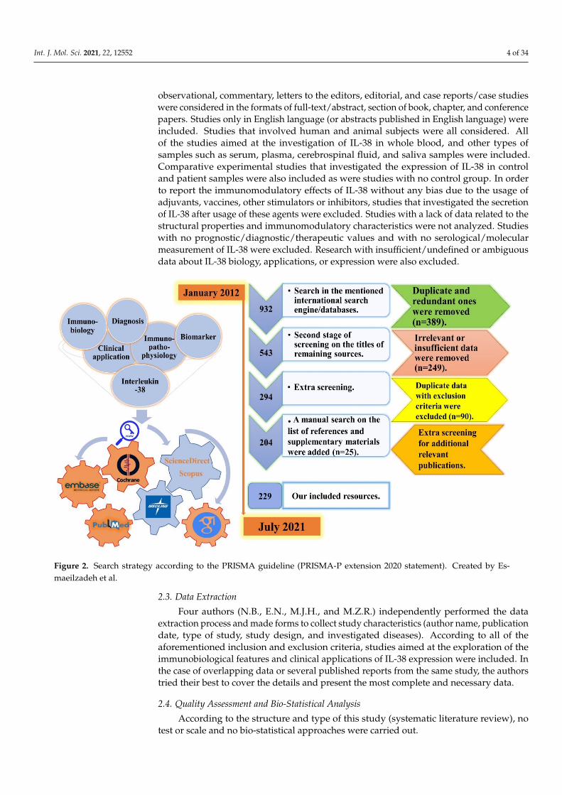
2. Methodology This present systematic literature review study was performed according to the Pre-
ferred Reporting Items for Systematic reviews and Meta-Analyses (PRISMA) statement guidelines (https://www.equator-network.org/reporting-guidelines/prisma/, accessed on 13 August 2021) (Figure 2).
Figure 1. Involvement of IL-38 in immune-mediated diseases. Created by Esmaeilzadeh et al.
2. Methodology
This present systematic literature review study was performed according to the Pre-ferred Reporting Items for Systematic reviews and Meta-Analyses (PRISMA) statementguidelines (https://www.equator-network.org/reporting-guidelines/prisma/, accessedon 13 August 2021) (Figure 2).
2.1. Literature Search Strategy, and Screening Process
An electronic comprehensive literature search was conducted with the time intervalstarting from January 2012 to July 2021 by using six main keywords (Biomarker, ANDClinical Applications, AND Diagnosis, AND Immunobiology, AND Immunopathophys-iology, AND Interleukin-38), and five complementary ones (Immunomodulation, ANDInflammatory Diseases, AND Interleukin-1 Superfamily, AND Interleukin-36 sub-family,AND IL-38). The selection was based on our inclusion and exclusion criteria, which arementioned in the next subsection.
In order to find potentially eligible resources, the screening process for our searchstrategy was independently conducted by N.B. in three main and one backward steps (onthe references and bibliographies of the included articles) according to all of the consideredkeywords and inclusion/exclusion criteria. Any uncommon points or disagreements werereferred to the corresponding author for consultations (Figure 2).
2.2. Inclusion and Exclusion Criteria
According to the aim of this study, all of the published original (experimental andnon-experimental) and review (mini-reviews, best evidence, narrative reviews, systematicreviews, systematic reviews, and meta-analysis), comparative, cross-sectional, cohort,
Int. J. Mol. Sci. 2021, 22, 12552 4 of 34
observational, commentary, letters to the editors, editorial, and case reports/case studieswere considered in the formats of full-text/abstract, section of book, chapter, and conferencepapers. Studies only in English language (or abstracts published in English language) wereincluded. Studies that involved human and animal subjects were all considered. Allof the studies aimed at the investigation of IL-38 in whole blood, and other types ofsamples such as serum, plasma, cerebrospinal fluid, and saliva samples were included.Comparative experimental studies that investigated the expression of IL-38 in controland patient samples were also included as were studies with no control group. In orderto report the immunomodulatory effects of IL-38 without any bias due to the usage ofadjuvants, vaccines, other stimulators or inhibitors, studies that investigated the secretionof IL-38 after usage of these agents were excluded. Studies with a lack of data related to thestructural properties and immunomodulatory characteristics were not analyzed. Studieswith no prognostic/diagnostic/therapeutic values and with no serological/molecularmeasurement of IL-38 were excluded. Research with insufficient/undefined or ambiguousdata about IL-38 biology, applications, or expression were also excluded.
Int. J. Mol. Sci. 2021, 22, x FOR PEER REVIEW 4 of 36
Figure 2. Search strategy according to the PRISMA guideline (PRISMA-P extension 2020 statement). Created by Esmaeil-zadeh et al.
2.1. Literature Search Strategy, and Screening Process An electronic comprehensive literature search was conducted with the time interval
starting from January 2012 to July 2021 by using six main keywords (Biomarker, AND Clinical Applications, AND Diagnosis, AND Immunobiology, AND Immunopathophys-iology, AND Interleukin-38), and five complementary ones (Immunomodulation, AND Inflammatory Diseases, AND Interleukin-1 Superfamily, AND Interleukin-36 sub-family, AND IL-38). The selection was based on our inclusion and exclusion criteria, which are mentioned in the next subsection.
In order to find potentially eligible resources, the screening process for our search strategy was independently conducted by N.B. in three main and one backward steps (on the references and bibliographies of the included articles) according to all of the consid-ered keywords and inclusion/exclusion criteria. Any uncommon points or disagreements were referred to the corresponding author for consultations (Figure 2).
2.2. Inclusion and Exclusion Criteria According to the aim of this study, all of the published original (experimental and
non-experimental) and review (mini-reviews, best evidence, narrative reviews, systematic reviews, systematic reviews, and meta-analysis), comparative, cross-sectional, cohort, ob-servational, commentary, letters to the editors, editorial, and case reports/case studies were considered in the formats of full-text/abstract, section of book, chapter, and confer-ence papers. Studies only in English language (or abstracts published in English language) were included. Studies that involved human and animal subjects were all considered. All of the studies aimed at the investigation of IL-38 in whole blood, and other types of sam-ples such as serum, plasma, cerebrospinal fluid, and saliva samples were included. Com-parative experimental studies that investigated the expression of IL-38 in control and pa-tient samples were also included as were studies with no control group. In order to report the immunomodulatory effects of IL-38 without any bias due to the usage of adjuvants,
Figure 2. Search strategy according to the PRISMA guideline (PRISMA-P extension 2020 statement). Created by Es-maeilzadeh et al.
2.3. Data Extraction
Four authors (N.B., E.N., M.J.H., and M.Z.R.) independently performed the dataextraction process and made forms to collect study characteristics (author name, publicationdate, type of study, study design, and investigated diseases). According to all of theaforementioned inclusion and exclusion criteria, studies aimed at the exploration of theimmunobiological features and clinical applications of IL-38 expression were included. Inthe case of overlapping data or several published reports from the same study, the authorstried their best to cover the details and present the most complete and necessary data.
2.4. Quality Assessment and Bio-Statistical Analysis
According to the structure and type of this study (systematic literature review), notest or scale and no bio-statistical approaches were carried out.

Int. J. Mol. Sci. 2021, 22, 12552 5 of 34
2.5. Ethical Statement
According to the structure and type of study (systematic literature review), there wasno need to register this project with the Research Ethical Committee (REC). It is worthmentioning that all of the data that support the findings of this study are openly availablein the context of this manuscript.
3. Results3.1. Immunobiology of IL-38
IL-1 superfamily members including IL-38 are involved in immune regulation, hostdefense, and inflammatory cascades. According to immunohistochemistry (IHC) analysis,IL-38 is secreted from many cells including keratinocytes, monocytes, and macrophages,and is found in many tissues such as tonsil, skin, spleen, thymus, heart, fetal liver, andplacenta. Recently, it has been demonstrated that IL-38 is mainly found in the cytoplasm ofhuman keratinocytes [23–28]. Secretion of IL-38 from apoptotic cells is not properly recog-nized. It has only been demonstrated that an impaired inflammation is considered as thesequel of IL-38 secretion from apoptotic cells. In other words, secretion and processing of IL-38 to truncated forms of IL-38 can lead to an immunoregulation by apoptotic cell-dependentsignaling, eventuating to the resolution of inflammation or autoimmunities [12,13,29,30].The IL-1 superfamily consists of several cytokines that are categorized into different groupsbased on their effects on receptors. In this sense, some of these cytokines are generallyknown as agonists (e.g., IL-1α, IL-1β, IL-18, IL-33, IL-36α, IL-36β, and IL-36γ), whereasothers are as considered as receptor antagonists (e.g., IL-1RA, IL-36RA, and IL-38). Anothermember, IL-37, is an anti-inflammatory agent (Figure 3) [31–52].
Int. J. Mol. Sci. 2021, 22, x FOR PEER REVIEW 6 of 36
Figure 3. Subtypes of the IL-1 family and their different functions. Created by Esmaeilzadeh et al. The immune functions of members of the IL-1 superfamily depend on their receptor.
There is another classification for the IL-1 superfamily, which is based on pro-inflam-matory and anti-inflammatory activities of members, dividing the IL-1 superfamily into two distinct groups. Among the members of the IL-1 superfamily, IL-38, IL-1Ra, IL-36Ra, and IL-37 are in the anti-inflammatory group [53]. IL-38 is the most recently known mem-ber of the IL-1 superfamily, originally named IL-1F10, IL-1HY2, FKSG75, IL-1-theta, MGC11983, MGC119832, and MGC119833 in various nomenclature systems, in which IL-1F10 is the most practical one. IL-33 and IL-36 subfamilies as well as IL-37 and IL-38 are discovered by an in silico gene identification system [54–57].
The genomic position of IL-38 in a cluster on chromosome 2 is between IL-36N and IL-1RN encoding genes (ch2q13_14.1). IL-38 shows 43% and 41% homology with IL-36N and IL-1RN, respectively. Biologically, IL-38 indicates a lower affinity in comparison with IL-1RA and IL-1β [26,27,53,58–62]. The IL-1F10 gene has five exon regions, encoding the 152 amino acid protein with 17–18 kDa molecular weight [54]. IL-38 is found in all types of vertebrates with developed immune responses [37]. Regarding purification and expres-sion of IL-38 in bacterial systems, there is one study conducted by Yuan et al. who suc-cessfully achieved this. They used a prokaryotic expression vector (named pET-44) to evaluate the expression of IL-38 in a one-step Ni2+-agarose affinity chromatography in a relatively large quantity of a C-terminus tagged IL-38 [27].
As previously mentioned, IL-38 is expressed in the skin, spleen, tonsils, thymus, heart, and liver tissues of the fetus in healthy people [63,64]. However, lower levels of this cytokine in these organs do not induce a specific role in the immunity processes [63]. It has been demonstrated that major functions of IL-38 are apoptosis-mediated phagocyte regulation [65], and inhibition of the induction of responses related to T Helper17 (TH17) cytokines. Additionally, IL-38 functions as an antagonist for IL-36, explaining the anti-inflammatory effects of IL-38 on the immune cells [53,66,67]. As IL-38 shows homology with IL-1Ra (an IL-1R1A) and with IL-36RA, it can be found that IL-38 has antagonistic functions with IL-1R1 and IL-36, respectively [20]. Eventually, pre-inflammatory and anti-
Figure 3. Subtypes of the IL-1 family and their different functions. Created by Esmaeilzadeh et al. The immune functions ofmembers of the IL-1 superfamily depend on their receptor.
There is another classification for the IL-1 superfamily, which is based on pro-inflammatoryand anti-inflammatory activities of members, dividing the IL-1 superfamily into twodistinct groups. Among the members of the IL-1 superfamily, IL-38, IL-1Ra, IL-36Ra, andIL-37 are in the anti-inflammatory group [53]. IL-38 is the most recently known member ofthe IL-1 superfamily, originally named IL-1F10, IL-1HY2, FKSG75, IL-1-theta, MGC11983,MGC119832, and MGC119833 in various nomenclature systems, in which IL-1F10 is the

Int. J. Mol. Sci. 2021, 22, 12552 6 of 34
most practical one. IL-33 and IL-36 subfamilies as well as IL-37 and IL-38 are discovered byan in silico gene identification system [54–57].
The genomic position of IL-38 in a cluster on chromosome 2 is between IL-36N andIL-1RN encoding genes (ch2q13_14.1). IL-38 shows 43% and 41% homology with IL-36Nand IL-1RN, respectively. Biologically, IL-38 indicates a lower affinity in comparison withIL-1RA and IL-1β [26,27,53,58–62]. The IL-1F10 gene has five exon regions, encoding the152 amino acid protein with 17–18 kDa molecular weight [54]. IL-38 is found in all types ofvertebrates with developed immune responses [37]. Regarding purification and expressionof IL-38 in bacterial systems, there is one study conducted by Yuan et al. who successfullyachieved this. They used a prokaryotic expression vector (named pET-44) to evaluate theexpression of IL-38 in a one-step Ni2+-agarose affinity chromatography in a relatively largequantity of a C-terminus tagged IL-38 [27].
As previously mentioned, IL-38 is expressed in the skin, spleen, tonsils, thymus, heart,and liver tissues of the fetus in healthy people [63,64]. However, lower levels of thiscytokine in these organs do not induce a specific role in the immunity processes [63]. Ithas been demonstrated that major functions of IL-38 are apoptosis-mediated phagocyteregulation [65], and inhibition of the induction of responses related to T Helper17 (TH17)cytokines. Additionally, IL-38 functions as an antagonist for IL-36, explaining the anti-inflammatory effects of IL-38 on the immune cells [53,66,67]. As IL-38 shows homologywith IL-1Ra (an IL-1R1A) and with IL-36RA, it can be found that IL-38 has antagonisticfunctions with IL-1R1 and IL-36, respectively [20]. Eventually, pre-inflammatory and anti-inflammatory effects of IL-38 were considered as dose-dependent immunological responses.As a prime instance, results acquired from a study on an IL-38 knock-out murine model ofasthma (KO) showed that severe clinical manifestations are associated with upregulationin the expression of IL-1β and IL-6 genes in the joints compared to the control ones,meanwhile, recombinant IL-38 could not be able to inhibit arthritis progression [68,69].
3.1.1. Activation of the IL-1 Superfamily Members
Some members in the IL-1 superfamily are activated and matured by a variety ofproteases including caspase 1, elastase, cathepsin G, and caspase 8 [54]. Biologically, IL-38is released from cells without peptide signaling. An inflammasome caspase-1-independentpathway is triggered for the activation of stimuli-induced intracellular IL-1 superfamilycytokines including IL-1α, IL-1Ra, IL-33, IL-36α, IL-36β, IL-36γ, IL-36RA, IL-37, and IL-38 [54,61]. From functional aspects, although the full-length molecule of IL-38 is bioactive,it lacks an integrated N-terminus to function as much as a processed cytokine [60], fur-thermore, there is even no nuclear localization signal (NLS) on IL-38 for caspase 1 andproteases [9,23].
3.1.2. IL-38 Receptors
Receptors of IL-1 superfamily members vary from IL-1R1 to IL-1R10. Following theinteraction with specific ligands, signaling processes result in the activation of severalimmunological pathways including nuclear factor-kappa B (NF-κB) or JNK/activatorprotein-1 (AP-1) [54,70]. Three are several receptors interacting with IL-38. Among them,there are three receptors that have shown great biological potentials to interact withIL-38 including IL-36R, IL-1R1, and interleukin-1 receptor accessory protein-like 1 (IL-1RAPL1) [54].
It is worth-mentioning that there were ambiguities in the immunobiological functionsof IL-38 when it was first discovered. On one hand, by binding to IL-36R and preventingthe interleukin-1 receptor accessory protein (IL-1RAcP) from functioning, IL-38 acts asan antagonist for pro-inflammatory properties of IL-36m especially in peripheral bloodmononuclear cells (PBMCs) (Figure 4). On the other hand, the dual functions of IL-38introduce IL-38 as an agonist for inflammatory responses, especially in dendritic cells(DCs). To be more precise, due to the existence of a wide array of documents related tothe inhibitory properties of IL-38 on IL-36 [71], IL-38 is mainly considered to be a potent

Int. J. Mol. Sci. 2021, 22, 12552 7 of 34
anti-inflammatory cytokine. This anti-inflammatory function of IL-38 is deeply rootedin the suppression of NF-κB and mitogen-activated protein kinases (MAPKs) signalingby MyD88 (Figure 4), which is normally found in some fungal infections (Candidiasis),psoriasis, and rheumatic diseases. It is noteworthy that IL-38 acts as a natural inhibitor ofcandida-induced production of IL-2 and IL-17 [23,26,44,52,60,67,72–74]. Additionally, it hasbeen demonstrated that IL-38 can play an indisputable role in the inhibition of β-glucan-induced trained immunity through blocking mTOR signaling, leading to long-lastinganti-inflammatory activities by IL-38 [75].
Int. J. Mol. Sci. 2021, 22, x FOR PEER REVIEW 8 of 36
Figure 4. Immunobiological activities of IL-38 in host immune system. Created by Esmaeilzadeh et al. IL-38 acts as an antagonist for the pro-inflammatory properties of IL-36 in PBMCs and as an agonist for inflammatory responses in DCs.
All in all, according to the controversial anti-inflammatory effects of IL-38, further investigations are needed for more crucial roles and more efficacious targeted therapies [76].
IL-1R1 IL-1R1 is related to IL-1α, IL-1β, and is produced in many cells that activate IL-1R3
following binding of the mentioned IL-1 cytokines [54]. Since IL-38 has 41% homology with IL-1Ra (an IL-1R1A), this cytokine also acts as an IL-1R1 inhibitor [54,62]. The affinity of IL-38 for binding to IL-1R1 is less than two other receptors, which is explained in the next subsections [54].
IL-36R IL-36R is related to cytokines IL-1α, β, γ, and members of the IL-36 subfamily [54].
After the binding of IL-36 subfamily members to IL-36R and IL-1RAPL1, activation of the NF-κB and MAPKs signaling pathways occurs (Figure 5) [54,77]. Since IL-38 has 43% ho-mology in the sequence with IL-36RA, both of them have similar antagonistic effects on IL-36 [54,59,78,79]. It has been demonstrated that the whole length of cDNA in IL-38 is 459 base pairs (bp), encoding the product of 152 amino acids [80].
Figure 4. Immunobiological activities of IL-38 in host immune system. Created by Esmaeilzadeh et al. IL-38 acts as anantagonist for the pro-inflammatory properties of IL-36 in PBMCs and as an agonist for inflammatory responses in DCs.
All in all, according to the controversial anti-inflammatory effects of IL-38, furtherinvestigations are needed for more crucial roles and more efficacious targeted therapies [76].
IL-1R1
IL-1R1 is related to IL-1α, IL-1β, and is produced in many cells that activate IL-1R3following binding of the mentioned IL-1 cytokines [54]. Since IL-38 has 41% homologywith IL-1Ra (an IL-1R1A), this cytokine also acts as an IL-1R1 inhibitor [54,62]. The affinityof IL-38 for binding to IL-1R1 is less than two other receptors, which is explained in thenext subsections [54].
IL-36R
IL-36R is related to cytokines IL-1α, β, γ, and members of the IL-36 subfamily [54].After the binding of IL-36 subfamily members to IL-36R and IL-1RAPL1, activation ofthe NF-κB and MAPKs signaling pathways occurs (Figure 5) [54,77]. Since IL-38 has 43%homology in the sequence with IL-36RA, both of them have similar antagonistic effects onIL-36 [54,59,78,79]. It has been demonstrated that the whole length of cDNA in IL-38 is 459base pairs (bp), encoding the product of 152 amino acids [80].

Int. J. Mol. Sci. 2021, 22, 12552 8 of 34Int. J. Mol. Sci. 2021, 22, x FOR PEER REVIEW 9 of 36
Figure 5. Interactions between IL-38 and involved immunological cells. Created by Esmaeilzadeh et al. Three important receptors of IL-38 and their immunomodulatory effects alter the secretion of pro-inflammatory and anti-inflammatory cytokines. Hence, IL-38 acts as a two-edged sword in immune microenvironments.
This receptor is widely expressed in human fibroblast cells and keratinocytes with various patterns of expression in mice and humans [54]. Murine macrophages can express this receptor continuously, whereas, the situation of IL-36R expression in humans is not the same [54]. Additionally, it has been reported that IL-38 is unable to show its immuno-biological effects due to the lack of IL-36R in THP1 cells [27,54].
IL-1 Receptor Accessory Protein-Like 1 (IL-1RAPL1) IL-1RAPL1 is extensively expressed in the brain tissues and there is no evidence of
the expression of IL-1RAPL1 in the lymph node, spleen, and bone marrow [54]. MORA et al. reported that IL-38 was also present on the macrophages and was more expressed in contact with the apoptotic conditioned media. They reported that binding of IL-38 to the receptors on the surface of the macrophages eventuated to inhibit the secretion of IL-6. This result supports the hypothesis that cleaved forms of IL-38 can decrease IL-6 expres-sion by binding to IL-1RAPL1, whereas, full-length forms of IL-38 have the potential to increase it (Figure 5) [54,81].
Figure 5. Interactions between IL-38 and involved immunological cells. Created by Esmaeilzadeh et al. Three importantreceptors of IL-38 and their immunomodulatory effects alter the secretion of pro-inflammatory and anti-inflammatorycytokines. Hence, IL-38 acts as a two-edged sword in immune microenvironments.
This receptor is widely expressed in human fibroblast cells and keratinocytes withvarious patterns of expression in mice and humans [54]. Murine macrophages can ex-press this receptor continuously, whereas, the situation of IL-36R expression in humansis not the same [54]. Additionally, it has been reported that IL-38 is unable to show itsimmunobiological effects due to the lack of IL-36R in THP1 cells [27,54].
IL-1 Receptor Accessory Protein-Like 1 (IL-1RAPL1)
IL-1RAPL1 is extensively expressed in the brain tissues and there is no evidence ofthe expression of IL-1RAPL1 in the lymph node, spleen, and bone marrow [54]. MORAet al. reported that IL-38 was also present on the macrophages and was more expressed incontact with the apoptotic conditioned media. They reported that binding of IL-38 to thereceptors on the surface of the macrophages eventuated to inhibit the secretion of IL-6. Thisresult supports the hypothesis that cleaved forms of IL-38 can decrease IL-6 expression bybinding to IL-1RAPL1, whereas, full-length forms of IL-38 have the potential to increase it(Figure 5) [54,81].

Int. J. Mol. Sci. 2021, 22, 12552 9 of 34
3.1.3. Inhibitory Properties of IL-38
As we reviewed earlier, on one hand, IL-38 binds to the IL-36R and provides inhibitoryeffects similar to IL-36RA including anti-inflammatory effects on PBMCs [82]. On the otherhand, the pro-inflammatory effects of IL-38 on the DCs are provided by increasing theproduction of IL-6 [57,82].
In several in vitro studies, it has been reported that IL-38 “directly” inhibits theproduction of cytokines from THP1 cells. IL-38, only at low doses (10 ng/mL), inhibitsthe production of IL-17 and IL-22 cytokines, rather than high doses (1 µg/mL). Similarto IL-36RA, IL-38 inhibits the production of IL-8, leading to inhibition of MAPK/NF-κBsignaling in keratinocytes (with 30% less potency than IL-36Ra). Truncated and purifiedforms of IL-38 inhibit IL-36γ-induced secretion of IL-8. Additionally, IL-38 can reducethe pro-inflammatory effects of IL-6, monocyte chemoattractant protein-1 (MCP-1), C–Cmotif chemokine ligand 2 (CCL2), and A proliferation-inducing ligand (APRIL) up to30 times [23,27,32,36,41,69,81–87].
In “indirect” processes, during lipopolysaccharide (LPS) stimulation, IL-38 diminishesToll-like receptor-4 (TLR-4)-mediated inflammation through decreasing levels of IL-6 andinterleukin-23 (IL-23) in THP 1 cells or primary M1 macrophages. Therefore, if targetingTLR signaling in this “indirect” pathway is recruited by an “IL-38-mediated targeting”, itcan be considered as a promising therapeutic approach in inflamed skin. However, thesignaling pathway of IL-38 still requires further investigation [81,88,89].
Results from several studies have reported that the concentration of IL-38 can be adetermining factor affecting the intensity of immunomodulatory properties by IL-38 [82].As a matter of fact, in high concentrations of IL-38, there is a decreasing trend for itsinhibitory effects. This point indicates a relative anthropometric function for IL-38. Toexplain this more clearly, there is a hypothetical inhibitory curve that IL-38 may use at lowconcentrations, but this crosstalk has not been scientifically detected yet [82].
3.2. Role of IL-38 in Immune-Mediated Diseases
Over the past few years, the tendency to investigate the immunobiological roles ofIL-38 in immune-mediated diseases has matured [90], which is shown by the schematicpresentation in Table 1.
Table 1. Role of IL-38 in Immune-Mediated Diseases.
Immune-Mediated Diseases Immunological Roles of IL-38 Reference
Inflammatory Bowel diseases (IBD)Protective effect in IBD, through production of pro-inflammatory
cytokines from macrophages, and a promising immunotherapeutictarget in IBD.
[91]
Acne Vulgaris Exacerbation of skin inflammation. [92]
Behcet’s Disease (BD) Exacerbation of eye involvement, and a protective anti-inflammatoryrole in BS. [93,94]
Intervertebral Disc Degeneration (IVDD)Therapeutic roles through alleviation of the inflammatory responsesand the degeneration of nucleus pulpous cells via inhibition of the
NF-κB signaling pathway.[95]
Alzheimer Novel biochemical marker with anti-inflammatory activities. [96]
Ischemic stroke Novel early predictor factor for ischemic stroke prognosis. [97]
Autism spectrum disorder Therapeutic role through inhibition of activation of human microglia. [98]
Thyroid-associated ophthalmopathy (TAO) Protective role in TAO, a promising marker of TAO disease activity,and a potential target for TAO therapy. [99]
Multiple sclerosis (MS) Development of through attenuated inflammatory conditions in earlystages of MS. [100]
Experimental autoimmuneencephalomyelitis (EAE) Promotion of inflammation in the central nervous system (CNS). [101]

Int. J. Mol. Sci. 2021, 22, 12552 10 of 34
Table 1. Cont.
Immune-Mediated Diseases Immunological Roles of IL-38 Reference
Candidiasis Dose-response reduction in Candida-induced T helper 17 responses. [102]
Arthritis Significant reduction in clinical inflammation and attenuated severityin mouse models of arthritis. [103]
Systemic sclerosis Role in the pathogenesis of systemic sclerosis. [104]
Atopic dermatitis (eczema) Prognostication of atopic severity and its inflammatory state inatopic sufferers. [105]
OsteoporosisInhibited proliferation of BMSCs and inhibited apoptosis of
osteoblasts by regulating the PI3K/Akt/GSK3β/NFATc1signaling pathway.
[106]
Asthma Development of a regulatory cytokine-based treatment forallergic asthma. [107]
Chronic inflammatory demyelinatingpolyneuropathy (CIDP)
Making a compensatory mechanism to reduce inflammatoryprocesses in these patients. [108]
Atopic dermatitis, allergic asthma, andallergic rhinitis
Therapeutic potential in the regulation of allergy asthma, andallergic rhinitis. [109]
Brucellosis Progression from acute into the chronic forms of brucellosis. [110]
inflammatory diseases (psoriasis,rheumatoid arthritis, gout, systemic lupus
erythematosus, and Crohn’s disease)Involved in the pathogenesis of inflammatory diseases. [111]
3.2.1. Role of IL-38 in Autoimmune DiseasesRheumatoid Arthritis (RA)
RA is a very common autoimmune disease with long-term chronic inflammation ofthe synovium, mainly causing swollen and painful joints in the wrist and hands. RA iscommonly manifested by a systemic inflammation in the hands, fingers, knees, ankles,elbows, hips, and shoulders. These clinical manifestations are related to the cellularinteractions between resident cells (fibroblast-like synoviocytes (FLS)) and immune cellsinvolved in the innate and adaptive immune system [112]. IL-38 is possibly inducedby synovial fibroblasts, monocytes, and macrophages in RA, and it can diminish theimmunobiological functions of IL-36 agonists in the pathogenesis of RA. IL-36 agonists,IL-36RA and IL-38, were induced in thee synovium of the majority of patients with RA, andthe total balance in the pathogenesis of RA was attributed to these potential antagonists.These are produced by many different cell types such as myeloid and plasma cells, andtheir expression correlates with macrophage-colony stimulating factor (M-CSF), CCL3,and CCL4. Other cells such as endothelial cells, fibroblasts, and enterocytes could alsoparticipate in RA pathogenesis [12,54,85].
Consequently, on one hand, overexpression of IL-38 induces anti-inflammatory ef-fects in mice with RA as well as human macrophages in vitro. In a clinical study, asignificant decrease in the filtration of macrophages as well as expression of TH17 cy-tokines were reported after an articular injection of IL-38-encoding adeno-associated virus(AAV). Additionally, there was a significant decrease in the clinical scoring of inflammationin collagen-induced arthritis (CIA) and serum transfer-induced arthritis (STIA) modelmice [12,113]. On the other hand, IL-23, whose expression is normally reduced by IL-38,is highly expressed by DCs. Therefore, in this study, it was reported that IL-38 couldtarget DCs as well as macrophages. At the same time, it did not affect cartilage and bonedestruction. Interestingly, over-expression of IL-38 may lead to an augmented expression ofosteogeneic factors, inhibited neovascularization, and improved damage in cartilage of RArabbits or murine models of clinical trials. Hence, IL-38 might be promising to consider asa potential seroimmunobiomarker, being targeted for the regenerative medicine purposesof RA [113–117].

Int. J. Mol. Sci. 2021, 22, 12552 11 of 34
Increased levels of the IL-38 cytokine in mouse models of RA has a positive correlationwith synovial fluid levels of IL-1β and a negative correlation with IL-17 responses as wellas with TNF-α production. IL-38 has the potential to suppress IL-17 and IL-22 secretion,similar to IL-36Ra. Upsurged levels of the IL-1 superfamily and IL-36 subfamily membersdepict a negative feedback for the inhibition of inflammatory responses in RA [24,63].Additionally, immunomodulatory properties of IL-38 in the pathogenesis of RA likelyreduced the filtration of macrophages into the synovium, and the reduced TH17 productionshould not be underestimated [12,54,103].
In CIA immunopathogenesis, it is noteworthy that the increase in IL-36/IL-36Raoccurs in the initial phases, whereas the elevation of articular IL-38 is postponed andaccurately will occur at the later phases. The excessive expression of IL-38 attenuatesthe severity of clinical manifestations in CIA and STIA types of RA, and improves theclinical scores of the disease through the reduction in TH17 cytokine patterns. Conversely,excessive expression of IL-38 led to more severe clinical manifestations in antigen-inducedarthritis (AIA). Additionally, overexpression of IL-38 did not indicate any significant effectson bone erosion (contrary to IL-1RA) [63,69,81,113,118].
To sum up, overexpression of IL-38 in the synovial membrane and sera leads tolessened functions of macrophages, TNF-α, IL-6, IL-10, TH17 cytokines (IL-17, IL-22), C-X-C motif chemokine ligand 1 (CXCL1), and CXCL8 expression [68,119]. Correspondingly, itwas demonstrated that IL-38 showed suppressive impacts on LPS-mediated TLR4 secretion,leading to an attenuated inflammation in patients with RA through inhibition of theactivation of NF-κB [120]. Therefore, if we consider both IL-36RA and IL-38 cytokines aspotential IL-36 antagonists, only a minor subpopulation of patients with RA (17–29%) havean elevated agonist/antagonist ratio [85,121].
Takenaka et al. reported higher serum and synovial levels of the IL-38 protein inpatients with RA as well as lower concentrations of IL-38 in osteoarthritis (OA) patientsand normal subjects. Serum levels of IL-38 in patients with RA did not correlate withtheir disease activity, treatment, or disease duration [122]. In another study, Wang-DongXu et al. found that there was a direct correlation between IL-38 expression and diseaseactivity in RA patients. Along with exacerbation in the inflammatory conditions, therewas a significant reduction in the levels of IL-38 expression. Hence, this cytokine wasidentified as an appropriate and promising seroimmunobiomarker for the diagnosis of RAand determination of RA severity [113,115,123].
In another newly conducted comparative study by Lifeng Jiang et al. [124], they re-ported promising findings on the association of IL-38 expression in the chondrocytes ofpatients with OA. Assessment of IL-38 was conducted on serum and synovial fluid of75 patients with OA, who had undergone joint replacement before the assay, and 25 age-and sex-matched healthy persons as a control group. They reported remarkably ele-vated serum and synovial fluid levels of IL-38 in patients with OA when compared tothe control group. These findings were proven by an attenuated expression of severalpro-inflammatory cytokines and were positively correlated with early disease activity.Altogether, they reported that IL-38 could promisingly serve as a novel immunotherapeutictarget, and a screening seroimmunobiomarker for patients with OA [124]. Results of thisstudy are in accordance with another study aimed at evaluating the anti-inflammatoryproperties of IL–38 in murine models of arthritis and systemic inflammation [125]. In thisstudy, inhibitory features of IL-38 in streptococcal cell wall (SCW)-induced arthritis andmonosodium urate (MSU) crystal-induced arthritis were recognized by a reduction injoint swelling, inflammatory cell influx, and synovial levels of several pro-inflammatorycytokines, clearly indicating the potential abilities of IL-38 as an acceptable seroimmuno-biomarker and immunotherapeutic target for patients with arthritis [125]. Additionally,Mitra Abbasifard et al. reported higher expression of IL-38 in patients with severe OA,which were approved by WOMAC scores > 40, VAS scores > 5, and BMI < 25 indices in 23newly-diagnosed patients with OA compared to 22 sex- and age-matched healthy personsas a control group [126].

Int. J. Mol. Sci. 2021, 22, 12552 12 of 34
Systemic Lupus Erythematosus (SLE)
Multiple genetic predispositions and environmental factors contribute to aggravatedpathogenesis of SLE as a multi-systemic autoimmune disease. In patients with SLE, a widerange of organs and tissues of the host (including skin, joints, blood, CNS, and kidneys)are clinically affected. Tissue damage and disruption of their normal functions are directconsequences of long-time inflammation in patients with SLE. Subsequently, the main goalof SLE therapy will be a reduction in inflammation in the immunopathogenesis of thediseases [118]. There are variations caused by complex single-nucleotide polymorphisms(SNPs) in the locus of the IL-1 superfamily, which are associated with a higher activity of IL-1 superfamily members (especially IL-1A activity), altering the susceptibility of individualsto SLE development [127].
In a C57/BL6 mouse model-based study, the immunobiological effect of IL-38 injectionin gouty arthritis was investigated through the administration of albumin-opsonized MSUcrystals. It was reported that intraperitoneal injection of 1µg recombinant IL-38 hadalleviated proteinuria, skin lesions, and nephritis two hours before the induction of goutyarthritis. Additionally, injection of 1µg recombinant IL-38 also reduced the serum levelsof IL-17 and IL-22 cytokines [63,81]. Recombinant IL-38 induces downregulatory impactson the secretion of pro-inflammatory cytokines (IL-17, IL-22, and IL-36γ), and increasesthe expression levels of IL-6, CCL2, and APRIL. These findings, all in all, indicate theanti-inflammatory activities of IL-38 on disease activity and organ involvement in a mousemodel of gouty arthritis [85].
Results of another study showed that concentration of serum IL-38 in patients withSLE was significantly higher compared to the healthy controls [63]. On the other hand,in patients with active SLE, the expression levels of IL-38 were higher than the ones inpatients with inactive SLE. Hence, the risk assessment for renal and CNS involvement isdirectly related to the level of IL-38 [55,63,85,118,128].
In their experimental study, Takeuchi et al. evaluated the serum levels of IL-38 in19 patients with SLE (juvenile SLE) through double-sandwich enzyme linked immunosor-bent assay (ELISA) and IHC (using a polyclonal anti-IL-38 antibody). They reportedundetectable serum levels of IL-38 in almost all cases throughout the disease course atthe diagnosis time before treatment (except one case). They suggested that IL-38 maynot be a sensitive disease-activity (disease-specific) biomarker in pediatrics with SLE. Inaddition, they suggested further investigation into the expression levels of IL-38 in largersamples of patients with juvenile SLE [129]. Another study introduced IL-38 as the firstmediator, being associated with disease severity, renal, and CNS involvement in patientswith SLE [130].
Chu et al. reported that intravenous administration of murine recombinant IL-38 intospecific kinds of mice can ameliorate skin inflammation and nephritis in mice with SLE,probably via suppressing the secretion of inflammatory cytokines such as IL-17 and IL-22.They suggest that IL-38 shows therapeutic potential for the recovery of skin and kidneyinvolvement in mice with SLE [131].
Psoriasis
As a common chronic inflammatory disorder, psoriasis is accompanied by an un-controlled proliferation of keratinocytes, several chemokines, and excessive cascades ofDCs infiltration, leading to a wide range of clinical manifestations such as dermatologiclesions, acanthosis, and cardiovascular comorbidities [5,38]. Severe clinical manifestationsin psoriatic patients emerge due to an aberrant expression of inflammatory agents suchas TNF-α, IL-2, IL-6, IL-12/IL-23p40, IL-17A, IL-17/IL-23, and interferon-gamma (IFN-γ)cytokines. Correspondingly, a wide array of novel monoclonal antibodies (Secukinumab,Briakinumab, Brodalumab) are going to be therapeutically used in clinical trials for psoria-sis according to the involved cytokines in the immunopathogenesis of psoriasis [5,72].
In a study accomplished by Han Y et al., they reported that in IL-38 KO mice, theprocess of disease resolution is postponed due to an aberrant secretion of IL-17, orches-

Int. J. Mol. Sci. 2021, 22, 12552 13 of 34
trating inflammation in the microenvironment of the diseases. Immune cells such as γδ Tcells are responsible for IL-17 cytokine production. There is a direct relationship betweenthe activation of γδ T cells and upregulation of IL-1RAPL1. Additionally, there are di-minished levels of IL-17 secretion and reduced inflammation by activated γδ T cells inpsoriatic IL-1RAPL1 KO mice. Additionally, clinical administration of mature IL-38 or γδT cell-receptor-blocking antibodies leads to an accelerated amelioration of the disease. Inanother study, higher expression levels of IL-38 were detected in pustular psoriasis in com-parison with psoriasis vulgaris and healthy controls [72,81,132]. Since IL-38 is reduced indifferentiated keratinocytes, the loss of IL-38 in the epidermal layers can play an importantrole in the severity of pathogenesis in psoriatic patients. Additionally, constitutive overex-pression of IL-38 decreases the proliferation and viability of normal human keratinocytes(NHKs), and is able to increase cell mortality, but due to the lack of sufficient experiments,it has not been fully understood yet [54,133,134]. Mast cells play a contributing role in theimmunobiology of IL-38, releasing IL-1 and stimulating the macrophages to express IL-36with its pro-inflammatory activities. Then, IL-38 and IL-36RA bind to IL-36R and inhibitinflammation in the psoriasis microenvironment, holding promising immune gene-basedtherapeutic capabilities of IL-38 and IL-36RA for psoriatic patients [135].
IL-38 polymorphism is associated with psoriatic arthritis, suggesting possible roles of ahigher expression levels of IL-38 in the lesional skin and exacerbated immunopathogenesisof this inflammatory skin disease. Conversely, in vitro addition of IL-38 in PBMC culturecould inhibit the production of IL-22 and IL-17A, presenting IL-38 as an agent involved inthe regulation of expression levels of IL-17 [61,136,137]. Although the exact role of IL-38 isnot completely elucidated, the correlations between SNPs in the regions encoding IL-38and susceptibility to psoriatic arthritis are thought to have contributed [138].
Boutet et al., in their mice model-based study, reported that IL-36RA is induced inpsoriatic skin, whereas IL-38 is reduced. Considering both IL-36RA and IL-38 as po-tential antagonists of IL-36, the majority of patients with psoriasis revealed an elevatedagonist/antagonist ratio, proposing key roles of IL-36 cytokines in psoriasis and theircorrelation with disease severity. In contrast, a reduction in IL-38 could have an impor-tant role in the immunopathogenesis of psoriasis through activated IL-36 agonists withpro-inflammatory properties. Hence, IL-38 can be promisingly considered as a prognosticseroimmunobiomarker for psoriasis [61,139].
Jennifer Palomo et al. measured the inhibitory effects of IL-38 on IL-36R in patientswith psoriasis based on the fact that IL-36R deficiency can decrease IMQ-induced skininflammation in psoriatic mice. Interestingly, their results showed no inhibitory effects ofIL-38 on IL-36R in patients with psoriasis [140]. Additionally, according to the results ofanother study, the inhibitory effects of IL-38 on inflammatory functions of IL-36 in the skinhave not been proven [64].
Atopic Dermatitis (AD)
As a chronic and itchy skin disorder, AD is immunologically recognized with anexacerbated inflammatory dermatosis, and intensified levels of TH2 cytokines. Althoughsome foods and environmental factors affect pathogenesis of AD, the real etiopathogenesisof AD is incompletely understood. Khattab et al., in their case-control study, surveyedthe serum levels of IL-38 in patients suffering from mild/moderate/severe atopic eczemacompared to the age- and sex-matched healthy controls. Results of ELISA revealed higherexpression levels of IL-38 in patients with atopic eczema. In addition, they reported asignificant correlation between serum levels of IL-38, disease severity, eosinophilia, and IgElevels. Altogether, elevated expression levels of IL-38 in the sera of patients with AD canpotentiate the specificity of eosinophilic count and IgE as inducer agents of TH2 responses,presenting IL-38 as a prognostic seroimmunobiomarker for disease severity [105].
In another most recently-published article, Lauritano et al. introduced allergic contactdermatitis as a provoker agent for AD immunopathogenesis. As is clear, involvement ofallergens and secretion of a majority of pro-inflammatory mediators (e.g., IL-1 superfamily

Int. J. Mol. Sci. 2021, 22, 12552 14 of 34
members) contribute to the aggravation of the pro-inflammatory situation in allergic con-tact dermatitis [141]. They reported that there are chains of anti-inflammatory activitiesrelated to IL-38, mainly including lessened allergic inflammation in the skin. Severalimmunobiological cascade processes by IL-38 are responsible for this reduced allergic in-flammation, and suppressed activities of IL-36, introducing IL-38 as an immunotherapeutictarget for allergic contact dermatitis. Altogether, the binding of mature IL-38 to IL-36R(IL-1R6) orchestrates the induction of inhibitory effects on the production of mast cell-derived proteases, prevention from the formation of biologically-active IL-36, expansion ofcommon proteases for IL-38 maturation, induction of inhibitory effects on the secretion ofIL-2, IL-17 cytokines, and inhibited production of IL-1, especially in macrophages [141].
Kawasaki Disease (KD)
KD is one of the inflammatory diseases that is commonly accompanied by vasculitis inchildren, increasing the risk of cardiovascular diseases (CVDs), especially when it remainsunresolved or mistreated. Environmental stimuli are also significant determinants in theclinical symptoms and manifestations of KD [79].
According to the immunological patterns of KD and major anti-inflammatory proper-ties of IL-38, it was demonstrated that in children with KD, the levels of IL-38 were higherin patients with an acute phase of KD compared to the healthy control group. These resultsreveal the immunobiological functions of IL-38, contributing to the inflammatory processof KD [121].
Gouty Arthritis
As one of the prototypes of IL-1β driven auto-inflammatory diseases, gouty arthritisclinically results from an excessive formation of acid uric crystals, especially in the joints ofthe toes. In this disease, neutrophils and other involved immune cells are collected in thesedimentation site following the deposition of MSU crystals in the joints [119]. A narrowrange of studies have been conducted to investigate the role of inflammatory factors andcytokines in the etiopathogenesis of this disease [142].
In an experimental study on a mouse model of gouty arthritis, it was hypothesized thatintraperitoneal administration of recombinant IL-38 leads to a reduction in inflammationin the joints and swelling. These profound alterations in the clinical manifestations ofthe disease are due to reduced secretion of IL-1β and IL-6 in the synovial membrane.These results indicate that IL-38 might be used as an acceptable cytokine-based targetedimmunotherapeutic approach for gouty arthritis [63].
Sjögren Syndrome (SS)
SS is a chronic systemic immune-mediated inflammatory disorder mainly character-ized by abnormal functions of exocrine glands, dryness, or irritability of the eyes, sensationof foreign bodies in the eyes, corneal scarring, dry mouth, and oral ulcers. The most recentlyknown immune axis involved in the immunopathogenesis of SS is the IL-23/IL-17/IL-22axis [143,144]. Not only do these patients suffer from poor lifestyle due to the abnormalclinical symptoms, but patients with SS are also prone to a shortened life expectancy byincreasing the risk of non-Hodgkin’s lymphoma [145].
In a study investigating the salivary gland levels of IL-38 in patients with primary SS(pSS), they reported higher levels of IL-38 in patients with pSS compared to the controlgroup [58].
In another study conducted by Ciccia F et al., it was reported that there was an upregu-lation in minor salivary gland and serum levels of IL-36α in a minority of patients with pSS,leading to a more severe disease activity. Additionally, in their minor salivary glands, therewas a down-regulation in the expression levels of IL-36RA, and an upregulation in theexpression levels of IL-38, aimed at counteracting the imbalanced activation of IL-36 [144].The latest molecular-based study conducted on the role of IL-38 in SS introduced IL-38 asan inhibitory agent for the secretion of chemokines involved in the TH17 signaling pathway,

Int. J. Mol. Sci. 2021, 22, 12552 15 of 34
preventing the exacerbation of immunopathophysiology involved in pSS, and acting as apromising targeted immunotherapeutic-based treatment for patients with pSS [146].
Crohn’s Disease (CD)
As a chronic inflammatory disease, the gastrointestinal tract is attacked by the hostimmune system in CD. Many studies have been conducted to understand the mechanismsof the inflammatory pathways in CD and significant results have been obtained [147]. Micewith colitis and patients with CD showed weakly augmented levels of IL-36α and IL-36γ.In a subgroup of patients with CD, there was a correlation between the expression levels ofIL-36α, IL-36γ, IL-38, and IL-17A. Hence, these results confirm a role for these cytokines inthe immunopathogenesis of CD, which can be used as prognostic or diagnostic approachesfor patients with CD [148].
In mouse and human inflamed colon, increased expression levels of IL-36α, IL-36γ,and IL-38 have been experimentally observed. Moreover, in an IL-36R-deficient mice, colitisdevelopment at early time points as well as defective recovery from mucosal injury werediminished. These results suggest major roles for IL-36γ in colonic inflammation and itsresolution. Further investigations are necessary to precisely clarify the immunobiologicalroles of other members of the IL-36 subfamily and IL-38 in the immunopathogenesis ofCD [149].
In another study aimed at the investigation of IL-38 levels in patients with RA andCD, it was reported that induced production of IL-38 (possibly by synovial fibroblasts,monocytes, and macrophages) could limit the immunobiological impacts of IL-36 agonistsin the immunopathogenesis of RA and CD. Future studies will certainly unravel more clueson the role of IL-38 in the immunopathophisiology of chronic inflammatory diseases suchas CD. Furthermore, there is an urgent need to precisely decipher the IL-38 antagonisticpotencies against members of the IL-36 subfamily or other triggered inflammatory agentsthrough in vitro and in vivo studies. In the colon of the majority of patients with CD,IL-36α, IL-36γ, and IL-38 were induced at low levels, potentiating IL-38 as an antagonist.These are produced by different cell types such as myeloid cells and plasma cells, and theirexpression correlates with TH17 cytokines [139].
In an original study designed by Fonseca Camarillo et al., they characterized tissueexpression of IL-38 and IL-36Ra, and their producing cells in patients with ulcerativecolitis (UC), CD, and other patients with remission of IBD. Colonic tissue levels of IL-38were increased in patients with inactive UC compared to active UC and control groups.Correspondingly, in patients with active CD, plasmacytoid DCs (pDCs) showed a highernumber on sub-mucosa, muscles, and adventitia compared to patients with active UC andnon-inflamed control tissue [148].
In another cross-sectional comparative study, Fonseca Camarillo et al. evaluated theexpression levels of IL-36α, IL-36β, IL-36γ, IL-36Ra, and IL-38 genes, their producingcells, and their correlation with clinical activity in IBD and non-inflamed non-IBD persons(as a control group) by reverse transcriptase-polymerase chain reaction (RT-PCT) anddouble-staining IHC techniques. In comparison with patients with CD and the controlgroup, there was an increase in the colonic mucosa levels of IL-36 subfamily members inpatients with active UC (except than IL-38) [150], whereas, laboratory data from real-timeRT-PCR depicted an increase in the tissue levels of IL-38 patients with inactive UC com-pared to patients with active IBD and non-inflamed non-IBD control groups. In addition,overexpression of IL-36α, IL-36β, IL-36γ, IL-36RA, and IL-38 was reported in intestinalepithelial cells, macrophages, and CD8+ T cells of patients with active IBD compared to thenon-inflamed non-IBD control group [150]. Altogether, elevated secretion of IL-38 fromimmune and non-immune cells in patients with active IBD introduces IL-38 as a practicalscreening and immunotherapeutic seroimmunobiomarker for gut inflammation [150].
Yuxia Zhao et al. [151] designed their comparative study to investigate the expressionlevels and molecular mechanisms of IL-38 in 115 patients with CD, 67 patients with UCas well as a control group (40 people) admitted to the referral Wuhan Children’s Hospital.

Int. J. Mol. Sci. 2021, 22, 12552 16 of 34
Several parameters such as C-Reactive protein (CRP), erythrocyte sedimentation rate (ESR),phosphorylated signal transduction and activator of transcription 3 (p-STAT3), IL-38, andNF-κB were measured in the intestinal mucosa of the studied population through IHCstaining [151]. Crohn’s Disease Activity Index (CDAI) for patients with CD, and Mayoscore system for patients with UC were also assessed [151]. The expression levels of IL-38 were quantitatively assessed by ELISA. For their mice-based molecular assessments,they established a dextran sulfate sodium (DSS) model for the induction of IBD in IL-38-C57BL/6 transgenic mice. Inflammatory markers and cells in the intestinal mucosa of thewildtype and IL-38-C57BL/6 transgenic mice were assessed by IHC and flowcytometrytechniques, respectively [151]. Results of this study reported lower intestinal mucosa levelsof IL-38 in both patient groups when compared to the control group (certified by IHCand ELISA), whereas decreased expression levels of mentioned inflammatory markerswere found in the control group when compared to both case groups. There was a reversecorrelation between disease activity, and expression levels of CRP, ESR, and IL-38. Resultsacquired from their mice-based study, showed similarity with the ones obtained from thepatients, indicating the therapeutic potentials of IL-38 in the immunopathogenesis of IBDby a reduction in inflammation through the inhibitory effects on p-STAT3 and NF-κB [151].
HLA-B27-Associated Anterior Uveitis and Idiopathic Anterior Uveitis (IAU)
IAU is an autoimmune systematic inflammatory disease, affecting anterior segment,iris, and ciliary body of the eye. Pathogenically autoreactive TH17 cells as well as increas-ing levels of IL-38 and IL-37 are of high prominence for investigation as diagnostic ortherapeutic tools for patients with IAU [152].
3.2.2. Role of IL-38 in Inflammatory DisordersInflammation-Induced Corneal Neovascularization
To study the immunobiological effects of IL-38 on corneal neovascularization, an alkali-induced corneal neovascularization mouse model has been used. It has been interrogatedthat if IL-38 is topically administrated in the injured cornea of the mentioned model,inflammation-induced angiogenesis is diminished. IL-38 is capable of attenuating theesecretion of IL-1β, IL-6, IL-8, and TNF-α cytokines and reducing angiogenesis-relatedactivities (e.g., proliferation, migration, and tube formation of human retinal endothelialcells) [153].
Hidradenitis Suppurativa (HS)
HS is chronic inflammatory disease presented with recurrent painful inflamed lesionsdue to the inflammatory processes in the terminal hair follicles. In a study, researchersaimed to evaluate the lesional and perilesional skin levels of the IL-36 subfamily members,IL-37 and IL-38 in patients with HS compared to the healthy controls [154]. Compared tohealthy controls, overexpression of IL-38 and reduced titration of IL-38 were reported inthe perilesional skin and lesional skin samples of patients with HS, respectively. Theseresults have been confirmed by real-time RT-PCR and IHC in these samples. Additionally,here, IL-38 showed anti-inflammatory properties by the suppression of IL-17 and IL-22cytokines [55,154].
Chronic Primary Angle Closure Glaucoma (CPACG)
As one of the worldwide leading causes of irreversible blindness, CPACG is mainly re-ported among female populations of Asian nations with a loss of visual functions [155–157].It has been demonstrated that there is an upregulated immune-mediated inflammationin acute primary angle closure glaucoma (APACG) and CPACG. For instance, there wereincreased levels of IL-6, IL-8, granulocyte-colony stimulating factor (G-CSF), MCP-1, andIP-10 in the aqueous humor of eyes or sera of patients with CPACG and APACG. In ad-dition, in an experimental study conducted by Jin-ling Zhang et al. (2019), there wereincreased levels of IL-38 in the aqueous humor of patients with CPACG in comparison

Int. J. Mol. Sci. 2021, 22, 12552 17 of 34
with age-related cataract (ARC) samples. In addition, a significant correlation was reportedbetween IL-38 levels and mean deviation of visual field (MDVF), proposing IL-38 as acritical factor in the immunopathophisiology of CPACG, even in the absence of a clinicallyconfirmed inflammation. Hence, it highlights the prognostic values of IL-38 in patientswith CPACG [157].
Retinal Ischemia and Proliferative Vascular Diseases
Damage from ischemic vascular diseases in the retina vary from a slow degradationof vision (due to the permanent ischemic retina) to a completely destroyed vision (dueto proliferative vascular disease). Oxygen-induced retinopathy (OIR) is a mouse modelfor studying retinal ischemia and proliferative vascular diseases in the retinal vasculature,demonstrating pivotal roles of several angiogenic factors and inflammatory cells in theaggravation of vascular immunopathogenesis [158].
Results acquired from a murine model-based study reported significant anti-angiogenicfunctions in the retina of the hyperoxia mice due to the injection of IL-38. This injection ledto an induction in the tube formation of vascular endothelial cells via the role of vascularendothelial cell growth factor (VEGF) and reduced severity [159].
3.2.3. Role of IL-38 in Metabolic DisordersType2 Diabetes Mellitus (T2DM) and Obesity
Obesity is clinically accompanied by chronic inflammatory responses. The mainmechanism accounting for obesity is a disturbed balance between receiving and consumingenergy. This imbalance may be due to the decreased physical activities or increased foodintake [160,161], and insulin resistance caused by T2DM [162]. T2DM is a metabolicdisorder in which several agents such as genetic factors, immunological agents, anddisturbed immune system play indispensable roles in aggravating the etiopathology ofT2DM. As a prime instance, overexpression of IL-6 can lead to complications such asdyslipidemia, which in turn, can cause obesity [163].
In a study, they found that SNPs in the IL-38 gene were associated with high serumlevels of CRP in patients with obesity and T2DM. Based on this evidence, IL-38 is likely toplay an important role in the inflammation involved in the immunopathogenesis of obesityand T2DM [164]. It has been demonstrated that there is a positive correlation betweenoverexpression of IL-38 and diabetic nephropathy in patients with T2DM [165].
In a newly conducted experimental study by Keye xu et al., the expression levels ofIL-38 were assessed in a mouse model of high-fat diet-induced obesity after hydrodynamic-based IL-38 gene delivery. Results of this study reported the anti-inflammatory effectsof IL-38 through the inhibition of IL-1β, IL-6, and MCP-1, and a reduction in liver fatcontent due to reduced adipose tissues [161]. Results of this study are in accordance withanother in vitro study that investigated the biological effects of IL-38 on adipogenesis andthe secretion of inflammatory cytokines from adipocytes [166]. IL-38 also decreased thenumber of lipid droplets in adipose precursor 3T3-L1 cells, inhibited the secretion of IL-1β,IL-6, and MCP-1, and reduced the number of differentiated adipocytes. These studiespromisingly indicate IL-38 as an immunotherapeutic target for patients with obesity [166].
Additionally, in the most recently accomplished comparative study by Ying Liu et al.,the expression levels of IL-38 and underlying inflammatory mechanisms were quanti-tively investigated in patients with T2DM and the ones with no abnormality in glucosemetabolism (as a control group). Expression levels of IL-38 were statistically related toT2DM and insulin resistance [167]. Afterward, they divided the study population into twocategories including the ones resistant and sensitive to insulin therapy. Higher expressionlevels of IL-38 were reported in children sensitive to insulin therapy. In another part ofthis study, the results from the mouse model comprised the suppressed expression levelsof IL-36 and improved clinical outcomes as inhibited development of T2DM due to theoverexpressed levels of IL-38 [167]. Results of this study were in accordance with otherstudy conducted on 21 Iraqi patients with T2DM, and age- and ethnic background-matched

Int. J. Mol. Sci. 2021, 22, 12552 18 of 34
people as a control group. Lower concentrations of IL-38 were presented with an increasedrisk of insulin resistance, obesity, and T2DM. Altogether, it seems that IL-38 can serve asan immunotherapeutic seroimmunobiomarker for cases with obesity and as a prognosticmarker for the determination of susceptibility to T2DM [168].
Pregnancy and Gestational Diabetes Mellitus (GDM)
As a form of diabetes, GDM appears for the first time after the first trimester ofpregnancy without any predisposition history of type 1 diabetes mellitus (T1DM) orT2DM [169]. Results of a study showed a significant increase in the expression levels of IL-38 at the arteries and veins of the umbilical cord and placenta in patients with GDM. Thesedata indicate that IL-38 plays an indispensable role in inhibiting localized inflammationin diabetic patients, although the underlying mechanisms for this role are not completelyknown as yet. However, the anti-inflammatory effects of IL-38 may be useful for patientswith controlled diabetes. There is a direct relationship between fasting blood sugar (FBS)and IL-38, reflecting the immunobiological roles of IL-38 in the development of GDM [133].
Recently, Southcombe et al. reported that IL-38 is expressed by the human placenta.They investigated the serum levels of IL-38 in women with normal pregnancy and withpre-eclampsia and determined IL-38 localization in the placenta and its release. In thisstudy, they found no notable differences in the expression levels of IL-38 between womenwith pre-eclamptic and normal placentas [170].
Hyperlipidemia
Hyperlipidemia, as increased levels of cholesterol or triglycerides or chylomicrons, en-circles several disorders that can be clinically manifested as glaucoma, ocular hypertension,and intraocular pressure. Hyperlipidemia can emerge familiarly or be caused by otherpredisposition diseases such as diabetes [171].
In their study, Ning Young et al. investigated the immunological roles of IL-38 inthe development of hyperlipidemia and reported higher blood levels of the IL-38 proteinin patients with hyperlipidemia compared to the control group. In addition, expressionlevels of IL-38 mRNA were higher in PBMC samples of the patients with hyperlipidemiain comparison with the control group. In this study, they found that in patients withhyperlipidemia who responded well to atorvastatin, the expression levels of IL-38 werehigher than those who were resistant to this drug. Results of the in vitro investigationsshowed the inhibitory effects of IL-38 on the expression levels of IL-1β, IL-6, and CRP inPBMCs. In addition, they reported increased expression levels of IL-38 and inhibited thedeterioration of hyperlipidemia. Finally, they concluded that IL-38 may be a promisingand effective targeted immunotherapeutic for hyperlipidemia [172].
3.2.4. Role of IL-38 in CVDsMyocardial Infarction (MI)
MI is considered as one of the direct consequences of consecutive hypo-perfusion inmyocardial cells. Following MI, the cellular immune system attempts to regenerate thedamaged tissues by producing various cytokines and increasing the levels of CRP [173].Polymorphisms in IL-38 were associated with CRP concentrations in the sera of patientswith stroke. In addition, IL-38 mRNA was found in human atheromatous plaques ofpatients with coronary artery disease (CADs) [32]. It has been reported that reperfusion-based strategies could decrease the above parameters, and the IL-38 levels were positivelycorrelated with CRP, cardiac troponin I (cTnI), and NT-proB-type natriuretic peptide(NTproBNP) whereas the expression levels of IL-38 were in a weakly negative correlationwith the left ventricular ejection fraction (LVEF) [32,83]. Therefore, it seems that IL-38 mightbe an efficient prognostic seroimmunobiomarker for the expansion of infarction, cardiacrupture, severity of MI, and risk assessment of mortality in patients with CVDs [174].
In another study, plasma levels and gene expression of IL-38 in PBMC samples weresignificantly increased in ST-elevated myocardial infarction (STEMI) patients in a time-

Int. J. Mol. Sci. 2021, 22, 12552 19 of 34
dependent manner compared to the control group (peaked at 24 h). In addition, plasmalevels of IL-38 were dramatically reduced in patients with hyper-perfusion who had under-gone treatments compared with the control group. Moreover, no significant differences inIL-38 level were reported between diabetic and non-diabetic STEMI patients. In addition,the assessment of plasma IL-38 levels could be a criterion in monitoring the response tojudging the successfulness of percutaneous coronary interventions (PCI) in patients withSTEMI as a seroimmunobiomarker for monitoring treatment [174].
In another newly published study, Yuzhen Wei et al. reported increased expressionlevels of IL-38 in the C57BL/6 mouse model of MI. In addition, after the injection ofrecombinant IL-38, there was ventricular remodeling post-MI, restricted inflammatoryresponses, attenuated myocardial injury, and diminished myocardial fibrosis throughaffecting (altering) the phenotype of DCs. Altogether, IL-38 could be considered as apromising and potential immunotherapeutic target for MI [175].
Another newly conducted study conducted by Dennis Marinus De Graaf et al. [176]aimed at investigating the association of plasma concentration of IL-38 in over-weightpatients with CVDs and 288 healthy European participants. Results of this study reportedlower expression levels of IL-38 in CD19+ B cells when compared to cellular immunity-associated expression. Hence, they concluded that patients with B cell deficiency haddecreased expression levels of IL-38 and increased systemic inflammation, accompaniedby increased risk for CVDs and other metabolic disorders as direct consequences [176].
3.2.5. IL-38 and Cancer Co-Relation
There is a correlation between cancer immunopathology and microenvironments withinflammatory factors. For instance, Lv et al. reported that tumor-associated transcriptionalfactors such as c-Fos, AP-1, c-Jun, and NF-κB could bind to the upstream region of theIL-36RN gene, encoding anti-inflammatory cytokine IL-36RA. This finding may indicatethat IL-38 is involved in carcinogenesis and tumor progression [177]. Additionally, in-creased expression levels of IL-38 in apoptotic cells showed the pro-tumoral behavior ofIL-38 [70]. As a matter of fact, on one hand, binding of mature IL-38 to IL-1RAPL1 with highaffinity can activate JNK signaling, leading to an indirect induction of tumor-promotinginflammation. Biologically, Mora et al. introduced an IL-38-related apoptotic process asa tolerogenic factor [178]. From functional aspects, precursors of IL-38 can upregulatethe expression levels of IL-6 by human macrophages in apoptotic tumor cell conditionedmedium (ACM), whereas the truncated forms of IL-38 downregulate expression levels ofIL-6 secretion through the attenuation of JNK/AP-1 signaling as a downstream process toIL-1RAPL1. On the other hand, as an antagonist for IL-36R, IL-38 shows inhibitory effectson anti-tumoral activities of IL-36γ [30,89,178].
In the latest clinical study conducted by Feng Wang et al., possible correlations betweenIL-38 and clinical outcomes of patients with non-small cell lung cancer (NSCLC) (includingsurvival rate, cancer progression, and sensitization to chemotherapy) were investigated.Overexpression of IL-38 suppressed the colony migration and cell proliferation in NSCLCcells and also increased the sensitization to chemotherapy drugs. These findings introduceIL-38 as an efficient immunotherapeutic target as well as a novel prognostic indicator forpatients with NSCLC [179].
In a study conducted by Feier Chen et al., they interestingly reported a 95% decreasein the expression levels of IL-38 in colorectal cancer (CRC) tissues compared to non-canceradjacent tissues [180]. In IHC techniques, stains were diffusely distributed within the cyto-plasm and on the cell membrane of the cancerous cells in CRC tissues. They promisinglyconcluded that IL-38 has a considerable specificity and sensitivity to be introduced asa prognostic seroimmunobiomarker for post-surgery longer survival rate as well as anefficient immunotherapeutic target for patients with CRC. Of note, IL-38 can be consideredas a potential therapeutic target for “precision medicine” in patients with CRC according tothe tumor size, invasion, metastasis, and position of the tumor for each patient [180]. Theresults of this study are in accordance with another study that investigated the role of IL-38

Int. J. Mol. Sci. 2021, 22, 12552 20 of 34
and the underlying mechanisms involved in immunopathophysiology in the microenvi-ronment of CRC [181]. They found that there was a significant correlation between IL-38and prognosis or overall survival rate in the advanced stages of CRC. Additionally, theyimplied that it is possible for staging and immunotherapeutic roles of IL-38 in “precisionmedicine”. Hence, the assessment of IL-38 can stop generalizing the results to all of thepatients and increase specificity, sensitivity, and efficacy of diagnosis and treatment forpatients with CRC [181].
3.2.6. Role of IL-38 in Lung and Respiratory DiseasesLung Fibrosis
Borthwick et al., in 2016, reported that there was no documentation regarding therole of IL-38 in lung fibrosis. Meanwhile, Takada et al., in 2017, published an articleinvestigating the role and expression levels of IL-38 in lung fibrosis [58]. The IL-38 proteinwas not strongly expressed in normal pulmonary alveolar tissues in all 22 control lungs.In contrast, IL-38 was overexpressed in the lungs of four out of five (80%) patients withacute exacerbation of idiopathic pulmonary fibrosis (IPF) and 100% (10/10) of the patientswith drug-induced interstitial lung disease (ILD). IL-38 overexpression was limited tohyperplastic type II pneumocytes, reflecting regenerative changes following diffuse alveolardamages in ILD. Altogether, they declared the contributing roles of IL-38 in acute orchronic inflammation in anti-cancer drug-induced ILD and acute exacerbation of IPF. Inother words, IL-38 is of immunopathological significance in both aforesaid respiratorydisorders [182].
In another recently conducted study by Zhiwei Xu et al. [183], the association ofIL-38 expression and chronic inflammation in a murine-based lung fibrosis model wasinvestigated. They reported that administration of a lentivirus-expressing IL-38 as atransgenic vector to the nasal cavity of a mouse with bleomycin-induced pulmonary fibrosishad led to lowered weight loss and an attenuated pulmonary inflammation. These findingswere proved by the reduced production of pro-inflammatory cytokines and increasedthe secretion of IL-1RA in the lungs by IL-38. Therefore, they believed that these dataindicate the anti-inflammatory activities of IL-38 in lung tissues, suggesting the promisingtherapeutic roles of IL-38 for patients with pulmonary fibrosis [183].
Yun-Hui SUN et al. [184] assessed the correlation between the expression levels ofIL-38 and MIP-2 in a bleomycin/dexamethasone-induced pulmonary fibrosis model, andcompared their results with the normal group (saline group) [184]. They used Hematoxylinand Eosin (H&E) staining, ELISA, and RT-PCR for the investigation of histopathologicalchanges and the expression levels of IL-38 and MIP-2, respectively. Increased inflammationaccompanied by MIP-2 and decreased expression of IL-38 were reported in both casegroups (bleomycin and dexamethasone-receiving groups). Due to increased levels of IL-38and decreased levels of MIP-2 expression in dexamethasone-receiving mice in comparisonwith bleomycin-receiving ones, they concluded that IL-38 can act as an immunomodulatoryagent in the immunopathogenesis of pulmonary fibrosis [184].
Results from a newly conducted study aimed at investigating circulating concentra-tions of IL-38 in patients with severe acute respiratory syndrome related coronavirus-2(SARS-CoV-2) infection, they reported that there was a negative correlation between serumlevels of IL-38 and disease severity [185]. Results from their in vitro study indicated thesignificant anti-inflammatory effects of IL-38 through inhibiting poly (I:C)-induced over-production of pro-inflammatory cytokines and amelioration in pulmonary inflammation.In summary, these results indicate the promising therapeutics roles of IL-38 in patientswith severe phases of SARS-CoV-2 infection [185]. Considering the inhibitory effects ofIL-38 on CRP in inflamed tissues, it could be a promising molecular therapeutic target forthe cases with SARS-CoV-2 infection [186,187].

Int. J. Mol. Sci. 2021, 22, 12552 21 of 34
Lung Adenocarcinoma
As a type of NSCLC, lung adenocarcinoma roughly accounts for a large part (50%)of lung malignancies. Thus, there is an urgent need for early diagnosis and effectiveimmunotherapeutic strategies for lung malignancies. Takada et al. carried out a study toinvestigate whether or not IL-38 plays a carcinogenic role in the immunopathogenesis oflung adenocarcinoma. They also reported high expression levels of IL-38 in high tumorgrades, the presence of pleural and vessel invasions, and advanced stages of NSCLC [188].
Results showed that the prognosis of lung adenocarcinoma has an inverse correlationwith the expression levels of IL-38. Accordingly, patients with positive PD-L1 showedhigher expression levels of IL-38, resulting in the impairment of acute inflammation bythe suppression of CD8+ and CD4+ T cells [55,58,189]. Thus, overexpression of IL-38 canlead to a poor prognosis for lung adenocarcinoma by showing inhibitory effects on IL-36.Contrary to lung adenocarcinoma, IL-38 is used as therapeutics aimed at attenuating thecytokine release syndrome in inflammatory respiratory disorders [188,189].
Results from another newly conducted study by Fumihiko Kinoshita et al. [190] thataimed at investigating the association of IL-38 and tumor growth in the microenvironmentof lung cancer demonstrated a correlation between higher concentrations of IL-38 andpoor prognosis in lung adenocarcinoma. In their experimental study, they establishedIL-38-plasmid-transfected Lewis lung carcinoma cells (LLC-IL-38), and CD8+ lymphocytedepletion models to examine possible relationships between the concentrations of IL-38and CD8+ lymphocytes. They reported in vivo increased tumor growth, decreased tumor-infiltrating CD8+ lymphocytes, and lower cell proliferation in the LLC-IL-38 model incomparison with the empty vector. Thereafter, they reported that there was an inverserelationship between the expression of IL-38 in cancerous cells and tumor-infiltrating CD8+
lymphocytes in lung tumor progression. Therefore, IL-38 can be promisingly recruited as ascreening seroimmunobiomarker and a targeted immunotherapeutic approach for lungtumors [190].
Chronic Obstructive Pulmonary Disease (COPD) and Acute Lung Injury (ALI)
COPD is a heterogeneous inflammatory disease with different immunopathologicalpathways (stable state or an acute exacerbated clinical phase). It has been demonstratedthat increased levels of IL-38 in the acute states of COPD (to a greater extent) have anegative correlation with plasma levels of CRP and fibrinogen, and a positive correlationwith body mass index (BMI) [159,191].
In addition to the diagnostic values of IL-38 in the determination of the severityof COPD [192], the results of a study showed downregulatory impacts of IL-38 on anelastase-induced mouse model of ALI and emphysema, but without changes in the chronicphase [193].
In a study conducted by Matsouka et al., it was revealed that in KO mice (previouslysensitized with ovalbumin (OVA)), there was a significant reduction in IL-5 and the numberof eosinophils in bronchoalveolar lavage fluid (BALF) [83,194]. Additionally, in anotherstudy, they investigated the OVA-induced eosinophilic airway inflammation in C57BL/6N(B6) mice (wild type) and KO mice. They reported higher mRNA expression of IL-38 inlung tissues of OVA challenged wild type mice compared to the saline challenged wild typemice. In addition, there were attenuated levels of eosinophils and IL-5 in BALF samples ofKO mice. In summary, it seems that IL-38 could be a promising seroimmunobiomarker toenhance the IL-5 associated OVA-induced eosinophilic inflammation [195]. Correspond-ingly, in a clinical study, it was reported that there was a negative correlation betweenthe expression levels of IL-38 and the immunopathogenesis of pulmonary embolism. Inaddition, binary logistic regressions depicted a negative correlation among the risk factorsof acute exacerbated COPD. Hence, increased levels of IL-38 could attenuate the expressionlevels of fibrinogen and CRP, and totally diminish inflammation. Therefore, the results ofthis study imply the predicting roles of IL-38 in acute exacerbated COPD with pulmonaryembolism [196].

Int. J. Mol. Sci. 2021, 22, 12552 22 of 34
These aforementioned results are in accordance with the results from a recently con-ducted study by DU Wei-Huan et al. [100] that aimed at investigating the correlation ofIL-38 concentration and pulmonary function in 107 patients with COPD. Compared tothe healthy group, a higher concentration of IL-38 in patients was correlated with lungmalfunction. Therefore, these results introduce IL-38 as a screening seroimmunobiomarkerfor the determination of the severity of the diseases, especially in patients with acuteexacerbated COPD [100].
Drug-Induced ILD
Lung diseases are classified into a diverse group of respiratory diseases with variousand complex clinical manifestations. One of the most prevalent clinical manifestations inpatients with ILD is worsened respiratory syndromes accompanied by IPF (progressive-fibrosing phenotype) [197]. As several drugs, some immunological reactions, and in-halation of asbestos or silica dust are involved in the etiopathogenesis of ILD, prognosisand diagnosis of ILD are complicated. Hence, rheumatologists, pulmonologists, immu-nologists, pathologists, radiologists, internal specialists, and even optometrists shouldcollaborate to provide an accurate diagnosis [198]. Infiltration of myofibroblasts and pneu-mocytes into the interstitial space of the lung tissues, followed by the secretion of thepro-inflammatory cytokines, can result in the demolition of functional lung tissue andreplacement of fibrosis [199].
IL-38 was immunohistochemically investigated in an experimental study after theadministration of an anti-human IL-38 monoclonal antibody (clone H127C) on lung tissuesof patients with IPF and drug-induced ILD. M. Tominaga et al. reported the overexpressionof IL-38 in both case groups in comparison with the control subjects, introducing IL-38 as ascreening factor for the deterioration of disease progression (acute/chronic inflammation)as well as a prognostic seroimmunobiomarker for alveolar tissue damage [200,201].
Results of another newly published study conducted by Yu-sen Chai et al. reportedelevated levels of IL-38 and TH17 cells of CD4+ in serum samples of patients with acuterespiratory distress syndrome (ARDS) compared to the control ones. The concentrationof IL-38 in serum was positively and significantly correlated with TNF-α, IFN-γ, CXCL-1,CXCL-8, IL-2, IL-4, IL-5, IL-6, IL-17A, and IL-27 [202]. Thereafter, in their cecal ligationpuncture (CLP) animal models, there was a remarkable increase in the expression levels ofIL-38 in comparison with the control and LPS model mice, concomitant with aggravatedARDS in the IL-38 blockade mouse and a significant decrease in the levels of p-STAT3. Insummary, these results showed the therapeutic potential of IL-38 in ARDS [202].
3.2.7. Role of IL-38 in Infectious DiseasesHepatitis
Hepatitis is considered as a life-threatening worldwide health problem. The WorldHealth Organization (WHO) and Centers for Disease Control and Prevention (CDC) presentbiostatistics about mortality and the morbidity rates of hepatitis, which are deeply rootedin the existence of aberrant genotypes for the hepatitis B virus (HBV) and lack of immuno-prophylaxis of infected mothers during their pregnancies [203]. Results of an experimentalstudy reported elevated serum levels of IL-38 in untreated patients with ongoing liverinjury. Higher serum levels of IL-38 before treatment indicate a greater probability ofvirologic response (VR) to telbivudine (LdT) treatment. It was demonstrated that theexpression levels of IL-38 diminished to their normal levels after the first 12 weeks oftreatment with LdT. Conclusively, IL-38 might be used as a target for monitoring the VR toLdT treatment in patients with hepatitis [204].
In another study, immunobiological factors such as vitamin E, IL-17, IL-37, and IL-38were assessed in Iraqi male patients with chronic hepatitis B (CHB). Although they reporteddiminished expression levels of IL-38 and vitamin E in patient groups compared to thehealthy controls, they did not recommend monitoring concentrations of IL-38 for theidentification of patients with CHB and the assessment of risk stratification [205].

Int. J. Mol. Sci. 2021, 22, 12552 23 of 34
Lethal Sepsis
As host systemic inflammatory responses to the invasion of microbial pathogens tothe bloodstream, sepsis is considered as one of the major causes of death in hospitalized pa-tients in developing and undeveloped countries mainly due to the indiscriminate usage ofantimicrobial drugs and antibiotic resistance. Aberrant inflammatory responses and organfailure after compensatory anti-inflammatory response syndrome (CARS) in septic patientsencourage us to investigate more efficacious immunotherapeutic-based solutions [36].
There are increased concentrations of IL-38 in the serum of sepsis patients. Like asth-matic patients, IL-38 decreases the expression levels of inflammatory agents such as IL-6,IL-10, IL-17, IL-27, TNF-α, CXCL1, CCL2, and Treg lymphocytes in septic patients. This sig-naling reduced organ damages in lung, liver (like concanavalin A-induced liver injury), andkidney in septic patients [36,206]. Elevated serum levels of IL-38 in experimental modelsand clinical trials of sepsis could alleviate the severity of sepsis and sepsis-related annualmortalities [60]. As such, improved survival rate, reduced tissue damage, and reducedinflammation have been reported in pediatric and adult patients with sepsis [60,207].
Therefore, IL-38 can be considered as a promising therapeutic and diagnostic approach,leading to the decreased invasion of severe pathogens to the bloodstream [60]. In a CLPmouse model-based study, there were increased serum levels of IL-38 in sepsis-inducedALI. Compared to sepsis-induced intrapulmonary ALI, higher serum levels of IL-38 werereported in sepsis-induced extrapulmonary ALI due to the high ratio of CD4+ and Treg. Inthis study, utilization of IL-38 to neutralize antibodies normally led to an exacerbation inthe severity of the disease. Hence, these findings certify the usage of IL-38 as a therapeutictarget [208].
In polymicrobial sepsis, IL-38 also leads to a controlled bacterial outgrowth anddown-regulation of inflammatory responses, leading to decreased mortality. Administra-tion of anti–IL-38 antibody and recombinant IL-38 for patients with polymicrobial sepsisshowed poor prognosis and improved survival rate, respectively. Clinically, anti-sepsistargeted therapeutic effects of IL-38 is amplified when combined with IL-5, IL-7, IL-30, andIL-33 [36].
In another newly published study, Yun Ge et al. conducted a murine-based studyaimed at investigating the immunobiological roles of IL-38 in immune responses mediatedby CD4+ CD25+ Tregs were involved in the immunopathophisiology of sepsis and theyreported remarkably enhanced immunosuppressive activities of the mentioned cells byIL-38 [209].
Aspergillosis
Pulmonary Aspergillosis is considered as a life-threatening fungal diseases in immuno-compromised patients. It has been clarified that IL-38 plays an important role in the hostdefense against Aspergillus fumigatus. IL-38 acts as an antagonist for IL-36R in Aspergillosismodels of infections. The action of this cytokine should be in balanced responses. IL-38 inregulated inflammation shows good results, however, IL-38 in an uncontrolled situationleads to the severity of pathogenesis [34,67].
4. Future Directions and Conclusions
Recruitment of the most efficient prognostic, diagnostic, and therapeutic approacheswith fewer side effects are of high clinical importance to eradicate immune-mediateddiseases, and improve quality of life (QOL) for the patients. Hence, it would be morepractically effective to focus on confirmed clinical and diagnostic applications of seroim-munobiomarkers acquired from laboratory-based results. IL-38 is the latest known memberof the IL-1 superfamily (IL-36 sub-family) [210,211]. Functionally, IL-38 is a dose (concen-tration), microenvironment, and receptor-dependent cytokine, acting as pro-inflammatoryand anti-inflammatory factors, although the exact role of IL-38 remains unclear. IL-38 bindsto the IL-36 receptor and mainly provides anti-inflammatory effects (on PBMCs) similar toIL-36RAs. According to the results acquired from the most updated studies, researchers

Int. J. Mol. Sci. 2021, 22, 12552 24 of 34
believe that several natural agents like luteolin (presented in olive) can stimulate the anti-inflammatory activities of IL-38 and can inhibit local inflammation through a reduction inthe intracellular levels of reactive oxygen species (ROS) [212].
As reviewed in this study, alterations in serum levels of IL-38 played a major role in theimmunopathogenesis of many diseases, in which patients had been previously faced witha wide array of problems due to the lack of prognosis or accurate diagnosis and impropertreatment [213–221]. At the time of the literature review for this systematic review study, asthere is a global pandemic of Coronavirus, it has been demonstrated that anti-inflammatoryproperties of IL-38 can be considered as recently-emerged pharmacotherapies for alleviationof the inflammatory symptoms of patients with SARS-CoV-2 infection [186,222].
Dealing with all of the above-mentioned frustrating challenges, manipulation of theimmune system by triggering several seroimmunobiomarkers such as IL-38 can be ra-tionalized. These immunomodulation-based approaches remind us of the proficiency ofimmunotherapeutics such as immune gene-based therapy of various pathologic states.In addition, they can open up promising windows to “precision medicine (personalizedmedicine/individualized medicine)”, especially for immune-mediated disorders with opti-mal clinical outcomes. Correspondingly, personalized medicine seeks life-long goals thatare reached by choosing the “right patients” (investigation of genetic predisposition) andthe “right treatment”, which should be administrated at the “right time”. To achieve thesepotential benefits, the clinical applications of promising seroimmunobiomarkers should beprioritized. Further investigations and collaborations between laboratory scientists, inter-nal medicine specialists, infectious diseases specialists, oncologists, cardiologists, pulmo-nologists, gynecologists, metabolic disorders specialists, skin specialists, rheumatologists,immunologists, molecular biologists, disease-specific biomarker scientists, gene therapists,basic medical researchers, and health system managers are highly recommended.
Author Contributions: Supervision, A.E.; Conceptualization: A.E. and N.B.; Methodology, N.B.;Search Strategy, N.B.; Validation, A.E. and N.B.; Investigation, N.B., E.N., M.J.H., and M.Z.R.;Literature review, N.B., E.N., M.J.H., and M.Z.R.; Visualization, image, and table designation, E.N.,M.J.H., and M.Z.R.; Data curation, N.B.; Academic, scientific, and grammatical revision, A.E. andN.B.; Formal analysis, A.E. and N.B.; Preparation of first draft of manuscript, E.N., M.J.H., andM.Z.R.; Preparation of the final draft of the manuscript, N.B. All authors have read and agreed to thepublished version of the manuscript.
Funding: This research received no external funding.
Conflicts of Interest: The authors declare that there are no conflicts of interest, financial or non-financial, and competing interest disclosure regarding the concept and publication of this article.
Abbreviations
Adeno-associated virus AAVActivator protein-1 AP-1Acute lung injury ALIAcute primary angle closure glaucoma APACGAcute respiratory distress syndrome ARDSAge related cataract ARCAntigen-induced arthritis AIAA proliferation-inducing ligand APRILBronchoalveolar lavage fluid BALFBehcet’s disease BDBody mass index BMICardiac troponin I cTnINTCardiovascular diseases CVDsC–C motif chemokine ligand CCLCecal ligation puncture CLPCenters for Disease Control and Prevention CDCCentral nervous system CNS

Int. J. Mol. Sci. 2021, 22, 12552 25 of 34
Chronic obstructive pulmonary disease COPDChronic primary angle closure glaucoma CPACGCollagen-induced arthritis CIAColorectal cancer CRCCompensatory anti-inflammatory responses syndrome CARSCoronary artery disease CADsC-reactive protein CRPCrohn’s disease CDC-X-C motif chemokine ligand CXCLDendritic cells DCsEnzyme linked immunosorbent assay ELISAFibroblast-like synoviocytes FLSsGranulocyte-colony stimulating factor G-CSFGestational diabetes mellitus GDMHepatitis B virus HBVHidradenitis suppurativa HSIdiopathic anterior uveitis IAUIdiopathic pulmonary fibrosis IPFImiquimod IMQInflammatory bowel disease IBDInterstitial lung disease ILDInterferon-gamma IFN-γInterleukin ILInterleukin-1 receptor accessory protein IL-1RAcPInterleukin-1 receptor accessory protein-like 1 IL-1RAcPL1Kawasaki disease KDLeft ventricular ejection fraction LVEFLipopolysaccharide LPSMacrophage-colony stimulating factor M-CSFMean deviation of visual field MDVFMonocyte chemoattractant protein-1 MCP-1Myocardial infarction MINon-small cell lung cancer NSCLCNormal human keratinocytes NHKsNuclear localization signal NLSNuclear factor-kappa B NF-κBOsteoarthritis OAOvalbumin OVAOxygen-induced retinopathy OIRPercutaneous coronary intervention PCIPeripheral blood mononuclear cells PBMCsPlasmacytoid dendritic cells pDCsPrimary Sjögren syndrome pSSProB-type natriuretic peptide NTproBNPReactive oxygen species ROSReceptor antagonist RAReverse transcriptase-polymerase chain reaction RT-PCRRheumatoid arthritis RASerum transfer-induced arthritis STIASingle nucleotide polymorphism SNPSjögren syndrome SSST-elevated myocardial infarction STEMISystematic lupus erythematosus SLET helper THPToll like receptor-4 TLR-4

Int. J. Mol. Sci. 2021, 22, 12552 26 of 34
Tumor necrosis factor TNFType1 diabetes mellitus T1DMType2 diabetes mellitus T2DMUlcerative colitis UCVascular endothelial cells growth factor VEGFVirologic response VRWorld Health Organization WHO
References1. Marshall, J.S.; Warrington, R.; Watson, W.; Kim, H.L. An introduction to immunology and immunopathology. Allergy Asthma Clin.
Immunol. 2018, 14, 1–10. [CrossRef] [PubMed]2. Tabas, I.; Glass, C.K. Anti-Inflammatory Therapy in Chronic Disease: Challenges and Opportunities. Science 2013, 339, 166–172.
[CrossRef] [PubMed]3. Khosh, E.; Bahmaie, N.; Esmaeilzadeh, A. Evolution in Immune Gene Therapy of Glioblastoma; Interleukin-37 as a Novel
Candidate. Clin Oncol. 2019, 4, 1618.4. Moghadam, S.; Erfanmanesh, M.; Esmaeilzadeh, A. Interleukin 35 and Hepatocyte Growth Factor; as a novel combined immune
gene therapy for Multiple Sclerosis disease. Med. Hypotheses 2017, 109, 102–105. [CrossRef]5. Esmaeilzadeh, A.; Mohammadzadeh, A.; Bahmaie, N. New Generation of Promising Immunotherapeutics Approaches for
Psoriasis Dilemma; IL-35 Gene as a Potentiated Candidate. Inflamm. Cell Signal. 2018, 5, 91171950.6. Bahmaie, N.; Faghihzadeh, S.; Esmaeilzadeh, A.; Amini, B. Immunomodulatory Effects of Helicobacter pylori on Pro and
Anti-inflammatory Cytokines Production in Peripheral Whole Blood Cells Culture. Biosci. Biotechnol. Res. Asia 2016, 13, 2221–2230.[CrossRef]
7. Bahmaie, N.; Esmaeilzadeh, A. Sepsis biomarkers; A new approach in the diagnosis and follow up treatment of infectiousdiseases. Lab. Diagn. 2016, 7, 53–64.
8. Elias, M.; Zhao, S.; Le, H.T.; Wang, J.; Neurath, M.F.; Neufert, C.; Fiocchi, C.; Rieder, F. IL-36 in chronic inflammation andfibrosis—bridging the gap? J. Clin. Investig. 2021, 131, e144336. [CrossRef]
9. Martin, P.; Goldstein, J.D.; Mermoud, L.; Diaz-Barreiro, A.; Palmer, G. IL-1 Family Antagonists in Mouse and Human SkinInflammation. Front. Immunol. 2021, 12, 652846. [CrossRef]
10. Iznardo, H.; Puig, L. The interleukin-1 family cytokines in psoriasis: Pathogenetic role and therapeutic perspectives.Expert Rev. Clin. Immunol. 2021, 17, 187–199. [CrossRef]
11. Fields, J.; Günther, S.; Sundberg, E.J. Structural Basis of IL-1 Family Cytokine Signaling. Front. Immunol. 2019, 10, 1412. [CrossRef][PubMed]
12. Jiang, K.; Guo, Y.; Xiao, L.; Pan, W.; Ma, J. Dendritic cells should not be overlooked when studying the effect of IL-38 administrationin arthritis. Ann. Rheum. Dis. 2017, 77, e12. [CrossRef] [PubMed]
13. Van de Veerdonk, F.L.; Stoeckman, A.K.; Wu, G.; Boeckermann, A.N.; Azam, T.; Netea, M.G.; Joosten, L.A.B.; van der Meer,J.W.M.; Hao, R.; Kalabokis, V.; et al. IL-38 binds to the IL-36 receptor and has biological effects on immune cells similar to IL-36receptor antagonist. Proc. Natl. Acad. Sci. USA 2012, 109, 3001–3005. [CrossRef] [PubMed]
14. Ahmadi, F.; Esmaeilzadeh, A. IL-1R2: A novel approach for gene therapy in atherosclerosis. Hypothesis J. 2016, 14, e1.15. Dinarello, C.A. Introduction to the interleukin-1 family of cytokines and receptors: Drivers of innate inflammation and acquired
immunity. Immunol. Rev. 2018, 281, 5–7. [CrossRef]16. Fietta, P.; Costa, E.; Delsante, G. Interleukins (ILs), a fascinating family of cytokines. Part II: ILs from IL-20 to IL-38.
Theor. Biol. Forum 2015, 108, 19–40.17. Tsutsui, H.; Cai, X.; Hayashi, S. Interleukin-1 Family Cytokines in Liver Diseases. Mediat. Inflamm. 2015, 2015, 1–19. [CrossRef]18. Khosh, E.; Bahmaie, N.; Elahi, R.; Esmaeilzadeh, A. Clinical Applications of Interleukin-37; As a Key Player in the Immunopatho-
genesis of Immune Disorders. Iran. J. Allergy Asthma Immunol. 2020, 19, 209–228. [CrossRef]19. Esmaeilzadeh, A.; Pouyan, S.; Erfanmanesh, M. Is Interleukin-38 a key player cytokine in atherosclerosis immune gene therapy?
Med. Hypotheses 2019, 125, 139–143. [CrossRef]20. Manesh, M.E.; Esmaeilzadeh, A.; Mirzaei, M.H. IL-24: A novel gene therapy candidate for immune system upregulation in
Hodgkin’s lymphoma. J. Medical Hypotheses Ideas 2015, 9, 61–66. [CrossRef]21. Mirzaei, M.H.; Esmaeilzadeh, A. Overexpression of MDA-7/IL-24 as an anticancer cytokine in gene therapy of thyroid carcinoma.
J. Med. Hypotheses Ideas 2014, 8, 7–13. [CrossRef]22. Piri, Z.; Esmaeilzadeh, A.; Hajikhanmirzaei, M. Interleukin-25 as a candidate gene in immunogene therapy of pancreatic cancer.
J. Med. Hypotheses Ideas 2012, 6, 75–79. [CrossRef]23. Gallenga, C.E.; Pandolfi, F.; Caraffa, A.; Kritas, S.K.; Ronconi, G.; Toniato, E.; Martinotti, S.; Conti, P. Interleukin-1 family cytokines
and mast cells: Activation and inhibition. J. Boil. Regul. Homeost. Agents 2019, 33, 1–6.24. Wang, M.; Wang, B.; Ma, Z.; Sun, X.; Tang, Y.; Li, X.; Wu, X. Detection of the novel IL-1 family cytokines by QAH-IL1F-1 assay in
rheumatoid arthritis. Cell. Mol. Boil. 2016, 62, 31–34.25. Vaillant, A.A.J.; Qurie, A. Interleukin. In StatPearls Internet; StatPearls Publishing: Bethesda, MD, USA, 2018.26. Conti, P. Impact of cytokines in mast cells allergic inflammation. Inter. Trends Immun. 2013, 1, 5–16.

Int. J. Mol. Sci. 2021, 22, 12552 27 of 34
27. Yuan, X.L.; Li, Y.; Pan, X.H.; Zhou, M.; Gao, Q.Y.; Li, M.C. Production of recombinant human interleukin-38 and its inhibitoryeffect on the expression of proinflammatory cytokines in THP-1 cells. Mol. Biol. 2016, 50, 405–411. [CrossRef]
28. Talabot-Ayer, D.; Mermoud, L.; Borowczyk, J.; Drukala, J.; Wolnicki, M.; Modarressi, A.; Boehncke, W.-H.; Brembilla, N.; Palmer, G.Interleukin-38 interacts with destrin/actin-depolymerizing factor in human keratinocytes. PLoS ONE 2019, 14, e0225782.[CrossRef]
29. Moorlag, S.J.; Röring, R.J.; Joosten, L.A.; Netea, M.G. The role of the interleukin-1 family in trained immunity. Immunol. Rev. 2017,281, 28–39. [CrossRef]
30. Mora, J.; Han, Y.; Wittig, I.; Putyrski, M.; Ernst, A.; Brüne, B.; Weigert, A. Apoptosis-induced Tolerance: Role of the NovelCytokine IL-38: P116. Cytokine 2016, 87, 109–110.
31. Mattii, M.; Ayala, F.; Balato, N.; Filotico, R.; Lembo, S.; Schiattarella, M.; Patruno, C.; Marone, G.; Balato, A. The balance betweenpro- and anti-inflammatory cytokines is crucial in human allergic contact dermatitis pathogenesis: The role of IL-1 familymembers. Exp. Dermatol. 2013, 22, 813–819. [CrossRef]
32. Garlanda, C.; Dinarello, C.A.; Mantovani, A. The Interleukin-1 Family: Back to the Future. Immunity 2013, 39, 1003–1018.[CrossRef]
33. Palomo, J.; Dietrich, D.; Martin, P.; Palmer, G.; Gabay, C. The interleukin (IL)-1 cytokine family—Balance between agonists andantagonists in inflammatory diseases. Cytokine 2015, 76, 25–37. [CrossRef] [PubMed]
34. Günther, S.; Sundberg, E.J. Molecular determinants of agonist and antagonist signaling through the IL-36 receptor. J. Immunol.2014, 193, 921–930. [CrossRef] [PubMed]
35. Schett, G.; Dayer, J.-M.; Manger, B. Interleukin-1 function and role in rheumatic disease. Nat. Rev. Rheumatol. 2016, 12, 14–24.[CrossRef] [PubMed]
36. Wang, L.; Quan, Y.; Yue, Y.; Heng, X.; Che, F. Interleukin-37: A crucial cytokine with multiple roles in disease and potentiallyclinical therapy (Review). Oncol. Lett. 2018, 15, 4711–4719. [CrossRef] [PubMed]
37. Mantovani, A.; Dinarello, C.A.; Molgora, M.; Garlanda, C. Interleukin-1 and Related Cytokines in the Regulation of Inflammationand Immunity. Immunity 2019, 50, 778–795. [CrossRef] [PubMed]
38. Xie, L.; Huang, Z.; Li, H.; Liu, X.; Zheng, S.G.; Su, W. IL-38: A New Player in Inflammatory Autoimmune Disorders. Biomolecules2019, 9, 345. [CrossRef]
39. Dinarello, C.; Arend, W.; Sims, J.; Smith, D.; Blumberg, H.; O’Neill, L.; Goldbach-Mansky, R.; Pizarro, T.; Hoffman, H.;Bufler, P.; et al. IL-1 family nomenclature. Nat. Immunol. 2010, 11, 973. [CrossRef]
40. Lukens, J.R.; Gross, J.M.; Kanneganti, T.-D. IL-1 family cytokines trigger sterile inflammatory disease. Front. Immunol. 2012,3, 315. [CrossRef]
41. Karumbaiah, L.; Norman, S.E.; Rajan, N.B.; Anand, S.; Saxena, T.; Betancur, M.; Patkar, R.; Bellamkonda, R.V. The upregulation ofspecific interleukin (IL) receptor antagonists and paradoxical enhancement of neuronal apoptosis due to electrode induced strainand brain micromotion. Biomaterials 2012, 33, 5983–5996. [CrossRef]
42. Italiani, P.; Carlesi, C.; Giungato, P.; Puxeddu, I.; Borroni, B.; Bossù, P.; Migliorini, P.; Siciliano, G.; Boraschi, D. Evaluating thelevels of interleukin-1 family cytokines in sporadic amyotrophic lateral sclerosis. J. Neuroinflammation 2014, 11, 94. [CrossRef][PubMed]
43. Kar, S.; Smith, D.W.; Gardiner, B.; Grodzinsky, A.J. Systems Based Study of the Therapeutic Potential of Small Charged Moleculesfor the Inhibition of IL-1 Mediated Cartilage Degradation. PLoS ONE 2016, 11, e0168047. [CrossRef] [PubMed]
44. Tilg, H.; Moschen, A.R.; Szabo, G. Interleukin-1 and inflammasomes in alcoholic liver disease/acute alcoholic hepatitis andnonalcoholic fatty liver disease/nonalcoholic steatohepatitis. Hepatology 2016, 64, 955–965. [CrossRef] [PubMed]
45. Harris, J. Autophagy and IL-1 family cytokines. Front. Immunol. 2013, 4, 83. [CrossRef] [PubMed]46. Dayer, J.-M.; Oliviero, F.; Punzi, L. A Brief History of IL-1 and IL-1 Ra in Rheumatology. Front. Pharmacol. 2017, 8, 293. [CrossRef]
[PubMed]47. Lamacchia, C.; Palmer, G.; Rodriguez, E.; Martin, P.; Vigne, S.; Seemayer, C.A.; Talabot-Ayer, D.; Towne, J.E.; Gabay, C. The
severity of experimental arthritis is independent of IL-36 receptor signaling. Arthritis Res. Ther. 2013, 15, R38. [CrossRef]48. Lopetuso, L.R.; Chowdhry, S.; Pizarro, T.T. Opposing Functions of Classic and Novel IL-1 Family Members in Gut Health and
Disease. Front. Immunol. 2013, 4, 181. [CrossRef]49. Martin, S.J. Cell death and inflammation: The case for IL-1 family cytokines as the canonical DAMP s of the im-mune system.
FEBS J. 2016, 283, 2599–2615. [CrossRef]50. Voronov, E.; Apte, R.N. IL-1 in Colon Inflammation, Colon Carcinogenesis and Invasiveness of Colon Cancer. Cancer Microenviron.
2015, 8, 187–200. [CrossRef]51. Frangogiannis, N.G. Interleukin-1 in cardiac injury, repair, and remodeling: Pathophysiologic and translational concepts.
Discoveries 2015, 3, e41. [CrossRef]52. Buhl, A.-L.; Wenzel, J. Interleukin-36 in Infectious and Inflammatory Skin Diseases. Front. Immunol. 2019, 10, 1162. [CrossRef]53. Striz, I. Cytokines of the IL-1 family: Recognized targets in chronic inflammation underrated in organ trans-plantations. Clin. Sci.
2017, 131, 2241–2256. [CrossRef]54. Garraud, T.; Harel, M.; Boutet, M.-A.; Le Goff, B.; Blanchard, F. The enigmatic role of IL-38 in inflammatory diseases.
Cytokine Growth Factor Rev. 2018, 39, 26–35. [CrossRef] [PubMed]

Int. J. Mol. Sci. 2021, 22, 12552 28 of 34
55. Catalan-Dibene, J.; McIntyre, L.L.; Zlotnik, A. Interleukin 30 to Interleukin 40. J. Interferon Cytokine Res. 2018, 38, 423–439.[CrossRef] [PubMed]
56. Gibson, M.S.; Kaiser, P.; Fife, M. The chicken IL-1 family: Evolution in the context of the studied vertebrate lineage. Immunogenetics2014, 66, 427–438. [CrossRef] [PubMed]
57. Dinarello, C.A. Overview of the interleukin-1 family of ligands and receptors. Semin. Immunol. 2013, 25, 389–393. [CrossRef][PubMed]
58. Borthwick, L.A. The IL-1 cytokine family and its role in inflammation and fibrosis in the lung. Semin. Immunopathol. 2016, 38,517–534. [CrossRef]
59. Li, J.; Liu, L.; Rui, W.; Li, X.; Xuan, D.; Zheng, S.; Yu, Y.; Zhang, J.; Kong, N.; Zhu, X.; et al. New Interleukins in Psoriasisand Psoriatic Arthritis Patients: The Possible Roles of Interleukin-33 to Interleukin-38 in Disease Activities and Bone Erosions.Dermatology 2017, 233, 37–46. [CrossRef] [PubMed]
60. Ge, Y.; Huang, M.; Yao, Y.-M. Recent advances in the biology of IL-1 family cytokines and their potential roles in development ofsepsis. Cytokine Growth Factor Rev. 2019, 45, 24–34. [CrossRef]
61. Kwak, A.; Lee, Y.; Kim, H.; Kim, S. Intracellular interleukin (IL)-1 family cytokine processing enzyme. Arch. Pharmacal. Res. 2016,39, 1556–1564. [CrossRef]
62. Gresnigt, M.S.; van de Veerdonk, F.L. Biology of IL-36 cytokines and their role in disease. Semin. Immunol. 2013, 25, 458–465.[CrossRef] [PubMed]
63. De Graaf, D.; van de Veerdonk, F.; Eisenmesser, E.; Joosten, L.A.B.; Dinarello, C. OP0170 Human il-38 reduces joint inflammationin a mouse model of gouty arthritis. Ann. Rheum. Dis. 2018, 77, 135.
64. Palomo, J.; Troccaz, S.; Talabot-Ayer, D.; Rodriguez, E.; Palmer, G. The severity of imiquimod-induced mouse skin inflammationis independent of endogenous IL-38 expression. PLoS ONE 2018, 13, e0194667. [CrossRef] [PubMed]
65. Chai, Y.-S.; Wang, C.-J.; Lin, S.-H.; Xu, F. Research progress of inflammatory immunity and regulation mechanism of interleukin-38.Chin. J. Immunol. 2019, 12, 230–235.
66. Akdis, M.; Aab, A.; Altunbulakli, C.; Azkur, K.; Costa, R.A.; Crameri, R.; Duan, S.; Eiwegger, T.; Eljaszewicz, A.; Ferstl, R.; et al.Interleukins (from IL-1 to IL-38), interferons, transforming growth factor β, and TNF-α: Receptors, functions, and roles indiseases. J. Allergy Clin. Immunol. 2016, 138, 984–1010. [CrossRef] [PubMed]
67. Gresnigt, M.S.; van de Veerdonk, F.L. The role of interleukin-1 family members in the host defence against Aspergillus fumigatus.Mycopathologia 2014, 178, 395–401. [CrossRef] [PubMed]
68. Xu, F.; Lin, S.; Yan, X.; Wang, C.; Tu, H.; Yin, Y.; Cao, J. Interleukin 38 Protects Against Lethal Sepsis. J. Infect. Dis. 2018, 218,1175–1184. [CrossRef]
69. Alunno, A.; Carubbi, F.; Giacomelli, R.; Gerli, R. Cytokines in the pathogenesis of rheumatoid arthritis: New players andtherapeutic targets. BMC Rheumatol. 2017, 1, 1–13. [CrossRef]
70. Mora, J.; Schlemmer, A.; Wittig, I.; Richter, F.; Putyrski, M.; Frank, A.-C.; Han, Y.; Jung, M.; Ernst, A.; Weigert, A.; et al.Interleukin-38 is released from apoptotic cells to limit inflammatory macrophage responses. J. Mol. Cell Biol. 2016, 8, 426–438.[CrossRef]
71. Shaik, Y.; Sabatino, G.; Maccauro, G.; Varvara, G.; Murmura, G.; Saggini, A.; Rosati, M.; Conti, F.; Cianchetti, E.; Caraffa, A.; et al.IL-36 Receptor Antagonist with Special Emphasis on IL-38; SAGE Publications Sage: London, UK, 2013.
72. Tsai, Y.-C.; Tsai, T.-F. Anti-interleukin and interleukin therapies for psoriasis: Current evidence and clinical use-fulness.Therapeutic Adv. Musculoskelet. Dis. 2017, 9, 277–294. [CrossRef]
73. Scheiermann, P.; Bachmann, M.; Härdle, L.; Pleli, T.; Piiper, A.; Zwissler, B.; Pfeilschifter, J.; Mühl, H. Application of IL-36 receptorn-tagonist weakens CCL20 expression and impairs recovery in the late phase of murine acetaminophen-induced liver injury.Sci. Rep. 2015, 5, 8521. [CrossRef] [PubMed]
74. Bensen, J.T.; Dawson, P.A.; Mychaleckyj, J.C.; Bowden, D.W. Identification of a novel human cytokine gene in the interleukingene cluster on chromosome 2q12-14. J. Interferon Cytokine Res. 2001, 21, 899–904. [CrossRef] [PubMed]
75. De Graaf, D.M.; Teufel, L.U.; van de Veerdonk, F.L.; Joosten, L.A.; Netea, M.G.; Dinarello, C.A.; Arts, R.J.W. IL-38 preventsinduc-tion of trained immunity by inhibition of mTOR signaling. J. Leukocyte Biol. 2021, 3, 23.
76. Pfeiler, S.; Winkels, H.; Kelm, M.; Gerdes, N. IL-1 family cytokines in cardiovascular disease. Cytokine 2019, 122, 154215. [CrossRef]77. Balato, A.; Schiattarella, M.; Lembo, S.; Mattii, M.; Prevete, N.; Balato, N.; Ayala, F. Interleukin-1 family members are enhanced in
psoriasis and suppressed by vitamin D and retinoic acid. Musculoskelet. Dermatol. Res. 2013, 305, 255–262. [CrossRef]78. Hu, Z.; Chen, Z.; Huang, N.; Teng, X.; Zhang, J.; Wang, Z.; Wei, X.; Qin, K.; Liu, X.; Wu, X.; et al. Expression, purification of IL-38
in Escherichia coli and production of polyclonal antibodies. Protein Expr. Purif. 2015, 107, 76–82. [CrossRef]79. Noorani, M.; Lakhani, N. Kawasaki disease: Two case reports from the Aga Khan Hospital, Dar es Salaam-Tanzania. BMC Pediatr.
2018, 18, 334. [CrossRef]80. Yuan, X.; Xu, Y.; Gao, X.; Li, Y.; Li, M.; Wang, Y.; Gao, Q. Cloning and Sequence Analysis of Human Inter-leukin-38 Gene. CNKI
2021, 7, 20099. (In Chinese)81. Boutet, M.-A.; Nerviani, A.; Pitzalis, C. IL-36, IL-37, and IL-38 Cytokines in Skin and Joint Inflammation: A Comprehensive
Review of Their Therapeutic Potential. Inter. J. Mol. Sci. 2019, 20, 1257. Available online: http://en.cnki.com.cn/Article_en/CJFDTotal-SWJS201302003.htm (accessed on 15 August 2021). [CrossRef]

Int. J. Mol. Sci. 2021, 22, 12552 29 of 34
82. Clavel, G.; Thiolat, A.; Boissier, M.-C. Interleukin newcomers creating new numbers in rheumatology: IL-34 to IL-38. Jt. Bone Spine2013, 80, 449–453. [CrossRef]
83. Chu, M.; Chu, I.M.; Yung, E.C.; Lam, C.W.; Leung, T.F.; Wong, G.W.; Wong, C.K. Aberrant Expression of Novel Cytokine IL-38and Regulatory T Lymphocytes in Childhood Asthma. Molecules 2016, 21, 933. [CrossRef] [PubMed]
84. Zhang, J.; Zhao, R.; Chen, J.; Jin, J.; Yu, Y.; Tian, Y.; Li, W.; Wang, W.; Zhou, H.; Su, S.B. The Effect of Interleukin 38 on Angiogenesisin a Model of Oxygen-induced Retinopathy. Sci. Rep. 2017, 7, 2756. [CrossRef]
85. Rudloff, I.; Godsell, J.; Nold-Petry, C.A.; Harris, J.; Hoi, A.; Morand, E.F.; Nold, M.F. Brief Report: Interleukin-38 Exerts Anti-inflammatory Functions and Is Associated with Disease Activity in Systemic Lupus Erythematosus. Arthritis Rheumatol. 2015, 67,3219–3225. [CrossRef] [PubMed]
86. Zdravkovic, N.; Rosic, M.; Lutovac, M.; Zdravkovic, V. Physiology and Pathology of Cytokine: Commercial Production andMedical Use. Physiol. Pathol. Immunol. 2017, 7, 20. [CrossRef]
87. Weigert, A.; Javier, M.; Brüne, B.; Dillmann, C.; Parnham, M.J.; Geisslinger, G. N-Terminally Truncated Interleukin-38.Eur. Patents EP2977384A1, 27 January 2016.
88. Yuan, X.; Peng, X.; Li, Y.; Li, M. Role of IL-38 and Its Related Cytokines in Inflammation. Mediat. Inflamm. 2015, 2015, 1–7.[CrossRef]
89. Mora, J.; Weigert, A. IL-1 family cytokines in cancer immunity–a matter of life and death. Biol. Chem. 2016, 397, 1125–1134.[CrossRef]
90. Xia, H.; Liu, Y.; Fu, Y.; Li, M.; Wu, Y. Biology of interleukin-38 and its role in chronic inflammatory diseases. Int. Immunopharmacol.2021, 95, 107528. [CrossRef]
91. Xie, C.; Yan, W.; Quan, R.; Chen, C.; Tu, L.; Hou, X.; Fu, Y. Interleukin-38 is elevated in inflammatory bowel diseases andsuppresses intestinal inflammation. Cytokine 2020, 127, 154963. [CrossRef]
92. El-Esawy, F.; Elhabak, D.; Mohamed, S.; Mostafa, S. Evaluating the Role of Interleukin-36 and Interleukin-38 in Acne VulgarisPatients. Benha J. Appl. Sci. 2020, 5, 1–6. [CrossRef]
93. Zarrabi, M.; Gholijani, N.; Shenavandeh, S.; Aflaki, E.; Amirghofran, Z. IL-38 serum levels in patients with Behcet’s disease andthe relationship with clinical features. Eur. Cytokine Netk. 2019, 30, 82–87.
94. Hatemi, G.; Seyahi, E.; Fresko, I.; Talarico, R.; Hamuryudan, V. One year in review 2020: Behçet’s syndrome. Clin. Exp. Rheumatol.2020, 38, S3–S10.
95. Yu, H.; Liu, Y.; Xie, W.; Xie, Q.; Liu, Q.; Cheng, L. IL-38 alleviates the inflammatory response and the degeneration of nucleuspulposus cells via inhibition of the NF-κB signaling pathway in vitro. Int. Immunopharmacol. 2020, 85, 106592. [CrossRef][PubMed]
96. Yahya, A.M.; Jasim, R.Z. Interleukin-38 (IL-38) is A Novel Biochemical Marker in Sera of Iraqi Patients with Alzheimers Disease.Biochem. Cell. Arch. 2021, 21, 1579–1584.
97. Rafie, M.Z.; Esmaeilzadeh, A.; Ghoreishi, A.; Tahmasebi, S.; Faghihzadeh, E.; Elahi, R. IL-38 as an early predictor of the ischemicstroke prognosis. Cytokine 2021, 146, 155626. [CrossRef] [PubMed]
98. Tsilioni, I.; Pantazopoulos, H.; Conti, P.; Leeman, S.E.; Theoharides, T.C. IL-38 inhibits microglial inflammatory me-diators and isdecreased in amygdala of children with autism spectrum disorder. Proc. Natl. Acad. Sci. USA 2020, 117, 16475–16480. [CrossRef]
99. Shi, L.; Ye, H.; Huang, J.; Li, Y.; Wang, X.; Xu, Z.; Chen, J.; Xiao, W.; Chen, R.; Yang, H. IL-38 Exerts Anti-Inflammatory andAntifibrotic Effects in Thyroid-Associated Ophthalmopathy. J. Clin. Endocrinol. Metab. 2021, 106, e3125–e3142. [CrossRef]
100. Zarrabi, M.; Nazarinia, M.; Jaberi, A.R.; Gholijani, N.; Amirghofran, Z. Elevated IL-38 Serum Levels in Newly Diagnosed MultipleSclerosis and Systemic Sclerosis Patients. Med. Princ. Pract. 2021, 29, 514–521.
101. Huard, A.; Do, H.N.; Frank, A.-C.; Sirait-Fischer, E.; Fuhrmann, D.; Hofmann, M.C.J.; Raue, R.; Palmer, G.; Brüne, B.; de Bruin,N.; et al. IL-38 Ablation Reduces Local Inflammation and Disease Severity in Experimental Autoimmune Encephalomyelitis.J. Immunol. 2021, 206, 1058–1066. [CrossRef]
102. Veerdonk, F.V.D.; Netea, M. New insights in the immunobiology of IL-1 family members. Front. Immunol. 2013, 4, 167. [CrossRef]103. Ummarino, D. Experimental arthritis: IL-38 promotes anti-inflammatory effects. Nat. Rev. Rheumatol. 2017, 13, 260. [CrossRef]104. Zhang, L.; Yan, J.-W.; Wang, Y.-J.; Wan, Y.-N.; Wang, B.-X.; Tao, J.-H.; Chen, B.; Li, B.-Z.; Yang, G.-J.; Wang, J. Association of
interleukin 1 family with systemic sclerosis. Inflammation 2014, 37, 1213–1220. [CrossRef] [PubMed]105. Khattab, F.M.; Khater, E.M.; Ibraheem, H.A. Serum levels of interleukin-38 in sufferers with atopic eczema. Egypt. J. Dermatol. Venerol.
2019, 39, 66. [CrossRef]106. Liu, Z.; Zhang, S.; Yang, Z. IL-38 inhibits osteoporosis via regulating PI3K/Akt/GSK3β/NFATc1 signaling pathway.
Chin. J. Immunol. 2018, 34, 251–255.107. Sun, X.; Hou, T.; Cheung, E.; Iu, T.N.-T.; Tam, V.W.-H.; Chu, I.M.-T.; Tsang, M.S.-M.; Chan, P.K.-S.; Lam, C.W.-K.; Wong, C.-K.
Anti-inflammatory mechanisms of the novel cytokine interleukin-38 in allergic asthma. Cell. Mol. Immunol. 2020, 17, 631–646.[CrossRef] [PubMed]
108. Ali, Z.P.M.; Ghafouri-Fard, S.; Komaki, A.; Mazdeh, M.; Taheri, M.; Eftekharian, M.M. Assessment of IL-38 Levels in Patients withAcquired Immune-Mediated Polyneuropathies. J. Mol. Neurosci. 2020, 70, 1385–1388. [CrossRef] [PubMed]
109. Tsang, M.S.-M.; Sun, X.; Wong, C.K. The Role of New IL-1 Family Members (IL-36 and IL-38) in Atopic Dermatitis, AllergicAsthma, and Allergic Rhinitis. Curr. Allergy Asthma Rep. 2020, 20, 1–8. [CrossRef] [PubMed]

Int. J. Mol. Sci. 2021, 22, 12552 30 of 34
110. Hiz, P.; Kanbur, E.; Demir, N.; Akalin, H.; Cagan, E.; Pashazadeh, M.; Bal, S.H.; Tezcan, G.; Oral, H.B.; Budak, F. Roles of novelIL-1 family (IL-36, IL-37, and IL-38) members in chronic brucellosis. Cytokine 2020, 135, 155211. [CrossRef]
111. Hahn, M.; Frey, S.; Hueber, A. The novel interleukin-1 cytokine family members in inflammatory diseases. Curr. Opin. Rheumatol.2017, 29, 208–213. [CrossRef]
112. Angelotti, F.; Parma, A.; Cafaro, G.; Capecchi, R.; Alunno, A.; Puxeddu, I. One year in review 2017: Pathogenesis of rheumatoidarthritis. Clin. Exp. Rheumatol. 2017, 35, 368–378.
113. Boutet, M.-A.; Najm, A.; Bart, G.; Brion, R.; Touchais, S.; Trichet, V.; Layrolle, P.; Gabay, C.; Palmer, G.; Blanchard, F.; et al. IL-38overexpression induces an-ti-inflammatory effects in mice arthritis models and in human macrophages in vitro. Ann. Rheum. Dis.2017, 76, 1304–1312. [CrossRef]
114. Boutet, M.-A.; Blanchard, F.; Le Goff, B. Response to:‘Does IL-38 act on macrophages and/or dendritic cells in ar-thritis?’ byJiang et al. Ann. Rheum. Dis. 2018, 77, e13. [CrossRef] [PubMed]
115. Rudd, P.A.; Herrero, L.J. Viruses: Friends and Foes. Cartil. Repair Regen. 2018, 8, 824. [CrossRef]116. Harel, M.; Garraud, T.; Le Goff, B.; Blanchard, F. P060 Il-38 in arthritis maturation and degradation of this new IL-1 family
anti-inflammatory cytokine. Poster Present. 2018, 77, A38–A39. [CrossRef]117. Pei, B.; Chen, K.; Zhou, S.; Min, D.; Xiao, W. IL-38 restrains inflammatory response of collagen-induced arthritis in rats via
SIRT1/HIF-1α signaling pathway. Biosci. Rep. 2020, 40, 2431. [CrossRef]118. Joosten, L.; Dinarello, C. Biology of IL-38 and its role in disease. Immunol. Rev. 2018, 281, 191–196.119. Fattori, V.; Zarpelon, A.C.; Staurengo-Ferrari, L.; Borghi, S.M.; Zaninelli, T.H.; Da Costa, F.B.; Alves-Filho, J.C.; Cunha, T.M.;
Cunha, F.Q.; Casagrande, R.; et al. Budlein A, a Sesquiterpene Lactone from Viguiera robusta, Alleviates Pain and Inflammationin a Model of Acute Gout Arthritis in Mice. Front. Pharmacol. 2018, 9, 1076. [CrossRef]
120. Hao, Z.; Juan, N.; Wang, H. Mechanism of IL-38 inhibits LPS/TLR4 induced inflammation in patients with rheumatoid arthritis.Chin. J. Immunol. 2017, 33, 1647–1651.
121. Zhang, X.; He, T.; Ling, J.; Hu, X.; Wen, Y.; Wei, J.; Lu, H.L. Serum levels of interleukin-38 and interleukin-1β in the acute phase ofKawasaki disease in children. Zhongguo Dang Dai Er Ke Za Zhi 2018, 20, 543–548. (In Chinese)
122. Takenaka, S.; Kaieda, S.; Kawayama, T.; Matsuoka, M.; Kaku, Y.; Kinoshita, T.; Sakazaki, T.; Okamoto, M.; Tominaga, M.;Kanesaki, K.; et al. IL-38: A new factor in rheuma-toid arthritis. Biochem. Biophys. Rep. 2015, 4, 386–391.
123. Xu, W.-D.; Su, L.-C.; He, C.-S.; Huang, A.-F. Plasma interleukin-38 in patients with rheumatoid arthritis. Int. Immunopharmacol.2018, 65, 1–7. [CrossRef]
124. Jiang, L.; Zhou, X.; Huang, C.; Bao, J.; Li, J.; Xu, K.; Dong, D.; Wu, L. The elevated expression of IL-38 serves as an an-ti-inflammatory factor in osteoarthritis and its protective effect in osteoarthritic chondrocytes. Int. Immunopharmacol. 2021, 94,107489. [CrossRef] [PubMed]
125. De Graaf, D.M.; Maas, R.J.; Smeekens, S.P.; Eisenmesser, E.; Redzic, J.S.; Helsen, M.M.; Powers, N.E.; Li, S.; Kalabokis, V.; Gresnigt,M.S.; et al. Human recombinant inter-leukin-38 suppresses inflammation in mouse models of local and systemic disease. Cytokine2021, 137, 155334. [CrossRef] [PubMed]
126. Abassifard, M.; Khorramdelazad, H.; Rezaee, S.; Jafarzadeh, A. Higher Circulating Concentration of Interleukin-38 in Patientswith Knee Osteoarthritis: Its Association with Disease Severity. Iran. J. Allergy Asthma Immunol. 2021, 20, 114–119. [CrossRef][PubMed]
127. Tahmasebi, Z.; Akbarian, M.; Mirkazemi, S.; Shahlaee, A.; Alizadeh, Z.; Amirzargar, A.A.; Jamshidi, A.R.; Ghoroghi, S.; Poursani,S.; Nourijelyani, K.; et al. Interleukin-1 gene cluster and IL-1 receptor polymorphisms in Iranian patients with systemic lupuserythematosus. Rheumatol. Int. 2013, 33, 2591–2596. [CrossRef]
128. Godsell, J.; Rudloff, I.; Harris, J.; Hoi, A.; Nold, M.; Morand, E. First Report of Il-38 In Systemic Lupus Erythematosus (sle): Ara8.Int. Med. J. 2014, 44, 3.
129. Takeuchi, Y.; Seki, T.; Kobayashi, N.; Sano, K.; Shigemura, T.; Shimojo, H.; Matsumoto, K.; Agematsu, K. Analysis of serum IL-38in juve-nile-onset systemic lupus erythematosus. Modern Rheum. 2018, 28, 1069–1072. [CrossRef] [PubMed]
130. Rudloff, I.; Godsell, J.; Nold-Petry, C.A.; Harris, J.; Hoi, A.; Morand, E.F.; Nold, M.F. Serum interleukin 38 is associated withdisease severity and organ involvement in systemic lupus erythematosus. Eur. J. Immunol. 2016, 46, 41.
131. Chu, M.; Tam, L.-S.; Zhu, J.; Jiao, D.; Liu, D.H.; Cai, Z.; Dong, J.; Lam, C.W.K.; Wong, C.K. In vivo anti-inflammatory activities ofnovel cytokine IL-38 in Murphy Roths Large (MRL)/lpr mice. Immunobiology 2017, 222, 483–493. [CrossRef]
132. Han, Y.; Mora, J.; Huard, A.; da Silva, P.; Wiechmann, S.; Putyrski, M.; Schuster, C.; Elwakeel, E.; Lang, G.; Scholz, A.; et al. IL-38Ameliorates Skin Inflammation and Limits IL-17 Production from γδ T Cells. Cell Rep. 2019, 27, 835–846.e5. [CrossRef]
133. Yu, Z.; Liu, J.; Zhang, R.; Huang, X.; Sun, T.; Wu, Y.; Hambly, B.; Bao, S. IL-37 and 38 signalling in gestational diabetes.J. Reprod. Immunol. 2017, 124, 8–14. [CrossRef]
134. Mermoud, L.; Talabot-Ayer, D.; Brembilla, N.; Borowczyk-Michalowska, J.J.; Palmer, G. P069 Constitutive overex-pressionof interleukin 38 has a negative impact on human NHK keratinocyte fitness. Ann. Rheum. Dis. 2019, 78, A29. Availableonline: https://scholar.google.com/scholar?hl=en&as_sdt=0%2C5&q=Serum+interleukin+38+is+associated+with+disease+severity+and+organ+involvement+in+systemic+lupus+erythematosus.+%E2%80%8E&btnG (accessed on 15 August 2021).
135. Conti, P.; Pregliasco, F.; Bellomo, R.G.; Gallenga, C.E.; Caraffa, A.; Kritas, S.K.; Lauritano, D.; Ronconi, G. Mast Cells MediateSkin Inflamma-tion in Psoriasis: A Novel Therapeutic Approach with the Anti-inflammatory Cytokines IL-37, IL-38 and IL-1Ra.Preprints 2021, 10, 520.

Int. J. Mol. Sci. 2021, 22, 12552 31 of 34
136. Wu, Y.; Li, H.; Jiang, Z.; Lai, Y. The interleukin-1 family: A key regulator in the pathogenesis of psoriasis. Austin J. Clin. Immunol.2014, 5, 1023.
137. Mercurio, L.; Morelli, M.; Scarponi, C.; Eisenmesser, E.Z.; Doti, N.; Pagnanelli, G.; Gubinelli, E.; Mazzanti, C.; Cavani, A.;Ruvo, M.; et al. IL-38 has an anti-inflammatory action in psoriasis and its expression correlates with disease severity andtherapeutic response to anti-IL-17A treatment. Cell Death Dis. 2018, 9, 1–13. [CrossRef] [PubMed]
138. Keermann, M.; Koks, S.; Reimann, E.; Abram, K.; Erm, T.; Silm, H.; Kingo, K. Expression of IL-36 family cytokines and IL-37 butnot IL-38 is altered in psoriatic skin. J. Dermatol. Sci. 2015, 80, 150–152. [CrossRef] [PubMed]
139. Boutet, M.-A.; Bart, G.; Penhoat, M.; Amiaud, J.; Brulin, B.; Charrier, C.; Morel, F.; Lecron, J.; Rolliderkinderen, M.;Bourreille, A.; et al. Distinct expression of interleukin (IL)-36α, βandγ, their antagonist IL-36Ra and IL-38 in psoriasis,rheumatoid arthritis and Crohn’s disease. Clin. Exp. Immunol. 2016, 184, 159–173. [CrossRef]
140. Palomo, J.; Troccaz, S.; Rodriguez, E.; Talabot-Ayer, D.; Gabay, C.; Palmer, G. 02.11 Il-38 is not involved in the modulation ofimq-induced skin inflammation. Ann. Rheum. Dis. 2017, 76, A12.
141. Lauritano, D.; Ronconi, G.; Caraffa, A.; Gallenga, C.E.; Kritas, S.K.; Di Emidio, P.; Martinotti, S.; Tetè, G.; Ross, R.; Conti, P. Newaspect of allergic contact dermatitis, an inflammatory skin disorder mediated by mast cells: Can IL-38 help? Med. Hypotheses 2020,139, 109687. [CrossRef]
142. Zhou, Q.; Lin, F.F.; Liu, S.M.; Sui, X.F. Influence of the total saponin fraction from Dioscorea nipponica Makino on TLR2/4-IL1Rreceptor singnal pathway in rats of gouty arthritis. J. Ethnopharmacol. 2017, 206, 274–282. [CrossRef]
143. Wei, W.; Syed Sayeed, A.; Shuang, C.; Yu, X.; Mohammad Amjad, K.; Jiang, L. From Molecular Mechanism to the Etiology ofSjogren Syndrome. Curr. Pharm. Des. 2018, 24, 1–9. [CrossRef]
144. Ciccia, F.; Accardo-Palumbo, A.; Alessandro, R.; Alessandri, C.; Priori, R.; Guggino, G.; Raimondo, S.; Carubbi, F.; Valesini, G.;Giacomelli, R.; et al. Interleukin-36α axis is modulated in patients with primary Sjögren’s syndrome. Clin. Exp. Immunol. 2015,181, 230–238. [CrossRef] [PubMed]
145. Zampeli, E.; Kalogirou, E.-M.; Piperi, E.; Mavragani, C.P.; Moutsopoulos, H.M. Tongue Atrophy in Sjögren Syndrome Patientswith Mucosa-associated Lymphoid Tissue Lymphoma: Autoimmune Epithelitis beyond the Epitheli-al Cells of Salivary Glands?J. Rheumatol. 2018, 45, 1565–1571. [CrossRef] [PubMed]
146. Luo, D.; Chen, Y.; Zhou, N.; Li, T.; Wang, H. Blockade of Th17 response by IL-38 in primary Sjögren’s syndrome. Mol. Immunol.2020, 127, 107–111. [CrossRef] [PubMed]
147. Dige, A.; Magnusson, M.K.; Uhrenholt, C.; Rasmussen, T.K.; Kragstrup, T.; Ohman, L.; Dahlerup, J.; Agnholt, J. Effects ofAnti-TNFalpha Treatment on Mucosal Expression of IL-17A, IL-21, and IL-22 and Cytokine-Producing T Cell Subsets in Crohn’sDisease. Mediators Inflamm. 2018, 26, 3279607.
148. Fonseca-Camarillo, G.; Iturriaga-Goyon, E.; Furuzawa-Carballeda, J.; Yamamoto-Furusho, J.K. Role of IL-38 and its Antagonist inPatients with Inflammatory Bowel Disease. Gastroenterology 2017, 152, S762. [CrossRef]
149. Fonseca-Camarillo, G.; Furuzawa-Carballeda, J.; Iturriaga-Goyon, E.; Yamamoto-Furusho, J.K. P042 Expression of IL-38 and theirantagonists in patients with inflammatory bowel disease. J. Crohn’s Colitis 2017, 11, S99. [CrossRef]
150. Fonseca-Camarillo, G.; Furuzawa-Carballeda, J.; Iturriaga-Goyon, E.; Yamamoto-Furusho, J.K. Differential Expres-sion ofIL-36 Family Members and IL-38 by Immune and Nonimmune Cells in Patients with Active Inflam-matory Bowel Disease.BioMed Res. Int. 2018, 2018, 5140691. [CrossRef]
151. Zhao, Y.; Mei, H.; Peng, H.; Gao, Y.; Chen, J. Molecular mechanisms of interleukin-38 inhibiting inflammatory bowel disease inchildren by regulating nuclear factor-κB and signal transduction and activator of transcrip-tion 3 pathway. Chin. J. Digesion. 2019,15, 237–243.
152. Zhao, B.; Chen, W.; Jiang, R.; Zhang, R.; Wang, Y.; Wang, L.; Gordon, L.; Chen, L. Expression profile of IL-1 family cytokines inaqueous humor and sera of patients with HLA-B27 associated anterior uveitis and idiopathic anterior uveitis. Exp. Eye Res. 2015,138, 80–86. [CrossRef]
153. Zhu, J.; Zhang, J.; Wang, Y.; Chen, J.; Li, X.; Liu, X.; Kong, E.; Su, S.B.; Zhang, Z. The Effect of Interleukin 38 on Inflammation-induced Corneal Neovascularization. Curr. Mol. Med. 2019, 19, 589–596. [CrossRef]
154. Hessam, S.; Sand, M.; Gambichler, T.; Skrygan, M.; Rüddel, I.; Bechara, F. Interleukin-36 in hidradenitis suppurativa: Evidencefor a distinctive proinflammatory role and a key factor in the development of an inflammatory loop. Br. J. Dermatol. 2018, 178,761–767. [CrossRef] [PubMed]
155. Izquierdo Villavicencio, J.C.; Agudelo Arbelaez, N.; Lastra, B.R.; Ramirez, I.; Quezada, F.; Ponte, M.C.; Cañola, L.; Smith, J.M.Primary Outcomes of Patients with Chronic Angle-Closure Glaucoma Treated with Combined Phacoemulsification, Viscogo-niosynechialysis, and Endocyclophotocoagulation. J. Ophthalmol. 2019, 2019, 173. [CrossRef] [PubMed]
156. Krishnadas, R. Current management options in primary angle closure disease. Indian J. Ophthalmol. 2019, 67, 321. [CrossRef]157. Zhang, J.; Song, X.; Chen, Y.; Nguyen, T.H.A.; Zhang, J.; Zhang, Y. Novel inflammatory cytokines (IL-36, 37, 38) in the aqueous
humor from patients with chronic primary angle closure glaucoma. Int. Immunopharmacol. 2019, 71, 164–168. [CrossRef] [PubMed]158. Scott, A.; Fruttiger, M. Oxygen-induced retinopathy: A model for vascular pathology in the retina. Eye 2010, 24, 416–421.
[CrossRef] [PubMed]159. Xu, W.-D.; Huang, A.-F. Role of Interleukin-38 in Chronic Inflammatory Diseases: A Comprehensive Review. Front. Immunol.
2018, 9, 1462. [CrossRef]

Int. J. Mol. Sci. 2021, 22, 12552 32 of 34
160. Gopalakrishnan, S.; Ganeshkumar, P.; Prakash, M.V.S.; Christopher, B.J.; Amalraj, V. Prevalence of overweight/obesity among themedical students, Malaysia. Med. J. Malays. 2012, 67, 442–444.
161. Xu, K.; Sun, J.; Chen, S.; Li, Y.; Peng, X.; Li, M. Hydrodynamic delivery of IL-38 gene alleviates obesity-induced inflammation andinsulin resistance. Biochem. Biophys. Res. Commun. 2019, 508, 198–202. [CrossRef]
162. Malone, J.I.; Hansen, B.C. Does obesity cause type 2 diabetes mellitus (T2DM)? Or is it the opposite? Pediatr. Diabetes 2018, 20,5–9. [CrossRef]
163. Pickup, J.C. Inflammation and Activated Innate Immunity in the Pathogenesis of Type 2 Diabetes. Diabetes Care 2004, 27, 813–823.[CrossRef]
164. Ballak, D.B.; Stienstra, R.; Tack, C.J.; Dinarello, C.A.; van Diepen, J.A. IL-1 family members in the pathogenesis and treatment ofmetabolic disease: Focus on adipose tissue inflammation and insulin resistance. Cytokine 2015, 75, 280–290. [CrossRef] [PubMed]
165. Gurău, F.; Silvestrini, A.; Matacchione, G.; Fazioli, F.; Bonfigli, A.R.; Olivieri, F.; Sabbatinelli, J. Plasma levels of interleukin-38 inhealthy aging and in type 2 diabetes. Diabetes Res. Clin. Pr. 2021, 171, 108585. [CrossRef] [PubMed]
166. Li, Y.; Chen, S.; Sun, J.; Yu, Y.; Li, M. Interleukin-38 inhibits adipogenesis and inflammatory cytokine production in 3T3-L1preadipocytes. Cell Biol. Int. 2020, 44, 2357–2362. [CrossRef] [PubMed]
167. Liu, Y.; Chen, T.; Zhou, F.; Mu, D.; Liu, S. Interleukin-38 increases the insulin sensitivity in children with the type 2 diabetes.Int. Immunopharmacol. 2020, 82, 106264. [CrossRef]
168. Abid, S.M.; Alaaraji, S.F.; Alrawi, K.F. Study of Interleukins 13, 18, 27, 32 and 38 Levels in Iraqi Type 2 diabetes with InsulinResistance. J. Educ. Sci. Stud. 2019, 4, 14.
169. Wang, C.; Guelfi, K.; Yang, H.-X. Exercise and its role in gestational diabetes mellitus. Chronic Dis. Transl. Med. 2016, 2, 208–214.[CrossRef]
170. Southcombe, J.H.; Redman, C.W.G.; Sargent, I.L.; Granne, I. Interleukin-1 family cytokines and their regulatory proteins in normalpregnancy and pre-eclampsia. Clin. Exp. Immunol. 2015, 181, 480–490. [CrossRef]
171. Zhou, X.; Zhang, W.; Liu, X.; Zhang, W.; Li, Y. Interrelationship between diabetes and periodontitis: Role of hyperlipidemia.Arch. Oral Biol. 2015, 60, 667–674. [CrossRef]
172. Yang, N.; Song, Y.; Dong, B.; Li, Y.; Kou, L.; Yang, J.; Qin, Q. Elevated Interleukin-38 Level Associates with Clinical Re-sponse toAtorvastatin in Patients with Hyperlipidemia. Cell. Physiol. Biochem. 2018, 49, 653–661. [CrossRef]
173. Frangogiannis, N.G. The inflammatory response in myocardial injury, repair, and remodelling. Nat. Rev. Cardiol. 2014, 11, 255–265.[CrossRef]
174. Zhong, Y.; Yu, K.; Wang, X.; Wang, X.; Ji, Q.; Zeng, Q. Elevated Plasma IL-38 Concentrations in Patients with Acute ST-SegmentElevation Myocardial Infarction and Their Dynamics after Reperfusion Treatment. Mediat. Inflamm. 2015, 2015, 1–10. [CrossRef][PubMed]
175. Wei, Y.; Lan, Y.; Zhong, Y.; Yu, K.; Xu, W.; Zhu, R.; Sun, H.; Ding, Y.; Wang, Y.; Zeng, Q. Interleukin-38 alleviates cardiacremodelling after myocardial infarction. J. Cell. Mol. Med. 2021, 24, 371–384. [CrossRef] [PubMed]
176. De Graaf, D.M.; Jaeger, M.; van den Munckhof, I.C.; Ter Horst, R.; Schraa, K.; Zwaag, J.; Kox, M.; Fujita, M.; Yamauchi, T.;Mercurio, L.; et al. Reduced concentrations of the B cell cytokine interleukin 38 are associated with cardiovascular disease risk inoverweight subjects. Eur. J. Immunol. 2021, 51, 662–671. [CrossRef] [PubMed]
177. Lv, Z.; Fan, J.; Zhang, X.; Huang, Q.; Han, J.; Wu, F.; Hu, G.; Guo, M.; Jin, Y. Integrative genomic analysis of interleukin-36RN andits prognostic value in cancer. Mol. Med. Rep. 2016, 13, 1404–1412. [CrossRef]
178. Han, Y.; Huard, A.; Mora, J.; da Silva, P.; Brüne, B.; Weigert, A. IL-36 family cytokines in protective versus destructive inflammation.Cell. Signal. 2020, 75, 109773. [CrossRef] [PubMed]
179. Wang, F.; Zhang, W.; Wu, T.; Chu, H. Reduced interleukin-38 in non-small cell lung cancer is associated with tumour progression.Royal Soc. Open Biol. 2018, 8, 180132. [CrossRef] [PubMed]
180. Chen, F.; Zhang, F.; Tan, Z.; Hambly, B.D.; Bao, S.; Tao, K. Interleukin-38 in colorectal cancer: A potential role in precision medicine.Cancer Immunol. Immunother. 2020, 69, 69–79. [CrossRef]
181. Bao, S.; Hu, R.; Hambly, B.D. IL-34, IL-36 and IL-38 in colorectal cancer—Key immunoregulators of carcinogenesis. Biophys. Rev.2020, 12, 925–930. [CrossRef]
182. Tominaga, M.; Okamoto, M.; Kawayama, T.; Matsuoka, M.; Kaieda, S.; Sakazaki, Y.; Kinoshita, T.; Mori, D.; Inoue, A.; Hoshino, T.Overexpression of IL-38 protein in anticancer drug-induced lung injury and acute exacerbation of idiopathic pulmonary fibrosis.Respir. Investig. 2017, 55, 293–299. [CrossRef]
183. Xu, Z.; Yuan, X.; Gao, Q.; Li, Y.; Li, M. Interleukin-38 overexpression prevents bleomycin-induced mouse pulmo-nary fibrosis.Naunyn-Schmiedeberg’s Arch. Pharmacol. 2021, 394, 391–399. [CrossRef]
184. Sun, Y.-H.; Liu, Z.-X.; Bao, W.-H.; Wang, Y.-X.; Niu, Y.-Y. Role of IL-38 and MIP-2 in pulmonary fibrosis in rats. Chin. J. Immunol.2019, 12, 151–155.
185. Gao, X.; Chan, P.K.S.; Lui, G.C.Y.; Hui, D.S.C.; Chu, I.M.-T.; Sun, X.; Tsang, M.S.-M.; Chan, B.C.L.; Lam, C.W.-K.; Wong, C.-K. Interleukin-38 ameliorates poly(I:C) induced lung inflammation: Therapeutic implications in respiratory viral infections.Cell Death Dis. 2021, 12, 1–18. [CrossRef] [PubMed]
186. Ross, R.; Conti, P. COVID-19 induced by SARS-CoV-2 causes Kawasaki-like disease in children: Role of pro-inflammatory andanti-inflammatory cytokines. J. Biol. Regul. Homeost. Agents. 2020, 34, 767–773.

Int. J. Mol. Sci. 2021, 22, 12552 33 of 34
187. Yalcin, A.D.; Yalcin, A.N. Future perspective: Biologic agents in patients with severe COVID-19. Immunopharmacol. Immunotoxicol.2021, 43, 1–7. [CrossRef] [PubMed]
188. Takada, K.; Okamoto, T.; Tominaga, M.; Teraishi, K.; Akamine, T.; Takamori, S.; Katsura, M.; Toyokawa, G.; Shoji, F.;Okamoto, M.; et al. Clinical implications of the novel cytokine IL-38 expressed in lung adenocarcinoma: Possible association withPD-L1 expression. PLoS ONE 2017, 12, e0181598. [CrossRef] [PubMed]
189. Anborgh, P.H.; Kolotilin, I.; Owens, N.; Mahboob, A.A. Prolific Induction of IL-6 in Human Cells by SARS-CoV-2-derived Peptideis Attenuated by Recombinant Human Anti-inflammatory Cytokines made in planta. bioRxiv. 2021. [CrossRef]
190. Kinoshita, F.; Tagawa, T.; Akamine, T.; Takada, K.; Yamada, Y.; Oku, Y.; Kosai, K.; Ono, Y.; Tanaka, K.; Wakasu, S.; et al. Interleukin-38 promotes tumor growth through regulation of CD8+ tumor-infiltrating lymphocytes in lung cancer tumor microenvironment.Cancer Immunol. Immunother. 2021, 70, 123–135. [CrossRef]
191. Zhu, Y.Q.; Ma, S.P.; Li, B.; Zheng, C.Y.; Ma, J. [Interleukin-38 expression and clinical significance in serum of patients with chronicobstructive pulmonary disease]. Zhonghua Yi Xue Za Zhi 2018, 98, 759–762. (In Chinese) [CrossRef]
192. Wei, H.D.; Hong, J.; De, S.L. Correlation Analysis of Serum Galectin-3, IL-38 Levels with Pulmonary Func-tion in Patients withChronic Obstructive Pulmonary Disease. J. Clin. Transfus. Lab. Med. 2020, 22, 152.
193. Kinoshita, T.; Sakazaki, Y.; Matsuoka, M.; Tokunaga, Y.; Tominaga, M.; Kaieda, S.; Okamoto, M.; Kawayama, T.; Hoshino, T.The Role of Interleukin-38 In Elastase-Induced Mouse Model. B80-A Mechanisms and Models of Acute Lung Injury; American ThoracicSociety: New York, NY, USA, 2017; p. A4410-A.
194. Matsuoka, M.; Kawayama, T.; Tominaga, M.; Kaieda, S.; Tokunaga, Y.; Kaku, Y.; Imaoka, H.; Kinoshita, T.; Okamoto, T.; Akiba,J.; et al. Attenuated Air-way Eosinophilic Inflammations in IL-38 Knockout Mouse Model. Kurume Med. J. 2019, 65, 37–46.[CrossRef]
195. Matsuoka, M.; Kawayama, T.; Tominaga, M.; Kaieda, S.; Tokunaga, Y.; Kaku, Y.; Imaoka, H.; Kinoshita, T.; Okamoto, M.; Hoshino,T. Il-38 May Enhance Ova-Induced Eosinophilic Inflammation in Mice. In C35 Asthma and Allergy Cellular Investigations; AmericanThoracic Society: New York, NY, USA, 2017; p. A5308-A.
196. Zhu, Y.; Ma, S.; Dong, W.; Zhang, J.; Zhang, Y. Correlation between serum interleukin-38 and acute exacerba-tion of chronicobstructive pulmonary disease with pulmonary embolism. Zhonghua Yi Xue Za Zhi 2019, 99, 929–933. (In Chinese) [PubMed]
197. Richeldi, L.; Launders, N.; Martinez, F.; Walsh, S.L.; Myers, J.; David-Wang, A.; Jones, M.; Chisholm, A.; Flaherty, K.R. Thecharacterisation of interstitial lung disease multidisciplinary team meetings: A global study. ERJ Open Res. 2019, 5, 00209–02018.[CrossRef]
198. Cottin, V.; Hirani, N.A.; Hotchkin, D.L.; Nambiar, A.M.; Ogura, T.; Otaola, M.; Skowasch, D.; Park, J.S.; Poonyagariyagorn, H.K.;Wuyts, W.; et al. Presentation, diagnosis and clinical course of the spectrum of progressive-fibrosing interstitial lung diseases.Eur. Respir. Rev. 2018, 27, 180076. [CrossRef] [PubMed]
199. Heukels, P.; Moor, C.; von der Thusen, J.; Wijsenbeek, M.; Kool, M. Inflammation and immunity in IPF pathogenesis and treatment.Respir. Med. 2019, 147, 79–91. [CrossRef] [PubMed]
200. Tominaga, M.; Okamoto, M.; Kawayama, T.; Kaieda, S.; Matsuoka, M.; Sakazaki, Y.; Kinoshita, T.; Hoshino, T. Overexpression ofIl-38 protein in the Lungs of drug induced Lung Injury. In C67 Acute Lung Injury; American Thoracic Society: New York, NY,USA, 2016; p. A5786-A.
201. Tominaga, M.; Okamoto, M.; Kinoshita, T.; Sakazaki, Y.; Matsuoka, M.; Kaieda, S.; Hoshino, T. New IL-1 family IL-38 is highlyexpressed in alveolar cells of drug-induced lung injury and idiopathic pulmonary fibrosis. Eur. Respir. J. 2016, 48, PA3088.[CrossRef]
202. Chai, Y.; Lin, S.; Zhang, M.; Deng, L.; Chen, Y.; Xie, K.; Wang, C.J.; Xu, F. IL-38 is a biomarker for acute respiratory distresssyn-drome in humans and down-regulates Th17 differentiation in vivo. Clin. Immunol. 2019, 210, 108315. [CrossRef] [PubMed]
203. Terrault, N.A.; Lok, A.S.; McMahon, B.J.; Chang, K.-M.; Hwang, J.; Jonas, M.M.; Brown, R.S., Jr.; Bzowej, N.H.; Wong, J.B. Updateon prevention, diagnosis, and treatment of chronic hepatitis B: AASLD 2018 hepatitis B guidance. Hepatology 2018, 67, 1560–1599.[CrossRef] [PubMed]
204. Wang, H.-J.; Jiang, Y.-F.; Wang, X.-R.; Zhang, M.-L.; Gao, P.-J. Elevated serum interleukin-38 level at baseline predicts virologicalresponse in telbivudine-treated patients with chronic hepatitis B. World J. Gastroenterol. 2016, 22, 4529–4537. [CrossRef] [PubMed]
205. Alaaraji, S.F.T. Exploration of the Relationship between Interleukins 17, 37 and 38 with Vitamin E in Iraqi Men with CHB.J. Phys. Ser. 2019, 1294, 052047. [CrossRef]
206. Yuan, X.; Li, Y.; Pan, X.; Peng, X.; Song, G.; Jiang, W.; Gao, Q.; Li, M. IL-38 alleviates concanavalin A-induced liver injury in mice.Int. Immunopharmacol. 2016, 40, 452–457. [CrossRef]
207. Xu, F.; Lin, S.; Yan, X.; Wang, C.; Tu, H.; Yin, Y.; Cao, J. Interleukin-38 is a Protective Cytokine for Lethal Sepsis. J. Infect. Dis. 2018,8, 24.
208. Wang, C.; Chai, Y.; Xie, K.; Lin, S.; Xu, F. Exploring Interleukin-38 As A Potential Therapeutic Tar-Get for Acute Lung InjuryInduced by Sepsis. Chest J. 2019, 155, 89A. [CrossRef]
209. Ge, Y.; Huang, M.; Wu, Y.; Dong, N.; Yao, Y.M. Interleukin-38 protects against sepsis by augmenting immunosuppressive activityof CD4+CD25+ regulatory T cells. J. Cell. Molecul. Med. 2020, 2, 2027–2039. [CrossRef] [PubMed]
210. Lissoni, P.; Messina, G.; Pelizzoni, F.; Rovelli, F.; Brivio, F.; Monzon, A.; Crivelli, N.; Lissoni, A.; Tassoni, S.; Sassola, A.; et al. TheFascination of Cytokine Immuno-logical Science. J. Infectiology 2020, 3, 18–28. [CrossRef]

Int. J. Mol. Sci. 2021, 22, 12552 34 of 34
211. Conti, P.; Caraffa, A.; Gallenga, C.E.; Ross, R.; Kritas, S.K.; Frydas, I.; Younes, A.; Di Emidio, P.; Ronconi, G.; Pandolfi, F.; et al.Powerful anti-inflammatory action of luteolin: Potential increase with IL-38. BioFactors 2021, 47, 165–169. [CrossRef]
212. Kaneko, N.; Kurata, M.; Yamamoto, T.; Morikawa, S.; Masumoto, J. The role of interleukin-1 in general pathology. Inflamm. Regen.2019, 39, 12. [CrossRef]
213. Sandhu, M.K. A Prospective Study to Assess The Role of Interleukin-38 in Inflammatory Bowel Disease. Ph.D. Thesis, MonashUniversity, Melbourne, Australia. [CrossRef]
214. Han, M.-M.; Yuan, X.-R.; Shi, X.; Zhu, X.-Y.; Su, Y.; Xiong, D.-K.; Zhang, X.-M.; Zhou, H.; Wang, J.-N. The Pathological Mechanismand Potential Application of IL-38 in Autoimmune Diseases. Front. Pharmacol. 2021, 12, 2306. [CrossRef]
215. Wang, M. The Role of IL-37 and IL-38 in Obstetrics Abnormalities. Front. Med. 2021, 8, 1465. [CrossRef]216. Wei, Q.; Chen, X.; Wang, C. Contribution of IL-38 in Lung Immunity During Pseudomonas Aeruginosa-Induced Pneumonia.
Res. Square 2021, 76, 639. [CrossRef]217. Huang, L.; Zhang, H.; Zhao, D.; Hu, H.; Lu, Z. Interleukin-38 Suppresses Cell Migration and Proliferation and Promotes
Apoptosis of Colorectal Cancer Cell Through Negatively Regulating Extracellular Signal-Regulated Kinases Signaling.J. Interferon Cytokine Res. 2021, 15, 47. [CrossRef]
218. Xu, W.D.; Su, L.C.; Liu, X.Y.; Wang, J.M.; Yuan, Z.C.; Qin, Z.; Zhou, X.P.; Huang, A.F. IL-38: A novel cytokine in systemic lupuserythematosus pathogenesis. J. Cell. Mol. Med. 2020, 24, 12379–12389. [CrossRef] [PubMed]
219. Klück, V.; Liu, R.; Joosten, L.A. The role of interleukin-1 family members in hyperuricemia and gout. Jt. Bone Spine 2021,88, 105092. [CrossRef] [PubMed]
220. Shaikh, M.A.; Roshan, S.; Singh, H.; Rawat, S.; Pathak, S.; Mishra, A.; Gupta, G. Role of Various Gene Expressions in Etiopatho-genesis of Type 2 Diabetes Mellitus. Adv. Mind Body Med. 2021, 35, 31–39.
221. Li, J.-M.; Lu, R.; Zhang, Y.; Lin, J.; Hua, X.; Pflugfelder, S.C.; Li, D.-Q. IL-36α/IL-36RA/IL-38 signaling mediates inflam-mationand barrier disruption in human corneal epithelial cells under hyperosmotic stress. Ocul. Surf. 2021, 22, 163–171. [CrossRef]
222. Salvi, R.; Patankar, P. Emerging pharmacotherapies for COVID-19. Biomed. Pharmacother. 2020, 128, 110267. [CrossRef]