Detecting cross-linked peptides by searching against a database of cross-linked peptide pairs
Upload
cicbioguneCategory
view
0download
0
Carbohydrate Research 383 (2014) 58–68
Contents lists available at ScienceDirect
Carbohydrate Research
journal homepage: www.elsevier .com/locate /carres
Synthesis and conformational analysis of phosphorylated b-(1?2)linked mannosides
0008-6215/$ - see front matter � 2013 Elsevier Ltd. All rights reserved.http://dx.doi.org/10.1016/j.carres.2013.10.017
⇑ Corresponding author. Tel.: +358 22154132.E-mail address: [email protected] (R. Leino).
Jani Rahkila a, Filip S. Ekholm a,b, Rajib Panchadhayee a, Ana Ardá c, Francisco Javier Cañada c,Jesús Jiménez-Barbero c, Reko Leino a,⇑a Laboratory of Organic Chemistry, Åbo Akademi University, FI-20500 Åbo, Finlandb Glykos Finland Ltd, Viikinkaari 6, 00790 Helsinki, Finlandc Centro de Investigaciones Biológicas, Ramiro de Maeztu 9, 28040 Madrid, Spain
a r t i c l e i n f o
Article history:Received 5 September 2013Received in revised form 30 October 2013Accepted 31 October 2013Available online 9 November 2013
Keywords:CarbohydratesMolecular modellingNMR-spectroscopyOrganic synthesisOligosaccharides
a b s t r a c t
Phosphorylated b-(1?2)-oligomannosides are found on the cell surface of several Candida species,including Candida albicans (an opportunistic pathogen). These molecules are believed to take part inthe invasion process of fungal infections, which in the case of C. albicans can lead to severe bloodstreaminfections and death, and can therefore be considered important from a biological standpoint. Under-standing the mechanism of their action requires access to the corresponding oligosaccharide model com-pounds in pure form. In the present work, synthesis of the model core structures involved in the invasionprocess of C. albicans, consisting of phosphorylated b-(1?2)-linked mannotriose and tetraose, is reported.In order to elucidate the nature of these molecules in more detail, an extensive NMR-spectroscopic studyencompassing complete spectral characterization, conformational analysis and molecular modelling wasperformed. The obtained results were also compared to similar chemical entities devoid of the chargedphosphate group.
� 2013 Elsevier Ltd. All rights reserved.
1. Introduction
Candida is a genus of fungi that causes almost 96% of all oppor-tunistic fungal infections. It is the fourth most commonly foundpathogen in North American hospitals and the seventh in Europeanhospitals.1a Candida albicans species is the most common onebelonging to this genus and part of the human microbial flora. Asa result, the majority of fungal infections are caused by endogenicC. albicans.1b In usual cases this does not cause a serious threat, butin people with compromized immune systems, such as HIV pa-tients and people undergoing prolonged chemotherapy or treat-ment with antibiotics, the fungi can grow uncontrollably andcause candidiasis leading to more serious infections.2a,b
The cell wall of C. albicans contains an N-linked phosphopepti-domannan glycoprotein. This glycoprotein consists of ana-(1?6)-linked mannan backbone and various side chains contain-ing mainly a-(1?2)- and a-(1?3)-linked mannoses, as well asadditional a-(1?6)-linked mannose units. In addition, the cell wallcontains a smaller amount of b-(1?2)-linked mannans bound tothe branches either through a glycosidic bond (acid stable b-man-nans) or through a phosphodiester bond (acid labile b-mannans)(Fig. 1).3
The b-linked mannosides isolated from the phosphopeptido-mannan by acid hydrolysis have been shown to stimulate the pro-duction of antibodies against C. albicans. Related compoundsisolated from natural sources have also been studied.4 Due to theinteresting properties of these molecules, they have often beensubjected to extensive biological and biomedical studies such asin vitro antibody binding assays and in vivo studies in rabbits, miceand rats.2,5–7 These studies have yielded promising results, andthus similar compounds have become attractive target moleculesfor the development of, for example, conjugate vaccines.2a,c–f
The inhibition of monoclonal antibodies caused by b-(1?2)-linkedmannosides is credited to the fact that these compounds adoptunique helical conformations in solution. Due to this, the glycosidicbonds are hidden inside the molecule thereby creating a hydropho-bic interior inside the helical conformation. In a similar way thehydrophobic faces of the internal mannoside units are hiddeninside the helical conformation.8 Binding to the monoclonalantibody C3.1 which protects mice against candidiasis takes placein conformations similar to the global minimum conformation insolution. The recognition of di- and trisaccharides depends on themannose unit at the reducing end, whereas in tetrasacchariderecognition, a frame shift is observed, as strong binding can be seento the mannose unit next to the reducing end.9 Frame shifting canalso be induced by modifying the carbohydrate structure and byremoving hydroxyl groups in certain key positions.5

Figure 1. Schematic illustration of a portion of the C. albicans cell wall, containingb-(1?2)-mannosides coupled to the backbone consisting of a-D-mannopyranan.
J. Rahkila et al. / Carbohydrate Research 383 (2014) 58–68 59
Recently Bundle et al.6 synthesized a conjugate vaccine by cou-pling b-(1?2)-linked mannobiose to a chicken serum albumin. Asa proof of concept, rabbits were immunized by a series of injectionsof the synthesized compound, after which the presence of antibod-ies could be detected. Later the immune systems of the rabbitswere suppressed and live C. albicans was injected. After euthaniz-ing the rabbits 8 days later a significant reduction in fungal burdenon the internal organs compared to that of non-immunized rabbitscould be detected (up to 140-fold in the spleen).6
Generally extracting the molecules from living organisms is te-dious, and does not usually yield sufficient amounts of material formore comprehensive biological studies. This is especially true forb-linked mannosides, as they occur rather scarcely, compared tosome other common sugars that can be extracted and purifiedfrom natural sources in larger scale. Indeed, only a few species pro-duce the b-linked mannosides at all, and even if they do, these mol-ecules only make up a small portion of the total carbohydrates inthese organisms. By further considering the problems associatedwith heterogeneity of the associated cell wall structures and thedifficulties in isolating pure compounds devoid of biological con-taminations, it often becomes preferable or even mandatory toturn towards synthetic organic chemistry for preparative solutions.
In the present work, we have focused on the synthesis ofb-(1?2)-linked mannotriose and tetraose containing an anomerica-linked phosphate group. Similar structures are found in natureon the cell surface of C. albicans and have been suggested to takepart in the invasion process of this fungus. The final products weresubjected to a detailed NMR spectroscopic and molecular model-ling study in order to enhance the understanding of their confor-mational behaviour in aqueous solutions.
2. Results and discussion
In synthetic carbohydrate chemistry, one of the most challeng-ing linkages is the b-mannoside linkage. Generally, the 1,2-cis-glycosylation is difficult with both the kinetic and thermody-namic factors preferring the formation of the corresponding a-product. Due to the anomeric effect, electronegative substituentsprefer to attain axial configurations at the anomeric position. Thisis due to stabilization, as the free electrons on the ring oxygencan shift towards the anti-bonding orbital of the C–O bond (inthe case of O-sugars). In addition, the b-face of mannose is steri-cally hindered due to the axial substituent on the C-2 carbon.10 Inrecent years, the synthesis of b-linked mannosides has receivedconsiderable attention and, consequently, reliable methodologiesfor their synthesis have been developed.11 Crich and Sun havedeveloped elegant methods for direct glycosylation to afford a
number of b-linkages using mannopyranosyl sulphoxides andthioglycoside donors.12 Their method is based on the activationof either mannopyranosyl sulfoxides or thioglycosides with Tf2Oin the presence of a hindered base 2,4,6-tri-tert-butyl pyrimidineto afford in situ an a-mannopyranosyl triflate.13 This triflate thenundergoes an SN2-like reaction with an acceptor molecule to formthe corresponding b-mannopyranoside.14,15 These methods havemade b-mannosides more accessible by synthetic methods.
2.1. Synthesis of phosphorylated mannotriose andmannotetraose
Synthesis of the monosaccharide building blocks 5, 6 and 7(Schemes 1 and 2) from mannose precursors 1–4 has been re-ported previously16,17 and is not described in detail here (for moreinformation, see Section 4 and Scheme S1 in the Supplementarydata).
The b-mannoside linkage was achieved by the previously de-scribed methods of Crich by first activating the donors with Tf2Oin the presence of BSP and the hindered base TTBP at �60 �C toyield an in situ a-triflate. To the solution of this triflate, was thenadded a solution of the acceptor alcohol at �78 �C.13 For prepara-tion of the mannotriose, the disaccharide 8 was first prepared frommonosaccharides 5 and 6 according to an earlier describedmethod.16 The obtained disaccharide was then coupled to mono-saccharide 6, again under similar conditions, affording the pro-tected trisaccharide 9 in 62% yield (Scheme 1).
For preparation of b-(1?2)-linked mannotetraoses, both linear3+1 and convergent 2+2 synthetic routes could be considered. Ithas been shown earlier, that a 2+2 addition is a viable alternativewith slightly decreased yields but with slightly increased selectiv-ities compared with the linear route.18 This could be assigned tothe helical character of b-(1?2)-linked oligomannosides, evidentalready at the trisaccharide stage, with the tetrasaccharide clearlyadopting a helix-like structure. This prompted us to utilize the 2+2addition route instead of 3+1, for which the disaccharide 11 had tobe synthesized. The synthesis of 11 was achieved by coupling ofthe monosaccharides 6 and 7 to afford the protected disaccharide10, from which 11 could be prepared by oxidative cleavage ofthe 4-methoxybenzyl group using DDQ in CH2Cl2/H2O. The result-ing protected disaccharide acceptor 11 was then coupled withdisaccharide 8, according to the aforementioned procedure, afford-ing the protected tetrasaccharide 12 in 31% overall yield over twosteps (Scheme 2).
In general, several methodologies can be utilized to introducephosphate groups to this type of compounds. The phosphorusbearing moiety can be added either as a nucleophile or an electro-phile, depending on the functional groups incorporated. In thepresent study, a preferential route would involve protection ofthe phosphate group with protecting groups similar to those usedfor the carbohydrate coupling partners, allowing the final removalof all protecting groups in a single deprotection step.
Phosphorylation of similar molecules has been earlier carriedout by hydrolysis of the anomeric thiophenyl group with NBS ina mixture of acetone and H2O.16 After the hydrolysis, the freehydroxyl group at the anomeric position can be reacted withdibenzyl(N,N-diisopropyl) phosphoramidite in the presence of1H-tetrazole, followed by oxidation to phosphate by m-CPBA(Scheme 3). Unfortunately the yields from this hydrolysis werevery low, possibly due to the formation of HBr, which might hydro-lyse the 4,6-O-benzylidene protecting groups.
Due to the low yields obtained with this methodology, analternative approach was sought. Seeberger has earlier publisheda methodology for phosphorylation by using a thioglycoside do-nor and a phosphate acceptor.19 With the protected trisaccharide9 and tetrasaccharide 12 already containing a thioglycoside

i
OBnO
OH
SPh
OO
Ph
OBnO
OPMB
SPh
OO
Ph
OBnO
OPMB
OO
OPh
OBnO
SPh
OO
Ph7
610
iiO
BnO
OH
OO
OPh
OBnO
SPh
OO
Ph
11
OBnO
OH
OO
OPh
OBnO
SPh
OO
Ph
OBnO
OBn
OO
OPh
OBnO
SPh
OO
Ph
OBnO
OBn
OO
OPh
OBnO O
OO
Ph
OBnO O
OO
Ph
OBnO
SPh
OO
Ph
iii
11
12
8
Scheme 2. Synthesis of the tetrasaccharide 12. Reagents and conditions: (i) TTBP, BSP, Tf2O, CH2Cl2, �60 �C, 0.5 h?�78 �C, 2 h (57%); (ii) DDQ, CH2Cl2/H2O, 0 �C, 1 h (88%);(iii) TTBP, BSP, Tf2O, CH2Cl2, �60 �C, 0.5 h?�78 �C, 2 h (35%).
OBnO
OBn
SPh
OO
Ph
OBnO
OH
SPh
OO
Ph
OBnO
OBn
OO
OPh
OBnO
SPh
OO
Ph
OBnO
OH
SPh
OO
Ph
OBnO
OBn
OO
OPh
OBnO O
OO
Ph
OBnO
SPh
OO
Phii
6
5
8
6
9
i
Scheme 1. Synthesis of the trisaccharide 9. Reagents and conditions: (i) TTBP, BSP, Tf2O, CH2Cl2,�60 �C, 0.5 h?�78 �C, 2 h (75%); (ii) TTBP, BSP, Tf2O, CH2Cl2,�60 �C?�78 �C,2 h (62%).
60 J. Rahkila et al. / Carbohydrate Research 383 (2014) 58–68
functionality, this seemed like a reasonable option. Accordingly,the molecules were then reacted with HOPO(OBn)2 in the pres-ence of NIS and TMSOTf or TfOH to afford the phosphorylatedtri- and tetrasaccharides in very good to excellent yields. Unfortu-nately, complete lack of stereoselectivity was observed in thisreaction, resulting in essentially 1:1 mixtures of the correspond-ing a and b products (Scheme 4). Fortunately, the two productscould easily be separated by column chromatography. It is worthnoting that the selectivities of the phosphorylation reactions de-crease significantly when moving from disaccharides to tri- andtetrasaccharides. With an analogous disaccharide, the previouslypublished method, where an anomeric hydroxyl group is reactedwith dibenzyl(N,N-diisopropyl) phosphoramidite, is essentiallyfully a-selective.16
After the phosphorylation step, the protecting groups were thenremoved in a single reaction in an autoclave reactor. The com-pounds were dissolved in dry methanol and stirred under 2 barsof H2 gas overnight in the presence of Pd after which the mixturewas filtered through celite (Scheme 5).
A compound similar to 17 with the next mannose residue of thecell wall linked by a phosphodiester bond has been described
earlier by Bundle and coworkers.3a The synthetic route was, how-ever, different, as in their work the b-coupling was achieved byusing glucose with neighbouring group participation for high ste-reoselectivity. After the glycosylation, the 2-OH was epimerizedby a two-step oxidation–reduction sequence using L-Selectride toafford the axial configuration. This method for making the trisac-charide has some benefits, as it does not involve a benzylidene pro-tecting group and thus the hydrolysis of the SPh does not causedegradation of the molecule (as was suspected to take place inour case). The yields for the glycosylation, and sequential epimer-ization of the C-2 carbon, however, were slightly lower than withthe Crich methodology utilized in the present study (42% overthree steps vs 62% in one step). The overall yields of adding thephosphate groups were quite similar. The method employed byBundle gave higher stereoselectivity but at the expense of yield,in two steps, whereas here high yields were obtained in one stepalthough in practice without selectivity. Comparison of the bindingof the tetrasaccharide containing a phosphodiester linkage be-tween two mannose units with a propyl capped b-(1?2)-linkedmannotriose suggests that the phosphodiester linkage is not cru-cial for binding to a monoclonal antibody.3a

OBnO
OBn
OO
OPh
OBnO O
OO
Ph
OBnO
SPh
OO
Ph
OBnO
OBn
OO
OPh
OBnO O
OO
Ph
OBnO
OH
OO
Ph
i
ii
OBnO
OBn
OO
OPh
OBnO O
OO
Ph
OBnO
O
OO
Ph
PO
OBnOBn
9: n = 112: n = 2
13: n = 115: n = 2
14: n = 116: n = 2
OBnO
OBn
OO
OPh
OBnO O
OO
Ph
OBnO
OO
PhO P
OOBn
OBn
+
n n
n n
Scheme 3. Phosphorylation of compounds 9 and 12 by method 1. Reagents andconditions: (i) NBS, acetone/H2O, 0 �C, 1.5 h (13: 28%, 15: 28%); (ii) (1) diben-zyl(N,N-diisopropyl) phosphoramidite, 1H-tetrazole, CH2Cl2, 0 �C 22 h, (2) m-CPBA,�60 �C, 2 h, (14: 41%, 16: 60%), a:b = 1:1 (14), a:b = 2:1 (16).
OBnO
OBn
OO
OPh
OBnO O
OO
Ph
OBnO
SPh
OO
Ph
i
9: n = 112: n = 2
n
OBnO
OBn
OO
OPh
OBnO O
OO
Ph
OBnO
O
OO
Ph
PO
OBnOBn14: n = 1
16: n = 2
n
Scheme 4. Phosphorylation of molecules 9 and 12, method 2. Reagents andconditions: (i) HOPO(OBn)2, NIS, TfOH (9), TMSOTf (15), CH2Cl2, �20 �C?0 �C, 1 h(9), �50 �C?�20 �C, overnight (12), (14: 42% a, 16: 47% a), a:b � 1:1.
OBnO
OBn
OO
OPh
OBnO O
OO
Ph
OBnO
O
OO
Ph
PO
OBnOBn14: n = 1
16: n = 2
n
OHO
OH
OHO
HO
OHO OHO
HO
OHO
O
HOHO
PO
OHOH17: n = 1
18: n = 2
n
i
Scheme 5. Deprotection of molecules 14 and 16. Reagents and conditions: (i): H2,MeOH, Pd/C (10% w/w), 2 bar, rt, overnight (17: 97%, 18: 67%).
J. Rahkila et al. / Carbohydrate Research 383 (2014) 58–68 61
2.2. Characterization by NMR spectroscopy
A number of NMR spectroscopic methods were then utilized forcharacterization and verification of the prepared substrates. Deter-mining the stereochemistry at the anomeric position in mannoseby NMR can be challenging due to the fact that H-1 and H-2 pro-tons always display either axial–equatorial (b-mannose) or equato-rial–equatorial (a-mannose) orientations. Consequently, thevicinal H1/H2 coupling constant is small in both cases and cannotbe utilized as a reliable measure of the anomeric configuration. Incontrast, the chemical shift of the H-5 proton appears at higherfield in b-linked residues when compared to the corresponding
a-compounds.20 The observed chemical shifts for trisaccharide17 were 3.86 ppm (H-5), 3.43 (H-50) and 3.37 ppm (H-500). The cor-responding values for the tetrasaccharide 18 were 3.84 ppm (H-5),3.42 ppm (H-50), 3.39 ppm (H-500) and 3.38 ppm (H-5000). Neverthe-less, the most reliable indicator of the stereochemistry in manno-pyranosides is the 1JC–H anomeric coupling constant. These valuesare usually �170 Hz for the a-anomer and �160 Hz for the b-ano-mer.20 For trisaccharide 17, the coupling constants were found tobe 173.4 Hz (1JC-1,H-1), 160.5 Hz (1JC-10–H-10) and 160.3 Hz (1JC-100–H-100).The corresponding values for tetrasaccharide 18 were 172.1 Hz(1JC-1,H-1), 159.9 Hz (1JC-10 ,H-10), 162.0 Hz (1JC-100–H-100) and 160.6 Hz(1JC-1000–H-1000), all in accordance with the literature values.21
Another problem associated with the NMR spectroscopic char-acterization of carbohydrates is that most of the carbohydrate pro-tons are surrounded by a very similar chemical and electronicenvironment. Therefore, most of the proton signals severely over-lap in 1H NMR spectra. 1D-TOCSY (1D-Total Correlation Spectros-copY) can be utilized to overcome these issues and to distinguishbetween protons from different residues. This reduces the com-plexity of the oligosaccharide 1H NMR spectrum to monosaccha-ride level (one residue at a time). After assigning the proton andcarbon signals of the individual monosaccharide units by use ofstandard NMR-techniques (COSY, HSQC, HMBC), the sequence ofresidues was then determined from 2D HMBC spectra. In orderto obtain accurate chemical shifts and coupling constants, the 1HNMR spectra were simulated with the PERCH (PEak reseaRCH)NMR simulation software.22
The accurate coupling constants acquired from the spectral sim-ulations were then used for confirming that the individual man-nose units in these molecules reside in 4C1 conformations (JH-2,H-
3 � 3.0–3.5 Hz, JH-3,H-4 � 9–10 Hz, JH-4,H-5 � 9–10 Hz). The distancesbetween two axial protons in such conformations are known23 andcan thus be used to calculate all other distances from the intensi-ties of the signals in NOESY and ROESY NMR spectra.
2.3. Conformational studies
Already in the 1970s, empirical force fields were used to predictthat b-(1?2)-linked mannosides would form crumpled conforma-tions.24 In previous studies, oligomannosides capped with variousalkyl groups at the reducing end, as well as fully unprotected oli-gomannosides, have been shown to attain helical-like conforma-tions.24,25 A particular goal of the present work was, therefore, toinvestigate the influence of the charged phosphate group at thereducing end on the conformational properties of these manno-sides. In particular we were interested in comparing the moleculeswith the fully deprotected b-(1?2)-mannotetraose recently inves-tigated by us.25 Similar mannoside oligomers have been shown toattain a contorted helical conformations in solution, with 3–4 res-idues per turn.5,24,25 Thus, both the phosphorylated mannotrioseand mannotetraose were here modelled and molecular dynamicssimulations performed on both compounds in order to elucidatetheir behaviour in water solution.
Obviously, one single geometry does not correctly represent theconformational behaviour of oligosaccharides in solution. Indeed,the existence of motion around the glycosidic linkages and addi-tional pendant groups has been extensively demonstrated. In thiscontext, it should be noted that the observed NMR signals and cor-relations correspond to the ensemble average of all conformationsthat the molecule adopts in solution during the timescale of theNMR experiment. At present moment, the different NMR time-scales are still much longer than those accessible by employingmolecular dynamics simulations. In any case, high energy confor-mations exist only for very short periods of time and, therefore,their contributions to the measured NMR parameters are basicallynegligible.

OHO
HOHO
O
HO
HOHO
HO O
Oφ
ψ
H
H1'
C2' O5'
C2φΗ
1
C1'
C3
H2
C1
ψΗ1
Figure 2. Definitions of the dihedral angles observed in the molecular dynamicssimulations.
Figure 3. 2D ROESY (300 ms mixing time, 500 MHz, 298 K) spectrum of 17. The keyinter-residual cross peaks are marked.
62 J. Rahkila et al. / Carbohydrate Research 383 (2014) 58–68
The definitions of the oligosaccharide dihedral angles are illus-trated in Figure 2, where UH
n refers to the angle H1n+1–C1n+1–O1n+1–C2n and wH
n refers to the angle C1n+1–O1n+1–C2n–H2n. Thelower index n shows which glycosidic linkage the angles refer to(i.e., 1, 2 or 3 in the molecules discussed herein). The upper indexH indicates that the torsion angle definitions also include the Hatoms, visible in NMR studies, in contrast crystallographic experi-ments where the corresponding definitions are based on heavyatoms. For the sake of clarity, this ‘H’ index will be omitted in
OHO
OH
O
HOHO
OHO O
HOHO
OHO
O
HOHO
PO
OHHO
H
H
HH
HH
H
Figure 4. The global minimum of trisaccharide 17, according to the molecular modellingin Figure 3. (For interpretation of the references to colour in this figure legend, the read
the following discussion, since no other dihedral angles are consid-ered herein. The carbon and hydrogen atoms of each residue arenumbered from 1 to 6 and the different residues distinguishedby adding a ‘prime’ symbol after the corresponding number whenprogressing from the reducing end towards the non-reducing end(the first residue is unprimed, the second contains one ‘prime’,the third is marked with two etc.).
NOE-based (NOESY or ROESY) NMR methods show correlationsbetween protons close to each other in space and can thus be usedto assess the presence of a given (modelled) conformation. SinceNOESY cross peaks for oligosaccharides are usually small at500 MHz and room temperature,24 the NOESY spectrum of the tet-rasaccharide was recorded at 700 MHz spectrometer frequencyand at 5 �C. In this manner, the observed NOEs entered into thenegative NOE regime. For the trisaccharide, this strategy was notsufficient (resulting in very weak NOE cross peaks) and, therefore,ROESY experiments were carried out at 25 �C. All the measuredNOE cross peaks under the two different experimental conditionswere employed to obtain information on the actual conformationaldistribution in solution for both molecules.
The 2D ROESY spectrum of trisaccharide 17 (Fig. 3) showed vic-inal correlations across the glycosidic bonds, which further con-firmed the correct sequential assignment. Furthermore, thecorrelation between protons H1 and H10 was also observed. Moreimportantly, correlations between H4 and H100, as well as correla-tions between H4 and H200 were also visible. Such correlationsbetween non-contiguous residues point to the existence of awell-defined and folded conformation. Nevertheless, due to severeoverlapping of signals, the proper quantification of the correspond-ing distances could not be achieved.
The glycosidic U angles of trisaccharide 17 remained mostlyaround 60� during the complete MD simulations, in agreementwith the exo-anomeric effect. The aglyconic w angles mostly ran-ged from 0� and 30�, consistent with the existence of an eclipsedsyn-type conformation. Nevertheless, the U/W plots revealed that,during the simulation, a number of minor conformations also ap-peared, although only momentarily. The most abundant low en-ergy conformations are illustrated in Figure 4. The correspondingtorsion angles are U1 = 58�, w1 = 41�, U2 = 56� and w2 = �1�, withthe associated key proton–proton distances collected in Table 1.These MD-based distances were then compared to those calculatedfrom the intensity of the cross peaks in the ROESY spectrum. Thereis a very good agreement between both sets.
The analysis of the 2D NOESY spectrum acquired for tetrasac-charide 18 (Fig. 5) leads to conclusions similar to those described
procedure. The arrows correspond to the correlations marked with identical colourser is referred to the web version of this article.)

Table 1Comparison of the proton–proton distances calculated from the MD simulations(weighted averages) for trisaccharide 17 to those estimated from the experimentalNMR data
Correlation MD-computed (Å) NMR-experimental (Å)
H2,H10 2.3 2.5H1,H10 2.3 2.8H20 ,H100 2.3 2.4
J. Rahkila et al. / Carbohydrate Research 383 (2014) 58–68 63
above for 17. Correlations between the H1/H10 and the possibleH10/H2 proton pairs across the glycosidic linkage, as well as crosspeaks between the non-contiguous residues were detected (H4/H200, H4/H100 and H40/H2000). Nevertheless, due to problems with sig-nal overlapping, not all the associated distances could be defini-tively quantified even at 700 MHz.
The inspection of the U/W plots over the complete MD indicatedthat the glycosidic torsions of tetrasaccharide 18 display less flexi-
OHO O
HOHO
OHO
O
HOHO
OHO
OH
O
HOHO
OHO O
HOHO
P
HO
O
OH
HH
HHH
H
HH
H
H
Figure 6. The modelled molecule 18. The coloured arrows indicate NOESY correlations shthe reader is referred to the web version of this article.)
Figure 5. 2D NOESY (250 ms mixing time, 700 MHz, 278 K) spec
bility than those of the analogous trisaccharide 17. In fact, accordingto the MD, the glycosidic linkage between the second and third man-nose unit is significantly more rigid than the two terminal ones. TheU angles are always in agreement with the exo-anomeric effect,while w1 and w3 angles again display syn-type geometry with posi-tive values (ca. 20–40�). The w2 angle, however, also displays a sig-nificant population in the negative side (ca. �15�), suggesting thatthe helical structure can be oriented in both directions. The mostpopulated low energy conformation is illustrated in Figure 6 withtorsion angles U1 = 58�, w1 = 17�, U2 = 31�, w2 = 8�, U3 = 51� andw3 = 16�. The corresponding computed distances were comparedto those estimated from the NMR data, (Table 2).
The comparison between the experimental and computed dis-tance values for both trisaccharide 17 and tetrasaccharide 18 ismore than satisfactory, indicating that the modelled structures cor-rectly represent the actual conformations of the molecules and thatthe MD provides a proper representation of the extent of motionaround the torsional degrees of freedom. Nevertheless, fairly large
own in Figure 5. (For interpretation of the references to colour in this figure legend,
trum of 18. The key inter-residual cross peaks are marked.
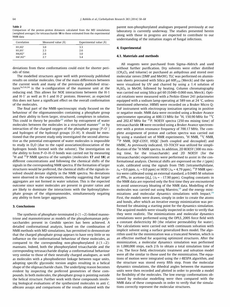
Table 2Comparison of the proton–proton distances calculated from the MD simulations(weighted averages) for tetrasaccharide 18 to those estimated from the experimentalNMR data
Correlation Measured value (Å) Experimental value (Å)
H1,H20 3.0 3.3H1,H10 2.3 2.6H4,H200 2.6 3.0H40 ,H2000 2.7 3.4
64 J. Rahkila et al. / Carbohydrate Research 383 (2014) 58–68
deviations from these conformations could exist for shorter peri-ods of time.
The modelled structures agree well with previously publishedresults on similar molecules. One of the main differences betweenthe current work and many of the previously published struc-tures3a,5,6,24 is the a-configuration of the mannose unit at thereducing end. This allows for NOE interactions between the H-1and H-10 as well as H-1 and H-20 protons. However, as expectedthis does not have a significant effect on the overall conformationof the molecules.
The final part of the NMR-spectroscopic study focused on thebehaviour of the oligomannosides containing a phosphate groupand their ability to form larger, structured, complexes in solution.This could in theory be possible26 either by entrapment of watermolecules between the molecules in a structured manner27 or byinteraction of the charged oxygen of the phosphate group (P–O�)and hydrogen of the hydroxyl groups (O–H). It should be men-tioned that the present study only investigated the second alterna-tive as the potential entrapment of water molecules is impossibleto study in D2O (due to the rapid association/dissociation of thehydrogen bonds formed with the solvent). The investigation onthe ability to form P–O–H–O bonds was carried out by measuring1H and 31P NMR spectra of the samples (molecules 17 and 18) atdifferent concentrations and following the chemical shifts of thesignals in the corresponding NMR spectra. If the formation of largerstructures would be preferred, the chemical shifts of the signals in-volved should deviate slightly in the NMR spectra. No deviationswere observed in the experiments, thereby suggesting that largeraggregates are not formed in water solution. This is the expectedoutcome since water molecules are present in greater ratios andare likely to dominate the interactions with the hydroxyl/phos-phate groups of the oligomannosides, simultaneously negatingany ability to form larger aggregates.
3. Conclusions
The synthesis of phosphate-terminated b-(1?2)-linked manno-triose and mannotetraose as models of the phosphomannan poly-saccharides present in Candida species has been achieved. Adetailed conformational analysis, based on the combination ofNMR methods with MD simulations, has permitted to demonstratethat the charged phosphate group appears to have very little or noinfluence on the conformational behaviour of these molecules, ascompared to the corresponding non-phosphorylated b-(1?2)-mannans. Indeed, both the phosphorylated trisaccharide and thecorresponding tetrasaccharide display a conformational behaviourvery similar to those of their neutrally charged analogues, as wellas molecules with a phosphodiester linkage between sugar units,adopting specific glycosidic torsion angles resulting in a helicalshape of the oligosaccharide chain. The underlying reason becomesevident by inspecting the preferred geometries of these com-pounds. In both molecules, the phosphate group is pointing outsidethe helical structure. Further work with these compounds involv-ing biological evaluations of the synthesized molecules in anti C.albicans assays and comparisons of the results obtained with the
parent non-phosphorylated analogues prepared previously at ourlaboratory is currently underway. The studies presented hereinalong with those in progress are expected to contribute to ourunderstanding of the initiation stages of fungal infections.
4. Experimental
4.1. Materials and methods
All reagents were purchased from Sigma–Aldrich and usedwithout further purification. Dry solvents were either distilled(CH2Cl2 and toluene) or purchased as anhydrous and stored overmolecular sieves (DMF and MeOH). TLC was performed on alumin-ium sheets precoated with Silica gel 60F254 (Merck) and the spotswere visualized by UV and charred by using a 1:4 solution ofH2SO4 in MeOH, followed by heating. Column chromatographywas carried out using Silica gel 60 (0.040–0.060 mm, Merck). Opti-cal rotations were measured with a Perkin–Elmer 241 polarimeterequipped with a sodium lamp operating at 589 nm at 24 �C, unlessmentioned otherwise. HRMS were recorded on a Bruker Micro Q-ToF instrument with electrospray ionisation operating in positiveor negative mode. NMR data were recorded using a Bruker Avancespectrometer operating at 600.13 MHz for 1H, 150.90 MHz for 13Cand 202.47 MHz for 31P. NOESY spectra (250 ms mixing time) oftetrasaccharide 18 were recorded using a Bruker Avance spectrom-eter with a proton resonance frequency of 700.17 MHz. The com-plete assignment of proton and carbon spectra was carried outby using a standard set of NMR experiments, 1H NMR, 13C NMR,31P NMR, DQF-COSY, HSQC (both coupled and decoupled) andHMBC. As previously indicated, 1D-TOCSY was utilized for simpli-fication of the 1H NMR spectra. In addition, 2D ROESY (300 ms mix-ing time, for the trisaccharide) and 2D NOESY (for thetetrasaccharide) experiments were performed to assist in the con-formational analysis. Chemical shifts are expressed on the d (ppm)scale, calibrated using the signals from TMS (tetramethylsilane,dH = 0.0 ppm, dC = 0.0 ppm) or HOD (dH = 4.79 ppm). 31P NMR spec-tra were calibrated using an external standard, a 0.0485 M solutionof PPh3 in acetone-[d6], (dP = �17.90 ppm). Coupling constants inthe NMR data are reported only the first time they are encounteredto avoid unnecessary bloating of the NMR data. Modelling of themolecules was carried out using Maestro,28 and the energy mini-mizations and molecular dynamics simulations with Impact.29
First, the models were drawn, simply in order to create the atomsand bonds, after which an iterative energy minimization was per-formed for obtaining a starting point for the dynamics simulation.The acquired models were visually inspected in order to verify thatthey were realistic. The minimizations and molecular dynamicssimulations were performed using the OPLS_2005 force field witha constant dielectricity 80 (the relative permeability for water).The calculations were carried out with continuum solvation withimplicit solvent using a surface generalized Born model. The algo-rithm used for the minimization was a truncated Newton, which isan effective method for acquiring optimized structures. After theminimization, a molecular dynamics simulation was performedin 1,000,000 steps, each 2 fs to obtain a total simulation time of2 ns. The force field, electrostatic treatment and solvation modelwere all the similar to those used for the minimization. The equa-tions of motion were integrated using the r-RESPA algorithm, andthe structure was stored every fifth step. From the moleculardynamics simulations, the dihedral angles between the mannoseunits were then recorded and plotted in order to provide a modelfor flexibility of the molecules. The low energy conformations ob-tained by molecular modelling were then compared with theNMR data of these compounds in order to verify that the simula-tions correctly represent the molecular structures.

J. Rahkila et al. / Carbohydrate Research 383 (2014) 58–68 65
The synthesis of monosaccharide precursors 1,2,3,4,6-penta-O-acetyl-D-mannopyranoside (1), phenyl 2,3,4,6-tetra-O-acetyl-1-thio-a-D-mannopyranoside (2), phenyl 1-thio-a-D-mannopyran-oside (3),phenyl 4,6-O-benzylidene-1-thio-a-D-mannopyranoside(4), phenyl 2,3-di-O-benzyl-4,6-O-benzylidene-1-thio-a-D-manno-pyranoside (5), phenyl 3-O-benzyl-4,6-O-benzylidene-1-thio-a-D-mannopyranoside (6) and phenyl 2-O-(4-methoxybenzyl)-3-O-benzyl-4,6-O-benzylidene-1-thio-a-D-mannopyranoside (7) hasbeen reported previously16,17 and is not discussed in detail in thispublication. A brief description is provided in the Supplementarydata (Scheme S1).
4.2. General procedure for b-mannosylation
To a solution of the donor (1 equiv) in dry CH2Cl2 (5 mL/0.1 mmol donor) under argon at �60 �C (acetone dry ice) wasadded pre-activated 4 Å molecular sieves, BSP (1.20 equiv), TTBP(1.5 equiv) and Tf2O (1.3 equiv). The reaction mixture was stirredfor 30 min until the activation was complete (confirmed by TLC),after which 1-octene (1 equiv) was added and the reaction mixturewas stirred for another 15 min. The reaction mixture was thencooled down to �78 �C and a solution of the acceptor (1.15 equiv)in dry CH2Cl2 (1 mL/0.1 mmol) was added dropwise. The reactionmixture was stirred for 2 h, at �78 �C and, after the reaction wascomplete, quenched by adding triethylphosphite (3 equiv) and stir-red for 1 h. The reaction mixture was then warmed to room tem-perature and diluted with CH2Cl2 (30 mL/100 mg) and washedwith a saturated solution of NaHCO3 in H2O (30 mL/100 mg). Thewater layer was extracted with CH2Cl2 (2 � 30 mL/100 mg) afterwhich the combined organic layers were washed with brine(30 mL/100 mg) and dried over Na2SO4. The solvent was removedand the crude product was purified by column chromatographyto yield the b-coupled product.
4.3. General procedure for hydrogenolysis of benzyl andbenzylidene protecting groups
To a solution of the protected sugar molecule in dry MeOH(1.5 mL/10 mg protected sugar) was added Pd/C 10% w/w (2 equivby mass) and the mixture was stirred in an autoclave under H2
(2 bar) overnight. The reaction mixture was then filtered throughcelite and evaporated to dryness to yield the corresponding unpro-tected product.
4.4. Phenyl O-(2,3-di-O-benzyl-4,6-O-benzylidene-b-D-mannopyranosyl)-(1?2)-O-(3-O-benzyl-4,6-O-benzylidene-b-D-mannopyranosyl)-(1?2)-3-O-benzyl-4,6-O-benzylidene-1-thio-a-D-mannopyranoside (9)
Synthesized from donor 8 (200 mg, 0.2 mmol) and acceptor 6(107 mg, 0.24 mmol) according to the general procedure forb-mannosylation. The product was purified by column chromatog-raphy (hexane/EtOAc 2:1, Rf = 0.45) to yield 9 as a white foam.Yield: 166 mg (62%). ½a�24
D �51.5� (c 1.0 CH2Cl2). 1H NMR(600.13 MHz, CDCl3, 25 �C): d = 7.25–6.90 (m, 40H, arom. H), 5.60(s, 1H, 4,6-OCH0Ph), 5.57 (s, 1H, 4,6-OCHPh), 5.51 (d, 1H, JH-1,H-2 =1.4 Hz, H-1), 5.37 (s, 1H, 4,6-OCH00Ph), 5.10 (d, 1H, JH-100 ,H-200 =0.5 Hz, H-100), 4.97 and 5.72 (each d, each 1H, J =�12.5 Hz, 2-OCH2Ph),4.82 and 4.77 (each d, each 1H, J = �12.6 Hz, 30-OCH2Ph), 4.67and 4.62 (each d, each 1H, J = �12.2 Hz, 3-OCH2Ph), 4.65 (d, 1H,JH-10 ,H-20 = 0.1 Hz, H-10), 4.56 (dd, 1H, JH-2,H-3 = 3.0 Hz, H-2), 4.46and 4.43 (each d, each 1H, J = �11.6 Hz, 300-OCH2Ph), 4.45 (dd,1H, JH-200 ,H-300 = 3.2 Hz, H-200), 4.36 (dd, 1H, JH-20 ,H-30 = 3.1 Hz, H-20)4.34 (ddd, 1H, JH-5,H-6a = 4.9 Hz, JH-5,H-4 = 9.2 Hz, JH-5,H-6b = 10.2 Hz,H-5), 4.33 (dd, 1H, JH-600a,H-500 = 4.9 Hz, JH-600a,H-600b = �10.5 Hz,
H-600a), 4.31 (dd, 1H, JH-60a,H-50 = 4.8 Hz, JH-60a,H-60b = �10.0 Hz,H-60a), 4.23 (dd, 1H, JH-6a,H-6b = �10.3 Hz, H-6a), 4.22 (dd, 1H,JH-400 ,H-500 = 9.2 Hz, JH-400 ,H-300 = 9.9 Hz, H-400), 4.06 (dd, 1H, JH-40 ,H-50 =9.3 Hz, JH-40 ,H-30 = 9.8 Hz, H-40), 3.99 (dd, 1H, JH-4, H-3 = 10.1 Hz,H-4), 3.98 (dd, 1H, H-3), 3.97 (dd, 1H, JH-600b,H-500 = 10.1 Hz, H-600b),3.77 (dd, 1H, JH-6b,H-5 = 10.2 Hz, H-6b), 3.72 (dd, JH-60b,H-50 =10.1 Hz, H-60b), 3.64 (dd, 1H, H-30), 3.55 (dd, 1H, H-300), 3.43(ddd, 1H, H-500), 3.33 (ddd, 1H, H-50) ppm.
13C NMR (150.9 MHz, CDCl3, 25 �C): d = 139.4–126.0 (arom. C),103.0 (C-100), 102.0 (4,6-OCHPh), 101.6 (4,6-OC00HPh), 101.3(4,6-OC0HPh), 98.5 (C-10), 85.6 (C-1), 79.3 (C-300), 79.0 (C-4), 78.2(C-40), 78.1 (C-400), 76.2 (C-20 and C-200), 75.7 (C-30), 74.9 (2-OCH2Ph),74.8 (C-2), 74.6 (C-3), 72.2 (300-OCH2Ph), 71.8 (3-OCH2Ph), 71.0(30-OCH2Ph), 68.7 (C-600), 68.6 (C-6 and C-60), 67.9 (C-500), 67.8(C-50), 65.2 (C-5) ppm.
1JC-1,H-1 = 165.3 Hz (a), 1JC-10 ,H-10 = 155.1 Hz (b), 1JC-100 ,H-100 = 158.5 Hz (b).HRMS: m/z Calcd for C73H73O15SNa [M+Na]+: 1243.4490. Found
1243.4502.
4.5. O-(2,3-Di-O-benzyl-4,6-O-benzylidene-b-D-mannopyranosyl)-(1?2)-O-(3-O-benzyl-4,6-O-benzylidene-b-D-mannopyranosyl)-(1?2)-3-O-benzyl-4,6-O-benzylidene-D-mannopyranose (13)
A solution of 9 (255 mg, 1 equiv) in 5 mL of a 50:1 mixture ofacetone/H2O was cooled on an ice bath and NBS (69 mg, 2 equiv)was added and the reaction mixture was stirred ad 0 �C for30 min after which additional NBS (35 mg, 1 equiv) was addedand the reaction mixture was again stirred for 30 min. AdditionalNBS (69 mg, 2 equiv) was added and the reaction mixture was stir-red for 30 min after which solid Na2S2O3 was added until the yel-low colour disappeared. The solvent was evaporated and theresulting oil was dissolved in 50 mL CH2Cl2 and washed with3 � 20 mL H2O and dried over Na2SO4 after which the solventwas removed. The crude product was purified by column chroma-tography (hexane/EtOAc 1:1, Rf = 0.36) to yield 13 as a white foam.Yield: 61 mg (28%). Due to the complexity of the NMR spectracaused by a mixture of anomers, the spectra were not fullyassigned.
HRMS: m/z Calcd for C67H68O16Na [M+Na]+: 1151.4405. Found1151.4401.
4.6. O-(2,3-Di-O-benzyl-4,6-O-benzylidene-b-D-mannopyranosyl)-(1?2)-O-(3-O-benzyl-4,6-O-benzylidene-b-D-mannopyranosyl)-(1?2)-3-O-benzyl-4,6-O-benzylidene-a-D-mannopyranosyl dibenzylphosphate (14)
Method 1: To a solution of 13 (40 mg, 1 equiv) in dry CH2Cl2 un-der argon was added 1H-tetrazole (9.3 mg, 3.8 equiv) after whichthe reaction mixture was cooled down to 0 �C. Dibenzyl(N,N-diiso-propyl) phosphoramidite (30 lL, 2.5 equiv) was added after whichthe reaction mixture was allowed to warm up to room temperatureand was stirred for 22 h. The reaction mixture was then cooled to�60 �C and 20 mg m-CPBA was added. The reaction mixture wasstirred at 0 �C for 1.5 h and at room temperature for 2 h after whichit was diluted with 40 mL CH2Cl2, washed with 2 � 15 mL of a sat-urated solution of Na2S2O3 in H2O, 2 � 15 mL of a saturated solu-tion of NaHCO3 in H2O and 2 � 10 mL H2O. The organic layer waspurified by column chromatography (hexane/EtOAc 1:1, Rf = 0.57)to yield 14 as a white foam. Yield of a-product: 20 mg (41%).½a�24
D �51.7� (c 1.0 CH2Cl2).Method 2: To a solution of 9 (140 mg, 1 equiv) in 3 mL dry CH2-
Cl2 under argon was added HOPO(OBn)2 (94.3 mg, 3 equiv) and 4 Åmolecular sieves. The reaction mixture was cooled to �20 �C andNIS (30.5 mg, 1.2 equiv) and TfOH, (2.5 lL, 0.12 equiv) were added.The reaction mixture was stirred at 0 �C for 1 h after which the

66 J. Rahkila et al. / Carbohydrate Research 383 (2014) 58–68
reaction was quenched by adding 0.5 mL pyridine. The reactionmixture was diluted with 20 mL CH2Cl2 and washed with2 � 15 mL of a saturated solution of Na2S2O3 in H2O, 2 � 15 mLof a saturated solution of NaHCO3 in H2O and 1 � 15 mL H2O.The organic layer was dried over Na2SO4 and the solvent wasremoved. The crude product was purified by column chromatogra-phy (hexane/EtOAc 1:1) to yield 14 as a white foam. Yield ofa-product: 64.2 mg (42%). 1H NMR (600.13 MHz, CDCl3, 25 �C):d = 7.60–7.00 (m, 45H, arom. H), 5.60 (s, 1H, 4,6-OCH00Ph), 5.58(dd, 1H, JH-1,H-2 = 1.5 Hz, JH-1,P = 6.2 Hz, H-1), 5.45 (s, 1H, 4,6-OCH0Ph), 5.33 (s, 1H, 4,6-OCHPh), 5.08 and 5.03 [each dd, each,1H, J = �11.9 Hz, JCH2a,P = 8.9 Hz, JCH2b,P = 8.2 Hz, PO(OCH2Ph)],5.02 and 4.99 [each dd, each, 1H, J = �11.8 Hz, JCH2a,P = 13.6 Hz,JCH2b,P = 9.6 Hz, PO(OCH2Ph)], 5.00 (d, 1H, JH-100 ,H-200 = 0.1 Hz, H-100),4.91 and 4.69 (each d, each 1H, J = �12.5 Hz, 200-OCH2Ph), 4.83and 4.78 (each d, each 1H, J = �12.5 Hz, 30-OCH2Ph), 4.60 and4.56 (each d, each 1H, J = �12.2 Hz, 3-OCH2Ph), 4.47 (s, 1H, H-10),4.45 and 4.42 (each d, each 1H, J = �11.7 Hz, 300-OCH2Ph), 4.39(dd, 1H, JH-200 ,H-300 = 3.2 Hz, H-200), 4.34 (dd, 1H, JH-600a,H-500 = 4.8 Hz,JH-600a,H-600b = �10.4 Hz, H-600a), 4.29 (d, 1H, JH-20 ,H030 = 3.1 Hz, H-20),4.26 (dd, 1H, JH-60a,H-50 = 4.8 Hz, JH-60a,H-60b = �10.2 Hz, H-60a), 4.22(dd, 1H, JH-400 ,500 = 9.2 Hz, JH-400 ,H-300 = 9.8 Hz, H-400), 4.15 (dd, 1H, JH-
2,H-3 = 3.3 Hz, H-2), 4.02 (dd, 1H, JH-40 ,H-50 = 9.4 Hz, JH-40 ,H-
30 = 9.7 Hz, H-40), 3.99 (dd, 1H, JH-6a,H-5 = 4.9 Hz, JH-6a,H-
6b = �10.3 Hz, H-6a), 3.97 (dd, 1H, JH-600b,500 = 10.2 Hz, H-600b), 3.85(dd, 1H, JH-4,H-5 = 9.0 Hz, JH-4,H-3 = 10.2 Hz, H-4), 3.84 (ddd, 1H, JH-
5,H-6b = 10.1 Hz, H-5), 3.83 (dd, 1H, H-3), 3.67 (dd, 1H, JH-60b,H-
50 = 10.1 Hz, H-60b), 3.60 (dd, 1H, H-6b), 3.59 (dd, 1H, H-30), 3.53(dd, 1H, -300), 3.41 (ddd, 1H, H-500), 3.24 (ddd, 1H, H-500) ppm.
13C NMR (150.9 MHz, CDCl3, 25 �C): d = 139.3–135.2(arom. C),103.0 (C-100), 101.9 (4,6-OC0HPh), 101.6 (4,6-OCHPh), 101.3 (4,6-OC00HPh), 99.6 (C-10), 95.7 (2JC-1,P = 5.7 Hz, C-1), 79.2 (C-300), 78.1(C-4, C-40, C-400), 76.2 (C-20), 76.0 (C-200), 75.5 (C-30), 74.9 (2-OCH2-
Ph), 73.7 (3JC-2,P = 9.9 Hz, C-2), 73.5 (C-3), 72.1 (300-OCH2Ph), 71.9(3-OCH2Ph), 71.1 (30-OCH2Ph), 69.9 [2JC,P = 6.1 Hz, PO(OCH2Ph)],69.8 (2JC,P = 6.1 Hz, PO(OCH2Ph)], 68.7 (C-600), 68.5 (C-60), 68.3 (C-6), 67.9 (C-500), 67.6 (C-50), 65.5 (C-5) ppm.
1JC-1,H-1 = 176.4 Hz (a), 1JC-10 ,H-10 = 156.2 Hz (b), 1JC-100 ,H-100 = 158.6 Hz (b).31P NMR (242.9 MHz, CDCl3, 25 �C): d = �2.88 ppm.HRMS: m/z Calcd for C81H81O19PNa [M+Na]+: 1411.5007. Found
1411.5025.
4.7. O-(b-D-Mannopyranosyl)-(1?2)-O-(b-D-mannopyranosyl)-(1?2)-a-D-mannopyranosylphosphate (17)
Synthesized from 14 (22 mg, 0.016 mmol) according to the gen-eral procedure for hydrogenolysis of benzyl and benzylidene pro-tecting group. Filtration through celite with water yielded theproduct as a colourless oil. Yield: 9 mg (97%). Purity: 95%, withan impurity possibly consisting of a molecule where the phosphategroup has been replaced by methyl. ½a�24
D �13� (c 1.0 H2O). 1H NMR(600.13 MHz, CDCl3, 25 �C): d = 5.45 (dd, 1H, JH-1,H-2 = 1.5 Hz,JH-1,P = 7.9 Hz, H-1), 4.96 (d, 1H, JH-10 ,H-20 = 0.2 Hz, H-10), 4.88 (d,1H, JH-100 ,H-200 = 0.5 Hz, H-100), 4.32 (dd, 1H, JH-20 ,H-30 = 3.4 Hz, H-20),4.18 (dd, 1H, JH-200 ,H-300 = 3.3 Hz, H-200), 4.15 (dd, JH-2,H-3 = 3.1 Hz,H-2), 4.01 (dd, JH-3,H-4 = 9.8 Hz, H-3), 3.93 (dd, 2 H, JH-60a,H-50 =2.1 Hz, JH-60a,H-60b = �11.9 Hz, JH-600a,H-500 = 2.2 Hz, JH-600a,H-600b =�12.6 Hz, H-60a and H-600a), 3.89 (dd, 1H, JH-6a,H-5 = 2.1 Hz,JH-600a,H-600b = �12.4 Hz, H-6a), 3.86 (ddd, JH-5,H-6b = 5.6 Hz, JH-5,H-4 =9.8 Hz, H-5), 3.77 (dd, 1H, H-6b), 3.75 (dd, 1H, JH-60b,H-50 = 5.9 Hz,H-60b), 3.73 (dd, 1H, JH-600b,H-500 = 6.4 Hz, H-600b), 3.70 (dd, 1H,JH-30 ,H-40 = 9.9 Hz, H-30), 3.64 (dd, 1H, JH-300 ,H-400 = 9.9 Hz, H-300),3.61 (each dd, each 1H, JH-40 ,H-50 = 9.7 Hz, H-4 and H-40), 3.57(dd, 1H, JH-400 ,H-500 = 9.8 Hz, H-400), 3.43 (ddd, 1H, H-50), 3.37 (ddd,1H, H-500) ppm.
13C NMR (150.9 MHz, CDCl3, 25 �C): d = 101.0 (C-100), 98.9 (C-10),92.9 (2JC-1,P = 4.0 Hz, C-1), 78.8 (3JC-2,P = 6.6 Hz, C-2), 78.7 (C-20),76.2 (C-500), 76.1 (C-50), 72.9 (C-5), 72.8 (C-300), 72.1 (C-30), 70.4(C-200), 69.1 (C-3), 67.3 and 66.9 (C-4 and C-40), 66.7 (C-400), 61.0(C-600), 60.7 (C-60), 60.5 (C-6) ppm.
1JC-1,H-1 = 173.4 Hz (a), 1JC-10 ,H-10 = 160.5 Hz (b), 1JC-100 ,H-100 = 160.3 Hz (b).31P NMR (242.9 MHz, CDCl3, 25 �C): d = 1.46 ppm.HRMS: m/z Calcd for C18H32O19P [M�H]�: 853.1276. Found
853.1252
4.8. Phenyl O-(2-O-(4-methoxybenzyl)-3-O-benzyl-4,6-O-benzylidene-b-D-mannopyranosyl)-(1?2)-3-O-benzyl-4,6-O-benzylidene-1-thio-a-D-mannopyranoside (10)
Synthesized from donor 7 (500 mg 0.876 mmol) and acceptor 6(455 mg 1.01 mmol) according to the general procedure for b-man-nosylation. The crude product was purified by column chromatog-raphy (hexane/EtOAc 2:1, Rf = 0.45) Yield: 455 mg (57%). ½a�24
D
�52.5� (c 1.0 CH2Cl2). 1H NMR (600.13 MHz, CDCl3, 25 �C):d = 7.75–7.20 (m, 29 H, arom. H), 5.59 (s, 1H, 4,6-OCH0Ph), 5.52(s, 1H, 4,6-OCHPh), 5.50 (d, 1H, JH-1,H-2 = 1.5 Hz, H-1), 4.97 and4.89 (each d, each 1H, J = �11.8, 20-OCH2Ph), 4.80 and 4.75 (eachd, each 1H, J = �12.1 Hz, 3-OCH2Ph), 4.67 and 4.59 (each d, each1H, J = �12.5 Hz, 30-OCH2Ph), 4.61 (d, 1H, JH-10 ,H-20 = 0.9 Hz, H-10),4.51 (dd, 1H, JH-2,H-3 = 3.2 Hz, H-2), 4.33 (ddd, 1H, JH-5,H-
6a = 4.8 Hz, JH-5,H-4 = 9.5 Hz, JH-5,H-6b = 10.2 Hz, H-5), 4.24 (dd, 2 H,JH-6a,H-6b = �10.2 Hz, JH-60a,H-50 = 4.9 Hz, JH-60a,H-60b = �10.4 Hz, H-6aand H-60a), 4.23 (dd, 1H, JH-40 ,H-50 = 9.3 Hz, JH-40 ,H-30 = 9.9 Hz, H-40),4.17 (dd, 1H, JH-4,H-3 = 10.0 Hz, H-4), 3.98 (dd, 1H, H-3), 3.96 (dd,1H, JH-20 ,H-30 = 3.2 Hz, H-20), 3.82 (dd, JH-60b,H-50 = 10.1 Hz, H-60b),3.79 (dd, 1H, H-6b), 3.73 (s, 3 H, OCH3), 3.57 (dd, 1H, H-30), 3.29(ddd, 1H, H-5) ppm.
13C NMR (150.9 MHz, CDCl3, 25 �C): d = 159.1–113.5 (arom. C),101.7 (4,6-OCHPh), 101.4 (4,6-OC0HPh), 99.8 (C-10), 86.4 (C-1),78.7 (C-4), 78.4 (C-40), 77.5 (C-30), 76.1 (C-2), 75.4 (C-20), 74.3(C-3), 74.2 (20-OCH2Ph), 72.2 (30-OCH2Ph), 71.4 (3-OCH2Ph), 68.6(C-6), 68.5 (C-60), 67.7 (C-50), 65.4 (C-5), 55.2 (OCH3) ppm.
HRMS: m/z Calcd for C54H54O11SNa [M+Na]+: 933.3285. Found933.3228; m/z Calcd for C54H58O11SN [M+NH4]+: 928.3731. Found928.3634.
4.9. Phenyl O-(3-O-benzyl-4,6-O-benzylidene-b-D-mannopyranosyl)-(1?2)-3-O-benzyl-4,6-O-benzylidene-1-thio-a-D-mannopyranoside (11)
A solution of 10 (200 mg, 1 equiv) in 3 mL CH2Cl2 was cooleddown to 0 �C and a suspension of DDQ (74 mg, 1.5 equiv) in 3 mLH2O was added dropwise. The reaction mixture was stirred for1 h at 0 �C after which it was diluted with 25 mL CH2Cl2 andwashed with 2 � 25 mL of a saturated solution of NaHCO3 inH2O. The water layer was extracted with 2 � 25 mL CH2Cl2, driedover Na2SO4 and evaporated to dryness. The crude product waspurified by column chromatography (hexane/EtOAc 2:1) to yieldthe 11 as a white foam. Yield: 153 mg (88%). NMR spectra are inaccordance with those published previously.16
4.10. Phenyl O-(2,3-di-O-benzyl-4,6-O-benzylidene-b-D-mannopyranosyl)-(1?2)-O-(3-O-benzyl-4,6-O-benzylidene-b-D-mannopyranosyl)-(1?2)-O-(3-O-benzyl-4,6-O-benzylidene-b-D-mannopyranosyl)-(1?2)-3-O-benzyl-4,6-O-benzylidene-1-thio-a-D-mannopyranoside (12)
Synthesized from donor 8 (290 mg, 0.33 mmol) and acceptor11 (300 mg, 0.38 mmol) according to the general procedure forb-mannosylation. The crude product was purified by column

J. Rahkila et al. / Carbohydrate Research 383 (2014) 58–68 67
chromatography (hexane/EtOAc 2:1, Rf = 0.50). Yield: 182 mg(35%). ½a�24
D �62.0� (c = 1.0 CH2Cl2). 1H NMR (600.13 MHz, CDCl3,25 �C): d = 7.75–7.00 (m, 50H, arom. H), 5.57 (s, 1H, 4,6-OCH00Ph),5.54 (s, 1H, 4,6-OCHPh), 5.49 (d, 1H, JH-1,H-2 = 1.3 Hz, H-1), 5.44 (s,1H, 4,6-OCH0Ph), 5.40 (s, 1H, 4,6-OCH000Ph), 5.27 (s, 1H, H-100), 5.07(d, 1H, JH-1000 ,H-2000 = 0.1 Hz, H-1000), 5.00 and 4.84 (each d, each 1H,J = �12.6 Hz, 2000-OCH2Ph), 4.70 (s, 2H, 3-OCH2Ph), 4.68 and 4.61(each d, each 1H, J = �12.4 Hz, 30-OCH2Ph), 4.68 (s, 1H, H-10),4.62 and 4.44 (each d, each 1H, J = �11.9 Hz, 300-OCH2Ph), 4.54(d, 1H, JH-200 ,H-300 = 3.0 Hz, H-200), 4.53 (dd, 1H, JH-2,H-3 = 3.1 Hz, H-2), 4.50 and 4.40 (each d, each 1H, J = �11.8 Hz, 3000-OCH2Ph),4.48 (d, 1H, JH-20 ,H-30 = 3.4 Hz, H-20), 4.41 (dd, 1H, JH-2000 ,H-3000 =3.3 Hz, H-2000), 4.33 (dd and ddd, 2H, JH-600a,H-500 = 4.8 Hz, JH-600a,H-600b =�10.2 Hz, JH-5,H-6a = 5.0 Hz, JH-5,H-4 = 9.2 Hz, JH-5,H-6b = 10.2 Hz, H-600aand H-5), 4.31 (dd, 1H, JH-60a,H-50 = 4.8 Hz, JH-60a,H-60b = �10.3 Hz,H-60a), 4.23 (dd, 1H, JH-6a,H-6b = �10.3 Hz, H-6a), 4.19 (dd anddd, 2 H, JH-4000 ,H-5000 = 9.3 Hz, JH-4000 ,H-3000 = 9.9 Hz, JH-6000a,H-5000 = 4.9 Hz,JH-6000a,H-6000b = �10.3 Hz, H-4000 and H-6000a), 4.06 (dd, 1H, JH-400 ,H-500 =9.3 Hz, JH-400 ,H-300 = 9.8 Hz, H-400), 3.99 (dd, 1H, JH-4,H-3 = 9.9 Hz, H-4),3.97 (dd, 1H, H-3), 4.93 (dd, 1H, JH-40 ,H-50 = 9.3 Hz, JH-40 ,H-30 = 9.8 Hz,H-40), 3.87 (dd, 1H, JH-6000b,H-5000 = 10.1 Hz, H-6000b), 3.78 (dd, 1H,JH-600b,H-500 = 10.1 Hz, H-600b), 4.77 (dd, 1H, JH-60b,H-50 = 10.1 Hz, H-60b),3.74 (dd, 1H, H-6b), 3.66 (dd, 1H, H-30), 5.54 (dd, 1H, H-3000), 3.53(dd, 1H, H-300), 3.43 (ddd, 1H, H-500), 3.36 (ddd, 1H, H-50), 3.32(ddd, 1H, H-5000) ppm.
13C NMR (150.9 MHz, CDCl3, 25 �C): d = 139.4–126.1 (arom. C),103.4 (C-1000), 102.1 (4,6-OCHPh), 102.0 (4,6-OC0HPh), 101.8 (C-100),101.5 (4,6-OC000HPh), 101.3 (4,6-OC00HPh), 99.1 (C-10), 85.7 (C-1),79.1 (C-4), 79.0 (C-3000), 78.4 (C-40), 78.3 (C-4000), 78.2 (C-400), 77.0(C-300), 76.3 (C-30), 76.1 (C-200), 76.0 (C-2000), 75.5 (C-2), 74.7
(2000-OCH2Ph), 74.5 (C-3), 74.0 (C-20), 72.2 (3000-OCH2Ph), 72.1(3-OCH2Ph), 71.3 (30-OCH2Ph), 70.9 (300-OCH2Ph), 68.8 (C-6000), 68.7(C-600, C-60), 68.6 (C-6), 68.0 (C-500), 67.9 (C-50), 67.5 (C-5000), 65.2(C-5) ppm.
1JC-1,H-1 = 168.2 Hz (a), 1JC-10 ,H-10 = 154.5 Hz (b), 1JC-100 ,H-
100 = 161.0 Hz (b), 1JC-1000 ,H-1000 = 159.6 Hz (b).HRMS: m/z Calcd for C93H92O20SNa [M+Na]+: 1583.5800. Found
1583.5812.
4.11. O-(2,3-Di-O-benzyl-4,6-O-benzylidene-b-D-mannopyranosyl)-(1?2)-O-(3-O-benzyl-4,6-O-benzylidene-b-D-mannopyranosyl)-(1?2)-O-(3-O-benzyl-4,6-O-benzylidene-b-D-mannopyranosyl)-(1?2)-3-O-benzyl-4,6-O-benzylidene-D-mannopyranose (15)
A solution of 12 (250 mg, 1 equiv) in 5 mL of a 6:1 mixture ofacetone/H2O was cooled on an ice bath and NBS (60 mg, 2 equiv)was added and the reaction mixture was stirred for 30 min. Addi-tional NBS (30 mg, 1 equiv) was added and the reaction mixturewas again stirred for 30 min after which additional NBS (30 mg,1 equiv) was added and the reaction mixture was stirred for30 min. The reaction was quenched by adding solid Na2S2O3 untilthe yellow colour had disappeared. The solvent was evaporatedand the residue was dissolved in 50 mL CH2Cl2 and washed with2 � 20 mL H2O. The organic layer was dried over Na2SO4 andevaporated to dryness. The crude product was purified by columnchromatography (hexane/EtOAc 1:1, Rf = 0.38) to yield 15 as awhite foam. Yield: 62 mg (28%). Due to the complexity of theNMR spectra, caused by a mixture of anomers, the spectra werenot fully assigned.
HRMS: m/z Calcd for C87H92O21N [M+NH4]+: 1486.6162. Found1486.6174.
4.12. O-(2,3-Di-O-benzyl-4,6-O-benzylidene-b-D-mannopyranosyl)-(1?2)-O-(3-O-benzyl-4,6-O-benzylidene-b-D-mannopyranosyl)-(1?2)-O-(3-O-benzyl-4,6-O-benzylidene-b-D-mannopyranosyl)-(1?2)-3-O-benzyl-4,6-O-benzylidene-D-mannopyranosyl dibenzylphosphate (16)
Method 1: To a solution of 15 (35 mg, 1 equiv) in 4 mL dryCH2Cl2 under argon was added 1H-tetrazole (6.3 mg, 3.8 equiv)after which the solution was cooled down to 0 �C. Dibenzyl(N,N-diisopropyl) phosphoramidite (22 lL, 2.5 equiv) was added andthe reaction mixture was allowed to return to room temperatureand was stirred for 2 h. The reaction mixture was cooled down to�60 �C and m-CPBA (13.5 mg, 3.8 equiv) was added and the mix-ture was stirred at 0 �C for 1 h and then at room temperature for1 h. The reaction mixture was diluted with 50 mL CH2Cl2 andwashed with 2 � 15 mL of a saturated solution of Na2S2O3 inH2O, 2 � 15 mL of a saturated solution of NaHCO3 in H2O and2 � 10 mL H2O after which the organic layer was dried over NasSO4
and concentrated to dryness. The crude product was purified bycolumn chromatography (hexane/EtOAc 1:1, Rf = 0.57) Yield ofa-product: 25 mg (60%). ½a�24
D �61.2� (c 1.0 CH2Cl2).Method 2: To a solution of 12 (50 mg, 1 equiv) in 3 mL dry
CH2Cl2 under argon was added PO(OBn)2Oh (26.7 mg, 3 equiv)and 4 Å molecular sieves. The reaction mixture was cooled downto �50 �C and NIS (8.6 mg, 1.2 equiv) and TMSOTf (0.7 lL,0.12 equiv) were added. The reaction mixture was stirred at�50 �C for 1 h and warmed to �20 �C and stirred for 3.5 h afterwhich additional TMSOTf (0.7 lL, 0.12 equiv) was added and thereaction mixture was stirred for another 17.5 h at �20 �C. The reac-tion was quenched by adding 0.5 mL pyridine after which the reac-tion mixture was diluted with 20 mL CH2Cl2 and washed with2 � 15 mL of a saturated solution of Na2S2O3 in H2O, 2 � 15 mLof a saturated solution of NaHCO3 in H2O and 1 � 15 mL H2O.The organic layer was dried over Na2SO4 and evaporated to dry-ness. The crude product was purified by column chromatography(hexane/EtOAc 1:1). Yield of a-product: 26 mg (47%). 1H NMR(600.13 MHz, CDCl3, 25 �C): d = 7.75–6.75 (m, 55H, arom. H), 5.59(dd, 1H, JH-1,H-2 = 1.7 Hz, JH-1,P = 6.3 Hz, H-1), 5.56 (s, 1H, 4,6-OCH000Ph), 5.43 (s, 1H, 4,6-OCHPh), 5.41 (s and s, 2H, 4,6-OCH0Phand 4,6-OCH00Ph), 5.14 (d, 1H, JH-100 ,H-200 = 0.1 Hz, H-100), 5.08 and5.02 [each dd, each, 1H, J = �11.9 Hz, JCH2a,P = 9.1 Hz, JCH2b,P = 8.2 -Hz, PO(OCH2Ph)], 5.06 (d, 1H, JH-1000 ,H-2000 = 0.1 Hz, H-1000), 5.01 and4.98 [each dd, each, 1H, J = �11.6 Hz, JCH2a,P = 8.4 Hz, JCH2b,P = 9.9 -Hz, PO(OCH2Ph)], 4.99 and 4.83 (each d, each 1H, J = �12.5 Hz,2000-OCH2Ph), 4.70 and 4.63 (each d, each 1H, J = �12.4 Hz, 30-OCH2-
Ph), 4.65 and 4.50 (each d, each 1H, J = �11.9 Hz, 300-OCH2Ph), 4.64and 4.61 (each d, each 1H, J = �12.4 Hz, 3-OCH2Ph), 4.51 (dd, JH-200 ,H-
300 = 3.1 Hz, H-200), 4.50 (d, 1H, JH-10 ,H-20 = H-10), 4.50 and 4.38 (each d,each 1H, J = �11.8 Hz, 3000-OCH2Ph), 4.42 (dd, 1H, JH-20 ,H-30 = 3.3 Hz,H-20), 4.39 (dd, 1H, JH-2000 ,H-3000 = 3.1 Hz, H-2000), 4.36 (dd, 1H, JH-600a,H-500 =4.6 Hz, JH-600a,H-600b =�10.2 Hz, H-600a), 4.26 (dd, 1H, JH-60a,H-50 = 4.8 Hz,JH-60a,H-60b = �10.2 Hz, H-60a), 4.18 (dd, 1H, JH-4000 ,H-5000 = 9.2 Hz,JH-4000 ,H-3000 = 9.9 Hz, H-4000), 4.17 (dd, 1H, JH-6000a,H-5000 = 4.8 Hz,JH-6000a,H-6000b = �10.4 Hz, H-6000a), 4.10 (dd, 1H, JH-2,H-3 = 3.7 Hz, H-2),4.07 (dd, 1H, JH-400 ,H-500 = 9.3 Hz, JH-400 ,H-300 = 9.8 Hz, H-400), 3.98 (dd,1H, JH-6a,H-5 = 4.9 Hz, JH-6a,H-6b = �10.3 Hz, H-6a), 3.88 (dd, 1H,JH-40 ,H-50 = 9.4 Hz, JH-40 ,H-30 = 9.9 Hz, H-40), 3.85 (dd and dd, 2H,JH-6000b,H-5000 = 10.1 Hz, JH-4,H-5 = 9.5 Hz, JH-4,H-3 = 10.1 Hz, H-6000b andH-4), 3.82 (ddd, 1H, JH-5,H-6b = 10.3 Hz, H-5), 3.80 (dd, 1H, H-3),3.78 (dd, 1H, JH-600b,H-500 = 10.0 Hz, H-60 0b), 3.73 (dd, 1H, JH-60b,H-50 =10.0 Hz, H-60b), 3.62 (dd, 1H, H-30), 3.55 (dd, 1H, H-6b), 3.53 (dd, 1H,H-300), 3.52 (dd, 1H, H-3000), 3.40 (ddd, 1H, H-500), 3.31 (ddd, 1H,H-5000), 3.27 (ddd, 1H, H-50) ppm.

68 J. Rahkila et al. / Carbohydrate Research 383 (2014) 58–68
13C NMR (150.9 MHz, CDCl3, 25 �C): d = 139.4–126.1 (arom. C),103.3 (C-1000), 101.9 (C-100, 4,6-OCHPh, 4,6-OC0 0 0HPh), 101.5 (4,6-OC00HPh), 101.2 (4,6-OC 0 0 0HPh), 100.5 (C-10), 95.7 (2JC-1,P = 4.9 Hz,C-1), 79.0 (C-3000), 78.4 (C-4000), 78.3 (C-40), 71.2 (C-4, C-400), 76.8(C-300), 76.2 (C-200), 76.1 (C-30), 75.9 (C-2000), 74.9 (3JC-2,P = 9.3 Hz,C-2), 74.6 (2000-OCH2Ph), 74.2 (C-20), 73.4 (C-3), 72.2 (3000-OCH2Ph),72.1 (3-OCH2Ph), 71.3 (30-OCH2Ph), 70.8 (300-OCH2Ph), 69.9[2JC,P = 5.4 Hz, 2JC,P = 5.8 Hz, PO(OCaH2Ph),PO(OCbH2Ph)], 68.8(C-600), 68.7 (C-6000), 68.6 (C-60), 68.3 (C-6), 68.0 (C-500), 67.7 (C-50),67.5 (C-5000), 65.5 (C-5) ppm.
1JC-1,H-1 = 175.3 Hz (a), 1JC-10 ,H-10 = 155.5 Hz (b), 1JC-100 ,H-100 = 161.0 Hz (b),1JC-1000 ,H-1000 = 158.9 Hz (b).
31P NMR (242.9 MHz, CDCl3, 25 �C): d = �2.92 ppm.HRMS: m/z Calcd for C101H101O24PNa [M+Na]+: 1751.6318.
Found 1751.6341.
4.13. O-(b-D-Mannopyranosyl)-(1?2)-O-(b-D-mannopyranosyl)-(1?2)-O-(b-D-mannopyranosyl)-(1?2)-a-D-mannopyranosylphosphate (18)
Synthesized from 16 (25 mg, 0.014 mmol) according to the gen-eral procedure for hydrogenolysis of benzyl and benzylidene pro-tecting groups. Filtration through celite with a 4:1 mixture ofMeOH/H2O yielded the product as a colourless oil. Yield: 7 mg(67%). Purity: 95%, with an impurity possibly consisting of a mole-cule where the phosphate group has been replaced by methyl. ½a�24
D
�15� (c 1.0 H2O). 1H NMR (600.13 MHz, CDCl3, 25 �C): d = 5.47(dd, 1H, JH-1,H-2 = 1.4 Hz, JH-1,P = 7.8 Hz, H-1), 4.96 (s, 1H, H-100), 4.94(s, 1H, H-1000), 4.93 (s, 1H, H-10), 4.43 (d, 1H, JH-200 ,H-300 = 3.4 Hz, H-200),4.29 (d, 1H, JH-20 ,H-30 = 3.4 Hz, H-20), 4.16 (dd and d, 2 H, JH-2,H-3 =3.2 Hz, JH-2000 ,H-3000 = 3.4 Hz, H-2 and H-2000), 4.00 (dd, 1H, JH-3,H-4 =9.9 Hz, H-3), 3.94 (dd, 1H, JH-60a,H-50 = 3.1 Hz, JH-60a,H-60b = �12.3 Hz,H-60a), 3.93 (dd, 1H, JH-6000a,H-5000 = 2.5 Hz, JH-6000a,H-6000b =�12.3 Hz, H-6000a),3.92 (dd, 1H, JH-600a,H-500 = 2.1 Hz, JH-600a,H-600b =�11.9 Hz, H-600a), 3.87(dd, 1H, JH-6a,H-5 = 2.0 Hz, JH-6a,H-6b =�12.3 Hz, H-6a), 3.84 (ddd, 1H,JH-5,H-6b = 4.6 Hz, JH-5,H-4 = 10.1 Hz, H-5), 3.77 (dd, 1H, H-6b), 3.75 (dd,1H, JH-600b,H-500 = 6.3 Hz, H-600b), 3.73 (dd and dd, JH-6000b,H-5000 = 6.5 Hz,JH-30 ,H-40 = 8.9 Hz, H-6000b and H-30), 3.72 (dd, 1H, JH-60b,H-50 = 5.5 Hz,H-60b), 3.66 (dd, 1H, JH-300 ,H-400 = 10.0 Hz, H-300), 3.62 (dd, 1H,JH-3000 ,H-4000 = 9.5 Hz, H-3000), 3.60 (dd 1H, H-4), 3.58 (dd, 1H, JH-400 ,H-500 =9.2 Hz, H-400), 3.56 (dd, 1H, JH-4000 ,H-5000 = 9.7 Hz, H-4000), 3.51 (dd, 1H,JH-40 ,H-50 = 10.2 Hz, H-40), 3.42 (ddd, 1H, H-50), 3.39 (ddd, 1H, H-5000),3.38 (ddd, 1H, H-500) ppm.
13C NMR (150.9 MHz, CDCl3, 25 �C): d = 101.2 (C-100), 101.0(C-1000), 99.1 (C-10), 93.2 (2JC-1,P = 5.8 Hz, C-1), 79.4 (C-20), 78.9(3JC-2,P = 8.4 Hz, C-2), 78.4 (C-200), 76.2, 76.2 (C-500, C-5000), 75.9(C-50), 72.9 (C-5, C-3000), 72.1 (C-300), 71.8 (C-30), 70.4 (C-2000), 68.9(C-3), 67.3 (C-4), 67.1 (C-40), 66.9, 66.7 (C-400, C-4000), 61.1 (C-60),60.7 (C-6000), 60.5 (C-600), 60.3 (C-6) ppm.
1JC-1,H-1 = 172.1 Hz (a), 1JC-10 ,H-10 = 159.9 Hz (b), 1JC-100 ,H-100 = 162.0 Hz (b),1JC-1000 ,H-1000 = 160.6 Hz (b).
31P NMR (242.9 MHz, CDCl3, 25 �C): d = 1.6 ppm.HRMS: m/z Calcd for C24H42O24P [M�H]�: 745.1804. Found
745.1835.
Acknowledgments
The authors thank the Academy of Finland (Research Grant#252371) and Stiftelsens för Åbo Akademi Forskningsinstitut for
financial support. The CMST COST Action CM1102 MultivalentGlycosystems for Nanoscience—MultiGlycoNano is thanked for STSMfunding to J.R., who also further acknowledges SuomalaistenKemistien Seura for Young Scientist Award granted for his M.Sc.thesis work based on this study.
Supplementary data
Supplementary data associated with this article can be found, inthe online version, at http://dx.doi.org/10.1016/j.carres.2013.10.017.
References
1. (a) Kourkoumpetis, T.; Manolakaki, D.; Velmahos, G. C.; Chang, Y.; Alam, H. B.;De Moya, M. M.; Sailhamer, E. A.; Mylonakis, E. Virulence 2012, 359–366; (b)Bergsson, G.; Arnfinnsson, J.; Steigrimsson, Ó.; Thormar, H. Antimicrob. AgentsChemother. 2001, 45, 3209–3212.
2. (a) Lipinski, T.; Wu, X.; Sadowska, J.; Kreiter, E.; Yasui, Y.; Chariaparambil, S.;Rennie, R.; Bundle, D. R. Vaccine 2012, 30, 6263–6369; (b) Martínez-Esparza,M.; Saracin, A.; Jouy, N.; Poulain, D.; Jouault, T. J. Immunol. Methods 2006, 314,90–102; (c) Han, Y.; Cutler, J. E. Infect. Immun. 1995, 63, 2714–2719; (d) Li, R.-K.; Cutler, J. E. J. Biol. Chem. 1993, 268, 18293–18299; (e) Han, Y.; Kanbe, T.;Cherniak, R.; Cutler, J. E. Infect. Immun. 1997, 4100–4107; (f) Bundle, D. R.; Nitz,M.; Wu, X.; Sadowska, J. M. Am. Chem. Soc., Symp. Ser. 2007, 989, 163–183.
3. (a) Dang, A.-T.; Johnson, M. A.; Bundle, D. R. Org. Biomol. Chem. 2012, 10, 8348–8360; (b) Jouault, T.; Fradin, C.; Dzierszinski, F.; Borg-Von-Zepelin, M.; Tomavo,S.; Corman, R.; Trinel, P.-A.; Kerckaert, J.-P.; Puolain, D. Glycobiology 2001, 11,693–701.
4. Shibata, N.; Kobayashi, H.; Suzuki, S. Proc. Jpn. Acad., Ser. B 2012, 88, 250–265.5. (a) Costello, C.; Bundle, D. R. Carbohydr. Res. 2012, 357, 7–15; (b) Nycholat, C.
M.; Bundle, D. R. Carbohydr. Res. 2009, 344, 555–569.6. Bundle, D. R.; Nycholat, C.; Costello, C.; Rennie, R.; Lipinski, T. ACS Chem. Biol.
2012, 7, 1754–1763.7. (a) Jouault, T.; Lepage, G.; Bernigaud, A.; Trinel, P.-A.; Fradin, C.; Wieruszeki, J.-
M.; Strecker, G.; Poulain, D. Infect. Immun. 1995, 63, 2378–2381; (b) Han, Y.;Riesselman, M. H.; Cutler, J. E. Infect. Immun. 2000, 68, 1649–1654.
8. Nitz, M.; Ling, C.-C.; Otter, A.; Cutler, J. E.; Bundle, D. R. J. Biol. Chem. 2002, 277,3440–3446.
9. Johnson, M. A.; Cartmell, J.; Weisser, N. E.; Woods, R. J.; Bundle, D. R. J. Biol.Chem. 2012, 287, 18078–18090.
10. Gridley, J. J.; Osborn, H. M. I. J. Chem. Soc., Perkin Trans. 1 2000, 1471–1491.11. Osborn, H. M. I. Carbohydrates, 1st ed.; Elsevier Science Ltd: Oxford, 2003. pp
241–242.12. Crich, D.; Sun, S. J. Am. Chem. Soc. 1998, 120, 435–436.13. Crich, D.; Smith, M. J. Am. Chem. Soc. 2001, 123, 9015–9020.14. Crich, D.; Chandrasekera, N. S. Angew. Chem., Int. Ed. 2004, 43, 5386–5389.15. Crich, D. Acc. Chem. Res. 2010, 43, 1144–1153.16. Poláková, M.; Roslund, M. U.; Ekholm, F. S.; Saloranta, T.; Leino, R. Eur. J. Org.
Chem. 2009, 870–888.17. Oshitari, T.; Kobayashi, S. Tetrahedron Lett. 1995, 36, 1089–1092.18. Crich, D.; Banerjee, A.; Yao, Q. J. Am. Chem. Soc. 2004, 126, 14930–14934.19. Martin, C. E.; Weishaupt, M. W.; Seeberger, P. H. Chem. Commun. 2011, 10260–
10262.20. Crich, D.; Sun, S. Tetrahedron 1998, 54, 8321–8348.21. Podlasek, C. A.; Wu, J.; Stripe, W. A.; Bondo, P. B.; Serianni, A. S. J. Am. Chem. Soc.
1995, 117, 8635–8644.22. PERCH Solution Ltd, Kuopio, Finland. 2010.23. Corey, E. J.; Feiner, N. F. J. Org. Chem. 1980, 45, 765–780.24. NMR Spectroscopy of Glycoconjugates; Jiménez-Barbero, J., Peters, T., Eds.;
WILEY-VCH GmbH & Co. KGaA: Weinheim, 2003; pp 145–184.25. Ekholm, F. S.; Sinkkonen, J.; Leino, R. New J. Chem. 2010, 34, 667–675.26. (a) Liu, Q.; Schmidt, R. K.; Teo, B.; Karplus, P. A.; Brady, J. W. J. Am. Chem. Soc.
1997, 119, 7851–7862; (b) Leroux, B.; Bizot, H.; Brady, J. W.; Tran, V. Chem.Phys. 1997, 216, 349–363.
27. Almond, A. Carbohydr. Res. 2005, 340, 907–920.28. Maestro, version 8.5; Schrödinger, LCC: New York, NY, 2008.29. Impact, version 5.0; Schrödinger, LCC: New York, NY, 2005.
Copyright © 2022 FDOKUMEN