Synaptic imbalance, stereotypies, and impaired social interactions in mice with altered neuroligin...
13
Cellular/Molecular Synaptic Imbalance, Stereotypies, and Impaired Social Interactions in Mice with Altered Neuroligin 2 Expression Rochelle M. Hines, 1 * Longjun Wu, 2 * Dustin J. Hines, 1 Hendrik Steenland, 2 Souraya Mansour, 1 Regina Dahlhaus, 1 Roshni R. Singaraja, 3 Xiaoyan Cao, 2 Esther Sammler, 4 Sheriar G. Hormuzdi, 4 Min Zhuo, 2 and Alaa El-Husseini 1 1 Department of Psychiatry, Brain Research Centre, Faculty of Medicine, University of British Columbia, Vancouver, British Columbia, Canada V6T 2B5, 2 Department of Physiology, Faculty of Medicine, University of Toronto, Toronto, Ontario, Canada M5S 1A8, 3 Department of Medical Genetics, Centre for Molecular Medicine and Therapeutics, University of British Columbia, Vancouver, British Columbia, Canada V5Z 4H4, and 4 Department of Neurology, Ninewells Hospital and Medical School, University of Dundee, Dundee DD1 9SY, United Kingdom The level of excitation in the brain is kept under control through inhibitory signals mainly exerted by GABA neurons. However, the molecular machinery that regulates the balance between excitation and inhibition (E/I) remains unclear. Candidate molecules implicated in this process are neuroligin (NL) adhesion molecules, which are differentially enriched at either excitatory or inhibitory contacts. In this study, we use transgenic mouse models expressing NL1 or NL2 to examine whether enhanced expression of specific NLs results in synaptic imbalance and altered neuronal excitability and animal behavior. Our analysis reveals several abnormalities selectively mani- fested in transgenic mice with enhanced expression of NL2 but not NL1. A small change in NL2 expression results in enlarged synaptic contact size and vesicle reserve pool in frontal cortex synapses and an overall reduction in the E/I ratio. The frequency of miniature inhibitory synaptic currents was also found to be increased in the frontal cortex of transgenic NL2 mice. These animals also manifested stereotyped jumping behavior, anxiety, impaired social interactions, and enhanced incidence of spike-wave discharges, as depicted by EEG analysis in freely moving animals. These findings may provide the neural basis for E/I imbalance and altered behavior associated with neurodevelopmental disorders. Key words: neuroligin; transgenic; synapse; excitatory/inhibitory ratio; neurodevelopmental disorder; autism Introduction Synapse maturation is a critical step in the generation of the complex neuronal networks of the CNS (Waites et al., 2005). Homeostatic control has been suggested to regulate neuronal function through synaptic efficacy, strength, and membrane ex- citability (Turrigiano and Nelson, 2004). The importance of tight regulation of synapses can be gleaned from the number of disor- ders arising from alterations to synapses (Holmes and McCabe, 2001; Zoghbi, 2003). Neuroligin (NL) adhesion molecules and their presynaptic binding partners, neurexins (Nrxs), are involved in regulating excitatory and inhibitory synapse function. NL function at the synapse is modulated by alternative splicing and association with binding partners (Ichtchenko et al., 1995; Irie et al., 1997; Prange et al., 2004; Boucard et al., 2005; Levinson et al., 2005; Chih et al., 2006; Graf et al., 2006). Consistent with a fundamental role for NLs in synapse maturation, a reduction in presynaptic terminal content, but not number, has been observed in NL1–3 triple knock-out mice (Varoqueaux et al., 2006). NLs are enriched at either excitatory (NL1,3) or inhibitory (NL2) synapses (Song et al., 1999; Graf et al., 2004; Varoqueaux et al., 2004; Chih et al., 2005; Levinson et al., 2005; Chubykin et al., 2007). Despite their specific localization, in vitro studies indicate that NLs can induce both excitatory and inhibitory presynaptic specializations (Scheiffele et al., 2000; Graf et al., 2004; Prange et al., 2004; Chih et al., 2005; Levinson et al., 2005; Gerrow et al., 2006). The ability of NLs to regulate excitatory and inhibitory synapses led to the proposal that NLs may comprise the molecu- lar machinery that maintains the ratio of excitation to inhibition (E/I ratio) (Cline, 2005; Levinson and El-Husseini, 2005). Work on cultured neurons shows that abnormal targeting of NLs to particular synapse types alters the E/I ratio (Levinson and El- Husseini, 2005; Levinson et al., 2005). Analysis of acute slices from NL knock-out mice also demonstrated that specific NLs differentially affect excitatory and inhibitory synapse function (Chubykin et al., 2007). These findings led to the question of whether altered expression of NLs could induce synaptic imbal- ance in vivo, leading to dysfunction of the CNS. Received Jan. 4, 2008; revised April 29, 2008; accepted April 30, 2008. This work was supported by Canadian Institutes for Health Research (CIHR) Grant 20R90479 (A.E.-H.), the Mi- chael Smith Foundation for Health Research (MSFHR) Grant 20R52464 (A.E.-H.), and the EJLB Foundation and Neuroscience Canada. A.E.-H. is an MSFHR senior scholar. R.M.H. is supported by CIHR and MSFHR doctoral scholar- ships. M.Z. is supported by grants from the CIHR, the EJLB–CIHR Michael Smith Chair in Neurosciences and Mental Health, and the Canada Research Chair. L.-J.W. is supported by postdoctoral Fellowship from CIHR and Fragile X Research Foundation of Canada. S.G.H. is supported by The Wellcome Trust Grant 078791. We thank Jennifer Wittmer for microinjection and assistance with ovary transplants and Kimberly Gerrow for her assistance in neuronal cell culture preparation. We also thank Drs. Yu Tian Wang, Brian MacVicar, and Tim Murphy for the valuable com- ments and suggestions. This work is dedicated to Dr. Alaa El-Husseini, a motivating and inspiring mentor and colleague. May he continue to inspire us. *R.M.H. and L.W. contributed equally to this work. Correspondence should be addressed to Rochelle M. Hines, 2255 Wesbrook Mall, University of British Columbia, Vancouver, British Columbia, Canada V6T 1Z3. E-mail: [email protected]. DOI:10.1523/JNEUROSCI.0032-08.2008 Copyright © 2008 Society for Neuroscience 0270-6474/08/286055-13$15.00/0 The Journal of Neuroscience, June 11, 2008 • 28(24):6055– 6067 • 6055
Transcript of Synaptic imbalance, stereotypies, and impaired social interactions in mice with altered neuroligin...
Cellular/Molecular
Synaptic Imbalance, Stereotypies, and Impaired SocialInteractions in Mice with Altered Neuroligin 2 Expression
Rochelle M. Hines,1* Longjun Wu,2* Dustin J. Hines,1 Hendrik Steenland,2 Souraya Mansour,1 Regina Dahlhaus,1
Roshni R. Singaraja,3 Xiaoyan Cao,2 Esther Sammler,4 Sheriar G. Hormuzdi,4 Min Zhuo,2 and Alaa El-Husseini1
1Department of Psychiatry, Brain Research Centre, Faculty of Medicine, University of British Columbia, Vancouver, British Columbia, Canada V6T 2B5,2Department of Physiology, Faculty of Medicine, University of Toronto, Toronto, Ontario, Canada M5S 1A8, 3Department of Medical Genetics, Centre forMolecular Medicine and Therapeutics, University of British Columbia, Vancouver, British Columbia, Canada V5Z 4H4, and 4Department of Neurology,Ninewells Hospital and Medical School, University of Dundee, Dundee DD1 9SY, United Kingdom
The level of excitation in the brain is kept under control through inhibitory signals mainly exerted by GABA neurons. However, themolecular machinery that regulates the balance between excitation and inhibition (E/I) remains unclear. Candidate molecules implicatedin this process are neuroligin (NL) adhesion molecules, which are differentially enriched at either excitatory or inhibitory contacts. In thisstudy, we use transgenic mouse models expressing NL1 or NL2 to examine whether enhanced expression of specific NLs results insynaptic imbalance and altered neuronal excitability and animal behavior. Our analysis reveals several abnormalities selectively mani-fested in transgenic mice with enhanced expression of NL2 but not NL1. A small change in NL2 expression results in enlarged synapticcontact size and vesicle reserve pool in frontal cortex synapses and an overall reduction in the E/I ratio. The frequency of miniatureinhibitory synaptic currents was also found to be increased in the frontal cortex of transgenic NL2 mice. These animals also manifestedstereotyped jumping behavior, anxiety, impaired social interactions, and enhanced incidence of spike-wave discharges, as depicted byEEG analysis in freely moving animals. These findings may provide the neural basis for E/I imbalance and altered behavior associatedwith neurodevelopmental disorders.
Key words: neuroligin; transgenic; synapse; excitatory/inhibitory ratio; neurodevelopmental disorder; autism
IntroductionSynapse maturation is a critical step in the generation of thecomplex neuronal networks of the CNS (Waites et al., 2005).Homeostatic control has been suggested to regulate neuronalfunction through synaptic efficacy, strength, and membrane ex-citability (Turrigiano and Nelson, 2004). The importance of tightregulation of synapses can be gleaned from the number of disor-ders arising from alterations to synapses (Holmes and McCabe,2001; Zoghbi, 2003).
Neuroligin (NL) adhesion molecules and their presynapticbinding partners, neurexins (Nrxs), are involved in regulatingexcitatory and inhibitory synapse function. NL function at the
synapse is modulated by alternative splicing and association withbinding partners (Ichtchenko et al., 1995; Irie et al., 1997; Prangeet al., 2004; Boucard et al., 2005; Levinson et al., 2005; Chih et al.,2006; Graf et al., 2006). Consistent with a fundamental role forNLs in synapse maturation, a reduction in presynaptic terminalcontent, but not number, has been observed in NL1–3 tripleknock-out mice (Varoqueaux et al., 2006).
NLs are enriched at either excitatory (NL1,3) or inhibitory(NL2) synapses (Song et al., 1999; Graf et al., 2004; Varoqueaux etal., 2004; Chih et al., 2005; Levinson et al., 2005; Chubykin et al.,2007). Despite their specific localization, in vitro studies indicatethat NLs can induce both excitatory and inhibitory presynapticspecializations (Scheiffele et al., 2000; Graf et al., 2004; Prange etal., 2004; Chih et al., 2005; Levinson et al., 2005; Gerrow et al.,2006). The ability of NLs to regulate excitatory and inhibitorysynapses led to the proposal that NLs may comprise the molecu-lar machinery that maintains the ratio of excitation to inhibition(E/I ratio) (Cline, 2005; Levinson and El-Husseini, 2005). Workon cultured neurons shows that abnormal targeting of NLs toparticular synapse types alters the E/I ratio (Levinson and El-Husseini, 2005; Levinson et al., 2005). Analysis of acute slicesfrom NL knock-out mice also demonstrated that specific NLsdifferentially affect excitatory and inhibitory synapse function(Chubykin et al., 2007). These findings led to the question ofwhether altered expression of NLs could induce synaptic imbal-ance in vivo, leading to dysfunction of the CNS.
Received Jan. 4, 2008; revised April 29, 2008; accepted April 30, 2008.This work was supported by Canadian Institutes for Health Research (CIHR) Grant 20R90479 (A.E.-H.), the Mi-
chael Smith Foundation for Health Research (MSFHR) Grant 20R52464 (A.E.-H.), and the EJLB Foundation andNeuroscience Canada. A.E.-H. is an MSFHR senior scholar. R.M.H. is supported by CIHR and MSFHR doctoral scholar-ships. M.Z. is supported by grants from the CIHR, the EJLB–CIHR Michael Smith Chair in Neurosciences and MentalHealth, and the Canada Research Chair. L.-J.W. is supported by postdoctoral Fellowship from CIHR and Fragile XResearch Foundation of Canada. S.G.H. is supported by The Wellcome Trust Grant 078791. We thank JenniferWittmer for microinjection and assistance with ovary transplants and Kimberly Gerrow for her assistance in neuronalcell culture preparation. We also thank Drs. Yu Tian Wang, Brian MacVicar, and Tim Murphy for the valuable com-ments and suggestions. This work is dedicated to Dr. Alaa El-Husseini, a motivating and inspiring mentor andcolleague. May he continue to inspire us.
*R.M.H. and L.W. contributed equally to this work.Correspondence should be addressed to Rochelle M. Hines, 2255 Wesbrook Mall, University of British Columbia,
Vancouver, British Columbia, Canada V6T 1Z3. E-mail: [email protected]:10.1523/JNEUROSCI.0032-08.2008
Copyright © 2008 Society for Neuroscience 0270-6474/08/286055-13$15.00/0
The Journal of Neuroscience, June 11, 2008 • 28(24):6055– 6067 • 6055
An important link to CNS disorderswas recently revealed when mutations insynaptic proteins were found to be associ-ated with autism, a neurodevelopmentaldisorder characterized by repetitive/ste-reotyped behavior, varying degrees of ab-normality in communication ability, andsocial interactions, along with high inci-dence of seizure (Konstantareas and Ho-matidis, 1999; Auranen et al., 2002; Tuch-man and Rapin, 2002; Jamain et al., 2003;Rubenstein and Merzenich, 2003; Zoghbi,2003; Laumonnier et al., 2004; Lise and El-Husseini, 2006; Christ et al., 2007; Doverand Le Couteur, 2007; Garber, 2007; Ruth-erford et al., 2007). Rearrangements ofchromosomal regions harboring NL1,2,and mutations in NL3,4 have been de-tected in families with autism. One partic-ular NL3 mutation associated with autism,Arg451Cys, when introduced into mice,leads to enhanced inhibition and impairedsocial interactions (Tabuchi et al., 2007).Single-copy chromosomal deletion of a re-gion containing �/�-Nrxs in families withautism demonstrates the influence ofchanges in gene dose in autism (Szatmariet al., 2007). In addition to single gene al-terations, recent work has shown thatmany autistic patients have novel deletionsand duplications in their genomes (Sebatet al., 2007). Thus, both reduced and en-hanced expression of affected genes cancontribute to the manifestation of autism.
Both loss- and gain-of-function studiesindicate that altered of amounts of NLs re-sult in aberrant synapse maturation andaltered neuronal excitability. It remainsunclear, however, whether changes in the expression of singleNLs in vivo will disturb the E/I balance and result in behavioraldeficits. In the present study, we use transgenic (Tg) mice toexamine whether expression of specific NLs results in synapticimbalance and altered neuronal excitability in vivo. Our resultsreveal that enhanced expression of NL2 induces aberrant synapsematuration and altered neuronal excitability, leading to behav-ioral deficits.
Materials and MethodsGeneration and genotyping of transgenic mice. NL1 and NL2 transgeneswere expressed under control of the Thy1 promoter for neuron-specificexpression (Fig. 1 A). Germ-line transmission of the transgenes was de-tected using PCR (supplemental Fig. 1 A, available at www.jneurosci.orgas supplemental material). For details of mouse generation and genotyp-ing, see supplemental Methods (available at www.jneurosci.org as sup-plemental material).
Western blotting. Whole-brain lysates were prepared as described pre-viously (Levinson et al., 2005). Briefly, extracted tissues were homoge-nized in 3 ml/g HEPES buffer, and cell debris was removed by centrifu-gation at 16,000 � g at 4°C for 1 h. Obtained supernatants were subjectedto SDS-PAGE and analyzed by immunoblotting. Coomassie stainingserved to assess the protein loaded for each sample. For antibodies used,see supplemental Methods (available at www.jneurosci.org as supple-mental material).
Behavioral assessments. All assessments of behavior were conducted by
experimenters and observers blinded to the genotype of the animal. Be-fore testing, animals used for all behavior experiments were arrangedinto matched sets containing at least one wild type (WT) and at least onetransgenic littermate that were housed together (minimum of three percage). These matched sets originated from three to four litters from dis-tinct parental mating pairs. Individuals within sets (including wild typeand TgNL2) were tested in the same day, and additional sets were testedat approximately the same time of day, on a subsequent day if required.
Preliminary screen. The preliminary screen were based on the modifiedSHIRPA protocol used by European Mouse Phenotyping Resource ofStandardized Screens (EMPReSS) designed to evaluate the basic pheno-type of transgenic mouse strains (Brown et al., 2005, 2006). For details ofthe scoring and methods of the phenotype screen, see supplementalMethods (available at www.jneurosci.org as supplemental material).
Open field behavior. The open field apparatus was based on that used inthe EMPReSS resource (Brown et al., 2005, 2006). Open field behavior ofmice was assessed using the Noldus Ethovision Tracking system fromvideo recordings taken from above. Results from tracking analysis wereanalyzed using ANOVA to compare means.
Assessment of anxiety-like behaviors. Protocols for both the light/darkexploration test and the elevated plus maze were based on those used byHolmes et al. (2002, 2003). Video recordings of test sessions were digi-tized and manually assessed by a genotype-blinded observer to quantifynumber of transitions between the light and dark compartments, and thetotal time spent in the light and dark compartments (light-dark), or thenumber of entries (all four paws into an arm) and time spent in open andclosed arms (elevated plus). For details of anxiety testing, see supplemen-tal Methods (available at www.jneurosci.org as supplemental material).
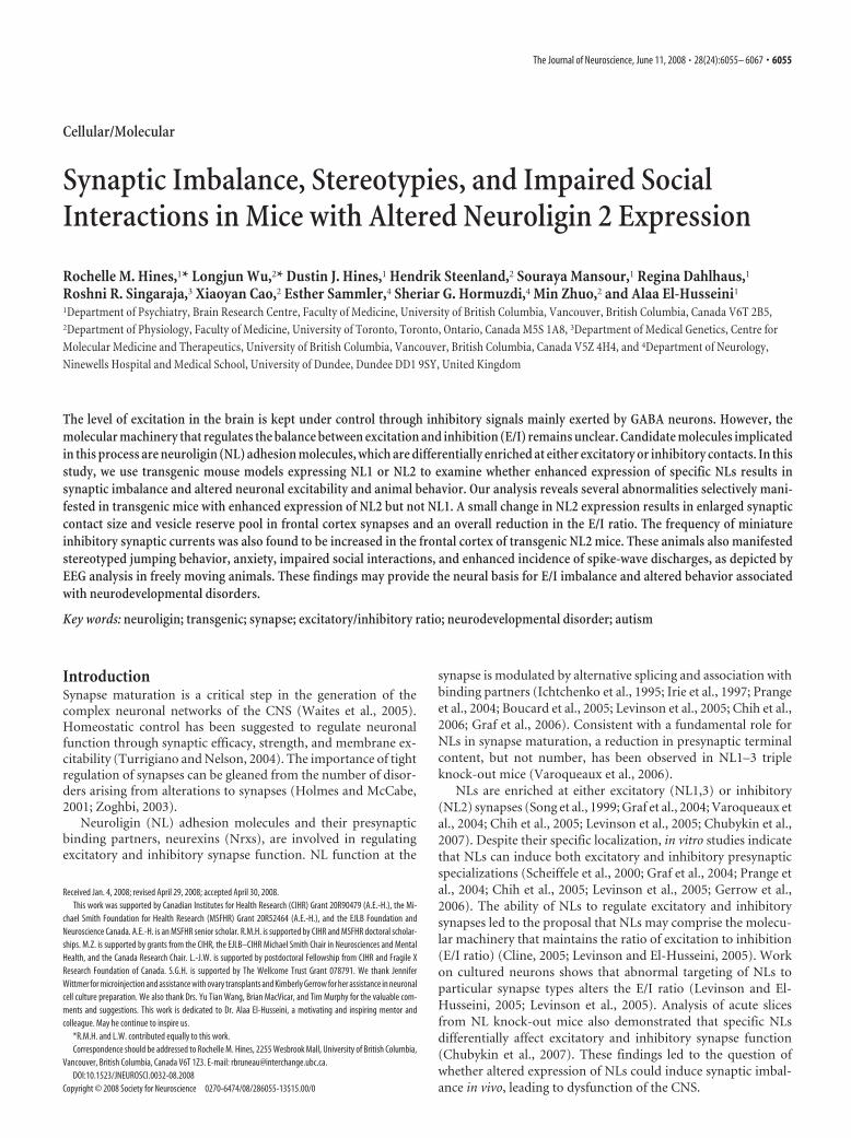
Figure 1. Mice expressing neuroligin 2 display a neurological phenotype related to levels of expression. A, Schematic diagramof HA-tagged NL1 and NL2 constructs in the Thy1.2 vector. B, Assessment of transgene expression level in TgNL1.6, TgNL1.7, andTgNL2.6 strains using immunoblotting for HA. Numbers in boxes show quantification of total protein load from Coomassie-stainedgels. C, Confirmation of generated NL2 rabbit antibody specificity (ABR) on COS-7 cell lysates transfected with HA-NL1, HA-NL2, orHA-NL3 (left 3 lanes) and on whole-brain lysates from wild-type and TgNL2.6 mice (right 2 lanes). D, Western blotting analysis ofwhole-brain lysates of endogenous and HA-NL2 expression using NL2 and HA antibodies. E, Limb clasping in TgNL2 mice (right)not seen in wild-type littermates or TgNL1 mice. F, Table comparing the level of HA-NL2 across TgNL2 strains relative to endog-enous NL2. The severity and frequency of limb clasping parallels the expression level of TgNL2 but not TgNL1 mice. Data shown aremeans � SEMs.
6056 • J. Neurosci., June 11, 2008 • 28(24):6055– 6067 Hines et al. • Characterization of Mice Expressing Neuroligin 2

Social behavior assessments. The reciprocal social interaction paradigmwas based on that used in previous studies (Tabuchi et al., 2007) andinvolved the introduction of a juvenile (6 weeks old) FVB male and eithera wild-type or TgNL2.6 mouse into a neutral environment. Video re-cordings of 5 min interaction session were then manually scored by ablinded observer for interaction parameters of total time, average length,total number, and proportion of initiations.
Social approach behavior was assessed in an apparatus modeled afterthat used by Dr. J. Crawley’s group (Moy et al., 2004; Nadler et al., 2004).Video recordings were assessed using the Noldus Ethovision Trackingsystem, and parameters, including time in zone, number of rears in zone,and speed traveled in zone, were compared. For more details on both thereciprocal social interaction and social approach behavior, see supple-mental Methods (available at www.jneurosci.org as supplementalmaterial).
In situ hybridization. In situ hybridization experiments were per-formed on postnatal day 28 (P28) mice as described previously (Wisdenand Morris, 2002). For details of in situ hybridization, see supplementalMethods (available at www.jneurosci.org as supplemental material).
Immunohistochemistry. For histology and immunohistochemistry(IHC), animals were either transcardially perfused with a 4% solution ofparaformaldehyde in PBS (nonfluorescent IHC) or brains were har-vested fresh and flash frozen in OCT embedding medium in liquid ni-trogen cooled isopentane (fluorescent synaptic protein IHC). Immuno-staining and colocalization was quantified using Northern eclipse, andresults were analyzed using t tests to compare means. For antibodies usedand additional details, see supplemental Methods (available at www.jneurosci.org as supplemental material).
Electron microscopy. Tissue for electron microscopy was harvestedfresh and cut using a vibratome in room temperature artificial CSF(ACSF), followed by rapid fixation in 6% glutaraldehyde, 1% parafor-maldehyde, 2 mM CaCl2, and 4 mM MgCl2 in 0.1 M cacodylate buffer.Results of synapse assessments were compared using ANOVA. For detailsof electron microscopy assessments, see supplemental Methods (avail-able at www.jneurosci.org as supplemental material).
Whole-cell patch-clamp recordings. Coronal slices of the frontal cortex(300 �m) were prepared using routine methods as described previously(Wu et al., 2007). For details of slice preparation and recording, seesupplemental Methods (available at www.jneurosci.org as supplementalmaterial).
In vivo electroencephalogram. Surgery was performed on 10 TgNL2and five wild-type mice (12–21 weeks old) for electroencephalogram(EEG) studies. For details of EEG surgery, recording, and analysis, seesupplemental Methods (available at www.jneurosci.org as supplementalmaterial).
Statistical methods. Results from experiments comparing wild-typecontrols with TgNL2.6 strains were analyzed by t tests to compare means.In the case of the social interaction test, in which t tests were applied tofour measures, a Bonferroni’s multiple testing correction was calculatedby dividing the standard significance level (0.05) by the number of t testsused (four), resulting in a Bonferroni’s corrected level of 0.0125. Exper-iments comparing wild-type, TgNL2.6, and TgNL2.7, or for the three-chambered social interaction paradigm, results were analyzed usingANOVA. The least squares difference post hoc test was applied to signif-icant ANOVA comparisons. All statistical analyses were calculated usingSPSS software.
ResultsLimb clasping reveals a neurological phenotype in miceexpressing neuroligin 2 that is not observed in miceexpressing neuroligin 1We used a gain-of-function approach to examine whether ma-nipulations of the levels of NLs in vivo influence synapse functionleading to altered behavior. For these experiments, transgenicanimals overexpressing NL1 (TgNL1) and NL2 (TgNL2) weregenerated (Fig. 1A,B). The transgenes were under the control ofThy1 promoter, which allows for expression in various brainregions at early stages of development. To facilitate detection of
the transgene, influenza hemagglutinin (HA) tags were inserted(between amino acids 45 and 46) immediately after the NL1 sig-nal sequence. To compare transgene expression and localizationwith wild-type NL2, a specific antibody was generated. This an-tibody was tested for specificity on lysates from Cos cells express-ing HA-NL1, HA-NL2, or HA-NL3 (Fig. 1C). In addition, theantibody was tested in cultured hippocampal neurons for pre-dominant colocalization with vesicular GABA transporter(VGAT) (supplemental Fig. 1B, available at www.jneurosci.orgas supplemental material) and in tissue from TgNL2 mice forcorrespondence with HA-tagged transgene (supplemental Fig.1C, available at www.jneurosci.org as supplemental material).
The generated TgNL1 and TgNL2 strains were viable; how-ever, TgNL2 mice exhibited a striking and distinct phenotype.TgNL2 mice with moderate to high levels of expression (Fig.1D,F) showed reduced lifespan and capacity to produce viableoffspring. One additional feature of the TgNL2 phenotype is limbclasping (Fig. 1E), a defect associated with mouse models of neu-rological disorders such as Rett syndrome (Gemelli et al., 2006).This feature was not observed at comparable levels in TgNL1strains with similar transgene expression (Fig. 1B) (1.25- to 1.71-fold TgNL2.6 HA expression).
Behavioral test battery demonstrates a consistent, dose-dependent phenotype in mice expressing neuroligin 2 notobserved in mice expressing neuroligin 1The preliminary behavioral screen was conducted on all foundersand strains generated based on the modified SHIRPA screen fromthe EMPReSS (Brown et al., 2005, 2006). The preliminary screenrevealed striking and consistent abnormalities among all TgNL2strains generated that increased with level of expression (Table 1,� indicates no difference from wild type). Animals with twofoldor greater expression of NL2 (determined by Western blot;TgNL2.1, TgNL2.4, and TgNL2.5) showed the most striking phe-notype with frequent limb clasping and death by 24 postnatalweeks, and these strains also failed to produce viable offspring.The primary strain has moderate expression level and phenotype(TgNL2.6; 1.6-fold wild-type NL2 expression) and has repro-duced successfully over nine generations; thus, most of the anal-ysis performed was on this strain. Whenever possible, results ob-tained from a higher expressing strain (TgNL2.7; 1.9-fold wild-type NL2) are included for comparison.
In general, TgNL2 mice are characterized by a reduction inbody weight (supplemental Fig. 2B–D, available at www.jneurosci.org as supplemental material), limb clasping (Fig.1E,F), Straub tail, transient episodes of kyphosis (supplementalFig. 2A, available at www.jneurosci.org as supplemental mate-rial), as well as enhanced basal activity and enhanced startle re-sponse to a 90 dB click (Table 1). However, no overt changes wereobserved in autonomic indicators such as palpebral closure andpiloerection (Table 1, Autonomic). In addition, somatosensory,visual, auditory, and olfactory systems were also found to be in-tact in TgNL2 mice using a battery of multiple tests (Table 1,Sensory). Basic muscle and motor assessments in TgNL2 micerevealed normal body and limb tone, and intact righting reflex (Ta-ble 1, Muscle and motor). The drastic phenotypic changes observedon relatively small alterations in transgene expression (1.6- to 2-foldendogenous NL2), combined with the dose-dependent change inthe severity of phenotype, indicates that the effects seen are not be-cause of excessive protein overexpression in vivo. Moreover, the lackof these abnormalities in TgNL1 further suggests that the observedchanges in TgNL2 mice have resulted from alterations attribut-able to specific manipulation of NL2 levels in vivo.
Hines et al. • Characterization of Mice Expressing Neuroligin 2 J. Neurosci., June 11, 2008 • 28(24):6055– 6067 • 6057

Expressed neuroligin 2 is distributed throughout theneuroaxis in neuronal cells and is predominantly localized toinhibitory synaptic contactsTo assess exogenous NL2 expression in detail, multiple ap-proaches were used. Both immunohistochemistry and in situ hy-bridization revealed broad distribution of the HA-NL2 transcriptand protein in TgNL2.6 (Fig. 2A–C). Despite varied levels ofexpression between transgenic strains as revealed by Westernblotting, the pattern of HA-NL2 staining was consistent. HA-NL2 was expressed throughout the neuroaxis, with high levels inthe cortex and limbic structures, such as amygdala and hip-pocampus (Fig. 2B,C). Staining of cortical cultures fromTgNL2.6 confirmed that HA-positive (HA�) labeling was exclu-sive to neuronal cells [4�,6�-diamidino-2-phenylindole-positive,MAP-2-negative (MAP-2�), HA�] and could be detected inlarge numbers of neurons (supplemental Fig. 1D, available atwww.jneurosci.org as supplemental material). Similar to wild-type NL2, HA-NL2 predominantly colocalized with inhibitory(gephyrin) and to a lesser extent with excitatory (PSD-95)postsynaptic markers, demonstrating that exogenous HA-NL2 isnot mislocalized (Fig. 2D,E).
Enhancement of markers of presynaptic terminals in miceexpressing neuroligin 2Given the implication of NLs in modulation of the E/I ratio, weanalyzed changes in the content of excitatory and inhibitory syn-apses in TgNL2 mice. Quantitative Western blotting analysis offorebrain lysates revealed significant changes in the amounts ofVGAT, vesicular glutamate transporter (VGluT) and syntaxinbut not in the expression of PSD-95 and gephyrin (Fig. 3A,B),suggesting a change in the content of presynaptic but notpostsynaptic elements of both excitatory and inhibitory synapses.Assessments of other NL family members revealed no change inNL1 but a significant decrease in NL3 expression levels in TgNL2
brain lysates (Fig. 3A,B). Interestingly, TgNL1 mice show oppos-ing alterations in related protein expression levels, with a signif-icant increase in both NL3 and PSD-95 (data not shown).
Immunostaining of frontal cortex sections from P28 TgNL2mice showed altered VGAT and VGluT staining intensity (Fig.3C,D). Consistent with the enrichment of NL2 at the majority ofGABAergic synapses, more robust increases were seen in the av-erage integrated intensity (VGAT, 1.7-fold; VGLuT, 1.3-fold) ofinhibitory contacts in the frontal cortex. The ratio of VGluT toVGAT staining intensity in the frontal cortex was significantlydecreased in TgNL2 when compared with wild-type littermates(Fig. 3E) revealing a bias toward increased inhibition during invivo expression of NL2.
EM analysis reveals changes in synapse morphology and anincrease in inhibitory contacts in frontal cortex of miceexpressing neuroligin 2Ultrastructural EM analysis of medial prefrontal cortex (MPFC)of TgNL2.6 and TgNL2.7 brain demonstrated changes in themorphology of synaptic components (Fig. 4C) and synapse den-sity. Marked increases were observed in the number of vesicles inthe reserve pool (Fig. 4A,D), as well as the area of symmetric(type II, typically inhibitory) presynaptic compartments (Fig.4A,E) and contact length of symmetric synapses (Fig. 4A,F) inTgNL2 mice. In addition, a small but significant change was ob-served in asymmetric (type I, typically excitatory) presynapticcompartment area (Fig. 4B,E). No change was observed in theaverage length of postsynaptic densities in TgNL2 mice (Fig.4B,D).
Using unbiased stereological analysis, a modest increase in thetotal density of synapses, as well as the density of symmetricalsynapses was observed in both TgNL2.6 and TgNL2.7 (Fig.4G,H). No changes were seen in the total number of neurons inthe frontal cortex (supplemental Fig. 3A,B, available at www.
Table 1. Summary of the preliminary screen conducted on all strains
WT TgNL1.6 TgNL1.7 TgNL2.6 TgNL2.7 TgNL2.1 TgNL2.3 TgNL2.4 TgNL2.5
GeneralViability � � � � 1 early adult 1 early adult 1 early adult 1 early adult 1 early adultOffspring viability � 1 incr # dead 1 incr # dead 1 incr # dead 2 high # dead 3 none viable 3 none viable 3 none viable 3 none viable
AutonomicPalpebral closure � � � � � � � � �Piloerection � � � � � � � � �Tail position � � � 2 Straub tail 2 Straub tail 2 Straub tail 2 Straub tail 2 Straub tail 2 Straub tail
SensorySomatosensory
Transfer arousal � � � � � � � � �Touch escape � � � � � � � � �
VisualCorneal reflex � � � � � � � � �Visual placing � � � � � � � � �
AuditoryPinna reflex � � � � � � � � �Acoustic startle (90 dB click) � � � 1 jump �1 cm 1 jump �1 cm 1 jump �1 cm 2 jump �1 cm 2 jump �1 cm 2 jump �1 cm
OlfactoryBuried food retrieval � � � � � � � � �
Muscle and motorBody position � � � 1 rare kyphosis 2 kyphosis 2 kyphosis 2 kyphosis 2 kyphosis 2 kyphosisBody tone and limb tone � � � � � � � � �Righting reflex � � � � � � � � �Basal activity � � � 2 rear/leap 2 rear/leap 2 rear/leap 1 rapid darting 1 rapid darting 1 rapid dartingOpen field speed/distance � � � � � � � � �
Table shows scores for WT, TgNL1, and TgNL2 strains; transgenic strains are arranged in order of increasing expression of transgene. � indicates normal or no impairment; in the case of impairment, increasing values indicate increasedimpairment, and a brief description is provided. For detailed description of methods, observations, and scoring, see supplemental Methods (available at www.jneurosci.org as supplemental material).
6058 • J. Neurosci., June 11, 2008 • 28(24):6055– 6067 Hines et al. • Characterization of Mice Expressing Neuroligin 2

jneurosci.org as supplemental material). By dividing the densityof asymmetric synapses by the density of symmetric synapses, wecan estimate a shift in the E/I ratio toward inhibition in TgNL2frontal cortex compared with littermate controls (Fig. 4 I), sup-
porting the finding of a decreased ratio ofVGluT to VGAT immunostaining inTgNL2.6 frontal cortex.
Together, these results suggest an effectof NL2 in vivo on the modulation of syn-apse morphology with primary effects onpresynaptic terminals and, in particular,symmetric synapses. The small effects ofNL2 on asymmetric presynaptic terminalsare not surprising because endogenousNL2 can be found at �20 –30% of excita-tory synapses (Fig. 2E). Given the effectson symmetrical synapse density, it is alsopossible that changes observed in the mor-phology of asymmetrical terminals are ameans of compensation. The greater de-gree of change observed in inhibitory ver-sus excitatory synapses suggest that en-hanced expression of NL2 results in anoverall reduction in the E/I ratio, revealingthat a small increase in NL2 expression re-sults in alterations in synaptic balance infrontal cortex.
Altered synaptic transmission inprefrontal cortex of mice expressingneuroligin 2Whole-cell patch-clamp recordings wereperformed in pyramidal neurons in layerII/III of wild-type and TgNL2.6 prefrontalcortex. Recording of spontaneous activityrevealed that the frequency of miniatureIPSCs (mIPSCs) is increased in TgNL2mice compared with those in wild-typemice (Fig. 5A,B). However, the mIPSCamplitude was not altered in the prefrontalcortex of TgNL2 mice (Fig. 5A,B). In con-trast to inhibitory currents, neither fre-quency (Fig. 5C,D) nor amplitude (Fig.5C,D) of mEPSCs in TgNL2 mice werefound to be significantly different fromthose in the prefrontal cortex of wild-typemice (Fig. 5A–C). These results are consis-tent with the EM data suggesting a primaryeffect of NL2 expression on modulatinginhibitory synapse function.
Altered neuroligin 2 expression leads tospontaneous stereotypies andanxiety behaviorTo assess the behavior of TgNL2 mice inmore detail, we next observed mice in theopen field paradigm. When in the openfield, TgNL2.6 mice display spontaneousjumping stereotypies in the corners of thearena (Fig. 6A) (supplemental Video 1,available at www.jneurosci.org as supple-mental material). Stereotyped patterns ofbehavior can be induced in animals by
treatment with amphetamines and cocaine or by deprivation(Powell et al., 1999; Wurbel, 2001) and have also been observed inmouse models of mental retardation such as the Down syndromemodel Ts65Dn (Turner et al., 2001). Stereotyped jumping behav-
Figure 2. Neuroligin 2 transgene distribution and localization. A, In situ hybridization analysis of HA-containing transcript inTgNL2.6 (left) and of NL2-containing RNA in TgNL2.6 (middle) and WT (right). Scale bar, 3 mm. B, DAB immunohistochemistry forHA in sagittal sections from WT and TgNL2.6. Scale bar, 3 mm. C, HA DAB immunostaining in coronal sections through the frontalcortex (bregma, 2.71–2.22 mm) and hippocampus/amygdala (bregma, �1.28 to �1.64 mm) of TgNL2.6 brain. Scale bar, 1 mm.D, Confocal analysis of colocalization of HA-NL2 (green) and excitatory (PSD-95, red, top panel) and inhibitory (gephyrin, red,bottom panel) synaptic markers in the frontal cortex. Scale bar, 2 �m. E, Quantification of excitatory (PSD-95; WT, 24.21 �5.50%; TgNL2.6, 35.20 � 3.51%; t test, p 0.181) and inhibitory (gephyrin; WT, 73.60 � 5.86%; TgNL2.6, 61.91 � 5.38%; ttest, p 0.176) marker colocalization with endogenous NL2 in wild-type frontal cortex compared with HA-NL2 in TgNL2.6 frontalcortex. Data shown are means � SEMs; WT and TgNL2.6, n 9. ns, Not significant.
Hines et al. • Characterization of Mice Expressing Neuroligin 2 J. Neurosci., June 11, 2008 • 28(24):6055– 6067 • 6059

ior is typically preceded by rearing against the arena wall and ischaracterized by repeated jumping vertically on hindlegs and bal-ancing on a rigid tail (supplemental Video 1, available at www.jneurosci.org as supplemental material) (Wurbel and Stauf-facher, 1996; Garner and Mason, 2002). The jumping stereotypywas also observed when TgNL2.6 mice were in the home cage (inthe presence of social and environmental enrichment); however,repetitive jumping was never observed in wild-type littermates(n 50). Not all TgNL2.6 mice showed stereotypies during openfield observation, and it was found that 44% of TgNL2.6 males(n 50) show more than one episode of stereotypy during a 5min open field exploration (Fig. 6B). TgNL2.6 mice showingstereotypies typically display high levels of this behavior, with anaverage of �10 episodes over the 5 min period (Fig. 6B). TgNL2.6mice displaying more than one episode of jumping stereotypyduring a 5 min prescreening session in the open field were ex-cluded from behavioral assessments of anxiety and social behav-ior to avoid possible confounds of this behavior (for details, seesupplemental Methods, available at www.jneurosci.org as sup-plemental material) (Garner, 2005). Other types of repeated be-havior patterns were also monitored in the open field, such asdigging and grooming, but the incidence of these types of behav-iors was not significantly different from wild-type littermates
(Fig. 6A). It has been shown that stereotyped patterns of behaviorthat result from drug sensitization can be blocked by antagonistsor induced by agonists of the GABAergic system (Karler et al.,1995); thus, it is possible that this behavior arises as a result of thealterations in GABAergic transmission observed in the frontalcortex (Karler et al., 1997) of TgNL2 mice.
During the open field test, we also observed indications ofanxiety behavior in TgNL2 mice. Both TgNL2.6 and TgNL2.7mice show thigmotaxis (Fig. 6C), as demonstrated by a decreasein both average (supplemental Fig. 4A, available at www.jneurosci.org as supplemental material) and cumulative (Fig.6D) distance from the arena border compared with wild-typemice. TgNL2 mice also show increased rearing frequency, whichis further indicative of anxiety (supplemental Fig. 4B, available atwww.jneurosci.org as supplemental material). However, nochanges were observed in average speed traveled while exploringthe open field (data not shown; wild type, 4.75 cm/s; TgNL2.6,6.07 cm/s; TgNL2.7, 6.18 cm/s; p 0.169) or total distance trav-eled in the open field (supplemental Fig. 4C, available at www.jneurosci.org as supplemental material), demonstrating thatTgNL2 animals do not have motor impairments or lack explor-atory motivation.
To confirm that the thigmotaxis observed in the open field
Figure 3. Western blotting and immunohistochemical assessment of synaptic proteins in mice expressing neuroligin 2. A, Representative blots for synaptic proteins assayed in wild-type andTgNL2.6 mice. B, Quantification of Western blot normalized to total protein load from Coomassie-stained gels and compared with WT expression level. C, Confocal microscopy of VGluT (middle,green) and VGAT (right, red) staining in wild-type (top) and TgNL2.6 (bottom) MPFC. Scale bar, 5 �m. D, Quantification of the average integrated intensity of VGluT (WT, 37.37 � 1.00; TgNL2.6,48.29 � 3.29; t test, p 0.033) and VGAT (WT, 30.26 � 2.02; TgNL2.6, 49.76 � 1.53; t test, p � 0.001) puncta in MPFC. E, The ratio of excitation to inhibition as expressed by the ratio of VGluTintensity/VGAT intensity (WT, 1.28 � 0.09; TgNL2.6, 1.00 � 0.07; t test, p 0.028). Data shown are means � SEMs; WT and TgNL2.6, n 9.
6060 • J. Neurosci., June 11, 2008 • 28(24):6055– 6067 Hines et al. • Characterization of Mice Expressing Neuroligin 2

was indicative of anxiety, we also compared the moderate ex-pressing TgNL2.6 strain with wild-type littermates in thelight-dark exploration test. Observation of TgNL2.6 mice inthe light/dark test revealed an increase in the percentage oftime spent in the dark compartment compared with wild-typelittermates (Fig. 6 E). However, no change was observed in thenumber of transitions between the light and dark compart-ments (supplemental Fig. 4 D, available at www.jneurosci.orgas supplemental material), which is an additional measure ofanxiety-like behavior in the light/dark test. It has been re-ported that the most consistent measure of anxiety using thelight/dark test is the percentage of time spent in each compart-ment, and transitions have been suggested to be influenced byactivity or exploration (Belzung et al., 1987; Hascoet andBourin, 1998; Bourin and Hascoet, 2003). Although it is un-
clear why TgNL2 mice display an anxiety-like phenotype ononly one of the two measures for the light/dark task, it ispossible that the high level of basal activity observed in TgNL2may obscure detection of differences in the number oftransitions.
To further confirm an anxiety-like phenotype, mice were alsotested in the elevated plus maze. TgNL2.6 mice demonstrated areduction in the percentage of time spent in the open arms (Fig.6F), as well as in the percentage of entries made into open arms(Fig. 6F) of the elevated plus maze compared with wild-typelittermate controls. No difference was observed in the total num-ber of entries (supplemental Fig. 4E, available at www.jneurosci.org as supplemental material), supporting the idea that TgNL2.6performance on this task is not limited by motor impairments orlack of exploratory motivation. Overall, these three assessments
Figure 4. Synaptic abnormalities in mice expressing neuroligin 2. A, B, Representative electron micrographs of symmetric (A) and asymmetric (B) synapses in wild-type and TgNL2.6 MPFC. Scalebars, 500 nm. C, Synaptic elements quantified are highlighted. D, E, Quantification of the length of the postsynaptic density (WT, 146.05 � 7.78; TgNL2.6, 180.00 � 15.14; TgNL2.7, 139.96 �10.35) and number of vesicles in the reserve pool (WT, 30.93 � 3.24; TgNL2.6, 51.87 � 6.37; TgNL2.7, 69.27 � 8.87; ANOVA, F(2,42) 8.52, p 0.001; post hoc tests, p 0.030 WT vs TgNL2.6,p � 0.001 WT vs TgNL2.7) and presynaptic compartment area (symmetric: WT, 501.54 � 71.79; TgNL2.6, 1020.11 � 131.01; TgNL2.7, 1436.04 � 157.72; ANOVA, F(2,65) 8.57, p � 0.001; posthoc tests, p 0.011 WT vs TgNL2.6, p � 0.001 WT vs TgNL2.7; asymmetric: WT, 572.95 � 44.52; TgNL2.6, 747.99 � 69.14; TgNL2.7, 934.83 � 145.27; ANOVA, F(2,138) 4.75, p 0.010; posthoc tests, p 0.048 WT vs TgNL2.6, p 0.003 WT vs TgNL2.7) in wild-type, TgNL2.6, and TgNL2.7 MPFC. F, Analysis of the length of contact between symmetric presynaptic and postsynapticcompartments (WT, 254.34 � 32.46; TgNL2.6, 493.87 � 57.00; TgNL2.7, 465.59 � 66.36; ANOVA, F(2,31) 4.03, p 0.028; post hoc tests, p 0.009 WT vs TgNL2.6, p 0.039 WT vs TgNL2.7)in wild-type, TgNL2.6, and TgNL2.7 symmetric synapses. G, H, Number of total (WT, 8.69 � 0.34; TgNL2.6, 10.42 � 0.34; TgNL2.7, 11.81 � 0.88; ANOVA, F(2,45) 7.28, p 0.002; post hoc tests,p 0.040 WT vs TgNL2.6, p � 0.001 WT vs TgNL2.7), symmetric (WT, 7.30 � 0.35; TgNL2.6, 7.99 � 0.44; TgNL2.7, 8.69 � 0.78; ANOVA, F(2,45) 1.58, p 0.219), and asymmetric (WT, 1.16 �0.28; TgNL2.6, 2.32 � 0.28; TgNL2.7, 3.13 � 0.49; ANOVA, F(2,45) 7.55, p 0.002; post hoc tests, p 0.028 WT vs TgNL2.6, p � 0.001 WT vs TgNL2.7) synapses per 10 �m3 field of MPFCneuropil. I, The ratio of excitation to inhibition as expressed by the ratio of asymmetric: symmetric synapses (WT, 5.89 � 0.60; TgNL2.6, 4.14 � 0.53; TgNL2.7, 3.67 � 0.60; ANOVA, F(2,45) 4.09,p 0.024; post hoc tests, p 0.039 WT vs TgNL2.6, p 0.010 WT vs TgNL2.7). Data shown are means � SEMs. Synapse density: WT and TgNL2.6, n 4 mice, n 16 fields; TgNL2.7, n 3 mice,n 16 fields.
Hines et al. • Characterization of Mice Expressing Neuroligin 2 J. Neurosci., June 11, 2008 • 28(24):6055– 6067 • 6061

indicate an increase in anxiety-like behavior in mice expressingNL2 compared with their littermate controls.
Mice expressing neuroligin 2 display abnormalities insocial behaviorTo assess reciprocal social interactions of TgNL2 mice, male micewere placed into a neutral home cage with a novel juvenile target
male of a different strain (FVB). The two animals were allowed tofreely interact over the course of 5 min, and the number andduration of interactions were manually assessed from video re-cordings. A striking reduction in the total time of interaction withthe novel target mouse was observed in TgNL2 mice when com-pared with wild-type littermates (Fig. 7A). Although TgNL2.6mice did not show a difference in the total number of interactionswith novel juveniles (Fig. 7C), the average duration of individualinteractions was significantly reduced (Fig. 7B). The lack ofchange in the total number of interactions is likely to result fromincreased initiation of interactions by the freely moving targetmouse (Fig. 7D).
To further assess social behavior and rule out a general defectin their interaction with novel stimuli, we assessed TgNL2.6 micein a counterbalanced social approach apparatus based on the taskdeveloped by the laboratory of Dr. J. N. Crawley (Fig. 7E) (Moy etal., 2004; Nadler et al., 2004). In this three-chambered apparatus,wild-type mice show a strong preference for the chamber con-taining the novel mouse over either the center or counterbal-anced novel object chamber. This effect can be seen in the totalamount of time spent in the chamber with the novel mouse (Fig.7F). In contrast, TgNL2.6 mice do not show this preference, withno significant difference between the time spent in the novelmouse chamber compared with the novel object chamber. Inaddition, wild-type mice also display preference for the novelmouse chamber by the number of investigative rears made in thischamber (Fig. 7G), indicating a high level of exploration of thenovel mouse. In contrast, TgNL2.6 mice do not demonstratepreference for the novel mouse chamber over the novel objectchamber in terms of high levels of rearing. This result of reducedsocial approach in the three-chambered apparatus, in conjunc-tion with reduced reciprocal social interactions, demonstratesthat TgNL2 animals are impaired in the natural preference forsocial interaction seen in wild-type mice.
Chronic EEG recording in freely moving neuroligin 2transgenic mice reveals bilateral spike-wave dischargesSynapse anomalies and behavioral observations prompted as-sessment of spike wave patterns via frontoparietal EEG record-ings in TgNL2 mice. Neck electromyogram (EMG) was recordedto establish whether spiking occurred during different sleep andwake states. Eight of the 10 TgNL2 animals studied demonstratedbrief bilateral bursting activity characterized by spike-wave dis-charge of �6 – 8Hz (Fig. 8A,B). The spike-wave episodes werebrief, did not always occur with specific behaviors, and could beidentified in both awake and sleep states (Fig. 8C). The resultsindicate that even a mild increase in the expression of NL2 resultsin spiking activity. Despite the detection of spiking activity byEEG, TgNL2.6 mice did not exhibit any overt visible signs ofseizure. The spiking activity observed by frontoparietal EEG maybe attributable to desynchronized cortical activity patterns or as aresult of spreading excitation from other brain regions.
DiscussionOur analysis reveals that manipulation of NL2 expression resultsin altered synapse morphology and function. Previous studiesindicate that altered levels of individual NLs at particular synapticsites modulate synapse maturation and neuronal excitability(Levinson and El-Husseini, 2005). Consistent with these find-ings, EM and electrophysiological analyses of frontal cortical ar-eas in transgenic NL2 mice reveal an increase in the size andnumber of inhibitory synaptic contacts and enhanced frequencyof presynaptic currents. Thus, the net result is potentiation of
Figure 5. Increased inhibitory synaptic transmission in pyramidal neurons of prefrontal cor-tex in mice expressing neuroligin 2. A, Representative traces showing the mIPSCs in wild-typeand TgNL2.6 mice. TTX (1 �M), CNQX (20 �M), and AP-5 (50 �M) was added in the ACSF duringmIPSC recordings. B, Statistical results showing the significant increase of mIPSC frequency(left; WT, 2.3 � 0.3, n 22; TgNL2.6, 3.6 � 0.4, n 23; t test, p � 0.050) but not amplitude(right; WT, 13.1 � 0.5, n 22; TgNL2.6, 13.5 � 0.3, n 23; t test, p 0.55) in TgNL2.6 micecompared with wild-type mice. C, Typical traces showing the mEPSCs recorded in wild-type(left) and TgNL2.6 (right) mice. TTX (1 �M), picrotoxin (100 �M), and AP-5 (50 �M) was addedin the ACSF during mEPSC recordings. D, Statistical results showing normal mEPSC frequency(left; WT, 1.6 � 0.3, n 16; TgNL2.6, 2.0 � 0.3, n 17; t test, p 0.24) or amplitude (right;WT, 9.2�0.3, n16; TgNL2.6, 8.7�0.2, n17; t test, p0.21) in TgNL2.6 mice comparedwith wild-type mice. Data shown are means � SEMs.
6062 • J. Neurosci., June 11, 2008 • 28(24):6055– 6067 Hines et al. • Characterization of Mice Expressing Neuroligin 2

inhibitory responses in the frontal cortex, revealing a shift in thebalance toward inhibition.
The observed phenotypic changes were attributable to smallalteration in NL2 expression (1.6- to 2-fold above endogenouslevels of NL2). These findings indicate that the effects seen are notattributable to excessive protein overexpression in vivo. Interest-ingly, the severity of behavioral changes correlates with the levelof NL2 expression, suggesting dose-dependent changes in syn-apse function and behavior. The lack of related abnormalities inTgNL1 further suggest that the observed behavioral changes inTgNL2 mice resulted from alterations in synaptic maturationand/or function attributable to specific manipulation of NL2 lev-els in vivo. Importantly, neither TgNL1 nor TgNL2 strains display
deficits in basic sensory, reflexive, or mo-tor function using the modified SHIRPAscreen, making them feasible candidatesfor additional behavioral characterization.Intact motor function and exploratory mo-tivation was further confirmed in TgNL2mice in open field, elevated plus, and socialapproach tasks in which no deficits were ob-served in control measures of speed, distancetraveled, or total entries.
The differential enrichment of spe-cific NLs to particular synaptic sites invivo suggests that NL3 and NL4 mainlymodulate excitatory synaptic function,whereas NL2 is associated with modula-tion of inhibitory synaptic transmission(Song et al., 1999; Dean et al., 2003;Prange et al., 2004; Varoqueaux et al.,2004, 2006; Chubykin et al., 2005, 2007;Levinson and El-Husseini, 2005; Levin-son et al., 2005; Sara et al., 2005; Deanand Dresbach, 2006). In particular, lossof NL2 has been shown to specificallyalter inhibitory synapse function in vivo.Consistent with these findings, our re-sults show that enhanced expression ofNL2 results in a significant increase ininhibitory synapse maturation andtransmission in cortical areas. However,it is important to note that a small butsignificant change in excitatory synapsemorphology was also observed in TgNL2mice, indicating that NL2 expression caninfluence both excitatory and inhibitorycontact maturation, although with pro-nounced effects on inhibitory contacts.These data suggest that NL2 function invivo is not fully restricted to inhibitorysynapse maturation. These findings alsohint to some overlapping and redundantfunctions of members of the NL familyin vivo. In support of some functionalredundancy between NLs, knock-out ofNL1–NL3 is lethal, whereas all of the sin-gle and double knock-out combinationsare viable (Varoqueaux et al., 2006). Thefinding that endogenous NL2 is found at�20 –30% of excitatory synapses and therecent work that demonstrates that NLscan form heteroligomers (Budreck and
Scheiffele, 2007) lend additional support to this notion.The mechanism underlying the lack of change in inhibitory
postsynaptic responses remains unclear. It is possible that re-cruitment of GABA receptors at inhibitory synapses is more con-strained and consequently the presynaptic changes seen did notlead to an increase in recruitment of postsynaptic GABA recep-tors. This could be caused by enhanced levels of NL2, which canact to disperse GABA receptors and reduce inhibitory currents(Graf et al., 2004).
The changes observed including limb clasping, repetitive be-haviors, anxiety, and social dysfunction are similar to those ob-served in animal models of Rett syndrome (Chen et al., 2001; Guyet al., 2001; Shahbazian et al., 2002; Pelka et al., 2006). Some of
Figure 6. Neuroligin 2-expressing mice display spontaneous jumping stereotypies and anxiety behavior. A, Graph plotting theincidence of stereotyped jumping (WT, 0; TgNL2.6, 5.58 � 2.53), digging (WT, 8.50 � 1.31; TgNL2.6, 10.75 � 2.63; t test, p 0.461), and grooming (WT, 3.63�0.78; TgNL2.6, 4.83�0.81; t test, p0.318) behaviors in the open field (WT, n7; TgNL2.6,n 8). � Signifies that wild-type animals did not demonstrate this behavior, under any of the conditions examined. B, Tableshowing the percentage of wild-type (0%) and TgNL2.6 (44%) mice showing stereotypies in a larger population (n 50) and, ofthese mice, the average number of stereotypies shown in a 5 min open field session. C, Representative paths of wild-type (left) andTgNL2 (right) mice in the open field arena. D, Assessment of the cumulative distance from the arena border (WT, 17,965.74 �379.79; TgNL2.6, 15,088.26�709.59; TgNL2.7, 9784.31�570.32; ANOVA, F(2,17) 30.51, p �0.001; post hoc tests, p 0.001WT vs TgNL2.6, p � 0.001 WT vs TgNL2.7) in the open field task. E, Percentage of time spent in the dark chamber (WT, 64.90 �4.21; TgNL2.6, 81.58 � 3.55; t test, p 0.022) by wild-type and TgNL2.6 mice during the light/dark exploration test. F,Assessment of the percentage of open arm time (WT, 34.17 � 2.92; TgNL2.6, 18.94 � 1.45; t test, p � 0.001) and open armentries (WT, 38.86 � 1.39; TgNL2.6, 27.46 � 2.15; t test, p � 0.001) by wild-type and TgNL2.6 mice in the elevated plus maze.Data shown are means � SEMs. Open field: wild-type, n 7; TgNL2.6, n 8; TgNL2.7, n 2. Light/dark exploration test:wild-type and TgNL2.6, n 8. Elevated plus maze: wild-type and TgNL2.6, n 8.
Hines et al. • Characterization of Mice Expressing Neuroligin 2 J. Neurosci., June 11, 2008 • 28(24):6055– 6067 • 6063

these phenotypic characteristics also re-semble aspects of human disorders, in-cluding Rett syndrome and autism (Hag-berg, 2002; Rubenstein and Merzenich,2003; Zoghbi, 2003; Dover and Le Cou-teur, 2007; Lewis et al., 2007). Autism, agenetically linked disorder, is character-ized by varying degrees of abnormality incommunication ability, social interac-tions, repetitive and stereotyped behavior,as well as high incidence of seizure (Tuch-man and Rapin, 2002; Rubenstein andMerzenich, 2003; Zoghbi, 2003; Christ etal., 2007; Dover and Le Couteur, 2007;Rutherford et al., 2007).
Of particular interest, recent studieshave shown rearrangement of chromo-somal regions harboring NL1 and NL2, aswell as mutations in NL3 and NL4 in fam-ilies with autism (Konstantareas and Ho-matidis, 1999; Auranen et al., 2002; Jamainet al., 2003; Laumonnier et al., 2004; Liseand El-Husseini, 2006). A single-copychromosomal deletion of a region con-taining �/�-Nrxs indicates that alteredamounts of these proteins is sufficient toconfer the behavioral changes associatedwith autism (Szatmari et al., 2007). In ad-dition to single gene alterations, recentstudies have shown that many autistic pa-tients have novel deletions and duplica-tions in their genomes (Sebat et al., 2007).
Neuroimaging and postmortem stud-ies of patients with autism suggest that analteration in the ratio of E/I in neural cir-cuits underlies the dysfunctions character-istic of autism (Rubenstein and Mer-zenich, 2003). The theory proposed byRubenstein and Merzenich suggests thatthe dysfunction in autism could resultfrom a shift toward excitation, but, inmouse models of related disorders such asRett syndrome (Dani et al., 2005), thetrend appears to favor inhibition. In addi-tion, increased inhibition was observed inmice expressing an autism-related muta-tion of NL3, and this was shown to resultin altered social interaction (Tabuchi et al., 2007). In this regard,a gain of NL2 function may result in a shift toward increasedinhibition, mimicking the reduced function of the mutant formsof NL3 or NL4 most commonly associated with autism. Thus, asalso observed for models of Rett syndrome (Collins et al., 2004;Gemelli et al., 2006), both reduced and enhanced expression ofaffected genes may model the underlying cause of neurodevelop-mental disorders such as autism.
Several lines of evidence indicate multiple deficits in the struc-ture and function of the brain in individuals with autism. Inparticular, there is strong evidence supporting that abnormalitiesin the frontal cortex and the amygdala contribute highly to defi-cits in social behavior and anxiety. In particular, previous studieshave shown a link between increased excitation in the amygdalaand anxiety (Davis et al., 1994). Amygdala excitability can also beincreased via disinhibition (decreasing inhibitory transmission
from other brain regions). For instance, increased inhibitionwithin the prefrontal cortex, which normally acts to inhibit theamygdala (Rosenkranz et al., 2003; Quirk and Gehlert, 2003;Quirk et al., 2003), can lead to increased excitation (disinhibi-tion) in the amygdala and, hence, anxiety (Davis et al., 1994;Berkowitz et al., 2007; Bishop, 2007). Changes in the morphologyand density of inhibitory synapses and increases in mIPSC fre-quency in the frontal cortex observed in the present work maytherefore disinhibit the amygdala and contribute, at least in part,to the enhanced anxiety in TgNL2 mice. In relation to alteredfrontal cortex–amygdala projections, our data may also shed lighton a hypothesis that suggests that the dysfunction in autism mayresult from excessive and unselective connectivity in local frontalcortex circuitry, paired with impoverished long-range connectiv-ity to other systems such as the amygdala (Courchesne andPierce, 2005).
Figure 7. Deficits in social interactions in mice expressing neuroligin 2. A, Assessment of the total time wild-type or TgNL2.6mice spent interacting with a novel mouse (WT, 124.01 � 9.04; TgNL2.6, 54.29 � 6.69; t test, p 0.001) in a neutral arena overa 5 min period. B, Graph showing the average length of individual interactions between wild-type or TgNL2.6 mice and a novelmouse (WT, 6.68 � 0.84; TgNL2.6, 2.84 � 0.26; t test, p 0.004). C, No difference was observed in the total number ofinteractions (WT, 20.40 � 1.40; TgNL2.6, 19.20 � 1.41; t test, p 0.527) between wild-type or TgNL2.6 mice and novel mice.D, Proportion of interactions initiated by either wild-type or TgNL2.6 mice with a novel mouse (WT, 52.80 � 1.67; TgNL2.6,23.35 � 3.59; t test, p � 0.001). A–D, Bonferroni’s corrected significance levels for multiple t tests used: 0.013, 0.003, and�0.001. E, Schematic of the three-chambered social behavior apparatus. F, Analysis of time spent (WT novel mouse, 654.78 �81.10; WT novel object, 350.60 � 51.71; TgNL2.6 novel mouse, 406.32 � 31.78; TgNL2.6 novel object, 539.75 � 45.10; ANOVA,F(3,28) 6.077, p 0.003; post hoc tests, p 0.001 WT novel mouse vs WT novel object, p 0.100 TgNL2.6 novel mouse vsTgNL2.6 novel object) in the novel mouse and novel object chambers of the social apparatus. G, Frequency of rearing (activeexploration) in the chambers of the social apparatus (WT novel mouse, 115.50 � 23.61; WT novel object, 59.50 � 7.84; TgNL2.6novel mouse, 94.00 � 12.20; TgNL2.6 novel object, 89.50 � 8.44; ANOVA, F(3,28) 2.54, p 0.077; post hoc tests, p 0.011WT novel mouse vs WT novel object, p 0.828 TgNL2.6 novel mouse vs TgNL2.6 novel object). Data shown are means � SEMs.Reciprocal social interaction, wild type and TgNL2.6, n 10; three-chamber social approach behavior, wild type and TgNL2.6,n 8.
6064 • J. Neurosci., June 11, 2008 • 28(24):6055– 6067 Hines et al. • Characterization of Mice Expressing Neuroligin 2

The incidence of frontoparietal seizure spiking, in light ofincreased prefrontal cortex inhibition, is particularly interestingbecause prevailing theories suggest that seizure activity resultsfrom hypersynchronous neuronal activity and is commonly as-sociated with increased E/I ratios (Kofke et al., 1997; Stief et al.,2007). However, recent reports have also shown that focal seizureactivity can result from desynchronization of firing caused byincreased inhibitory feedback (Netoff and Schiff, 2002; Mor-mann et al., 2003; Klaassen et al., 2006). Thus, increased inhibi-tion observed in frontal cortex of TgNL2 mice could underlie thecortical seizure spiking activity detected by freely moving EEGrecording.
Clinically apparent seizures occur in �30% of autistic indi-viduals (Gillberg and Billstedt, 2000), whereas 50 –70% of autisticindividuals display ongoing “sharp-spike” activity during sleep(Lewine et al., 1999; Wheless et al., 2002). The incidence of sei-zure spiking in TgNL2 mice may relate to cortical–limbic dys-function discussed above in relation to autism, which is also im-plicated in temporal lobe epilepsy (Aroniadou-Anderjaska et al.,2007). In keeping with the idea of frontal cortex inhibition lead-ing to amygdala disinhibition in TgNL2 mice, altered neuronalexcitability in the amygdala could generate spontaneous epilep-
tiform activity, which will subsequentlyspread to the other brain areas(Aroniadou-Anderjaska et al., 2007).Overall, the spiking activity observed maybe directly attributable to alterations incortical synchrony cause by increased in-hibition or indirectly as a result of overex-citation in the amygdala spreading to thecortex. Future studies are needed to deter-mine whether enhanced inhibition in thefrontal cortex can be related to alterationsin the excitability of the amygdala.
Our new findings combined with thelink between mutations in NLs/Nrxs andautism may provide the neuronal basis foralterations in neuronal excitability, seizurespiking activity, stereotypies, anxiety, andimpaired social interactions associatedwith autism (Konstantareas and Homati-dis, 1999; Auranen et al., 2002; Jamain etal., 2003; Laumonnier et al., 2004; Lise andEl-Husseini, 2006; Szatmari et al., 2007).In conclusion, we have discovered synapseand behavioral abnormalities in mice withaltered expression of particular membersof the NL family that recapitulate multipleaspects of behavioral changes associatedwith neurodevelopmental disorders suchas Rett syndrome and autism. These in-clude impaired social interactions, stereo-typed patterns of behavior, and enhancedincidence of seizure spiking, as depicted byEEG analysis in freely moving animals. Wealso show that a small change in NL2 ex-pression results in synaptic abnormalitiesin cortical networks, which are thought tounderlie the complex behavioral alter-ations seen in autism. These findings mayprovide the neural basis for synaptic im-balance and altered behavior associatedwith autism.
ReferencesAroniadou-Anderjaska V, Qashu F, Braga MF (2007) Mechanisms regulat-
ing GABAergic inhibitory transmission in the basolateral amygdala: im-plications for epilepsy and anxiety disorders. Amino Acids 32:305–315.
Auranen M, Vanhala R, Varilo T, Ayers K, Kempas E, Ylisaukko-Oja T, Sin-sheimer JS, Peltonen L, Jarvela I (2002) A genomewide screen forautism-spectrum disorders: evidence for a major susceptibility locus onchromosome 3q25–27. Am J Hum Genet 71:777–790.
Belzung C, Misslin R, Vogel E, Dodd RH, Chapouthier G (1987) Anxiogeniceffects of methyl-beta-carboline-3-carboxylate in a light/dark choice sit-uation. Pharmacol Biochem Behav 28:29 –33.
Berkowitz RL, Coplan JD, Reddy DP, Gorman JM (2007) The human di-mension: how the prefrontal cortex modulates the subcortical fear re-sponse. Rev Neurosci 18:191–207.
Bishop SJ (2007) Neurocognitive mechanisms of anxiety: an integrative ac-count. Trends Cogn Sci 11:307–316.
Boucard AA, Chubykin AA, Comoletti D, Taylor P, Sudhof TC (2005) Asplice code for trans-synaptic cell adhesion mediated by binding of neu-roligin 1 to alpha- and beta-neurexins. Neuron 48:229 –236.
Bourin M, Hascoet M (2003) The mouse light/dark box test. Eur J Pharma-col 463:55– 65.
Brown SD, Chambon P, de Angelis MH (2005) EMPReSS: standardizedphenotype screens for functional annotation of the mouse genome. NatGenet 37:1155.
Figure 8. Seizure spiking activity as observed via freely moving EEG recording in mice expressing NL2. A, B, Representativetraces of differential recordings from neck EMG and left and right frontoparietal (FP) EEG from wild-type (A) and TgNL2 (B) mice.TgNL2 mice exhibit bilaterally synchronous bursting activity characterized by spike-wave discharge �7 Hz. C, Spiking activity wasobserved in 8 of 10 TgNL2 mice tested and was detected in all stages of sleep but most consistently in wakefulness and rapid eyemovement sleep. REM, Rapid eye movement; NREM, non-rapid eye movement.
Hines et al. • Characterization of Mice Expressing Neuroligin 2 J. Neurosci., June 11, 2008 • 28(24):6055– 6067 • 6065

Brown SD, Hancock JM, Gates H (2006) Understanding mammalian ge-netic systems: the challenge of phenotyping in the mouse. PLoS Genet2:e118.
Budreck EC, Scheiffele P (2007) Neuroligin-3 is a neuronal adhesion pro-tein at GABAergic and glutamatergic synapses. Eur J Neurosci26:1738 –1748.
Chen RZ, Akbarian S, Tudor M, Jaenisch R (2001) Deficiency of methyl-CpG binding protein-2 in CNS neurons results in a Rett-like phenotype inmice. Nat Genet 27:327–331.
Chih B, Engelman H, Scheiffele P (2005) Control of excitatory and inhibi-tory synapse formation by neuroligins. Science 307:1324 –1328.
Chih B, Gollan L, Scheiffele P (2006) Alternative splicing controls selectivetrans-synaptic interactions of the neuroligin-neurexin complex. Neuron51:171–178.
Christ SE, Holt DD, White DA, Green L (2007) Inhibitory control in chil-dren with autism spectrum disorder. J Autism Dev Disord 37:1155–1165.
Chubykin AA, Liu X, Comoletti D, Tsigelny I, Taylor P, Sudhof TC (2005)Dissection of synapse induction by neuroligins: effect of a neuroliginmutation associated with autism. J Biol Chem 280:22365–22374.
Chubykin AA, Atasoy D, Etherton MR, Brose N, Kavalali ET, Gibson JR,Sudhof TC (2007) Activity-dependent validation of excitatory versusinhibitory synapses by neuroligin-1 versus neuroligin-2. Neuron54:919 –931.
Cline H (2005) Synaptogenesis: a balancing act between excitation and in-hibition. Curr Biol 15:R203–R205.
Collins AL, Levenson JM, Vilaythong AP, Richman R, Armstrong DL,Noebels JL, David Sweatt J, Zoghbi HY (2004) Mild overexpression ofMeCP2 causes a progressive neurological disorder in mice. Hum MolGenet 13:2679 –2689.
Courchesne E, Pierce K (2005) Why the frontal cortex in autism might betalking only to itself: local over-connectivity but long-distance disconnec-tion. Curr Opin Neurobiol 15:225–230.
Dani VS, Chang Q, Maffei A, Turrigiano GG, Jaenisch R, Nelson SB (2005)Reduced cortical activity due to a shift in the balance between excitationand inhibition in a mouse model of Rett syndrome. Proc Natl Acad SciUSA 102:12560 –12565.
Davis M, Rainnie D, Cassell M (1994) Neurotransmission in the rat amyg-dala related to fear and anxiety. Trends Neurosci 17:208 –214.
Dean C, Dresbach T (2006) Neuroligins and neurexins: linking cell adhe-sion, synapse formation and cognitive function. Trends Neurosci29:21–29.
Dean C, Scholl FG, Choih J, DeMaria S, Berger J, Isacoff E, Scheiffele P(2003) Neurexin mediates the assembly of presynaptic terminals. NatNeurosci 6:708 –716.
Dover CJ, Le Couteur A (2007) How to diagnose autism. Arch Dis Child92:540 –545.
Garber K (2007) Neuroscience. Autism’s cause may reside in abnormalitiesat the synapse. Science 317:190 –191.
Garner JP (2005) Stereotypies and other abnormal repetitive behaviors: po-tential impact on validity, reliability, and replicability of scientific out-comes. Ilar J 46:106 –117.
Garner JP, Mason GJ (2002) Evidence for a relationship between cage ste-reotypies and behavioural disinhibition in laboratory rodents. BehavBrain Res 136:83–92.
Gemelli T, Berton O, Nelson ED, Perrotti LI, Jaenisch R, Monteggia LM(2006) Postnatal loss of methyl-CpG binding protein 2 in the forebrain issufficient to mediate behavioral aspects of Rett syndrome in mice. BiolPsychiatry 59:468 – 476.
Gerrow K, Romorini S, Nabi SM, Colicos MA, Sala C, El-Husseini A (2006)A preformed complex of postsynaptic proteins is involved in excitatorysynapse development. Neuron 49:547–562.
Gillberg C, Billstedt E (2000) Autism and Asperger syndrome: coexistencewith other clinical disorders. Acta Psychiatr Scand 102:321–330.
Graf ER, Zhang X, Jin SX, Linhoff MW, Craig AM (2004) Neurexins inducedifferentiation of GABA and glutamate postsynaptic specializations vianeuroligins. Cell 119:1013–1026.
Graf ER, Kang Y, Hauner AM, Craig AM (2006) Structure function andsplice site analysis of the synaptogenic activity of the neurexin-1� LNSdomain. J Neurosci 26:4256 – 4265.
Guy J, Hendrich B, Holmes M, Martin JE, Bird A (2001) A mouse Mecp2-null mutation causes neurological symptoms that mimic Rett syndrome.Nat Genet 27:322–326.
Hagberg B (2002) Clinical manifestations and stages of Rett syndrome.Ment Retard Dev Disabil Res Rev 8:61– 65.
Hascoet M, Bourin M (1998) A new approach to the light/dark test proce-dure in mice. Pharmacol Biochem Behav 60:645– 653.
Holmes A, Yang RJ, Crawley JN (2002) Evaluation of an anxiety-relatedphenotype in galanin overexpressing transgenic mice. J Mol Neurosci18:151–165.
Holmes A, Kinney JW, Wrenn CC, Li Q, Yang RJ, Ma L, Vishwanath J,Saavedra MC, Innerfield CE, Jacoby AS, Shine J, Iismaa TP, Crawley JN(2003) Galanin GAL-R1 receptor null mutant mice display increasedanxiety-like behavior specific to the elevated plus-maze. Neuropsychop-harmacology 28:1031–1044.
Holmes GL, McCabe B (2001) Brain development and generation of brainpathologies. Int Rev Neurobiol 45:17– 41.
Ichtchenko K, Hata Y, Nguyen T, Ullrich B, Missler M, Moomaw C, SudhofTC (1995) Neuroligin 1: a splice site-specific ligand for beta-neurexins.Cell 81:435– 443.
Irie M, Hata Y, Takeuchi M, Ichtchenko K, Toyoda A, Hirao K, Takai Y,Rosahl TW, Sudhof TC (1997) Binding of neuroligins to PSD-95. Sci-ence 277:1511–1515.
Jamain S, Quach H, Betancur C, Rastam M, Colineaux C, Gillberg IC, Sod-erstrom H, Giros B, Leboyer M, Gillberg C, Bourgeron T (2003) Muta-tions of the X-linked genes encoding neuroligins NLGN3 and NLGN4 areassociated with autism. Nat Genet 34:27–29.
Karler R, Calder LD, Thai LH, Bedingfield JB (1995) The dopaminergic,glutamatergic, GABAergic bases for the action of amphetamine and co-caine. Brain Res 671:100 –104.
Karler R, Bedingfield JB, Thai DK, Calder LD (1997) The role of the frontalcortex in the mouse in behavioral sensitization to amphetamine. BrainRes 757:228 –235.
Klaassen A, Glykys J, Maguire J, Labarca C, Mody I, Boulter J (2006) Sei-zures and enhanced cortical GABAergic inhibition in two mouse modelsof human autosomal dominant nocturnal frontal lobe epilepsy. Proc NatlAcad Sci USA 103:19152–19157.
Kofke WA, Tempelhoff R, Dasheiff RM (1997) Anesthetic implications ofepilepsy, status epilepticus, and epilepsy surgery. J Neurosurg Anesthesiol9:349 –372.
Konstantareas MM, Homatidis S (1999) Chromosomal abnormalities in aseries of children with autistic disorder. J Autism Dev Disord 29:275–285.
Laumonnier F, Bonnet-Brilhault F, Gomot M, Blanc R, David A, MoizardMP, Raynaud M, Ronce N, Lemonnier E, Calvas P, Laudier B, Chelly J,Fryns JP, Ropers HH, Hamel BC, Andres C, Barthelemy C, Moraine C,Briault S (2004) X-linked mental retardation and autism are associatedwith a mutation in the NLGN4 gene, a member of the neuroligin family.Am J Hum Genet 74:552–557.
Levinson JN, El-Husseini A (2005) Building excitatory and inhibitory syn-apses: balancing neuroligin partnerships. Neuron 48:171–174.
Levinson JN, Chery N, Huang K, Wong TP, Gerrow K, Kang R, Prange O,Wang YT, El-Husseini A (2005) Neuroligins mediate excitatory and in-hibitory synapse formation: involvement of PSD-95 and neurexin-1betain neuroligin-induced synaptic specificity. J Biol Chem 280:17312–17319.
Lewine JD, Andrews R, Chez M, Patil AA, Devinsky O, Smith M, Kanner A,Davis JT, Funke M, Jones G, Chong B, Provencal S, Weisend M, Lee RR,Orrison Jr WW (1999) Magnetoencephalographic patterns of epilepti-form activity in children with regressive autism spectrum disorders. Pe-diatrics 104:405– 418.
Lewis MH, Tanimura Y, Lee LW, Bodfish JW (2007) Animal models ofrestricted repetitive behavior in autism. Behav Brain Res 176:66 –74.
Lise MF, El-Husseini A (2006) The neuroligin and neurexin families: fromstructure to function at the synapse. Cell Mol Life Sci 63:1833–1849.
Mormann F, Kreuz T, Andrzejak RG, David P, Lehnertz K, Elger CE (2003)Epileptic seizures are preceded by a decrease in synchronization. EpilepsyRes 53:173–185.
Moy SS, Nadler JJ, Perez A, Barbaro RP, Johns JM, Magnuson TR, Piven J,Crawley JN (2004) Sociability and preference for social novelty in fiveinbred strains: an approach to assess autistic-like behavior in mice. GenesBrain Behav 3:287–302.
Nadler JJ, Moy SS, Dold G, Trang D, Simmons N, Perez A, Young NB, Bar-baro RP, Piven J, Magnuson TR, Crawley JN (2004) Automated appa-ratus for quantitation of social approach behaviors in mice. Genes BrainBehav 3:303–314.
6066 • J. Neurosci., June 11, 2008 • 28(24):6055– 6067 Hines et al. • Characterization of Mice Expressing Neuroligin 2

Netoff TI, Schiff SJ (2002) Decreased neuronal synchronization during ex-perimental seizures. J Neurosci 22:7297–7307.
Pelka GJ, Watson CM, Radziewic T, Hayward M, Lahooti H, Christodoulou J,Tam PP (2006) Mecp2 deficiency is associated with learning and cogni-tive deficits and altered gene activity in the hippocampal region of mice.Brain 129:887– 898.
Powell SB, Newman HA, Pendergast JF, Lewis MH (1999) A rodent modelof spontaneous stereotypy: initial characterization of developmental, en-vironmental, and neurobiological factors. Physiol Behav 66:355–363.
Prange O, Wong TP, Gerrow K, Wang YT, El-Husseini A (2004) A balancebetween excitatory and inhibitory synapses is controlled by PSD-95 andneuroligin. Proc Natl Acad Sci USA 101:13915–13920.
Quirk GJ, Gehlert DR (2003) Inhibition of the amygdala: key to pathologi-cal states? Ann NY Acad Sci 985:263–272.
Quirk GJ, Likhtik E, Pelletier JG, Pare D (2003) Stimulation of medial pre-frontal cortex decreases the responsiveness of central amygdala outputneurons. J Neurosci 23:8800 – 8807.
Rosenkranz JA, Moore H, Grace AA (2003) The prefrontal cortex regulateslateral amygdala neuronal plasticity and responses to previously condi-tioned stimuli. J Neurosci 23:11054 –11064.
Rubenstein JL, Merzenich MM (2003) Model of autism: increased ratio ofexcitation/inhibition in key neural systems. Genes Brain Behav2:255–267.
Rutherford MD, Clements KA, Sekuler AB (2007) Differences in discrimi-nation of eye and mouth displacement in autism spectrum disorders.Vision Res 47:2099 –2110.
Sara Y, Biederer T, Atasoy D, Chubykin A, Mozhayeva MG, Sudhof TC,Kavalali ET (2005) Selective capability of SynCAM and neuroligin forfunctional synapse assembly. J Neurosci 25:260 –270.
Scheiffele P, Fan J, Choih J, Fetter R, Serafini T (2000) Neuroligin expressedin nonneuronal cells triggers presynaptic development in contacting ax-ons. Cell 101:657– 669.
Sebat J, Lakshmi B, Malhotra D, Troge J, Lese-Martin C, Walsh T, Yamrom B,Yoon S, Krasnitz A, Kendall J, Leotta A, Pai D, Zhang R, Lee YH, Hicks J,Spence SJ, Lee AT, Puura K, Lehtimaki T, Ledbetter D, et al. (2007)Strong association of de novo copy number mutations with autism. Sci-ence 316:445– 449.
Shahbazian M, Young J, Yuva-Paylor L, Spencer C, Antalffy B, Noebels J,Armstrong D, Paylor R, Zoghbi H (2002) Mice with truncated MeCP2recapitulate many Rett syndrome features and display hyperacetylation ofhistone H3. Neuron 35:243–254.
Song JY, Ichtchenko K, Sudhof TC, Brose N (1999) Neuroligin 1 is a
postsynaptic cell-adhesion molecule of excitatory synapses. Proc NatlAcad Sci USA 96:1100 –1105.
Stief F, Zuschratter W, Hartmann K, Schmitz D, Draguhn A (2007) En-hanced synaptic excitation-inhibition ratio in hippocampal interneuronsof rats with temporal lobe epilepsy. Eur J Neurosci 25:519 –528.
Szatmari P, Paterson AD, Zwaigenbaum L, Roberts W, Brian J, Liu XQ, Vin-cent JB, Skaug JL, Thompson AP, Senman L, Feuk L, Qian C, Bryson SE,Jones MB, Marshall CR, Scherer SW, Vieland VJ, Bartlett C, Mangin LV,Goedken R, et al. (2007) Mapping autism risk loci using genetic linkageand chromosomal rearrangements. Nat Genet 39:319 –328.
Tabuchi K, Blundell J, Etherton MR, Hammer RE, Liu X, Powell CM, SudhofTC (2007) A neuroligin-3 mutation implicated in autism increases in-hibitory synaptic transmission in mice. Science 318:71–76.
Tuchman R, Rapin I (2002) Epilepsy in autism. Lancet Neurol 1:352–358.Turner CA, Presti MF, Newman HA, Bugenhagen P, Crnic L, Lewis MH
(2001) Spontaneous stereotypy in an animal model of Down syndrome:Ts65Dn mice. Behav Genet 31:393– 400.
Turrigiano GG, Nelson SB (2004) Homeostatic plasticity in the developingnervous system. Nat Rev Neurosci 5:97–107.
Varoqueaux F, Jamain S, Brose N (2004) Neuroligin 2 is exclusively local-ized to inhibitory synapses. Eur J Cell Biol 83:449 – 456.
Varoqueaux F, Aramuni G, Rawson RL, Mohrmann R, Missler M, GottmannK, Zhang W, Sudhof TC, Brose N (2006) Neuroligins determine synapsematuration and function. Neuron 51:741–754.
Waites CL, Craig AM, Garner CC (2005) Mechanisms of vertebrate synap-togenesis. Annu Rev Neurosci 28:251–274.
Wheless JW, Simos PG, Butler IJ (2002) Language dysfunction in epilepticconditions. Semin Pediatr Neurol 9:218 –228.
Wisden W, Morris BJ (2002) In situ hybridization with oligonucleotideprobes. Int Rev Neurobiol 47:3–59.
Wu LJ, Ko SW, Toyoda H, Zhao MG, Xu H, Vadakkan KI, Ren M, Knifed E,Shum F, Quan J, Zhang XH, Zhuo M (2007) Increased anxiety-like be-havior and enhanced synaptic efficacy in the amygdala of GluR5 knockoutmice. PLoS ONE 2:e167.
Wurbel H (2001) Ideal homes? Housing effects on rodent brain and behav-iour. Trends Neurosci 24:207–211.
Wurbel H, Stauffacher M (1996) Prevention of stereotypy in laboratorymice: effects on stress physiology and behaviour. Physiol Behav59:1163–1170.
Zoghbi HY (2003) Postnatal neurodevelopmental disorders: meeting at thesynapse? Science 302:826 – 830.
Hines et al. • Characterization of Mice Expressing Neuroligin 2 J. Neurosci., June 11, 2008 • 28(24):6055– 6067 • 6067