Surgical Management of Impacted Lower Second Molars
13
Surgical Management of Impacted Lower Second Molars: A Comprehensive Review Diane Isabel Selvido 1 Nattharin Wongsirichat 2 Pratanporn Arirachakaran 1 Dinesh Rokaya 1 Natthamet Wongsirichat 1 1 Clinical Science Department, International College of Dentistry, Walailak University, Bangkok, Thailand 2 Department of Preventive Dentistry, Division of Orthodontics, Faculty of Dentistry, Khon Kaen University, Khon Kaen, Thailand Eur J Dent Address for correspondence Natthamet Wongsirichat, BSc, DDS, International College of Dentistry, Walailak University, 73 PhraRam 6 Road, Phaya Thai District, Bangkok, 10400, Thailand (e-mail: [email protected]). Introduction The nature of impaction of second molars has been observed to be an outcome of inadequate skeletal development to permit normal and undisturbed eruption. 1,2 The manage- ment of this occurrence has been challenging for orthodont- ists and oral and maxillofacial surgeons because of the technique-sensitive treatment planning, varying prognosis, and limited access to the tooth. 3,4 Objectives of imposing the tooth to be placed and positioned in its ideal position in the dentition are to avoid dental caries and possible inflamma- tion of the periodontium in proximity to the distal of the first molar to inhibit arch discrepancies that may lead to insta- bilities. 3 Additionally, mastication muscles can be function- ally affected by impacted second molars. 5 Second molar impaction is rarely reported in the litera- ture, mainly because of its sporadic occurrence. 2 Together with first molars, it has a prevalence of 0.01 to 0.8%. 6,7 Moreover, among patients undergoing orthodontic treat- ment, the prevalence of this impaction was reported to range between 2 and 3%. 2,8 This type of impaction is frequent in the mandible compared with the maxilla and is described as unilaterally occurring in the dental arch. In addition, second molar impaction leans toward predilection in males com- pared with females, and is usually mesially inclined. 8–10 Management of impacted second molars has been dis- cussed in the literature to be employing surgical or nonsur- gical approaches. 5 As per Boynton and Lieblich, there is a consensus that conventional nonsurgical treatment, even if noninvasive, requires a long treatment period to complete. In addition, considerations like patient’s adherence to proper oral hygiene and protocols and consistent orthodontic visits, especially in severe cases, are emphasized in the nonsurgical approach. 11 The surgical approach can be an optimum solu- tion to some of the clinical dilemmas being faced by nonsur- gical methodologies. 12 According to Anderson et al, surgical Keywords ► impacted lower second molars ► molar impaction ► lower third molars ► surgical uprighting ► surgery ► extraction ► impaction ► second molars Abstract Impacted lower second molars (ILM2) are rarely reported in the literature, but various studies have been done for its treatment. Apart from solely orthodontic approaches, different surgical management techniques were reported to have successful out- comes. Surgical intervention of ILM2 can help expose the tooth for further orthodontic purposes, simplifying complex treatment methods, and reducing treatment time. This review illustrates the comprehensive evaluation and updated methods of surgical uprighting, repositioning, and transplantation of ILM2 with future directions for better understanding and treatment planning in the clinical setting. The successful outcome of surgical intervention depends on case selection, root development of ILM2, careful surgical manipulation, and adherence to sound biological principles. DOI https://doi.org/ 10.1055/s-0041-1739443. ISSN 1305-7456. © 2022. The Author(s). This is an open access article published by Thieme under the terms of the Creative Commons Attribution License, permitting unrestricted use, distribution, and reproduction so long as the original work is properly cited. (https://creativecommons.org/licenses/by/4.0/) Thieme Medical and Scientific Publishers Pvt. Ltd., A-12, 2nd Floor, Sector 2, Noida-201301 UP, India THIEME Review Article Published online: 2022-01-11
-
Upload
khangminh22 -
Category
Documents
-
view
7 -
download
0
Transcript of Surgical Management of Impacted Lower Second Molars
Surgical Management of Impacted LowerSecond Molars: A Comprehensive ReviewDiane Isabel Selvido1 Nattharin Wongsirichat2 Pratanporn Arirachakaran1 Dinesh Rokaya1
Natthamet Wongsirichat1
1Clinical Science Department, International College of Dentistry,Walailak University, Bangkok, Thailand
2Department of Preventive Dentistry, Division of Orthodontics,Faculty of Dentistry, Khon Kaen University, Khon Kaen, Thailand
Eur J Dent
Address for correspondence Natthamet Wongsirichat, BSc, DDS,International College of Dentistry, Walailak University, 73 PhraRam 6Road, Phaya Thai District, Bangkok, 10400, Thailand(e-mail: [email protected]).
Introduction
The nature of impaction of secondmolars has been observedto be an outcome of inadequate skeletal development topermit normal and undisturbed eruption.1,2 The manage-ment of this occurrence has been challenging for orthodont-ists and oral and maxillofacial surgeons because of thetechnique-sensitive treatment planning, varying prognosis,and limited access to the tooth.3,4 Objectives of imposing thetooth to be placed and positioned in its ideal position in thedentition are to avoid dental caries and possible inflamma-tion of the periodontium in proximity to the distal of thefirstmolar to inhibit arch discrepancies that may lead to insta-bilities.3 Additionally, mastication muscles can be function-ally affected by impacted second molars.5
Second molar impaction is rarely reported in the litera-ture, mainly because of its sporadic occurrence.2 Togetherwith first molars, it has a prevalence of 0.01 to 0.8%.6,7
Moreover, among patients undergoing orthodontic treat-ment, the prevalence of this impactionwas reported to rangebetween 2 and 3%.2,8 This type of impaction is frequent in themandible compared with the maxilla and is described asunilaterally occurring in the dental arch. In addition, secondmolar impaction leans toward predilection in males com-pared with females, and is usually mesially inclined.8–10
Management of impacted second molars has been dis-cussed in the literature to be employing surgical or nonsur-gical approaches.5 As per Boynton and Lieblich, there is aconsensus that conventional nonsurgical treatment, even ifnoninvasive, requires a long treatment period to complete. Inaddition, considerations like patient’s adherence to properoral hygiene and protocols and consistent orthodontic visits,especially in severe cases, are emphasized in the nonsurgicalapproach.11 The surgical approach can be an optimum solu-tion to some of the clinical dilemmas being faced by nonsur-gical methodologies.12 According to Anderson et al, surgical
Keywords
► impactedlower second molars
► molar impaction► lower third molars► surgical uprighting► surgery► extraction► impaction► second molars
Abstract Impacted lower second molars (ILM2) are rarely reported in the literature, but variousstudies have been done for its treatment. Apart from solely orthodontic approaches,different surgical management techniques were reported to have successful out-comes. Surgical intervention of ILM2 can help expose the tooth for further orthodonticpurposes, simplifying complex treatment methods, and reducing treatment time. Thisreview illustrates the comprehensive evaluation and updated methods of surgicaluprighting, repositioning, and transplantation of ILM2 with future directions for betterunderstanding and treatment planning in the clinical setting. The successful outcomeof surgical intervention depends on case selection, root development of ILM2, carefulsurgical manipulation, and adherence to sound biological principles.
DOI https://doi.org/10.1055/s-0041-1739443.ISSN 1305-7456.
© 2022. The Author(s).This is an open access article published by Thieme under the terms of the
Creative Commons Attribution License, permitting unrestricted use,
distribution, and reproduction so long as the original work is properly cited.
(https://creativecommons.org/licenses/by/4.0/)
Thieme Medical and Scientific Publishers Pvt. Ltd., A-12, 2nd Floor,Sector 2, Noida-201301 UP, India
THIEME
Review Article
Published online: 2022-01-11
manipulation of impacted molars helps access the impactedtooth to bondwith orthodontic brackets or buttons, assists insimplifying complicated treatment methods, and aids inreduction of treatment timelines.3 The specific managementvaries from case to case and demands special attention sincean impacted mandibular second molar can cause caries,periodontal problems, and resorption of the roots of thedistal root of the first molar.13–15 Several authors agree thatsurgery is an effective strategy in accessing, uprighting, andpositioning impacted second molars.5,16,17 These methodsare deemed necessary since the result of manipulatingimpacted second molars, in general, can be unpredictable,and depends on timely detection and treatment.7
The literature on ILM2 can be divided into two entities:surgical and nonsurgical. Although numerous case studiesand retrospective studies are available about the surgicalaspect, there has not been a review done exclusively aboutthe various surgical techniques. In line with that, this reviewaims to illustrate a comprehensive account of the surgicalmanagement of ILM2 used in clinical settings.
Materials and Methods
Electronic databases were utilized, such as Scopus, PubMed,PubMed Central, and EBSCO using associated keywords. Thecomputerized search comprises case reports, literaturereviews, clinical trials, retrospective studies related to man-dibular or lower second molar impaction management, andsurgical treatment modalities. All related topics searchedwere “surgical management” plus keywords such as man-dibular or lower second impactedmolars, surgical impactionmanagements, surgical uprighting, surgical repositioning,transplantation, autotransplantation, and other related sub-jects about lower second molar impactions. Publications inthe English language were collected. Most of the literaturesgathered in the indices were peer-reviewed articles relatedto impactedmandibular secondmolars, with 18 case reportsand 6 retrospective studies.
Initial AssessmentsSimilar to nonsurgical methods, a panoramic radiograph hasbeen the imaging of choice ofmany authors in their respectivereports. Panoramic radiographic imaging should be in con-junction with clinical examination to accurately identify theoccurrence of impacted second molars and for postoperativeassessment, especially when surgical intervention is consid-ered necessary. Retrospective studies have relied upon thesepanoramic radiographs in ILM2.14,18 Some studies also usedperiapical radiographs for a more detailed viewpoint.19,20
According to Kravitz et al, a panoramic image can predictan upcoming ILM2 in a preadolescent patient when thethird molar tooth follicles are placed on top of theincomplete second molar.4 In surgical uprighting or repo-sitioning and transplantation, numerous authors have syn-onymously noted the ideal age of treatment to be between11 and 15 years old, with one-half to two-thirds of rootformation. These parameters are best identified by pan-oramic imaging.2,4,10,12
Another assessment being used today is cone beam com-puterized tomography [CBCT] which can identify obstruc-tions in the eruption pathway. This particular evaluationwashelpful in a report presented by Lorente et al21 in amaxillaryimpacted second molar. CBCT can easily distinguish molarinfraocclusions, molar angulations, and anomalies such asdilacerations that can deter the eruption of second impactedmolars. Particularly, in the case of the ILM2, it serves as avaluable tool.22 Since this method can identify any possibleobstructions preventing eruptions of the said teeth in manyplanes of view, its utilization can assist significantly inmaxillary and mandibular second molar impaction cases.
Surgical Exposure of the Impacted Lower SecondMolarsA careful assessment for surgical exposure of the ILM2includes clinical and radiographic means. If the clinicianobserves that the ILM2 are not erupting within 6 to12 months, a radiographic assessment can be performed tovalidate if there is an aberration or hindrance in the physio-logic eruption pattern.20,23,24On the condition that the toothis situated deep in its position, only surgical exposuremay beindicated, and placing bonding attachments is unfeasiblebecause of its difficult isolation.25
Exposure is usually the first step in every surgicalapproach. It can be combined with other methods ofuprighting second molars, including surgical luxation, sur-gical uprighting with miniscrews/miniplates, and ortho-dontic-assisted uprighting.14
Kenrad et al23 reported that surgical exposure for ILM2 hadsolved the problem regarding crown follicle retention, whichcauses the impacted secondmolar tonot erupt at the expectedrange of time. They also stated that surgical exposure had thehighest success rate of 90% among all surgical methods per-formed as spontaneous eruption was achieved. According toMagnusson and Kjellberg,26 surgical exposure is the mosteffective treatment for impacted or retained second mandib-ular molars, with a 71% success rate for maxillary andmandibular second molars, particularly gaining positiveresults with a total of seven impacted second mandibularmolars. Both maxillary and mandibular impacted secondmolars were discussed in their study, but the latter was usedin the majority of the surgical exposure group that demon-strated favorable outcomes compared with other surgicalapproaches of impacted or retained impactions.26 In a recentstudy by Abate et al,27 operculectomy, a procedure where theclinician eliminates the soft tissue covering of an impacted orpartially erupted tooth, which also resembles surgical expo-sure, successfully stimulated physiologic eruption when per-formed on 30 counts of ILM2. Their study also advised thatvertically positioned and mesioangularly inclined second im-pacted molars benefit more from the case selection of thisprocedure.
Thenumberof ILM2 in thestudiesmentionedearlier is veryfew, so further studies are needed. Nevertheless, overall out-comes have shown that surgical exposure is the easiestmanagement that can be combined with other methods ofsurgical uprighting because of the simplicity of the procedure.A summary of retrospective studies regarding second
European Journal of Dentistry © 2022. The Author(s).
Surgical Management of Lower Impacted Second Molar Teeth Selvido et al.
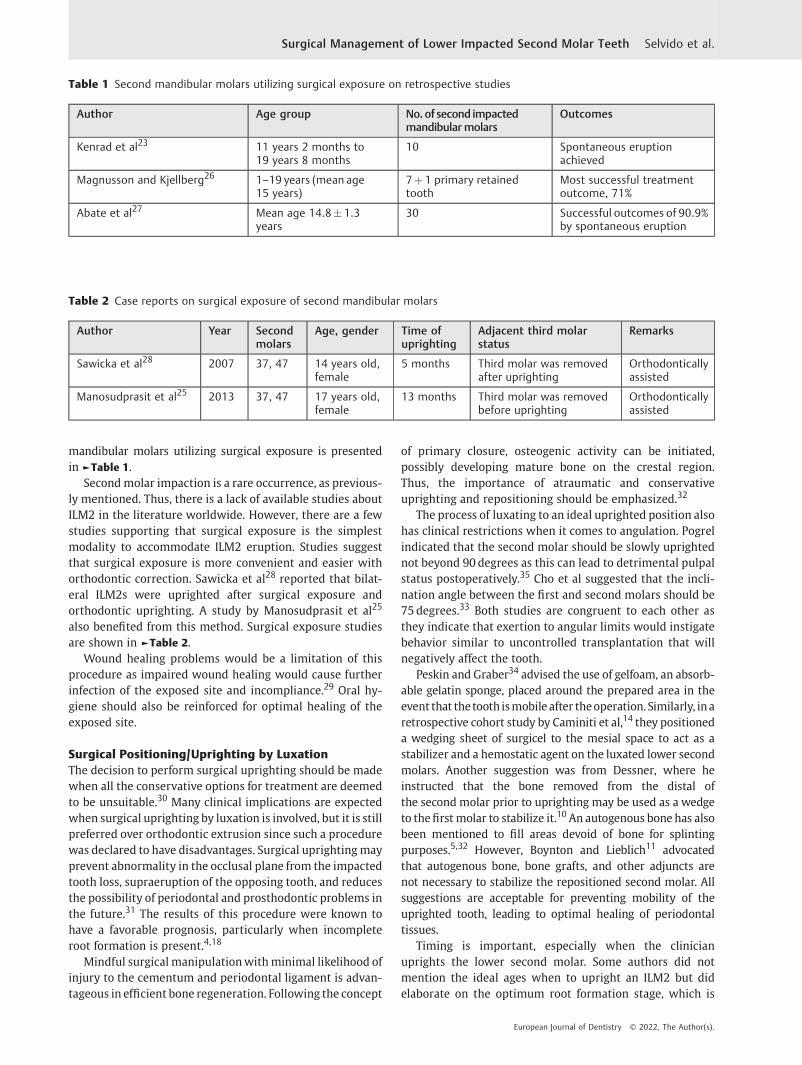
mandibular molars utilizing surgical exposure is presentedin ►Table 1.
Secondmolar impaction is a rare occurrence, as previous-ly mentioned. Thus, there is a lack of available studies aboutILM2 in the literature worldwide. However, there are a fewstudies supporting that surgical exposure is the simplestmodality to accommodate ILM2 eruption. Studies suggestthat surgical exposure is more convenient and easier withorthodontic correction. Sawicka et al28 reported that bilat-eral ILM2s were uprighted after surgical exposure andorthodontic uprighting. A study by Manosudprasit et al25
also benefited from this method. Surgical exposure studiesare shown in ►Table 2.
Wound healing problems would be a limitation of thisprocedure as impaired wound healing would cause furtherinfection of the exposed site and incompliance.29 Oral hy-giene should also be reinforced for optimal healing of theexposed site.
Surgical Positioning/Uprighting by LuxationThe decision to perform surgical uprighting should be madewhen all the conservative options for treatment are deemedto be unsuitable.30 Many clinical implications are expectedwhen surgical uprighting by luxation is involved, but it is stillpreferred over orthodontic extrusion since such a procedurewas declared to have disadvantages. Surgical uprightingmayprevent abnormality in the occlusal plane from the impactedtooth loss, supraeruption of the opposing tooth, and reducesthe possibility of periodontal and prosthodontic problems inthe future.31 The results of this procedure were known tohave a favorable prognosis, particularly when incompleteroot formation is present.4,18
Mindful surgical manipulationwithminimal likelihood ofinjury to the cementum and periodontal ligament is advan-tageous in efficient bone regeneration. Following the concept
of primary closure, osteogenic activity can be initiated,possibly developing mature bone on the crestal region.Thus, the importance of atraumatic and conservativeuprighting and repositioning should be emphasized.32
The process of luxating to an ideal uprighted position alsohas clinical restrictions when it comes to angulation. Pogrelindicated that the second molar should be slowly uprightednot beyond 90 degrees as this can lead to detrimental pulpalstatus postoperatively.35 Cho et al suggested that the incli-nation angle between the first and second molars should be75 degrees.33 Both studies are congruent to each other asthey indicate that exertion to angular limits would instigatebehavior similar to uncontrolled transplantation that willnegatively affect the tooth.
Peskin and Graber34 advised the use of gelfoam, an absorb-able gelatin sponge, placed around the prepared area in theevent that thetooth ismobile after theoperation. Similarly, inaretrospective cohort study by Caminiti et al,14 they positioneda wedging sheet of surgicel to the mesial space to act as astabilizer and a hemostatic agent on the luxated lower secondmolars. Another suggestion was from Dessner, where heinstructed that the bone removed from the distal ofthe second molar prior to uprighting may be used as a wedgeto thefirst molar to stabilize it.10An autogenous bone has alsobeen mentioned to fill areas devoid of bone for splintingpurposes.5,32 However, Boynton and Lieblich11 advocatedthat autogenous bone, bone grafts, and other adjuncts arenot necessary to stabilize the repositioned second molar. Allsuggestions are acceptable for preventing mobility of theuprighted tooth, leading to optimal healing of periodontaltissues.
Timing is important, especially when the clinicianuprights the lower second molar. Some authors did notmention the ideal ages when to upright an ILM2 but didelaborate on the optimum root formation stage, which is
Table 2 Case reports on surgical exposure of second mandibular molars
Author Year Secondmolars
Age, gender Time ofuprighting
Adjacent third molarstatus
Remarks
Sawicka et al28 2007 37, 47 14 years old,female
5 months Third molar was removedafter uprighting
Orthodonticallyassisted
Manosudprasit et al25 2013 37, 47 17 years old,female
13 months Third molar was removedbefore uprighting
Orthodonticallyassisted
Table 1 Second mandibular molars utilizing surgical exposure on retrospective studies
Author Age group No.of second impactedmandibular molars
Outcomes
Kenrad et al23 11 years 2 months to19 years 8 months
10 Spontaneous eruptionachieved
Magnusson and Kjellberg26 1–19 years (mean age15 years)
7þ 1 primary retainedtooth
Most successful treatmentoutcome, 71%
Abate et al27 Mean age 14.8� 1.3years
30 Successful outcomes of 90.9%by spontaneous eruption
European Journal of Dentistry © 2022. The Author(s).
Surgical Management of Lower Impacted Second Molar Teeth Selvido et al.
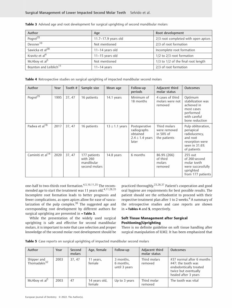
one-half to two-thirds root formation.4,5,10,11,35 The recom-mended age to start the treatment was 11 years old.4,11,28,35
Incomplete root formation leads to better prognosis andfewer complications, as open apices allow for ease of vascu-larization of the pulp complex.36 The suggested age andcorresponding root development by different authors forsurgical uprighting are presented in ►Table 3.
While the presentation of the widely used surgicaluprighting is safe and effective for second mandibularmolars, it is important to note that case selection and properknowledge of the second molar root development should be
practiced thoroughly.23,28,37 Patient’s cooperation and goodoral hygiene are requirements for best possible results. Thepatient should see the orthodontist to proceed with theirrespective treatment plan after 1 to 2 weeks.4 A summary ofthe retrospective studies and case reports are shownin ►Tables 4 and 5, respectively.
Soft Tissue Management after SurgicalPositioning/UprightingThere is no definite guideline on soft tissue handling aftersurgical manipulation of ILM2. It has been emphasized that
Table 3 Advised age and root development for surgical uprighting of second mandibular molars
Author Age Root development
Pogrel35 11.7–17.9 years old 2/3 root completed with open apices
Dessner10 Not mentioned 2/3 of root formation
Sawicka et al28 11–14 years old Incomplete root formation
Kravitz et al4 11–15 years old 1/2 to 2/3 root formation
McAboy et al5 Not mentioned 1/3 to 1/2 of the final root length
Boynton and Leiblich11 11–14 years 2/3 of root formation
Table 4 Retrospective studies on surgical uprighting of impacted mandibular second molars
Author Year Tooth # Sample size Mean age Follow-upperiods
Adjacent thirdmolar status
Outcomes
Pogrel35 1995 37, 47 16 patients 14.1 years Minimum of18 months
4 cases of thirdmolars were notremoved
Optimumstabilization wasachieved inmost casesperformedwith carefulbone reduction
Padwa et al18 2017 37, 47 16 patients 13� 1.1 years Postoperativeradiographsobtained2.4�1.4 yearslater
Third molarswere removedin 50% ofthe patients
Pulp obliteration,periapicalradiolucency,and rootresorption wereseen in 31.6%of patients
Caminiti et al14 2020 37, 47 177 patientswith 260mandibularsecond molars
14.8 years 6 months 86.9% (266)of thirdmolarsremoved
255 outof 260 secondmolar teethwere successfullyuprightedfrom 177 patients
Table 5 Case reports on surgical uprighting of impacted mandibular second molars
Author Year Secondmolars
Age, female Follow-up Adjacent thirdmolar status
Outcomes
Shipper andThomadakis32
2003 37, 47 11 years,female
3 months,6 months,until 3 years
Third molarsremoved
#37 normal after 6 months#47: the tooth wasendodontically treatedtwice but eventuallyhealed after 3 years
McAboy et al5 2003 47 14 years old,female
Up to 3 years Third molarremoved
The tooth was vital
European Journal of Dentistry © 2022. The Author(s).
Surgical Management of Lower Impacted Second Molar Teeth Selvido et al.
effective soft tissue management warrants the success of theoverall treatment outcomes. However, according to Ander-son et al,3 there is a shortage of studies because of timelimitations. The clinician can then acquire careful tissuehandling that is consistent with other surgical proceduresperformed in the lower arch.
Preferably, the uprighted tooth should be reduced on theocclusal surface tobe slightly out of contact to preventocclusaland soft tissue trauma.5 In a technical note by Anderson et al,3
they recommended flap repositioning and occlusal compositebuttonplacement in termsof soft tissuemanagement. Theflaprepositioning is needed to minimize tissue trauma of theexpected buccal flap created after the said modality. Thisprocedure can be accomplished by doing conservative osteot-omy fromthemarginal buccalboneof therepositioned ILM2tothe thirdmolarextractionarea.Onceadequatebone removal isachieved, the flap will be approximated from the distal ofthe second molar then sutured to close.
An occlusal composite button is an added accessorywhichwill raise the bite to keep the flap undisturbed, therebypromoting proper healing. The orthodontist can removethe button during their treatment time, 10 to 14 days aftersurgical manipulation. Indeed, this method will be difficultto achieve when the third molar is preserved. This methodwas stated to have advantages like preserving the keratinizedbuccal gingiva, preventing plaque accumulation, and avoid-ing hyperocclusion.3
The occurrence of pseudopockets can also be a majorproblem for the clinician since there is unpredictable softtissue behavior after second mandibular molar uprighting.One possible technique to prevent periodontal problems isdistal wedge surgery which can be performed in the area ofthe third lower molars to the second mandibular molar ifremoval of the third molar is indicated. In this procedure, atriangular incision is made with the apex of the incisionlocated in the direction of the retromolar area and the baselocated in the distal part of the uprighted second molar.38
This technique is favorable for cases where the third lowermolar is indicated to be extracted. Additionally, it facilitateshealthy healing around the second lower molar tissues. Thismethod was applied by Leechanavanichpan et al39 in 2019,where impacted mandibular third molar was surgicallyremoved with distal wedge incision, and it created positiveperiodontal clinical outcomes. The distal wedge technique ismostly used during odontectomy procedures and can bebeneficial in second mandibular molar uprighting, prevent-ing clinical attachment loss and plaque retention to the distalpart of the molar.
Another soft tissue management method is covering theoperated site with a periodontal dressing of choice that canbe placed to splint the tooth for stabilization and avoid buccaltissue trauma.4 The dressing can also serve an additionalpurpose of inhibiting the soft tissue from slowly covering thecrown of the uprighted tooth.14 Prevention of this adverseevent is important as its removal can be tedious. In addition,problems such as inflammation of the gingiva and cariesformation in the second molar are possible complications ofthis condition.
Irrigation of the exposed area is also advised postopera-tively, using a syringe to prevent food debris accumulation onthe area, making the site infection-free.20 Any other adjunctsfor proper care of surgical sites are strongly recommended sothat the tooth will not be indicated for subsequentextraction.
Uprighting with Miniplate and Miniscrew ApplicationOver the last decade, there have been increases in thenumber of case studies and reviews regarding miniplatesand miniscrews in uprighting ILM2.40 These devices havebeen chosen for molar uprighting procedures with ortho-dontic appliances as they offer stability and anchorageduring uprighting.41 Both anchorage devices were knownto have special considerations and shortcomings.
Miniscrews provide optimal stability and the necessaryforce to move the tooth. They are conservative and inexpen-sive as well. However, the placement should be done by anexperienced clinician, and the precision of placement shouldbe supplemented by three-dimensional imaging for properinstallation.42 In the mandible, special considerations areemphasized since it has a higher amount of cortical bone thatcorrelates with the compromised stability of miniscrews.43
An interesting finding by Samrit et al44 revealed that theminiscrew application is predominantly successful in themaxilla than the mandible to a general extent. Nevertheless,many case studies report successful outcomes with upright-ing using miniscrews in ILM2.
On the other hand, miniplates are stronger and sturdierthanminiscrews, but are expensive. In addition, they requireinvasive surgery rendering them to be traumatizing.42
In general, bothminiscrews andminiplates are efficient inretraction and distalization on both dental arches.41 Howev-er, there is less concern when it comes to placement withminiplates due to the fact that there is less risk of damagingvital structures.45
There are two types of miniscrew anchorage: directanchorage, which involves mounting the miniscrew in theretromolar region; and indirect anchorage, in which place-ment is situated between the roots of the teeth.15 Of the twotypes, the direct anchorage was mentioned to be morefavorable for immediate orthodontic loading.46 An exampleof direct anchorage is from a 2020 case study by Altieriet al,16 who used a miniscrew anchorage drilling in theretromolar area along with orthodontic assistance to uprightbilateral ILM2. Similarly, Giancotti et al47 in 2004 placed a 7-mm-long miniscrew in the retromolar triangle to upright aleft ILM2 in the same day, with 50 g of force applied by theelastics. Furthermore, from the same author, a 150 g wasexerted for a right ILM2 after miniscrew installation on thesame area as well.48 Hence, the force of 50 to 150 g can thenbe applied on ILM2 uprighting, with cautious manipulation.
In indirect anchorage, an example would be from a caseseries by Lee et al,15 where they placed the miniscrew in thebuccal cortical bone between the first mandibular molarand second premolars. They used an open coiled spring tomaximize the distalizing force to fully upright the ILM2 in itsrightful position. Three of the cases presented were
European Journal of Dentistry © 2022. The Author(s).
Surgical Management of Lower Impacted Second Molar Teeth Selvido et al.
adolescents aged 12, 13, and 16 years old, respectively. All ofthe cases reported success in uprighting via indirect anchor-age with miniscrews, even in the case of a 13-year-oldpatient who did not undergo lower third molar removal.Therefore, miniscrews can be used even if third molarremoval is not warranted.
Two miniscrews with slots placed carefully on the buccalalveolus between mandibular first molar and second pre-molar, and between second premolar and first premolar, canalso be installed to increase retention, making the wholesetup impervious to orthodontic forces.49 Compared withthe case report by Lee et al, correction with added minis-crews took almost double the time as compared with oneminiscrewwith open coil springs. The cause of the prolongedtime was possibly because some patients in this case serieshad dislodged miniscrews before the ILM2 got uprighted.Orthodontic biomechanics needed in the use of miniscrews,however, is beyond the scope of this review.
Complex uprighting with miniscrews is also an option, aswith Celebi and colleagues’50 approach on uprighting. Theminiscrewwas installed in the upper posterior alveolus areafor maximum traction from themaxillary arch. Regardless ofthe location of the miniscrew, uprighting time is the same asthe miniscrews placed in the mandibular arch.
Similarly, with miniscrews, miniplates should also bemounted by a clinician with proficient knowledge of themandibular anatomy as proper flap reflection is required.51
It is usually performed by oral surgeons. Regardless of theinvasiveness of the procedure, the chance of iatrogenic nerveimpairment is less concerning.52 As it stands, there are noknown fractures involving miniplates and their subsequentscrews as they are known to be very rigid.42
Most miniplates in the mandible for molar uprighting areplaced in the retromolar region, and they can be used fordisimpacting deep ILM2. An instance is a clinical report byTseng et al52 that utilized a four-hole L-shaped miniplatedrilled by miniscrews in the buccal cortex near the retro-molar area as an anchorage for orthodontic ILM2 uprighting.The casewas corrected in 8months. Miyahira et al53 also hada case with a 12-year-old patient in which they used an L-shapedminiplate positioned distal to the ILM2.With thehelpof optimum orthodontic forces, an ILM2 was nearlyuprighted for 3 months. This technique can be consideredinvasive, but in spite of the trauma it entails, outcomes havebeen reported to be acceptable.
In the studies mentioned, it is clear that miniscrews havebeen mostly utilized in ILM2 uprighting cases. Miniplates,however, with their limited studies, should not be concludedlightly. Sherwood et al51 claimed that miniplates are morestable than other anchorage devices. In contrast, severalminiplates in the mandible in a study by Choi et al54 hadfailed, with a 7% failure rate. Thus,more studies are needed inthis type of modality in ILM2 to determine the exact successrate of this procedure.
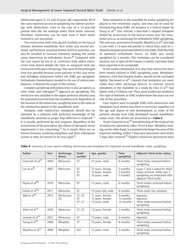
Case reports used to upright ILM2 with miniscrews andminiplates have shown that there is correction regardless ofthe age and degree of root development, as some of thepatients already have fully developed second mandibularmolar roots. The details are presented in ►Table 6.
As per Giancotti et al,48 wound healing of the surgical siteof miniscrew placement takes 10 to14 days. Miniplate heal-ing, on the other hand, is assumed to be longer because of theexpected swelling within 7 days post placement and within7 days after removal.55 Flapless miniscrew placements have
Table 6 Summary of case reports utilizing miniscrews and miniplates for impacted second mandibular molar uprighting
Author Year Anchoragedevice
Toothnumber
Age, gender Timeuprighted
Adjacent third molar status
Giancotti et al47 2004 Miniscrew 37 27 years, male 8 months Third molar removed beforeuprighting
Lee et al15 2007 Miniscrew 473747
12 years, female13 years, female16 years, male
5 months5 months2 months
Cases 1 and 3 opted for thirdmolar removal, while case 2uprighting accomplished withadjacent third molar
Nęcka et al46 2010 Miniscrew 47 15 years, male 6 months Third molars removed beforeuprighting
Celebi et al50 2011 Miniscrew 37 15 years old, male 8 months Third molar not removed
Mah et al49 2015 Miniscrews(2)
37, 474737, 47
11 years, female13 years, female13 years, male
9 months13 months12 months
AgenesisAgenesisThird molar extracted beforeuprighting
Altieri et al16 2020 Miniscrew 37, 47 12 years, female 3 months Third molar extracted beforeuprighting
Lorente et al21 2021 Miniscrew 47 13 years, male 4 months Agenesis
Tseng et al52 2008 Miniplate 37 19 years, female 8 months Third molar extracted beforeuprighting
Miyahira et al53 2008 Miniplate #47 12 years, male 3 months Third molar extracted beforeuprighting
European Journal of Dentistry © 2022. The Author(s).
Surgical Management of Lower Impacted Second Molar Teeth Selvido et al.
higher success rates with reduced pain and discomfort thanthose performed with full-thickness flap surgery. In the caseof miniplate installation, postoperative pain is experiencedwith or without flap operation.56 Patients were also knownto be comfortable in the placement and removal of minis-crews, but in the case of miniplates are believed to beotherwise.47,57 Nevertheless, best oral hygiene practicesare advised since these are plaque-retentive fixtures. Patientcooperation is also key to avoid dislodgement and subse-quent swallowing of the small components.
Extraction of the Second Mandibular MolarThe etiologic factors of ILM2 according to Kenrad et al23
are secondary retention, decreased space, and anomalousinclination. Gooris et al58 proposed the removal of theseILM2 as there is anticipation that the third molar will eventu-ally erupt toward the extracted second molar’s position.Interestingly, Orton-Gibbs et al asserted that third molars,evenwith severe angulation, can replace these ILM2effectivelyafter extraction. There is an immense risk as the space of theextracted site does not ensure the eruption of the thirdmolar.59,60 Known cases of successful replacement wereseen in the maxillary arch.61 However, in the mandibulararch, failed replacements were observed.62
Clinically, the option to extract the ILM2 is advised ifuprighting/repositioning has been deemed impossible be-cause of its detrimental position in the mandibular arch.63
While radiographically, there are factors to be considered toutilize extraction for effective replacements: proximity of thetooth follicle to the ILM2 roots, theminimal amount of tipping,and crown completion of the third mandibular molar bud.58
Another contributing indicator for ILM2 extraction is thepatient’s age, wherein the more advanced the age, thepossibility of root closure is anticipated.63 Despite the diffi-culties mentioned earlier, mandibular third molar teeth canstill erupt in the position of the second molars, but notwithout deficiencies. A retrospective study by De-la-Rosa-Gay et al62 resulted only in 66.2% of third mandibular molareruption to its correct positionwhen the ILM2was extracted.The failure was accounted for by the later Nolla’s stages ofdevelopment, which is the root closure and completion. Thisfinding is crucial along with Gooris and colleagues’58 study,inwhich the optimal time of ILM2 extraction depends on theadjacent third molar’s root development. Such time shouldbe from the crown completion to two-thirds root develop-ment. The thirdmolars that failed to erupt successfully either
had a severe mesial tilt or the proximal contact was notachieved, thereby requiring more orthodontic correction.62
Correct eruption of third mandibular molars has beenmentioned as unpredictable and inconsistent.5 In terms ofarch length, Richardson and Richardson64 proposed that onecan opt for second mandibular molar extraction if the archlength is deficient, with the certainty that the third molarwould be impacted and the proper eruption is assumed.However, Magnusson and Kjellberg26 concluded the oppo-site for this condition, as they advised that the patient shouldbe cautioned that the final position of the third molars inboth archesmay be unfavorable, leading tomore orthodonticproblems such as crossbites. Among all the surgical treat-ment modalities that transpired in their retrospective study,the second molar extraction with emphasis on themandibular second molars received the most unsatisfactoryresults. To address these problems, an additional study byDe-la-rosa-Gay et al65 in 2010 developed a predictive modelto estimate the degree of tilting of the eruption of the thirdmolars in both upper and lower teeth by using panoramicradiographs, and by calculating angles and coefficients. Thepredictive models were successful after several years, exceptfor some mandibular molars mentioned in the study. Al-though the mandibular results in the study were not assuccessful as the maxillary results, this can still be an addedprognostic value for treatment planning that willinvolve second molar extractions.
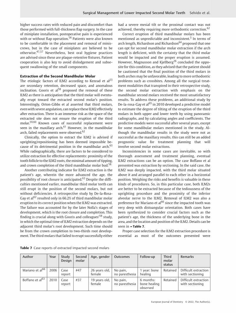
Inconsistencies in some cases are inevitable, so withthorough assessment and treatment planning, eventualILM2 extractions can be an option. The case Boffano et alpresented was extraction by sectioning. In the said case, theILM2 was deeply impacted, with the third molar situatedabove it and arranged parallel to each other in a horizontalposition. Weighing the risks and benefits is valuable in thesekinds of procedures. So, in this particular case, both ILM2sare better to be extracted because of the tediousness of theuprighting procedure and the proximity of the inferioralveolar nerve to the ILM2. Removal of ILM2 was also apreference for Mariano et al66 since the impacted tooth wasvery deep with distoangular orientation. Both cases havebeen synthesized to consider crucial factors such as thepatient’s age, the thickness of the underlying bone in thearea, and the location and position of the ILM2. Details can beseen in ►Table 7.
Proper case selection for the ILM2 extraction procedure isessential as most of the outcomes presented were
Table 7 Case reports of extracted impacted second molars
Author Year StudyDesign
Secondmolar
Age, gender Outcomes Follow-up Thirdmolarstatus
Remarks
Mariano et al66 2006 Casereport
#47 26 years old,female
No pain,no paresthesia
1 year: bonehealing
Retained Difficult extractionwith sectioning
Boffano et al63 2010 Casereport
#37 19 years old,female
No pain,no paresthesia
6 months:bone healingobserved
Retained Difficult extractionwith sectioning
European Journal of Dentistry © 2022. The Author(s).
Surgical Management of Lower Impacted Second Molar Teeth Selvido et al.
unpredictable. The clinician must also be careful as onedisadvantage is possible supraeruption of the opposing toothwhich occurs if the tooth is left without contact from theantagonist for prolonged periods, leading to further chal-lenging orthodontic correction.67 To a large extent, thelength of time for correction, the proximity to the inferioralveolar canal, themorphological anomalies of the ILM2, andthe overall health of the patient have to be considered. Morestudies are needed, with suggested emphasis on position andclassification of the ILM2 and third molars, and their corre-sponding management, to assist the clinician in the clinicalsetting if such a need arises.
AutotransplantationAs early as the 1950s, autotransplantations or transplanta-tions have been proposed as a replacement modality forextracted teeth. It is broadly known as relocating the toothfrom its alveolar socket to another area in the same person.68
Choosing this treatment option is viable when surgicalexposure and orthodontic traction have been unsuccessful.69
Autotransplantations are often suggested to have a goodprognosis as they preserve function, mastication, and pro-prioception, particularly in young patients.70 It is a feasibleoption since dental implants are contraindicated for growingpatients due to their maturing alveolar arches.71,72 Further-more, orthodontic treatment is more or less unnecessary inthis type of modality.73
Currently, there are no specific guidelines when it comesto the transplantation of teeth.74,75 Many case reports haveshown that it is especially beneficial to second mandibularmolar space rehabilitation, due to the general claim thattransplantations reduce alveolar resorptions.76 Moreover, itwas attested that thismethod is less traumatic than perform-ing odontectomy.77 It is also important that when doing thistechnique, the donor and recipient teeth should be morpho-logically analogous to each other tominimize extraoral time,thus maximizing positive results.13
Case selection for third molar transplantation to the ILM2site is of utmost importance. Similar to uprighting surgicalprocedures of the ILM2, autotransplantations of the adjacentthird molars were believed to require a certain root develop-ment stage for successful outcomes. Radiographic measure-ments of the third molar root length had been used before,disputing that 2 to 3mm or 3 to 5mm are the optimal lengthsfor a good transplantation prognosis.78 Similar to root devel-opment standards in surgical uprighting of ILM2, one-half tothree-fourths of root development of the thirdmolar have alsobeen reinforced as an acceptable guideline.13,26,76,79,80 How-ever, compelling evidence also demonstrated a high successrate with mature third molars with complete root forma-tion.81–84 The actual success rates were established onmatureteeth because of atraumatic and swift transplantation meth-ods, sufficient alveolar bonedimensions, and aseptic protocolsutilized. Despite the success rates, autotransplantations stillentail immense risk like periodontal and pulpal complicationsafter the procedure, specifically for mature teeth.4,5
To date, there are only case reports and retrospectivestudies that showcased transplantation of third molars to
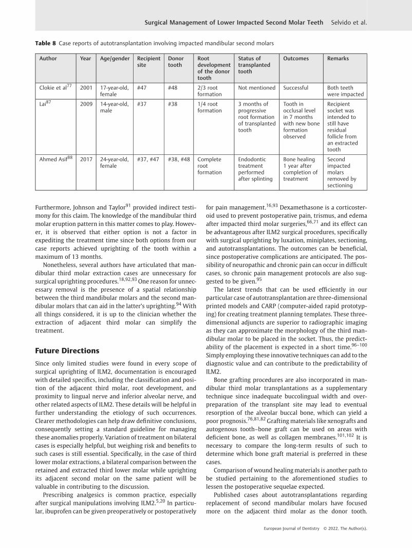
their adjacent second molar extraction sites. Most of themwere about extracted second molars that were eitherdecayed or with pulpal pathosis and were not extracteddue to impaction or uneruption.72,79,81,84–86 Regardless,three case reports matched our criteria. The first report isone of Clokie and team’s77 case series, where impacted lowerright third and second molars were evaluated.
The decision to extract the ILM2 was reinforced sinceorthodontic correction cannot be performed. To rehabilitatethe space, they transplanted the impacted lower thirdmolarstooth adjacent to the ILM2 since it has two-thirds rootdevelopment. Lai,87 on the other hand, reported a casewherehe extracted a left ILM2 with a residual follicle left in thesocket. He transplanted the adjacent unerupted third molarwith one-fourth root development. Both cases yielded posi-tive results as both teeth proceeded with their root develop-ment, and maintained their pulp vitality and function.
Ahmed Asif et al88 reported a transplantation case withbilateral impacted third and second mandibular molars withcompletely formed roots. The ILM2s were sectioned andreplaced with the atraumatically removed third molarsunder general anesthesia. As expected, the autotransplantedteeth were endodontically treated after splinting, which wasrelated to the previous statement about complications oftransplanting mature teeth. The outcome explains that cau-tion should be prompted in such clinical situations, nomatter how short the extraoral time and how atraumaticthe procedure was performed. The details of these cases areshown in ►Table 8.
Three existing case reports in managing ILM2 are notsufficient to draw a significant conclusion. However, we canderive from long-term case studies and retrospective studiesthat immature third mandibular molars transplantedto second molar recipient sites were more successful ingrowing patients with good oral hygiene.83,86,89
Overall, proper case selection and patient’s cooperationare essential to autotransplantations as the outcomes arevariable and unpredictable. Many contributing factors canaffect the treatment as there is no conclusive evidence andno prevailing guidelines to date.75,90 Undoubtedly, the ben-efits outweigh the risks of performing the transplantation.Constantly reminding the patient about maintaining oralhygiene, attending yearly recalls, and checking the status ofthe implanted tooth are minor inconveniences but willtremendously help in the success of the treatment.
Third Molar ExtractionsTerry and Hegtvedt12 in 1993 stated that mandibular thirdmolars are one of the contributing factors ofmalpositioned second mandibular molars; therefore, itsremoval is required. Pogrel35 favored the idea as the easeof uprighting can be attained without the presence of thethird molar beside the ILM2. He did not extract the thirdmolars in his cases because their proximity does not affectthe ILM2 and simply because the third molar buds areinvisible radiographically. According to Going and Reyes-Lois,20 the presence of a third molar does not affect theresults promised in the surgical uprighting procedure.
European Journal of Dentistry © 2022. The Author(s).
Surgical Management of Lower Impacted Second Molar Teeth Selvido et al.
Furthermore, Johnson and Taylor91 provided indirect testi-mony for this claim. The knowledge of the mandibular thirdmolar eruption pattern in this matter comes to play. Howev-er, it is observed that either option is not a factor inexpediting the treatment time since both options from ourcase reports achieved uprighting of the tooth within amaximum of 13 months.
Nonetheless, several authors have articulated that man-dibular third molar extraction cases are unnecessary forsurgical uprighting procedures.18,92,93One reason for unnec-essary removal is the presence of a spatial relationshipbetween the third mandibular molars and the second man-dibular molars that can aid in the latter’s uprighting.94 Withall things considered, it is up to the clinician whether theextraction of adjacent third molar can simplify thetreatment.
Future Directions
Since only limited studies were found in every scope ofsurgical uprighting of ILM2, documentation is encouragedwith detailed specifics, including the classification and posi-tion of the adjacent third molar, root development, andproximity to lingual nerve and inferior alveolar nerve, andother related aspects of ILM2. These details will be helpful infurther understanding the etiology of such occurrences.Clearer methodologies can help draw definitive conclusions,consequently setting a standard guideline for managingthese anomalies properly. Variation of treatment on bilateralcases is especially helpful, but weighing risk and benefits tosuch cases is still essential. Specifically, in the case of thirdlower molar extractions, a bilateral comparison between theretained and extracted third lower molar while uprightingits adjacent second molar on the same patient will bevaluable in contributing to the discussion.
Prescribing analgesics is common practice, especiallyafter surgical manipulations involving ILM2.5,20 In particu-lar, ibuprofen can be given preoperatively or postoperatively
for pain management.16,93 Dexamethasone is a corticoster-oid used to prevent postoperative pain, trismus, and edemaafter impacted third molar surgeries,66,71 and its effect canbe advantageous after ILM2 surgical procedures, specificallywith surgical uprighting by luxation, miniplates, sectioning,and autotransplantations. The outcomes can be beneficial,since postoperative complications are anticipated. The pos-sibility of neuropathic and chronic pain can occur in difficultcases, so chronic pain management protocols are also sug-gested to be given.95
The latest trends that can be used efficiently in ourparticular case of autotransplantation are three-dimensionalprinted models and CARP (computer-aided rapid prototyp-ing) for creating treatment planning templates. These three-dimensional adjuncts are superior to radiographic imagingas they can approximate the morphology of the third man-dibular molar to be placed in the socket. Thus, the predict-ability of the placement is expected in a short time.96–100
Simply employing these innovative techniques can add to thediagnostic value and can contribute to the predictability ofILM2.
Bone grafting procedures are also incorporated in man-dibular third molar transplantations as a supplementarytechnique since inadequate buccolingual width and over-preparation of the transplant site may lead to eventualresorption of the alveolar buccal bone, which can yield apoor prognosis.76,81,82Graftingmaterials like xenografts andautogenous tooth–bone graft can be used on areas withdeficient bone, as well as collagen membranes.101,102 It isnecessary to compare the long-term results of such todetermine which bone graft material is preferred in thesecases.
Comparison of wound healingmaterials is another path tobe studied pertaining to the aforementioned studies tolessen the postoperative sequelae expected.
Published cases about autotransplantations regardingreplacement of second mandibular molars have focusedmore on the adjacent third molar as the donor tooth.
Table 8 Case reports of autotransplantation involving impacted mandibular second molars
Author Year Age/gender Recipientsite
Donortooth
Rootdevelopmentof the donortooth
Status oftransplantedtooth
Outcomes Remarks
Clokie et al77 2001 17-year-old,female
#47 #48 2/3 rootformation
Not mentioned Successful Both teethwere impacted
Lai87 2009 14-year-old,male
#37 #38 1/4 rootformation
3 months ofprogressiveroot formationof transplantedtooth
Tooth inocclusal levelin 7 monthswith new boneformationobserved
Recipientsocket wasintended tostill haveresidualfollicle froman extractedtooth
Ahmed Asif88 2017 24-year-old,female
#37, #47 #38, #48 Completerootformation
Endodontictreatmentperformedafter splinting
Bone healing1 year aftercompletion oftreatment
Secondimpactedmolarsremoved bysectioning
European Journal of Dentistry © 2022. The Author(s).
Surgical Management of Lower Impacted Second Molar Teeth Selvido et al.
However, it will be interesting to see outcomes of the trans-planted site from an upper molar as a donor tooth or thecontralateral mandibular molar. The results can be furtheredas one of the treatment options if the clinician is faced with asituation where the adjacent third molar is not available. Thiscan expand the knowledge domain on autotransplantation asan excellent elective procedure asmore options are presented.
Advancements in autotransplantation using piezosurgeryhave also been used in a third molar to be transplanted intoa second molar socket. Atraumatic harvesting of the donortooth to the recipient site using this device leads to unharmedperiodontal ligament fibers that can count for less occurrenceof complications. The piezosurgery can also be used fornoninvasive socket preparationbecauseof itsability to removeunnecessary bone judiciously.19,79More studies on utilizationof this device on ILM2 and third molars are recommended.
Quality-of-life assessments should also be undertaken asmost of these procedures are rather invasive. Theminiscrewsand miniplates located in the retromolar area seem to beuncomfortable to patients during function. Thus, the quality-of-life assessment studies can help the clinician choosewhich modality the patient will comply with.
Updated studies on surgical techniques regarding ILM2are required since advanced trends in the orthodontic andsurgical fields are evolving. It is also needed for a betterunderstanding of these occurrences. Lastly, the necessity forlong-term follow-up studies is also advised to ensure theeffectiveness of every modality discussed.
Limitations
Few case reports and retrospective studies spanning morethan 20 years are available regarding the management ofimpacted second molars. The authors of each study madeassumptions that are all substantial in contributing to man-agement and case selection. The pointsmade are sufficient toguide the decision-making process, but robust standardscannot be made. Although most of the findings are notcurrent, the case reports and retrospective studies are stillrelevant to techniques applied in the modern setting, rein-forcing the effectiveness of the surgical modality.
Conclusion
This review covered the surgical aspects of uprighting,repositioning, and autotransplantation, with novelapproaches regarding ILM2. Various works have alreadydocumented well-known success rates and prognoses forsurgical exposures, surgical uprighting, and transplantation,especially presented by retrospective studies. In addition,surgical approaches regarding ILM2 are less complicatedthan orthodontic uprighting techniques because they re-quire lesser tools with fewer steps. Hence, these surgicalmodalities are not time-consuming. All in all, the outcome ofsurgical intervention depends on case selection, root devel-opment of the ILM2, careful surgical manipulation, andadherence to sound biological principles. Patient’s compli-ance, general health, and oral hygiene should also be
highlighted. Nevertheless, there is a constant need for otherinnovative, advanced, and atraumatic surgical approaches forILM2 to give patients more options to improve complianceand comfort.
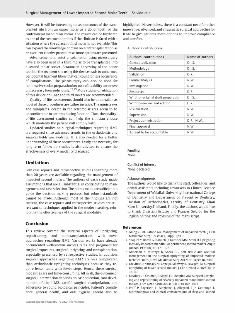
Authors’ Contributions
FundingNone.
Conflict of InterestNone declared.
AcknowledgmentsThe authors would like to thank the staff, colleagues, anddental assistants including coworkers in Clinical ScienceDepartment of Walailak University International Collegeof Dentistry and Department of Preventive Dentistry,Division of Orthodontics, Faculty of Dentistry KhonKaen University,Thailand. Finally, the authors would liketo thank Christian Estacio and Frances Selvido for theEnglish editing and revising of the manuscript.
References1 Alling CC III, Catone GA. Management of impacted teeth. J Oral
Maxillofac Surg 1993;51(1, Suppl 1):3–62 Shapira Y, Borell G, Nahlieli O, Kuftinec MM, Stom D. Uprighting
mesially impacted mandibular permanent second molars. AngleOrthod 1998;68(02):173–178
3 Anderson K, Murtagh K, Sacks HG. Soft tissue and occlusalmanagement in the surgical uprighting of impacted molars:technical note. J Oral Maxillofac Surg 2012;70(08):e438–e440
4 Kravitz ND, Yanosky M, Cope JB, Silloway K, Favagehi M. Surgicaluprighting of lower second molars. J Clin Orthod 2016;50(01):33–40
5 McAboy CP, Grumet JT, Siegel EB, Iacopino AM. Surgical upright-ing and repositioning of severely impacted mandibular secondmolars. J Am Dent Assoc 2003;134(11):1459–1462
6 Proff P, Bayerlein T, Fanghänel J, Allegrini S Jr, Gedrange T.Morphological and clinical considerations of first and second
Authors’ contributions Name of authors
Conceptualization D.I.S.
Methodology D.I.S.
Validation D.R.
Formal analysis N.W.
Investigation N.W.
Resources D.R.
Writing—original draft preparation D.I.S.
Writing—review and editing D.R.
Visualization N.W.
Supervision N.W.
Project administration D.R., N.W.
Final approval N.W.
Agreed to be accountable N.W.
European Journal of Dentistry © 2022. The Author(s).
Surgical Management of Lower Impacted Second Molar Teeth Selvido et al.
permanent molar eruption disorders. Ann Anat 2006;188(04):353–361
7 Valmaseda-Castellón E, De-la-Rosa-Gay C, Gay-Escoda C. Erup-tion disturbances of the first and second permanent molars:results of treatment in 43 cases. Am J Orthod Dentofacial Orthop1999;116(06):651–658
8 Varpio M, Wellfelt B. Disturbed eruption of the lower secondmolar: clinical appearance, prevalence, and etiology. ASDC J DentChild 1988;55(02):114–118
9 Frank CA. Treatment options for impacted teeth. J AmDent Assoc2000;131(05):623–632
10 Dessner S. Surgical uprighting of second molars: rationale andtechnique. Oral Maxillofac Surg Clin North Am 2002;14(02):201–212
11 Boynton T, Lieblich SE. Surgical uprighting of second molars.Atlas Oral Maxillofac Surg Clin North Am 2013;21(02):235–237
12 Terry BC, Hegtvedt AK. Self-stabilizing approach to surgicaluprighting of the mandibular second molar. Oral Surg OralMed Oral Pathol 1993;75(06):674–676
13 Tsukiboshi M. Autotransplantation of teeth: requirements forpredictable success. Dent Traumatol 2002;18(04):157–180
14 CaminitiMF, El-RabbanyM, Lou T, Reinish EI. Surgical uprightingof mandibular second molars: a single-group retrospectivecohort study. Am J Orthod Dentofacial Orthop 2020;158(06):849–855
15 Lee K-J, Park Y-C, Hwang W-S, Seong E-H. Uprightingmandibular second molars with direct miniscrew anchorage.J Clin Orthod 2007;41(10):627–635
16 Altieri F, Guarnieri R, Mezio M, et al. Uprighting impactedmandibular second molar using a skeletal anchorage: a casereport. Dent J (Basel) 2020;8(04):E129
17 Zeitler DL. Management of impacted teeth other than thirdmolars. Oral Maxillofac Surg Clin North Am 1993;5(01):95–103
18 Padwa BL, Dang RR, Resnick CM. Surgical uprighting is a success-ful procedure for management of impacted mandibular secondmolars. J Oral Maxillofac Surg 2017;75(08):1581–1590
19 Nagori SA, Jose A, Bhutia O, Roychoudhury A. Evaluating successof autotransplantation of embedded/impacted third molarsharvested using piezosurgery: a pilot study. Acta Odontol Scand2014;72(08):846–851
20 Going RE Jr, Reyes-Lois DB. Surgical exposure and bracketingtechnique for uprighting impacted mandibular second molars. JOral Maxillofac Surg 1999;57(02):209–212
21 Lorente C, Perez-Vela M, Lorente P, Lorente T. Miniscrew-sup-ported pole technique: surgical-orthodontic approach for im-pacted or retained second molars in adolescents. Int Orthod2021;19(01):147–158
22 Fan W, Gao D, Wang Y, et al. Three-dimensional measurementand analysis of mandibular characteristics in subjects withimpacted mandibular second molars. Orthod Craniofac Res2020;23(03):332–341
23 Kenrad J, Vedtofte H, Andreasen JO, Kvetny MJ, Kjær I. A retro-spective overview of treatment choice and outcome in 126 caseswith arrested eruption of mandibular second molars. Clin OralInvestig 2011;15(01):81–87
24 la Monaca G, Cristalli MP, Pranno N, Galluccio G, Annibali S, PippiR. First and second permanent molars with failed or delayederuption: clinical and statistical analyses. Am J Orthod Dento-facial Orthop 2019;156(03):355–364
25 Manosudprasit M, Wangsrimongkol T, Pisek P, Chantaramung-korn M. Management of bilateral severely impactedmandibular second molars: a case report. J Med Assoc Thai2013;96(Suppl 4):S157–S161
26 Magnusson C, Kjellberg H. Impaction and retention of secondmolars: diagnosis, treatment and outcome. A retrospectivefollow-up study. Angle Orthod 2009;79(03):422–427
27 Abate A, Cavagnetto D, Fama A, Matarese M, Bellincioni F,Assandri F. Efficacy of operculectomy in the treatment of 145cases with unerupted second molars: a retrospective case-control study. Dent J (Basel) 2020;8(03):E65
28 Sawicka M, Racka-Pilszak B, Rosnowska-Mazurkiewicz A.Uprighting partially impacted permanent second molars. AngleOrthod 2007;77(01):148–154
29 Guo S, Dipietro LA. Factors affecting wound healing. J Dent Res2010;89(03):219–229
30 Sangha TK, Sangha SK, Hanna B, Tayab T, Padala S. Surgicaluprighting of impacted mandibular second molar: a narrativereview. J Oral Med Oral Surg. 2021;27(02):1–8
31 ShellhartWC, Oesterle LJ. Uprightingmolars without extrusion. JAm Dent Assoc 1999;130(03):381–385
32 Shipper G, Thomadakis G. Bone regeneration after surgicalrepositioning of impacted mandibular second molars: a casereport. Dent Traumatol 2003;19(02):109–114
33 Cho SY, Ki Y, Chu V, Chan J. Impaction of permanent mandibularsecond molars in ethnic Chinese schoolchildren. J Can Dent Assoc2008;74(06):521
34 Peskin S, Graber TM. Surgical repitioning of teeth. J Am DentAssoc 1970;80(06):1320–1326
35 PogrelMA. The surgical uprighting ofmandibular secondmolars.Am J Orthod Dentofacial Orthop 1995;108(02):180–183
36 Holland DJ. The surgical positioning of unerupted, impactedteeth (surgical orthodontics). Oral Surg Oral Med Oral Pathol1956;9(02):130–140
37 Fu PS, Wang JC, Wu YM, et al. Impacted mandibular secondmolars. Angle Orthod 2012;82(04):670–675
38 Johnson TM, Herold RW, Akers JA, DiPirro DA, Berridge JP,McGary RT. Decisions before incisions: technique selection forthe distal wedge procedure. Clinic Adv Periodontics 2020;10(02):94–102
39 Leechanavanichpan P, Rodanant P, Leelarungsun R,WongsirichatN. Postoperative pain perception and patient’s satisfaction aftermandibular third molar surgery by primary closure with distalwedge surgery. J Clin Med Res 2019;11(07):489–494
40 Magkavali-Trikka P, Emmanouilidis G, Papadopoulos MA. Man-dibularmolar uprighting using orthodonticminiscrew implants:a systematic review. Prog Orthod 2018;19(01):1–12
41 Leung MTC, Lee TCK, Rabie ABM, Wong RWK. Use of miniscrewsand miniplates in orthodontics. J Oral Maxillofac Surg 2008;66(07):1461–1466
42 Moon CH. Pros and cons of miniscrews and miniplates fororthodontic treatment. In: Park JH, ed. Temporary AnchorageDevices in Clinical Orthodontics. 2020:731–738
43 Ntolou P, Tagkli A, Pepelassi E. Factors related to the clinicalapplication of orthodonticmini-implants. J Int Oral Health 2018;10(03):103–110
44 Samrit V, Kharbanda OP, Duggal R, Seith A, Malhotra V. Bonedensity and miniscrew stability in orthodontic patients. AustOrthod J 2012;28(02):204–212
45 Chung KR, Kim SH, Kang YG, Nelson G. Orthodontic miniplatewith tube as an efficient tool for borderline cases. Am J OrthodDentofacial Orthop 2011;139(04):551–562
46 Nęcka A, Skrzypczyński J, Antoszewska J. Miniscrew-anchoragein treatment of impacted second molar in mandible - casereport. Dent Med Probl 2010;47(03):379–383
47 Giancotti A, Arcuri C, Barlattani A. Treatment of ectopicmandibular second molar with titanium miniscrews. Am JOrthod Dentofacial Orthop 2004;126(01):113–117
48 Giancotti A, Muzzi F, Santini F, Arcuri C. Miniscrew treatment ofectopic mandibular molars. J Clin Orthod 2003;37(07):380–383
49 Mah SJ, Won PJ, Nam JH, Kim EC, Kang YG. Uprighting mesiallyimpacted mandibular molars with 2 miniscrews. Am J OrthodDentofacial Orthop 2015;148(05):849–861
50 Celebi AA, Gelgor IE, Catalbas B. Correction of mesially impactedlower second molar. J Med Cases 2011;2(31):236–239
European Journal of Dentistry © 2022. The Author(s).
Surgical Management of Lower Impacted Second Molar Teeth Selvido et al.
51 Sherwood KH, Burch JG, Thompson WJ. Closing anterior openbites by intruding molars with titanium miniplate anchorage.Am J Orthod Dentofacial Orthop 2002;122(06):593–600
52 Tseng YC, Chen CM, Chang HP. Use of a miniplate for skeletalanchorage in the treatment of a severely impactedmandibular second molar. Br J Oral Maxillofac Surg 2008;46(05):406–407
53 Miyahira YI, Maltagliati LÁ, Siqueira DF, Romano R. Miniplates asskeletal anchorage for treating mandibular second molar impac-tions. Am J Orthod Dentofacial Orthop 2008;134(01):145–148
54 Choi BH, Zhu SJ, Kim YH. A clinical evaluation of titaniumminiplates as anchors for orthodontic treatment. Am J OrthodDentofacial Orthop 2005;128(03):382–384
55 Cornelis MA, Scheffler NR, Mahy P, Siciliano S, De Clerck HJ,Tulloch JFC. Modified miniplates for temporary skeletal anchor-age in orthodontics: placement and removal surgeries. J OralMaxillofac Surg 2008;66(07):1439–1445
56 Kuroda S, Sugawara Y, Deguchi T, Kyung HM, Takano-YamamotoT. Clinical use of miniscrew implants as orthodontic anchorage:success rates and postoperative discomfort. Am J Orthod Dento-facial Orthop 2007;131(01):9–15
57 Sugawara J. Temporary skeletal anchorage devices: the case forminiplates. Am J Orthod Dentofacial Orthop 2014;145(05):559–565
58 Gooris CG, Artun J, Joondeph DR. Eruption of mandibular thirdmolars after second-molar extractions: a radiographic study. AmJ Orthod Dentofacial Orthop 1990;98(02):161–167
59 Orton-Gibbs S, Crow V, Orton HS. Eruption of third permanentmolars after the extraction of second permanent molars. Part 1:assessment of third molar position and size. Am J OrthodDentofacial Orthop 2001;119(03):226–238
60 Russell B, Skvara M, Draper E, Proffit WR, Philips C, White RP Jr.The association between orthodontic treatment with removal ofpremolars and the angulation of developing mandibular thirdmolars over time. Angle Orthod 2013;83(03):376–380
61 Moffitt AH. Eruption and function ofmaxillary thirdmolars afterextraction of second molars. Angle Orthod 1998;68(02):147–152
62 De-la-Rosa-Gay C, Valmaseda-Castellón E, Gay-Escoda C. Spon-taneous third-molar eruption after second-molar extraction inorthodontic patients. Am J Orthod Dentofacial Orthop 2006;129(03):337–344
63 Boffano P, Gallesio C, Bianchi F, Roccia F. Surgical extraction ofdeeply horizontally impacted mandibular second and thirdmolars. J Craniofac Surg 2010;21(02):403–406
64 Richardson ME, Richardson A. Lower third molar developmentsubsequent to secondmolar extraction. Am J Orthod DentofacialOrthop 1993;104(06):566–574
65 De-la-Rosa-Gay C, Valmaseda-Castellón E, Gay-Escoda C. Predic-tivemodel of thirdmolar eruption after secondmolar extraction.Am J Orthod Dentofacial Orthop 2010;137(03):346–353
66 Mariano RC, Mariano LdeC, de Melo WM. Deep impactedmandibular second molar: a case report. Quintessence Int2006;37(10):773–776
67 Compagnon D, Woda A. Supraeruption of the unopposed maxil-lary first molar. J Prosthet Dent 1991;66(01):29–34
68 Apfel H. Transplantation of the unerupted thirdmolar tooth. OralSurg Oral Med Oral Pathol 1956;9(01):96–98
69 Arikan F, Nizam N, Sonmez S. 5-year longitudinal study ofsurvival rate and periodontal parameter changes at sites ofmaxillary canine autotransplantation. J Periodontol 2008;79(04):595–602
70 Kumar R, Khambete N, Priya E. Successful immediate autotrans-plantation of tooth with incomplete root formation: case report.Oral Surg Oral Med Oral Pathol Oral Radiol 2013;115(05):e16–e21
71 Huth KC, Nazet M, Paschos E, Linsenmann R, Hickel R, Nolte D.Autotransplantation and surgical uprighting of impacted or
retained teeth: a retrospective clinical study and evaluation ofpatient satisfaction. Acta Odontol Scand 2013;71(06):1538–1546
72 Mendes RA, Rocha G. Mandibular third molar autotransplanta-tion–literature reviewwith clinical cases. J Can Dent Assoc 2004;70(11):761–766
73 Turley PK. The management of mesially inclined/impacted man-dibular permanent second molars. J World Fed Orthod 2020;9(3S):S45–S53
74 Plotino G, Abella Sans F, Duggal MS, et al. Clinical procedures andoutcome of surgical extrusion, intentional replantation andtooth autotransplantation - a narrative review. Int Endod J2020;53(12):1636–1652
75 Armstrong L, O’Reilly C, Ahmed B. Autotransplantation of thirdmolars: a literature review and preliminary protocols. Br Dent J2020;228(04):247–251
76 Thomas S, Turner SR, Sandy JR. Autotransplantation of teeth: isthere a role? Br J Orthod 1998;25(04):275–282
77 Clokie CM, Yau DM, Chano L. Autogenous tooth transplantation:an alternative to dental implant placement? J Can Dent Assoc2001;67(02):92–96
78 Hernandez SL, Cuestas-Carnero R. Autogenic tooth transplanta-tion: a report of ten cases. J Oral Maxillofac Surg 1988;46(12):1051–1055
79 Bauss O, Zonios I, Rahman A. Root development of immaturethird molars transplanted to surgically created sockets. J OralMaxillofac Surg 2008;66(06):1200–1211
80 Kvint S, Lindsten R, Magnusson A, Nilsson P, Bjerklin K. Auto-transplantation of teeth in 215 patients. A follow-up study. AngleOrthod 2010;80(03):446–451
81 Mejàre B, Wannfors K, Jansson L. A prospective study on trans-plantation of third molars with complete root formation. OralSurg Oral Med Oral Pathol Oral Radiol Endod 2004;97(02):231–238
82 YuHJ, Jia P, Lv Z, Qiu LX. Autotransplantation of thirdmolarswithcompletely formed roots into surgically created sockets andfresh extraction sockets: a 10-year comparative study. Int JOral Maxillofac Surg 2017;46(04):531–538
83 Tang H, Shen Z, HouM,Wu L. Autotransplantation ofmature andimmature third molars in 23 Chinese patients: a clinical andradiological follow-up study. BMC Oral Health 2017;17(01):163
84 Bae JH, Choi YH, Cho BH, Kim YK, Kim SG. Autotransplantation ofteeth with complete root formation: a case series. J Endod 2010;36(08):1422–1426
85 Sugai T, Yoshizawa M, Kobayashi T, et al. Clinical study onprognostic factors for autotransplantation of teeth with com-plete root formation. Int J Oral Maxillofac Surg 2010;39(12):1193–1203
86 Erdem NF, Gümüşer Z. Retrospective evaluation of immediateimpacted third molars autotransplantation after extractions ofmandibular first and/or second molars with chronic periapicallesions. J Oral Maxillofac Surg 2021;79(01):37–48
87 Lai FS. Autotransplantation of an unerupted wisdom tooth germwithout its follicle immediately after removal of an impactedmandibular second molar: a case report. J Can Dent Assoc 2009;75(03):205–208
88 Ahmed Asif J, Yusuf Noorani T, Khursheed Alam M. Tooth auto-transplantation: an alternative treatment. Bull Tokyo Dent Coll2017;58(01):41–48
89 Tsukiboshi M, Yamauchi N, Tsukiboshi Y. Long-term outcomes ofautotransplantation of teeth: a case series. Dent Traumatol 2019;35(06):358–367
90 Almpani K, Papageorgiou SN, Papadopoulos MA. Autotransplan-tation of teeth in humans: a systematic review and meta-analysis. Clin Oral Investig 2015;19(06):1157–1179
91 Johnson E, Taylor RC. A surgical-orthodontic approach inuprighting impacted mandibular second molars. Am J Orthod1972;61(05):508–514
European Journal of Dentistry © 2022. The Author(s).
Surgical Management of Lower Impacted Second Molar Teeth Selvido et al.

92 Wellfelt B, Varpio M. Disturbed eruption of the permanentlower second molar: treatment and results. ASDC J Dent Child1988;55(03):183–189
93 Cassetta M, Altieri F. The influence of mandibular third molargermectomy on the treatment time of impactedmandibular secondmolars using brasswire: a prospective clinicalpilot study. Int J Oral Maxillofac Surg 2017;46(07):905–911
94 Melsen B, Fiorelli G, Bergamini A. Uprighting of lower molars. JClin Orthod 1996;30(11):640–645
95 Selvido DI, Bhattarai BP, Rokaya D, Niyomtham N, WongsirichatN. Pain in oral and maxillofacial surgery and implant dentistry:types and management. Eur J Dent 2021;15(03):588–598
96 Kim E, Jung JY, Cha IH, Kum KY, Lee SJ. Evaluation of theprognosis and causes of failure in 182 cases of autogenous toothtransplantation. Oral Surg Oral Med Oral Pathol Oral RadiolEndod 2005;100(01):112–119
97 Lee S-J, Kim E. Minimizing the extra-oral time in autogeneoustooth transplantation: use of computer-aided rapid prototyping
(CARP) as a duplicate model tooth. Restor Dent Endod 2012;37(03):136–141
98 Wu Y, Chen J, Xie F, Liu H, Niu G, Zhou L. Autotransplantation ofmature impacted tooth to a freshmolar socket using a 3D replicaand guided bone regeneration: two years retrospective caseseries. BMC Oral Health 2019;19(01):248
99 Cross D, El-Angbawi A, McLaughlin P, et al. Developments inautotransplantation of teeth. Surgeon 2013;11(01):49–55
100 Pecci Lloret MP, Martínez EP, Rodríguez Lozano FJ, et al.Influencing factors in autotransplantation of teeth withopen apex: a review of the literature. Appl Sci (Basel)2021;11(09):
101 Jang JH, Lee SJ, Kim E. Autotransplantation of immature thirdmolars using a computer-aided rapid prototyping model: areport of 4 cases. J Endod 2013;39(11):1461–1466
102 Kim Y-K, Choi Y-H. Tooth autotransplantation with autogenoustooth- bone graft: a case report. J Korean Dent Sci 2011;4(02):79–84
European Journal of Dentistry © 2022. The Author(s).
Surgical Management of Lower Impacted Second Molar Teeth Selvido et al.