Study of optical anisotropy in thin molecular layers by total internal reflection ellipsometry
6
Sensors and Actuators B 181 (2013) 119–124 Contents lists available at SciVerse ScienceDirect Sensors and Actuators B: Chemical journa l h o mepage: www.elsevier.com/locate/snb Study of optical anisotropy in thin molecular layers by total internal reflection ellipsometry Z. Balevicius a,b,∗ , A. Makaraviciute a,c , G.-J. Babonas d , S. Tumenas d , V. Bukauskas d , A. Ramanaviciene c , A. Ramanavicius c,d a Institute of Physics, State Research Institute Center for Physical and Technological Sciences, Savanoriu Ave. 231, LT-01108 Vilnius, Lithuania b Faculty of Electronics, Vilnius Gediminas Technical University, Sauletekio 11, LT-10223 Vilnius, Lithuania c Faculty of Chemistry, Vilnius University, Naugarduko st. 24, LT-03225 Vilnius, Lithuania d Semiconductor Physics Institute, State Research Institute Center for Physical and Technological Sciences, A. Gostauto st. 11, LT-01108 Vilnius, Lithuania a r t i c l e i n f o Article history: Received 5 October 2012 Received in revised form 24 January 2013 Accepted 25 January 2013 Available online xxx Keywords: Total internal reflection ellipsometry Spectroscopic ellipsometry Surface plasmons Antibody–antigen a b s t r a c t Variable angle total internal reflection ellipsometry (TIRE) has been utilized to reveal anisotropy in the optical response of fragmented antibody (frag-Ab) monolayer. Optical anisotropy has been taken into account in determination of optical constants of an organic layer. The orientation of frag-Ab on the surface has been characterized by using the uniaxial model in the analysis of TIRE data. Regression results have shown that the largest dimension of frag-Ab molecule (11.5 nm) is oriented parallel to the gold surface with a slight tilt (13 ◦ ). Due to the tilt, the distance between the recognition sites of frag-Ab and the gold surface increases resulting in an enhanced reactivity of the molecules and better steric accessibility. © 2013 Elsevier B.V. All rights reserved. 1. Introduction Nowadays biosensors based on the optical signal interrogation are used for detection of biochemical interactions at solid–liquid interfaces. For this purpose, surface plasmon resonance (SPR) biosensors are most widely used [1–3]. SPR-type optical biosensors are able to detect kinetic features of different biochemical interac- tions, such as antibody–antigen reactions [4], and to determine the surface-adsorbed mass, e.g., in monolayers of proteins [5]. Total internal reflection ellipsometry (TIRE) [6] is a technique combining spectroscopic ellipsometry and SPR. The sensitivity of this method is higher in comparison to conventional ellipsometry or SPR [7,8]. In fact, TIRE utilizes the analytical power of ellipsome- try and increases the sensitivity by introducing the SPR effect into the operation scheme of the ellipsometer. Large sensitivity of TIRE enables one to analyze in detail the structure and properties of thin organic layers [9]. ∗ Corresponding author at: Institute of Physics, State Research Institute Center for Physical and Technological Sciences, Savanoriu Ave. 231, LT-01108 Vilnius, Lithua- nia. Tel.: +370 52644866; fax: +370 52644866. E-mail addresses: zbalevicius@ar.fi.lt (Z. Balevicius), [email protected] (A. Makaraviciute), jgb@pfi.lt (G.-J. Babonas), tumenas@pfi.lt (S. Tumenas), virgis@pfi.lt (V. Bukauskas), [email protected] (A. Ramanaviciene), [email protected] (A. Ramanavicius). It is well known [10] that molecules of organic compounds most frequently generate an anisotropic optical response to exter- nal electromagnetic field. However, optical anisotropy is masked in liquids. This phenomenon has been neglected in the analysis per- formed by means of commercially available SPR-based biosensors, although optical anisotropy is supposed to manifest itself due to partially regular arrangement of molecules on a substrate. There- fore, some important features of biologically active monolayers are lost, and, as a result, structural aspects of investigated molecules cannot be completely characterized. In the case of immunosensors, the appropriate site-specific orientation of antibodies on the sensor surface is important for their sensitivity and ability to interact with antigen [11–13]. Several publications [14–17] have been dedicated for studies of molecular anisotropy in vacuum deposited films for organic electronics, using a conventional geometry of variable angle spec- troscopic ellipsometry. Optical anisotropy has been reported for polymer layers [18,19], and biomolecular films of DNA [20–22]. The aim of this work is dual. Firstly, the variable angle TIRE has been utilized to demonstrate the influence of anisotropy on the optical response of fragmented antibodies (frag-Ab) monolayer and, as a result, on the determination of optical constants of an organic layer. Secondly, the uniaxial anisotropic model has been applied to get information about the orientation of frag-Ab on the surface and to clarify the layer structure, i.e., to estimate the average distance between the active sides of frag-Ab and the gold-coated surface of the substrate. 0925-4005/$ – see front matter © 2013 Elsevier B.V. All rights reserved. http://dx.doi.org/10.1016/j.snb.2013.01.059
Transcript of Study of optical anisotropy in thin molecular layers by total internal reflection ellipsometry
Se
ZAa
b
c
d
a
ARRAA
KTSSA
1
aibats
ctotteo
Pn
ataa
0h
Sensors and Actuators B 181 (2013) 119– 124
Contents lists available at SciVerse ScienceDirect
Sensors and Actuators B: Chemical
journa l h o mepage: www.elsev ier .com/ locate /snb
tudy of optical anisotropy in thin molecular layers by total internal reflectionllipsometry
. Baleviciusa,b,∗, A. Makaraviciutea,c, G.-J. Babonasd, S. Tumenasd, V. Bukauskasd, A. Ramanavicienec,. Ramanaviciusc,d
Institute of Physics, State Research Institute Center for Physical and Technological Sciences, Savanoriu Ave. 231, LT-01108 Vilnius, LithuaniaFaculty of Electronics, Vilnius Gediminas Technical University, Sauletekio 11, LT-10223 Vilnius, LithuaniaFaculty of Chemistry, Vilnius University, Naugarduko st. 24, LT-03225 Vilnius, LithuaniaSemiconductor Physics Institute, State Research Institute Center for Physical and Technological Sciences, A. Gostauto st. 11, LT-01108 Vilnius, Lithuania
r t i c l e i n f o
rticle history:eceived 5 October 2012eceived in revised form 24 January 2013ccepted 25 January 2013
a b s t r a c t
Variable angle total internal reflection ellipsometry (TIRE) has been utilized to reveal anisotropy in theoptical response of fragmented antibody (frag-Ab) monolayer. Optical anisotropy has been taken intoaccount in determination of optical constants of an organic layer. The orientation of frag-Ab on the surfacehas been characterized by using the uniaxial model in the analysis of TIRE data. Regression results have
vailable online xxx
eywords:otal internal reflection ellipsometrypectroscopic ellipsometry
shown that the largest dimension of frag-Ab molecule (11.5 nm) is oriented parallel to the gold surfacewith a slight tilt (13◦). Due to the tilt, the distance between the recognition sites of frag-Ab and the goldsurface increases resulting in an enhanced reactivity of the molecules and better steric accessibility.
© 2013 Elsevier B.V. All rights reserved.
urface plasmonsntibody–antigen
. Introduction
Nowadays biosensors based on the optical signal interrogationre used for detection of biochemical interactions at solid–liquidnterfaces. For this purpose, surface plasmon resonance (SPR)iosensors are most widely used [1–3]. SPR-type optical biosensorsre able to detect kinetic features of different biochemical interac-ions, such as antibody–antigen reactions [4], and to determine theurface-adsorbed mass, e.g., in monolayers of proteins [5].
Total internal reflection ellipsometry (TIRE) [6] is a techniqueombining spectroscopic ellipsometry and SPR. The sensitivity ofhis method is higher in comparison to conventional ellipsometryr SPR [7,8]. In fact, TIRE utilizes the analytical power of ellipsome-
ry and increases the sensitivity by introducing the SPR effect intohe operation scheme of the ellipsometer. Large sensitivity of TIREnables one to analyze in detail the structure and properties of thinrganic layers [9].∗ Corresponding author at: Institute of Physics, State Research Institute Center forhysical and Technological Sciences, Savanoriu Ave. 231, LT-01108 Vilnius, Lithua-ia. Tel.: +370 52644866; fax: +370 52644866.
E-mail addresses: [email protected] (Z. Balevicius),[email protected] (A. Makaraviciute), [email protected] (G.-J. Babonas),[email protected] (S. Tumenas), [email protected] (V. Bukauskas),[email protected] (A. Ramanaviciene),[email protected] (A. Ramanavicius).
925-4005/$ – see front matter © 2013 Elsevier B.V. All rights reserved.ttp://dx.doi.org/10.1016/j.snb.2013.01.059
It is well known [10] that molecules of organic compoundsmost frequently generate an anisotropic optical response to exter-nal electromagnetic field. However, optical anisotropy is masked inliquids. This phenomenon has been neglected in the analysis per-formed by means of commercially available SPR-based biosensors,although optical anisotropy is supposed to manifest itself due topartially regular arrangement of molecules on a substrate. There-fore, some important features of biologically active monolayers arelost, and, as a result, structural aspects of investigated moleculescannot be completely characterized. In the case of immunosensors,the appropriate site-specific orientation of antibodies on the sensorsurface is important for their sensitivity and ability to interact withantigen [11–13].
Several publications [14–17] have been dedicated for studiesof molecular anisotropy in vacuum deposited films for organicelectronics, using a conventional geometry of variable angle spec-troscopic ellipsometry. Optical anisotropy has been reported forpolymer layers [18,19], and biomolecular films of DNA [20–22].
The aim of this work is dual. Firstly, the variable angle TIREhas been utilized to demonstrate the influence of anisotropy onthe optical response of fragmented antibodies (frag-Ab) monolayerand, as a result, on the determination of optical constants of anorganic layer. Secondly, the uniaxial anisotropic model has been
applied to get information about the orientation of frag-Ab on thesurface and to clarify the layer structure, i.e., to estimate the averagedistance between the active sides of frag-Ab and the gold-coatedsurface of the substrate.
120 Z. Balevicius et al. / Sensors and Actuators B 181 (2013) 119– 124
asure
2
2
iAi
2
c(Gfitiagdioi
a(oicTcAfcia
smbcpot
layer. After injection of BSA, the spectra of � (�) and �(�) did notchange indicating the formation of the complete frag-Ab mono-layer. Ellipsometric spectra of � (�) and �(�) of the monolayer weremeasured at the same angles of incidence as in the case of pure
Fig. 1. Experimental setup of TIRE me
. Materials and methods
.1. Materials
Bovine blood serum containing antibodies specifically interact-ng with gp51 (anti-gp51) were obtained from BIOK (Kursk, Russia).ll aqueous solutions were prepared in HPLC-grade water purified
n a Purator-B Glas Keramic (Berlin, Germany).
.2. Methods
The experimental setup (Fig. 1) used for present investigationsonsisted of a variable angle spectral ellipsometer SOPRA GES-5SemiLab) with a rotating polarizer. The BK7 glass slide (Xantec,ermany) covered with a gold film of thickness of about 40 nm wasxed into a custom-made cell filled with a 0.01 M PBS buffer solu-ion of pH 7.4. Variable angle TIRE measurements were performedn the spectral range from 400 nm to 900 nm for light incidencengles in the range from 68◦ to 72◦ to characterize both the pureold layer and the complex system with frag-Ab layer. A hemicylin-rical BK7 glass prism connected to the glass slide by the refraction
ndex (RI) matching fluid (Cargille, USA) was installed to keep theptical pathway constant in experiments at various angles of lightncidence.
For antibody fragmentation (reduction), the mixed solution ofntibody, reducing agent 0.1 M 2-mercaptoethanol and 0.01 M PBSpH 7.4) was incubated at 37 ◦C for 90 min. The Y-shaped moleculef antibody comprises two Fab regions (Fragment, antigen bind-ng) used for specific antigen binding and one Fc region (fragment,rystallizable) that ensures the generation of the immune response.he antibody molecule consists of two couples of polypeptidehains. Each couple contains the heavy chain and the light chain.ll the chains are linked together via disulfide bonds. During the
ragmentation process the disulfide bonds conjoining the heavyhains are reduced and subsequently two separated identical “half-mmunoglobulin” fragments, i.e., fragmented antibodies (frag-Ab)re produced [5,11].
The structure of an antibody is presented in Fig. 2. The crystaltructure of an intact IgG1 antibody is characterized [23] by sym-etry of space group P21. The monoclinic unit cell (Fig. 2) is defined
y the following lattice parameters: a = 6.665 nm, b = 19.066 nm,
= 7.310 nm, = 109.66◦, Z = 1. A unique symmetry axis 21, which isarallel to the crystallographic y-axis, determines the orientationf biaxial optical indicatrix. Therefore, considering symmetry onlyhe orientation of nˇ axis of biaxial indicatrix is fixed.ments at variable angles of incidence.
To form a thin molecular layer, the solution of reduced antibodyfragments (0.44 mg/mL) in PBS (pH 7.4) was injected into the cell.The RI dispersion of PBS (pH 7.4) was obtained from the measure-ments of pure gold layer with buffer solution. The values of Cauchycoefficients were A = 1.322 ± 0.002, B = 3.2 ± 1.4 × 10⁻⁴ �m2, C = 0(�633 nm = 1.3301). The in situ real time data of ellipsometric param-eters � (t) and �(t) were recorded for 60 min at a fixed wavelengthof 645 nm and the angle of incidence of 71◦ in order to charac-terize the binding reaction of fragmented antibodies to the goldsurface via SH groups. The formed structure was rinsed with PBS(pH 7.4). Bovine serum albumin (BSA) solution was successivelyinjected in order to check the formation of the complete frag-Ab
Fig. 2. The diagram of a monoclinic unit cell of IGg crystal (bottom view) [28].
Z. Balevicius et al. / Sensors and Actuators B 181 (2013) 119– 124 121
Fig. 3. The height profile cross sections of untouched (red line) and the “shaved”(bc
gCam
wTpm0idlgtctcph(r“tTeb
3
wTniosawlweahlat
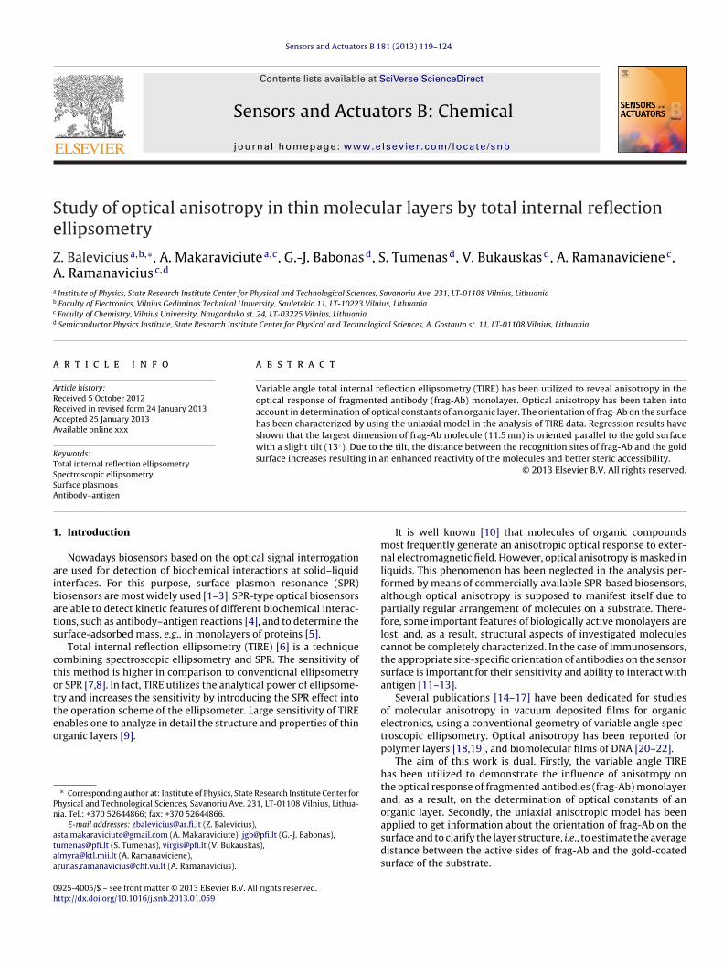
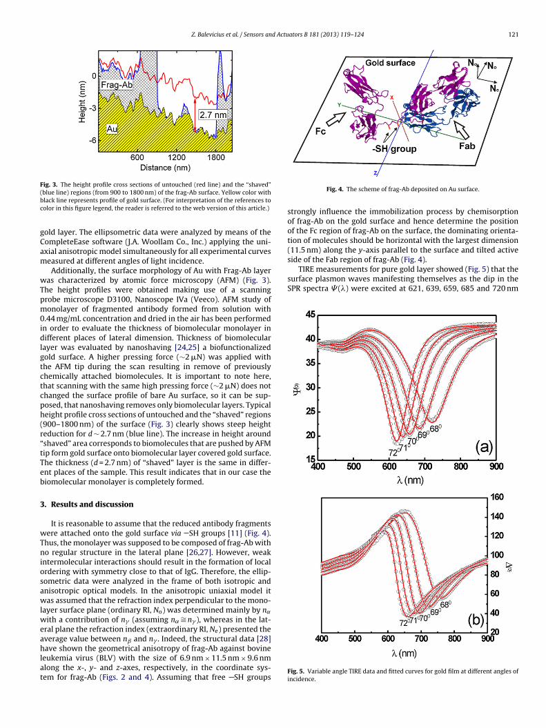
side of the Fab region of frag-Ab (Fig. 4).TIRE measurements for pure gold layer showed (Fig. 5) that the
surface plasmon waves manifesting themselves as the dip in theSPR spectra � (�) were excited at 621, 639, 659, 685 and 720 nm
blue line) regions (from 900 to 1800 nm) of the frag-Ab surface. Yellow color withlack line represents profile of gold surface. (For interpretation of the references toolor in this figure legend, the reader is referred to the web version of this article.)
old layer. The ellipsometric data were analyzed by means of theompleteEase software (J.A. Woollam Co., Inc.) applying the uni-xial anisotropic model simultaneously for all experimental curveseasured at different angles of light incidence.Additionally, the surface morphology of Au with Frag-Ab layer
as characterized by atomic force microscopy (AFM) (Fig. 3).he height profiles were obtained making use of a scanningrobe microscope D3100, Nanoscope IVa (Veeco). AFM study ofonolayer of fragmented antibody formed from solution with
.44 mg/mL concentration and dried in the air has been performedn order to evaluate the thickness of biomolecular monolayer inifferent places of lateral dimension. Thickness of biomolecular
ayer was evaluated by nanoshaving [24,25] a biofunctionalizedold surface. A higher pressing force (∼2 �N) was applied withhe AFM tip during the scan resulting in remove of previouslyhemically attached biomolecules. It is important to note here,hat scanning with the same high pressing force (∼2 �N) does nothanged the surface profile of bare Au surface, so it can be sup-osed, that nanoshaving removes only biomolecular layers. Typicaleight profile cross sections of untouched and the “shaved” regions900–1800 nm) of the surface (Fig. 3) clearly shows steep heighteduction for d ∼ 2.7 nm (blue line). The increase in height aroundshaved” area corresponds to biomolecules that are pushed by AFMip form gold surface onto biomolecular layer covered gold surface.he thickness (d = 2.7 nm) of “shaved” layer is the same in differ-nt places of the sample. This result indicates that in our case theiomolecular monolayer is completely formed.
. Results and discussion
It is reasonable to assume that the reduced antibody fragmentsere attached onto the gold surface via SH groups [11] (Fig. 4).
hus, the monolayer was supposed to be composed of frag-Ab witho regular structure in the lateral plane [26,27]. However, weak
ntermolecular interactions should result in the formation of localrdering with symmetry close to that of IgG. Therefore, the ellip-ometric data were analyzed in the frame of both isotropic andnisotropic optical models. In the anisotropic uniaxial model itas assumed that the refraction index perpendicular to the mono-
ayer surface plane (ordinary RI, No) was determined mainly by n˛ith a contribution of n� (assuming n˛∼= n� ), whereas in the lat-
ral plane the refraction index (extraordinary RI, Ne) presented theverage value between nˇ and n� . Indeed, the structural data [28]
ave shown the geometrical anisotropy of frag-Ab against bovineeukemia virus (BLV) with the size of 6.9 nm × 11.5 nm × 9.6 nmlong the x-, y- and z-axes, respectively, in the coordinate sys-em for frag-Ab (Figs. 2 and 4). Assuming that free SH groups
Fig. 4. The scheme of frag-Ab deposited on Au surface.
strongly influence the immobilization process by chemisorptionof frag-Ab on the gold surface and hence determine the positionof the Fc region of frag-Ab on the surface, the dominating orienta-tion of molecules should be horizontal with the largest dimension(11.5 nm) along the y-axis parallel to the surface and tilted active
Fig. 5. Variable angle TIRE data and fitted curves for gold film at different angles ofincidence.
122 Z. Balevicius et al. / Sensors and Actuators B 181 (2013) 119– 124
Fm
aagi6nssltts
trsbfimoniolig
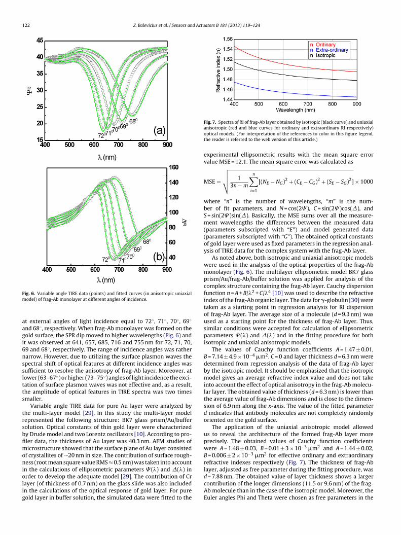
Fig. 7. Spectra of RI of frag-Ab layer obtained by isotropic (black curve) and uniaxial
d = 7.88 nm. The obtained value of layer thickness shows a larger
ig. 6. Variable angle TIRE data (points) and fitted curves (in anisotropic uniaxialodel) of frag-Ab monolayer at different angles of incidence.
t external angles of light incidence equal to 72◦, 71◦, 70◦, 69◦
nd 68◦, respectively. When frag-Ab monolayer was formed on theold surface, the SPR dip moved to higher wavelengths (Fig. 6) andt was observed at 641, 657, 685, 716 and 755 nm for 72, 71, 70,9 and 68◦, respectively. The range of incidence angles was ratherarrow. However, due to utilizing the surface plasmon waves thepectral shift of optical features at different incidence angles wasufficient to resolve the anisotropy of frag-Ab layer. Moreover, atower (63–67◦) or higher (73–75◦) angles of light incidence the exci-ation of surface plasmon waves was not effective and, as a result,he amplitude of optical features in TIRE spectra was two timesmaller.
Variable angle TIRE data for pure Au layer were analyzed byhe multi-layer model [29]. In this study the multi-layer modelepresented the following structure: BK7 glass prism/Au/bufferolution. Optical constants of thin gold layer were characterizedy Drude model and two Lorentz oscillators [10]. According to pro-ler data, the thickness of Au layer was 40.3 nm. AFM studies oficrostructure showed that the surface plane of Au layer consisted
f crystallites of ∼20 nm in size. The contribution of surface rough-ess (root mean square value RMS ≈ 0.5 nm) was taken into account
n the calculations of ellipsometric parameters � (�) and �(�) inrder to develop the adequate model [29]. The contribution of Cr
ayer (of thickness of 0.7 nm) on the glass slide was also includedn the calculations of the optical response of gold layer. For pureold layer in buffer solution, the simulated data were fitted to theanisotropic (red and blue curves for ordinary and extraordinary RI respectively)optical models. (For interpretation of the references to color in this figure legend,the reader is referred to the web version of this article.)
experimental ellipsometric results with the mean square errorvalue MSE = 12.1. The mean square error was calculated as
MSE =
√√√√ 13n − m
n∑i=1
[(NE − NG)2 + (CE − CG)2 + (SE − SG)2] × 1000
where “n” is the number of wavelengths, “m” is the num-ber of fit parameters, and N = cos(2� ), C = sin(2� )cos(�), andS = sin(2� )sin(�). Basically, the MSE sums over all the measure-ment wavelengths the differences between the measured data(parameters subscripted with “E”) and model generated data(parameters subscripted with “G”). The obtained optical constantsof gold layer were used as fixed parameters in the regression anal-ysis of TIRE data for the complex system with the frag-Ab layer.
As noted above, both isotropic and uniaxial anisotropic modelswere used in the analysis of the optical properties of the frag-Abmonolayer (Fig. 6). The multilayer ellipsometric model BK7 glassprism/Au/frag-Ab/buffer solution was applied for analysis of thecomplex structure containing the frag-Ab layer. Cauchy dispersionfunction n = A + B/�2 + C/�4 [10] was used to describe the refractiveindex of the frag-Ab organic layer. The data for �-globulin [30] weretaken as a starting point in regression analysis for RI dispersionof frag-Ab layer. The average size of a molecule (d = 9.3 nm) wasused as a starting point for the thickness of frag-Ab layer. Thus,similar conditions were accepted for calculation of ellipsometricparameters � (�) and �(�) and in the fitting procedure for bothisotropic and uniaxial anisotropic models.
The values of Cauchy function coefficients A = 1.47 ± 0.01,B = 7.14 ± 4.9 × 10⁻⁴ �m2, C = 0 and layer thickness d = 6.3 nm weredetermined from regression analysis of the data of frag-Ab layerby the isotropic model. It should be emphasized that the isotropicmodel gives an average refractive index value and does not takeinto account the effect of optical anisotropy in the frag-Ab molecu-lar layer. The obtained value of thickness (d = 6.3 nm) is lower thanthe average value of frag-Ab dimensions and is close to the dimen-sion of 6.9 nm along the x-axis. The value of the fitted parameterd indicates that antibody molecules are not completely randomlyoriented on the gold surface.
The application of the uniaxial anisotropic model allowedus to reveal the architecture of the formed frag-Ab layer moreprecisely. The obtained values of Cauchy function coefficientswere A = 1.48 ± 0.03, B = 0.01 ± 3 × 10⁻³ �m2 and A = 1.44 ± 0.02,B = 0.006 ± 2 × 10⁻³ �m2 for effective ordinary and extraordinaryrefractive indexes respectively (Fig. 7). The thickness of frag-Ablayer, adjusted as free parameter during the fitting procedure, was
contribution of the longer dimensions (11.5 or 9.6 nm) of the frag-Ab molecule than in the case of the isotropic model. Moreover, theEuler angles Phi and Theta were chosen as free parameters in the
d Actu
usotiI(esTaattarAcdit
mot(tta(tTm
4
miofsuogds[
dfitgltb
A
pesS
[
[
[
[
[
[
[
[
[
[
[
[
[
[
[
[
[
Z. Balevicius et al. / Sensors an
niaxial anisotropic model. As noted above, the axis nˇ is fixed andhould be oriented close to the gold-coated surface. The best fit wasbtained at Phi = 0 and Theta = 13 ± 6◦. It is reasonable to assumehat the optical axis of the refractive index ellipsoid of frag-Ab layers tilted by about 13◦ from the plane parallel to the substrate. ThegG antibody can be presented as a three-dimensional ellipsoid inx, y, z) coordinates. The equation representing this refractive indexllipsoid is x2/εx + y2/εy + z2/εz = 1, where the semiaxes of this ellip-oid correspond to the refractive indices along the x, y, and z-axes.hus, this ellipsoid is referred to as the index ellipsoid. The x, y,nd z-axes are generally called the principal axes, and nx, ny, nz
re the principal indexes of refraction [10]. However, in our case,he principal axes were not parallel to the (x, y, z) coordinates. Theransformations of coordinates (x, y, z) were performed via Eulerngles (�, �, ) to (˛, ˇ, �). The three Euler angles represented theotation angles of the coordinates (x, y, z) (Fig. 2). In the case of frag-b attached via SH groups to the gold surface, the molecule wasonsidered as a uniaxial crystal with the refraction index perpen-icular to the monolayer surface plane (ordinary RI, no), whereas
n the lateral plane the refraction index (extraordinary RI, ne) washe average value between nˇ and n� .
The obtained values of parameters in the uniaxial anisotropicodel confirmed the assumption that the horizontal orientation
f frag-Ab molecules was dominating. It should be noted that inhe case of uniaxial anisotropic model both adjusted parametersthickness and refractive indices) give additional information abouthe layer structure in comparison to the isotropic model, in whichhe refractive index represents the average value. The regressionnalysis of the spectra of ellipsometric parameters � (�) and �(�)Fig. 6) showed that the MSE-value (14.7) was smaller in the case ofhe uniaxial anisotropic model than that (21.8) of isotropic model.hese MSE-values demonstrated that in the uniaxial anisotropicodel the refractive indexes were obtained with a higher accuracy.
. Conclusions
Summarizing, the structural and optical parameters deter-ined from regression analysis of ellipsometric data provide the
mportant information about the molecular monolayer, such asrientation of biomolecules with respect to the substrate sur-ace, the distance of active sites of biological objects from theurface. In particular, regression results obtained by applying theniaxial anisotropic model showed that the largest dimensionf frag-Ab molecule (11.5 nm) was horizontally oriented on theold surface with a slight tilt. The tilt leads to an increase of theistance between the recognition sites of frag-Ab and the goldurface and results their enhanced activity and steric accessibility26].
To the best of our knowledge TIRE method has been used forefine optical anisotropy of biorecognition molecule layer for therst time. These results allowing not only to evaluate the orienta-ion of the molecules resulting in analyte accessibility but also toet some insight on the molecule conformation after the immobi-ization on the sensor surface leading to its remaining activity andhis way affecting the sensitivity, selectivity and stability of theiosensor.
cknowledgements
The work was supported by Research Council of Lithuania, sup-
ort to research of scientists and other researchers (Global Grant),nzymes functionalized by polymers and biorecognition unit forelective treatment of target cells (NanoZim’s), Project Nr. VP1-3.1-ˇMM-07-K-02-042.
[
[
ators B 181 (2013) 119– 124 123
References
[1] J. Homola, S.S. Yee, G. Gauglitz, Surface plasmon resonance sensors: review,Sensors and Actuators B 54 (1999) 3–15.
[2] W. Lee, B.K. Oh, W.H. Lee, J.W. Choi, Immobilization of antibody fragment forimmunosensor application based on surface plasmon resonance, Colloids andSurfaces B: Biointerfaces 40 (2005) 143–148.
[3] X.D. Hao, A.G. Kirk, M. Tabrizian, Towards integrated and sensitive surfaceplasmon resonance biosensors: a review of recent progress, Biosensors andBioelectronics 23 (2007) 151–160.
[4] A. Kausaite-Minkstimiene, A. Ramanaviciene, A. Ramanavicius, Surface plas-mon resonance biosensor for direct detection of antibodies against humangrowth hormone, Analyst 134 (2009) 2051–2057.
[5] A. Kausaite-Minkstimiene, A. Ramanaviciene, J. Kirlyte, A. Ramanavicius, Acomparative study of random and oriented antibody immobilization tech-niques on the binding capacity of immunosensor, Analytical Chemistry 82(2010) 6401–6408.
[6] H. Arwin, M. Poksinski, K. Johansen, Total internal reflection ellipsometry –principles and applications, Applied Optics 43 (2004) 3028–3036.
[7] H. Arwin, M. Poksinski, K. Johansen, Enhancement in ellipsometric thin filmsensitivity near surface plasmon resonance conditions, Physica Status Solidi(A) 205 (2008) 817–820.
[8] A.V. Nabok, A. Tsargorodskaya, A.K. Hassan, N.F. Starodub, Registration of T-mycotoxin with total internal reflection ellipsometry and QCM impedancemethods, Applied Surface Science 246 (2005) 381–386.
[9] A. Nabok, A. Tsargorodskaya, M.K. Mustafa, I. Szekacs, N.F. Starodub, A. Szekacs,Detection of low molecular weight toxins using an optical phase method ofellipsometry, Sensors and Actuators B 154 (2011) 232–237.
10] H. Fujiwara, Spectroscopic Ellipsometry Principles and Applications, John Wiley& Sons, Chichester, 2007 (Chapter 7).
11] A.A. Karyakin, G.V. Presnova, M.Y. Rubtsova, A.M. Egorov, Oriented immobiliza-tion of antibodies onto the gold surfaces via their native thiol groups, AnalyticalChemistry 72 (2000) 3805–3811.
12] Y.M. Bae, B.-K. Oh, W. Lee, W.H. Lee, J.-W. Choi, Study on orientation ofimmunoglobulin G on protein G layer, Biosensors and Bioelectronics 21 (2005)103–110.
13] T. Dubrovsky, A. Tronin, S. Dubrovskaya, O. Guryev, C. Nicolini, Langmuir filmsof Fc binding receptors engineered from protein A and protein G as a sublayerfor immunoglobulin orientation, Thin Solid Films 284–285 (1996) 698–702.
14] D. Yokoyama, A. Sakaguchi, M. Suzuki, C. Adachi, Horizontal molecular ori-entation in vacuum-deposited organic amorphous films of hole and electrontransport materials, Applied Physics Letters 93 (2008) 173302.
15] D. Yokoyama, A. Sakaguchi, M. Suzuki, C. Adachi, Horizontal orientationof linear-shaped organic molecules having bulky substituents in neat anddoped vacuum-deposited amorphous films, Organic Electronics 10 (2009)127–137.
16] H.W. Lin, C.L. Lin, H.H. Chang, Y.T. Lin, C.C. Wu, Anisotropic optical propertiesand molecular orientation in vacuum-deposited ter(9,9-diarylfluorene)s thinfilms using spectroscopic ellipsometry, Journal of Applied Physics 95 (2004)881–886.
17] D. Yokoyama, C. Adachi, In situ real-time spectroscopic ellipsometry mea-surement for the investigation of molecular orientation in organic amorphousmultilayer structures, Journal of Applied Physics 107 (2010) 123512.
18] K. Hinrichs, S.D. Silaghi, C. Cobet, N. Esser, D.R.T. Zahn, Ellipsometry frominfrared to vacuum ultraviolet: structural properties of thin anisotropic gua-nine films on silicon, Physica Status Solidi (B) 242 (2005) 2681–2687.
19] L.A.A. Pettersson, S. Ghosh, O. Inganas, Optical anisotropy in thin filmsof poly(3,4-ethylenedioxythiophene)–poly(4-styrenesulfonate), Organic Elec-tronics 3 (2002) 143–148.
20] M. Losurdo, G. Bruno, E.A. Irene, Anisotropy of optical properties of conjugatedpolymer thin films by spectroscopic ellipsometry, Journal of Applied Physics94 (2003) 4923–4929.
21] S. Wenmackers, S.D. Pop, K. Roodenko, V. Vermeeren, O.A. Williams, M. Dae-nen, O. Douheret, J. D’Haen, A. Hardy, M.K. Van Bael, K. Hinrichs, C. Cobet, M.vande Ven, M. Ameloot, K. Haenen, L. Michiels, N. Esser, P. Wagner, Structuraland optical properties of DNA layers covalently attached to diamond surfaces,Langmuir 24 (2008) 7269–7277.
22] S. Wenmackers, P. Christiaens, M. Daenen, K. Haenen, M. Nesladek, M. vandeVen, V. Vermeeren, L. Michiels, M. Ameloot, P. Wagner, DNA attachment tonanocrystalline diamond films, Physica Status Solidi (A) 202 (2005) 2212–2216.
23] L.J. Harris, E. Skaletsky, A. McPherson, Crystallographic structure of an intactIgG1 monoclonal antibody, Journal of Molecular Biology 275 (1998) 861–872.
24] L.G. Rosa, J. Jiang, O.V. Lima, J. Xiao, E. Utreras, P.A. Dowben, L.Tan, Selective nanoshaving of self-assembled monolayers of 2-(4-pyridylethyl)triethoxysilane, Materials Letters 63 (2009) 961–964.
25] L.G. Rosa, J. Liang, Atomic force microscope nanolithography: dip-pen,nanoshaving, nanografting, tapping mode, electrochemical and thermal nano-lithography, Journal of Physics: Condensed Matter 21 (2009) 483001.
26] Z. Balevicius, A. Ramanaviciene, I. Baleviciute, A. Makaraviciute, L. Mikoliu-naite, A. Ramanavicius, Evaluation of intact- and fragmented-antibody basedimmunosensors by total internal reflection ellipsometry, Sensors and Actuators
B 160 (2011) 555–562.27] I. Baleviciute, Z. Balevicius, A. Makaraviciute, A. Ramanaviciene, A. Ramanavi-cius, Study of antibody/antigen binding kinetics by total internal reflectionellipsometry, Biosensors and Bioelectronics 39 (2013) 170–176.
28] www.rcsb.org/pdb
1 d Actu
[
[
B
DtRiFfo
Ai–UTn
DePsTs
Sa
24 Z. Balevicius et al. / Sensors an
29] Z. Balevicius, V. Vaicikauskas, G.-J. Babonas, The role of surface roughness intotal internal reflection ellipsometry of hybrid systems, Applied Surface Science256 (2009) 640–644.
30] H. Arwin, Optical properties of thin layers of bovine serum albumin, gamma-globulin, and hemoglobin, Applied Spectroscopy 40 (1986) 313–318.
iographies
r. Zigmas Balevicius received his Ph.D. degree in 2008 in spectroscopy and laserechnology in Institute of Physics, Vilnius/Lithuania. He is a researcher in Stateesearch Institute Centre for Physical and Technological Sciences in Nanoengineer-
ng Department, Plasmonic Lab., Vilnius, Lithuania. In addition he is working inaculty of Electronics, Vilnius Gediminas Technical University as associated pro-essor. He is an expert in spectroscopic ellipsometry. He focuses his research onptical biosensing, plasmonics, optical measurement systems, optics in biology.
sta Makaraviciute is a Ph.D. student; in 2010 she finished MSc studies in biochem-stry at Vilnius University and now Asta is doing her Ph.D. studies at Nanotechnas
Centre of Nanotechnology and Materials Science, Faculty of Chemistry, Vilniusniversity. She is also working in State Research Institute Centre for Physical andechnological Sciences in Nanoengineering Department, Plasmonic Laboratory, Vil-ius, Lithuania.
r. Gintautas Jurgis Babonas is a senior researcher in the Department of Opto-lectronics, Institute of Semiconductor Physics, State Research Institute Center forhysical and Technological Sciences. He received Dr. Sci. degree at Vilnius Univer-ity in 1991. In 1995 the professor title was granted to him at Vilnius Gediminas
echnical University. His latest interest is in the area of optical studies of complexystems.aulius Tumenas is a Ph.D. student in 2008 finished MS studies in solid state physicst Vilnius University. Now he is finishing his Ph.D. studies at State Scientific Research
ators B 181 (2013) 119– 124
Institute Center for Physical Sciences and Technology in Department of Optoelec-tronics, Semiconductor Optics Laboratory, Vilnius, Lithuania. Saulius focuses hisresearch on advanced semiconductors, quasicrystals, nanocomposites, solar cellsand conductive organics.
Dr. Virginijus Bukauskas is a research associate in the Department of Phys-ical Technologies of State Research Institute Center for Physical Sciences andTechnology. He received Dr. Sci. degree at Vilnius University in 2010. His lat-est interest areas are: Scanning probe microscopy and spectroscopy; integrationof biomolecules on solid surfaces; formation and investigation of gas sensitivematerials.
Dr. Almira Ramanaviciene is an associate professor, head of the division ofImmunotechnology at the newly established State Research Institute Centre of Inno-vative Medicine. Prior to that she headed the Laboratory of Immunoanalysis andNanotechnology at the Institute of Immunology of Vilnius University, Lithuania. Shereceived her Ph.D. degree in Biomedicine in 2002 from the Institute of Immunologyand Vilnius University. In 2008 she completed habilitation procedure in PhysicalSciences at Vilnius University. She is serving as an expert of FP7 program at the Euro-pean Commission and other international foundations. Now her primary researchwork is in fields related to nanotechnology.
Dr. Arunas Ramanavicius is a professor at Vilnius University. Recently he hasbeen heading the Department of Physical Chemistry and the Research Cen-tre for Nanotechnology and Materials Science – NanoTechnas at Faculty ofChemistry, Vilnius University, Lithuania. At the same time he is also a seniorresearcher in Department of Materials Science and Electronics, Institute of Semi-
conductor Physics, State Scientific Research Institute Centre for Physical Sciencesand Technology, Vilnius, Lithuania. In 1998 he received a Ph.D. degree andin 2002 he received the doctor habilitus degree from Vilnius University. From2002 he has been working as a professor at the Faculty of Chemistry, VilniusUniversity.