Stress-Induced Visceral Hypersensitivity in Maternally Separated Rats Can Be Reversed by...
8
Stress-Induced Visceral Hypersensitivity in Maternally Separated Rats Can Be Reversed by Peripherally Restricted Histamine-1-Receptor Antagonists Oana I. Stanisor 1 , Sophie A. van Diest 1 , Zhumei Yu 1,2 , Olaf Welting 1 , Noor Bekkali 1 , Jing Shi 2 , Wouter J. de Jonge 1 , Guy E. Boeckxstaens 3 , Rene M. van den Wijngaard 1 * 1 Tytgat Institute for Liver and Intestinal Research, Academic Medical Center, Amsterdam, The Netherlands, 2 Department of Neurobiology, Tongji Medical College, HUST, Wuhan, People’s Republic of China, 3 Division of Gastroenterology, University Hospital Gasthuisberg, Catholic University of Leuven, Leuven, Belgium Abstract Background: The histamine-1 receptor (H1R) antagonist ketotifen increased the threshold of discomfort in hypersensitive IBS patients. The use of peripherally restricted and more selective H1R antagonists may further improve treatment possibilities. We examined the use of fexofenadine and ebastine to reverse post-stress visceral hypersensitivity in maternally separated rats. Methods: The visceromotor response to colonic distension was assessed in adult maternally separated and nonhandled rats pre- and 24 hours post water avoidance. Subsequently rats were treated with vehicle alone or different dosages of fexofenadine (1.8 and 18 mg/kg) or ebastine (0.1 and 1.0 mg/kg) and re-evaluated. Colonic tissue was collected to assess relative RMCP-2 and occludin expression levels by Western blot and histamine-1 receptor by RT-qPCR. b-hexosaminidase release by RBL-2H3 cells was used to establish possible mast cell stabilizing properties of the antagonists. Key results: Water avoidance only induced enhanced response to distension in maternally separated rats. This response was reversed by 1.8 and 18 mg/kg fexofenadine. Reversal was also obtained by 1.0 but not 0.1 mg/kg ebastine. RMCP-2 expression levels were comparable in these two ebastine treatment groups but occludin was significantly higher in 1.0 mg/ kg treated rats. There were no differences in histamine-1 receptor expression between nonhandled and maternally separated rats. Fexofenadine but not ebastine showed mast cell stabilizing quality. Conclusions: Our results indicate that the peripherally restricted 2 nd generation H1-receptor antagonists fexofenadine and ebastine are capable of reversing post stress visceral hypersensitivity in rat. These data justify future IBS patient trials with these well tolerated compounds. Citation: Stanisor OI, van Diest SA, Yu Z, Welting O, Bekkali N, et al. (2013) Stress-Induced Visceral Hypersensitivity in Maternally Separated Rats Can Be Reversed by Peripherally Restricted Histamine-1-Receptor Antagonists. PLoS ONE 8(6): e66884. doi:10.1371/journal.pone.0066884 Editor: Julie A. Chowen, Hosptial Infantil Universitario Nin ˜o Jesu ´ s, CIBEROBN, Spain Received February 12, 2013; Accepted May 14, 2013; Published June 12, 2013 Copyright: ß 2013 Stanisor et al. This is an open-access article distributed under the terms of the Creative Commons Attribution License, which permits unrestricted use, distribution, and reproduction in any medium, provided the original author and source are credited. Funding: OIS was supported by funding for the IPODD consortium under Grant Agreement 202020 of the Seventh Research Framework Programme of the European Union (http://www.IPODD.eu). SAvD was supported by the Netherlands Top Institute Pharma, grant number T1-215-1 (www.TIPharma.com) and the Netherlands Digestive Diseases Foundation (MLDS), project number WO10-12 (www.MLDS.NL/). ZY was supported by The China Exchange Programme (CEP) of the Royal Netherlands Academy of Arts and Sciences (KNAW), project number 11CDP005 (www.KNAW.NL). WJdJ was funded by a grant of the Dutch Organization for Scientific Research (NWO-VIDI). The funders had no role in study design, data collection and analysis, decision to publish, or preparation of the manuscript. Competing Interests: The authors have declared that no competing interests exist. * E-mail: [email protected] Introduction The functional gastrointestinal disorder irritable bowel syn- drome (IBS) is characterized by abdominal pain or discomfort associated with defecation or change in bowel habit.[1] Increased perception to gastrointestinal stimuli, so called visceral hypersen- sitivity, and barrier dysfunction are considered important patho- physiological mechanisms in IBS. Stress is an important trigger for IBS-symptoms and preclinical investigations suggest that barrier- and sensitivity changes may relate to stress-induced degranulation of intestinal mucosal mast cells.[2–4] A recent clinical trial with the mast cell stabilizer and histamine-1-receptor (H1R) antagonist ketotifen confirmed the possible relevance of this cell type.[5] Ketotifen not only decreased abdominal pain and other IBS symptoms but also improved health related quality of life and increased the threshold of discomfort in hypersensitive patients. However, the exact working mechanism of ketotifen remained elusive. Investigations comparing pre- and post-therapy mediator release by submerged rectal biopsies did not support a role for mast cell stabilization. Consequently, it was suggested that H1R antagonism was the main molecular mode of action in this trial. Ex vivo investigations performed by Barbara et al. indicated that a mediator present in IBS biopsy-supernatants induced H1R- dependent mesenteric afferent nerve discharge and Ca 2+ -mobili- sation in cultured rat DRG neurons.[6] In addition, mucosal biopsies from IBS patients showed a significant increase in H1R mRNA levels over controls.[7] Similar to the ketotifen trial, these results suggested that H1R-targeting may be an attractive PLOS ONE | www.plosone.org 1 June 2013 | Volume 8 | Issue 6 | e66884
-
Upload
independent -
Category
Documents
-
view
3 -
download
0
Transcript of Stress-Induced Visceral Hypersensitivity in Maternally Separated Rats Can Be Reversed by...

Stress-Induced Visceral Hypersensitivity in MaternallySeparated Rats Can Be Reversed by PeripherallyRestricted Histamine-1-Receptor AntagonistsOana I. Stanisor1, Sophie A. van Diest1, Zhumei Yu1,2, Olaf Welting1, Noor Bekkali1, Jing Shi2, Wouter J. de
Jonge1, Guy E. Boeckxstaens3, Rene M. van den Wijngaard1*
1 Tytgat Institute for Liver and Intestinal Research, Academic Medical Center, Amsterdam, The Netherlands, 2 Department of Neurobiology, Tongji Medical College, HUST,
Wuhan, People’s Republic of China, 3 Division of Gastroenterology, University Hospital Gasthuisberg, Catholic University of Leuven, Leuven, Belgium
Abstract
Background: The histamine-1 receptor (H1R) antagonist ketotifen increased the threshold of discomfort in hypersensitiveIBS patients. The use of peripherally restricted and more selective H1R antagonists may further improve treatmentpossibilities. We examined the use of fexofenadine and ebastine to reverse post-stress visceral hypersensitivity in maternallyseparated rats.
Methods: The visceromotor response to colonic distension was assessed in adult maternally separated and nonhandled ratspre- and 24 hours post water avoidance. Subsequently rats were treated with vehicle alone or different dosages offexofenadine (1.8 and 18 mg/kg) or ebastine (0.1 and 1.0 mg/kg) and re-evaluated. Colonic tissue was collected to assessrelative RMCP-2 and occludin expression levels by Western blot and histamine-1 receptor by RT-qPCR. b-hexosaminidaserelease by RBL-2H3 cells was used to establish possible mast cell stabilizing properties of the antagonists.
Key results: Water avoidance only induced enhanced response to distension in maternally separated rats. This response wasreversed by 1.8 and 18 mg/kg fexofenadine. Reversal was also obtained by 1.0 but not 0.1 mg/kg ebastine. RMCP-2expression levels were comparable in these two ebastine treatment groups but occludin was significantly higher in 1.0 mg/kg treated rats. There were no differences in histamine-1 receptor expression between nonhandled and maternallyseparated rats. Fexofenadine but not ebastine showed mast cell stabilizing quality.
Conclusions: Our results indicate that the peripherally restricted 2nd generation H1-receptor antagonists fexofenadine andebastine are capable of reversing post stress visceral hypersensitivity in rat. These data justify future IBS patient trials withthese well tolerated compounds.
Citation: Stanisor OI, van Diest SA, Yu Z, Welting O, Bekkali N, et al. (2013) Stress-Induced Visceral Hypersensitivity in Maternally Separated Rats Can Be Reversedby Peripherally Restricted Histamine-1-Receptor Antagonists. PLoS ONE 8(6): e66884. doi:10.1371/journal.pone.0066884
Editor: Julie A. Chowen, Hosptial Infantil Universitario Nino Jesus, CIBEROBN, Spain
Received February 12, 2013; Accepted May 14, 2013; Published June 12, 2013
Copyright: � 2013 Stanisor et al. This is an open-access article distributed under the terms of the Creative Commons Attribution License, which permitsunrestricted use, distribution, and reproduction in any medium, provided the original author and source are credited.
Funding: OIS was supported by funding for the IPODD consortium under Grant Agreement 202020 of the Seventh Research Framework Programme of theEuropean Union (http://www.IPODD.eu). SAvD was supported by the Netherlands Top Institute Pharma, grant number T1-215-1 (www.TIPharma.com) and theNetherlands Digestive Diseases Foundation (MLDS), project number WO10-12 (www.MLDS.NL/). ZY was supported by The China Exchange Programme (CEP) ofthe Royal Netherlands Academy of Arts and Sciences (KNAW), project number 11CDP005 (www.KNAW.NL). WJdJ was funded by a grant of the Dutch Organizationfor Scientific Research (NWO-VIDI). The funders had no role in study design, data collection and analysis, decision to publish, or preparation of the manuscript.
Competing Interests: The authors have declared that no competing interests exist.
* E-mail: [email protected]
Introduction
The functional gastrointestinal disorder irritable bowel syn-
drome (IBS) is characterized by abdominal pain or discomfort
associated with defecation or change in bowel habit.[1] Increased
perception to gastrointestinal stimuli, so called visceral hypersen-
sitivity, and barrier dysfunction are considered important patho-
physiological mechanisms in IBS. Stress is an important trigger for
IBS-symptoms and preclinical investigations suggest that barrier-
and sensitivity changes may relate to stress-induced degranulation
of intestinal mucosal mast cells.[2–4] A recent clinical trial with the
mast cell stabilizer and histamine-1-receptor (H1R) antagonist
ketotifen confirmed the possible relevance of this cell type.[5]
Ketotifen not only decreased abdominal pain and other IBS
symptoms but also improved health related quality of life and
increased the threshold of discomfort in hypersensitive patients.
However, the exact working mechanism of ketotifen remained
elusive. Investigations comparing pre- and post-therapy mediator
release by submerged rectal biopsies did not support a role for
mast cell stabilization. Consequently, it was suggested that H1R
antagonism was the main molecular mode of action in this trial.
Ex vivo investigations performed by Barbara et al. indicated that
a mediator present in IBS biopsy-supernatants induced H1R-
dependent mesenteric afferent nerve discharge and Ca2+-mobili-
sation in cultured rat DRG neurons.[6] In addition, mucosal
biopsies from IBS patients showed a significant increase in H1R
mRNA levels over controls.[7] Similar to the ketotifen trial, these
results suggested that H1R-targeting may be an attractive
PLOS ONE | www.plosone.org 1 June 2013 | Volume 8 | Issue 6 | e66884

treatment option in IBS. However, ketotifen has low H1R
selectivity and is known to cross the blood-brain barrier and
cause central side effects.[8,9] Consequently, possibilities to
increase therapeutic dose for enhanced effectiveness are limited
and evaluation of other, peripherally restricted, H1-receptor
antagonists may proof beneficial. In the nineteen eighties second
generation non-sedating H1-antihistamines became available and
by now this group of antihistamines comprises more than 45
different compounds, including fexofenadine and ebastine.[8] In
clinical trials these compounds appeared to be safe, effective and
well tolerated and they are now routinely being used in the
treatment of allergic rhinitis and urticaria.[10,11] To establish
whether these antagonists hold promise for therapeutical inter-
ventions in IBS we evaluated them in the IBS-like rat model of
maternal separation. Similar to patients, acute stress induces
enhanced sensitivity to colorectal distension in previously separat-
ed Long Evans rats.[12] This change in sensitivity was shown to be
long lasting, one hour of water avoidance induced enhanced
sensitivity for up to one month, and could be reversed by the mast
cell stabilizer doxantrazole.[13] In the present study we investi-
gated whether fexofenadine and ebastine were also capable of
reversing post stress, mast cell dependent, visceral hypersensitivity
in the rat maternal separation model. Our results suggest that
peripheral H1Rs may be a safe new target for therapeutical
intervention in IBS.
Materials and Methods
Ethics statementAll procedures were conducted in accordance with the
institutional guidelines and approved by the Animal Ethical
Committee of the AMC/University of Amsterdam (reference
protocol number 100998).
Animals and maternal separation (MS) protocolLong-Evans rats (Harlan, Horst, The Netherlands) were bred
and housed at the animal facility of the AMC (Amsterdam
Medical Centrum, Amsterdam, The Netherlands). Rats were
maintained on a normal 12:12-h dark/light cycle and temperature
(20–22uC) and provided with food and water ad libitum. Separation
was accomplished by placing the dams into another cage in a
separate room for 180 minutes per day from postnatal day 2 to 14.
During separation, cages were placed on a heating pad (30–34uC)
to help pups regulate normal body temperature. Pups were
weaned on day 22 and subsequently raised in pairs of two. NH
pups were nursed normally.
Colonic distension protocol and water avoidance (WA)In IBS patients investigations of visceral sensitivity are
performed by colorectal distensions: hypersensitive patients
perceive pain during luminal distensions at lower volumes or
pressures than normal controls.[14] In our investigations in rat,
colonic distensions were performed with a latex balloon (Ultra-
cover 8F, International Medical Products, Zutphen, The Nether-
lands) at the minimum age of 4 months and carried out as
described before.[12,13,15] A catheter was placed during short
isoflurane anesthesia 20 minutes before distensions with graded
volumes of water (1.0, 1.5 and 2.0 mL). Length and diameter of
the balloon during maximum volume distension were 18 mm and
15 mm respectively. After each 20 second distension period, water
was quickly removed and 80 seconds rest was exercised. At adult
age rats were subjected to WA stress during which they were
positioned on a pedestal surrounded by water for one hour. Earlier
investigations indicated that, in contrast to NH rats, WA induces
enhanced sensitivity to colonic distension in MS rats.[12]
Measurement of the visceromotor response to colonicdistension and data analysis
Distension of the colon induces contractions of the abdominal
musculature, the so called visceromotor response. Quantification
of these contractions by electromyography (EMG) is often used to
assess visceral pain responses in rodents. We used radiotelemetric
transmitters (Physiotel Implant TA10AE-F20; DSI, St Paul, MN,
USA) to record these EMG signals in freely moving rats.[12] In
short, the transmitter was positioned in the abdominal cavity and
two connected electrodes were placed in abdominal muscles.
During distension protocols, animals were placed in a standard
macralon cage (exact size of the receiver) that was positioned on
top of a receiver (Data Sciences International). The receiver was
linked to a Biopac MP100 data acquisition system (Biopac Systems
Inc., Santa Barbara, CA, USA) and a personal computer via a raw
data analog converter (Data Sciences International). Data were
acquired with AcqKnowledge software (Biopac Systems Inc.,
Santa Barbara, CA, USA) and analyzed as described before.
Briefly, each 20-s distension period and its preceding 20-s of
baseline recording were extracted from the original raw EMG
data file. After correction for movement and breathing, data were
rectified and integrated. Absolute data sets were then obtained by
subtracting the 20-s baseline recording from the 20-s distension
result. Similar to earlier publications the final results are given as
normalized data sets, which were calculated from the absolute
data by setting the 2 mL value of the first (pre-stress) distension at
100%.[12,13,15] Area under the curve (AUC) of relative responses
was calculated for individual rats and used to show possible
changes in visceromotor response within treatment groups.
Relative response data were also used to evaluate possible changes
on a per volume basis.
Design of in vivo pharmacological intervention protocolsAnimal experiments were performed while the investigator was
blinded to administration of drug or vehicle alone (disclosed after
evaluation of all tracings). Directly after measuring baseline
sensitivity to distension (10:00 AM, day 0), rats were subjected
to WA stress and measured again 24 hours post-WA. Subse-
quently, rats were treated with intraperitoneal fexofenadine
hydrochloride (Tocris Bioscience, Bristol, UK), ebastine (Sigma-
Aldrich, Zwijndrecht, The Netherlands) or vehicle alone (10%
alcohol). Compounds were administered two times at day 1 (11:00
AM and 05.00 PM) and one time at day 2 (30 minutes before the
last distension protocol at 09:30 AM). Cumulative dosages (total of
3 intraperitoneal injections in 24 hour timeframe) were 1.8 mg/kg
or 18 mg/kg for fexofenadine and 0.1 mg/kg or 1 mg/kg for
ebastine.
RT-qPCRTo avoid possible distension related effects on H1R expression
levels, vehicle treated NH and MS rats were sacrificed 7 days after
the last distension protocol. Total RNA was isolated from colonic
tissue of NH and MS rats using TRIzol (Invitrogen, Breda, The
Netherlands) according to manufacturer’s protocol. Following
DNAse treatment, cDNA was obtained by using RevertAid First
Strand cDNA Synthesis Kit (Fermentas, Waltham, MA, USA)
Quantitative PCR was performed with SYBR Green in the
LightCycler480 system (Roche) using a default 60u program.
Primer pairs used for H1R were; sense, CTTCTACCTCCC-
CACTTTGCT, antisense: TTCCCTTTCCCCCTCTTG and
H1R Antagonism in Visceral Hypersensitivity
PLOS ONE | www.plosone.org 2 June 2013 | Volume 8 | Issue 6 | e66884

H1R Antagonism in Visceral Hypersensitivity
PLOS ONE | www.plosone.org 3 June 2013 | Volume 8 | Issue 6 | e66884

for the housekeeping gene Ppib[15]: sense, GCAAG-
CACGTGGTTTTCGGC, antisense: TGTGAGGGAATCGA-
CAGGACCC.
Western blottingIn contrast to tissues used for RT-qPCR H1R evaluation,
ebastine treated rats were sacrificed directly after the last
distension protocol. This tissue was than used to semi quantita-
tively assess direct effects of ebastine treatment on RMCP-2 and
occludin expression levels. Distal colon was dissected, homoge-
nized in lysis buffer (Cell Signaling, Danvers, MA, USA) and
assessed by SDS-polyacrylamide gel electrophoresis and Western
blotting. Blots were cut at appropriate kD and evaluated for
expression of the rat chymase analogue RMCP-2 (polyclonal anti-
RMCP-2, Moredun Scientific, Penicuik, Scotland), the tight
junction protein occludin (rabbit-anti-occludin, Zymed, San
Francisco (CA), USA) and GAPDH (mouse-anti-GAPDH, Milli-
pore, Amsterdam, The Netherlands). Peroxidase-labeled second-
ary antibody was visualized with Lumi-light plus (Roche Diag-
nostics, Almere, The Netherlands) and densitometric analyses
were carried out with the image processing program ImageJ
(http://rsb.info.nih.gov/ij/).
In vitro mast cell degranulation experiments and beta-hexosaminidase asay
RBL-2H3 cells were used to evaluate the possible mast cell
stabilizing effect of fexofenadine and the active metabolite of
ebastine; carebastine[11] (Santa Cruz, Heidelberg, Germany).
After 30 minutes pre-treatment with these compounds (10 mM,
100 mM or vehicle alone)[16] cells were stimulated with
compound C48/80 (Sigma-Aldrich, 100 mg/ml, 500 mg/ml,
1 mg/ml or vehicle alone) for 1 hour. b-hexosaminidase release
was quantified by using 4-methylumbelliferyl glucaosaminide as a
substrate. Fluorescence was measured at an emmision wavelength
of 450 nm and an excitation wavelength of 360 nm. Release of b-
hexosaminidase was calculated as a percentage of total cellular
content.
Statistical analysisStatistical calculations were performed using SPSS for windows
(version 11.5.2). VMR data were analysed with the Wilcoxon
signed ranks test which was applied for the area under the curve
(AUC) of the relative response (normalized data) to colonic
distension. Possible statistical differences in Western blot and RT-
qPCR evaluations were assessed by Mann-Whitney test.
Results
In vivo fexofenadine treatmentWe established whether a) WA induced post stress hypersen-
sitivity to distension in NH and MS rats and b) whether
fexofenadine was capable of reversing sensitivity changes. As
published before[12], WA was unable to induce post stress visceral
hypersensitivity in NH rats (figures 1A and 1B, white vs black bars)
and intraperitoneal post stress administration of high dose
fexofenadine (18 mg/kg) did not induce sensitivity changes in
these animals (figure 1B, black vs grey bar). In MS rats WA led to
increased post-WA AUC in all 3 treatment groups (figure 1C, D
and E; *P,0.05, **P,0.01). Enhanced post-stress sensitivity levels
were not affected by vehicle treatment alone (figure 1C) but
treatment with 18 and 1.8 mg fexofenadine/kg effectively reversed
visceral hypersensitivity (figure 1D and 1E respectively).
Per volume comparisons (right side line-diagrams and accom-
panying statistics-boxes in figure 1A–E) corroborated AUC-data
for all MS groups except rats treated with 1.8 mg fexofenadine/kg.
In the latter group fexofenadine-induced reversal of hypersensi-
tivity was not significant for 1.0 and 2.0 ml distension volumes.
Antagonist treatment did not lead to changes in compliance as
assessed by pressure-volume curves (data not shown).
H1R gene expression was then determined in colonic tissue of
vehicle treated NH and MS rats. Tissue was collected 7 days after
the final distension series to avoid protocol induced effects on
receptor expression levels. Sufficient yield of RNA was obtained
from all but 2 vehicle treated nonhandled rats. As shown in figure
2 there were no significant differences between NH and MS rats.
In vivo ebastine treatmentPost WA hypersensitivity to distension did not occur in NH rats
and remained unaltered upon fexofenadine treatment. Further,
due to their broad clinical use, we know that fexofenadine as well
as ebastine are well tolerated in the human setting. Therefore, we
choose not to sacrifice additional NH rats to reconfirm results
obtained with fexofenadine; ebastine was only evaluated in MS
rats. AUC comparisons indicated that WA-induced hypersensitiv-
ity to distension could be reversed by an accumulative dose of
1.0 mg ebastine/kg (figure 3B, black vs grey bars, **P,0.01) but
not 0.1 mg/kg (figure 3A). Statistical evaluations on a per volume
basis (line-diagrams and statistics boxes in figures 3 C and D)
showed similar results: we observed a significant post-WA increase
Figure 1. In vivo post stress fexofenadine treatment. The visceromotor response to distension was measured pre-WA and 24 and 48 hourspost-WA in NH and MS rats. Fexofenadine or vehicle was administered 3 times between 24- and 48 hours measurements (cumulative dosages 18 and1.8 mg/kg). Responses to distension are depicted as AUC (left side histograms) and per volume (right side line-diagrams, corresponding statistics inlower right side tables). NH rats did not become hypersensitive to distension and fexofenadine treatment did not change sensitivity levels (figures Aand B). In MS rats WA induced enhanced sensitivity to distension in all 3 treatment groups (figures C, D and E). Treatment with 18 and 1.8 mgfexofenadine/kg (figure D and E respectively) but not vehicle alone (C) was able to reverse stress induced visceral hypersensitivity. All data arepresented as mean 6 SEM, all groups n = 8 or 9 rats, *P,0.05 and **P,0.01.doi:10.1371/journal.pone.0066884.g001
Figure 2. Relative colonic expression values for the histamineH1 receptor gene. H1R mRNA expression was evaluated relative to thehousekeeping gene Ppib in colonic samples of NH and MS rats. Tissuewas collected 7 days post vehicle treatment and distensions. Therewere no significant differences.doi:10.1371/journal.pone.0066884.g002
H1R Antagonism in Visceral Hypersensitivity
PLOS ONE | www.plosone.org 4 June 2013 | Volume 8 | Issue 6 | e66884

H1R Antagonism in Visceral Hypersensitivity
PLOS ONE | www.plosone.org 5 June 2013 | Volume 8 | Issue 6 | e66884
for all 3 distension volumes in both treatment groups but these
responses could only be reversed in 1.0 mg/kg treated rats.
Rats were sacrificed directly after distensions at the 48 hours
time point and selected tissue samples (i.e. tissue not distended by
balloon) from distal colon were gathered and evaluated by semi
quantitative Western blot. Densitometric analysis of RMCP-2
expression levels (figure 3E) showed no differences between two
treatment groups. However, compared to 0.1 mg/kg treated rats,
occludin levels were significantly higher in rats treated with 1.0 mg
ebastine/kg (figure 3F, ***P,0.001).
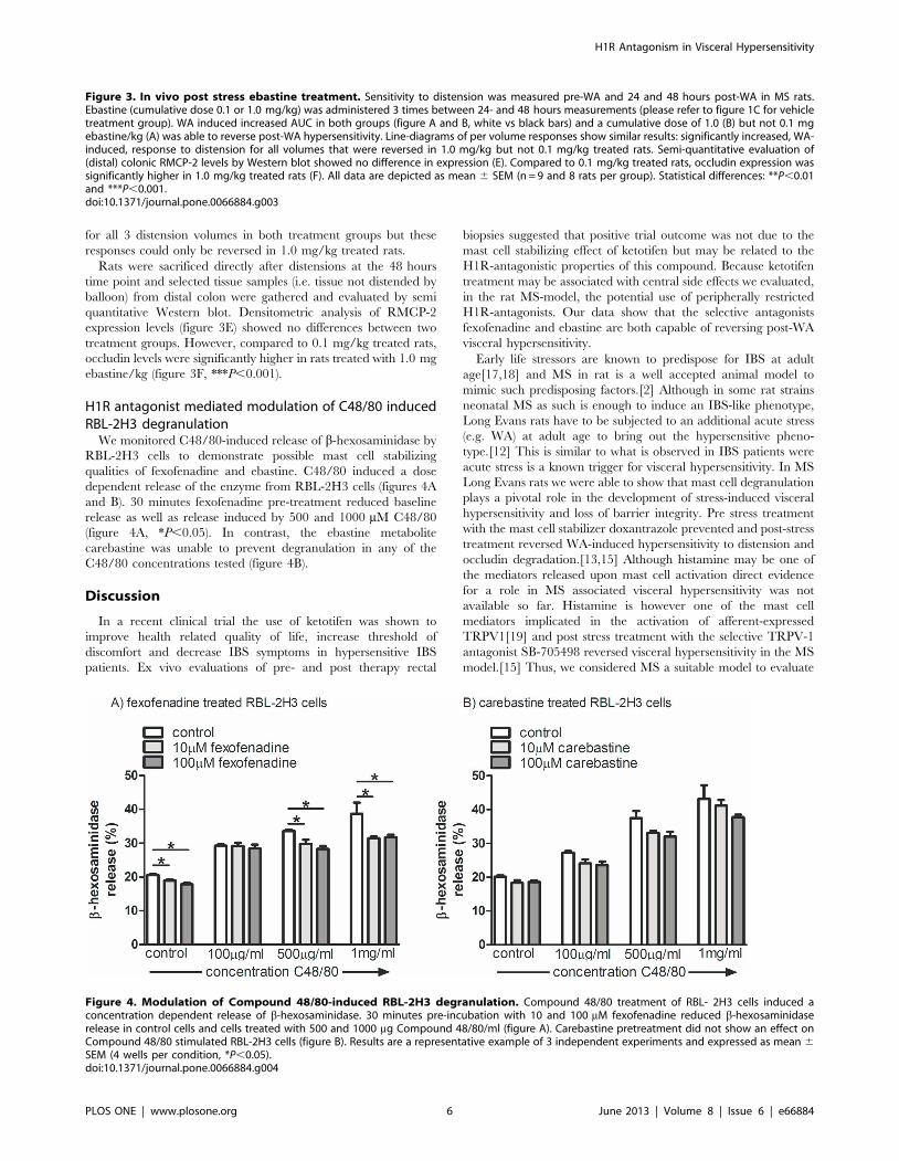
H1R antagonist mediated modulation of C48/80 inducedRBL-2H3 degranulation
We monitored C48/80-induced release of b-hexosaminidase by
RBL-2H3 cells to demonstrate possible mast cell stabilizing
qualities of fexofenadine and ebastine. C48/80 induced a dose
dependent release of the enzyme from RBL-2H3 cells (figures 4A
and B). 30 minutes fexofenadine pre-treatment reduced baseline
release as well as release induced by 500 and 1000 mM C48/80
(figure 4A, *P,0.05). In contrast, the ebastine metabolite
carebastine was unable to prevent degranulation in any of the
C48/80 concentrations tested (figure 4B).
Discussion
In a recent clinical trial the use of ketotifen was shown to
improve health related quality of life, increase threshold of
discomfort and decrease IBS symptoms in hypersensitive IBS
patients. Ex vivo evaluations of pre- and post therapy rectal
biopsies suggested that positive trial outcome was not due to the
mast cell stabilizing effect of ketotifen but may be related to the
H1R-antagonistic properties of this compound. Because ketotifen
treatment may be associated with central side effects we evaluated,
in the rat MS-model, the potential use of peripherally restricted
H1R-antagonists. Our data show that the selective antagonists
fexofenadine and ebastine are both capable of reversing post-WA
visceral hypersensitivity.
Early life stressors are known to predispose for IBS at adult
age[17,18] and MS in rat is a well accepted animal model to
mimic such predisposing factors.[2] Although in some rat strains
neonatal MS as such is enough to induce an IBS-like phenotype,
Long Evans rats have to be subjected to an additional acute stress
(e.g. WA) at adult age to bring out the hypersensitive pheno-
type.[12] This is similar to what is observed in IBS patients were
acute stress is a known trigger for visceral hypersensitivity. In MS
Long Evans rats we were able to show that mast cell degranulation
plays a pivotal role in the development of stress-induced visceral
hypersensitivity and loss of barrier integrity. Pre stress treatment
with the mast cell stabilizer doxantrazole prevented and post-stress
treatment reversed WA-induced hypersensitivity to distension and
occludin degradation.[13,15] Although histamine may be one of
the mediators released upon mast cell activation direct evidence
for a role in MS associated visceral hypersensitivity was not
available so far. Histamine is however one of the mast cell
mediators implicated in the activation of afferent-expressed
TRPV1[19] and post stress treatment with the selective TRPV-1
antagonist SB-705498 reversed visceral hypersensitivity in the MS
model.[15] Thus, we considered MS a suitable model to evaluate
Figure 3. In vivo post stress ebastine treatment. Sensitivity to distension was measured pre-WA and 24 and 48 hours post-WA in MS rats.Ebastine (cumulative dose 0.1 or 1.0 mg/kg) was administered 3 times between 24- and 48 hours measurements (please refer to figure 1C for vehicletreatment group). WA induced increased AUC in both groups (figure A and B, white vs black bars) and a cumulative dose of 1.0 (B) but not 0.1 mgebastine/kg (A) was able to reverse post-WA hypersensitivity. Line-diagrams of per volume responses show similar results: significantly increased, WA-induced, response to distension for all volumes that were reversed in 1.0 mg/kg but not 0.1 mg/kg treated rats. Semi-quantitative evaluation of(distal) colonic RMCP-2 levels by Western blot showed no difference in expression (E). Compared to 0.1 mg/kg treated rats, occludin expression wassignificantly higher in 1.0 mg/kg treated rats (F). All data are depicted as mean 6 SEM (n = 9 and 8 rats per group). Statistical differences: **P,0.01and ***P,0.001.doi:10.1371/journal.pone.0066884.g003
Figure 4. Modulation of Compound 48/80-induced RBL-2H3 degranulation. Compound 48/80 treatment of RBL- 2H3 cells induced aconcentration dependent release of b-hexosaminidase. 30 minutes pre-incubation with 10 and 100 mM fexofenadine reduced b-hexosaminidaserelease in control cells and cells treated with 500 and 1000 mg Compound 48/80/ml (figure A). Carebastine pretreatment did not show an effect onCompound 48/80 stimulated RBL-2H3 cells (figure B). Results are a representative example of 3 independent experiments and expressed as mean 6SEM (4 wells per condition, *P,0.05).doi:10.1371/journal.pone.0066884.g004
H1R Antagonism in Visceral Hypersensitivity
PLOS ONE | www.plosone.org 6 June 2013 | Volume 8 | Issue 6 | e66884

the possible use of H1R antagonists in the treatment of stress-
induced IBS-like phenotypical changes. Importantly, because a
possible future treatment protocol would aim to reverse complaints
in IBS patients we evaluated these compounds in a post stress
treatment protocol. Fexofenadine as well as ebastine were capable
of reversing post-WA visceral hypersensitivity.
The outcome of these experiments contradicts earlier investi-
gations involving the in vivo use of the calcium ionophore BrX-
537A (Bromolasolacid). Coelho et al. showed that intraperitoneal
administration of BrX-537A led to mast cell degranulation and
enhanced sensitivity to colorectal distension.[20] The observed
visceral hypersensitivity was prevented by 5-HT1a receptor
antagonist but not by histamine receptor-1, -2 and -3 antagonists.
In these experiments only one dosage of H1R antagonist (1 mg
chlorophenizamine/kg) was used and we can not rule out the
possibility that it was to low to be effective. Alternatively, histamine
release may occur in both experimental conditions but release only
has consequences relevant to visceral sensitivity when rats are
predisposed to react to this mediator (e.g. by increased H1R
expression). In relation to this, mucosal biopsies of IBS patients
were indeed shown to have increased H1R mRNA over
controls.[7] Therefore, we investigated the possibility of enhanced
post-WA H1R expression in MS rats but expression was not
increased over NH rats. Although the same approach to H1R
evaluation was successfully used by Sander et al.[7], we can not
exclude the possibility that existing differences between groups
were diluted out because we evaluated whole tissue specimens
instead of isolated sensory neurons. Another explanation for the
observed discrepancy with the earlier BrX-537A investigations
may be found in an often neglected aspect of in vivo visceral
sensitivity investigations. The calcium ionophore study evaluated
prevention of mast cell induced hypersensitivity whereas our H1R
antagonist data describe reversal of mast cell dependent hyper-
sensitivity. The difference is not ‘just semantics’. Recent data on
the use of a-helical-CRF (9–41) showed that pre-WA targeting of
CRF receptors prevented, but post-WA targeting could not reverse
stress induced visceral hypersensitivity.[13] Similarly, histamine
may play a role in prolonged mast cell dependent hypersensitivity
but not during an acute phase such as investigated in the BrX-
537A experiments.
Because ketotifen that was used in the IBS clinical trial has H1R
antagonistic as well as mast cell stabilizing qualities we also
evaluated fexofenadine and carebastine (the active metabolite of
ebastine) for possible mast cell stabilizing effects. Data obtained
with RBL-2H3 cells indicated that fexofenadine had some weak
mast cell stabilizing quality and carebastine, although results did
not reach significance, showed the same trend. However, the level
of stabilization was far from complete and can never explain the
successful in vivo reversal of post stress visceral hypersensitivity by
these compounds. Further, our results on RMCP-2 tissue
expression levels suggest that in vivo mast cell degranulation is
not altered by their use. In an earlier study we showed that in vivo
post stress mast cell degranulation is associated with a decrease in
tissue RMCP-2 levels.[13] In the current investigations high
(1.0 mg/kg) but not low dose (0.1 mg/kg) ebastine effectively
reversed visceral hypersensitivity whereas semi-quantitative eval-
uation of corresponding colonic tissues did not show differences in
RMCP-2 expression levels. The latter data suggest equal level of
mast cell degranulation in treatment groups and confirmed the
lack of in vitro stabilization by ebastine. Therefore, at least for
ebastine, we suggest that H1R antagonism rather than mast cell
stabilization was the in vivo mechanism of action.
In addition to RMCP-2, homogenized colonic tissue samples of
ebastine-treated rats were evaluated for occludin expression. In a
previous study we observed post-stress degradation of this tight
junction protein in stripped colonic mucosa of MS Long Evans
rats.[13] Here, ebastine-induced reversal of visceral hypersensitiv-
ity was associated with high- and failure to reverse with low- level
occludin expression. How this change is relevant for the observed
visceral hypersensitivity is not clear yet. However, barrier
dysfunction is thought to be an important pathophysiological
mechanism in IBS and in patient biopsies occludin degradation
was shown to correlate with abdominal pain intensity
scores.[21,22] The latter may be explained by enhanced mucosal
influx of luminal antigens and/or bacteria leading to subsequent
immune cell and afferent activation.[4] In relation to this, in vivo
occludin depletion by selective siRNA-induced knock down in
mouse intestine was indeed shown to enhance macromolecular
flux across intestinal epithelial cells.[23] A direct link between
barrier dysfunction and hypersensitivity to distension was shown in
rats where intra-colonic infusion of a tight junction blocker
prevented stress induced rectal hypersensitivity.[24] In the present
investigations we only obtained a limited dataset on occludin
expression. Future investigations should aim to establish whether
ebastine treatment, next to possible effects on afferent expressed
H1R[6], can also lead to antagonist mediated restoration of
barrier function.
At present peripherally restricted H1R-selective antihistamines
are the most broadly used medications in the treatment of allergic
diseases. In consequence, compounds like ebastine and fexofena-
dine have been extensively investigated regarding clinical phar-
macology and safety. The present study indicates that these
compounds are capable of reversing post stress visceral hypersen-
sitivity. Since this trait may be relevant to IBS we suggest that
peripheral H1Rs can be a safe new target for IBS therapy.
Author Contributions
Conceived and designed the experiments: GEB RvdW. Performed the
experiments: OIS SAvD ZY OW NB. Analyzed the data: OIS SAvD ZY
OW NB. Wrote the paper: OIS RvdW. Critically revised the article for
important intellectual content: JS WJdJ GEB.
References
1. Longstreth GF, Thompson WG, Chey WD, Houghton LA, Mearin F, et al.
(2006) Functional bowel disorders. Gastroenterology 130: 1480–1491.
2. Barreau F, Ferrier L, Fioramonti J, Bueno L (2007) New insights in the etiology
and pathophysiology of irritable bowel syndrome: contribution of neonatal stress
models. Pediatr Res 62: 240–245.
3. Gareau MG, Silva MA, Perdue MH (2008) Pathophysiological mechanisms of
stress-induced intestinal damage. Curr Mol Med 8: 274–281.
4. Van Den Wijngaard RM, Klooker TK, De Jonge WJ, Boeckxstaens GE (2010)
Peripheral relays in stress-induced activation of visceral afferents in the gut.Auton Neurosci 153: 99–105.
5. Klooker TK, Braak B, Koopman KE, Welting O, Wouters MM, et al. (2010)The mast cell stabiliser ketotifen decreases visceral hypersensitivity and improves
intestinal symptoms in patients with irritable bowel syndrome. Gut 59: 1213–
1221.
6. Barbara G, Wang B, Stanghellini V, de GR, Cremon C, et al. (2007) Mast cell-dependent excitation of visceral-nociceptive sensory neurons in irritable bowel
syndrome. Gastroenterology 132: 26–37.
7. Sander LE, Lorentz A, Sellge G, Coeffier M, Neipp M, et al. (2006) Selectiveexpression of histamine receptors H1R, H2R, and H4R, but not H3R, in the
human intestinal tract. Gut 55: 498–504. gut.2004.061762 [pii];10.1136/gut.2004.061762 [doi].
8. Simons FE, Simons KJ (2011) Histamine and H1-antihistamines: celebrating a
century of progress. J Allergy Clin Immunol 128: 1139-1150. S0091-6749(11)01408-4 [pii];10.1016/j.jaci.2011.09.005 [doi].
9. Tashiro M, Mochizuki H, Sakurada Y, Ishii K, Oda K, et al. (2006) Brainhistamine H receptor occupancy of orally administered antihistamines measured
by positron emission tomography with (11)C-doxepin in a placebo-controlled
crossover study design in healthy subjects: a comparison of olopatadine and
H1R Antagonism in Visceral Hypersensitivity
PLOS ONE | www.plosone.org 7 June 2013 | Volume 8 | Issue 6 | e66884
ketotifen. Br J Clin Pharmacol 61: 16-26. BCP2514 [pii];10.1111/j.1365-
2125.2005.02514.x [doi].
10. Simpson K, Jarvis B (2000) Fexofenadine: a review of its use in the management
of seasonal allergic rhinitis and chronic idiopathic urticaria. Drugs 59: 301–321.
11. Van Cauwenberge P, De Belder T, Sys L (2004) A review of the second-
generation antihistamine ebastine for the treatment of allergic disorders. Expert
Opin Pharmacother 5: 1807-1813. EOP050814 [pi i] ;10.1517/
14656566.5.8.1807 [doi].
12. Welting O, Van Den Wijngaard RM, De Jonge WJ, Holman R, Boeckxstaens
GE (2005) Assessment of visceral sensitivity using radio telemetry in a rat model
of maternal separation. Neurogastroenterol Motil 17: 838–845.
13. Van Den Wijngaard RM, Stanisor OI, van Diest SA, Welting O, Wouters MM,
et al. (2012) Peripheral alpha-helical CRF (9-41) does not reverse stress-induced
mast cell dependent visceral hypersensitivity in maternally separated rats.
Neurogastroenterol Motil 24: 274-82, e111. 10.1111/j.1365-2982.2011.01840.x
[doi].
14. Azpiroz F, Bouin M, Camilleri M, Mayer EA, Poitras P, et al. (2007)
Mechanisms of hypersensitivity in IBS and functional disorders. Neurogas-
troenterol Motil 19: 62–88.
15. Van Den Wijngaard RM, Klooker TK, Welting O, Stanisor OI, Wouters MM,
et al. (2009) Essential role for TRPV1 in stress-induced (mast cell-dependent)
colonic hypersensitivity in maternally separated rats. Neurogastroenterol Motil
21: 1107–1e94.
16. Marone G, Gentile M, Petraroli A, De Rosa N, Triggiani M (2001) Histamine-
induced activation of human lung macrophages. Int Arch Allergy Immunol 124:
249–252.
17. Chitkara DK, van Tilburg MA, Blois-Martin N, Whitehead WE (2008) Early life
risk factors that contribute to irritable bowel syndrome in adults: a systematicreview. Am J Gastroenterol 103: 765–774.
18. Klooker TK, Braak B, Painter RC, de Rooij Sr., van Elburg RM, et al. (2009)
Exposure to severe wartime conditions in early life is associated with anincreased risk of irritable bowel syndrome: a population-based cohort study. Am
J Gastroenterol 104: 2250–2256.19. Shim WS, Oh U (2008) Histamine-induced itch and its relationship with pain.
Mol Pain 4: 29. 1744-8069-4-29 [pii];10.1186/1744-8069-4-29 [doi].
20. Coelho AM, Fioramonti J, Bueno L (1998) Mast cell degranulation inducesdelayed rectal allodynia in rats: role of histamine and 5-HT. Dig Dis Sci 43:
727–737.21. Bertiaux-Vandaele N, Youmba SB, Belmonte L, Lecleire S, Antonietti M, et al.
(2011) The expression and the cellular distribution of the tight junction proteinsare altered in irritable bowel syndrome patients with differences according to the
disease subtype. Am J Gastroenterol 106: 2165-2173. ajg2011257 [pii];10.1038/
ajg.2011.257 [doi].22. Coeffier M, Gloro R, Boukhettala N, Aziz M, Lecleire S, et al. (2009) Increased
Proteasome-Mediated Degradation of Occludin in Irritable Bowel Syndrome.Am J Gastroenterol.
23. Al-Sadi R, Khatib K, Guo S, Ye D, Youssef M, et al. (2011) Occludin regulates
macromolecule flux across the intestinal epithelial tight junction barrier. Am JPhysiol Gastrointest Liver Physiol 300: G1054-G1064. ajpgi.00055.2011
[pii];10.1152/ajpgi.00055.2011 [doi].24. Ait-Belgnaoui A, Bradesi S, Fioramonti J, Theodorou V, Bueno L (2005) Acute
stress-induced hypersensitivity to colonic distension depends upon increase inparacellular permeability: role of myosin light chain kinase. Pain 113: 141–147.
H1R Antagonism in Visceral Hypersensitivity
PLOS ONE | www.plosone.org 8 June 2013 | Volume 8 | Issue 6 | e66884