Inhibition of histamine-mediated signaling confers significant protection against severe malaria in...
14
The Journal of Experimental Medicine ARTICLE JEM © The Rockefeller University Press $30.00 Vol. 205, No. 2, February 18, 2008 395-408 www.jem.org/cgi/doi/ 395 10.1084/jem.20071548 Our previous research demonstrated that the saliva of Anopheles stephensi induces a rapid de- granulation of cutaneous mast cells (MCs) in mice, followed by an influx of neutrophils at the bite site and lymph node hyperplasia (1). Moreover, this mosquito saliva–induced inflammatory re- sponse leads to a down-regulation of subsequent T cell–mediated immune responses, as assessed by a model of the delayed hypersensitivity reac- tion. This down-regulation of antigen-specific T cell responses is MC dependent (2). These results led us to hypothesize that inflammatory responses influence the course of infection with Plasmodium parasites. Some reports indicate that specific compo- nents of the innate immune system, including eosinophils (3), basophils (4), and MCs (5), could play important roles in the pathogenesis of ma- laria. Increased levels of histamine in plasma and tissue, derived from basophils and MCs, are as- sociated with the severity of disease in humans infected with P. falciparum and in several animal models of infection with Plasmodium (6–8). In addition, higher levels of IgE, which binds to basophils and MCs and can trigger histamine release, are associated with the severity of in- fection with P. falciparum (9). The effects of histamine are exerted through three classical G protein–coupled histamine re- ceptor subtypes termed H1R, H2R, and H3R, which are thoroughly described pharmacologi- cally (10), and a recently identified fourth mem- ber of the histamine receptor family, H4R (11). H1R mediates most of the proinflammatory CORRESPONDENCE Salaheddine Mécheri: [email protected] Abbreviations used: APC, allophycocyanin; BBB. blood– brain barrier; CM, cerebral malaria; HDC, histidine decar- boxylase; ICAM-1, intercellular adhesion molecule 1; MC, mast cell; MFI, mean fluorescence intensity; MGG, May- Grünwald Giemsa; mRNA, messenger RNA; Pb, Plasmodium berghei; TCTP, translationally controlled tumor protein; VCAM-1, vascular cell adhesion molecule 1. A. Porcherie and B.S. Schneider contributed equally to this work. Inhibition of histamine-mediated signaling confers significant protection against severe malaria in mouse models of disease Walid Beghdadi, 1 Adeline Porcherie, 1 Bradley S. Schneider, 1 David Dubayle, 3 Roger Peronet, 1 Michel Huerre, 2 Takeshi Watanabe, 4 Hiroshi Ohtsu, 5 Jacques Louis, 1 and Salaheddine Mécheri 1 1 Unité des Réponses Précoces aux Parasites et Immunopathologie and 2 Unité de Recherche et d’Expertise Histotechnologie et Pathologie, Institut Pasteur, Paris 75015, France 3 Centre National de la Recherche Scientifique, UMR 8119, Université Paris Descartes, 75270 Paris, Cedex 06, France 4 Research Center for Allergy and Immunology, RIKEN, Yokohama 230-0045, Japan 5 Tohoku University School of Engineering, Sendai 980-8579, Japan From the inoculation of Plasmodium sporozoites via Anopheles mosquito bites to the development of blood-stage parasites, a hallmark of the host response is an inflammatory reaction characterized by elevated histamine levels in the serum and tissues. Given the proinflammatory and immunosuppressive activities associated with histamine, we postu- lated that this vasoactive amine participates in malaria pathogenesis. Combined genetic and pharmacologic approaches demonstrated that histamine binding to H1R and H2R but not H3R and H4R increases the susceptibility of mice to infection with Plasmodium. To further understand the role of histamine in malaria pathogenesis, we used histidine de- carboxylase–deficient (HDC / ) mice, which are free of histamine. HDC / mice were highly resistant to severe malaria whether infected by mosquito bites or via injection of infected erythrocytes. HDC / mice displayed resistance to two lethal strains: Plasmodium berghei ( Pb) ANKA, which triggers cerebral malaria (CM), and Pb NK65, which causes death with- out neurological symptoms. The resistance of HDC / mice to CM was associated with preserved blood–brain barrier integrity, the absence of infected erythrocyte aggregation in the brain vessels, and a lack of sequestration of CD4 and CD8 T cells. We demonstrate that histamine-mediated signaling contributes to malaria pathogenesis. Understanding the basis for these biological effects of histamine during infection may lead to novel therapeutic strategies to alleviate the severity of malaria.
-
Upload
independent -
Category
Documents
-
view
5 -
download
0
Transcript of Inhibition of histamine-mediated signaling confers significant protection against severe malaria in...
The
Journ
al o
f Exp
erim
enta
l M
edic
ine
ARTICLE
JEM © The Rockefeller University Press $30.00
Vol. 205, No. 2, February 18, 2008 395-408 www.jem.org/cgi/doi/
395
10.1084/jem.20071548
Our previous research demonstrated that the saliva of Anopheles stephensi induces a rapid de-granulation of cutaneous mast cells (MCs) in mice, followed by an infl ux of neutrophils at the bite site and lymph node hyperplasia ( 1 ). Moreover, this mosquito saliva – induced infl ammatory re-sponse leads to a down-regulation of subsequent T cell – mediated immune responses, as assessed by a model of the delayed hypersensitivity reac-tion. This down-regulation of antigen-specifi c T cell responses is MC dependent ( 2 ). These results led us to hypothesize that infl ammatory responses infl uence the course of infection with Plasmodium parasites.
Some reports indicate that specifi c compo-nents of the innate immune system, including
eosinophils ( 3 ), basophils ( 4 ), and MCs ( 5 ), could play important roles in the pathogenesis of ma-laria. Increased levels of histamine in plasma and tissue, derived from basophils and MCs, are as-sociated with the severity of disease in humans infected with P. falciparum and in several animal models of infection with Plasmodium ( 6 – 8 ). In addition, higher levels of IgE, which binds to basophils and MCs and can trigger histamine release, are associated with the severity of in-fection with P. falciparum ( 9 ).
The eff ects of histamine are exerted through three classical G protein – coupled histamine re-ceptor subtypes termed H1R, H2R, and H3R, which are thoroughly described pharmacologi-cally ( 10 ), and a recently identifi ed fourth mem-ber of the histamine receptor family, H4R ( 11 ). H1R mediates most of the proinfl ammatory
CORRESPONDENCE
Salaheddine M é cheri:
Abbreviations used: APC,
allophycocyanin; BBB. blood –
brain barrier; CM, cerebral
malaria; HDC, histidine decar-
boxylase; ICAM-1, intercellular
adhesion molecule 1; MC, mast
cell; MFI, mean fl uorescence
intensity; MGG, May-
Gr ü nwald Giemsa; mRNA,
messenger RNA; Pb , Plasmodium
berghei ; TCTP, translationally
controlled tumor protein;
VCAM-1, vascular cell adhesion
molecule 1.
A. Porcherie and B.S. Schneider contributed equally to this work.
Inhibition of histamine-mediated signaling confers signifi cant protection against severe malaria in mouse models of disease
Walid Beghdadi , 1 Adeline Porcherie , 1 Bradley S. Schneider , 1 David Dubayle , 3 Roger Peronet , 1 Michel Huerre , 2 Takeshi Watanabe , 4 Hiroshi Ohtsu , 5 Jacques Louis , 1 and Salaheddine M é cheri 1
1 Unit é des R é ponses Pr é coces aux Parasites et Immunopathologie and 2 Unit é de Recherche et d ’ Expertise Histotechnologie et
Pathologie, Institut Pasteur, Paris 75015, France
3 Centre National de la Recherche Scientifi que, UMR 8119, Universit é Paris Descartes, 75270 Paris, Cedex 06, France
4 Research Center for Allergy and Immunology, RIKEN, Yokohama 230-0045, Japan
5 Tohoku University School of Engineering, Sendai 980-8579, Japan
From the inoculation of Plasmodium sporozoites via Anopheles mosquito bites to the
development of blood-stage parasites, a hallmark of the host response is an infl ammatory
reaction characterized by elevated histamine levels in the serum and tissues. Given the
proinfl ammatory and immunosuppressive activities associated with histamine, we postu-
lated that this vasoactive amine participates in malaria pathogenesis. Combined genetic
and pharmacologic approaches demonstrated that histamine binding to H1R and H2R
but not H3R and H4R increases the susceptibility of mice to infection with Plasmodium .
To further understand the role of histamine in malaria pathogenesis, we used histidine de-
carboxylase – defi cient (HDC � / � ) mice, which are free of histamine. HDC � / � mice were highly
resistant to severe malaria whether infected by mosquito bites or via injection of infected
erythrocytes. HDC � / � mice displayed resistance to two lethal strains: Plasmodium berghei
( Pb ) ANKA, which triggers cerebral malaria (CM), and Pb NK65, which causes death with-
out neurological symptoms. The resistance of HDC � / � mice to CM was associated with
preserved blood – brain barrier integrity, the absence of infected erythrocyte aggregation in
the brain vessels, and a lack of sequestration of CD4 and CD8 T cells. We demonstrate that
histamine-mediated signaling contributes to malaria pathogenesis. Understanding the basis
for these biological effects of histamine during infection may lead to novel therapeutic
strategies to alleviate the severity of malaria.

396 HISTAMINE-MEDIATED SIGNALING IS DETRIMENTAL DURING MALARIA INFECTION | Beghdadi et al.
In vascular endothelial cells, H1R stimulation leads to several cellular responses, including the release of nitric oxide ( 19 ), and enhancements in vascular permeability, particularly in post-capillary venules as a result of endothelial cell contraction ( 20, 21 ). Several of these eff ects of histamine might be exploited by Plasmodium to survive in its mammalian host. The increase in vascular permeability appears to be a component of malaria pathogenesis and could be advantageous for the parasites, as sporozoites or blood-stage parasites, because it facilitates their entry and exit from blood vessels. The vasodilatory eff ects of histamine might promote the spread of the parasite through the vasculature, and histamine can increase endothelial ex-pression of thrombomodulin, which is both an anticoagulant and a receptor for parasitized erythrocyte sequestration. Finally, the putative benefi t of histamine signaling to Plasmo-dium is strongly supported by the existence of a parasite-derived homologue of the mammalian histamine-releasing factor known as translationally controlled tumor protein (TCTP).
The aim of this work was to directly assess the rele-vance of histamine in malaria pathogenesis and its association with disease severity. Using mice genetically defi cient in H1R (H1R � / � ), H2R (H2R � / � ), and histidine decarboxylase (HDC � / � ), as well as targeting the four histamine receptors (H1R, H2R, H3R, and H4R) by antihistamine drugs, we dem-onstrate a deleterious eff ect of histamine-associated infl ammatory
eff ects of histamine ( 12 ). The antiinfl ammatory and immuno-suppressive eff ects of histamine, such as inhibition of poly-morphonuclear chemotaxis ( 13 ), IL-12 secretion by monocytes, and induction of IL-10 production ( 14 ), are largely depen-dent on stimulation of H2R, which is coupled to the adeny-lyl cyclase pathway. IL-10, on the other hand, is a suppressor cytokine and a major regulatory agent of infl ammatory re-sponses ( 15 ). H3R elicits an increase in intracellular calcium concentration and regulates cytokine release in alveolar mac-rophages and MCs ( 16 ). Histamine enhances intracellular Ca 2+ concentration and actin polymerization in immature DCs via activation of H1R and H3R, which also enhances chemo-taxis of these cells. In maturing DCs, histamine promotes an increase of cAMP and IL-10 production, whereas IL-12 se-cretion is inhibited ( 17 ). These histamine-mediated eff ects on cAMP and IL-10, as well as inhibition of IL-12 secretion, are mainly mediated by H2R and H3R. In addition, histamine inhibits the capacity of mature DCs to induce allogeneic Th1 responses, suggesting that histamine might infl uence the polari -zation of Th cell development ( 17 ) .
Histamine also modulates several biological functions of vascular endothelial cells. During an acute infl ammatory re-sponse, the histamine-induced increase of P-selectin (CD62P) expression on endothelial cells mediates the initial capture of infl ammatory cells, including neutrophils, from the blood ( 18 ).
Figure 1. Role of H1R and H2R in Pb infection. Histamineamia was measured by ELISA in naive and infected C57BL/6 mice at different time points
after inoculation of Pb NK65 (A) or Pb ANKA (B) through mosquito bites. Values represent the mean ± SD. ***, P = 0.003 (A); * and ***, P < 0.04 and 0.004 (B),
respectively, versus plasma histamine levels in uninfected mice. (C) The determination of histamine levels was performed in the brains from naive and
infected C57BL/6 mice 7 d after the inoculation of Pb ANKA through mosquito bites. Data represent the mean ± SD. **, P < 0.003. Wild-type C57BL/6 mice
(squares) or H1R � / � (diamonds) and H2R � / � (triangles) mice were infected either with infectious mosquito bites (seven to nine per mouse; D and E) or
with 10 6 infected erythrocytes per mouse (F and G). Survival and parasitemia were recorded. Signifi cant differences were observed between C57BL/6 and
H1R � / � and H2R � / � mice after infection by mosquito bites ( n = 7; P < 0.042 and 0.035, respectively; D), and after infection with erythrocytes ( n = 7;
P < 0.008 and 0.007, respectively; F). Differences in parasitemia between groups were not signifi cant. Each group consisted of seven mice. Data shown
are from three independent experiments.

JEM VOL. 205, February 18, 2008
ARTICLE
397
three groups ( Fig. 1 E ). To determine whether the relative resistance of H1R � / � and H2R � / � mice was confi ned to the preerythrocytic stage of infection, mice were also inoculated with infected erythrocytes, and mortality and parasitemia were monitored. Although mice from all groups infected in this manner died earlier than when infected through mosquito bites, a similar pattern of divergence between wild-type C57BL/6 mice and H1R � / � and H2R � / � mice was observed ( Fig. 1, F and G ). These data indicate that regardless of the develop-mental stage of the parasites used for infection, histamine in-teraction with either H1R or H2R hasten the fatal outcome, thus suggesting that the detrimental eff ects of histamine on the course of disease operate during the late stage of infection.
These fi ndings were confi rmed using a pharmacological intervention consisting of repeated administration of four his-tamine inhibitors — levocetirizine (H1R inhibitor), cimetidine (H2R inhibitor), imetit (H3R inhibitor), and JNJ 7777120 (H4R inhibitor) — before infection with 5 × 10 5 blood-stage parasites ( Pb NK65). Mice treated with either levocetirizine or cimetidine died signifi cantly later than similarly infected but untreated mice ( Fig. 2 A ). In contrast, imetit and JNJ 7777120 had no eff ect on mice survival as compared with untreated mice. Again, there was no diff erence in the parasitemia between antihistamine-treated or untreated mice when infected with blood-stage parasites ( Fig. 2 B ).
Susceptibility to infection with Pb is associated
with the HDC gene
Because histamine is known to act through four receptors (H1R, H2R, H3R, and H4R), obstructing signaling of only a single histamine receptor still permits manifestations of the histamine eff ects by the other receptors. Therefore, to deter-mine the eff ect of histamine on the course of disease induced by Plasmodium in a comprehensive manner, we used mice defi cient in HDC, the enzyme that converts histidine into histamine. As a result of this mutation introduced into the
responses in malaria infection. Therefore, suppressing the bio-logical eff ects of histamine may prove to be a new strategy to reduce the severity of diseases resulting from infections with Plasmodium parasites.
RESULTS
H1R- and H2R-mediated signaling play a role in the
pathogenesis of infection with Plasmodium berghei ( Pb )
In a preliminary attempt to appraise the impact of histamine on the course of infection with Plasmodium , we compared histamine levels in the plasma of mice infected or not with Pb . As shown in Fig. 1 , the levels of histamine in plasma increased signifi cantly over time in C57BL/6 mice inoculated with Pb NK65 and Pb ANKA by mosquito bites. It can be observed that although histamine levels increased relatively late at day 15 (P = 0.045) and became signifi cantly high only at the on-set of disease (day 20; P = 0.003; see Fig. 2 A ) after infection with Pb NK65, the rise in histaminemia occurred much ear-lier and became signifi cantly higher at day 5 after infection (P < 0.04) and constantly increased up to the time of death of most of the mice (day 8; P < 0.004; see Fig. 3 C ) after infec-tion with Pb ANKA. A similar increase in histamine levels has been reported in humans infected with P. falciparum ( 22 ). We also determined the levels of histamine in the brains of infected mice after infection with Pb ANKA, which causes cerebral malaria (CM), and found signifi cantly higher amounts of histamine as compared with control mice ( Fig. 1 C ). To assess whether histamine signaling plays a role in the patho-genesis of malaria, we fi rst determined the mortality and para-sitemia of mice genetically defi cient for H1R or H2R infected with parasites from a lethal strain of Pb NK65 via An. stephensi mosquito bites. Results illustrate that, compared with wild-type C57BL/6 mice, H1R � / � and H2R � / � mice displayed a delayed mortality; specifi cally, death was delayed by 3 d in histamine receptor – defi cient mice ( Fig. 1 D ). Interestingly, comparable parasitemia was observed between mice from the
Figure 2. Prolonged survival after treatment with antihistamine drugs. Wild-type C57BL/6 mice were left untreated (squares) or treated (triangles)
with the histamine inhibitors levocetirizine, cimetidine, imetit, and JNJ7777123 (for H1R, H2R, H3R, and H4R, respectively) before and during infection
with 10 6 infected erythrocytes per mouse. Survival (A) and parasitemia (B) were monitored over time. These data are from three independent experiments.
No signifi cant difference was observed for parasitemia between groups (7 – 15 mice per group). Data are shown as means ± SD.
398 HISTAMINE-MEDIATED SIGNALING IS DETRIMENTAL DURING MALARIA INFECTION | Beghdadi et al.
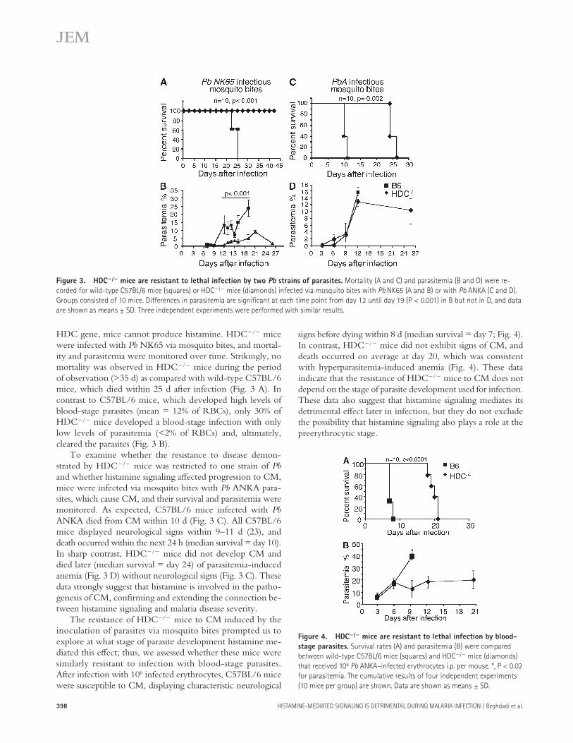
signs before dying within 8 d (median survival = day 7; Fig. 4 ). In contrast, HDC � / � mice did not exhibit signs of CM, and death occurred on average at day 20, which was consistent with hyperparasitemia-induced anemia ( Fig. 4 ). These data indicate that the resistance of HDC � / � mice to CM does not depend on the stage of parasite development used for infection. These data also suggest that histamine signaling mediates its detrimental eff ect later in infection, but they do not exclude the possibility that histamine signaling also plays a role at the preerythrocytic stage.
HDC gene, mice cannot produce histamine. HDC � / � mice were infected with Pb NK65 via mosquito bites, and mortal-ity and parasitemia were monitored over time. Strikingly, no mortality was observed in HDC � / � mice during the period of observation ( > 35 d) as compared with wild-type C57BL/6 mice, which died within 25 d after infection ( Fig. 3 A ). In contrast to C57BL/6 mice, which developed high levels of blood-stage parasites (mean = 12% of RBCs), only 30% of HDC � / � mice developed a blood-stage infection with only low levels of parasitemia ( < 2% of RBCs) and, ultimately, cleared the parasites ( Fig. 3 B ).
To examine whether the resistance to disease demon-strated by HDC � / � mice was restricted to one strain of Pb and whether histamine signaling aff ected progression to CM, mice were infected via mosquito bites with Pb ANKA para-sites, which cause CM, and their survival and parasitemia were monitored. As expected, C57BL/6 mice infected with Pb ANKA died from CM within 10 d ( Fig. 3 C ). All C57BL/6 mice displayed neurological signs within 9 – 11 d ( 23 ), and death occurred within the next 24 h (median survival = day 10). In sharp contrast, HDC � / � mice did not develop CM and died later (median survival = day 24) of parasitemia-induced anemia ( Fig. 3 D ) without neurological signs ( Fig. 3 C ). These data strongly suggest that histamine is involved in the patho-genesis of CM, confi rming and extending the connection be-tween histamine signaling and malaria disease severity.
The resistance of HDC � / � mice to CM induced by the inoculation of parasites via mosquito bites prompted us to explore at what stage of parasite development histamine me-diated this eff ect; thus, we assessed whether these mice were similarly resistant to infection with blood-stage parasites. After infection with 10 6 infected erythrocytes, C57BL/6 mice were susceptible to CM, displaying characteristic neurological
Figure 3. HDC � / � mice are resistant to lethal infection by two Pb strains of parasites. Mortality (A and C) and parasitemia (B and D) were re-
corded for wild-type C57BL/6 mice (squares) or HDC � / � mice (diamonds) infected via mosquito bites with Pb NK65 (A and B) or with Pb ANKA (C and D).
Groups consisted of 10 mice. Differences in parasitemia are signifi cant at each time point from day 12 until day 19 (P < 0.001) in B but not in D, and data
are shown as means ± SD. Three independent experiments were performed with similar results.
Figure 4. HDC � / � mice are resistant to lethal infection by blood-
stage parasites. Survival rates (A) and parasitemia (B) were compared
between wild-type C57BL/6 mice (squares) and HDC � / � mice (diamonds)
that received 10 6 Pb ANKA – infected erythrocytes i.p. per mouse. *, P < 0.02
for parasitemia. The cumulative results of four independent experiments
(10 mice per group) are shown. Data are shown as means ± SD.

JEM VOL. 205, February 18, 2008
ARTICLE
399
did not show signifi cant extravasation of the dye in the cere-bral cortex, similar to uninfected C57BL/6 and HDC � / � con-trol mice. Thus, infected C57BL/6 mice have a disruption of the BBB integrity concomitant with the appearance of neu-rological signs, whereas similarly infected HDC � / � mice do not exhibit detectable alteration of their BBB integrity.
The neurological signs that characterize CM are generally accompanied by the sequestration of infected erythrocytes in the cerebral vasculature. Histological analysis of brain sections obtained from naive or infected C57BL/6 mice revealed large erythrocyte aggregates observed in sections from olfactory bulbs and other anatomical parts of the brain ( Fig. 5 B , panels 1 and 2). In sharp contrast, such aggregates were not detected in sam-ples from infected HDC � / � mice ( Fig. 5 B , panels 5 and 6).
Role of the blood – brain barrier (BBB) during CM
A central feature of CM pathology after infection with Pb ANKA is the alteration of the BBB ( 24 ). Increased BBB per-meability may be mediated through the action of vasoactive amines, such as histamine ( 25 ), released from perivascular brain MCs ( 26, 27 ). To assess the status of the BBB during infection in our model system, C57BL/6 and HDC � / � mice were infected with 10 6 Pb ANKA – parasitized erythrocytes, and the BBB integrity was determined by an Evans blue dye exclusion assay. At day 7, when C57BL/6 mice develop neu-rological signs, the BBB permeability was evaluated by visu-alization of the dye extravasation in the brain, as refl ected by the appearance of a blue color in the cerebral cortex ( Fig. 5 A ). In contrast to infected C57BL6 mice, infected HDC � / � mice
Figure 5. Preserved BBB and a lack of sequestration of infected erythrocytes in the brain of HDC � / � mice. (A) Wild-type C57BL/6 mice and
HDC � / � mice were inoculated i.p. with 10 6 infected erythrocytes of Pb ANKA per mouse. At day 7 after infection, mice were injected with a solution of
Evans blue dye and were perfused with PBS 1 h later, and macroscopic observation was made of the brains. Brains from uninfected mice were used as
controls. (B) Sections from the brains of the same mice were stained with MGG. Blood-stage parasites associated with sequestered erythrocytes (panels
1 and 2) in the brain could be visualized by fl uorescence because of their expression of GFP (white, arrows; panel 2). No fl uorescence could be observed in
brain sections from uninfected and infected HDC � / � mice (panels 5 and 6). Data shown are representative of six mice per group. (C) The density of erythro-
cyte aggregates in brain sections of infected and control HDC � / � and C57BL/6 mice were expressed as the mean of aggregates per fi eld and counted in
50 consecutive microscopic fi elds. These data are from two different experiments. *, P < 0.05 between infected HDC � / � and C57BL/6 mice. Data are
shown as means ± SD. Bar, 100 � m.
400 HISTAMINE-MEDIATED SIGNALING IS DETRIMENTAL DURING MALARIA INFECTION | Beghdadi et al.
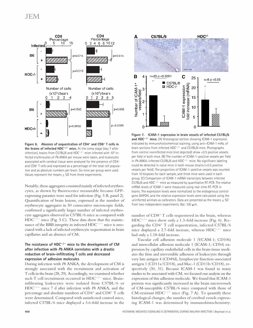
number of CD8 + T cells sequestered in the brain, whereas HDC � / � mice show only a 1.3-fold increase ( Fig. 6 ). Re-garding the CD4 + T cell sequestration, infected C57BL/6 mice displayed a 2.7-fold increase, whereas HDC � / � mice had only a 1.18-fold increase.
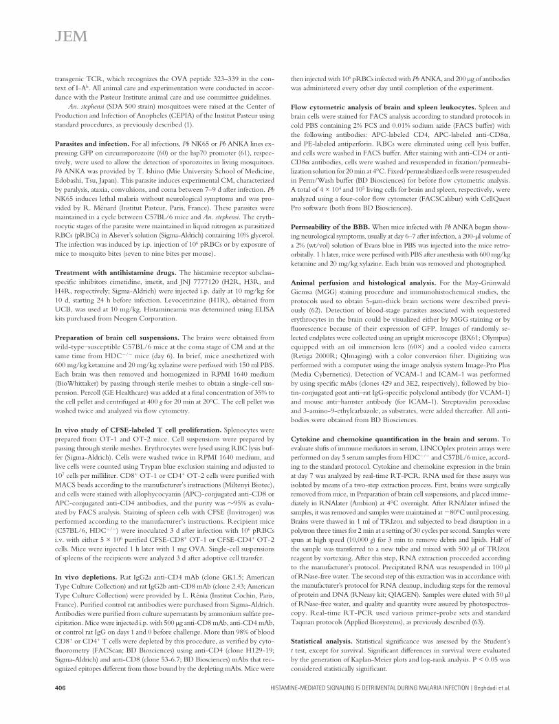
Vascular cell adhesion molecule 1 ( VCAM-1; CD106) and intercellular adhesion molecule 1 ( ICAM-1; CD54) ex-pression by capillary endothelial cells in the brain tissue medi-ates the fi rm and irreversible adhesion of leukocytes through very late antigen 4 (CD49d), lymphocyte function-associated antigen 1 (CD11a/CD18), and Mac-1 (CD11b/CD18), re-spectively ( 30, 31 ). Because ICAM-1 was found in many studies to be associated with CM, we focused our analysis on the expression of this adhesion molecule. We found that ICAM-1 protein was signifi cantly increased in the brain microvessels of CM-susceptible C57BL/6 mice compared with those of CM-resistant HDC � / � mice ( Fig. 7 A ). To quantify these histological changes, the number of cerebral vessels express-ing ICAM-1 was determined by immunohistochemistry.
Notably, these aggregates consisted mainly of infected erythro-cytes, as shown by fl uorescence measurable because GFP-expressing parasites were used for infection ( Fig. 5 B , panel 2). Quantifi cation of brain lesions, expressed as the number of erythrocyte aggregates in 50 consecutive microscopic fi elds, confi rmed a signifi cantly larger number of infected erythro-cyte aggregates observed in C57BL/6 mice as compared with HDC � / � mice ( Fig. 5 C ). These data show that the mainte-nance of the BBB integrity in infected HDC � / � mice is asso-ciated with a lack of infected erythrocyte sequestration in brain capillaries and an absence of CM.
The resistance of HDC � / � mice to the development of CM
after infection with Pb ANKA correlates with a drastic
reduction of brain-infi ltrating T cells and decreased
expression of adhesion molecules
During infection with Pb ANKA, the development of CM is strongly associated with the recruitment and activation of T cells in the brain ( 28, 29 ). Accordingly, we examined whether such T cell recruitment occurred in HDC � / � mice. Brain-infiltrating leukocytes were isolated from C57BL/6 or HDC � / � mice 7 d after infection with Pb ANKA, and the percentage and absolute numbers of CD4 + and CD8 + T cells were determined. Compared with uninfected control mice, infected C57BL/6 mice displayed a 3.6-fold increase in the
Figure 6. Absence of sequestration of CD4 + and CD8 + T cells in
the brains of infected HDC � / � mice. At the coma stage (day 7 after
infection), brains from C57BL/6 and HDC � / � mice infected with 10 6 in-
fected erythrocytes of Pb ANKA per mouse were taken, and leukocytes
associated with cerebral tissue were analyzed for the presence of CD4 +
and CD8 + T cells and expressed as a percentage of the total cell popula-
tion and as absolute numbers per brain. Six mice per group were used.
Values represent the means ± SD from three experiments.
Figure 7. ICAM-1 expression in brain vessels of infected C57BL/6
and HDC � / � mice. (A) Histological section showing ICAM-1 expression
indicated by immunohistochemical staining, using anti – ICAM-1 mAb, of
brain sections from infected HDC � / � and C57BL/6 mice. Photographs
from control noninfected mice (not depicted) show < 0.5 positive vessels
per fi eld in both mice. (B) The number of ICAM-1 – positive vessels per fi eld
in Pb ANKA – infected C57BL/6 and HDC � / � mice. No signifi cant labeling
could be detected in naive mice in both mouse strains ( < 0.5 positive
vessels per fi eld). The proportion of ICAM-1 – positive vessels was counted
from 10 biopsies for each sample, and three mice were used in each
group. (C) Comparison of ICAM-1 mRNA transcripts between infected
C57BL/6 and HDC � / � mice as measured by quantitative RT-PCR. The relative
mRNA levels of ICAM-1 were measured using real-time RT-PCR in
brains. The expression levels were normalized to the endogenous control
gene GAPDH, and the relative expression levels were calculated using the
uninfected animals as calibrators. Data are presented as the means ± SD
from two independent experiments. Bar, 100 � m.

JEM VOL. 205, February 18, 2008
ARTICLE
401
between C57BL/6 and HDC � / � mice. Of the proteins quan-tifi ed, the most striking diff erences were observed in infl am-mation-associated proteins. Serum IL-5 levels in infected C57BL/6 mice were 10-fold higher than infected HDC � / � mice (P < 0.05). Similarly, IFN- � and keratinocyte-derived chemokine were, respectively, two- and threefold higher in infected C57BL/6 mice (P < 0.05). Although not statistically signifi cant, IL-6 and monocyte chemotactic protein 1 were markedly reduced in infected HDC � / � mice as compared with infected C57BL/6 mice.
Second, the expression of mRNA specifi c for immune mediators was quantifi ed. Real-time RT-PCR was used to evaluate cytokines of particular interest in the brains of mice infected for 7 d. IL-10, IFN- � , and IL-4 expressions were 2.4, 3, and 3 times higher, respectively, in infected C57BL/6 mice than in HDC � / � mice ( Fig. 8 B ). In support of the serum protein data, IL-5 mRNA levels in the brain were consistently lower in HDC � / � mice ( Fig. 8 ), and treatment of C57BL/6 mice with levocetirizine reduced cerebral IL-5 levels to those seen in HDC � / � mice (not depicted). Collectively, these re-sults exemplify a divergence between C57BL/6 and HDC � / � mice in the expression of several cytokines. Further work is clearly required to determine whether or not these diff erences are at the basis of the resistance of HDC � / � mice to CM.
The antigen-induced response of specifi c CD8 + and CD4 +
T cells is similarly affected by infection with Pb ANKA
in C57BL/6 and HDC � / � mice
Diff erences in the magnitude of the CD4 and CD8 responses to infection between C57BL/6 and HDC � / � mice could result
Signifi cantly fewer ICAM-1 – positive vessels per fi eld ( Fig. 7 B ) were observed in infected HDC � / � mice, which are refrac-tory to CM development. Inasmuch as ICAM-1 expression is known to be modulated by histamine ( 32, 33 ), we also de-termined the ICAM-1 messenger RNA (mRNA) expression in the brains of infected mice and found that it was 2.2 times higher in the brains of C57BL/6 mice than in HDC � / � mice (P < 0.05; Fig. 7 C ). Unexpectedly, VCAM-1 mRNA ex-pression was slightly elevated in HDC � / � brain samples (see Fig. 8 B ). These results could suggest that the lower level of CD4 + and CD8 + T cells sequestered within the brain capil-laries of infected HDC � / � mice could be at the basis of the failure of these mice to develop CM. Furthermore, the near absence of T cell sequestration in the brain of CM-resistant HDC � / � mice correlates with the low ICAM-1 expression by brain endothelial cells.
Differential production of immunoregulatory and effector
molecules between C57BL/6 and HDC � / � mice infected
with Pb ANKA
The possibility exists that the resistance of HDC � / � mice to CM resulted from an impaired or improved capacity of these mice to produce some immune mediators in response to in-fection with Pb ANKA. Therefore, the levels of expression of various relevant molecules at the mRNA and protein levels were compared between HDC � / � and C57BL/6 mice in-fected with Pb ANKA.
First, serum samples at 5 d after infection with Pb ANKA were analyzed by the LINCOplex protein array ( Fig. 8 A ) to evaluate diff erences in the expression of some relevant proteins
Figure 8. Differential expression of immune signaling genes in C57BL/6 and HDC � / � mice infected with Pb ANKA. (A) Serum levels of specifi c
immune and infl ammatory proteins were quantifi ed at day 5 after infection by LINCOplex protein assay, and proteins with the highest variation between
groups are displayed. (B) Transcription of cytokines and chemokines in the brain ( n > 6 per group) 7 d after infection as evaluated by real-time RT-PCR.
Gene mRNA expression is normalized relative to uninfected controls for each mouse strain, and control mRNA levels did not differ signifi cantly between
groups. Data are shown as means ± SD. *, P < 0.05; #, P = 0.05.

402 HISTAMINE-MEDIATED SIGNALING IS DETRIMENTAL DURING MALARIA INFECTION | Beghdadi et al.
2.52 ± 0.01 – fold decrease in MFI, respectively). In contrast, in noninfected mice from both strains, CD8 OT-1 cells and CD4 OT-2 cells exhibited a much higher proliferative re-sponse (a 27.7 ± 0.06 – and 31.5 ± 0.9 – fold decrease in MFI for C57BL/6 and HDC � / � mice, respectively, for CD8 + T cells; and a 12.4 ± 0.09 – and 16.4 ± 0.07 – fold decrease in MFI for C57BL/6 and HDC � / � mice, respectively, for CD4 + T cells). These data indicate that the specifi c responses of CD4 + and CD8 + T cells are equally aff ected by infection with Plasmodium in mice from both strains and, thus, render unlikely the possibility that diff erences in T cell expansion be-tween mice from the two strains account for the observed dissimilarity in the susceptibility to develop CM.
The resistance of HDC � / � mice to CM is not affected
by depletion of CD4 + and CD8 + T cells
It is well established that CD4 + and CD8 + T cells contribute to CM pathogenesis in wild-type C57BL/6 and CBA mice, in view of the fact that mice depleted of CD4 + and CD8 + T cells fail to develop CM ( 23 – 25 ). Furthermore, our results have shown that the resistance of HDC � / � mice to CM corre-sponds to a lack of sequestration of T cells in brain capillaries ( Fig. 6 ). Nevertheless, we also decided to assess the possibility that peculiar T cell responses triggered within the distinct en-vironment of HDC � / � mice might account for the inability
in dissimilar manifestations of CM between mice from these two strains. Therefore, in an attempt to assess whether infec-tion with Plasmodium interferes diff erently with the develop-ment of T cell responses in C57BL/6 and HDC � / � mice, the proliferative capacity of OVA-specifi c CD4 and CD8 cells in response to specifi c stimulation was examined in the synge-neic environment of either infected C57BL/6 or HDC � / � mice. 3 d after infection with Pb ANKA, C57BL/6 and HDC � / � mice were injected with either CD8 OT-1 or CD4 OT-2 cells labeled with CFSE and treated or not with OVA. The proliferation of these OVA-specifi c T cells, as refl ected by a decrease in the mean fl uorescence intensity (MFI) of CFSE-labeled cells, was determined by FACS analysis 3 d later and expressed as the ratio between the MFI of CFSE-labeled cells recovered from mice receiving OVA to the MFI of similarly labeled cells recovered from mice not injected with OVA. Fig. 9 A shows a FACS quadrant analysis of rep-resentative samples in which the numbers of CD4 + or CD8 + T cells were plotted against CFSE levels. This was quantita-tively analyzed in Fig. 9 B , where no signifi cant diff erence in the proliferative response of CD8 OT-1 cells could be observed between infected C57BL/6 and HDC � / � mice (a 3 ± 0.09 – and 2.9 ± 0.1 – fold decrease in MFI, respectively). Similarly, there was no diff erence in the proliferative response of CD4 OT-2 cells between mice from both strains (a 3.1 ± 0.2 – and
Figure 9. Similar expansion of OT-1 and OT-2 cells in infected C57BL/6 and HDC � / � mice. Wild-type C57BL/6 and HDC � / � mice were infected
i.p. with 10 6 infected erythrocytes of Pb ANKA per mouse, and 3 d later they received 5 × 10 6 CFSE-labeled OT-1 or OT-2 cells, followed by injection or not
of 1 mg OVA 1 h later. At day 6 after infection, spleen cells were harvested and stained either with APC – anti-CD8 or APC – anti-CD4 mAb, and quadrant
FACS analysis was performed (A). Numbers in the top right quadrants indicate the fraction of CFSE/CD8 or CFSE/CD4 double-positive cells and the MFI,
respectively. The proliferative response of OT-1 and OT-2 cells was expressed as a ratio between the MFI of CFSE-labeled cells from mice that received OVA
to the MFI of those that did not (B). These data were obtained using three mice per group, and the experiment was replicated twice. Values are expressed
as the means ± SD.

JEM VOL. 205, February 18, 2008
ARTICLE
403
H1R and H2R had a signifi cant but limited eff ect with re-gard to the increase in the mean survival time, possibly be-cause the other factors or histamine receptors, H3R and H4R, also play a role. This is unlikely, however, because imetit and JNJ7777120, H3 and H4 antihistamines known to target H3R and H4R, neither infl uenced parasitemia nor survival. Thioperamide, a nonselective H3/H4R inhibitor, was also tested and showed no protective eff ect (unpublished data). To unequivocally determine the eff ect of histamine on the course of infection with Plasmodium , histamine-free mice were used. HDC � / � mice infected with Pb NK65 through mosquito bites were highly resistant to malaria disease (90 – 100% survival up to 35 d after infection), with only 30% of the mice developing blood-stage parasites, which were ulti-mately cleared. These data strongly suggest that the absence of histamine either resulted in the generation of effi cient ef-fector responses against the preerythrocytic and blood stages of the parasite or limited the immunopathology associated with the disease. To expand these results to CM, HDC � / � mice were infected with parasites from Pb ANKA. Although HDC � / � mice eventually succumbed from the infection with this strain of Pb , they died at much later times than similarly infected control C57BL/6 mice and did not exhibit any signs of CM. These data implicate histamine in the progression to CM, demonstrate that the enhanced resistance of histamine-free mice is not restricted to parasites from a particular strain, and suggest that histamine signaling could lead to enhanced
of these mice to develop CM. Such T cells could, for exam-ple, interfere with the triggering of eff ector T cell responses mediating CM pathologies in wild-type C57BL/6 mice. Thus, to establish whether distinct parasite-induced CD4 + and CD8 + T cells interfere with the development of CM in infected HDC � / � mice, we studied the course of disease in mice de-pleted of either of these subpopulations of T cells. As expected, depletion of both CD4 + and CD8 + T cells ( Fig. 10 A ) com-pletely abrogated the development of CM and improved the survival rate in C57BL/6 mice, which then died from anemia (median survival = 7, 21, and 37 d for mice that were untreated, treated with anti-CD8, or treated with anti-CD4, respectively). Depletion of either CD8 + or CD4 + T cells did not allow CM to manifest in HDC � / � mice ( Fig. 10 B ). However, CD4 T cell depletion resulted in signifi cantly enhanced survival of infected HDC � / � mice (median survival = 20 and 34 d for untreated and anti-CD4 – treated mice, respectively; Fig. 10 B ). These data do not clearly support a role for CD8 and CD4 T cells in the resistance of HDC � / � mice to CM, but they draw attention to a possible involvement of CD4 + T cells in malaria pathologies other than CM in HDC � / � and C57BL/6 mice.
DISCUSSION
Histamine is recognized as a key factor in the pathogenesis of allergic diseases. Levocetirizine, a selective H1 antihistamine, controls seasonal allergic rhinitis symptoms in children ( 34 ). Use of sedating H1R blockers was also associated with a de-creased risk of developing multiple sclerosis ( 35 ) and improved the quality of life of patients with chronic idiopathic urticaria ( 36 ), suggesting a possible benefi cial eff ect of antihistamines on the onset of some autoimmune diseases. Increased histamine in plasma and tissues is associated with disease severity in human infections with P. falciparum and in animal models of malaria ( 6 – 8, 22 ).
Apart from the correlation with immunopathogenesis in other diseases, little is known of the role of histamine in malaria. In this study, we analyzed the infl uence of histamine on the course of disease in mice infected with Pb from two diff erent strains that induce distinct pathologies. This work evaluated the hypothesis that the histamine signaling during infection with Plasmodium results in an infl ammatory process that exacerbates disease by inducing immunopathology or facilitating the dis-semination, adherence, and sequestration of parasites in tissues. Based on divergent approaches using H1R � / � and H2R � / � mice, antihistamine drugs, and histamine-defi cient mice, we demonstrate that histamine signaling is associated with the severity of disease.
The mechanisms by which histamine contributes to dis-ease severity and at which stage of the parasite life cycle they operate is unclear. Mice genetically defi cient in H1R and H2R, as well as mice treated with H1 and H2 antihistamines, have delayed mortality as compared with similarly infected wild-type mice untreated with the antihistamines, suggesting that the production and binding of histamine to these two re-ceptors are deleterious to the host. However, antagonizing the eff ects of histamine by interference only at the levels of
Figure 10. In vivo depletion of CD4 or CD8 T cells did not alter
resistance of HDC � / � mice to lethal infection by Pb ANKA. Wild-type
C57BL/6 (A and C) and HDC � / � (B and D) mice were left untreated
(squares) or treated with neutralizing anti-CD4 (triangles) or anti-CD8
mAb (diamonds) before and during infection with 10 6 Pb ANKA – infected
erythrocytes per mouse. Survival rates (A and B) and parasitemia (C and D)
were recorded over time. Six mice per group were used, and data
are shown as means ± SD. This experiment was replicated twice.
Significant differences in A were observed between untreated and
anti-CD8 – treated (*, P < 0.001) and anti-CD4 – treated (**, P < 0.0001)
mice. A signifi cant difference in B was observed only with anti-CD4 – treated
mice (*, P = 0.034).

404 HISTAMINE-MEDIATED SIGNALING IS DETRIMENTAL DURING MALARIA INFECTION | Beghdadi et al.
stop the adverse eff ects of hyperparasitemia. In addition to its distinctive effi ciency in producing CM in mice, the possi-bility exists that Pb ANKA also elicits infl ammatory media-tors, other than histamine, that promote the progression of infection even in HDC � / � mice. Indeed, analysis of several infl ammatory proteins and other immune response – associ-ated molecules, including IFN- � , IL-6, IL-10, keratinocyte-derived chemokine (KC), monocyte chemotactic protein 1 (MCP-1), macrophage infl ammatory protein 1 � (MIP-1 � ), cytotoxic T lymphocyte – associated protein 4, and inducible co-stimulator ( Fig. 8 ; and not depicted), indicates that an infl ammatory response, although attenuated, was induced in HDC � / � mice. Several studies indicate that the CM patholo-gies seen in mice after infection with Pb ANKA result from the induced proinfl ammatory immune response. In agreement, IFN- � , which was shown to be implicated in the pathogene-sis of experimental CM ( 38 ), reached signifi cantly higher lev-els in the brain tissue and plasma of CM-sensitive C57BL/6 mice than in HDC � / � mice ( Fig. 8, A and B ). Similarly, our results showing higher levels of IL-6 in the plasma and in-creased expression of IL-6 transcripts in the brain of CM-sensitive C57BL/6 than in HDC � / � mice are also in agreement with studies showing higher production of IL-6 in response to TNF- � by endothelial cells of brain capillaries of mice ge-netically sensitive to CM development ( 39 ). Our results, which diverge from murine CM experiments that suggest a protec-tive eff ect for IL-10 in CM ( 40 ) but are in accordance with observations in humans that showed an increase in IL-10 with CM as compared with mild malaria patients ( 41 ), show a lower cerebral expression of IL-10 by HDC � / � mice. IL-4 expression corresponds with CM in our model, in agreement with previous studies that demonstrate that defi ciency in IL-4 or IL-4R � leads to higher resistance to Pb ANKA ( 42 ).
Analogous to the present data that reveal the adverse ef-fects of histamine for the host during infection with Plasmo-dium , a human study demonstrated that the concentration of histamine in plasma was increased by almost fi vefold in chil-dren suff ering from malaria ( 22 ). Additionally, in this study, higher levels of histamine were observed in the brains of chil-dren with severe malaria as compared to lesser disease presen-tations. Among the histamine-associated biological activities relevant to features predictive of severe CM in humans are increased intracerebral blood fl ow ( 43 ), increased vascular and BBB permeability ( 44 ), and edema ( 45 ). Strikingly, the resistance of histamine-defi cient mice to CM was clearly identifi ed by a preserved BBB integrity, in contrast to the greatly increased permeability of the brain vasculature with large clusters of infected erythrocytes observed in infected C57BL/6 mice.
In the absence of histamine signaling, profound altera-tions of the immune response may also occur. A characteris-tic feature of CM is the sequestration of CD8 + and CD4 + T cells in the brain capillaries ( 46 – 48 ). This was confi rmed by results in this study showing a 3.7- and 2.7-fold increase of CD8 + and CD4 + T cells, respectively, in the brain of C57BL/6 mice infected with Pb ANKA. Such sequestration in the brain was not observed in similarly infected HDC � / � mice ( Fig. 6 ).
immunopathological responses. Importantly, results showing comparable parasitemia between groups infected with para-sites from Pb ANKA do not support the possibility that the inability of histamine-free mice to develop CM resulted from a reduced parasite burden.
In an attempt to better determine at which stage of parasite development histamine mediates its adverse eff ects, HDC � / � mice were also infected with Pb ANKA – parasitized RBCs, thus bypassing the preerythrocytic phase. Identical to what was observed after infectious mosquito bites, histamine-free mice did not develop CM and survived signifi cantly longer than similarly infected control C57BL/6 mice. These data indicate that although histamine is produced during all stages of infection with Plasmodium parasites (i.e., by histamine-pro-ducing MCs in the skin and in the liver during the preerythro-cytic stage of parasites development, and by histamine-producing basophils in the blood where infected RBCs circulate), the pathogenic eff ects of histamine are likely prevailing in the latter stages of infection. However, the possibility that hista-mine also plays a role in the skin and/or the liver during the initial phases of the infection cannot be excluded. Indeed, during the initial phases of the infection, specifi cally the pre-erythrocytic stages of parasite development, histamine release may be triggered in the dermis when sporozoites are inocu-lated via mosquito bites. In this context, Anopheles saliva in-duces MC degranulation ( 1 ). During later phases of infection, particularly during the blood stage of parasite development, histamine production can be elicited from circulating baso-phils either via the cross-linking of parasite-specifi c IgE anti-bodies or by TCTP, a parasite-derived homologue of the histamine-releasing factor. TCTP has been found in the plasma of patients infected with P. falciparum and was shown to trigger histamine release from basophils and IL-8 secretion from eosinophils ( 37 ). The existence of a Plasmodium protein that stimulates histamine release lends support to the hypoth-esis that histamine signaling is advantageous to the pathogen and, thus, harmful to the host. These fi ndings could provide a rational basis for higher levels of histamine in blood and in tissues during malaria as a result of the activity of vector- or parasite-derived constituents. Our results ( Fig. 1 ) clearly revealed a progressive increase in histaminemia during the course of in-fection in C57BL/6 mice. Amplifying the host infl ammatory response via histamine signaling may be a strategy developed by the parasites to create conditions advantageous for their own survival and persistence. Although the parasites from the two strains used in our study are lethal for mice, the disease courses that they elicit are quite diff erent. Our results in HDC � / � mice show that in the absence of histamine, the disease severity induced by both parasites was signifi cantly attenuated. The adverse eff ect of histamine is highlighted by the survival induced after infection with Pb NK65 and a delayed non-CM death from Pb ANKA resulting from the interruption of histamine signaling. The reason that infection with Pb ANKA remains lethal in histamine-free HDC � / � mice is unclear. It seems plausible that even though inhibiting histamine signaling may abrogate immunopathology, it cannot

JEM VOL. 205, February 18, 2008
ARTICLE
405
of the Pb -specifi c T cell response is currently being compared between C57BL/6 and HDC � / � mice. Our results are con-sistent with previous fi ndings showing that during an acute blood-stage malaria infection T cell responses to Plasmodium parasites and other bystander antigens are inhibited ( 53 ). Thus, the mechanism by which HDC � / � mice resist CM does not appear to stem from a Pb ANKA – induced modifi cation of the peripheral T cell responses, though one could argue that the absence of HDC expression may alter histamine-mediated infl ammatory responses that are necessary for the manifesta-tion of some eff ector functions exerted by CD4 + and CD8 + T cell responses.
To demonstrate directly and unequivocally the implica-tion of histamine in the pathogenesis of CM, attempts were made to revert the CM-resistant phenotype (HDC � / � ) by frequent injections of histamine into Pb ANKA – infected mice. Such treatment failed to induce signifi cant alterations in mor-tality (unpublished data). This method of repleting mice may, however, not be suffi cient because of the low bioavailability and the lability of histamine ( 54 ). Nevertheless, our results with antihistamines and genetic knockouts strongly suggest that antihistamine could have a therapeutic value in the treat-ment of malaria infection, particularly by reducing the likeli-hood of adverse complications. This is supported by the fact that antihistaminic drugs such as chlorpheniramine potentiate the anti- Plasmodium eff ect of mefl oquine, quinine, or pyronar-idine ( 55 ). It should be pointed out that the antihistamine drugs do not very likely exert their eff ect directly on the par-asite. We performed an experiment in which infected eryth-rocytes treated with the H1R blocker levocetirizine were compared with untreated infected erythrocytes for their ca-pacity to infect mice. No signifi cant diff erence regarding sur-vival or parasitemia was observed between the two groups of mice, suggesting that H1 antihistamine drugs exert no direct eff ect on the parasite (unpublished data).
The present work represents the fi rst comprehensive study documenting the role of histamine in the development of severe pathologies during infection with Plasmodium . Al-though malaria vaccine development remains a central and ultimate goal, alternative chemotherapy-based approaches such as histamine receptor antagonists have the potential to be highly valuable. Such treatment could be part of an integrated control strategy and could also be a useful candidate in inter-mittent preventive treatment strategies that target specifi c high-risk groups, such as children and pregnant women. Fur-thermore, given the availability of antihistamines, such treat-ment could be implemented rapidly to alleviate the burden of malaria in endemic areas.
MATERIALS AND METHODS Animals. 6 – 8-wk-old female C57BL/6 mice were purchased from Charles
River Laboratories. HDC � / � ( 56 ) and H1R and H2R knockout (H1R � / �
and H2R � / � ) ( 57, 58 ) mice were provided by H. Ohtsu and T. Watanabe,
respectively. All knockout mice originated from the C57BL/6 background.
The mice, including OT-1 and OT-2 transgenic mice ( 59 ), were bred
in our animal facility. OT-1 mice are transgenic for a TCR that recognizes
the OVA peptide 257 – 264 in the context of H2K b . OT-2 mice express a
These data suggest that the histamine produced after infection with Pb ANKA triggers infl ammation that leads to the seques-tration of T cells in brain capillaries, a prominent component of CM pathogenesis. In this context, it has been reported that histamine is essential for the recruitment of antigen-specifi c CD4 + and CD8 + T cells into the site of antigen delivery and the subsequent generation of infl ammatory responses ( 12 ).
Several studies reported a crucial role of ICAM-1 in ma-laria pathogenesis, and notably, ICAM-1 – defi cient mice are protected from CM ( 49 ). In this model, ICAM-1 � / � mice survived > 15 d despite a similar level of parasitemia in wild-type and knockout mice, a phenotype that is comparable to HDC � / � mice. Results from our study confi rmed the corre-lation between ICAM-1 expression, both at the protein and transcript levels, and susceptibility to CM. The low levels of ICAM-1 expression in brain microvascular endothelial cells in histamine-defi cient HDC � / � mice ( Fig. 8 ) could represent a mechanism leading to the lack of T cell sequestration in the brain capillaries of these mice. It is interesting to note that ICAM-1 � / � and HDC � / � mice, which express similar phe-notypes, have in common a reduced synthesis of ICAM-1 molecules, suggesting a control mechanism exerted by hista-mine on ICAM-1 expression. Indeed, histamine has been shown to stimulate endothelial cells to express ICAM-1 and VCAM-1 ( 32 ), and cetirizine, a H1R antagonist, has recently been shown to have antiinfl ammatory properties through the inhibition of leukocyte recruitment and activation, and by the reduction of ICAM-1 expression on keratinocytes ( 50 ) and on conjunctival and nasal epithelial cells ( 33 ). Interest-ingly, a reduction in ICAM-1 and VCAM-1 expression was observed in the brain tissue of infected C57BL/6 mice treated with levocetirizine (unpublished data). In one study, VCAM-1 was identifi ed by microarray analysis as a candidate gene that discriminates between CM-resistant and -sensitive mice in a Pb model of CM ( 51 ). Our data do not support the assertion that elevated VCAM-1 corresponds to CM, as we observed slightly higher VCAM-1 mRNA levels in HDC � / � mice (P < 0.05) as compared with C57BL/6 mice, suggesting an uncertain or, perhaps, protective role for this adhesion mole-cule in our model. In accordance with our observation of a predominant role for ICAM-1 as compared with VCAM-1 in malaria pathogenesis, infusion of anti – ICAM-1 but not anti – VCAM-1 mAb prevents cytoadherence of infected erythro-cytes in a P. yoelii model of CM ( 52 ).
A possible dissimilarity in the magnitude and quality of the CD4 and CD8 responses elicited by infection with Pb ANKA in C56BL/6 and HDC � / � mice could account for the lack of CM in HDC � / � mice. Such diff erences in the magnitude of the T cell responses could be the result of a di-vergent ability to suppress antigen-specifi c T cell responses in C57BL/6 and HDC � / � mice.
Our results showing a similar eff ect of infection with Plas-modium on the specifi c proliferative response of OT-1 and OT-2 cells in C57BL/6 and HDC � / � mice suggest that the magnitude of T cell responses is similarly aff ected in C57BL/6 and HDC � / � mice. The eff ect of infection on the magnitude
406 HISTAMINE-MEDIATED SIGNALING IS DETRIMENTAL DURING MALARIA INFECTION | Beghdadi et al.
then injected with 10 6 pRBCs infected with Pb ANKA, and 200 μ g of antibodies
was administered every other day until completion of the experiment.
Flow cytometric analysis of brain and spleen leukocytes. Spleen and
brain cells were stained for FACS analysis according to standard protocols in
cold PBS containing 2% FCS and 0.01% sodium azide (FACS buff er) with
the following antibodies: APC-labeled CD4, APC-labeled anti-CD8 � ,
and PE-labeled antiperforin. RBCs were eliminated using cell lysis buff er,
and cells were washed in FACS buff er. After staining with anti-CD4 or anti-
CD8 � antibodies, cells were washed and resuspended in fi xation/permeabi-
lization solution for 20 min at 4 ° C. Fixed/permeabilized cells were resuspended
in Perm/Wash buff er (BD Biosciences) for before fl ow cytometric analysis.
A total of 4 × 10 4 and 10 5 living cells for brain and spleen, respectively, were
analyzed using a four-color fl ow cytometer (FACSCalibur) with CellQuest
Pro software (both from BD Biosciences).
Permeability of the BBB. When mice infected with Pb ANKA began show-
ing neurological symptoms, usually at day 6 – 7 after infection, a 200- μ l volume of
a 2% (wt/vol) solution of Evans blue in PBS was injected into the mice retro-
orbitally. 1 h later, mice were perfused with PBS after anesthesia with 600 mg/kg
ketamine and 20 mg/kg xylazine. Each brain was removed and photographed.
Animal perfusion and histological analysis. For the May-Gr ü nwald
Giemsa (MGG) staining procedure and immunohistochemical studies, the
protocols used to obtain 5- � m-thick brain sections were described previ-
ously ( 62 ). Detection of blood-stage parasites associated with sequestered
erythrocytes in the brain could be visualized either by MGG staining or by
fl uorescence because of their expression of GFP. Images of randomly se-
lected endplates were collected using an upright microscope (BX61; Olympus)
equipped with an oil immersion lens (60 × ) and a cooled video camera
(Retiga 2000R; QImaging) with a color conversion fi lter. Digitizing was
performed with a computer using the image analysis system Image-Pro Plus
(Media Cybernetics). Detection of VCAM-1 and ICAM-1 was performed
by using specifi c mAbs (clones 429 and 3E2, respectively), followed by bio-
tin-conjugated goat anti – rat IgG-specifi c polyclonal antibody (for VCAM-1)
and mouse anti – hamster antibody (for ICAM-1). Streptavidin peroxidase
and 3-amino-9-ethylcarbazole, as substrates, were added thereafter. All anti-
bodies were obtained from BD Biosciences.
Cytokine and chemokine quantifi cation in the brain and serum. To
evaluate shifts of immune mediators in serum, LINCOplex protein arrays were
performed on day 5 serum samples from HDC � / � and C57BL/6 mice, accord-
ing to the standard protocol. Cytokine and chemokine expression in the brain
at day 7 was analyzed by real-time RT-PCR. RNA used for these assays was
isolated by means of a two-step extraction process. First, brains were surgically
removed from mice, in Preparation of brain cell suspensions, and placed imme-
diately in RNAlater (Ambion) at 4 ° C overnight. After RNAlater infused the
samples, it was removed and samples were maintained at � 80 ° C until processing.
Brains were thawed in 1 ml of TRI zol and subjected to bead disruption in a
polytron three times for 2 min at a setting of 30 cycles per second. Samples were
spun at high speed (10,000 g ) for 3 min to remove debris and lipids. Half of
the sample was transferred to a new tube and mixed with 500 μ l of TRI zol
reagent by vortexing. After this step, RNA extraction proceeded according
to the manufacturer ’ s protocol. Precipitated RNA was resuspended in 100 μ l
of RNase-free water. The second step of this extraction was in accordance with
the manufacturer ’ s protocol for RNA cleanup, including steps for the removal
of protein and DNA (RNeasy kit; QIAGEN). Samples were eluted with 50 μ l
of RNase-free water, and quality and quantity were assured by photospectros-
copy. Real-time RT-PCR used various primer-probe sets and standard
Taqman protocols (Applied Biosystems), as previously described ( 63 ).
Statistical analysis. Statistical signifi cance was assessed by the Student ’ s
t test, except for survival. Signifi cant diff erences in survival were evaluated
by the generation of Kaplan-Meier plots and log-rank analysis. P < 0.05 was
considered statistically signifi cant.
transgenic TCR, which recognizes the OVA peptide 323 – 339 in the con-
text of I-A b . All animal care and experimentation were conducted in accor-
dance with the Pasteur Institute animal care and use committee guidelines.
An. stephensi (SDA 500 strain) mosquitoes were raised at the Center of
Production and Infection of Anopheles (CEPIA) of the Institut Pasteur using
standard procedures, as previously described ( 1 ).
Parasites and infection. For all infections, Pb NK65 or Pb ANKA lines ex-
pressing GFP on circumsporozoite ( 60 ) or the hsp70 promoter ( 61 ), respec-
tively, were used to allow the detection of sporozoites in living mosquitoes.
Pb ANKA was provided by T. Ishino (Mie University School of Medicine,
Edobashi, Tsu, Japan). This parasite induces experimental CM, characterized
by paralysis, ataxia, convulsions, and coma between 7 – 9 d after infection. Pb
NK65 induces lethal malaria without neurological symptoms and was pro-
vided by R. M é nard (Institut Pasteur, Paris, France). These parasites were
maintained in a cycle between C57BL/6 mice and An. stephensi . The eryth-
rocytic stages of the parasite were maintained in liquid nitrogen as parasitized
RBCs (pRBCs) in Alsever ’ s solution (Sigma-Aldrich) containing 10% glycerol.
The infection was induced by i.p. injection of 10 6 pRBCs or by exposure of
mice to mosquito bites (seven to nine bites per mouse).
Treatment with antihistamine drugs. The histamine receptor subclass-
specifi c inhibitors cimetidine, imetit, and JNJ 7777120 (H2R, H3R, and
H4R, respectively; Sigma-Aldrich) were injected i.p. daily at 10 mg/kg for
10 d, starting 24 h before infection. Levocetirizine (H1R), obtained from
UCB, was used at 10 mg/kg. Histamineamia was determined using ELISA
kits purchased from Neogen Corporation.
Preparation of brain cell suspensions. The brains were obtained from
wild-type – susceptible C57BL/6 mice at the coma stage of CM and at the
same time from HDC � / � mice (day 6). In brief, mice anesthetized with
600 mg/kg ketamine and 20 mg/kg xylazine were perfused with 150 ml PBS.
Each brain was then removed and homogenized in RPMI 1640 medium
(BioWhittaker) by passing through sterile meshes to obtain a single-cell sus-
pension. Percoll (GE Healthcare) was added at a fi nal concentration of 35% to
the cell pellet and centrifuged at 400 g for 20 min at 20 ° C. The cell pellet was
washed twice and analyzed via fl ow cytometry.
In vivo study of CFSE-labeled T cell proliferation. Splenocytes were
prepared from OT-1 and OT-2 mice. Cell suspensions were prepared by
passing through sterile meshes. Erythrocytes were lysed using RBC lysis buf-
fer (Sigma-Aldrich). Cells were washed twice in RPMI 1640 medium, and
live cells were counted using Trypan blue exclusion staining and adjusted to
10 7 cells per milliliter. CD8 + OT-1 or CD4 + OT-2 cells were purifi ed with
MACS beads according to the manufacturer ’ s instructions (Miltenyi Biotec),
and cells were stained with allophycocyanin (APC)-conjugated anti-CD8 or
APC-conjugated anti-CD4 antibodies, and the purity was � 95% as evalu-
ated by FACS analysis. Staining of spleen cells with CFSE (Invitrogen) was
performed according to the manufacturer ’ s instructions. Recipient mice
(C57BL/6, HDC � / � ) were inoculated 3 d after infection with 10 6 pRBCs
i.v. with either 5 × 10 6 purifi ed CFSE-CD8 + OT-1 or CFSE-CD4 + OT-2
cells. Mice were injected 1 h later with 1 mg OVA. Single-cell suspensions
of spleens of the recipients were analyzed 3 d after adoptive cell transfer.
In vivo depletions. Rat IgG2a anti-CD4 mAb (clone GK1.5; American
Type Culture Collection) and rat IgG2b anti-CD8 mAb (clone 2.43; American
Type Culture Collection) were provided by L. R é nia (Institut Cochin, Paris,
France). Purifi ed control rat antibodies were purchased from Sigma-Aldrich.
Antibodies were purifi ed from culture supernatants by ammonium sulfate pre-
cipitation. Mice were injected i.p. with 500 μ g anti-CD8 mAb, anti-CD4 mAb,
or control rat IgG on days 1 and 0 before challenge. More than 98% of blood
CD8 + or CD4 + T cells were depleted by this procedure, as verifi ed by cyto-
fl uorometry (FACScan; BD Biosciences) using anti-CD4 (clone H129-19;
Sigma-Aldrich) and anti-CD8 (clone 53-6.7; BD Biosciences) mAbs that rec-
ognized epitopes diff erent from those bound by the depleting mAbs. Mice were

JEM VOL. 205, February 18, 2008
ARTICLE
407
Expression and function of histamine receptors in human monocyte-derived dendritic cells. J. Allergy Clin. Immunol. 109 : 839 – 846 .
18 . Burns , A.R. , R.A. Bowden , Y. Abe , D.C. Walker , S.I. Simon , M.L. Entman , and C.W. Smith . 1999 . P-selectin mediates neutrophil adhe-sion to endothelial cell borders. J. Leukoc. Biol. 65 : 299 – 306 .
19 . Van de Voorde , J. , and I. Leusen . 1983 . Role of the endothelium in the vasodilator response of rat thoracic aorta to histamine. Eur. J. Pharmacol. 87 : 113 – 120 .
20 . Majno , G. , S.M. Shea , and M. Leventhal . 1969 . Endothelial contraction induced by histamine-type mediators: an electron microscopic study. J. Cell Biol. 42 : 647 – 672 .
21 . Svensjo , E. , and G.J. Grega . 1986 . Evidence for endothelial cell-mediated regulation of macromolecular permeability by postcapillary venules. Fed. Proc. 45 : 89 – 95 .
22 . Enwonwu , C.O. , B.M. Afolabi , L.O. Salako , E.O. Idigbe , and N. Bashirelah . 2000 . Increased plasma levels of histidine and histamine in falciparum malaria: relevance to severity of infection. J. Neural Transm. 107 : 1273 – 1287 .
23 . Belnoue , E. , M. Kayibanda , J.C. Deschemin , M. Viguier , M. Mack , W.A. Kuziel , and L. Renia . 2003 . CCR5 defi ciency decreases suscepti-bility to experimental cerebral malaria. Blood . 101 : 4253 – 4259 .
24 . Medana , I.M. , and G.D. Turner . 2006 . Human cerebral malaria and the blood-brain barrier. Int. J. Parasitol. 36 : 555 – 568 .
25 . Schilling , L. , and M. Wahl . 1994 . Opening of the blood-brain barrier during cortical superfusion with histamine. Brain Res. 653 : 289 – 296 .
26 . Kermode , A.G. , A.J. Thompson , P. Tofts , D.G. MacManus , B.E. Kendall , D.P. Kingsley , I.F. Moseley , P. Rudge , and W.I. McDonald . 1990 . Breakdown of the blood-brain barrier precedes symptoms and other MRI signs of new lesions in multiple sclerosis. Pathogenetic and clinical implications. Brain . 113 : 1477 – 1489 .
27 . Theoharides , T.C. 1990 . Mast cells: the immune gate to the brain. Life Sci. 46 : 607 – 617 .
28 . Bauer , P.R. , H.C. Van Der Heyde , G. Sun , R.D. Specian , and D.N. Granger . 2002 . Regulation of endothelial cell adhesion molecule ex-pression in an experimental model of cerebral malaria. Microcirculation . 9 : 463 – 470 .
29 . Finley , R.W. , L.J. Mackey , and P.H. Lambert . 1982 . Virulent P. berghei malaria: prolonged survival and decreased cerebral pathology in cell-dependent nude mice. J. Immunol. 129 : 2213 – 2218 .
30 . McEver , R.P. 1992 . Leukocyte – endothelial cell interactions. Curr. Opin. Cell Biol. 4 : 840 – 849 .
31 . Carlos , T.M. , and J.M. Harlan . 1994 . Leukocyte-endothelial adhesion molecule. Blood . 84 : 2068 – 2101 .
32 . Kimura , S. , K.Y. Wang , A. Tanimoto , Y. Murata , Y. Nakashima , and Y. Sasaguri . 2004 . Acute infl ammatory reactions caused by histamine via monocytes/macrophages chronically participate in the initiation and progression of atherosclerosis. Pathol. Int. 54 : 465 – 474 .
33 . Ciprandi , G. , M.A. Tosca , C. Cosentino , A.M. Riccio , G. Passalacqua , and G.W. Canonica . 2003 . Eff ects of fexofenadine and other antihis-tamines on components of the allergic response: adhesion molecules. J. Allergy Clin. Immunol. 112 : S78 – S82 .
34 . de Blic , J. , U. Wahn , E. Billard , R. Alt , and M.C. Pujazon . 2005 . Levocetirizine in children: evidenced effi cacy and safety in a 6-week random-ized seasonal allergic rhinitis trial. Pediatr. Allergy Immunol. 16 : 267 – 275 .
35 . Alonso , A. , S.S. Jick , and M.A. Hernan . 2006 . Allergy, histamine 1 recep-tor blockers, and the risk of multiple sclerosis. Neurology . 66 : 572 – 575 .
36 . Lachapelle , J.M. , J. Decroix , A. Henrijean , P.P. Roquet-Gravy , A. De Swerdt , H. Boonen , M. Lecuyer , E. Suys , G. Speelman , and N. Vastesaeger . 2006 . Desloratadine 5 mg once daily improves the quality of life of patients with chronic idiopathic urticaria. J. Eur. Acad. Dermatol. Venereol. 20 : 288 – 292 .
37 . MacDonald , S.M. , J. Bhisutthibhan , T.A. Shapiro , S.J. Rogerson , T.E. Taylor , M. Tembo , J.M. Langdon , and S.R. Meshnick . 2001 . Immune mimicry in malaria: Plasmodium falciparum secretes a functional histamine-releasing factor homolog in vitro and in vivo. Proc. Natl. Acad. Sci. USA . 98 : 10829 – 10832 .
38 . Amani , V. , A.M. Vig á rio , E. Belnoue , M. Marussig , L. Fonseca , D. Mazier , and L. R é nia . 2000 . Involvement of IFN-gamma receptor-medi-cated signaling in pathology and anti-malarial immunity induced by Plasmodium berghei infection. Eur. J. Immunol. 30 : 1646 – 1655 .
We thank Dr. T. Ishino and R. M é nard for providing GFP – Plasmodium berghei
parasite strains. We also thank all of the members of CEPIA, at the Institut Pasteur,
for providing us with Anopheles mosquitoes, as well as H. Kuhn for assisting us
with histological work. We are grateful to L. R é nia for his generous gift of anti-CD4
and anti-CD8 mAbs, to E. Schneider for histamine measurements in brain tissues,
and to UCB for providing levocetirizine. We thank R. M é nard and S. Pied for their
critical readings of the manuscript before publication.
This work was supported by the Institut Pasteur, Sanofi -aventis, and the
Ministry of Research Initiative for Fighting against Parasitic Diseases. W. Beghdadi
was supported by the Foundation M é rieux.
The authors have no confl icting fi nancial interests.
Submitted: 25 July 2007
Accepted: 4 January 2008
REFERENCES 1 . Demeure , C.E. , K. Brahimi , F. Hacini , F. Marchand , R. Peronet ,
M. Huerre , P. St-Mezard , J.F. Nicolas , P. Brey , G. Delespesse , and S. Mecheri . 2005 . Anopheles mosquito bites activate cutaneous mast cells leading to a local infl ammatory response and lymph node hyperplasia. J. Immunol. 174 : 3932 – 3940 .
2 . Depinay , N. , F. Hacini , W. Beghdadi , R. Peronet , and S. Mecheri . 2006 . Mast cell-dependent down-regulation of antigen-specifi c immune re-sponses by mosquito bites. J. Immunol. 176 : 4141 – 4146 .
3 . Kurtzhals , J.A. , C.M. Reimert , E. Tette , S.K. Dunyo , K.A. Koram , B.D. Akanmori , F.K. Nkrumah , and L. Hviid . 1998 . Increased eosino-phil activity in acute Plasmodium falciparum infection – association with cerebral malaria. Clin. Exp. Immunol. 112 : 303 – 307 .
4 . Nyakeriga , M.A. , M. Troye-Blomberg , S. Bereczky , H. Perlmann , P. Perlmann , and G. ElGhazali . 2003 . Immunoglobulin E (IgE) containing complexes induce IL-4 production in human basophils: eff ect on Th1-Th2 balance in malaria. Acta Trop. 86 : 55 – 62 .
5 . Furuta , T. , T. Kikuchi , Y. Iwakura , and N. Watanabe . 2006 . Protective roles of mast cells and mast cell-derived TNF in murine malaria. J. Immunol. 177 : 3294 – 3302 .
6 . Bhattacharya , U. , S. Roy , P.K. Kar , B. Sarangi , and S.C. Lahiri . 1988 . Histamine & kinin system in experimental malaria. Indian J. Med. Res. 88 : 558 – 563 .
7 . Maegraith , B. , and A. Fletcher . 1972 . The pathogenesis of mammalian malaria. Adv. Parasitol. 10 : 49 – 75 .
8 . Srichaikul , T. , N. Archararit , T. Siriasawakul , and T. Viriyapanich . 1976 . Histamine changes in Plasmodium falciparum malaria. Trans. R. Soc. Trop. Med. Hyg. 70 : 36 – 38 .
9 . Perlmann , P. , H. Perlmann , G. ElGhazali , and M.T. Blomberg . 1999 . IgE and tumor necrosis factor in malaria infection. Immunol. Lett. 65 : 29 – 33 .
10 . Hill , S.J. , C.R. Ganellin , H. Timmerman , J.C. Schwartz , N.P. Shankley , J.M. Young , W. Schunack , R. Levi , and H.L. Haas . 1997 . International Union of Pharmacology. XIII. Classifi cation of histamine receptors. Pharmacol. Rev. 49 : 253 – 278 .
11 . Hofstra , C.L. , P.J. Desai , R.L. Thurmond , and W.P. Fung-Leung . 2003 . Histamine H4 receptor mediates chemotaxis and calcium mobilization of mast cells. J. Pharmacol. Exp. Ther. 305 : 1212 – 1221 .
12 . Bryce , P.J. , C.B. Mathias , K.L. Harrison , T. Watanabe , R.S. Geha , and H.C. Oettgen . 2006 . The H1 histamine receptor regulates allergic lung responses. J. Clin. Invest. 116 : 1624 – 1632 .
13 . Bury , T.B. , J.L. Corhay , and M.F. Radermecker . 1992 . Histamine-induced inhibition of neutrophil chemotaxis and T-lymphocyte prolif-eration in man. Allergy . 47 : 624 – 629 .
14 . Elenkov , I.J. , E. Webster , D.A. Papanicolaou , T.A. Fleisher , G.P. Chrousos , and R.L. Wilder . 1998 . Histamine potently suppresses human IL-12 and stimulates IL-10 production via H2 receptors. J. Immunol. 161 : 2586 – 2593 .
15 . Shevach , E.M. 2002 . CD4+ CD25+ suppressor T cells: more questions than answers. Nat. Rev. Immunol. 2 : 389 – 400 .
16 . Bissonnette , E.Y. 1996 . Histamine inhibits tumor necrosis factor alpha release by mast cells through H2 and H3 receptors. Am. J. Respir. Cell Mol. Biol. 14 : 620 – 626 .
17 . Idzko , M. , A. la Sala , D. Ferrari , E. Panther , Y. Herouy , S. Dichmann , M. Mockenhaupt , F. Di Virgilio , G. Girolomoni , and J. Norgauer . 2002 .

408 HISTAMINE-MEDIATED SIGNALING IS DETRIMENTAL DURING MALARIA INFECTION | Beghdadi et al.
39 . Lou , J. , Y. Gasche , L. Zheng , B. Critico , C. Monso-Hinard , P. Juillard , P. Morel , W.A. Buurman , and G.E. Grau . 1998 . Diff erential reactiv-ity of brain microvascular endothelial cells to TNF refl ects the genetic susceptibility to cerebral malaria. Eur. J. Immunol. 28 : 3989 – 4000 .
40 . Kossodo , S. , C. Monso , P. Juillard , T. Velu , M. Goldmani , and G.E. Grau . 1997 . Interleukin-10 modulates susceptibility in experimental cere-bral malaria. Immunology . 91 : 536 – 540 .
41 . Baptista , J.L. , G. Vanham , M. Wery , and E. Van Marck . 1997 . Cytokine levels during mild and cerebral falciparum malaria in children living in a mesoendemic area. Trop. Med. Int. Health . 2 : 673 – 679 .
42 . Saeftel , M. , A. Krueger , S. Arriens , V. Heussler , P. Racz , B. Fleischer , F. Brombacher , and A. Hoerauf . 2004 . Mice defi cient in interleukin-4 (IL-4) or IL-4 receptor alpha have higher resistance to sporozoite infec-tion with Plasmodium berghei (ANKA) than do naive wild-type mice. Infect. Immun. 72 : 322 – 331 .
43 . Edvinsson , L. , and B.B. Fredholm . 1983 . Characterization of adenosine receptors in isolated cerebral arteries of cat. Br. J. Pharmacol. 80 : 631 – 637 .
44 . Wahl , M. , and L. Schilling . 1993 . Regulation of cerebral blood fl ow – a brief review. Acta Neurochir. Suppl. (Wien) . 59 : 3 – 10 .
45 . Newton , C.R. , N. Peshu , B. Kendall , F.J. Kirkham , A. Sowunmi , C. Waruiru , I. Mwangi , S.A. Murphy , and K. Marsh . 1994 . Brain swell-ing and ischaemia in Kenyans with cerebral malaria. Arch. Dis. Child. 70 : 281 – 287 .
46 . Belnoue , E. , M. Kayibanda , A.M. Vigario , J.C. Deschemin , N. van Rooijen , M. Viguier , G. Snounou , and L. Renia . 2002 . On the patho-genic role of brain-sequestered alphabeta CD8+ T cells in experimental cerebral malaria. J. Immunol. 169 : 6369 – 6375 .
47 . Grau , G.E. , P.F. Piguet , H.D. Engers , J.A. Louis , P. Vassalli , and P.H. Lambert . 1986 . L3T4+ T lymphocytes play a major role in the patho-genesis of murine cerebral malaria. J. Immunol. 137 : 2348 – 2354 .
48 . Yanez , D.M. , D.D. Manning , A.J. Cooley , W.P. Weidanz , and H.C. van der Heyde . 1996 . Participation of lymphocyte subpopulations in the pathogenesis of experimental murine cerebral malaria. J. Immunol. 157 : 1620 – 1624 .
49 . Favre , N. , C. Da Laperousaz , B. Ryff el , N.A. Weiss , B.A. Imhof , W. Rudin , R. Lucas , and P.F. Piguet . 1999 . Role of ICAM-1 (CD54) in the development of murine cerebral malaria. Microbes Infect. 1 : 961 – 968 .
50 . Shimizu , T. , J. Nishihira , H. Watanabe , R. Abe , T. Ishibashi , and H. Shimizu . 2004 . Cetirizine, an H1-receptor antagonist, suppresses the expression of macrophage migration inhibitory factor: its potential anti-infl ammatory action. Clin. Exp. Allergy . 34 : 103 – 109 .
51 . Delahaye , N.F. , N. Coltel , D. Puthier , L. Flori , R. Houlgatte , F.A. Iraqi , C. Nguyen , G.E. Grau , and P. Rihet . 2006 . Gene-expression
profi ling discriminates between cerebral malaria (CM)-susceptible mice and CM-resistant mice. J. Infect. Dis. 193 : 312 – 321 .
52 . Kaul , D.K. , X.D. Liu , R.L. Nagel , and H.L. Shear . 1998 . Microvascular hemodynamics and in vivo evidence for the role of intercellular adhesion molecule-1 in the sequestration of infected red blood cells in a mouse model of lethal malaria. Am. J. Trop. Med. Hyg. 58 : 240 – 247 .
53 . Wilson , N.S. , G.M. Behrens , R.J. Lundie , C.M. Smith , J. Waithman , L. Young , S.P. Forehan , A. Mount , R.J. Steptoe , K.D. Shortman , et al . 2006 . Systemic activation of dendritic cells by Toll-like receptor ligands or malaria infection impairs cross-presentation and antiviral immunity. Nat. Immunol. 7 : 165 – 172 .
54 . Kownatzki , E. 1984 . Clearance of histamine from the peritoneal cavity of rats. Agents Actions . 15 : 249 – 253 .
55 . Nakornchai , S. , and P. Konthiang . 2006 . Potentiation of antimalarial drug action by chlorpheniramine against multidrug-resistant Plasmodium falciparum in vitro. Parasitol. Int. 55 : 195 – 199 .
56 . Ohtsu , H. , S. Tanaka , T. Terui , Y. Hori , Y. Makabe-Kobayashi , G. Pejler , E. Tchougounova , L. Hellman , M. Gertsenstein , N. Hirasawa , et al . 2001 . Mice lacking histidine decarboxylase exhibit abnormal mast cells. FEBS Lett. 502 : 53 – 56 .
57 . Inoue , I. , K. Yanai , D. Kitamura , I. Taniuchi , T. Kobayashi , K. Niimura , T. Watanabe , and T. Watanabe . 1996 . Impaired locomotor activity and exploratory behavior in mice lacking histamine H1 receptors. Proc. Natl. Acad. Sci. USA . 93 : 13316 – 13320 .
58 . Kobayashi , T. , S. Tonai , Y. Ishihara , R. Koga , S. Okabe , and T. Watanabe . 2000 . Abnormal functional and morphological regulation of the gastric mucosa in histamine H2 receptor-defi cient mice. J. Clin. Invest. 105 : 1741 – 1749 .
59 . Hogquist , K.A. , S.C. Jameson , W.R. Heath , J.L. Howard , M.J. Bevan , and F.R. Carbone . 1994 . T cell receptor antagonist peptides induce positive selection. Cell . 76 : 17 – 27 .
60 . Natarajan , R. , V. Thathy , M.M. Mota , J.C. Hafalla , R. Menard , and K.D. Vernick . 2001 . Fluorescent Plasmodium berghei sporozoites and pre-erythrocytic stages: a new tool to study mosquito and mammalian host interactions with malaria parasites. Cell. Microbiol. 3 : 371 – 379 .
61 . Ishino , T. , Y. Orito , Y. Chinzei , and M. Yuda . 2006 . A calcium-depen-dent protein kinase regulates Plasmodium ookinete access to the midgut epithelial cell. Mol. Microbiol. 59 : 1175 – 1184 .
62 . Serviere , J. , D. Dubayle , and D. Menetrey . 2003 . Increase of rat medial habenular mast cell numbers by systemic administration of cyclophospha-mide. Toxicol. Lett. 145 : 143 – 152 .
63 . Schneider , B.S. , L. Soong , N.S. Zeidner , and S. Higgs . 2004 . Aedes ae-gypti salivary gland extracts modulate anti-viral and TH1/TH2 cytokine responses to sindbis virus infection. Viral Immunol. 17 : 565 – 573 .