Histamine signalling in Schistosoma mansoni: Immunolocalisation and characterisation of a new...
12
Histamine signalling in Schistosoma mansoni: Immunolocalisation and characterisation of a new histamine-responsive receptor (SmGPR-2) q Fouad El-Shehabi, Paula Ribeiro * Institute of Parasitology, McGill University, Macdonald Campus, 21,111 Lakeshore Road, Ste. Anne de Bellevue, Quebec, Canada H9X 3V9 article info Article history: Received 18 February 2010 Received in revised form 1 April 2010 Accepted 5 April 2010 Keywords: Schistosoma mansoni Histamine Biogenic amines GPCR Receptor Platyhelminthes Neurotransmitter Confocal immunofluorescence abstract In parasitic platyhelminthes, including Schistosoma mansoni, biogenic amines play several important roles in the control of motility, metabolism and reproduction. A bioinformatics analysis of the S. mansoni gen- ome identified approximately 16 full-length G protein-coupled receptors (GPCRs) that share significant homology with aminergic receptors from other species. Six of these sequences are structurally related to SmGPR-1 (formerly SmGPCR), a previously described histamine receptor of S. mansoni, and constitute a new clade of amine-like GPCRs. Here we report the cloning of a second member of this clade, named SmGPR-2. The full-length receptor cDNA was expressed in Saccharomyces cerevisiae and shown to be acti- vated by histamine and 1-methylhistamine, whereas other common biogenic amines had no significant effect. Antagonist assays showed that SmGPR-2 was inhibited by classical biogenic amine antagonists but the pharmacological profile was unlike those of known mammalian histamine receptors. Confocal immu- nolocalisation studies revealed that SmGPR-2 was expressed in the nervous system and was particularly enriched in the subtegumental neuronal plexus of adult S. mansoni and larvae. The ligand, histamine, was found to be widely distributed, mainly in the peripheral nervous system including the subtegumental plexus where the receptor is also expressed. Finally, SmGPR-2 was shown to be developmentally regu- lated at the RNA level. Quantitative PCR studies showed it was up-regulated in the parasitic stages com- pared with cercaria and expressed at the highest level in young schistosomula. The widespread distribution of histamine and the presence of at least two receptors in S. mansoni suggest that this trans- mitter is an important neuroactive substance in schistosomes. Ó 2010 Australian Society for Parasitology Inc. Published by Elsevier Ltd. All rights reserved. 1. Introduction Schistosoma mansoni (Platyhelminthes, Trematoda) is a major cause of human schistosomiasis, a disease that afflicts over 200 mil- lion people worldwide. S. mansoni exists where its intermediate host, the freshwater snail Biomphalaria glabrata, is available, nota- bly in Africa, the Middle East, South America and the Caribbean. Praziquantel is the drug of choice for treatment of schistosomiasis but drug-resistant strains have emerged and thus alternative che- motherapeutic agents should be designed and tested (Fallon and Doenhoff, 1994; Ismail et al., 1994; William et al., 2001). Many pharmaceutical drugs exert their effects by interacting with G pro- tein-coupled receptors (GPCRs) (Wise et al., 2002; Eglen, 2005), in particular Family A (Rhodopsin-like) GPCRs, which include the vast majority of small transmitter and hormone receptors. While a few GPCRs have been cloned from schistosomes (Hoffmann et al., 2001; Hamdan et al., 2002; Pearson et al., 2007; Taman and Ribeiro, 2009), there are many more predicted sequences in the S. mansoni gene database that have yet to be characterised (Berriman et al., 2009). These GPCRs are potentially good targets for new anti-schis- tosomal drugs, especially if their pharmacological profiles prove to be parasite-specific. Biogenic amines (BAs) are derivatives of amino acids (trypto- phan, tyrosine or histidine) and act as neurotransmitters, hor- mones and modulators. They include such ubiquitous substances as serotonin (5-hydroxytryptamine, 5HT), catecholamines (dopa- mine and noradrenaline) and histamine (HA). In platyhelminthes, BAs play many vital roles in metabolism, the control of motility and therefore survival within the host (Ribeiro et al., 2005; Maule et al., 2006; Ribeiro and Geary, 2010). The most widespread and best studied BA is 5HT. Serotonergic neurons are distributed abun- dantly in the CNS and peripheral nervous system (PNS) of every flatworm tested to date, including S. mansoni. Moreover, 5HT is strongly myoexcitatory (Day et al., 1994; Pax et al., 1996; Walker et al., 1996; Ribeiro et al., 2005; Maule et al., 2006) and there is evi- dence both for endogenous biosynthesis (Hamdan and Ribeiro, 1999) and carrier-mediated transport (Boyle and Yoshino, 2005; Patocka and Ribeiro, 2007). By comparison, little is known about other BAs, particularly HA. HA is variably distributed among 0020-7519/$36.00 Ó 2010 Australian Society for Parasitology Inc. Published by Elsevier Ltd. All rights reserved. doi:10.1016/j.ijpara.2010.04.006 q Nucleotide sequence data reported in this paper are available in the GenBank database under the Accession No. GQ397114. * Corresponding author. Tel.: +1 514 398 7607; fax: +1 514 398 7857. E-mail address: [email protected] (P. Ribeiro). International Journal for Parasitology 40 (2010) 1395–1406 Contents lists available at ScienceDirect International Journal for Parasitology journal homepage: www.elsevier.com/locate/ijpara
Transcript of Histamine signalling in Schistosoma mansoni: Immunolocalisation and characterisation of a new...

Histamine signalling in Schistosoma mansoni: Immunolocalisationand characterisation of a new histamine-responsive receptor (SmGPR-2) q
Fouad El-Shehabi, Paula Ribeiro *
Institute of Parasitology, McGill University, Macdonald Campus, 21,111 Lakeshore Road, Ste. Anne de Bellevue, Quebec, Canada H9X 3V9
a r t i c l e i n f o
Article history:Received 18 February 2010Received in revised form 1 April 2010Accepted 5 April 2010
Keywords:Schistosoma mansoniHistamineBiogenic aminesGPCRReceptorPlatyhelminthesNeurotransmitterConfocal immunofluorescence
a b s t r a c t
In parasitic platyhelminthes, including Schistosoma mansoni, biogenic amines play several important rolesin the control of motility, metabolism and reproduction. A bioinformatics analysis of the S. mansoni gen-ome identified approximately 16 full-length G protein-coupled receptors (GPCRs) that share significanthomology with aminergic receptors from other species. Six of these sequences are structurally relatedto SmGPR-1 (formerly SmGPCR), a previously described histamine receptor of S. mansoni, and constitutea new clade of amine-like GPCRs. Here we report the cloning of a second member of this clade, namedSmGPR-2. The full-length receptor cDNA was expressed in Saccharomyces cerevisiae and shown to be acti-vated by histamine and 1-methylhistamine, whereas other common biogenic amines had no significanteffect. Antagonist assays showed that SmGPR-2 was inhibited by classical biogenic amine antagonists butthe pharmacological profile was unlike those of known mammalian histamine receptors. Confocal immu-nolocalisation studies revealed that SmGPR-2 was expressed in the nervous system and was particularlyenriched in the subtegumental neuronal plexus of adult S. mansoni and larvae. The ligand, histamine, wasfound to be widely distributed, mainly in the peripheral nervous system including the subtegumentalplexus where the receptor is also expressed. Finally, SmGPR-2 was shown to be developmentally regu-lated at the RNA level. Quantitative PCR studies showed it was up-regulated in the parasitic stages com-pared with cercaria and expressed at the highest level in young schistosomula. The widespreaddistribution of histamine and the presence of at least two receptors in S. mansoni suggest that this trans-mitter is an important neuroactive substance in schistosomes.
� 2010 Australian Society for Parasitology Inc. Published by Elsevier Ltd. All rights reserved.
1. Introduction
Schistosoma mansoni (Platyhelminthes, Trematoda) is a majorcause of human schistosomiasis, a disease that afflicts over 200 mil-lion people worldwide. S. mansoni exists where its intermediatehost, the freshwater snail Biomphalaria glabrata, is available, nota-bly in Africa, the Middle East, South America and the Caribbean.Praziquantel is the drug of choice for treatment of schistosomiasisbut drug-resistant strains have emerged and thus alternative che-motherapeutic agents should be designed and tested (Fallon andDoenhoff, 1994; Ismail et al., 1994; William et al., 2001). Manypharmaceutical drugs exert their effects by interacting with G pro-tein-coupled receptors (GPCRs) (Wise et al., 2002; Eglen, 2005), inparticular Family A (Rhodopsin-like) GPCRs, which include the vastmajority of small transmitter and hormone receptors. While a fewGPCRs have been cloned from schistosomes (Hoffmann et al.,2001; Hamdan et al., 2002; Pearson et al., 2007; Taman and Ribeiro,
2009), there are many more predicted sequences in the S. mansonigene database that have yet to be characterised (Berriman et al.,2009). These GPCRs are potentially good targets for new anti-schis-tosomal drugs, especially if their pharmacological profiles prove tobe parasite-specific.
Biogenic amines (BAs) are derivatives of amino acids (trypto-phan, tyrosine or histidine) and act as neurotransmitters, hor-mones and modulators. They include such ubiquitous substancesas serotonin (5-hydroxytryptamine, 5HT), catecholamines (dopa-mine and noradrenaline) and histamine (HA). In platyhelminthes,BAs play many vital roles in metabolism, the control of motilityand therefore survival within the host (Ribeiro et al., 2005; Mauleet al., 2006; Ribeiro and Geary, 2010). The most widespread andbest studied BA is 5HT. Serotonergic neurons are distributed abun-dantly in the CNS and peripheral nervous system (PNS) of everyflatworm tested to date, including S. mansoni. Moreover, 5HT isstrongly myoexcitatory (Day et al., 1994; Pax et al., 1996; Walkeret al., 1996; Ribeiro et al., 2005; Maule et al., 2006) and there is evi-dence both for endogenous biosynthesis (Hamdan and Ribeiro,1999) and carrier-mediated transport (Boyle and Yoshino, 2005;Patocka and Ribeiro, 2007). By comparison, little is known aboutother BAs, particularly HA. HA is variably distributed among
0020-7519/$36.00 � 2010 Australian Society for Parasitology Inc. Published by Elsevier Ltd. All rights reserved.doi:10.1016/j.ijpara.2010.04.006
q Nucleotide sequence data reported in this paper are available in the GenBankdatabase under the Accession No. GQ397114.
* Corresponding author. Tel.: +1 514 398 7607; fax: +1 514 398 7857.E-mail address: [email protected] (P. Ribeiro).
International Journal for Parasitology 40 (2010) 1395–1406
Contents lists available at ScienceDirect
International Journal for Parasitology
journal homepage: www.elsevier .com/locate / i jpara

parasitic flatworms. Some species are capable of endogenous HAbiosynthesis and have very high tissue levels of the amine(Mettrick and Telford, 1963; Eriksson et al., 1996), whereas inother parasites HA is present at low levels and may be entirely ofhost origin (Yonge and Webb, 1992). The biological role of HA inflatworms is unclear but it probably affects the musculature andthe outcome is concentration-dependent. It was reported that HAsignificantly modulates movement in the posterior region of thestrobila in Hymenolepis diminuta (Sukhdeo et al., 1984) andstimulates motility in S. mansoni (Ercoli et al., 1985). HA-contain-ing neurons innervate the somatic musculature and the suckersin some species (Wikgren et al., 1990; Eriksson et al., 1996), whichfurther supports a role in the control of muscle function and move-ment. The distribution of HA neurons in S. mansoni has not beeninvestigated.
Previously, a GPCR from S. mansoni, named SmGPR-1 (formerlySmGPCR), was cloned in our laboratory and was shown to be selec-tively activated by HA (Hamdan et al., 2002). Further analysis ofthis receptor revealed that it was expressed in the tegument andmusculature of larval and adult parasites (El-Shehabi et al.,2009). Following completion of the S. mansoni genome project,we detected several new sequences that are structurally relatedto SmGPR-1. Bioinformatics analyses suggest these sequences haveevolved from a common ancestor and constitute a new structuraltype of BA receptor. Given their novelty, we have adopted the sys-tem of classification used for human orphan GPCRs and designatedthese sequences as S. mansoni GPR receptors (SmGPR). In the pres-ent study, we report the cloning, functional analysis and immuno-localisation of a new member of this clade, named SmGPR-2. Theresults indicate that SmGPR-2 is a second histaminergic receptorof S. mansoni and is expressed in close proximity to HA-containingneurons in the subtegumental neuronal plexus. We further demon-strate that histaminergic neurons are abundantly distributed inschistosomes, suggesting that HA is an important neuroactive sys-tem in this parasite.
2. Materials and methods
2.1. The parasite
B. glabrata snails infected with a Puerto Rican strain of S. man-soni were kindly provided by Dr. Fred Lewis, Biomedical ResearchInstitute, Rockville, MD, USA. S. mansoni cercaria were collected35–45 days p.i. (Lewis et al., 1986, 2001) and were mechanicallytransformed to produce schistosomula (Basch, 1981) as describedby El-Shehabi et al. (2009). In vitro transformed schistosomulawere cultured at 37 �C and 5% CO2 in OPTI-MEM I medium (Invit-rogen) supplemented with 10% FBS, streptomycin 100 lg/ml, pen-icillin 100 U/ml and fungizone 0.25 lg/ml (El-Shehabi et al., 2009).To obtain adult parasites, 28-day-old female CD-1 mice were in-fected with 150 cercaria/animal by skin penetration. Adult S. man-soni worms were recovered 6–7 weeks p.i. by perfusion of the liver(Basch and Humbert, 1981), washed extensively and either flash-frozen in liquid nitrogen for subsequent RNA extraction or fixedin 4% paraformaldehyde (PFA) for immunolocalisation experi-ments. Animal care was conducted according to the protocol ap-proved by the Animal Care Committee of McGill University,Canada (Protocol No. 3346).
2.2. Cloning of S. mansoni SmGPR-2
The full-length SmGPR-2 cDNA was cloned from adult S. man-soni based on a predicted coding sequence (Smp_043340) obtainedfrom the S. mansoni Genome database (S. mansoni GeneDB; http://www.genedb.org/genedb/smansoni/). Total RNA was purified from
25 to 30 adult S. mansoni worms (Qiagen RNeasy kit) and was oli-go-dT reverse-transcribed with M-MLV reverse transcriptase(Invitrogen), according to standard procedures. To clone SmGPR-2, we designed primers that targeted the beginning and end ofthe predicted coding sequence. The primer sequences were asfollows: 50-ATGAAACAAGTGTTTTTAAATGACAACAG-30 (sense) and50-TTATATATTCCTTCCAATATGTAATAAACG-30 (antisense). A proof-reading Platinum Pfx DNA polymerase (Invitrogen) was used toamplify the cDNA in a standard PCR reaction (35 cycles of 94 �C/15 s, 55.6 �C/30 s and 68 �C/90 s). The resulting amplicon(1656 bp) was gel excised, purified (QIAquick spin kit, Qiagen), li-gated to a pGEM-T Easy vector (Promega) and verified by DNAsequencing.
2.3. Yeast functional expression assays
The SmGPR-2 coding sequence was sub-cloned between theNcoI/XbaI restriction sites of the yeast expression vector Cp4258(kindly provided by Dr. J. Broach, Princeton University, NJ, USA)and the resulting construct was confirmed by DNA sequencing.The functional expression assay was adapted from the protocolof Wang et al. (2006) as described by Kimber et al. (2009). Thereceptor was expressed in Saccharomyces cerevisiae strain YEX108(MATa PFUS1-HIS3 PGPA1-Gaq(41)-GPA1-Gaq(5) can1 far1D 1442his3 leu2 lys2 sst2D2 ste14::trp1::LYS2 ste18D6-3841 ste3D1156tbt1-1 trp1 ura3; kindly provided by J. Broach, Princeton University,NJ, USA). This strain expresses the HIS3 reporter gene under thecontrol of the FUS1 promoter (Stevenson et al., 1992) and containsan integrated copy of a chimeric Ga gene in which the first 31 andlast five codons of native yeast Ga (GPA1) were replaced withthose of human Gaq (Wang et al., 2006). Strains carrying chimerasof GPA1 and human Gai2, Ga12, Gao or Gas were also tested inpreliminary experiments but were found to yield lower or noreceptor activity compared with strain YEX108. S. cerevisiae werecultured in yeast YPD medium (1% yeast extract, 2% peptone and2% dextrose), according to standard conditions and transformationwas performed by the lithium acetate method (Gietz et al., 1995),using 200 ll mid-log phase cells, 200 lg carrier single stranded(ssDNA) (Invitrogen) and 1 lg Cp4258-SmGPR-2 or empty plasmidas a negative control. Positive transformants were selected onsynthetic complete (SC) 2% glucose solid medium lacking leucine(SC/leu�). For the agonist assay, single colonies of transformantscarrying plasmid Cp4258-SmGPR-2 or vector alone (mock control)were cultured overnight in SC/leu� liquid medium at 250 rpm/30 �C. The next day, cells were washed three times in SC 2% glucoseliquid medium that lacked both leucine and histidine (SC/leu�/his�). Cells were finally resuspended in SC/leu�/his� medium sup-plemented with 50 mM 3-(N-morpholino)propanesulfonic acid(MOPS), pH 6.8 and 1.5 mM 3-Amino-1, 2, 4-Triazole (3-AT). 3-AT inhibits the gene product of HIS3 and was used to reduce basalgrowth due to endogenous background signalling (Wang et al.,2006). Aliquots of cell culture containing approximately 3000 cellswere added to each well of a 96-well plate containing test agonistor vehicle plus additional medium for a total reaction volume of100 ll. The plates were incubated at 30 �C for 22–26 h, after which10 ll of Alamar blue (Invitrogen) was added to each well. Theplates were returned to the 30 �C incubator until the Alamar bluebegan to change to pink (approximately 1–4 h) and fluorescence(560 nm excitation/590 nm emission) was measured at 30 �C every30 min for 3–4 h using a plate fluorometer (FlexStation II, Molecu-lar Devices, USA) Antagonist assays were done in the same way, ex-cept that each well contained 10�4 M agonist (HA or 1-methylHA,as indicated) and the antagonist at the specified concentration.Data analyses and dose–response curve fits were performed usingPrism v5.0 (GraphPad software Inc.).
1396 F. El-Shehabi, P. Ribeiro / International Journal for Parasitology 40 (2010) 1395–1406

2.4. Quantitative PCR (qPCR) analyses
Total RNA was purified from S. mansoni cercaria, schistosomulaand adult worms using RNeasy micro or mini kits, as required(Qiagen, Mississauga, Ontario, Canada). The concentration of RNAwas measured with a Nanodrop ND1000 spectrophotometer(Wilmington, USA) and equal amounts of RNA from the variousdevelopmental stages were used for the reverse transcription(RT). The RT was performed according to standard protocols in a20 ll reaction volume containing purified total RNA (130–180 ng), 200 U M-MLV reverse transcriptase (Invitrogen), 40 URNaseOUT ribonuclease inhibitor (Invitrogen), 0.5 lM oligo(dT)12–18, 0.5 mM dNTPs and 10 mM DTT in 1� first strand buffer(Invitrogen). The real-time qPCR was carried out with the PlatinumSYBR Green qPCR SuperMix-UDG kit (Invitrogen) in a final volumeof 25 ll containing 2 ll of cDNA and 0.2 lM of each primer. Theprimers for qPCR were designed so as to amplify approximately200 bp of either SmGPR-2 (Smp_043340; Accession #GQ397114)or S. mansoni glyceraldehyde-3-phosphate dehydrogenase GAPDH(Accession #M92359), which was used as a housekeeping genefor data normalisation. The SmGPR-2 primer pair was: 50-CGTATCAAGAGGTATCTC-30 (sense) and 50-CATTCCACTCTGGTTGTAC-30
(antisense) and the GAPDH primers were: 50-GTTGATCTGACATGTAGGTTAG-30 (sense) and 50-ACTAATTTCACGAAGTTGTTG-30
(antisense). The reactions were performed in a Rotor-GeneRG3000 instrument (Corbbett Research, Australia) and the cyclingconditions were as follows: 50 �C/2 min, 95 �C/2 min followed by45 cycles of 94 �C/15 s; 53 �C/30 s; 72 �C/30 s. PCR products wereverified at the end of each reaction by melting curve analysesand DNA gel electrophoresis. Expression levels of the different par-asite stages were normalised to the internal GAPDH control. Fold-changes in expression were calculated relative to the cercarialstage by the comparative DDCT method (Livak and Schmittgen,2001; Cikos et al., 2007).
2.5. Immunolocalisation studies
A polyclonal anti-SmGPR-2 antibody was purchased from 21stCentury Biochemicals (Marlboro, MA, USA). The antibody wasraised in rabbits against two unique SmGPR-2 peptides which wereconjugated to ovalbumin as a carrier. The peptides correspond tothe first 16 amino acids in the predicted extracellular N-terminaldomain and positions 416–437 (of the third intracellular loop re-gion). Peptide sequences were examined by BLAST analysis againstthe Schistosome Gene Database as well as the general protein data-base at NCBI to ensure specificity (http://www.ncbi.nlm.nih.gov/).The antiserum was tested first by ELISA and shown to be of hightitre. The IgG fraction was subsequently purified by protein A se-pharose affinity chromatography (Sigma, Canada), dialysed againstPBS, pH 7.4, and the protein concentration was adjusted to 5 mg/ml. Confocal immunolocalisation studies were performed both inadult worms and in vitro transformed schistosomula. The proce-dure is based on the protocols of Halton and co-workers (Mairet al., 2000, 2003) and El-Shehabi et al. (2009). Adult worms andlarvae were washed three times in PBS, pH 7.2 and were fixed in4% PFA in PBS overnight at 4 �C. The samples were washed threeto four times in PBS and blocked overnight in blocking solution(PBS containing 0.5% TritonX-100 and 5% goat serum). The nextday, samples were incubated with purified anti-SmGPR-2 primaryantibody (1:150 dilution in blocking solution) for 3–4 days at 4 �Cwith end-over-end rotation. Animals were washed three times inPBS supplemented with 0.5% TritonX-100 (PBST) and were incu-bated in the secondary antibody labelled either with FITC or rhoda-mine, as required (1:300 dilution in blocking solution) for 3 days at4 �C with rotation. When phalloidin was used as a counterstain,400 ng of tetramethylrhodamine B isothiocyanate (TRITC)-labelled
phalloidin (Sigma, USA) was added during the last 2 days of incu-bation with secondary antibody. Following incubation, the wormswere washed in PBST, mounted on a slide and examined using aBio-Rad Radiance confocal laser scanning microscope equippedwith Nikon E800 fluorescence microscope for confocal imageacquisition and the LaserSharp 2000 software package. The follow-ing negative controls were used routinely: (i) omission of theprimary antibody, (ii) replacing the primary antibody with pre-im-mune serum and (iii) using purified anti-SmGPR-2 antibody thatwas preadsorbed overnight at 4 �C with 1 mg/ml of pooled peptideantigens (0.5 mg/ml of each peptide). Labelling was considered tobe specific if it was consistently absent in the minus primary anti-body and/ or pre-immune controls, and it was blocked by an excessof peptide antigens (preadsorbed control). For HA immunolocalisa-tion experiments, we used a commercial monoclonal anti-HA anti-body (mouse anti-HA conjugated to BSA, Millipore) in blockingsolution (1:150 dilution) for 3 days and the secondary antibodywas a rhodamine-labelled goat anti-mouse antibody (1:300), incu-bated for 3 days at 4 �C with gentle rotation. The monoclonal anti-body is highly specific for HA; it does not recognise the HAprecursor, histidine, or any of the related BAs. As controls for theseexperiments, we omitted the primary anti-HA antibody and testedtwo irrelevant monoclonal antibodies that target other BA neuro-transmitters (rat anti-serotonin (5HT)-BSA, Millipore; mouseanti-dopamine-BSA, Millipore). The pattern of HA immunoreactiv-ity was clearly distinct from that of 5HT or dopamine, and it waseliminated by omission of the primary antibody, indicating thatthe signal was specific.
2.6. Bioinformatics analyses
Homology searches were performed by BLAST analyses(tBLASTn or BLASTp) of the S. mansoni Genome database (S. man-soni GeneDB; www.genedb.org/genedb/smansoni/) (Berrimanet al., 2009), the Schistosoma japonicum Transcriptome and Prote-ome Database (SjTPdb) (The Schistosoma japonicum GenomeSequencing and Functional Analysis Consortium, 2009), the mostcurrent genome annotations of the planarians, Schmidtea mediter-ranea (SmedGD version 1.3.14) (Robb et al., 2007), Macrostomumlignano (www.macgenome.org/index.html) and the general data-base available at the National Centre for Biotechnology Informa-tion (NCBI). Sequences showing significant homology withSmGPR-2 were aligned with ClustalW and inspected manually forthe presence of conserved Class A (rhodopsin-like) GPCR motifs(Roth and Kristiansen, 2004). Radial phylogenetic trees were gen-erated with MEGA4 (Tamura et al., 2007) using two different meth-ods, neighbour-joining and Unweighted Pair Group Method withArithmetic mean (UPGMA) with similar results. The trees weretested by bootstrap analysis with 1000 replicates. Predictions oftransmembrane (TM) regions were made using the TMpred server(http://www.ch.embnet.org) and by comparison with the crystalstructures of bovine rhodopsin (1f88) and the human b2 adrenergicGPCR (2r4s). To facilitate identification, S. mansoni sequences aredescribed using both their S. mansoni GeneDB designation (Berri-man et al., 2009) and the corresponding GenBank Accession num-bers. S. mediterranea sequences are identified by their SmedGDdesignation (Robb et al., 2007). All other sequences are identifiedby their GenBank Accession numbers. Specific GPCR amino acidresidues are described according to the system of Ballesteros andWeinstein (1995). Each amino acid within a TM region is identifiedby the TM number (1–7) followed by the position in the TM helixrelative to an invariant reference residue, which is arbitrarily as-signed the number 50. Residues E3.20 and N3.32 of SmGPR-2 corre-spond to positions E128 and N140, respectively, in the primarysequence.
F. El-Shehabi, P. Ribeiro / International Journal for Parasitology 40 (2010) 1395–1406 1397

2.7. Other methods
Protein content was measured with a Lowry assay, using acommercial kit (Bio-Rad). Indirect ELISA was performed in 96-wellplates coated with individual or pooled SmGPR-2 peptides(50–500 ng/well) and incubated with a serial dilution of rabbitanti-SmGPR-2 antiserum or pre-immune serum (1:30,000–1:100), followed by incubation with a horseradish peroxidase(HRP)-labelled secondary antibody (goat anti-rabbit IgG, 1:2000),according to standard protocols. Statistical comparisons weredone with Student t-tests or a one-way ANOVA, followed by a Tu-key pairwise comparison. P 6 0.05 was considered statisticallysignificant.
3. Results
3.1. SmGPR-2 belongs to a cluster of novel amine-like receptors
A bioinformatics search of the S. mansoni GeneDB identified asequence that was closely related to SmGPR-1 (SmGPCR; Accession#AF031196; Smp_043260), a previously described HA receptor of
S. mansoni (Hamdan et al., 2002). The new predicted receptor cDNAwas cloned from adult S. mansoni by RT-PCR, verified by DNAsequencing, submitted to the GenBank (Accession #GQ397114;Smp_043340) and was designated SmGPR-2. BLAST analyses ofthe general protein database at NCBI confirmed the identity of thisreceptor as a member of the BA GPCR family. Among the sequencesproducing significant alignments with SmGPR-2, the top 100 wereall BA GPCRs (E values <�25). The most closely related sequencesare predicted S. mansoni and S. japonicum receptors that also sharehigh homology with SmGPR-1. These include six S. mansonisequences, Smp_043270 (XP_002575669; 62.4% homology), Smp_043300 (XP_002575672; 73.5% homology), Smp_145520 (XP_002575670; 55.6% homology), Smp_043290 (ACT36165; 46.0%homology), the prototype, SmGPR-1 (43.0% homology), Smp_043460 (XP_002573729; 38.0% homology) and two sequencesfrom S. japonicum (AAX28307 and FN328430). SmGPR-2 is also re-lated to BA GPCRs from other organisms, particularly planarians,insects and mammals, but the level of homology is generally lower(�30%).
Shown in Fig. 1 is an unrooted phylogenetic tree of 83 receptorsequences from various species, including 13 BA GPCRs from S.mansoni, two from S. japonicum, eight planarian sequences and
Fig. 1. Dendogram analysis of biogenic amine G protein-coupled receptors (GPCRs). A radial tree of 83 invertebrate and vertebrate biogenic amine (BA) receptors wasconstructed from a ClustalW sequence alignment, using MEGA4 (Tamura et al., 2007). Included in the alignment are 15 predicted Schistosoma mansoni and Schistosomajaponicum BA GPCR sequences, of which nine clustered together into a separate clade (SmGPR-like). These receptors share sequence homology with SmGPR-1 (N), a previouslydescribed histamine-activated receptor and also include SmGPR-2 (d), the receptor described in this paper. Schistosoma mansoni receptor sequences that do not align withinthis clade are marked with a square (j). Sequences are identified by their accession numbers and the species names are abbreviated as follows: A.e. (Aedes aegypti), A.i.(Agrotis ipsilon), A.m. (Apis mellifera), B.m. (Bombyx mori), B.t. (Bos taurus), C.e. (Caenorhabditis elegans), C.f. (Canis familiaris), C.p. (Cavia porcellus), D.m. (Drosophilamelanogaster), D.j. (Dugesia japonica), D.r. (Danio rerio), H.s. (Homo sapiens), H.v. (Heliothis virescens), M.b. (Mamestra brassicae), M.m. (Mus musculus), M.mul. (Macaca mulatta),P.a. (Periplaneta americana), P.x. (Papilio xuthus), R.n. (Rattus norvegicus), S.j. (S. japonicum), S.med. (Schmidtea mediterranea), S.l. (Spodoptera littoralis) and S.s. (Sus scrofa).Predicted S. mansoni coding sequences are identified by their ‘‘Smp” designation obtained from the S. mansoni Genome database (S. mansoni GeneDB) and the correspondingGenBank Accession number. H1–H4, histamine type 1–4 receptors; D1–D5, dopamine type 1–5 receptors; Adr, adrenergic receptors; 5HT, serotonin (5-hydroxytryptamine)receptors; mACh, muscarinic acetylcholine receptors; OA/TA, octopamine/tyramine receptors.
1398 F. El-Shehabi, P. Ribeiro / International Journal for Parasitology 40 (2010) 1395–1406

the remaining are vertebrate and invertebrate representatives ofall major types of BA receptors, namely dopaminergic (D1–D5),5HT, adrenergic, histaminergic (H1–H4), tyramine/octopamine(TA/OA) and muscarinic (mACh) receptors. With the exception ofone partial S. japonicum sequence (AAX28307), only full-lengthcDNAs or full-length genomic predictions were used in the align-ment. The results suggest that SmGPR-2 belongs to a new cladeof BA receptor that also includes SmGPR-1 and the structurally re-lated schistosome sequences described above. These receptorscluster together into a separate branch of the tree and appear tohave evolved from a common ancestor. Approximately half of allthe BA GPCRs encoded in the S. mansoni genome (Berriman et al.,2009) align within this clade, suggesting this is an important typeof receptor for this parasite.
SmGPR-2 and other members of the clade have the character-istic heptahelical topology and all of the signature motifs of classA GPCRs, including a DRY motif at the intracellular boundary ofTM3 and the NPxxY motif of TM7. We also identified several res-idues that have been implicated in BA binding and receptor acti-vation, notably the aromatic cluster FxxCWxPFF of TM6(Choudhary et al., 1993; Kristiansen et al., 2000; Ballesteros andPalczewski, 2001; Roth and Kristiansen, 2004). The SmGPR-likereceptors are unusual, however, in that they lack an important
functional aspartate (D3.32) of TM3 (Fig. 2). This residue is con-served in every BA GPCR identified to date, both vertebrate andinvertebrate, and it is considered to be essential for receptor activ-ity (Shi and Javitch, 2002; Roth and Kristiansen, 2004; Roth,2006). In modelling studies, D3.32 serves as an anchoring pointfor the different amines (Massotte and Kieffer, 2005). The otherpredicted BA receptors in the S. mansoni database also carry thisconserved aspartate (D), whereas the majority of the SmGPR se-quences, including the novel SmGPR-2, have an asparagine at thisposition (Fig. 2). Even conservative mutations of D3.32 are suffi-cient to abolish receptor activity in other species (Muntasiret al., 2006). Thus the asparagine substitution marks a significantdeparture from current models of receptor structure. Interest-ingly, we found the same asparagine substitution in the twoS. japonicum members of this clade but we could not detect it inany of the planarian BA receptors examined. This is based on ananalysis of eight full-length Dugesia and S. mediterranea se-quences, in addition to 35 partial receptor sequences (containingTM3), which were identified in the current annotations of the S.mediterranea (SmedGD v1.3.14) (Robb et al., 2007) and M. lignano(www.macgenome.org/index.html) genomes. Recognising theremay be other planarian sequences that have yet to be annotated,these results nonetheless suggest that the asparagine substitution
Fig. 2. Novel Schistosoma mansoni biogenic amine receptors lack the conserved aspartate (D3.32) of transmembrane domain 3. A ClustalW alignment was performed asdescribed in Fig. 1. Shown is a portion of the alignment representing transmembrane (TM) domains II and III. SmGPR-like receptors (see Fig. 1 above) are boxed (horizontalbox) and the positions of SmGPR-1 (Schistosoma mansoni G protein-coupled receptor; SmGPCR) and SmGPR-2 are marked. Invariant residues in each TM segment (Ballesterosand Weinstein, 1995) are identified by asterisks. Also shown in vertical boxes is the highly conserved aspartate (D3.32) of TMIII, which is replaced with an asparagine in all butone of the SmGPR-like sequences (right box) and the unique glutamate (E3.20) of the SmGPR-like receptors (left box). Species names are abbreviated as described above. DA,dopamine; 5HT, serotonin (5-hydroxytryptamine); OA, octopamine; TA, tyramine; H, histamine; Ach M, acetylcholine, muscarinic type. Shaded areas mark regions ofidentical or conserved sequences.
F. El-Shehabi, P. Ribeiro / International Journal for Parasitology 40 (2010) 1395–1406 1399
occurred after the separation of free-living and parasitic flat-worms and thus could be parasite-specific.
3.2. Functional assays: SmGPR-2 is a second HA receptor of S. mansoni
To test for receptor activity, the full-length SmGPR-2 cDNA wasligated to a yeast expression plasmid and introduced into S. cerevi-sae. We used a histidine auxotrophic strain that expresses a HIS3reporter gene under the control of the FUS1 promoter. Activationof a recombinant GPCR in this system in the presence of the appro-priate ligand increases expression of the HIS3 reporter via theyeast’s endogenous pheromone response, which in turn allowsthe cells to grow in histidine-deficient medium (Wang et al.,2006). Thus receptor activity was quantified based on measure-ments of yeast growth in the selective medium, using a fluoromet-ric Alamar Blue assay. Cells transformed with SmGPR-2 or emptyvector were initially tested with all different biogenic amines, eachat 10�4 M (Fig. 3A). The results showed that SmGPR-2 was selec-tively activated by HA. The receptor exhibited significant constitu-tive activity in the absence of agonist but it was further activated
by addition of HA (P < 0.01), whereas other biogenic amines hadno significant effect. Experiments were repeated with differentconcentrations of HA and the response was shown to be dose-dependent (Fig. 3B). Moreover, SmGPR-2 could be activated by 1-methylhistamine (1-methylHA), a common HA metabolite, andwas strongly inhibited by the histaminergic antagonist, prometha-zine (Fig. 4). 1-MethylHA was a more powerful agonist than HA it-self in two separate clones of SmGPR-2-expressing cells, causingsignificant 5- to 6.5-fold increase in growth compared with the un-treated (no agonist) control (P < 0.001) (Fig. 4A). As for prometha-zine, the addition of the drug at 10�4 M inhibited all receptoractivity either in the presence of 10�4 M HA (P < 0.001) or 10�4 M1-methylHA (P < 0.001) and the effect was dose-dependent(Fig. 4B). Because the assay is based on cell growth, we questionedwhether the inhibitory effect of promethazine was due to drug-in-duced toxicity leading to cell death. To test this possibility, we re-peated the assay in medium supplemented with histidine(10�4 M), which enables cell growth irrespective of receptor activa-tion. The results showed normal growth in promethazine-treatedcells in the presence of histidine (Fig. 4B), indicating that the inhib-itory effect of the drug was receptor-mediated and not the productof generalised toxicity.
In addition to promethazine, we tested three classical (mamma-lian) HA antagonists (diphenhydramine, cimetidine and ranitidine)as well as a battery of drugs that normally target other BA recep-tors (Fig. 5). Among the histaminergics, only promethazine wasable to significantly inhibit HA-induced activity in three separateclones of the receptor (P < 0.001). The other three drugs had noantagonist activity and produced, instead, a small stimulation.Aside from promethazine, we observed significant inhibition bycyproheptadine (P < 0.001), flupenthixol (P < 0.001) and, to a lesserextent, buspirone (P < 0.01). Cyproheptadine has broad specificityand has been shown to target HA receptors as well 5HT receptorsin vertebrates. Flupenthixol and buspirone are classical antagonistsof DA and 5HT receptors, respectively, and are not known to haveantihistaminic activity. As in the case of promethazine, these drugsdid not inhibit normal cell growth in histidine-containing mediumat the concentrations tested (data not shown) and therefore theinhibition is presumed to be specific. Mianserin, a mixed adrener-gic/5HT antagonist had no effect on SmGPR-2, nor did sulpiride, aclassical DA antagonist.
3.3. .SmGPR-2 expression is up-regulated in schistosomula
We have previously shown that SmGPR-1 is markedly up-regu-lated at the RNA level in young schistosomula compared witheither cercaria or adult worms (El-Shehabi et al., 2009). BecauseSmGPR-2 is structurally related, and to address whether its expres-sion is developmentally regulated, we compared mRNA levels indifferent developmental stages of S. mansoni by real-time qPCR.The data were calculated according to the comparative DDCTmethod, using the housekeeping gene GAPDH as an internal con-trol and the cercarial stage as the calibrator reference. The resultsshow that the receptor mRNA is expressed in all stages testedbut the level of expression is developmentally regulated. SmGPR-2 expression increased immediately after transformation from cer-caria to stage 0 schistosomula (S0) and the expression level contin-ued to increase up to about 60-fold at day 7 (P < 0.001) (Fig. 6). Asthe animals aged beyond 7 days, SmGPR-2 levels were down-regu-lated first in the 14-day schistosomula and more so in the adultworms, where the level of expression is comparable with that ofthe newly transformed S0 larvae. This developmental pattern issimilar to that of SmGPR-1 (El-Shehabi et al., 2009) and suggeststhat HA receptors are particularly important during early schisto-somula development.
ND HA TA5-H
T OA A DA NA0
30000
60000
90000
120000
150000
mockSmGPR-2
SmG
PR-2
act
ivity
(RFU
)
-6 -5 -4 -3 -20
100000
200000
300000
400000
500000
SmGPR-2mock
Log [HA], M
SmG
PR-2
act
ivity
(RFU
)
A
B
Fig. 3. Functional expression studies of the Schistosoma mansoni receptor, SmGPR-2in yeast. (A) The full-length SmGPR-2 cDNA was expressed in Saccharomycescerevisae strain YEX108 and grown in selective leu/histidine-deficient (leu�/his�)medium containing 10�4 M test agonist or vehicle (no drug control, ND). Yeast cellstransformed with empty plasmid were used as a negative control (mock). Receptoractivation was quantified from measurements of yeast growth in relative fluores-cence units (RFU), using an Alamar blue fluorescence assay. The results are themeans ± S.E.M. of three individual experiments, each performed in triplicate.SmGPR-2 exhibits constitutive activity in the absence of ligand, but is furtheractivated by histamine (HA). Other common biogenic amines tested had no effect,including: DA, dopamine; 5HT, serotonin (5-hydroxytryptamine); OA, octopamine;TA, tyramine; A, adrenaline; NA, noradrenaline. (B) Functional assays were repeatedwith the same yeast strains, using variable concentrations of HA. The data are themeans ± S.E.M. of two experiments, each in triplicate.
1400 F. El-Shehabi, P. Ribeiro / International Journal for Parasitology 40 (2010) 1395–1406

3.4. In situ localisation of HA in S. mansoni
HA is present in schistosomes (Perez-Keep and Payares, 1978;Ercoli et al., 1985) but its tissue distribution is unknown. Here
we used a commercial monoclonal anti-HA antibody to localisethe amine in S. mansoni (Fig. 7). The results revealed abundantand widespread HA immunoreactivity in the nervous system ofthe parasite, particularly the PNS. HA labelling was identified in
ND HA
1-metH
A012345678
mockclone Aclone B
SmG
PR-2
Fol
d ch
ange
in R
FU(r
elat
ive
to n
o ag
onis
t CT)
- - 11.2
5 2.5 5 10 - 1 10 - 100
1
2
3
4
5
6
7
8
clone Aclone B
ND HA 1-metHA +ve
[Promethazine], x 10-5 M
SmG
PR-2
Fol
d ch
ange
in R
FU(r
elat
ive
to n
o ag
onis
t CT)
A
B
Fig. 4. Pharmacological studies of the Schistosoma mansoni receptor, SmGPR-2. (A) SmGPR-2 expressed in yeast strain YEX108 is activated by 100 lM of either histamine (HA)or its metabolite 1-methylhistamine (1-metHA). Two independent clones were tested with similar results. Measurements of receptor activity were obtained from yeastgrowth assays in restrictive leu�/his� medium, as described in Fig. 3 and the results were normalised relative to the untreated (ND) control. The results are the means ± S.E.M.of a minimum of three separate experiments, each performed in triplicate. (B) Dose-dependent inhibition of SmGPR-2 activity by the antihistaminic drug promethazine. Twoindependent clones of SmGPR-2 expressed in yeast were treated with 100 lM agonist (HA or 1-metHA) and increasing concentrations of promethazine or vehicle (�). Thedata were normalised relative to the untreated control (ND) that lacked both agonist and promethazine. To test for drug-induced toxicity, assays were repeated in thepresence of 100 lM HA and 100 lM promethazine or vehicle in histidine-supplemented (his+) medium, which enables the cell to grow irrespective of receptor activation (+vecontrol; see text for details). The results are the means ± S.E.M. of three individual experiments, each performed in triplicate. RFU, relative fluorescence units; CT, control.
PMZ DPH
CMTRNT
CPHBUS
FLPMNS
SLP0
50
100
150
200
clone1clone2clone3
% S
mG
PR-2
act
ivity
(rel
ativ
e to
no
anta
goni
st c
ontr
ol)
Fig. 5. The Schistosoma mansoni receptor, SmGPR-2 has an atypical drug profile. Three independent clones of SmGPR-2 were tested for activity in the presence of 100 lMhistamine (HA) and a test antagonist or vehicle. Drugs were used at 100 lM except for flupenthixol (FLP), which was tested at 10 lM. The data are shown as the percentage ofa control sample that contained HA but no antagonist (control, dotted line). Error bars are derived from the means ± S.E.M. values of three individual experiments, each intriplicate. Tested drugs were promethazine (PMZ), diphenhydramine (DPH), cimetidine (CMT), ranitidine (RNT), cyproheptadine (CPH), buspirone (BUS), flupenthixol (FLP),mianserin (MNS) and sulpiride (SLP).
F. El-Shehabi, P. Ribeiro / International Journal for Parasitology 40 (2010) 1395–1406 1401

minor nerve cords and an extensive subtegumental nerve plexusthat runs along the entire length of the body (Fig. 7A–C). The bodywall muscles are amply innervated with histaminergic fibres,which are varicose in appearance (Fig. 7D). We also see fibresinnervating elements of the female reproductive tract (Fig. 7E)and, in some animals, the excretory ducts (not shown). Anotherdistinctive area of HA immunoreactivity is the neural plexuses thatsupply the suckers, both ventral (Fig. 7F) and oral (Fig. 7G). Hista-minergic processes can be seen along the periphery of the oralsucker and anastomose throughout the sucker musculature(Fig. 7H). We did not observe significant immunoreactivity in cen-tral elements of the nervous system, neither the brain region normajor nerve cords, suggesting that HA may be restricted to thePNS in this animal. Outside the nervous system, we observed somefluorescence in the female reproductive tract and the caecum butthis is presumed to be non-specific, since it was also present inthe negative (minus primary antibody) control.
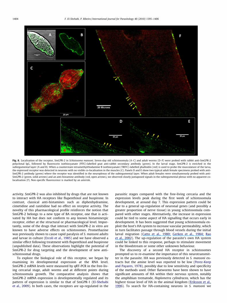
3.5. Confocal immunofluorescence analysis of SmGPR-2 in S. mansoni
The tissue localisation of the receptor SmGPR-2 was examinedin adult and larval S. mansoni. We selected in vitro cultured7 day-old schistosomula for these studies because they wereshown in the qPCR analysis to have the highest SmGPR-2 expres-sion level. The larvae and adult worms were probed with rabbitpolyclonal anti-SmGPR-2 IgG, followed by a FITC-labelled goatanti-rabbit secondary antibody. Some animals were also treatedwith TRITC-conjugated phalloidin to label cytoskeletal elementsand muscle (Mair et al., 2000, 2003). The results showed strongSmGPR-2 green fluorescence in the subtegumental region of thelarvae (Fig. 8A and B). The signal could be seen along the entirelength of the body and there was no apparent co-localisation ofSmGPR-2 (green) and muscle (red) in larvae that were counter-stained with TRITC-conjugated phalloidin (Fig. 8C), suggestingthe receptor is associated with the nervous system rather thanthe musculature. SmGPR-2 immunoreactivity in the adults wasgenerally weaker than in the larvae but the distribution pattern
was similar. Most of the expression was restricted to the peripheralneuronal plexus of the subtegumental region (Fig. 8D and E).Importantly, we observed that the localisation of the receptor inthis area closely resembles that of HA itself. Animals probed withthe two antibodies showed distinctive anti-SmGPR-2 immunofluo-rescence (green) in close proximity to anti-HA immunofluores-cence (red) in the nerve plexus (Fig. 8F). No co-localisation couldbe seen, however, indicating that the transmitter and its receptorare present on different cells. Aside from this region, we observedweak expression of the receptor in the oral and ventral suckers (notshown) but not in the CNS. Some non-specific fluorescence was de-tected in the caecum of adult worms (Fig. 8D, asterisk) and the fe-male reproductive tract.
4. Discussion
Previously, our laboratory described the first HA-responsivereceptor of S. mansoni, named SmGPR-1 (SmGPCR; Hamdan et al.,2002). In the present study, we report the cloning and expressionof a structurally related receptor, which we have named SmGPR-2. The bioinformatics analysis identified two S. japonicum se-quences and a total of six orphan receptors in the genome of S.mansoni that share high homology with SmGPR-2. These sequencesdo not align within the known clades of the biogenic amine GPCRtree and thus appear to constitute a new type of amine receptor.The SmGPR homologues are characterised in part by the absenceof the highly conserved aspartate D3.32 of TM3, which is replacedwith an asparagine in all but one of these receptors(Smp_043290; SmGPR-3, Accession #GQ259333). As mentionedearlier, D3.32 is a critical residue in the ligand-binding pocket ofBA receptors (Shi and Javitch, 2002; Roth and Kristiansen, 2004;Roth, 2006). The side-chain carboxylate of D3.32 is believed to formdirect contact with the protonated amino moiety of the differentBAs via a salt bridge interaction (Strader et al., 1987, 1991; Man-sour et al., 1992; Boess et al., 1998; Shi and Javitch, 2002; Jongejanet al., 2008). D3.32N and D3.32A single-point mutations were shownto abrogate or greatly diminish ligand binding to a variety of recep-tors, including histaminergic (H1 and H4) GPCRs (Ohta et al., 1994;Nonaka et al., 1998; Muntasir et al., 2006). Thus the D3.32N substi-tution of the schistosome sequences is surprising and suggests afundamental difference in the organisation of the binding pocket.The three-dimensional models of SmGPR-1 and SmGPR-2 do notshow any obvious acidic residues on the TM3 helix that could sub-stitute for D3.32. There is, however, a unique glutamate (SmGPR-2residue E3.20) in the first extracellular loop region, near the bound-ary of TM3. This glutamate residue is present in all SmGPR-likereceptors but is not conserved in other aminergic receptors(Fig. 2). If this residue contributes to the binding pocket, it couldbe a schistosome-specific substitution that compensates for theabsence of D3.32.
SmGPR-2 was tested for activity by expressing the cDNA inyeast. The system used in this study is designed for functionalexpression of GPCRs (Dowell and Brown, 2002) and offers manyadvantages over other heterologous expression systems, particu-larly for receptor deorphanisation. Besides low cost of growthand maintenance, yeast cells have robust translational and foldingmechanisms for expression of foreign eukaryotic proteins and theycan be easily adapted to high-throughput activity assays (Dowelland Brown, 2002; Ladds et al., 2005). Many GPCRs have been suc-cessfully expressed in yeast, including helminth receptors (Kimberet al., 2009; Taman and Ribeiro, 2009).
SmGPR-2 expressed in yeast was selectively activated by HA.The receptor showed intrinsic activity in the absence of ligand.However, in the presence of HA or a methylated derivative, thatactivity was several fold greater and the stimulation was dose-
Fig. 6. Developmental expression of the receptor, SmGPR-2 in Schistosoma mansoni.Quantitative PCR (qPCR) was performed on reverse-transcribed RNA from S.mansoni cercaria (C), adult worms (A) and in vitro transformed schistosomulasampled immediately after transformation (S0), 3 days (S3), 7 days (S7) or 14 days(S14) post-transformation. The qPCR data were standardised by simultaneousamplification of internal housekeeping controls (GAPDH) and differences inexpression data were calculated according to the comparative DDCT method. Theresults are shown as the fold-change in SmGPR-2 expression relative to the cercariaand error bars are the means ± S.E.M. of a minimum of three experiments, eachperformed in triplicate.
1402 F. El-Shehabi, P. Ribeiro / International Journal for Parasitology 40 (2010) 1395–1406

dependent. Other common BAs had no effect on this receptor, indi-cating the response was specific. The half-maximum effective con-centration (EC50) for HA was in the micromolar range, a valuehigher than that of mammalian HA receptors. This difference couldbe due to the aforementioned D3.32N substitution, which mightlower binding affinity, or it could be an artefact caused by heterol-ogous expression in yeast. Although the yeast system has manyadvantages for GPCR expression, the cell wall hinders access of li-gands to the receptor, with the result that more ligand is neededfor activation. Agonist potency is often greatly reduced in yeastGPCR expression systems compared with mammalian cells (Laddset al., 2005), in some instances by more than 100-fold (Taman andRibeiro, 2009).
One distinctive feature of this receptor is its high constitutiveactivity in yeast. Cells expressing SmGPR-2 exhibited significantactivity compared with the mock control, even in the absence ofHA. It is not uncommon for GPCRs to show some spontaneous (li-gand-independent) activity when they are expressed in a heterol-ogous environment, possibly due to protein over-expression inthe foreign cell. Some receptors, however, have a natural propen-sity towards spontaneous activation and the resulting basal activ-ity is biologically relevant in vivo. Mammalian receptors such asthe H3 histaminergic receptor, 5HT4, 5HT2C, b1-adrenoceptorand the parathyroid hormone (PTH) receptor all show spontaneous
activation in vivo, which, in some instances, has been linked to dis-ease (Bond and Ijzerman, 2006). Viral GPCRs, for example, are con-stitutively activated and this is thought to contribute to theinfection mechanism (Leurs et al., 2003; Vischer et al., 2006; Can-non, 2007). The cause of high basal activity has been linked to sin-gle nucleotide polymorphisms (SNPs), splicing and/or RNA editingevents that disrupt the normal constraints on GPCR activation(Huang and Chen, 2005). Many of the amino acids implicated inthese constraints are conserved in SmGPR-2 (e.g. D3.49, R3.50, E6.30
and T6.34) but there may be additional interactions among neigh-bouring residues that destabilise the inactive conformation, allow-ing the receptor to spontaneously activate. The absence of D3.32, inparticular, could be a contributing factor. Single-point mutations ofD3.32 were shown to increase agonist-independent activity in someGPCRs (Porter et al., 1996; Befort et al., 1999; Huang and Chen,2005). Whether the high basal activity of SmGPR-2 is relevant tothe parasite in vivo or simply a function of heterologous expressionremains to be determined.
The yeast antagonist assays suggest that SmGPR-2 has an unu-sual pharmacological profile, which is quite different from those ofmammalian HA receptors. The HA or 1-methylHA activated recep-tor was strongly inhibited by promethazine, a classical (mamma-lian) H1 antagonist and, to a lesser degree, by cyproheptadine, amixed antagonist that has both serotonergic and histaminergic
Fig. 7. Localisation of histamine (HA) in adult Schistosoma mansoni. Adult worms were probed with an anti-HA monoclonal antibody followed by a rhodamine-labelledsecondary antibody. (A) HA immunoreactivity was seen in both genders and occurs in the peripheral subtegumental nerve plexus and minor nerve cords that run along theentire length of the worm (arrows). A close-up view of the subtegumental region shows numerous HA-containing cell bodies (arrowheads) and processes scatteredthroughout the nerve plexus (B). Going from the surface to the interior of the worm, we see a well-developed surface nerve net arranged in a criss-cross pattern (C) followedby multiple varicose nerve fibers, which lie deep in the somatic musculature (D). Histaminergic processes innervate the reproductive tract of an adult female (arrowhead) (E)and are highly abundant in the male acetabulum (F) and oral sucker (G). Anastomosing fibers can be seen throughout the musculature of the oral sucker (H). Non-specificautofluorescence in the female reproductive tract is marked by asterisks (A and E).
F. El-Shehabi, P. Ribeiro / International Journal for Parasitology 40 (2010) 1395–1406 1403
activity. SmGPR-2 was also inhibited by drugs that are not knownto interact with HA receptors like flupenthixol and buspirone. Incontrast, classical anti-histamines such as diphenhydramine,cimetidine and ranitidine had no effect on receptor activity. Thenovelty of this pharmacological profile reinforces the notion thatSmGPR-2 belongs to a new type of BA receptor, one that is acti-vated by HA but does not conform to any known histaminergicreceptor, either at the structural or pharmacological level. Impor-tantly, some of the drugs that interact with SmGPR-2 in vitro areknown to have adverse effects on schistosomes. Promethazinewas previously shown to cause rapid paralysis of S. mansoni adultsand larvae in culture (Ercoli et al., 1985) and we have observed asimilar effect following treatment with flupenthixol and buspirone(unpublished data). These observations highlight the potential ofSmGPR-2 for drug targeting and the development of new anti-schistosomal drugs.
To explore the biological role of this receptor, we began byexamining its developmental expression at the RNA level.SmGPR-2 mRNA levels were measured by qRT-PCR in the free-liv-ing cercarial stage, adult worms and at different points duringschistosomula growth. The comparative analysis shows thatSmGPR-2 mRNA expression is developmentally regulated and itspattern of expression is similar to that of SmGPR-1 (El-Shehabiet al., 2009). In both cases, the receptors are up-regulated in the
parasitic stages compared with the free-living cercaria and theexpression levels peak during the first week of schistosomuladevelopment, at around day 7. This expression pattern could bedue to a general up-regulation of neuronal genes (and possibly agreater proportion of nerve tissue) in young schistosomula com-pared with other stages. Alternatively, the increase in expressioncould be tied to some aspect of HA signalling that occurs early indevelopment. It has been suggested that young schistosomula ex-ploit the host’s HA system to increase vascular permeability, whichin turn facilitates passage through blood vessels during the initiallarval migration (Catto et al., 1980; Gerken et al., 1984; Raoet al., 2002). The up-regulation of the parasite’s own HA systemcould be linked to this response, perhaps to stimulate movementin the bloodstream or some other unknown behaviour.
The discovery of a second HA receptor in schistosomesprompted us to re-examine the importance of this neurotransmit-ter in the parasite. HA was previously detected in S. mansoni ex-tracts but the amine level was reported to be low (Perez-Keepand Payares, 1978), possibly due to low sensitivity and specificityof the methods used. Other flatworms have been shown to havesignificant amounts of HA within their nervous system, notablythe amphibian trematode, Haplometra cylindracea, which has thehighest tissue level of HA in the animal kingdom (Eriksson et al.,1996). To search for HA-containing neurons in S. mansoni we
Fig. 8. Localisation of the receptor, SmGPR-2 in Schistosoma mansoni. Seven-day old schistosomula (A–C) and adult worms (D–F) were probed with rabbit anti-SmGPR-2polyclonal IgG, followed by fluorescein isothiocyanate (FITC)-labelled goat anti-rabbit secondary antibody (green). In the larval stage, SmGPR-2 is enriched in thesubtegumental layer (A and B). When a counterstain tetramethylrhodamine B isothiocyanate (TRITC)-labelled phalloidin (red) is used to probe the musculature of the larva,the expressed receptor was detected in neurons with no visible co-localisation in the muscles (C). Panels D and E show two typical adult female specimens probed with anti-SmGPR-2 antibody (green) where the receptor was identified in the neuroplexus of the subtegumental layer. When adult females were simultaneously probed with anti-SmGPR-2 (green, solid arrows) and an anti-histamine antibody (red, open arrows), we observed closely juxtaposed signals in the subtegumental plexus with no apparent co-localisation (F). Non-specific fluorescence is marked by an asterisk.
1404 F. El-Shehabi, P. Ribeiro / International Journal for Parasitology 40 (2010) 1395–1406

probed adult male and female worms with a commercial anti-HAmonoclonal antibody by confocal immunofluorescence. The resultsshowed widespread HA immunoreactivity in the PNS of S. mansoni.Histaminergic cell bodies and processes are enriched in the nerveplexus that supplies the body wall musculature and they are alsoprominent in the innervation of the suckers of both male and fe-male worms. The prevalence of these neurons in the suckers andsubtegumental plexus resembles that seen in H. cylindracea (Eriks-son et al., 1996), which also has significant levels of HA in these re-gions. Unlike H. cylindracea, however, S. mansoni exhibited little HAimmunoreactivity in the brain region or major nerve cords of theCNS. These results add to the notion that HA plays different rolesin different parasites within the phylum (Eriksson et al., 1996). Inthe case of S. mansoni, HA is most likely acting as a transmitter/modulator of the peripheral nerve plexuses rather than the CNS.
To investigate the role of HA further we examined the tissuedistribution of the receptor, SmGPR-2, using a specific peptide anti-body. SmGPR-2 was detected in the subtegumental nerve plexus ofboth adult worms and larvae. The level of protein expression wasstronger and more widespread in the schistosomula than in theadults, consistent with the findings of the qPCR analysis. In bothstages, expression was restricted to the nervous system; we couldnot detect specific SmGPR-2 immunoreactivity in other tissues,including the musculature. Importantly, SmGPR-2 was expressedin close proximity to HA-containing neurons, where the receptorcould be activated by endogenously released amine. The HA andSmGPR-2 signals did not co-localise, however, indicating they wereassociated with different neurons. Based on these results, we con-clude that HA is acting through SmGPR-2 to modulate the activityof other neurotransmitters in the subtegumental region. The plex-uses are rich in serotonergic, peptidergic and cholinergic neurons,any one of which could be regulated in this manner.
Together with previous studies of SmGPR-1, these results arebeginning to shed new light on the mode of action of HA in schis-tosomes. HA was previously reported to modulate schistosomemotility (Ercoli et al., 1985) and this is supported by the discoveryof numerous histaminergic neurons in the peripheral plexus thatsupplies the body wall musculature. Effects on motility could beachieved through direct activation of SmGPR-1, which is presenton the body wall muscles (El-Shehabi et al., 2009) or, indirectly,through SmGPR-2-mediated modulation of neuromuscular cir-cuits, as discussed above. Besides effects on motility, the presentresults suggest a probable role for HA in the musculature of thesuckers, both oral and the acetabulum, as evidenced by the abun-dance of HA-containing fibres in these structures. These effects arelikely mediated, at least in part by SmGPR-1, which is strongly ex-pressed in both suckers (El-Shehabi et al., 2009). We also observedsome histaminergic innervation of the female reproductive tractand excretory ducts in some animals, which might suggest addi-tional role(s) for HA in the parasite. SmGPR-1 and -2 could notbe detected in these tissues but there may be other HA-activatedreceptors that have yet to be identified.
Given the importance of HA in S. mansoni, one important ques-tion that needs to be answered is whether the parasite synthesisesits own amine or whether it is obtained from the host. In the caseof serotonin (5HT), the amine is synthesised endogenously (Ham-dan and Ribeiro, 1999) but it can also be derived from the host,through a specific transporter system (Patocka and Ribeiro,2007). HA can be synthesised by some parasites (Mettrick and Tel-ford, 1963; Eriksson et al., 1996), whereas in other worms it is ta-ken up by simple diffusion (Yonge and Webb, 1992). Bioinformaticanalyses of the S. mansoni genome identified a potential histidinedecarboxylase, the enzyme responsible for histamine biosynthesisbut this has yet to be cloned and characterised enzymatically.Additional research is needed to fully characterise the HA systemof schistosomes and possibly to identify additional receptors. Ef-
forts are underway to examine whether other members of theSmGPR clade are also activated by HA or other biogenic amines.
Acknowledgements
The authors would like to thank Dr. J. Broach (Princeton Univer-sity, NJ, USA), who kindly provided us with the yeast expressionstrains. We also thank Dr. Fred Lewis (Biomedical Research Insti-tute, Rockville, MD, USA), who supplied the infected snails. Thiswork was supported by a grant from the Natural Sciences and Engi-neering Research Council of Canada (NSERC) to P.R.
References
Ballesteros, J., Palczewski, K., 2001. G protein-coupled receptor drug discovery:implications from the crystal structure of rhodopsin. Curr. Opin. Drug Discov.Dev. 4, 561–574.
Ballesteros, J.A., Weinstein, H., 1995. Integrated methods for the construction ofthree-dimensional models and computational probing of structure–functionrelations in G protein coupled receptors. Method Neurosci. 25, 366–428.
Basch, P.F., 1981. Cultivation of Schistosoma mansoni in vitro. I. Establishment ofcultures from cercaria and development until pairing. J. Parasitol. 67, 179–185.
Basch, P.F., Humbert, R., 1981. Cultivation of Schistosoma mansoni in vitro. III.Implantation of cultured worms into mouse mesenteric veins. J. Parasitol. 67,191–195.
Befort, K., Zilliox, C., Filliol, D., Yue, S., Kieffer, B.L., 1999. Constitutive activation ofthe delta opioid receptor by mutations in transmembrane domains III and VII. J.Biol. Chem. 274, 18574–18581.
Berriman, M., Haas, B.J., LoVerde, P.T., Wilson, R.A., Dillon, G.P., Cerqueira, G.C.,Mashiyama, S.T., Al-Lazikani, B., Andrade, L.F., Ashton, P.D., Aslett, M.A.,Bartholomeu, D.C., Blandin, G., Caffrey, C.R., Coghlan, A., Coulson, R., Day, T.A.,Delcher, A., DeMarco, R., Djikeng, A., Eyre, T., Gamble, J.A., Ghedin, E., Gu, Y.,Hertz-Fowler, C., Hirai, H., Hirai, Y., Houston, R., Ivens, A., Johnston, D.A.,Lacerda, D., Macedo, C.D., McVeigh, P., Ning, Z., Oliveira, G., Overington, J.P.,Parkhill, J., Pertea, M., Pierce, R.J., Protasio, A.V., Quail, M.A., Rajandream, M.A.,Rogers, J., Sajid, M., Salzberg, S.L., Stanke, M., Tivey, A.R., White, O., Williams,D.L., Wortman, J., Wu, W., Zamanian, M., Zerlotini, A., Fraser-Liggett, C.M.,Barrell, B.G., El-Sayed, N.M., 2009. The genome of the blood fluke Schistosomamansoni. Nature 460, 352–358.
Boess, F.G., Monsma Jr., F.J., Sleight, A.J., 1998. Identification of residues intransmembrane regions III and VI that contribute to the ligand binding site ofthe serotonin 5-HT6 receptor. J. Neurochem. 71, 2169–2177.
Bond, R.A., Ijzerman, A.P., 2006. Recent developments in constitutive receptoractivity and inverse agonism, and their potential for GPCR drug discovery.Trends Pharmacol. Sci. 27, 92–96.
Boyle, J.P., Yoshino, T.P., 2005. Serotonin-induced muscular activity in Schistosomamansoni larval stages: importance of 5HT transport and role in daughtersporocyst production. J. Parasitol. 91, 542–550.
Cannon, M., 2007. The KSHV and other human herpesviral G protein-coupledreceptors. Curr. Top. Microbiol. Immunol. 312, 137–156.
Catto, B.A., Lewis, F.A., Ottesen, E.A., 1980. Cercaria-induced histamine release: afactor in the pathogenesis of schistosome dermatitis? Am. J. Trop. Med. Hyg. 29,886–889.
Choudhary, M.S., Craigo, S., Roth, B.L., 1993. A single point mutation(Phe340 ? Leu340) of a conserved phenylalanine abolishes 4-[125I]iodo-(2,5-dimethoxy)phenylisopropylamine and [3H]mesulergine but not [3H]ketanserinbinding to 5-hydroxytryptamine2 receptors. Mol. Pharmacol. 43, 755–761.
Cikos, S., Bukovska, A., Koppel, J., 2007. Relative quantification of mRNA:comparison of methods currently used for real-time PCR data analysis. BMCMol. Biol. 8, 113.
Day, T.A., Bennett, J.L., Pax, R.A., 1994. Serotonin and its requirement formaintenance of contractility in muscle fibers isolated from Schistosomamansoni. Parasitology 108, 425–432.
Dowell, S.J., Brown, A.J., 2002. Yeast assays for G-protein-coupled receptors.Receptors Channels 8, 343–352.
Eglen, R.M., 2005. Emerging concepts in GPCR function—the influence of cellphenotype on GPCR pharmacology. Proc. West Pharmacol. Soc. 48, 31–34.
El-Shehabi, F., Vermeire, J., Timothy, P., Yoshino, T.P.R., 2009. Developmentalexpression analysis and immunolocalization of a biogenic amine receptor inSchistosoma mansoni. Exp. Parasitol. 122, 17–27.
Ercoli, N., Payares, G., Nunez, D., 1985. Schistosoma mansoni: neurotransmitters andthe mobility of cercaria and schistosomules. Exp. Parasitol. 59, 204–216.
Eriksson, K.S., Johnston, R.N., Shaw, C., Halton, D.W., Panula, P.A., 1996. Widespreaddistribution of histamine in the nervous system of a trematode flatworm. J.Comp. Neurol. 373, 220–227.
Fallon, P.G., Doenhoff, M.J., 1994. Drug-resistant schistosomiasis: resistance topraziquantel and oxamniquine induced in Schistosoma mansoni in mice is drugspecific. Am. J. Trop. Med. Hyg. 51, 83–88.
Gerken, S.E., Mota-Santos, T.A., Vaz, N.M., Correa-Oliveira, R., Dias-da-Silva, W.,Gazzinelli, G., 1984. Recovery of schistosomula of Schistosoma mansoni frommouse skin: involvement of mast cells and vasoactive amines. Braz. J. Med. Biol.Res. 17, 301–307.
F. El-Shehabi, P. Ribeiro / International Journal for Parasitology 40 (2010) 1395–1406 1405

Gietz, R.D., Schiestl, R.H., Willems, A.R., Woods, R.A., 1995. Studies on thetransformation of intact yeast cells by the LiAc/SS-DNA/PEG procedure. Yeast11, 355–360.
Hamdan, F.F., Ribeiro, P., 1999. Characterization of a stable form of tryptophanhydroxylase from the human parasite Schistosoma mansoni. J. Biol. Chem. 274,21746–21754.
Hamdan, F.F., Abramovitz, M., Mousa, A., Xie, J., Durocher, Y., Ribeiro, P., 2002.A novel Schistosoma mansoni G protein-coupled receptor is responsive tohistamine. Mol. Biochem. Parasitol. 119, 75–86.
Hoffmann, K.F., Davis, E.M., Fischer, E.R., Wynn, T.A., 2001. The guanine proteincoupled receptor rhodopsin is developmentally regulated in the free-livingstages of Schistosoma mansoni. Mol. Biochem. Parasitol. 112, 113–123.
Huang, P., Chen, C., 2005. Molecular mechanisms involved in the activation ofrhodopsin-like seven-transmembrane receptors. In: Devi, Lakshmi A., Engel,Andreas, Palczewski, Krzysztof (Eds.), The G Protein-Coupled ReceptorsHandbook. Humana Press Inc., New Jersey, USA, pp. 33–70 (Chapter 2).
Ismail, M.M., Taha, S.A., Farghaly, A.M., el-Azony, A.S., 1994. Laboratory inducedresistance to praziquantel in experimental schistosomiasis. J. Egypt Soc.Parasitol. 24, 685–695.
Jongejan, A., Lim, H.D., Smits, R.A., de Esch, I.J., Haaksma, E., Leurs, R., 2008.Delineation of agonist binding to the human histamine H4 receptor usingmutational analysis, homology modeling, and ab initio calculations. J. Chem. Inf.Model. 48, 1455–1463.
Kimber, M.J., Sayegh, L., El-Shehabi, F., Song, C., Zamanian, M., Woods, D.J., Day, T.A.,Ribeiro, P., 2009. Identification of an Ascaris G protein-coupled acetylcholinereceptor with atypical muscarinic pharmacology. Int. J. Parasitol. 39, 1215–1222.
Kristiansen, K., Kroeze, W.K., Willins, D.L., Gelber, E.I., Savage, J.E., Glennon, R.A.,Roth, B.L., 2000. A highly conserved aspartic acid (Asp-155) anchors theterminal amine moiety of tryptamines and is involved in membrane targeting ofthe 5-HT(2A) serotonin receptor but does not participate in activation via a‘‘salt-bridge disruption” mechanism. J. Pharmacol. Exp. Ther. 293, 735–746.
Ladds, G., Goddard, A., Davey, J., 2005. Functional analysis of heterologous GPCRsignaling pathways in yeast. Trends Biotechnol. 23, 367–373.
Leurs, C., Jansen, M., Pollok, K.E., Heinkelein, M., Schmidt, M., Wissler, M.,Lindemann, D., Von Kalle, C., Rethwilm, A., Williams, D.A., Hanenberg, H.,2003. Comparison of three retroviral vector systems for transduction ofnonobese diabetic/severe combined immunodeficiency mice repopulatinghuman CD34+ cord blood cells. Hum. Gene Ther. 14, 509–519.
Lewis, F.A., Stirewalt, M.A., Souza, C.P., Gazzinelli, G., 1986. Large-scale laboratorymaintenance of Schistosoma mansoni, with observations on three schistosome/snail host combinations. J. Parasitol. 72, 813–829.
Lewis, F.A., Patterson, C.N., Knight, M., Richards, C.S., 2001. The relationshipbetween Schistosoma mansoni and Biomphalaria glabrata: genetic and molecularapproaches. Parasitology 123 (Suppl.), S169–S179.
Livak, K.J., Schmittgen, T.D., 2001. Analysis of relative gene expression data usingreal-time quantitative PCR and the 2(�Delta Delta C(T)) method. Methods 25,402–408.
Mair, G.R., Maule, A.G., Day, T.A., Halton, D.W., 2000. A confocal microscopical studyof the musculature of adult Schistosoma mansoni. Parasitology 121 (Pt. 2), 163–170.
Mair, G.R., Maule, A.G., Fried, B., Day, T.A., Halton, D.W., 2003. Organization of themusculature of schistosome cercaria. J. Parasitol. 89, 623–625.
Mansour, A., Meng, F., Meador-Woodruff, J.H., Taylor, L.P., Civelli, O., Akil, H., 1992.Site-directed mutagenesis of the human dopamine D2 receptor. Eur. J.Pharmacol. 227, 205–214.
Massotte, D., Kieffer, B., 2005. Structure–function relationships in G protein-coupledreceptors: ligand binding and receptor activation. In: Devi, Lakshmi A. (Ed.), TheG Protein-Coupled Receptors Handbook. Humana Press Inc., NJ, pp. 3–31(Chapter 1).
Maule, A., Marks, N., Day, T., 2006. Signaling molecules and nerve–muscle function.In: Maule, A., Marks, N. (Eds.), Parasitic Flatworms: Molecular Biology,Biochemistry, Immunology and Physiology. CABI Publishing, UK, pp. 369–386(Chapter 19).
Mettrick, D.F., Telford, J.M., 1963. Histamine in the phylum platyhelminthes. J.Parasitol. 49, 653–656.
Muntasir, H.A., Takahashi, J., Rashid, M., Ahmed, M., Komiyama, T., Hossain, M.,Kawakami, J., Nashimoto, M., Nagatomo, T., 2006. Site-directed mutagenesis ofthe serotonin 5-Hydroxytryptamine2c receptor: identification of amino acidsresponsible for sarpogrelate binding. Biol. Pharm. Bull. 29, 1645–1650.
Nonaka, H., Otaki, S., Ohshima, E., Kono, M., Kase, H., Ohta, K., Fukui, H., Ichimura,M., 1998. Unique binding pocket for KW-4679 in the histamine H1 receptor.Eur. J. Pharmacol. 345, 111–117.
Ohta, K., Hayashi, H., Mizuguchi, H., Kagamiyama, H., Fujimoto, K., Fukui, H., 1994.Site-directed mutagenesis of the histamine H1 receptor: roles of aspartic
acid107, asparagine198 and threonine194. Biochem. Biophys. Res. Commun.203, 1096–1101.
Patocka, N., Ribeiro, P., 2007. Characterization of a serotonin transporter in theparasitic flatworm, Schistosoma mansoni: cloning, expression and functionalanalysis. Mol. Biochem. Parasitol. 154, 125–133.
Pax, R.A., Day, T.A., Miller, C.L., Bennett, J.L., 1996. Neuromuscular physiology andpharmacology of parasitic flatworms. Parasitology 113 (Suppl.), S83–S96.
Pearson, M.S., McManus, D.P., Smyth, D.J., Jones, M.K., Sykes, A.M., Loukas, A., 2007.Cloning and characterization of an orphan seven transmembrane receptor fromSchistosoma mansoni. Parasitology 134, 2001–2008.
Perez-Keep, O., Payares, G., 1978. Histoquimica de la cercaria de Schistosomamansoni. Determinaci6n de la histamina e histaminoxidasa. Acta CientificaVenezolana 29 (Suppl. l), 147.
Porter, J.E., Hwa, J., Perez, D.M., 1996. Activation of the alpha1b-adrenergic receptoris initiated by disruption of an interhelical salt bridge constraint. J. Biol. Chem.271, 28318–28323.
Rao, K.V., Chen, L., Gnanasekar, M., Ramaswamy, K., 2002. Cloning andcharacterization of a calcium-binding, histamine-releasing protein fromSchistosoma mansoni. J. Biol. Chem. 277, 31207–31213.
Ribeiro, P., El-Shehabi, F., Patocka, N., 2005. Classical transmitters and theirreceptors in flatworms. Parasitology 131 (Suppl.), S19–S40.
Ribeiro, P., Geary, T.G., 2010. Neuronal signaling in schistosomes: current status andprospects for postgenomics. Can. J. Zool. 88, 1–22.
Robb, S.M.C., Ross, E., Sánchez Alvarado, A., 2007. SmedGD: the Schmidteamediterranea genome database. Nucleic Acids Res. 36, D599–D606.
Roth, B., Kristiansen, K., 2004. Molecular mechanisms of ligand binding, signalingand regulation within the superfamily of G protein-coupled receptors:molecular modeling and mutagenesis approaches to receptor structure andfunction. Pharmacol. Ther. 103, 21–80.
Roth, B.L., 2006. The Serotonin Receptors: From Molecular Pharmacology to HumanTherapeutics. Humana Press Inc., NJ.
Shi, L., Javitch, J.A., 2002. The binding site of aminergic G protein-coupled receptors:the transmembrane segments and second extracellular loop. Annu. Rev.Pharmacol. Toxicol. 42, 437–467.
Stevenson, B.J., Rhodes, N., Errede, B., Sprague Jr., G.F., 1992. Constitutive mutants ofthe protein kinase STE11 activate the yeast pheromone response pathway in theabsence of the G protein. Genes Dev. 6, 1293–1304.
Strader, C.D., Sigal, I.S., Register, R.B., Candelore, M.R., Rands, E., Dixon, R.A., 1987.Identification of residues required for ligand binding to the beta-adrenergicreceptor. Proc. Natl. Acad. Sci. USA 84, 4384–4388.
Strader, C.D., Gaffney, T., Sugg, E.E., Candelore, M.R., Keys, R., Patchett, A.A., Dixon,R.A., 1991. Allele-specific activation of genetically engineered receptors. J. Biol.Chem. 266, 5–8.
Sukhdeo, M.V., Hsu, S.C., Thompson, C.S., Mettrick, D.F., 1984. Hymenolepis diminuta:behavioral effects of 5-hydroxytryptamine, acetylcholine, histamine andsomatostatin. J. Parasitol. 70, 682–688.
Taman, A., Ribeiro, P., 2009. Investigation of a dopamine receptor in Schistosomamansoni: functional studies and immunolocalization. Mol. Biochem. Parasitol.
Tamura, K., Dudley, J., Nei, M., Kumar, S., 2007. MEGA4: Molecular EvolutionaryGenetics Analysis (MEGA) software version 4.0. Mol. Biol. Evol 24, 1596–1599.
The Schistosoma japonicum Genome Sequencing and Functional AnalysisConsortium, 2009. The Schistosoma japonicum genome reveals features ofhost–parasite interplay. Nature 460, 345–351.
Vischer, H.F., Hulshof, J.W., de Esch, I.J., Smit, M.J., Leurs, R., 2006. Virus-encoded G-protein-coupled receptors: constitutively active (dys)regulators of cell functionand their potential as drug target. Ernst Schering Found. Symp. Proc. 2, 187–209.
Walker, R.J., Brooks, H.L., Holden-Dye, L., 1996. Evolution and overview of classicaltransmitter molecules and their receptors. Parasitology 113 (Suppl.), S3–S33.
Wang, Z., Broach, J.R., Peiper, S.C., 2006. Functional expression of CXCR4 inSaccharomyces cerevisiae in the development of powerful tools for thepharmacological characterization of CXCR4. In: Ali, H., Haribabu, B. (Eds.),Methods in Molecular Biology: Transmembrane Signaling Protocols, vol. 332.Humana Press Inc., NJ, pp. 115–127.
Wikgren, M., Reuter, M., Gustafsson, M.K., Lindroos, P., 1990. Immunocytochemicallocalization of histamine in flatworms. Cell Tissue Res. 260, 479–484.
William, S., Botros, S., Ismail, M., Farghally, A., Day, T.A., Bennett, J.L., 2001.Praziquantel-induced tegumental damage in vitro is diminished inschistosomes derived from praziquantel-resistant infections. Parasitology 122(Pt. 1), 63–66.
Wise, A., Gearing, K., Rees, S., 2002. Target validation of G-protein coupled receptors.Drug Discov. Today 7, 235–246.
Yonge, K.A., Webb, R.A., 1992. Uptake and metabolism of histamine by the rattapeworm Hymenolepis diminuta: an in vitro study. Can. J. Zool. 70, 43–50.
1406 F. El-Shehabi, P. Ribeiro / International Journal for Parasitology 40 (2010) 1395–1406