STAT Protein Interference and Suppression of Cytokine Signal Transduction by Measles Virus V Protein
10
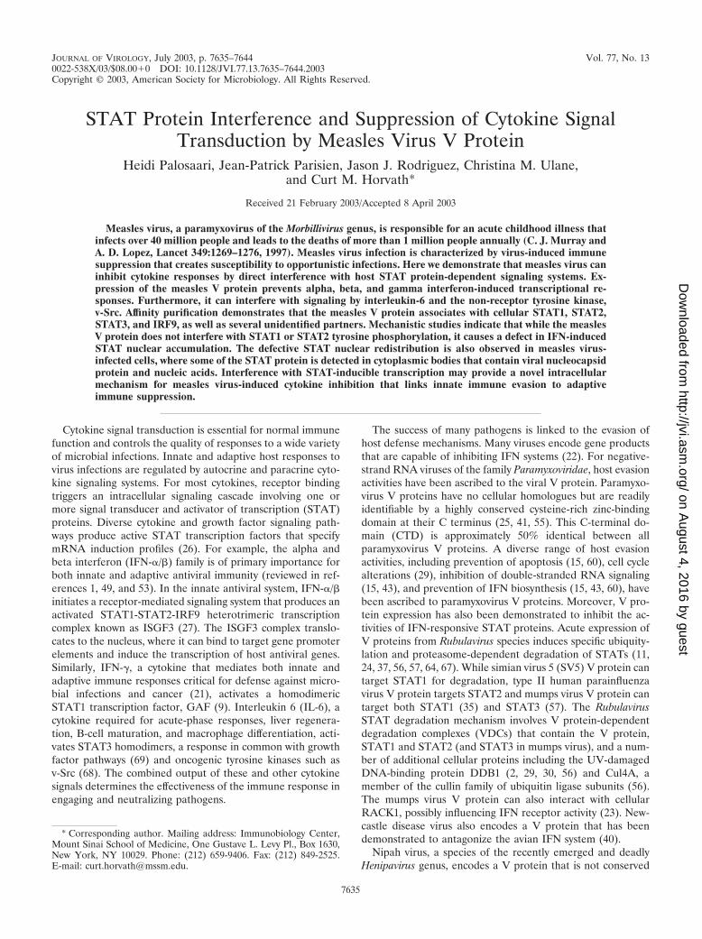
JOURNAL OF VIROLOGY, July 2003, p. 7635–7644 Vol. 77, No. 13 0022-538X/03/$08.000 DOI: 10.1128/JVI.77.13.7635–7644.2003 Copyright © 2003, American Society for Microbiology. All Rights Reserved. STAT Protein Interference and Suppression of Cytokine Signal Transduction by Measles Virus V Protein Heidi Palosaari, Jean-Patrick Parisien, Jason J. Rodriguez, Christina M. Ulane, and Curt M. Horvath* Received 21 February 2003/Accepted 8 April 2003 Measles virus, a paramyxovirus of the Morbillivirus genus, is responsible for an acute childhood illness that infects over 40 million people and leads to the deaths of more than 1 million people annually (C. J. Murray and A. D. Lopez, Lancet 349:1269–1276, 1997). Measles virus infection is characterized by virus-induced immune suppression that creates susceptibility to opportunistic infections. Here we demonstrate that measles virus can inhibit cytokine responses by direct interference with host STAT protein-dependent signaling systems. Ex- pression of the measles V protein prevents alpha, beta, and gamma interferon-induced transcriptional re- sponses. Furthermore, it can interfere with signaling by interleukin-6 and the non-receptor tyrosine kinase, v-Src. Affinity purification demonstrates that the measles V protein associates with cellular STAT1, STAT2, STAT3, and IRF9, as well as several unidentified partners. Mechanistic studies indicate that while the measles V protein does not interfere with STAT1 or STAT2 tyrosine phosphorylation, it causes a defect in IFN-induced STAT nuclear accumulation. The defective STAT nuclear redistribution is also observed in measles virus- infected cells, where some of the STAT protein is detected in cytoplasmic bodies that contain viral nucleocapsid protein and nucleic acids. Interference with STAT-inducible transcription may provide a novel intracellular mechanism for measles virus-induced cytokine inhibition that links innate immune evasion to adaptive immune suppression. Cytokine signal transduction is essential for normal immune function and controls the quality of responses to a wide variety of microbial infections. Innate and adaptive host responses to virus infections are regulated by autocrine and paracrine cyto- kine signaling systems. For most cytokines, receptor binding triggers an intracellular signaling cascade involving one or more signal transducer and activator of transcription (STAT) proteins. Diverse cytokine and growth factor signaling path- ways produce active STAT transcription factors that specify mRNA induction profiles (26). For example, the alpha and beta interferon (IFN-/) family is of primary importance for both innate and adaptive antiviral immunity (reviewed in ref- erences 1, 49, and 53). In the innate antiviral system, IFN-/ initiates a receptor-mediated signaling system that produces an activated STAT1-STAT2-IRF9 heterotrimeric transcription complex known as ISGF3 (27). The ISGF3 complex translo- cates to the nucleus, where it can bind to target gene promoter elements and induce the transcription of host antiviral genes. Similarly, IFN-, a cytokine that mediates both innate and adaptive immune responses critical for defense against micro- bial infections and cancer (21), activates a homodimeric STAT1 transcription factor, GAF (9). Interleukin 6 (IL-6), a cytokine required for acute-phase responses, liver regenera- tion, B-cell maturation, and macrophage differentiation, acti- vates STAT3 homodimers, a response in common with growth factor pathways (69) and oncogenic tyrosine kinases such as v-Src (68). The combined output of these and other cytokine signals determines the effectiveness of the immune response in engaging and neutralizing pathogens. The success of many pathogens is linked to the evasion of host defense mechanisms. Many viruses encode gene products that are capable of inhibiting IFN systems (22). For negative- strand RNA viruses of the family Paramyxoviridae, host evasion activities have been ascribed to the viral V protein. Paramyxo- virus V proteins have no cellular homologues but are readily identifiable by a highly conserved cysteine-rich zinc-binding domain at their C terminus (25, 41, 55). This C-terminal do- main (CTD) is approximately 50% identical between all paramyxovirus V proteins. A diverse range of host evasion activities, including prevention of apoptosis (15, 60), cell cycle alterations (29), inhibition of double-stranded RNA signaling (15, 43), and prevention of IFN biosynthesis (15, 43, 60), have been ascribed to paramyxovirus V proteins. Moreover, V pro- tein expression has also been demonstrated to inhibit the ac- tivities of IFN-responsive STAT proteins. Acute expression of V proteins from Rubulavirus species induces specific ubiquity- lation and proteasome-dependent degradation of STATs (11, 24, 37, 56, 57, 64, 67). While simian virus 5 (SV5) V protein can target STAT1 for degradation, type II human parainfluenza virus V protein targets STAT2 and mumps virus V protein can target both STAT1 (35) and STAT3 (57). The Rubulavirus STAT degradation mechanism involves V protein-dependent degradation complexes (VDCs) that contain the V protein, STAT1 and STAT2 (and STAT3 in mumps virus), and a num- ber of additional cellular proteins including the UV-damaged DNA-binding protein DDB1 (2, 29, 30, 56) and Cul4A, a member of the cullin family of ubiquitin ligase subunits (56). The mumps virus V protein can also interact with cellular RACK1, possibly influencing IFN receptor activity (23). New- castle disease virus also encodes a V protein that has been demonstrated to antagonize the avian IFN system (40). Nipah virus, a species of the recently emerged and deadly Henipavirus genus, encodes a V protein that is not conserved * Corresponding author. Mailing address: Immunobiology Center, Mount Sinai School of Medicine, One Gustave L. Levy Pl., Box 1630, New York, NY 10029. Phone: (212) 659-9406. Fax: (212) 849-2525. E-mail: [email protected]. 7635 on August 4, 2016 by guest http://jvi.asm.org/ Downloaded from
-
Upload
independent -
Category
Documents
-
view
2 -
download
0
Transcript of STAT Protein Interference and Suppression of Cytokine Signal Transduction by Measles Virus V Protein
JOURNAL OF VIROLOGY, July 2003, p. 7635–7644 Vol. 77, No. 130022-538X/03/$08.00�0 DOI: 10.1128/JVI.77.13.7635–7644.2003Copyright © 2003, American Society for Microbiology. All Rights Reserved.
STAT Protein Interference and Suppression of Cytokine SignalTransduction by Measles Virus V Protein
Heidi Palosaari, Jean-Patrick Parisien, Jason J. Rodriguez, Christina M. Ulane,and Curt M. Horvath*
Received 21 February 2003/Accepted 8 April 2003
Measles virus, a paramyxovirus of the Morbillivirus genus, is responsible for an acute childhood illness thatinfects over 40 million people and leads to the deaths of more than 1 million people annually (C. J. Murray andA. D. Lopez, Lancet 349:1269–1276, 1997). Measles virus infection is characterized by virus-induced immunesuppression that creates susceptibility to opportunistic infections. Here we demonstrate that measles virus caninhibit cytokine responses by direct interference with host STAT protein-dependent signaling systems. Ex-pression of the measles V protein prevents alpha, beta, and gamma interferon-induced transcriptional re-sponses. Furthermore, it can interfere with signaling by interleukin-6 and the non-receptor tyrosine kinase,v-Src. Affinity purification demonstrates that the measles V protein associates with cellular STAT1, STAT2,STAT3, and IRF9, as well as several unidentified partners. Mechanistic studies indicate that while the measlesV protein does not interfere with STAT1 or STAT2 tyrosine phosphorylation, it causes a defect in IFN-inducedSTAT nuclear accumulation. The defective STAT nuclear redistribution is also observed in measles virus-infected cells, where some of the STAT protein is detected in cytoplasmic bodies that contain viral nucleocapsidprotein and nucleic acids. Interference with STAT-inducible transcription may provide a novel intracellularmechanism for measles virus-induced cytokine inhibition that links innate immune evasion to adaptiveimmune suppression.
Cytokine signal transduction is essential for normal immunefunction and controls the quality of responses to a wide varietyof microbial infections. Innate and adaptive host responses tovirus infections are regulated by autocrine and paracrine cyto-kine signaling systems. For most cytokines, receptor bindingtriggers an intracellular signaling cascade involving one ormore signal transducer and activator of transcription (STAT)proteins. Diverse cytokine and growth factor signaling path-ways produce active STAT transcription factors that specifymRNA induction profiles (26). For example, the alpha andbeta interferon (IFN-�/�) family is of primary importance forboth innate and adaptive antiviral immunity (reviewed in ref-erences 1, 49, and 53). In the innate antiviral system, IFN-�/�initiates a receptor-mediated signaling system that produces anactivated STAT1-STAT2-IRF9 heterotrimeric transcriptioncomplex known as ISGF3 (27). The ISGF3 complex translo-cates to the nucleus, where it can bind to target gene promoterelements and induce the transcription of host antiviral genes.Similarly, IFN-�, a cytokine that mediates both innate andadaptive immune responses critical for defense against micro-bial infections and cancer (21), activates a homodimericSTAT1 transcription factor, GAF (9). Interleukin 6 (IL-6), acytokine required for acute-phase responses, liver regenera-tion, B-cell maturation, and macrophage differentiation, acti-vates STAT3 homodimers, a response in common with growthfactor pathways (69) and oncogenic tyrosine kinases such asv-Src (68). The combined output of these and other cytokinesignals determines the effectiveness of the immune response inengaging and neutralizing pathogens.
The success of many pathogens is linked to the evasion ofhost defense mechanisms. Many viruses encode gene productsthat are capable of inhibiting IFN systems (22). For negative-strand RNA viruses of the family Paramyxoviridae, host evasionactivities have been ascribed to the viral V protein. Paramyxo-virus V proteins have no cellular homologues but are readilyidentifiable by a highly conserved cysteine-rich zinc-bindingdomain at their C terminus (25, 41, 55). This C-terminal do-main (CTD) is approximately 50% identical between allparamyxovirus V proteins. A diverse range of host evasionactivities, including prevention of apoptosis (15, 60), cell cyclealterations (29), inhibition of double-stranded RNA signaling(15, 43), and prevention of IFN biosynthesis (15, 43, 60), havebeen ascribed to paramyxovirus V proteins. Moreover, V pro-tein expression has also been demonstrated to inhibit the ac-tivities of IFN-responsive STAT proteins. Acute expression ofV proteins from Rubulavirus species induces specific ubiquity-lation and proteasome-dependent degradation of STATs (11,24, 37, 56, 57, 64, 67). While simian virus 5 (SV5) V protein cantarget STAT1 for degradation, type II human parainfluenzavirus V protein targets STAT2 and mumps virus V protein cantarget both STAT1 (35) and STAT3 (57). The RubulavirusSTAT degradation mechanism involves V protein-dependentdegradation complexes (VDCs) that contain the V protein,STAT1 and STAT2 (and STAT3 in mumps virus), and a num-ber of additional cellular proteins including the UV-damagedDNA-binding protein DDB1 (2, 29, 30, 56) and Cul4A, amember of the cullin family of ubiquitin ligase subunits (56).The mumps virus V protein can also interact with cellularRACK1, possibly influencing IFN receptor activity (23). New-castle disease virus also encodes a V protein that has beendemonstrated to antagonize the avian IFN system (40).
Nipah virus, a species of the recently emerged and deadlyHenipavirus genus, encodes a V protein that is not conserved
* Corresponding author. Mailing address: Immunobiology Center,Mount Sinai School of Medicine, One Gustave L. Levy Pl., Box 1630,New York, NY 10029. Phone: (212) 659-9406. Fax: (212) 849-2525.E-mail: [email protected].
7635
on August 4, 2016 by guest
http://jvi.asm.org/
Dow
nloaded from
outside of the CTD. Nipah virus V protein can nonethelessantagonize IFN responses in both human and avian systems(40, 48). Rather than degrading STATs, Nipah virus V proteinsequesters both STAT1 and STAT2 in high-molecular-weightcytoplasmic complexes, thereby preventing IFN-dependentand basal STAT protein nuclear accumulation, an efficientmeans of stopping host antiviral responses (48).
The discovery of functional diversity within the context ofSTAT inhibition suggested the possibility that other paramyxo-viruses might encode V proteins that possess distinct STAT-directed interference properties. Measles virus, a prototype ofthe Morbillivirus genus, encodes a V protein distinct from thoseof the Rubulavirus and Henipavirus genera. The measles virusV protein has 37% overall amino acid sequence identity to theSV5 V protein (7% outside the CTD) but is homologous to theNipah virus V protein only within the CTD.
The virulence and pathogenicity of measles virus have beenwell correlated with cellular IFN responses. It has been re-ported that infection with some strains of measles virus caninduce IFN biosynthesis (34) and that measles infection (18) orexpression of the measles virus N protein (54) can activateNF-�B and IRF3 transcription factors that are required forIFN-� gene induction. In addition, infection with measles virusactivates the IFN-induced STAT pathways to yield activatedISGF3 and GAF signaling complexes (18). Both wild-typemeasles virus and vaccine strains induce immediate-early genesand genes linked to antiviral responses (5, 17). Recombinantmeasles viruses with defective V proteins can grow in cell linesthat do not produce IFN (51), but in vivo studies demonstratean important role of the V proteins as virulence factors (42),and analysis of thymic xenografts revealed that V-deficientvirus replication was delayed and prolonged compared to thatof wild-type or V-overproducing viruses (58). These pheno-types are consistent with a role for V protein in measles patho-genesis and might be explained by defective host evasion ca-pabilities in the V-deficient virus.
To test the hypothesis that measles virus might also haveevolved to use V-dependent mechanisms to inhibit STAT sig-naling, assays for V-dependent IFN signaling inhibition wereperformed. The results demonstrate that measles virus V pro-tein is an efficient inhibitor of IFN signal transduction but actsvia a mechanism distinct from the previously characterizedactivities of both Rubulavirus and Henipavirus V proteins. Mea-sles virus V protein prevents both IFN-�/�- and IFN-�-in-duced transcriptional responses and inhibits STAT3-depen-dent signaling via IL-6 or v-Src. Measles virus V proteincopurifies STAT1, STAT2, STAT3, and IRF9 but does notcopurify other known cellular components of RubulavirusVDC ubiquitin ligases. It does not eliminate IFN-inducedSTAT activation by tyrosine phosphorylation but effectivelyprevents IFN-induced STAT nuclear translocation. In additionto these effects revealed by ectopic expression of measles virusV protein, a dramatic redistribution of cellular STAT proteinsis observed in measles virus-infected cells, suggesting that ad-ditional virus-encoded factors may also participate in immuneevasion.
MATERIALS AND METHODS
Cells and viruses. Human 2fTGH, 293T, and 293 Tet-On (Clontech) cell linesand murine NIH 3T3 cells were maintained in Dulbecco’s modified Eagle’s
medium (Gibco-BRL) supplemented with 10% Cosmic calf serum (HyClone)and 1% penicillin-streptomycin (Gibco-BRL) as described previously (38). Mea-sles virus (Edmonston strain; ATCC VR-24) was cultivated and subjected to titerdetermination in Vero cells.
For the tetracycline-inducible measles virus V cell line, 293 Tet-On cells(Clontech) were stably transfected with a pBI-FLAG-measles virus V plasmidand a puromycin-resistant plasmid (pBABE-puro [32]). Individual clones wereselected and then probed by FLAG immunoblotting for inducible V expressionregulated by 1 �g of doxycycline per ml. Positive clones were further screened fordoxycycline-inducible suppression of IFN-�-induced ISRE-Luciferase reportergene activity (data not shown). The Tet-On measles virus V cell line was main-tained in Dulbecco’s modified Eagle’s medium supplemented with 10% Cosmiccalf serum, 1% penicillin-streptomycin, 2 �g of puromycin per ml, and 500 �g ofG418 per ml.
Plasmids, transfection, and reporter gene assays. A cDNA copy of the mea-sles virus V mRNA was amplified by PCR from reverse-transcribed polyadenyl-ate RNA isolated from infected Vero cells by oligo(dT) chromatography. Prim-ers were used to add restriction endonuclease recognition sequences for directcloning of the PCR products into a mammalian expression plasmid downstreamof in-frame N-terminal FLAG or HA epitope tags. Several independently tran-scribed and amplified cDNAs were sequenced, and V cDNAs were recognized by
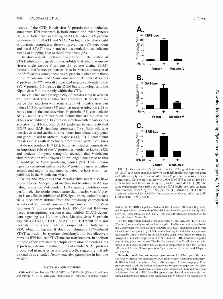
FIG. 1. Measles virus V protein blocks IFN signal transduction.(A) 293T cells were transfected with an ISRE-luciferase reporter geneand either empty vector or measles virus V protein expression vectoras indicated. Cells were treated with 1,000 U of IFN-� per ml for 14 hprior to lysis and luciferase assays (�) or left untreated (�). (B) Thesame experiment was carried out using a GAS-luciferase reporter geneand treatment with 5 ng of IFN-� per ml. (C) Murine NIH3T3 fibro-blasts were subject to ISRE-luciferase assays as above, but using 1,000U of murine IFN-� per ml.
7636 PALOSAARI ET AL. J. VIROL.
on August 4, 2016 by guest
http://jvi.asm.org/
Dow
nloaded from
the presence of non-templated nucleotides at the “editing site.” All full-length VcDNAs were found to differ from the GenBank database sequence entry at twopositions (C at nucleotide 423; G at nucleotide 825) without influencing theamino acid sequence.
Luciferase assays in 293T and 2fTGH cells were carried out exactly as de-scribed previously (57). In all cases, data represent the average values for trip-licate samples normalized to cotransfected �-galactosidase activity, expressed asa percentage of the value for the control stimulated sample. In the experimentsin some figures, P values were calculated by the t test using SigmaPlot software.
Indirect immunofluorescence. For immunofluorescence, cells were grown onPermanox chamber slides (Nalgene Nunc) and transfected and stained exactly asdescribed previously (48). Images were obtained using a Leica TCSSP confocalmicroscope, and representative fields illustrating transfected and untransfectedcells in the same field are illustrated whenever possible. For infections, 2fTGHcells were infected on chamber slides with 2 or 10 PFU of measles virus per celland stained with measles virus N antibody (10 �g/ml; Chemicon International).Nucleic acids were visualized by staining with 48 nM TOTO3 (MolecularProbes). Representative fields containing both transfected and untransfectedcells are illustrated, but effects of measles virus V protein on STATs wereobserved with 100% penetrance in several independent experiments.
Cell extracts, immunoblotting, and immunoprecipitation. For preparation ofcell extracts, samples were washed once with ice-cold phosphate-buffered salineand subsequently lysed with whole-cell extract buffer (WCEB) as describedpreviously (38, 56). For immunoblotting, proteins were separated, transferred tonitrocellulose, probed with antibodies, and visualized by chemiluminescence(NEN Life Sciences). For immunoprecipitation, lysates were prepared in WCEBand precleared with protein A-agarose. Antibody-protein complexes were puri-fied with protein A beads and washed with WCEB. After elution with sodiumdodecyl sulfate (SDS), proteins were separated by SDS-polyacrylamide gel elec-trophoresis (PAGE) and processed for immunoblotting. The antibodies usedwere STAT1 (Santa Cruz C20), STAT2 (Santa Cruz, C24), STAT3 (Santa CruzC20), IRF9 (Santa Cruz 496), FLAG (Sigma), DDB1 (Abcam 9194), Cullin 4A(Santa Cruz 8557), STAT1 phosphotyrosine 701 (Cell Signaling Technologies9171), and STAT2 phosphotyrosine 690 (Upstate Biotechnology 07-224).
Affinity purification. Affinity purification was carried out exactly as describedpreviously (56) using FLAG affinity gel (Sigma). After binding, the beads werewashed with WCEB and protein complexes were eluted with 100 �g of FLAGpeptide per ml. Eluates were denatured in SDS loading buffer, separated by
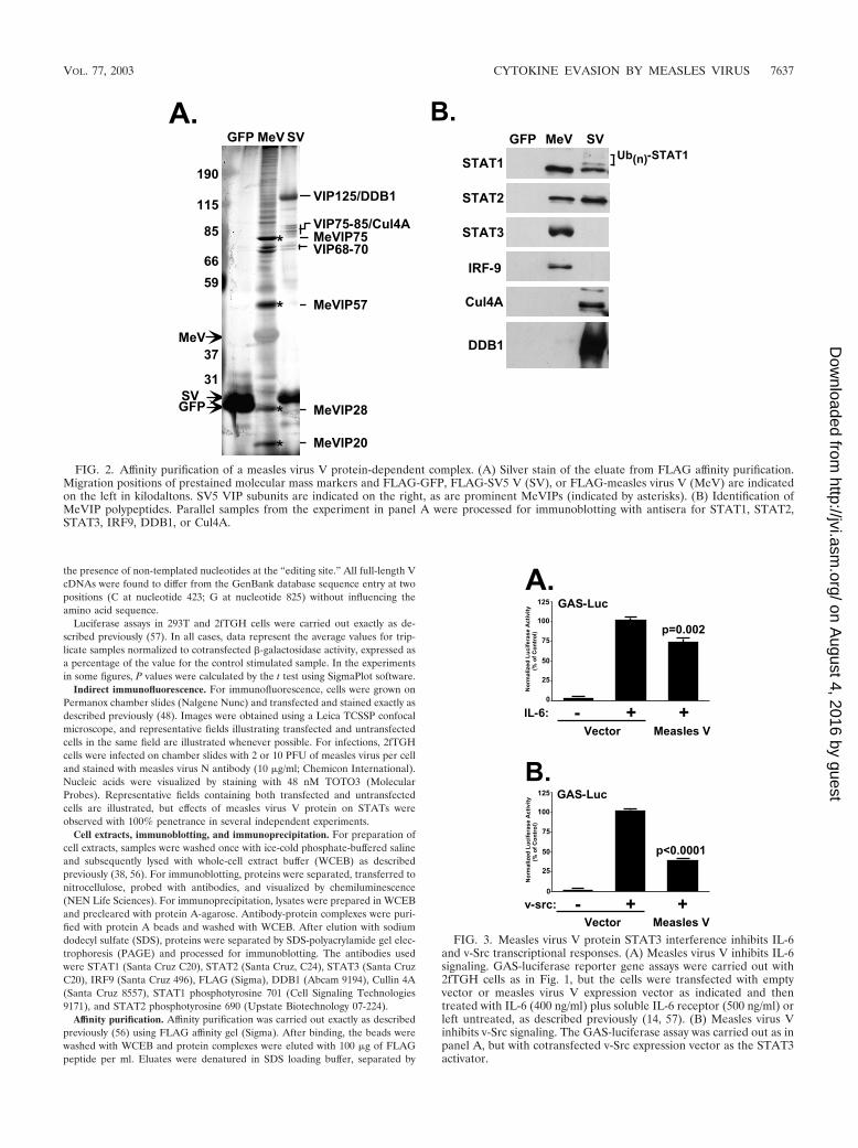
FIG. 2. Affinity purification of a measles virus V protein-dependent complex. (A) Silver stain of the eluate from FLAG affinity purification.Migration positions of prestained molecular mass markers and FLAG-GFP, FLAG-SV5 V (SV), or FLAG-measles virus V (MeV) are indicatedon the left in kilodaltons. SV5 VIP subunits are indicated on the right, as are prominent MeVIPs (indicated by asterisks). (B) Identification ofMeVIP polypeptides. Parallel samples from the experiment in panel A were processed for immunoblotting with antisera for STAT1, STAT2,STAT3, IRF9, DDB1, or Cul4A.
FIG. 3. Measles virus V protein STAT3 interference inhibits IL-6and v-Src transcriptional responses. (A) Measles virus V inhibits IL-6signaling. GAS-luciferase reporter gene assays were carried out with2fTGH cells as in Fig. 1, but the cells were transfected with emptyvector or measles virus V expression vector as indicated and thentreated with IL-6 (400 ng/ml) plus soluble IL-6 receptor (500 ng/ml) orleft untreated, as described previously (14, 57). (B) Measles virus Vinhibits v-Src signaling. The GAS-luciferase assay was carried out as inpanel A, but with cotransfected v-Src expression vector as the STAT3activator.
VOL. 77, 2003 CYTOKINE EVASION BY MEASLES VIRUS 7637
on August 4, 2016 by guest
http://jvi.asm.org/
Dow
nloaded from
SDS-PAGE, and processed for either silver staining (Bio-Rad Silver Stain Plus)or immunoblotting.
RESULTS
Measles virus V protein inhibits IFN-� and IFN-� tran-scriptional responses. A cDNA copy of the measles virus VmRNA from the Edmonston strain was isolated and tested inassays for IFN signaling inhibition (Fig. 1). Stimulation of cellswith IFN-� potently activated the transcription of an ISGF3-responsive ISRE-luciferase reporter gene. In contrast, expres-sion of measles virus V protein interfered with the ability ofIFN-� to induce a transcriptional response (Fig. 1A). Similarly,the response to IFN-� was tested using a STAT1-dependent�-IFN activation sequence (GAS)-luciferase reporter gene.Expression of measles virus V protein also inhibited the tran-scriptional response to IFN-� (Fig. 1B). These results indicatethat isolated expression of the measles virus V protein is suf-ficient to suppress STAT-dependent signaling by IFNs. ForSV5, differences between human and murine STAT2 ortho-logues provide a barrier to SV5-induced STAT1 degradation inthe mouse system (39). To test the measles virus V protein for
species-restricted IFN signaling inhibition, mouse NIH 3T3fibroblast cells were used for the ISGF3 transcription assay.While IFN stimulation induced the ISRE-luciferase reportergene, expression of the measles virus V protein efficiently an-tagonized IFN-� signaling in the murine system (Fig. 1C).Together, these results indicate that measles virus V protein isan effective inhibitor of IFN-�/� and IFN-� signaling and func-tions in both human and murine systems.
Measles virus V protein forms complexes with STAT1,STAT2, STAT3, and IRF9. STAT protein targeting by SV5,HPIV2, and mumps viruses requires a multisubunit VDC Ubligase complex that is composed of cellular proteins includingSTATs, DDB1, and Cul4A (56, 57), and Nipah virus V proteinassembles high-molecular-weight STAT-containing complexes(48). To isolate a measles virus V protein STAT-targetingcomplex, FLAG-tagged measles virus V was subjected to af-finity chromatography along with FLAG-tagged SV5 V andgreen fluorescent protein (GFP) controls. Analysis of the af-finity-purified material by SDS-PAGE and silver staining re-vealed the expected pattern of polypeptide species for SV5 Vthat ranged in apparent molecular mass between 40 and 300
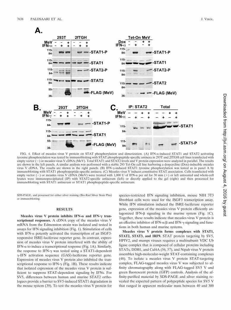
FIG. 4. Effect of measles virus V protein on STAT phosphorylation and dimerization. (A) IFN-�-induced STAT1 and STAT2 activatingtyrosine phosphorylation was tested by immunoblotting with STAT phosphopeptide-specific antisera in 293T and 2fTGH cell lines transfected withempty vector (�) or measles virus V cDNA (MeV). Total STAT1 and STAT2 levels and V protein expression were analyzed in parallel. The resultsare shown in the left panels. A similar analysis was performed with a stable 293 Tet-On cell line harboring a doxycycline (Dox)-inducible measlesvirus V cDNA. The results are shown in the right panels. (B) IFN-�-induced STAT1 tyrosine phosphorylation was tested as in panel A byimmunoblotting with STAT1 phosphopeptide-specific antisera. (C) Measles virus V induces constitutive STAT association. Cells transfected withempty vector (�) or measles virus V cDNA (MeV) were treated with 1,000 U of IFN-� per ml for 30 min (�) or left untreated and whole-celllysates were immunoprecipitated (IP) with STAT2-specific antiserum (left) or directly applied to the gel (right) and then processed forimmunoblotting with STAT1 antiserum or STAT1 phosphopeptide-specific antiserum.
7638 PALOSAARI ET AL. J. VIROL.
on August 4, 2016 by guest
http://jvi.asm.org/
Dow
nloaded from
kDa, in agreement with previously described SV5 V-interact-ing protein (VIP) complexes (56, 57). The measles virus Vaffinity preparation had a distinct composition, with only a fewbands in common with SV5 V, notably in the range of VIP68to VIP70. In addition, the eluted material contained severalunique measles virus V-interacting protein (MeVIP) specieswith the most abundant MeVIP bands migrating in the rangeof �20, �28, �57, and �75 kDa (Fig. 2A). Interestingly, amore heterogenous ladder of higher-molecular-mass proteinsthat migrate between �90 and 200 kDa were found in themeasles virus V affinity preparation. Overall, the patterns ofcellular proteins associated with the two viral proteins arestrikingly distinct, consistent with a hypothesis that these com-plexes function differently in STAT signaling inhibition.
Immunoblotting with specific antisera (Fig. 2B) was used toprobe the composition of the measles virus and SV5 V proteinaffinity preparations. The STAT1� immunoblot revealed a sin-gle band for measles virus V, in contrast to the STAT1 lad-dering pattern observed for SV5 V protein, consistent with thereported SV5 V-mediated STAT1 polyubiquitylation (56, 57).
STAT2 was also present in both V protein affinity prepara-tions. In contrast, antiserum specific for IRF9 reacted with theaffinity preparation from measles virus V protein but not withthat from the SV5 V protein. Similarly, immunoblotting withantiserum specific for STAT3 revealed that measles virus Vformed associations with STAT3. Analysis for the VDC ubiq-uitin ligase subunits DDB1 and Cul4A revealed that theseproteins were not significant components of the measles virusV affinity preparation, although a trace amount of DDB1 couldbe detected. Importantly, none of these partner proteins weredetected in the FLAG-GFP control. These results support amodel wherein the measles virus V protein induces the forma-tion of STAT1-, STAT2-, STAT3-, and IRF9-containing com-plex(es) distinct in composition from the interactions nucle-ated by the SV5 V protein.
Measles virus V protein interferes with STAT3-dependentIL-6 and v-Src signaling. The discovery of STAT3 as a MeVIPprompted us to investigate the impact of V protein expressionon STAT3-dependent transcription. STAT3 activation andtranscription factor activity has been well studied for cytokine
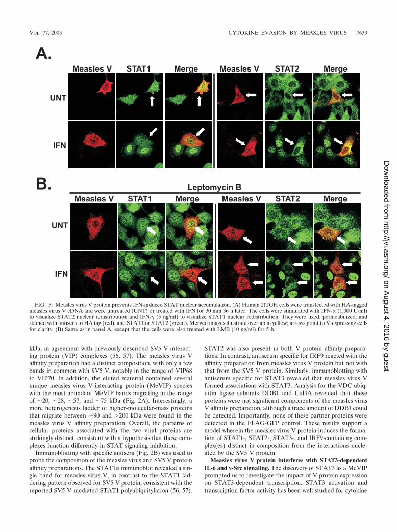
FIG. 5. Measles virus V protein prevents IFN-induced STAT nuclear accumulation. (A) Human 2fTGH cells were transfected with HA-taggedmeasles virus V cDNA and were untreated (UNT) or treated with IFN for 30 min 36 h later. The cells were stimulated with IFN-� (1,000 U/ml)to visualize STAT2 nuclear redistribution and IFN-� (5 ng/ml) to visualize STAT1 nuclear redistribution. They were fixed, permeabilized, andstained with antisera to HA tag (red), and STAT1 or STAT2 (green). Merged images illustrate overlap in yellow; arrows point to V-expressing cellsfor clarity. (B) Same as in panel A, except that the cells were also treated with LMB (10 ng/ml) for 5 h.
VOL. 77, 2003 CYTOKINE EVASION BY MEASLES VIRUS 7639
on August 4, 2016 by guest
http://jvi.asm.org/
Dow
nloaded from
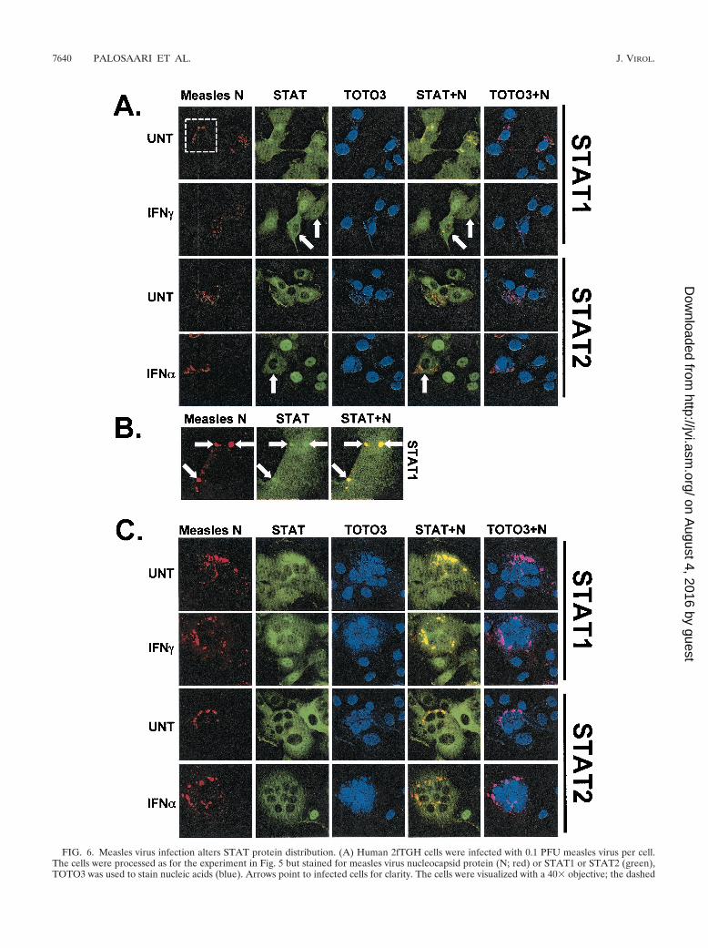
FIG. 6. Measles virus infection alters STAT protein distribution. (A) Human 2fTGH cells were infected with 0.1 PFU measles virus per cell.The cells were processed as for the experiment in Fig. 5 but stained for measles virus nucleocapsid protein (N; red) or STAT1 or STAT2 (green),TOTO3 was used to stain nucleic acids (blue). Arrows point to infected cells for clarity. The cells were visualized with a 40 objective; the dashed
7640 PALOSAARI ET AL. J. VIROL.
on August 4, 2016 by guest
http://jvi.asm.org/
Dow
nloaded from
signaling systems similar to IL-6 (1, 16), a cytokine induced bymeasles virus infections (3, 18). Treatment of 2fTGH cells withIL-6 potently induced reporter gene expression from a STAT3-responsive GAS-luciferase construct, but expression of mea-sles virus V protein reduced IL-6-induced reporter gene activ-ity to 72% of control values (P � 0.002 [Fig. 3A]). STAT3 isalso an important effector for intracellular tyrosine kinase sig-naling (6, 7). To determine the ability of measles virus Vprotein to block STAT3 signaling induced by an intracellularstimulus, the v-Src oncogenic tyrosine kinase was used to ac-tivate STAT3. Expression of v-Src potently induced reportergene activity, but this activity was reduced to 38% of controlvalues by expression of measles virus V protein (P � 0.0001[Fig. 3B]). Together, these results demonstrate that the mea-sles virus V protein is capable of inhibiting both extracellularand intracellular STAT3-dependent signaling.
Effect of measles virus V protein on STAT activation anddimerization. It has been reported that ISGF3 activation isinduced by measles virus infection (18). However, since mul-tiple STAT proteins were found to copurify as MeVIPs andV-dependent IFN signaling inhibition was observed, we moreclosely examined the early steps of IFN signaling: STAT pro-tein activating tyrosine phosphorylation and STAT1–STAT2heterodimerization. Stimulation of cells with IFN inducesphosphorylation of STAT2 on tyrosine 690 and of STAT1 ontyrosine 701. Immunoblotting with STAT phosphotyrosine-specific antisera revealed that the expression of measles virusV protein did not detectably reduce IFN-induced tyrosinephosphorylation of either STAT1 or STAT2 in transfected2fTGH cells or 293T cells or in a stable cell line harboring atetracycline-inducible measles virus V protein (Fig. 4A). Sim-ilarly, no inhibition of IFN-�-induced STAT1 tyrosine 701phosphorylation was observed (Fig. 4B). These data indicatethat IFN-dependent STAT protein activating tyrosine phos-phorylation is not targeted by measles virus V protein.
Following activation, STAT proteins dimerize by reciprocalSH2 domain-phosphotyrosine interactions (52). A coimmuno-precipitation assay was used to test STAT1–STAT2 het-erodimerization. Cell lysates were immunoprecipitated withSTAT2-specific antiserum, and immune complexes were pro-cessed for STAT1 immunoblotting. In control cells, STAT1 isdetected in the STAT2 immune complex only following IFNstimulation (Fig. 4C). In contrast, in cells expressing the mea-sles virus V protein, STAT1 was found in STAT2 immunecomplexes regardless of IFN stimulation (Fig. 4C), consistentwith V-dependent complex formation. Immunoblotting withthe STAT1 phosphotyrosine-specific antiserum indicates thatactivated STAT1 is not excluded from the coimmunoprecipi-tated material. One interpretation of these results is that themeasles virus V-STAT complex(es) is capable of being pre-sented to the IFN receptor-kinase complex but is deficient in asubsequent step of STAT signaling.
Measles virus V protein prevents IFN-induced STAT nu-clear accumulation. IFN signaling inhibition by the Nipah virus
V protein relies on alteration of the subcellular distribution ofSTAT1 and STAT2 (48). To determine the effects of measlesvirus V protein on STAT protein distribution, indirect immu-nofluorescence was used to visualize the subcellular localiza-tion of measles virus V protein, STAT1, and STAT2. Epitope-tagged V protein was detected with tag-specific antibodies, andSTAT1 and STAT2 were detected in the same cells by doublestaining. The measles virus V protein was found in both thenucleus and cytoplasm, and the distribution was not altered byIFN stimulation (Fig. 5A).
STAT1 is found in both the nucleus and cytoplasm in un-stimulated cells, a result of balanced signal-independent basalnuclear shuttling (31) (Fig. 5). STAT2 also dynamically shut-tles between the nucleus and cytoplasm, but with net export(48), resulting in cytoplasmic accumulation in unstimulatedcells (50). In response to IFN stimulation, both STAT1 andSTAT2 rapidly translocate to and accumulate within the nu-cleus (48, 50), as also observed here in control cells that did notexpress measles virus V protein (Fig. 5A). In cells expressingmeasles virus V protein, the basal STAT1 and STAT2 distri-bution did not change. However, IFN stimulation failed toinduce STAT nuclear redistribution in measles virus V-ex-pressing cells, which retained the distribution pattern of un-stimulated cells. The defective STAT protein nuclear translo-cation was observed uniformly in cells expressing measles virusV protein in several independent experiments (Fig. 5A pre-sents illustrative examples). These results verify that measlesvirus V protein does not induce STAT protein degradation. Inthe context of the tyrosine phosphorylation results, the resultsdemonstrate that the measles virus V protein inhibits IFNsignaling at a point between STAT activation and nuclearimport.
The cytoplasmic distribution of many proteins, including theNipah virus V protein, relies on Crm1-dependent nuclear ex-port, which can be inhibited by a fungal metabolite, leptomycinB (LMB) (65). A role for Crm1-dependent nuclear export inthe distribution of measles virus V protein and its effects onSTAT redistribution were tested in LMB-treated cells (Fig.5B). No redistribution of measles virus V protein toward nu-clear accumulation was observed in LMB-treated cells. Evenfollowing LMB treatment, IFN stimulation failed to induceSTAT nuclear redistribution in measles virus V-expressingcells, which retained the distribution patterns of unstimulatedcells. These results demonstrate that neither the distribution ofmeasles virus V protein nor its effects on STAT redistributiondepend on Crm1-mediated nuclear shuttling.
STAT protein redistribution in measles virus-infected cells.Indirect immunofluorescence was used to evaluate STAT pro-tein distribution in the context of measles virus infections. Anantibody that recognizes the measles virus nucleocapsid (N)protein was used to identify infected cells (Fig. 6). The Nprotein staining was localized to discrete cytoplasmic bodies, apattern that has been observed for other paramyxoviruses (12,13, 36, 44–46, 57, 61). All cells that stained positive for N also
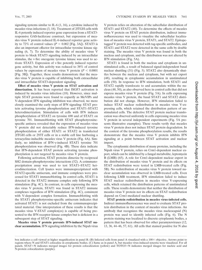
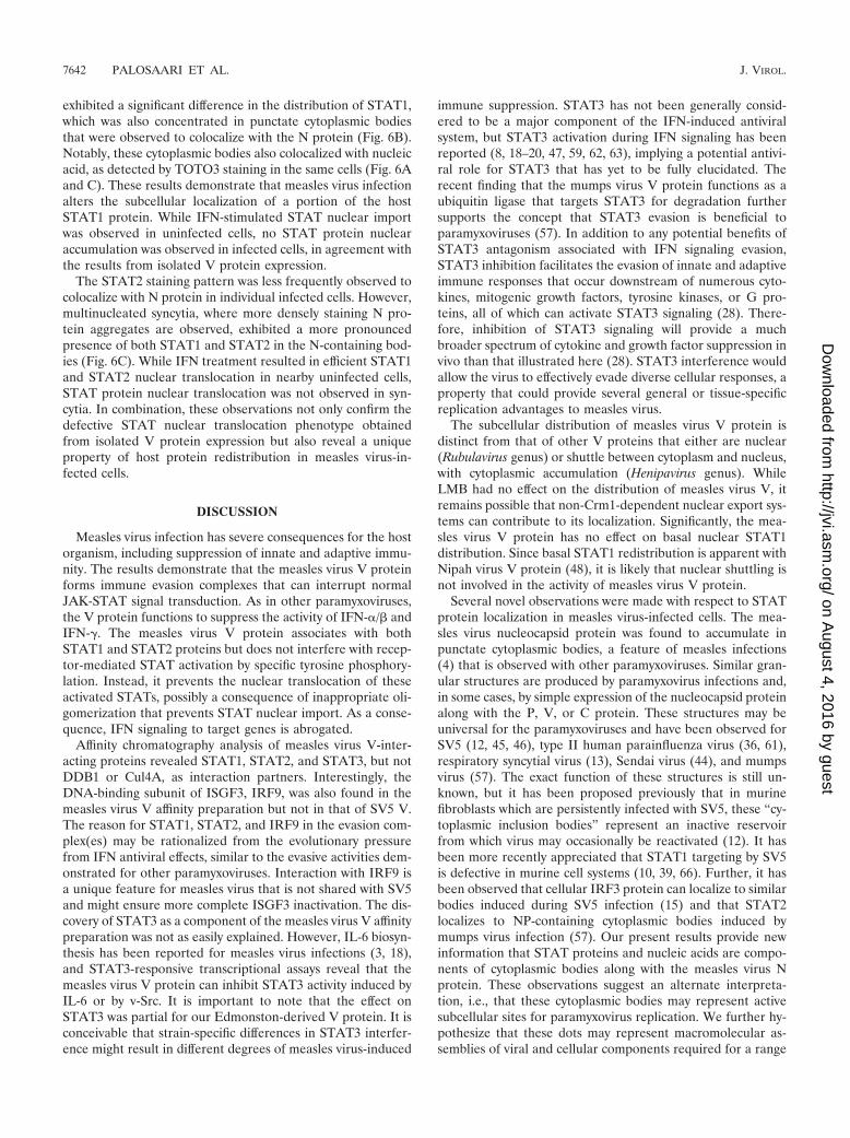
box indicates a cell viewed at higher magnification in panel B. (B) Infected cells from panel A visualized with a 100 objective. Arrows point toregions where N and STAT1 colocalize in cytoplasmic bodies. (C) Same as in panel A, but measles virus-induced syncytia were visualized. For allpanels. STAT�N indicates merged images for protein colocalization (yellow) and TOTO3�N indicates merged images for nucleic acid andN-protein colocalization (purple).
VOL. 77, 2003 CYTOKINE EVASION BY MEASLES VIRUS 7641
on August 4, 2016 by guest
http://jvi.asm.org/
Dow
nloaded from
exhibited a significant difference in the distribution of STAT1,which was also concentrated in punctate cytoplasmic bodiesthat were observed to colocalize with the N protein (Fig. 6B).Notably, these cytoplasmic bodies also colocalized with nucleicacid, as detected by TOTO3 staining in the same cells (Fig. 6Aand C). These results demonstrate that measles virus infectionalters the subcellular localization of a portion of the hostSTAT1 protein. While IFN-stimulated STAT nuclear importwas observed in uninfected cells, no STAT protein nuclearaccumulation was observed in infected cells, in agreement withthe results from isolated V protein expression.
The STAT2 staining pattern was less frequently observed tocolocalize with N protein in individual infected cells. However,multinucleated syncytia, where more densely staining N pro-tein aggregates are observed, exhibited a more pronouncedpresence of both STAT1 and STAT2 in the N-containing bod-ies (Fig. 6C). While IFN treatment resulted in efficient STAT1and STAT2 nuclear translocation in nearby uninfected cells,STAT protein nuclear translocation was not observed in syn-cytia. In combination, these observations not only confirm thedefective STAT nuclear translocation phenotype obtainedfrom isolated V protein expression but also reveal a uniqueproperty of host protein redistribution in measles virus-in-fected cells.
DISCUSSION
Measles virus infection has severe consequences for the hostorganism, including suppression of innate and adaptive immu-nity. The results demonstrate that the measles virus V proteinforms immune evasion complexes that can interrupt normalJAK-STAT signal transduction. As in other paramyxoviruses,the V protein functions to suppress the activity of IFN-�/� andIFN-�. The measles virus V protein associates with bothSTAT1 and STAT2 proteins but does not interfere with recep-tor-mediated STAT activation by specific tyrosine phosphory-lation. Instead, it prevents the nuclear translocation of theseactivated STATs, possibly a consequence of inappropriate oli-gomerization that prevents STAT nuclear import. As a conse-quence, IFN signaling to target genes is abrogated.
Affinity chromatography analysis of measles virus V-inter-acting proteins revealed STAT1, STAT2, and STAT3, but notDDB1 or Cul4A, as interaction partners. Interestingly, theDNA-binding subunit of ISGF3, IRF9, was also found in themeasles virus V affinity preparation but not in that of SV5 V.The reason for STAT1, STAT2, and IRF9 in the evasion com-plex(es) may be rationalized from the evolutionary pressurefrom IFN antiviral effects, similar to the evasive activities dem-onstrated for other paramyxoviruses. Interaction with IRF9 isa unique feature for measles virus that is not shared with SV5and might ensure more complete ISGF3 inactivation. The dis-covery of STAT3 as a component of the measles virus V affinitypreparation was not as easily explained. However, IL-6 biosyn-thesis has been reported for measles virus infections (3, 18),and STAT3-responsive transcriptional assays reveal that themeasles virus V protein can inhibit STAT3 activity induced byIL-6 or by v-Src. It is important to note that the effect onSTAT3 was partial for our Edmonston-derived V protein. It isconceivable that strain-specific differences in STAT3 interfer-ence might result in different degrees of measles virus-induced
immune suppression. STAT3 has not been generally consid-ered to be a major component of the IFN-induced antiviralsystem, but STAT3 activation during IFN signaling has beenreported (8, 18–20, 47, 59, 62, 63), implying a potential antivi-ral role for STAT3 that has yet to be fully elucidated. Therecent finding that the mumps virus V protein functions as aubiquitin ligase that targets STAT3 for degradation furthersupports the concept that STAT3 evasion is beneficial toparamyxoviruses (57). In addition to any potential benefits ofSTAT3 antagonism associated with IFN signaling evasion,STAT3 inhibition facilitates the evasion of innate and adaptiveimmune responses that occur downstream of numerous cyto-kines, mitogenic growth factors, tyrosine kinases, or G pro-teins, all of which can activate STAT3 signaling (28). There-fore, inhibition of STAT3 signaling will provide a muchbroader spectrum of cytokine and growth factor suppression invivo than that illustrated here (28). STAT3 interference wouldallow the virus to effectively evade diverse cellular responses, aproperty that could provide several general or tissue-specificreplication advantages to measles virus.
The subcellular distribution of measles virus V protein isdistinct from that of other V proteins that either are nuclear(Rubulavirus genus) or shuttle between cytoplasm and nucleus,with cytoplasmic accumulation (Henipavirus genus). WhileLMB had no effect on the distribution of measles virus V, itremains possible that non-Crm1-dependent nuclear export sys-tems can contribute to its localization. Significantly, the mea-sles virus V protein has no effect on basal nuclear STAT1distribution. Since basal STAT1 redistribution is apparent withNipah virus V protein (48), it is likely that nuclear shuttling isnot involved in the activity of measles virus V protein.
Several novel observations were made with respect to STATprotein localization in measles virus-infected cells. The mea-sles virus nucleocapsid protein was found to accumulate inpunctate cytoplasmic bodies, a feature of measles infections(4) that is observed with other paramyxoviruses. Similar gran-ular structures are produced by paramyxovirus infections and,in some cases, by simple expression of the nucleocapsid proteinalong with the P, V, or C protein. These structures may beuniversal for the paramyxoviruses and have been observed forSV5 (12, 45, 46), type II human parainfluenza virus (36, 61),respiratory syncytial virus (13), Sendai virus (44), and mumpsvirus (57). The exact function of these structures is still un-known, but it has been proposed previously that in murinefibroblasts which are persistently infected with SV5, these “cy-toplasmic inclusion bodies” represent an inactive reservoirfrom which virus may occasionally be reactivated (12). It hasbeen more recently appreciated that STAT1 targeting by SV5is defective in murine cell systems (10, 39, 66). Further, it hasbeen observed that cellular IRF3 protein can localize to similarbodies induced during SV5 infection (15) and that STAT2localizes to NP-containing cytoplasmic bodies induced bymumps virus infection (57). Our present results provide newinformation that STAT proteins and nucleic acids are compo-nents of cytoplasmic bodies along with the measles virus Nprotein. These observations suggest an alternate interpreta-tion, i.e., that these cytoplasmic bodies may represent activesubcellular sites for paramyxovirus replication. We further hy-pothesize that these dots may represent macromolecular as-semblies of viral and cellular components required for a range
7642 PALOSAARI ET AL. J. VIROL.
on August 4, 2016 by guest
http://jvi.asm.org/
Dow
nloaded from
of enzymatic activities contributing to host immune evasionand virus replication.
The mechanisms described here for measles virus-inducedSTAT inhibition are unprecedented among the Paramyxoviri-dae genera, adding to the catalog of STAT inhibition by Vproteins alongside the Rubulavirus-encoded E3 ubiquitin ligaseactivities (56) and the Henipavirus-encoded STAT cytoplasmicsequestration mechanisms (48). Together, these suppressionactivities contribute to the severity of measles virus pathoge-nicity.
ACKNOWLEDGMENTS
We are grateful to Huabao Xiong, Sergio Lira, Adrian Ting, PatriciaCortes, and Horvath laboratory members for helpful discussions andcomments about the manuscript.
This work was supported by research grants to C.M.H. from theAmerican Cancer Society (Research Scholar Grant GMC-103079) andthe NIH (AI-48722 and AI-50707). J.J.R. is a trainee in the integratedtraining program in pharmacological sciences (GM-62754). Confocalmicroscopy at Mt. Sinai is partially supported by NIH Shared Instru-mentation grant 1S10 RR0 9145 and NSF Major Research Instrumen-tation grant DBI-9724504.
REFERENCES
1. Aaronson, D. S., and C. M. Horvath. 2002. A road map for those who don’tknow JAK-STAT. Science 296:1653–1655.
2. Andrejeva, J., E. Poole, D. F. Young, S. Goodbourn, and R. E. Randall. 2002.The p127 subunit (DDB1) of the UV-DNA damage repair binding protein isessential for the targeted degradation of STAT1 by the V protein of theparamyxovirus simian virus 5. J. Virol. 76:11379–11386.
3. Bieback, K., E. Lien, I. M. Klagge, E. Avota, J. Schneider-Schaulies, W. P.Duprex, H. Wagner, C. J. Kirschning, V. Ter Meulen, and S. Schneider-Schaulies. 2002. Hemagglutinin protein of wild-type measles virus activatestoll-like receptor 2 signaling. J. Virol. 76:8729–8736.
4. Bohn, W., F. Ciampor, R. Rutter, and K. Mannweiler. 1990. Localization ofnucleocapsid associated polypeptides in measles virus-infected cells by im-munogold labelling after resin embedding. Arch. Virol. 114:53–64.
5. Bolt, G., K. Berg, and M. Blixenkrone-Moller. 2002. Measles virus-inducedmodulation of host-cell gene expression. J. Gen. Virol. 83:1157–1165.
6. Bowman, T., R. Garcia, J. Turkson, and R. Jove. 2000. STATs in oncogen-esis. Oncogene 19:2474–2488.
7. Bromberg, J., and J. E. Darnell, Jr. 2000. The role of STATs in transcrip-tional control and their impact on cellular function. Oncogene 19:2468–2473.
8. Caldenhoven, E., M. Buitenhuis, T. B. van Dijk, J. A. Raaijmakers, J. W.Lammers, L. Koenderman, and R. P. de Groot. 1999. Lineage-specific acti-vation of STAT3 by interferon-gamma in human neutrophils. J. Leukoc.Biol. 65:391–396.
9. Decker, T., D. J. Lew, J. Mirkovitch, and J. E. Darnell, Jr. 1991. Cytoplasmicactivation of GAF, an IFN-� regulated DNA binding factor. EMBO J.10:927–932.
10. Didcock, L., D. F. Young, S. Goodbourn, and R. E. Randall. 1999. Sendaivirus and simian virus 5 block activation of interferon- responsive genes:importance for virus pathogenesis. J. Virol. 73:3125–3133.
11. Didcock, L., D. F. Young, S. Goodbourn, and R. E. Randall. 1999. The Vprotein of simian virus 5 inhibits interferon signalling by targeting STAT1 forproteasome-mediated degradation. J. Virol. 73:9928–9933.
12. Fearns, R., D. F. Young, and R. E. Randall. 1994. Evidence that theparamyxovirus simian virus 5 can establish quiescent infections by remaininginactive in cytoplasmic inclusion bodies. J. Gen. Virol. 75:3525–3539.
13. Garcia, J., B. Garcia-Barreno, A. Vivo, and J. A. Melero. 1993. Cytoplasmicinclusions of respiratory syncytial virus-infected cells: formation of inclusionbodies in transfected cells that coexpress the nucleoprotein, the phospho-protein, and the 22K protein. Virology 195:243–247.
14. Guschin, D., N. Rogers, J. Briscoe, B. Witthuhn, D. Watling, F. Horn, S.Pellegrini, K. Yasukawa, P. Heinrich, G. R. Stark, J. N. Ihle, and I. M. Kerr.1995. A major role for the protein tyrosine kinase JAK1 in the JAK/STATpathway in response to interleukin-6. EMBO J. 14:1421–1429.
15. He, B., R. G. Paterson, N. Stock, J. E. Durbin, R. K. Durbin, S. Goodbourn,R. E. Randall, and R. A. Lamb. 2002. Recovery of paramyxovirus simianvirus 5 with a V protein lacking the conserved cysteine-rich domain: themultifunctional V protein blocks both interferon-beta induction and inter-feron signaling. Virology 303:15–32.
16. Heinrich, P. C., I. Behrmann, G. Muller-Newen, F. Schaper, and L. Graeve.1998. Interleukin-6-type cytokine signalling through the gp130/Jak/STATpathway. Biochem. J. 334:297–314.
17. Helin, E., S. Matikainen, I. Julkunen, J. Heino, T. Hyypia, and R. Vainion-paa. 2002. Measles virus enhances the expression of cellular immediate-earlygenes and DNA-binding of transcription factor AP-1 in lung epithelial A549cells. Arch. Virol. 147:1721–1732.
18. Helin, E., R. Vainionpaa, T. Hyypia, I. Julkunen, and S. Matikainen. 2001.Measles virus activates NF-kappa B and STAT transcription factors andproduction of IFN-alpha/beta and IL-6 in the human lung epithelial cell lineA549. Virology 290:1–10.
19. Horvath, C. M., and J. E. Darnell, Jr. 1996. The antiviral state induced byalpha interferon and gamma interferon requires transcriptionally activeStat1 protein. J. Virol. 70:647–650.
20. Horvath, C. M., Z. Wen, and J. E. Darnell, Jr. 1995. A STAT protein domainthat determines DNA sequence recognition suggests a novel DNA-bindingdomain. Genes Dev. 9:984–994.
21. Ikeda, H., L. J. Old, and R. D. Schreiber. 2002. The roles of IFN gamma inprotection against tumor development and cancer immunoediting. CytokineGrowth Factor Rev. 13:95–109.
22. Katze, M. G., Y. He, and M. Gale, Jr. 2002. Viruses and interferon: a fightfor supremacy. Nat. Rev. Immunol. 2:675–687.
23. Kubota, T., N. Yokosawa, S. Yokota, and N. Fujii. 2002. Association ofmumps virus V protein with RACK1 results in dissociation of STAT-1 fromthe alpha interferon receptor complex. J. Virol. 76:12676–12682.
24. Kubota, T., N. Yokosawa, S. Yokota, and N. Fujii. 2001. C terminal CYS-RICH region of mumps virus structural V protein correlates with block ofinterferon alpha and gamma signal transduction pathway through decreaseof STAT 1-alpha. Biochem. Biophys. Res. Commun. 283:255–259.
25. Lamb, R. A., and D. Kolakofsky. 1996. Paramyxoviridae: the viruses and theirreplication, p. 1177–1204. In B. N. Fields, D. M. Knipe, and P. M. Howley(ed.), Fields virology, 3rd ed. Lippincott-Raven Publishers, Philadelphia, Pa.
26. Levy, D. E., and J. E. Darnell, Jr. 2002. Stats: transcriptional control andbiological impact. Nat. Rev. Mol. Cell Biol. 3:651–662.
27. Levy, D. E., D. S. Kessler, R. I. Pine, and J. E. Darnell, Jr. 1989. Cytoplasmicactivation of ISGF3, the positive regulator of interferon-� stimulated tran-scription, reconstituted in vitro. Genes Dev. 3:1362–1372.
28. Levy, D. E., and C. K. Lee. 2002. What does Stat3 do? J. Clin. Investig.109:1143–1148.
29. Lin, G. Y., and R. A. Lamb. 2000. The paramyxovirus simian virus 5 Vprotein slows progression of the cell cycle. J. Virol. 74:9152–9166.
30. Lin, G. Y., R. G. Paterson, C. D. Richardson, and R. A. Lamb. 1998. The Vprotein of the paramyxovirus SV5 interacts with damage-specific DNA bind-ing protein. Virology 249:189–200.
31. Meyer, T., A. Begitt, I. Lodige, M. van Rossum, and U. Vinkemeier. 2002.Constitutive and IFN-gamma-induced nuclear import of STAT1 proceedthrough independent pathways. EMBO J. 21:344–354.
32. Morgenstern, J. P., and H. Land. 1990. Advanced mammalian gene transfer:high titre retroviral vectors with multiple drug selection markers and acomplementary helper-free packaging cell line. Nucleic Acids Res. 18:3587–3596.
33. Murray, C. J., and A. D. Lopez. 1997. Mortality by cause for eight regions ofthe world: Global Burden of Disease Study. Lancet 349:1269–1276.
34. Naniche, D., A. Yeh, D. Eto, M. Manchester, R. M. Friedman, and M. B.Oldstone. 2000. Evasion of host defenses by measles virus: wild-type measlesvirus infection interferes with induction of alpha/beta interferon production.J. Virol. 74:7478–7484.
35. Nishio, M., D. Garcin, V. Simonet, and D. Kolakofsky. 2002. The carboxylsegment of the mumps virus V protein associates with Stat proteins in vitrovia a tryptophan-rich motif. Virology 300:92–99.
36. Nishio, M., M. Tsurudome, M. Kawano, N. Watanabe, S. Ohgimoto, M. Ito,H. Komada, and Y. Ito. 1996. Interaction between nucleocapsid protein (NP)and phosphoprotein (P) of human parainfluenza virus type 2: one of the twoNP binding sites on P is essential for granule formation. J. Gen. Virol.77:2457–2463.
37. Parisien, J.-P., J. F. Lau, J. J. Rodriguez, B. M. Sullivan, A. Moscona, G. D.Parks, R. A. Lamb, and C. M. Horvath. 2001. The V protein of humanparainfluenza virus 2 antagonizes type I interferon responses by destabilizingsignal transducer and activator of transcription 2. Virology 283:230–239.
38. Parisien, J.-P., J. F. Lau, J. J. Rodriguez, C. M. Ulane, and C. M. Horvath.2002. Selective STAT protein degradation induced by paramyxoviruses re-quires both STAT1 and STAT2, but is independent of alpha/beta interferonsignal transduction. J. Virol. 76:4190–4198.
39. Parisien, J. P., J. F. Lau, and C. M. Horvath. 2002. STAT2 acts as a hostrange determinant for species-specific paramyxovirus interferon antagonismand simian virus 5 replication. J. Virol. 76:6435–6441.
40. Park, M. S., M. L. Shaw, J. Munoz-Jordan, J. F. Cros, T. Nakaya, N.Bouvier, P. Palese, A. Garcia-Sastre, and C. F. Basler. 2003. Newcastledisease virus (NDV)-based assay demonstrates interferon-antagonist activityfor the NDV V protein and the Nipah virus V, W, and C proteins. J. Virol.77:1501–1511.
41. Paterson, R. G., G. P. Leser, M. A. Shaughnessy, and R. A. Lamb. 1995. Theparamyxovirus SV5 V protein binds two atoms of zinc and is a structuralcomponent of virions. Virology 208:121–131.
42. Patterson, J. B., D. Thomas, H. Lewicki, M. A. Billeter, and M. B. Oldstone.
VOL. 77, 2003 CYTOKINE EVASION BY MEASLES VIRUS 7643
on August 4, 2016 by guest
http://jvi.asm.org/
Dow
nloaded from
2000. V and C proteins of measles virus function as virulence factors in vivo.Virology 267:80–89.
43. Poole, E., B. He, R. A. Lamb, R. E. Randall, and S. Goodbourn. 2002. TheV proteins of simian virus 5 and other paramyxoviruses inhibit induction ofinterferon-beta. Virology 303:33–46.
44. Portner, A., K. C. Gupta, J. M. Seyer, E. H. Beachey, and D. W. Kingsbury.1986. Localization and characterization of Sendai virus nonstructural C andC proteins by antibodies against synthetic peptides. Virus Res. 6:109–121.
45. Precious, B., D. F. Young, A. Bermingham, R. Fearns, M. Ryan, and R. E.Randall. 1995. Inducible expression of the P, V, and NP genes of theparamyxovirus simian virus 5 in cell lines and an examination of NP-P andNP-V interactions. J. Virol. 69:8001–8010.
46. Randall, R. E., and A. Bermingham. 1996. NP:P and NP:V interactions ofthe paramyxovirus simian virus 5 examined using a novel protein:proteincapture assay. Virology 224:121–129.
47. Rani, M. R., D. W. Leaman, Y. Han, S. Leung, E. Croze, E. N. Fish, A.Wolfman, and R. M. Ransohoff. 1999. Catalytically active TYK2 is essentialfor interferon-beta-mediated phosphorylation of STAT3 and interferon-al-pha receptor-1 (IFNAR-1) but not for activation of phosphoinositol 3-ki-nase. J. Biol. Chem. 274:32507–32511.
48. Rodriguez, J. J., J. P. Parisien, and C. M. Horvath. 2002. Nipah virus Vprotein evades alpha and gamma interferons by preventing STAT1 andSTAT2 activation and nuclear accumulation. J. Virol. 76:11476–11483.
49. Samuel, C. E. 2001. Antiviral actions of interferons. Clin. Microbiol. Rev.14:778–809.
50. Schindler, C., K. Shuai, V. R. Prezioso, and J. E. Darnell, Jr. 1992. Inter-feron-dependent tyrosine phosphorylation of a latent cytoplasmic transcrip-tion factor. Science 257:809–813.
51. Schneider, H., K. Kaelin, and M. A. Billeter. 1997. Recombinant measlesviruses defective for RNA editing and V protein synthesis are viable incultured cells. Virology 227:314–322.
52. Shuai, K., C. M. Horvath, L. H. Tsai-Huang, S. Qureshi, D. Cowburn, andJ. E. Darnell, Jr. 1994. Interferon activation of the transcription factor Stat91involves dimerization through SH2-phosphotyrosyl peptide interactions. Cell76:821–828.
53. Stark, G. R., I. M. Kerr, B. R. Williams, R. H. Silverman, and R. D.Schreiber. 1998. How cells respond to interferons. Annu. Rev. Biochem.67:227–264.
54. tenOever, B. R., M. J. Servant, N. Grandvaux, R. Lin, and J. Hiscott. 2002.Recognition of the measles virus nucleocapsid as a mechanism of IRF-3activation. J. Virol. 76:3659–3669.
55. Thomas, S. M., R. A. Lamb, and R. G. Paterson. 1988. Two mRNAs thatdiffer by two nontemplated nucleotides encode the amino coterminal pro-teins P and V of the paramyxovirus SV5. Cell 54:891–902.
56. Ulane, C. M., and C. M. Horvath. 2002. Paramyxoviruses SV5 and HPIV2assemble STAT protein ubiquitin ligase complexes from cellular compo-nents. Virology 304:160–166.
57. Ulane, C. M., J. J. Rodriguez, J.-P. Parisien, and C. M. Horvath. 2003.
STAT3 ubiquitylation and degradation by mumps virus suppresses cytokineand oncogene signaling. J. Virol. 77:6385–6393.
58. Valsamakis, A., H. Schneider, P. G. Auwaerter, H. Kaneshima, M. A. Bill-eter, and D. E. Griffin. 1998. Recombinant measles viruses with mutations inthe C, V, or F gene have altered growth phenotypes in vivo. J. Virol.72:7754–7761.
59. Velichko, S., T. C. Wagner, J. Turkson, R. Jove, and E. Croze. 2002. STAT3Activation by type I interferons is dependent on specific tyrosines located inthe cytoplasmic domain of interferon receptor chain 2c. Activation of mul-tiple STATs proceeds through the redundant usage of two tyrosine residues.J. Biol. Chem. 277:35635–35641.
60. Wansley, E. K., and G. D. Parks. 2002. Naturally occurring substitutions inthe P/V gene convert the noncytopathic paramyxovirus simian virus 5 into avirus that induces alpha/beta interferon synthesis and cell death. J. Virol.76:10109–10121.
61. Watanabe, N., M. Kawano, M. Tsurudome, M. Nishio, M. Ito, S. Ohgimoto,S. Suga, H. Komada, and Y. Ito. 1996. Binding of the V proteins to thenucleocapsid proteins of human parainfluenza type 2 virus. Med. Microbiol.Immunol. 185:89–94.
62. Yang, C. H., A. Murti, and L. M. Pfeffer. 1998. STAT3 complements defectsin an interferon-resistant cell line: evidence for an essential role for STAT3in interferon signaling and biological activities. Proc. Natl. Acad. Sci. USA95:5568–5572.
63. Yang, C. H., W. Shi, L. Basu, A. Murti, S. N. Constantinescu, L. Blatt, E.Croze, J. E. Mullersman, and L. M. Pfeffer. 1996. Direct association ofSTAT3 with the IFNAR-1 chain of the human type I interferon receptor.J. Biol. Chem. 271:8057–8061.
64. Yokosawa, N., S. Yokota, T. Kubota, and N. Fujii. 2002. C-terminal region ofSTAT-1alpha is not necessary for its ubiquitination and degradation causedby mumps virus V protein. J. Virol. 76:12683–12690.
65. Yoshida, M., and S. Horinouchi. 1999. Trichostatin and leptomycin. Inhibi-tion of histone deacetylation and signal-dependent nuclear export. Ann.N. Y. Acad. Sci. 886:23–36.
66. Young, D. F., N. Chatziandreou, B. He, S. Goodbourn, R. A. Lamb, and R. E.Randall. 2001. Single amino acid substitution in the V protein of simian virus5 differentiates its ability to block interferon signaling in human and murinecells. J. Virol. 75:3363–3370.
67. Young, D. F., L. Didcock, S. Goodbourn, and R. E. Randall. 2000. Paramyxo-viridae use distinct virus-specific mechanisms to circumvent the interferonresponse. Virology 269:383–390.
68. Yu, C. L., D. J. Meyer, G. S. Campbell, A. C. Larner, C. Carter-Su, J.Schwartz, and R. Jove. 1995. Enhanced DNA-binding activity of a Stat3-related protein in cells transformed by the Src oncoprotein. Science 269:81–83.
69. Zhong, Z., Z. Wen, and J. E. Darnell, Jr. 1994. Stat3: a STAT family memberactivated by tyrosine phosphorylation in response to epidermal growth factorand interleukin-6. Science 264:95–98.
7644 PALOSAARI ET AL. J. VIROL.
on August 4, 2016 by guest
http://jvi.asm.org/
Dow
nloaded from