Spore Dispersal of Slime Molds and Higher Fungi via Animal ...
70
University of Arkansas, Fayetteville University of Arkansas, Fayetteville ScholarWorks@UARK ScholarWorks@UARK Graduate Theses and Dissertations 5-2021 Spore Dispersal of Slime Molds and Higher Fungi via Animal Spore Dispersal of Slime Molds and Higher Fungi via Animal Vectors Vectors Courtney Trimble University of Arkansas, Fayetteville Follow this and additional works at: https://scholarworks.uark.edu/etd Part of the Plant Biology Commons, Plant Pathology Commons, and the Terrestrial and Aquatic Ecology Commons Citation Citation Trimble, C. (2021). Spore Dispersal of Slime Molds and Higher Fungi via Animal Vectors. Graduate Theses and Dissertations Retrieved from https://scholarworks.uark.edu/etd/4085 This Thesis is brought to you for free and open access by ScholarWorks@UARK. It has been accepted for inclusion in Graduate Theses and Dissertations by an authorized administrator of ScholarWorks@UARK. For more information, please contact [email protected].
-
Upload
khangminh22 -
Category
Documents
-
view
0 -
download
0
Transcript of Spore Dispersal of Slime Molds and Higher Fungi via Animal ...

University of Arkansas, Fayetteville University of Arkansas, Fayetteville
ScholarWorks@UARK ScholarWorks@UARK
Graduate Theses and Dissertations
5-2021
Spore Dispersal of Slime Molds and Higher Fungi via Animal Spore Dispersal of Slime Molds and Higher Fungi via Animal
Vectors Vectors
Courtney Trimble University of Arkansas, Fayetteville
Follow this and additional works at: https://scholarworks.uark.edu/etd
Part of the Plant Biology Commons, Plant Pathology Commons, and the Terrestrial and Aquatic
Ecology Commons
Citation Citation Trimble, C. (2021). Spore Dispersal of Slime Molds and Higher Fungi via Animal Vectors. Graduate Theses and Dissertations Retrieved from https://scholarworks.uark.edu/etd/4085
This Thesis is brought to you for free and open access by ScholarWorks@UARK. It has been accepted for inclusion in Graduate Theses and Dissertations by an authorized administrator of ScholarWorks@UARK. For more information, please contact [email protected].

Spore Dispersal of Slime Molds and Higher Fungi via Animal Vectors
A thesis submitted in partial fulfilment
of the requirements for the degree of
Master of Science in Biology
by
Courtney Trimble
University of Arkansas-Fort Smith
Bachelor of Science in Biology, 2007
May 2021
University of Arkansas
This thesis is approved for recommendation to the Graduate Council
_______________________________________
Steven L. Stephenson, Ph.D.
Thesis Director
_______________________________________
Frederick Spiegel, Ph.D.
Committee Member
_______________________________________
J. D. Wilson, Ph.D.
Committee Member

Abstract
Myxomycetes and dictyostelids are Amoebozoans that are cosmopolitan inhabitants of a
variety of habitats, particularly forest environments. Both groups reproduce using spores which
are primarily dispersed via wind in myxomycetes but this characteristic poses a problem for
dictyostelids. The spores of dictyostelids are incased in a mucilaginous matrix that makes wind
ineffective except in exceptional cases. It has been suggested that animals such as birds may play
an important yet understudied role in the dispersal of these organisms. This study investigated
how animals could potentially serve as vectors for spore dispersal of dictyostelids and
myxomycetes with some limited data obtained of higher fungi. The ecology of these organisms
is understudied and the potential interactions between these and other animals is largely
unknown. Animals may disperse spores to different areas by consuming spores or other animals
such as insects that have consumed spores, or by moving across areas where myxomycetes and
dictyostelids occur. Coprophilous myxomycetes occur primarily on dung and data was collected
from the northwest Arkansas area investigating potential differences in the species composition
of myxomycetes isolated on the dung of large herbivorous mammals. There is limited previous
data indicating that birds, amphibians, small mammals, and bats may disperse the spores of
dictyostelids and this study was the first recorded instance where dictyostelids have been isolated
from reptiles.

Acknowledgements
This study would not have been possible without the generous help of a number of
people. I am grateful to Autumn Coffey, Dr. Adam Rollins, and Dr. Francis Onduso for their
help in collecting samples from deer and other animals. Beth Kegley from the University of
Arkansas Experimental Farm was a great help in obtaining samples of horse and cow dung.
Nazrana Payal and Gurpreet Kaur Thiara helped take pH readings of moist chamber cultures.
Apulu Ndotimi took photographs of specimens that were isolated for this study. Marla Steele and
Jason Ortega were helpful in gathering samples for the reptile data. A special acknowledgment
goes to Dr. Steve Stephenson for his help in finding some relevant publications and for verifying
the identification of some specimens.

Table of Contents
I. Introduction…………………………………..…………………………………………………1
II. Spore Dispersal of Slime Molds and Higher Fungi via Animal Vectors………………………3
III. Dispersal of Dictyostelid Spores by Reptilian Vectors………………………………………25
IV. Large Herbivorous Mammals as Vectors of Coprophilous Myxomycetes …………..………40
V. Conclusions…………………………………………………………………………………...64

1
Introduction
This study investigated the role that animals play in the dispersal of myxomycetes
and dictyostelids with some limited data also obtained from higher fungi. All three groups
produce spores that can be dispersed by animals such as birds, insects, and large herbivorous
mammals via either direct ingestion or when the animal moves across the fruiting body of a
slime mold or fungus. These ecological relationships are understudied and may be particularly
significant for dictyostelids. Dictyostelids are Amobozoans that are commonly found in the
soil/litter layer on the forest floor. The spores of dictyostelids are incased in a mucilaginous
matrix, which makes wind dispersal less effective than it is for myxomycetes. Previous studies
have indicated that animals such as birds may pick up the spores of dictyostelids from eating
insects or from foraging on the forest floor and that the spores can pass through the digestive
system unharmed. Prior to this study there had been no published studies investigating whether
reptiles can serve in a similar role. In 2014, a series of samples obtained from reptiles was
evaluated for the presence of dictyostelids. These samples were collected from Lake Fayetteville,
The Ozark Natural Science Center, and Lake Leatherwood in northwest Arkansas. Swabs were
taken from lizards, turtles, and snakes and then taken back to the Eumycetozoan Laboratory at
the University of Arkansas for processing. It was hypothesized that the species of dictyostelids
isolated from these reptiles could have related to the habitats of these animals. Myxomycetes are
a much larger group of Amoebozoans that are cosmopolitan and found in virtually every
ecosystem investigated to date. Although myxomycetes can disperse their spores via wind a
small number of myxomycete are found primarily and in some cases virtually only on the dung
of large herbivorous mammals. Coprophilous myxomycetes are the most understudied ecological
group of myxomycetes and this association may be particularly important in arid regions where

2
there are extreme temperatures and alternative substrates are not available. During the fall and
winter of 2019 and 2020, a series of dung samples was collected from horses, cows, deer and
other animals from Pennsylvania, West Virginia, the University of Arkansas Experimental Farm,
and the North Dakota Zoo. Dung samples were air-dried, and several series of moist chambers
were prepared. The moist chambers were checked approximately once a week with a dissecting
microscope for up to five months. Fruiting bodies that formed in culture were identified and
placed in small paper boxes for storage. During the fall of 2019, deer pellets were collected and
examined for the spores of higher fungi. Each deer pellet was suspended in a vial of distilled
water and agitated to separate any spores present from the substrate. A series of photographs was
generated to show fungal spores from some of the samples, but this project could not be
completed due to the historic COVID-19 pandemic.

3
Spore Dispersal of Slime molds and Higher Fungi via Animal Vectors
Abstract
The purpose of this study was to investigate the role that animals play in the dispersal of
myxomycetes and dictyostelids. Dictyostelids (cellular slime molds) and myxomycetes
(plasmodial slime molds) are protists that are cosmopolitan and reproduce through the
production of spores. Animals such as birds have been shown to disperse their spores in previous
studies, in some cases over vast distances. The role that animals play as spore vectors is still
largely understudied and more research is needed to understand the importance of this dispersal
strategy and its potential effects on the ecology and genetics of these organisms. This association
may be particularly important in dictyostelids since their spores are encased in a mucilaginous
matrix which makes wind dispersal largely ineffective except in exceptional cases.
Introduction
Such unassuming places as dried leaf litter, soil, tree bark, and animal dung reveal a rich
diversity of small, amoeboid organisms informally referred to as slime molds or mycetozoans.
Although they resemble certain microfungi, slime molds are classified as protists belonging to
the domain Amoebozoa. Most people have never heard of slime molds, but there exists a varied
world of these organisms that goes largely unnoticed since most are too small to be observed
directly with the naked eye. Amoebozoa is extremely diverse and includes important model
organisms and significant pathogens. Slime molds are a large group of over 1000 species and
were traditionally placed in the Eumycetozoa with three major groups Dictyostelia, Myxogastria,
and Protosporangiida (formerly Protostelia). Within Amoebozoa there are two groups. These are
the sorocarpic slime molds (dictyostelids, social amoebae, aggregative amoebae, or cellular slime
molds) and the sporocarpic myxomycetes (plasmodial slime molds or myxomycetes) along with

4
the protosteloid amoebae (formally referred to as protostelids now classified as
protosproangiids). In sporocarpic organisms a single amoeboid cell develops into a usually
stalked, subaerial structure (fruiting body) that supports one to many propagules called spores, a
characteristic unique to the Amoebozoa. Sorocarpic amoebae aggregate into a multicellular mass
that develops into a subaerial fruiting body consisting of either distinct stalk cells and spores or
non-differentiated encysted cells (usually also called spores). Within the Amoebozoa sorocarpic
cells are found in two lineages the Dictyostelia (Eumycetozoa) and in Copromyxa (Tubulinea).
Sorocarpic taxa are found outside the Amoebozoa among Opisthokonta, Excavata,
Stramenopiles, Alveolata, and Rhizaria (Adle et al. 2019: Kang, 2017: Olive 1975: Sheikh et al.
2018: Raper 1984: Spiegel et al. 2017).
The plasmodial slime molds, which are commonly referred to as myxomycetes, “true”
slime molds, or acellular slime molds are more familiar since most are macroscopic and some
species can form large fruitings that are easily observed. Plasmodial slime molds were first
referred to as myxomycetes by Heinrich Link in 1833, and their name is derived from the two
Greek words myxa (slime) and myketes (fungi). He regarded them as fungi, but this view was
later challenged by Anton de Bary, who felt that they were more closely related to protozoans
and named the group Mycetozoa, from the Greek words zoon (animal) and mykes (fungus). Olive
(1975) named this group Eumycetozoa with the monophyletic taxa Myxogastria and Dictyostelia
arising from the paraphyletic taxon Protostelia (Shadwick et al., 2009). Myxomycetes are
cosmopolitan and species have been found on every continent, including Antarctica (Olive,
1975). There are over 1,000 species of these organisms that traditionally have been divided into
six different taxonomic orders. These are the Ceratiomyxales, Echinosteliales, Liceales,
Physarales, Stemonitales, and Trichiales. These groups were based primarily on morphological

5
characteristics such as spore color, but the advent of phylogenetic analysis has shown that the
classical grouping of taxa, especially at the genus, family and order levels were artificial and, in
many cases, not monophyletic. The domain Eukaryota was revised several times to better reflect
evolutionary relationships among protists. Adle et al. (2005, 2019) divided the Eukaryotes into
two domains called the Amorphea and the Diaphoretickes. Amorphea was further divided into
the Amoebozoa, Nucletmycea and Holozoa. Amoebozoa is considered the sister super group to
Nucletmycea (fungi) and Holozoa (animals). Phylogenetic analysis has revised the class
Myxomycetes into bright-spored and dark-spored clades composing the subclasses
Lucisporomycetidae and Columellomycetidae, respectively. The bright-spored clade is
composed of four orders Cribrariales, Reticulariales, Liceales and Trichiales. The dark-spored
clade contains five orders. These are the Echinosteliales (considered the basal group),
Clastodermatales and Meridermatales, Stemonitidales, and Physarales (Leontyev et al., 2019).
Members of the former group Ceratiomyxales along with some protosteloid amoebae were
grouped in the class Ceratiomyxomycetes but are now considered Protosproangiids (Spiegel,
2017).
All three groups of slime molds play an important role in the nutrient cycling within the
microhabitats in which they occur by feeding on bacteria and maintaining the nutrient balance
that exists with bacteria and other microorganisms. Bacteria are regarded as the main item in the
diet of slime molds, and these organisms can affect bacterial population size through predation.
Myxomycete amoebae have also been documented to ingest fungal spores and plasmodia have
been observed feeding on yeasts, algae and fungal fruiting bodies (Rollins & Stephenson, 2011).
Myxomycetes and dictyostelids vary dramatically in their life cycles and these
differences can play a role in their distribution. The myxomycete life cycle consists of two

6
trophic stages (plasmodia and amoeboflagellate cells) and a reproductive stage (spore-producing
fruiting bodies). Most of the myxomycete life cycle is spent as microscopic amoeboflagellates;
the plasmodium is a macroscopic slimy mass of protoplasm that creeps like an amoeba over
surfaces and engulfs bacteria and small pieces of organic matter. Plasmodia are usually too
inconspicuous to be noticed by the casual observer, but the reproductive structures called fruiting
bodies are usually macroscopic and, in some cases, can reach a considerable size. Myxomycetes
began their life cycle as a single microscopic spore that is released by the fruiting body (see
Figure 1B) In unfavorable environmental conditions such as when low temperatures or limited
moisture occur, amoeboflagellate cells can form a resistant structure called a microcyst that can
remain dormant for significant periods of time (See Figure 1D). Under favorable environmental
conditions two compatible amoeboflagellate cells fuse to form a diploid zygote from the fusion
of the protoplast (plasmogamy) and nuclei (karyogamy). After fusion the zygote feeds and grows
by repeated mitotic divisions, which leads to a giant multinucleated cell not delimited by cell
walls. The plasmodium can form another resistant structure, called a sclerotium, during adverse
environmental conditions (See Figure 1I). The mature plasmodium will eventually form one or
more fruiting bodies that will sporulate and begin the cycle anew. It is not completely understood
what causes a plasmodium to form fruiting bodies. but factors such as pH, the exhaustion of
available food supply, and changes in moisture are believed to affect sporulation. The fruiting
body is formed from the whole plasmodium or from fragmentation of the latter into smaller
multinucleate units. Once the fruiting body is fully formed the multinucleate protoplast usually
cleaves into uninucleate cells that develop into spores with spore walls. Meiosis in sexual strains
takes place after the spore walls are mature. Some myxomycetes never undergo meiosis and

7
spend their entire life cycle in the diploid condition and are referred to as apomictic (Clark, 1984,
Olive, 1975).
Dictyostelids are a much smaller group than myxomycetes with only about 160 species,
many of which cosmopolitan They are common inhabitants of the humus-leaf litter layer on the
forest floor. Like myxomycetes dictyostelids have a life cycle with both unicellular and
multicellular structures. Most of the dictyostelid life cycle is spent as free-living one-celled
myxamoebae that feed on bacteria, growing and multiplying until their food source is exhausted
(Figure 2). Chemical attractants are then excreted causing the myxamoebae to stream together
into an aggregation center. The aggregation center eventually forms a slug-like structure called a
pseudoplasmodium that may remain stationary or moves a small distance across the substrate,
often towards a light source. Each individual amoeboid cell remains distinct but no longer acts
independently and moves in unison. Either immediately or after some movement across the
substrate the pseudoplasmodium forms a fruiting body called a sorocarp where spores develop at
the tip. Remarkably, each myxamoeba differentiates into a different part of the sorocarp to form
the stalk and sorocarp. Cells near the anterior or the pseudoplasmodium begin to secrete
cellulose and rise upward to form the stalk (sorophore) while cells near the posterior end are
lifted off the surface on the end of the extending stalk and begin to differentiate into a mass of
spores (sorus). This is an asexual process, but sexual reproduction also occurs in the microcyst
state, resulting from the fusion of compatible mating types. Dispersal in dictyostelids is
particularly problematic due to the morphology of the sorocarp. Wind dispersal is regarded as the
most common method of distributing myxomycete spores, but this is not the case with
dictyostelids. Unlike the fruiting bodies of myxomycetes the spores of a dictyostelid sorocarp are
encased on a mucilaginous matrix that usually makes wind dispersal ineffective. Wind can only

8
disperse dictyostelid spores in instances when the soil is dry and blown into the atmosphere as
dust (Cavender, 1973). Although dictyostelids will sometimes migrate across the substrate
during the pseudoplasmodal stage the distance traveled is limited, probably no further than
several cm over a 3-4 d life cycle. How is it possible for dictyostelids to effectively distribute
their spores? It is possible that animal vectors play an important but understudied role in the
dispersal of these organisms (Landolt et al., 2009: Raper, 1984: Stephenson and Landolt, 1992).
Keller and Smith (1978) observed that the spores of myxomycetes can be picked up by insects
and mites crawling over herbarium specimens and that the spores can survive passage through
the digestive system unharmed. A later study by Suthers (1985) indicated that dictyostelid spores
can also survive the digestive system of birds for up to ten days which indicates that their spores
could be transported over vast distances. Studies of earthworms by Huss (1989) and of small
mammals, birds, moths and salamanders by Stephenson and Landolt (1994) have also indicated
that animals can potentially disperse the spores of dictyostelids either by ingestion (either
directly such as in insects or indirectly when an animal such as a bird ingests an invertebrate that
has fed on dictyostelids) or by picking up spores when passing over fruiting bodies. Dictyostelids
have also been cultured from the boots of humans which indicate that anthropogenic dispersal
may also influence dispersal (Perrigo et al., 2012).
The protosteloid amoebae (formerly considered protostelids) are a non-monophyletic
assemblage within Amoebozoa characterized by sporocarps that bear one or a few terminal
spores. This is a much smaller group of about 100 species, with roughly 40 that are formally
named. Protosteloid amoebae were not formally described until the 1960's and there have been
few studies on this group due to a lack of interest. Protosteloid amoebae have a more
inconspicuous morphology than myxomycetes and dictyostelids and this may have resulted in

9
fewer studies even though protosteloid amoebae appear to be cosmopolitan and share the same
environments as other Amoebozoans. Like myxomycetes and dictyostelids, protosteloid
amoebae have an ameboid trophic stage and a spore-producing sporocarp stage. During the
amoeboid stage, the amoebae consume bacterial and fungal cells while moving through decaying
vegetation. The amoeboid states of this group vary widely in morphology and many species have
life cycles that include both amoeboflagellates and obligate amoebae. Protosteloid amoebae are
found primarily in primary (leaves, stems and inflorescences) or secondary (wood or bark)
decaying vegetation but can occasionally be found in herbivore dung and the humus layer of the
soil. Most species of protosteloid amoebae form single-spored sporocarps but some species
contain two, four or eight spores. Most species appear to be cosmopolitan with fewer occurring
in high latitudes. Species assemblages of protosteloid amoebae in different habitats may be
similar but differences often exist for different microhabitats in the same habitat. Some species
are present on bark or on dead aerial plant material but not on both, although this distinction
seems less pronounced in tropical forests. Protostelid-like organisms have been suggested as the
progenitors of myxogastrids and dictyostelids since their sporocarps are simpler. Originally
referred to as protostelids, phylogenetic studies have shown that these organisms belong to seven
clades that contained protostelids but did not appear be related to one another and were scattered
among groups of amoebae that were never observed to fruit. For this reason, a monophyletic
Eumycetozoa was rejected and protostelids were renamed to protosteloid amoebae (Shadwick et
al. 2009: Stephenson, 2010: Spiegel et al. 2007).
The varied life cycles of slime molds pose a major challenge to biodiversity assessments.
Unlike larger organisms, describing a “population” and delimiting patterns of species occurrence
are difficult since most of a slime molds life cycle is spent as inconspicuous single-celled

10
myxamoebae. Even macroscopic structures such as fruiting bodies tend to be ephemeral and
seldom last for an extended period. It can be difficult to define an “individual” and it is not
possible to observe and record all trophic stages. Even within a single species there can be
marked differences in appearance due to morphological plasticity. Furthermore, species
occurrence is affected by a variety of biotic and abiotic factors including seasonality, prevailing
weather conditions, microclimate variability, substrate pH, vegetation gradients, variation in food
sources, and competitive interactions (Walker & Stephenson, 2016).
Although slime molds have traditionally been viewed as cosmopolitan, more recent
studies suggest that slime molds show some biogeographical patterns. Temperature, moisture,
pH, and the presence of decomposing plant material are regarded as the main factors that cause
these patterns. Slime molds have been divided into four major ecological groups depending upon
what substrate they occur lignicolous, corticolous, litter-inhabiting, and coprophilous species.
Lignicolous species are found on dead and decaying wood and are regarded to be the most
abundant group although this could in fact be partly because many lignicolous myxomycetes
produce conspicuous fruiting bodies. Changes in species composition have been observed
depending on the level of wood decay, moisture levels and the amount of bark still present
(Rollins & Stephenson, 2011, Stephenson & Rojas, 2017). Corticolous species occur on the bark
of living trees and were virtually unknown until the moist chamber technique was introduced.
Factors such as surface texture, epiphyte load, water holding capacity, and pH impact what
species occurs on the living bark microhabitat. In general, the highest numbers of species occur
on bark with circumneutral pH and decreases with a lower pH. Litter-inhabiting species occur on
dead plant material such as fallen leaves and twigs, and the species assemblage is still poorly
documented. It has been demonstrated that some species of myxomycetes prefer the litter of

11
either broadleaf or coniferous trees. The depth of the litter layer has also been shown to produce
clear patterns of species diversity. Of the four ecological groups coprophilous myxomycetes are
the least studied and much remains unknown on the ecology of slime molds that inhabit this
substrate. In general, coprophilous myxomycetes seem to be more abundant in arid climates such
as deserts and grasslands, less common in temperate forests and extremely rare in tropical
forests. In addition to these four ecological groups, myxomycetes have also been found in soil,
aerial litter, twigs, bryophytes and associated with alpine snowbanks. There is also some
indication that myxomycetes exhibit seasonal trends. For example, in temperate regions they are
found from early summer until late fall and are usually not present in the winter. In tropical
regions temperature and moisture affect when species are visible. Unlike many other organisms
that follow the latitudinal species concept with species richness increasing with decreasing
latitude myxomycete species richness is highest in temperate areas and decreases as one travels
north or south. Tropical, Arctic and boreal forests tend to have lower species diversity while
deserts and grasslands have surprisingly high species diversity (Rollins & Stephenson, 2011).
Several noteworthy ecological associations have coevolved between myxomycetes and
animals, most notably insect-myxomycete associations and myxomycetes occurring on the dung
of herbivorous animals (coprophilous myxomycetes). Many of these associations are poorly
understood but there is some evidence that these associations help with the dispersal of
myxomycetes. Birds may further aid in dispersal of myxomycetes via ingestion of insects
associated with slime molds. These insect-myxomycete associations have been widely recorded,
but more research is needed to further investigate these relationships. Several insects, especially
members of the order Coleoptera (beetles) will often consume slime mold spores and may exert a
considerable influence on dispersal. Consumption of slime mold spores and plasmodia by beetles

12
has evolved independently in at least six Coleopteran phyletic lines. Spores can survive passage
through the gut, which may increase germination success due to pH shock. Some species of
Coleoptera feed exclusively on slime molds while others are accidental feeders who happen to
share the same microhabitat of rotting forest litter with the mycetozoans. The plasmodia of slime
molds also serve as a substrate for some species of to lay their eggs, acting as an incubator and a
food-rich nursery for larvae (Ing, 1967, Russell, 1979). Beetles are incredibly numerous and
widespread throughout the world in a variety of habitats and make up over 25% of all described
animal species (Stephenson et al., 1989). There is clearly evidence that some groups of
Coleoptera are strongly associated with myxomycetes, in some cases being obligate spore or
plasmodial feeders. The dependence of myxomycetes on beetles for dispersal is less clear, and
more research is needed to understand this association. There is evidence that spores can adhere
to the exoskeleton of beetles and can be left behind, such as in the study by Blackwell & Laman
(1982), which was the first study indicating the possible role Coleoptera played in spore
dispersal of myxomycetes. The authors observed the lathridiid beetle Enicmus feeding on the
spores of the myxomycete Fuligo septica and successfully grew colonies on plated agar where
the beetles had crawled. In one Petri dish, bacteria and yeast from the beetle’s interior also
formed colonies on which Fuligo septica fed, indicating that the myxomycete could make use of
food sources from the beetle’s digestive tract. Fecal samples of the beetles were also plated on
agar but most of the spores were cracked and flattened and none germinated. The authors
concluded that spores could attach to the beetle’s exoskeleton but could be lost by contact with
the agar, thus distributing the spores to a new area. Wheeler (1984) suggested that the pH shock
of going through the alimentary canal might aid in spore germination although this hypothesis
needs more research. Species of mycetophagous beetles have demonstrated different

13
morphologies based on their feeding preferences although some groups contain both spore and
plasmodial feeders (Lawrence and Newton, 1980). Morphological adaptations in some beetles
such as the dorsal mandibular cavities of Sphinus increase the success of spore adherence and
subsequent dispersal. Plasmodial feeding by beetles may be more common than observed
because plasmodia are cryptic and can escape detection underneath tree bark, leaf litter and other
substrates. Many species of beetles have been found associated with Fuligo septica, which is the
largest species of North American myxomycete. The large size of this species could allow for
more observations than more cryptic species, creating an inaccurate indication of what species
are consumed. (Olive, 1975; Wheeler, 1984).
It is unlikely that myxomycetes rely on animal vectors to disperse their spore because
wind is an effective dispersal agent (Alexopoulos 1963, Grey and Alexopoulos 1968, Ing 1994,
Kamano et al., 2009). However, animals may act as an important aid to dispersing spores to areas
where conditions are more favorable and may be particularly important for dictyostelid spore
dispersal. Huss (1989) examined the gut contents of earthworms (Aporrectodea calignosa and
Octolasion tyrtaeum) and pillbugs (Armadillium nasatum and A. vulgare) and found high
abundances of living dictyostelids. The author hypothesized that earthworms may disperse the
spores of dictyostelids to better microsites, since earthworms avoided drought, heat, and cold.
The author also suggested that such dispersal could aid in increasing genetic diversity through
the dispersal of different mating strains. Several species of flies are also commonly found on
myxomycetes including members of the family Mycetophilidae, Sciaridae and Drosophilidae and
some species are only known to breed on the fruiting bodies and/or plasmodia of myxomycetes.
Although data on slime mold flies is extremely limited it has been observed that when the larvae
of these flies pupate they come into contact with the spores of fruiting bodies which adhere to

14
their exoskeleton and are carried away when the fly leaves the mature myxomycete, indicating
that flies as well as beetles may play a role in myxomycete spore dispersal (Russell, 1979,
Wheeler, 1979).
Myxomycetes that occur predominantly on dung are referred to as coprophilous or
fimicolous. These myxomycetes are considered one of the least studied and most specialized of
myxomycetes, with over 100 species occurring on this substratum and 16 species are considered
obligate coprobionts. Four species of obligate coprobionts (Licea alexpoulii, Kelleromyxa
fimicola, Perichaena taimyriensis and Trichia brunnea) have distinctive thick-walled spores that
may be an adaptation to passing through the intestinal tract of herbivorous animals (Eliasson,
2013, Eliasson and Keller, 1999). It is believed that coprobiont myxomycetes evolved because
dung was the only suitable substrate on which they could grow on due to extreme temperatures.
It has also been observed that the pH of desert substrates tends to be higher than other types of
substrates. Dung tends to have a higher pH than bark or litter and this may be less of a limiting
factor in environments with higher substrate pH values.
Dictyostelids are common in many types of soil (forest, cultivated, prairie, desert, and
marshland), dung, decaying plant material, wood, and decaying fungi (Raper, 1984, Stephenson,
2010). They were once considered primarily coprophilous since they were originally isolated
from dung, but later studies indicated that they are common inhabitants of the leaf litter
decomposition zone on the forest floor. In addition to forests, dictyostelids have been isolated
from the soil of cultivated areas, grasslands, the Arctic, alpine regions, and “canopy soil” the soil
associated with epiphytes on trees. They appear to be more common in soil/humus layer on the
forest floor than in the soil in other areas. More species of dictyostelids are found at lower
latitudes than higher latitudes and at given latitude more species are found at lower elevations

15
than higher elevations. Dictyostelids also appear to prefer moist to dry soil but are rare where the
ground is saturated. Species competition has also been found to vary with forest composition and
in some cases, populations can be affected via competitive exclusion between species dependent
on the same kind of bacteria. Some species of dictyostelids occur solely in tropical regions,
others are strictly temperate and others that are considered cosmopolitan are more abundant in
temperate regions than in tropical ones (Stephenson and Feest, 2012).
Romeralo et al. (2010) conducted the first large-scale investigation of the genetic diversity of
a single species of cellular slime mold using sequences from the rDNA internal transcribed
spacer (ITS1 and ITS2) and the 5.8S gene for Dictyostelid rosarium and found little difference
between specimens from Europe, the United States, Mexico, Pakistan, Hawaii and New Zealand.
This result was not expected since populations separated by vast distances would be expected to
show more diversity due to divergence and samples have frequently been isolated in caves that
are separated by considerable distances (Stephenson et al., 2006). Animal vectors could be one
possible explanation for how spores travel from cave to cave. Five species of dictyostelids were
isolated from six of twelve cave crickets (Ceuthophilus gracilipes (Haldeman)) by Stephenson et
al. (2007). These invertebrates forage outside of caves during the night and may introduce spores
to these environments when they return from feeding. A survey of over 100 caves in Alabama,
Arkansas (17 caves sampled), Indiana, Missouri, New York, Oklahoma, South Carolina,
Tennessee, West Virginia, Puerto Rico, and San Salvador in the Bahamas by Stephenson et al.
(2006) revealed about 17 species. Although dictyostelids were not found in all caves there was
high diversity and density in the caves where they were detected. Dictyostelid rosarium was
common in sampled caves, even though this species does not commonly occur above ground and
may indicate introduction by animal vectors, especially bats. Bats could verve as vectors by

16
consuming insects that were feeding on dictyostelids. Previous studies by Huss (1989) and
Suthers (1985) have shown that the spores of dictyostelids can survive passage through the
digestive tract of animals and form fruiting bodies once deposited in fecal matter. During the
summer of 2011, the senior author isolated the spores of D. rosarium from the droppings of an
Eastern Pipistrelle (Pipistrellus subflavus) from a private cave in Benton County, Arkansas,
further indicating that there may be a correlation between this species and animal vectors. Prior
to this study there had been little data on the species diversity of dictyostelids in Arkansas.
Cavender and Raper (1965b) reported species from only one area in western central Arkansas.
Waddell (1982) recorded eight species from Blanchard Springs Caverns including Dictyostelium
caveatum, a species new to science that was isolated from bat guano. A survey of over 100 caves
in Alabama, Arkansas, Indiana, Missouri, New York, Oklahoma, South Carolina, Tennessee,
West Virginia, Puerto Rico, and San Salvador in the Bahamas by Landolt et al. (2006) reported
17 species from over 25 caves. In Arkansas eight species were isolated from 17 caves including
Dictyostelium sphaerocephalum, D. mucroroides, D. rosarium, D. giganteum, D. discoideum, D.
purpureum, D. caveatum, Polysphondylium violaceum and P. pallidium. Dictyostelium
caveatum, which had been isolated by Weddell in 1982, was not found and this species has not
been isolated either above ground or in caves since this one record from Blanchard Springs
Caverns. Apart from D. rosarium which is common in cave studies but rare in other
environments, the general pattern observed was that caves contained similar species diversity
when compared to samples collected outside of caves.
The most complete study of dictyostelid diversity in Arkansas was from a study conducted
from 2003-2008 by Landolt et al. (2009) who isolated 13 species from the six geographic regions
of Arkansas to bring the total species count for the state to 16. Polysphondylium pallidum was

17
the most abundant species, making up 47% of isolated clones. Another 30% of species identified
included Dictyostelium minutum, P. violaceum, and D. purpureum. The other nine species were
considered uncommon to rare. There did not seem to be any distinct patterns of species richness
and density observed for the different geographic areas and each area yielded at least eight
species. The species isolated in this study were similar to those reported from other studies in
North America and other temperate regions of the Northern hemisphere. Compared to Ohio
where dictyostelid diversity is best known there are differences in species composition that may
be ecologically significant but are still not fully understood. Cavender and Vadell (2006)
reported that D. mucoroides was the most common followed by P. pallidum. In contrast, five
other species of dictyostelids were more common in Arkansas than D. mucoroides (P. pallidum,
D. minutum, P. violaceum, D. purpureum, D. giganteum). Another pattern observed was that D.
discoideum was common in Ohio but made up less than 1% of clones. Landolt (1986) reported
that P. pallidum, D. murcoroides and D. purpureum being the most abundant species in southern
Oklahoma and north central Texas with P. violaceum and D. minutum also occurring as a
common isolate.
In contrast to slime molds, ecological associations of higher fungi and animals are more fully
understood. Mycophagy, the consumption of fungi, is well documented in a variety of animals
such as deer, squirrels, mice, voles, and other small mammals. This ecological association may
be particularly important in the dispersal of ectomycorrhizal fungi. Ectomycorrhizal fungi form
symbiotic relationships with trees and other plants and are crucial to the health of a forest by
increasing surface area on plant roots and consequently increasing nutrient and water intake.
Surface area is increased via hyphae, threadlike fungal projections that compose most of the
fungal body and serve as extensions of plant roots (Gehring et al., 2002). There is a second type

18
of mycorrhizal fungi, the arbuscular fungi (AM or VAM). Ectomycorrhizal fungi form a sheath
on the outside of plant roots without entering the cell. In contrast, arbuscular mycorrhizal fungi
form a network consisting of pouch-like vesicles that store lipids and branching structures called
arbuscules that exchange material between the fungus and the host plant. AM fungi disrupt the
cell interior although the cells seldom die from it. Most ectomycorrhizal fungi belong to the
phylum Basidiomycota, but some are found in Ascomycota. The endomycorrhizal fungi belong
to a different phylum, the Glomeromycota.
Like myxomyctes, the spores of fungi are widely regarded as wind dispersed but animals
may aid in introducing fungi to favorable environments via mycophagy. Seedling and plant
establishment during primary succession depends on inoculation of mycorrhizal spores to form
this relationship and animals may be an important aid in dispersing mycorrhizal spores. If no
mycorrhizal inoculum is introduced the seedling will eventually die (Ashkannejhad & Horton,
2006). Mammals such as mice, squirrels and deer have been commonly demonstrated as vectors
of mycorrhizal fungi by feeding on toadstools and dispersing them in fecal material (Cázares &
Trappe, 1994: Shchipanov et al. 2006: Terwilliger & Pastor, 1999).

19
Figure 1: Typical life Cycle of Myxomycetes (species illustrated is Diachea leucopodia): A.
Spore. B, Germinating spore. If conditions are favorable, then the spore germinates by cracking
open or a small pore forms in the cell wall to release one to four haploid protoplasts. Protoplasts
may be flagellated (swarm cells) or amoeboid (myxamoebae) and are also called
amoeboflagellate cells since the two forms are convertible. Flagella can develop after
germination and the flagellated form is more commonly found in wet conditions. Both forms
divide by binary fission with environmental conditions affecting what stage forms next. C.
Unicellular stage as a myxamoeba (left) or swarm cell (right). D, Microcyst. E-F, Fusion of
compatible amoeboflagellate cell to form a single multinucleated cell. G, Zygote. H, Early
plasmodium. I, Sclerotium. Consists of irregular masses of small cell-like units called
macrocysts. The sclerotium will revert into a plasmodium once conditions become favorable. J,
Mature plasmodium. K, Mature fruiting bodies with spores enclosed. Illustration by the senior
author.

20
Figure 2: Typical Life Cycle of Dictyostelids (Illustration shows Dictyostelium Discoideum). A,
Spore. B, Germinating spore. C, myxamoebae. D, Beginning of cell aggregation. E, Streams of
aggregating myxamoebae. F, Late aggregation. G, Pseudoplasmodium. H-L, Progressive stages
in the formation of the sorocarp. M, Mature sorocarp bearing a spore mass. Illustration by the
senior author.

21
Literature Cited
Adl, S. M., Simpson, A. G., Farmer, M. A., Anderson, R. A., et al. (2005) The new higher level
classification of eukaryotes with emphasis on the taxonomy of protists. Journal of Eukaryotic
Microbiology 52: 399-451.
Adl, S. M., et al. (2019) Revisions to the classification, nomenclature, and diversity of
eukaryotes. Journal of Eukaryotic Microbiology 66: 4-119.
Alexopoulos, C. J. (1963) The myxomycetes II. The Botanical Review 29:1-78.
Ashkannejhad, S. & Horton, T. R. (2006). Ectomycorrhizal ecology under primary succession on
coastal sand dunes: interactions involving Pinus contorta, sulloid fungi and deer. New
Phytologist 169:345-354.
Blackwell, M., Laman, T. G., & Gilbertson, R. (1982). Spore Dispersal in Fuligo septica
(Myxomycetes) by Lathridiid Beetles. Mycotaxon 14: 58-60.
Cavender, J. C. and Vadell, E. (2006) Cellular slime molds of Ohio. Ohio Biological Survey
Bulletin New series. 16.
Cavender, J. C. & Raper, K. B. (1965b). The Acrasieae in nature. III. Occurence and distribution
in forests of eastern North America. American Journal of Botany 52:302-308.
Cázares, E. & Trappe, J. M. (1994) Spore dispersal of ectomycorrhizal fungi on a glacier
forefront by mammal mycophagy. Mycologia 86:507-510.
Clark, J. (1984) Lifespans and senescence in six slime molds. Mycologia 76: 366-369.
Eliasson, U. H. and Keller, H. W. (1999) Coprophilous myxomycetes: updated summary, key to
species, and taxonomic observations on Trichia brunnea, Arcyria elaterensis, and Arcyria
stipata. Karstenia 39:1-10.
Eliasson, U. (2013) Coprophilous myxomycetes: Recent advances and future research
directions. Fungal Diversity 59:85–90.
Gray, W. D. & Alexopoulos, C. J. (1968) Biology of the myxomycetes. The Ronald Press
Company, New York.
Gehring, C. A., et al. (2002) Terrestrial vertebrates promote arbuscular mycorrhizal fungal
diversity and inoculum potential in a rain forest soil. Ecology Letters 5:540-548.
Huss, M. J., (1989). Dispersal of cellular slime molds by two soil invertebrates. Mycologia
81:677-682.

22
Ing, B. (1967). Myxomycetes as food for other organisms. Proceedings of the South London
Entomological and Natural History Society 18-23.
Ing, B. (1994) The phytosociology of myxomycetes. New Phytologist 126:175-201.
Kamono, A., et al. (2009) Airborne myxomycete spores: detection using molecular
techniques. Naturwissenschaften 96: 147-151.
Kang, S., et al. (2017) Between a pod and a hard test: the deep evolution of amoebae. Molecular
Biology and Evolution 34: 2258-2270.
Keller, H. W., & Smith, D. M. (1978). Dissemination of myxomycete spores through the feeding
activities (ingestion-defecation) of an acarid mite. Mycologia 70:1239-1241.
Landolt, J. C. (1986) Cellular slime molds (Dictyosteliales) from selected plant communities of
southern Oklahoma and northern Texas. Southwestern Naturalist 31:267-269.
Landolt, J. C., Stephenson, S. L., & Slay, M. E. (2006) Dictyostelid cellular slime molds from
caves. Journal of Cave and Karst Studies 68:22-26.
Landolt, J. C., Stephenson, S. L., & Rollins, A. W. (2009). Dictyostelid cellular slime molds of
Arkansas. Southern Appalachian Botanical Society 74:353-359.
Lawrence, J.F., & Newton, A. F. (1980) Coleoptera associated with the fruiting bodies of slime
molds (Myxomycetes). The Coelopterists Bulletin 34: 129-143.
Leontyev, D. V., Schnittler, M., Stephenson, S. L., Novozhilov, Y. K., & Schepin, O. N. (2019)
Towards a phylogenetic classification of the Myxomycetes. Phytotaxa 399: 209-238.
Maser, S. A., Claridge, A. W., & Trappe, J. M. (2008) Trees, Truffles and Beasts How Forests
Function. Rutgers University Press, New Jersey.
Olive, L. S. 1975. The Mycetozoans. Academic Press, New York
Perrigo, A. L., et al. (2012), What’s on your boots: an investigation into the role we play in
protist dispersal. Journal of Biogeography, 39: 998–1003.
Raper, K. B. (1984) The Dictyostelids. Princeton University Press: Princeton, New Jersey.
Romeralo, M. et al. (2010). Population structure of the social amoeba Dictyostelium rosarium
based on rDNA. Fungal Ecology 3:379-385.
Russell,L.K. 1979: Beetles associated with slime molds (Mycetozoa) in Oregon and California
(Coeloptera: Leioidae, Sphindidae, Lathridiidae). Pan-Pacific Entomologist 55: 1-9.

23
Shadwick, L. L., Spiegel, F. W., Shadwick, J. D., Brown, M. W., Silberman, J. D. (2009)
Eumycetozoa= Amoebozoa?: SSUrDNA phylogeny of protosteloid slime molds and its
significance for the amoebozoan supergroup. PloS one 4: e6754.
Shchipanov, N. A. et al. (2006). Dispersal of micromycete spores by small mammals.
Zoologichesky Zhurnal 85: 101-113.
Sheikh, Sanea, et al. (2018) A new classification of the dictyostelids. Protist 169: 1-28.
Stephenson, S. L. & Rollins, A. W. (2011) Global distribution and ecology of myxomycetes.
Current Topics in Plant Biology 12:1-14.
Stephenson, S. L., & Landolt, J. C. (1992) Vertebrates as vectors of cellular slime-molds in
temperate forests. Mycological Research 96:670-672.
Spiegel, F.W., Shadwick, J. D., Lindley, L. A., Brown, M. W., Nderitu, G. (2007) A beginner’s
guide to identifying the protostelids. University of Arkansas, Fayetteville. Revised, July.
Spiegel, F.W., Shadwick, L. L., Ndiritu, G. G., Brown, M. W., Aguilar, M. A., Shadwick, J. D.
(2017) Protosteloid Amoeboazoa (Protosteliids, Protosporangiida, Cavostellida,
Schizoplasmodiida, Fractoviteliida, and sporcarpic members of Vanellida, Centramoebida, and
Pellitida) In: Archibald J. M., Simpson A. G. B. & Slamovits C. (ed.), Handbook of the
Protists (Second Edition of the Handbook of Protoctista by Margulis et al.). Springer; Reference
Works (e‐book). 10.1007/978-3-319-32669-6_12-1
Stephenson, S. L. (2010) The Kingdom Fungi. Timber Press. Portland
Stephenson, S. L. & Feest, A. (2012) Ecology of soil Eumycetozoans ACTA Protozoologica
51:201-208.
Stephenson, S. L. & Landolt, L. C. (1992) Vertebrates as vectors of cellular slime moulds in
temperate forests. Mycological Research 96:670-672.
Stephenson, S.L., Wheeler, Q. D., McHugh, J. V., Fraissinet, P. R. (1989) New North American
associations of coleoptera with myxomycetes. Journal of Natural History 28:921-936.
Stephenson, S. L., Slay, M. E., Tuggle, A. E. (2007) Cave crickets (Orthoptera:
Rhaphidophoridae) as vectors of dictyostelids (Protista: Dictyosteliida). Entomological News
118: 292-295.
Suthers, H.B. (1985). Ground-feeding migratory songbirds as cellular slime mold distribution
vectors. Oecologia 65:526-530.
Terwilliger, J., & Pastor, J. (1999) Small mammals, ectomycorrhizae, and conifer succession in
beaver meadows. OIKOS 85:83-94.

24
Waddell, D. R. (1982) A predatory slime mould. Nature 298:464-466.
Walker, L. M. & Stephenson, S. L. (2016) The species problem in myxomycetes revisited.
Protist 167:319-338.
Wheeler, Q. D. (1979) Slime mold beetles of the genus Anisotoma (Leiodidae): classification
and evolution. Systematic Enromology 4:251-309.
Wheeler, Q. D. (1984) Associations of beetles with slime molds: Ecological patterns in the
Anisotomini (Leiodidae). Bulletin of the Entomological Society of America: 15-18.
Wheeler, Q. D. & Blackwell, M. (1984). Fungus-insect relationships: perspectives in ecology
and evolution. Columbia University Press.

25
Dispersal of Dictyostelid Spores by Reptilian Vectors
Abstract
A study of reptilian vectors for the presence of dictyostelids (cellular slime molds) spores
was carried out as part of a broader study of animal vectors to determine if reptiles can aid in
spore dispersal. Dictyostelid spores cannot easily be dispersed by wind and there is evidence that
animal vectors may aid in dispersal and perhaps even impact their genetic diversity. Dictyostelid
spores have been shown to survive passage through the digestive tract of birds for up to ten days
and may consequently be dispersed over vast distances during that time. The effect of reptile and
amphibian vectors on dictyostelid spore dispersal is largely unknown and more research is
needed.
Introduction
Dictyostelids or cellular slime molds are Amoebozoans that are commonly associated
with the humus-litter layer of the forest floor. Dictyostelids have both unicellular and
multicellular trophic stages. Most of their life cycle is spent as free-living myxamoebae but their
food supply becomes depleted individual cells will aggregate to form a slug-like structure called
a pseudoplasmodium, which in some cases will move a short distance. Ultimately, a spore-
producing fruiting body is formed called a sorocarp (Chapter 1, Figure 2). Unlike the fruiting
bodies of myxomycetes the spores of a dictyostelid sorocarp are encased in a mucilaginous
matrix that makes wind dispersal less effective although it may occur under extraordinary
circumstances. Although dictyostelids will sometimes migrate across the substrate during the
pseudoplasmodum stage the distance traveled is limited, probably no further than several cm
over a 3-4 d life cycle. A small number of studies have demonstrated that animals may play a

26
role in the dispersal of these organisms (Raper, 1984, Suthers, 1985, Stephenson and Landolt,
1992).
The first published study demonstrating mycetozoan spore dissemination via digestion
was observed in Tyrophagus putrescentiae, an acarid mite (Keller & Smith, 1978). A film
segment by Koevenig in 1961 had already shown that myxomycete spores can be picked up by
insects and mites crawling over herbarium specimens (Keller & Smith, 1978). Tyrophagus
putrescentiae was observed feeding on an undescribed species of Didymium (a plasmodial slime
mold) ingesting mature spores, which passed unharmed through the mites’ digestive tracts. Fecal
pellets were collected and mounted in clear lactophenol. Spores germinated, demonstrating that
myxomycete spores can survive passage through another organism’s digestive system (Keller &
Smith, 1978).
Suthers (1985) conducted the first study to investigate what possible role animals play in
dictyostelid spore dispersal. She isolated spores from fecal samples and foot swabs of seventy
species of migratory and breeding birds in overgrown fields and nearby forests in New Jersey
and the rainforest of Central America and found a correlation between birds that were ground
feeders and the presence of dictyostelid spores. Ground-feeding finches and sparrows had the
highest concentration of spores, followed by thrushes and ground-feeding warblers. Occasional
ground feeders had a lower percentage of positive samples, and dictyostelids were virtually
absent from arboreal species. The author also hand-raised starling, robin, grackle and pigeon
fledglings and fed them different forms of dictyostelid propagules to test spore viability after
digestion. Amoebae, spores and macrocysts all survived passage through the digestive tract for
up to ten days, a significant find since birds could potentially disperse spores over vast distances
during migration. Rare species not found in the soil of the study area were isolated from birds.

27
The rare dictyostelid Polysphondylium filamentosum, which had first been described in
Switzerland and Ohio the previous year, was found unexpectedly on an Ovenbird (Seiurus
aurocapillus) and a Rufous-sided Towhee (Pipilo erythrophthalmus). Suthers indicated that such
rare species could serve as markers, indicating where migratory birds had traveled and birds with
known habitats could indicate where rare dictyostelids may occur. Spores of dictyostelids could
have been picked up by birds through contact with the forest litter or through ingestion of
invertebrates. Beetles and other invertebrates eat dictyostelids and may also serve as vectors.
When larger animals consume these organisms, the spores are consequently ingested and may be
dispersed in the fecal matter (Blackwell & Laman, 1982; Russell, 1979).
Only one published paper exists on dictyostelid spore dispersal by reptiles and
amphibians, and only one species of amphibian was sampled. Reptiles were not sampled due to
the scarcity of species found at the Mountain Lake Biological Station in southwestern Virginia,
where the study was carried out. Mountain Lake occurs at an elevation where there are very few
reptiles (samples were collected between 1127.76 and 1249.68 meters (3,700 to 4,100 feet))
(Stephenson, personal communication). Thirteen out of fourteen Red-Backed Salamanders
(Plethodon cinereus) sampled in the study by Stephenson and Landolt (1992) yielded
dictyostelid spores. Dictyostelids were also found in the droppings of a big brown bat (Eptesicus
fuscus), an eastern woodrat (Neotoma floridana Ord), a white-footed deer mouse (Peromyscus
leucopus Rafinesque), a pine vole (Pitymys pinetorum Le Conte) and an eastern chipmunk
(Tamias striatus Linnaeus). Of the three species of birds sampled, dictyostelids were isolated
from the two that were ground feeders. These were a wood thrush (Hylocichla mustelina
Gmelin) and a slate-colored junco (Junco hyemalis Linnaeus). Dictyostelium discoideum was
isolated from a woody angle moth (Semiothisa aequiferaria Walker), which spends the pupal
28
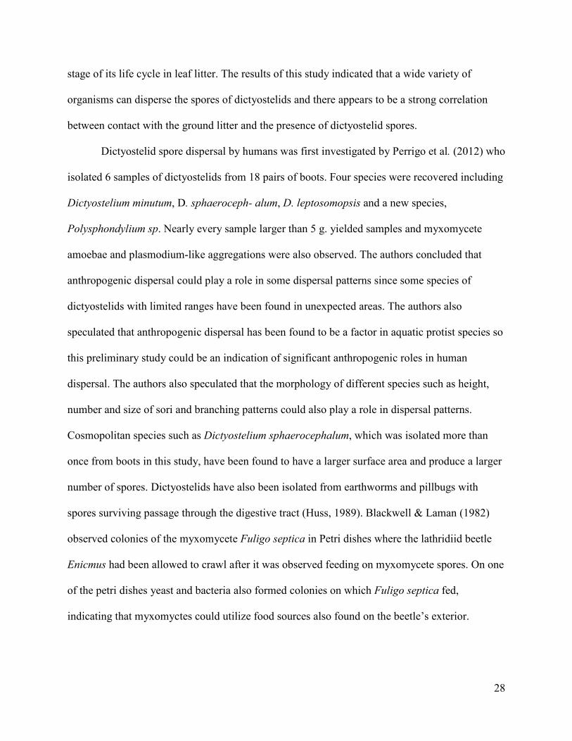
stage of its life cycle in leaf litter. The results of this study indicated that a wide variety of
organisms can disperse the spores of dictyostelids and there appears to be a strong correlation
between contact with the ground litter and the presence of dictyostelid spores.
Dictyostelid spore dispersal by humans was first investigated by Perrigo et al. (2012) who
isolated 6 samples of dictyostelids from 18 pairs of boots. Four species were recovered including
Dictyostelium minutum, D. sphaeroceph- alum, D. leptosomopsis and a new species,
Polysphondylium sp. Nearly every sample larger than 5 g. yielded samples and myxomycete
amoebae and plasmodium-like aggregations were also observed. The authors concluded that
anthropogenic dispersal could play a role in some dispersal patterns since some species of
dictyostelids with limited ranges have been found in unexpected areas. The authors also
speculated that anthropogenic dispersal has been found to be a factor in aquatic protist species so
this preliminary study could be an indication of significant anthropogenic roles in human
dispersal. The authors also speculated that the morphology of different species such as height,
number and size of sori and branching patterns could also play a role in dispersal patterns.
Cosmopolitan species such as Dictyostelium sphaerocephalum, which was isolated more than
once from boots in this study, have been found to have a larger surface area and produce a larger
number of spores. Dictyostelids have also been isolated from earthworms and pillbugs with
spores surviving passage through the digestive tract (Huss, 1989). Blackwell & Laman (1982)
observed colonies of the myxomycete Fuligo septica in Petri dishes where the lathridiid beetle
Enicmus had been allowed to crawl after it was observed feeding on myxomycete spores. On one
of the petri dishes yeast and bacteria also formed colonies on which Fuligo septica fed,
indicating that myxomyctes could utilize food sources also found on the beetle’s exterior.

29
Digestion of dictyostelid spores by turtles may also provide a suitable habitat for a similar
increase in growth (Murray et al. 1985).
Methods and Materials
This study was carried out in the Ozark Plateau region of northwest Arkansas, an area
primarily composed of oak-hickory forests and consisting of low plateaus and valleys with an
average annual temperature between 14° and 17° C and year-round precipitation between 104-
152 cm. Historically northern red oak (Quercus rubra), white oak (Quercus alba), pignut hickory
(Carya alba), mockernut hickory (C. tomentosa), shortleaf pine (Pinus echinate) and eastern red
cedar (Juniperus virginiana) were the dominant trees in this ecosystem (Ley et al., 2019).
Figure 2 Swabbing a Three-toed Box Turtles (Terrapene carolina triunguis) at the Ozark
Natural Science Center, Huntsville Arkansas.

30
Figure 3 Swabbing a Little Brown Skink (Scincella lateralis).
Samples were collected on 4 April 2012 at Lake Fayetteville from two Three-toed Box
Turtles (Terrapene carolina triunguis) one Western Rat Snake (Pantherophis obsoletus) and two
Eastern Fence Lizards (Sceloporus undulates). On 10 April, a sample was taken from one
Timber Rattlesnake (Crotalus horridus), one Eastern Gartersnake (Thamnophis s. sirtalis), one
Little Brown Skink (Scincella lateralis) (Figure 3), one Western Rat Snake (P. obsoletus) and
one Three-toed Box Turtle (T. Carolina triunguis) (Figure 2) at the Ozark Natural Science
Center. Animals sampled at Lake Leatherwood on April 24 included one Eastern Hognosed
Snake (Heterodon platirhinos) and Four Eastern Collard Lizards (Crotaphytus collaris).
Sampling consisted of dipping both ends of a Q-tip in a vial of distilled water and swabbing the
feet, plastron (if present) and cloaca of the animal and placing the swab in a sterile sandwich bag

31
(Figures 2-3). Individual swabs were used for each body part sampled. Once collected, the swabs
were taken back to the lab and plated out according to the method of Cavender and Raper
(1965a). Petri dishes filled with hay infusion agar were labeled, inoculated with three drops of
Escherichia coli (a standard food source for dictyostelids), the swab was brushed across the
streak and three drops of distilled water were added to aid in dispersing any propagules present.
The Petri dishes were stacked and left to incubate for four days, after which they were examined
for the presence of dictyostelids with the aid of a dissecting scope (Figure 4). The plates were
then checked twice a week for four additional weeks. Six additional samples from Three-toed
Box Turtles (Terrapene carolina triunguis) were collected by a fellow graduate student and
yielded samples although identification was hindered by contamination in the lab.
Results
Of the five samples collected at Lake Fayetteville, four (excluding one Eastern Fence Lizard
(Sceloporus undulates) sample) yielded at least one species of dictyostelid, a success rate of
80%. Both swabs from the Three-toed Box Turtle (Terrapene Carolina triunguis) were positive
for dictyostelids. Dictyostelium discoideum was detected after one week from one sample and
Polysphondylium violaceum was found after four weeks from the other sample. Dictyostelium
discoideum was detected after one week from the Western Rat Snake (Pantherophis obsoletus)
swab, P. violaceum and D. mucoroides were both detected from one of the two Eastern Fence
Lizard (S. undulates) samples after four weeks. No results were obtained from the samples
collected at the Ozark Natural Science Center. Of the five samples collected at Lake
Leatherwood, two had positive colonies, a success rate of 40%. Colonies of D. sphaerocephalum
were found from two of the Eastern Collard Lizards (Crotaphytus collaris), one male and one

32
female and Dictyostelium mucoroides was isolated from an Eastern Hognosed Snake (Heterodon
platirhinos).

33
Figure 4. Checking for clones with a dissecting microscope.
Discussion
This study is the first recorded incidence of dictyostelids being isolated from reptiles and
demonstrates that this group of vertebrates apparently plays a role in the dispersal of these
organisms. There was a high success rate (80%) of samples collected from Lake Fayetteville and
samples from Lake Leatherwood showed a relatively high success rate of 40%, indicating that
reptiles can serve as vectors of dictyostelids. The samples in this study took up to four weeks to
appear which is not typical of dictyostelids, which appear within a week on average. The reasons
for this may have been due to spore germination being inhibited by soil factors or certain soil
bacteria or perhaps bacterial supply was too low. If there is a poor supply of bacterial food then it
would take longer for amoebae to proliferate to an aggregating population. It is also possible that
the spores could have been incased in a material from the lizard’s skin that retards germination or

34
that the dictyostelids isolated were microcyst producers, since macrocysts are sometimes
reluctant to germinate releasing amoebae (John Landolt, personal communication).
Contamination of samples by spores in the lab is unlikely because plates were kept tightly closed
and the lid was not removed after inoculation. Prior to this study there had been very little data
on the species diversity of dictyostelids in Arkansas. In this study the most common species
isolated was Dictyostelium mucoroides (3), followed by D. discoideum (2), Polysphondylium
violaceum (2), and D. sphaerocephalum (1). Compared to the study of dictyostelids in Arkansas
by Landolt, et al. (2009) the species isolated in this study, although they do occur in Arkansas are
considered less common. The previous study indicated that P. pallidum is the most abundant
(47% of cultures isolated) with three others (D. minutum, P. violaceum and D. purpureum)
making up 30% of remaining species. Of the four species isolated from reptiles only one (P.
violaceum) was considered common in the previous study with 205 clones observed. Of the
2,082 clones recovered in the previous study D. murcoroides yielded only 96, D. discoideum 24
and D. sphaerocephalum 16. The four species isolated in this study were also found in caves by
Landolt et al. (2006) who isolated two clones each of D. sphaerocephalum and D. murcoroides,
one clone of D. discoideum and four clones of P. violaceum Although the number of samples
collected in this study were small this was the first recorded instance of dictyostelids to be
isolated from reptiles and indicated that this group of organisms, along with other animals may
play a role in the dispersal of these organisms. Three of the four species isolated were considered
rare in the survey of Arkansas dictyostelids by Landolt et al. (2009). One possibility for these
findings might be due to the micro and macro habitat preferences of these reptiles. Three-toed
Box Turtles (Terrapene Carolina triunguis) are commonly found in grasslands in late spring and
early fall but prefer forested habitats during the summer, early spring and late fall. During these
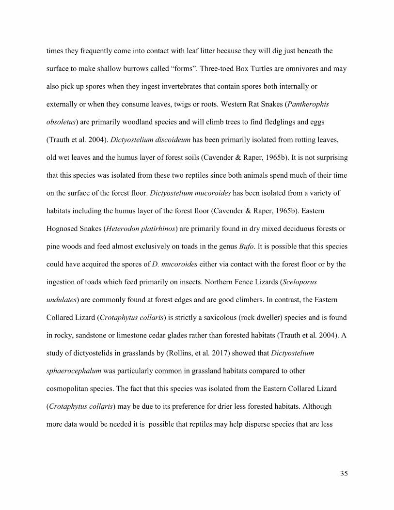
35
times they frequently come into contact with leaf litter because they will dig just beneath the
surface to make shallow burrows called “forms”. Three-toed Box Turtles are omnivores and may
also pick up spores when they ingest invertebrates that contain spores both internally or
externally or when they consume leaves, twigs or roots. Western Rat Snakes (Pantherophis
obsoletus) are primarily woodland species and will climb trees to find fledglings and eggs
(Trauth et al. 2004). Dictyostelium discoideum has been primarily isolated from rotting leaves,
old wet leaves and the humus layer of forest soils (Cavender & Raper, 1965b). It is not surprising
that this species was isolated from these two reptiles since both animals spend much of their time
on the surface of the forest floor. Dictyostelium mucoroides has been isolated from a variety of
habitats including the humus layer of the forest floor (Cavender & Raper, 1965b). Eastern
Hognosed Snakes (Heterodon platirhinos) are primarily found in dry mixed deciduous forests or
pine woods and feed almost exclusively on toads in the genus Bufo. It is possible that this species
could have acquired the spores of D. mucoroides either via contact with the forest floor or by the
ingestion of toads which feed primarily on insects. Northern Fence Lizards (Sceloporus
undulates) are commonly found at forest edges and are good climbers. In contrast, the Eastern
Collared Lizard (Crotaphytus collaris) is strictly a saxicolous (rock dweller) species and is found
in rocky, sandstone or limestone cedar glades rather than forested habitats (Trauth et al. 2004). A
study of dictyostelids in grasslands by (Rollins, et al. 2017) showed that Dictyostelium
sphaerocephalum was particularly common in grassland habitats compared to other
cosmopolitan species. The fact that this species was isolated from the Eastern Collared Lizard
(Crotaphytus collaris) may be due to its preference for drier less forested habitats. Although
more data would be needed it is possible that reptiles may help disperse species that are less

36
common in Arkansas such as D. sphaerocephalum and impact species diversity in ways that are
still not fully understood.
This study showed that dictyostelid spores can be found on reptiles and that these animals
may play a role in dispersal but the sample size taken was too small to make any assumptions
other than the fact that dictyostelid clones can be isolated from the bodies of reptiles. Going
forward I would need to increase the number of organisms swabbed and make more detailed
observations of differences in body parts swabbed (feet, cloaca, etc.) to see how these factors
might influence the species diversity isolated. It might be interesting to sample stomach contents
of swabbed reptiles to see what organisms the animal consumed since dictyostelid spores are a
food source for some arthropods. A more detailed analysis of other non-fruiting protists on
isolated samples could indicate whether or not fruiting has any effect on dispersal by reptiles.
The samples in this study took an atypically long time to germinate for dictyostelids and this
observation could be further studied by doing a series of isolations comparing swabs from
reptiles to isolates from the soil/leaf litter in the surrounding habitat to see if the body/surface of
the reptile may have some factor that could inhibit growth or if the difference may have been due
to variations in the bacterial microfauna of both habitats (soil surface vs. reptile body). To
investigate this the bacteria found in each habitat would need to be identified and compared as
well as the chemical composition of both swabbed animals and the soil material collected.

37
Table 1. Presence/Absence of Dictyostelids in Reptile Samples €
Animal Species Unknown Dictyostelium discoideum Dictyostelium mucoroides Polysphondylium violaceumDictyostelium
sphaerocephalum
Three-toed Box
Turtle
(Terrapene
Carolina
triunguis )
+ + +
Western Rat
Snake
(Pantherophis
obsoletus )
+
Eastern Fence
Lizard
(Sceloporus
undulates )
+ +
Eastern Hognosed
Snake
(Heterodon
platirhinos )
+
Eastern Collard
Lizard
(Crotaphytus
collaris )
+
Three-toed Box
Turtle
(Terrapene
Carolina
triunguis ) 023P0
+
Three-toed Box
Turtle
(Terrapene
Carolina
triunguis ) 024FL
+
Three-toed Box
Turtle
(Terrapene
Carolina
triunguis )
sample 025CH
+
Three-toed Box
Turtle
(Terrapene
Carolina
triunguis )
sample 026P0
+
Three-toed Box
Turtle
(Terrapene
Carolina
triunguis ) hybrid
sample 021P0
+
Three-toed Box
Turtle
(Terrapene
Carolina
triunguis )
sample 016CP
+
Eastern Collard
Lizard
(Crotaphytus
collaris )
+

38
Literature Cited
Blackwell, M., & Laman, T. G. (1982). Spore dispersal of Fuligo septica (Myxomycetes) by
lathridiid beetles. Mycotaxon 14:58-60.
Cavender, J. C. (1973) Geographical Distribution of Acrasieae. Mycologia, 65:1044–1054.
Cavender, J. C. & Raper, K. B. (1965a). The Acrasieae in nature. I. Isolation. American Journal
of Botany 52:294-296.
Cavender, J. C., & Raper, K. B. (1965b). The Acrasieae in Nature. II. Forest Soil as a Primary
Habitat. American Journal of Botany, 52:297-302.
Huss, M. J., (1989). Dispersal of cellular slime molds by two soil invertebrates. Mycologia
81:677-682.
Keller, H. W., & Smith, D. M. (1978). Dissemination of myxomycete spores through the feeding
activities (ingestion-defecation) of an acarid mite. Mycologia 70:1239-1241.
Landolt, J. C., Stephenson, S. L., & Slay, M. E. (2006) Dictyostelid cellular slime molds from
caves. Journal of Cave and Karst Studies 68:22-26.
Landolt, J. C., Stephenson, S. L., & Rollins, A. W. (2009). Dictyostelid cellular slime molds of
Arkansas. Southern Appalachian Botanical Society 74:353-359.
Ley, E. L. et al. (accessed 2020) Selecting plants for pollinators a regional guide for farmers,
land managers and gardeners in the Ozark broadleaf forest meadow province including parts of
Arkansas and Oklahoma. The Pollinator Partnership/North American Pollinator Protection
Campaign San Francisco, CA www.pollinator.org
Murray, P. M., et al. (1985). The numbers of viable myxomycete cells in the alimentary tracts of
earthworms and in earthworm casts. Botanical Journal of the Linnean Society 91:359-366.
Perrigo, A. L., et al. (2012), What’s on your boots: an investigation into the role we play in
protist dispersal. Journal of Biogeography, 39: 998–1003
Raper, K. B. (1984). The Dictyostelids. Princeton University Press: Princeton, New Jersey.
Rollins, A. W., Landolt, J. C., & Stephenson, S. L. (2010) Dictyostelid cellular slime molds
associated with grasslands of the central and western United States. Mycologia, 102:996-1003.
Stephenson, S. L., & Landolt, J. C. (1992) Vertebrates as vectors of cellular slime-molds in
temperate forests. Mycological Research 96:670-672.Stephenson, S. L., & Rojas, C. (2017)
Myxomycetes: Biology, Systematics, Biogepgraphy, and Ecology. Academic Press, Cambridge,
Massachusetts. 474 pp.
39
Suthers, H. B. (1985) Ground-feeding migratory songbirds as cellular slime mold distribution
vectors. Oecologia 65:526-530.
Trauth, S. E., Robinson, H. W., & Plummer, M. V. (2004) The Amphibians and Reptiles of
Arkansas. University of Arkansas Press, Fayetteville, AR.

40
Large Herbivorous Mammals as Vectors of Coprophilous Myxomycetes
Abstract
The objective of this study was first to document what species of myxomycetes are
associated with the dung of large herbivorous mammals and then to evaluate the effect of
differences in pH on the species diversity of these organisms in this microhabitat. Although wind
is the main dispersal mechanism for myxomycete spores, animals may play an important
although little studied role in this process. Myxomycetes that occur on dung are considered
coprophilous and this substrate may be particularly important in arid regions such as deserts and
grasslands due to the lack of alternative substrates. A series of moist chambers was prepared with
dung samples collected from cows, horses, deer and several other species of herbivores and
observed over a period of several months for the occurrence of fruiting bodies. The pH and any
corresponding differences were noted and recorded.
Introduction
Myxomycetes occur in a wide variety of habitats, especially decaying wood, bark and
leaf litter. Many species appear to prefer growing in specific microhabitats and myxomycetes
that occur predominantly on dung are referred to as coprophilous or fimicolous. A small number
of species are considered truly coprophilous since they have been recorded only on this substrate
(Eliasson, 2013, Eliasson and Keller, 1999). In arid areas such as deserts and steppes, there are
myxomycetes that occur on weathered dung and are considered coprobionts. These
myxomycetes are considered one of the least studied and most specialized of myxomycetes, with
over 100 species occurring on this substrate and 16 species are considered obligate coprobionts.
It is believed that coprobiont myxomycetes evolved because dung contained adequate moisture
for this substrate to serve as an ideal microhabitat (Vlasenko et al. 2017).

41
Dung is a very complex substrate, and its physical and chemical properties vary
depending on the animal it came from, remaining plant fragments, age, grade of decomposition,
and the variety of bacteria and fungi present. The occurrence of myxomycetes on dung may be
related more to what bacteria are present than the physical properties of the substrate although
more research is needed to explore this relationship. This high microbial content along with high
nutrient richness and moisture create a favorable substrate and microhabitat for myxomycetes
(Eliasson, 2013, Hudson, 1986, Stephenson, 2011). Compared to bark and other woody
substrates on which lignicolous myxomycetes commonly occur, dung has a much higher
nitrogen content (up to 4%) and also has a pH much higher than most of the other substrates that
myxomycetes usually grow on (Stephenson, 1989). The pH of a myxomycete substrate is one of
the most important factors in determining what species will grow on them and can be used to
divide different species into groups (Eliasson, 2013). Although many species of myxomycetes
can tolerate a broad range of pH this can become a limiting factor in some cases. Previous
studies have shown pH to affect species diversity on bark, with the most acidic bark containing
the lowest species richness (Liu et al. 2015). Most of the species observed on dung occur on
other types of substrates and their occurrence of dung is considered accidental. For example,
many species that usually grow on decaying wood occur on other substrates as well. Examples
include Comatricha nigra, Cribraria cancellata, C. microcarpa, Diderma radiatum, Lycogala
epidendrum, Physarum album, Trichia botrytis and T. varia. Other coprophilous myxomycetes
include such species as Cribraria violacea and Echinostelium minutum that both inhabit the bark
of living trees as well as Didymium nigripes, which usually grows on leaf litter (Eliasson, 2013).
Although it is believed that there are no truly obligate coprophilous myxomycetes in the strictest
sense, there are 16 coprophilous species that usually and in a few cases have been found only on

42
dung. These include Badhamia apiculospora, B. rhytidosprema, B. spinispora, Didymium
annulisporum, D. rugulosporum, Kelleromyxa fimicola, Licea alexopouli, L pescadorensis,
Macbrideola indica, Perichaena liceoides, P. luteola, Trichia brunnea, T. elaterensis, T.
fimicola, and T. papillata. Among these the most common species to occur on dung include
Badhamia apiculospora, B. spinispora, Licea alexpoulii, Perichaena liceoides and P. luteola. Of
these 16 species some have been shown to have thick-walled spores. This is the case for: Licea
alexpoulii, Kelleromyxa fimicola, Perichaena taimyriensis and Trichia brunnea. This
morphology may be an adaptation for passing through the intestinal tract of herbivorous
mammals and the action of intestinal juices may be required for germination to take place
(Eliasson, 2013). All four species with thick-walled spores have been found exclusively on dung.
Licea alexpoulii was first described by Blackwell (1974), who isolated cultures from horse and
cow dung. This species was set apart from other members of its genus by its smooth shiny black
peridium and its hemispherical to subglobose sporangial shape. Eliasson et. al. (1991) proposed a
new genus Kelleromyxa, renaming the myxomycete Licea fimicola to Kelleromyxa fimicola. This
species was set apart from other members of the order Liceales by the presence of a true
capillitium composed of short unbranched threads and occasional longer branched threads; a
phaneroplasmodial plasmodium type giving rise to many fruiting bodies per plasmodium;
clustered fruiting bodies, dark spores with uniformly thickened walls; and the presence of
calcium in the peridium. This species was found only on the dung of herbivorous animals and
has been isolated from horse, cow, bison, pronghorn and rabbit in the New World and from dik-
dik, cow or yak in the Old World. The evenly thick spore walls and lack of a thin region where
germination occurs also sets Kelleromyxa fimicola apart as unique. Prior to this study, there was
evidence that Liceales was an unnatural taxon defined by the lack of a characteristic (a

43
capillitium) rather the presence of one and these differences marked Licea fimicola apart as an
anomaly in need of reclassification. Trichia brunnea was first described by Cox (1981) in his
survey of coprophilous myxomycetes of the western United States.
Myxomycetes have also been found on the droppings of birds that feed on seeds, buds
and other plant parts. This is significant because in theory a bird could transport spores across
vast distances. Myxomycetes have been isolated from the droppings of the Capercaillie (Tetrao
urogallus) (Arcyria cinerea, Physarum confertum, Didymium difforme); grouse (Lagopus?)
(Physarum bitectum, Didymium difforme); black grouse (Lyrutus tetrix) (Didymium diffome);
goose (Anser?) (Perichaena corticalis); and rock dove (Columba livia) (Physarum compressum)
(Eliasson, 2013). The myxomycete species assemblage was investigated in a colony of great
cormorants (Phalacrocorax carbosinensis) and compared to the surrounding pine forest in
western Lithuania (Adamontytė et al. 2013). The authors found that the lowest species richness
was found in the most active part of the colony with the newest and most abundant nests as well
as the largest difference in species composition compared to the surrounding pine forest. They
also observed species of myxomycetes that were not present in the surrounding forest, several of
which were considered rare (Comatricha mirabilis and Arcyria leiocarpa). The authors cited
previous studies that piscivorous birds act as vectors in the transfer of material between aquatic
and terrestrial environments and influence the biogeographical cycling of nitrogen and
phosphorous that occurs in guano. Compared to the surrounding forest soil collected from the
colony had a higher pH due to cormorant guano and also contained species resistant to a higher
pH. This study indicated that myxomycete species composition could change dramatically in
hypertoficated environments. Coprophilous myxomycetes were first described in 1803 by
Schumacher, who collected Physarum fimetarium from cow dung. There were relatively few

44
studies on this group of myxomycetes until Eliasson and Lundqvist (1979) whose study aimed to
show what species of myxomycetes occurred on dung. This monograph is significant due to the
large variety of animal species sampled and the fact that samples were left to culture for at least
several months (Eliasson, 2013). Over 160 samples from 25 species of animals were set up using
the moist chamber technique. It was found that myxomycetes growing on this substrate always
developed late, after at least three weeks and concluded that at least a month was probably
needed to complete a life cycle. Of the samples collected, cow dung was the most favored
substrate (55 species collected), followed by the dung from hares (5 species) 23, horse 21, rabbit
18, elk and moose 14, roe deer 7, goat 5, sheep 5, capercaillie 3, donkey 3, camel 2, lemming 2,
and grouse 2. The authors admitted that these distribution patterns were partially reflective of
what substrates are more readily available than others but did find some patterns of substrate
preference for some species. About 80% of positive samples for Perichaena liceoides were
found on cow dung and this species favored the dung of domesticated animals (cow, horse,
sheep, donkey; 32 samples) to wildlife such as deer. Didymium difforme was also common on
cow dung (38% of samples) but had a wider host range of nine species. Arcyria cinerea was
found on 50% of horse dung samples and a total of 6 host species. Stemonitis fusca was found
only on the dung of forest animals (elk, roe deer, hare; 10 occurrences). A similar pattern can be
found in fungi. Lundquist (1972) found that some fungi favored the dung from certain species
and divided his findings into three groups: (1) fungi with a wide ecological range and low
substrate preference; (2) those with high substrate specificity and a wide ecological range; and
(3) a small number of fastidious species that were restricted to certain substrates. Among these it
was found that all common Nordic species of Sordariaceae sampled were found only on the
dung of three species out of a total of 100 (Richardson, 2001).

45
Since the number of studies focusing on coprophilous myxomycetes has been limited, a
number of coprophilous species have been discovered fairly recently. Perichaena taimyriensis
was not discovered until 2000 when Novozhilov & Schnittler (2000) described a species of
myxomycete with thick-walled spores from reindeer (Rangifer tarandus) dung collected from the
Arctic tundra. The species was placed in the genus Perichaena and was described as an
intermediate species between the genus Perichaena and Licea since it lacked a capillitium. Since
reindeer dung was the only substrate from which this species was isolated, the authors suggested
that reindeer might play an important role in the dispersal of this species due to the large number
of distances this animal travels (as much as 10,700,000 sq. km annually). Adamonyte (2003)
isolated a new species of Trichia from the dung of hare (Lepus sp.) and roe deer (Capreolus
capreolus) in southern Lithuania. The species was named Trichia papillata and differed from
similar species of Trichia by having a yellow plasmodium and a papillate peridium divided by
yellow bands into angular segments.
A comprehensive study of myxomycetes by Stephenson (1989) revealed that different
species of myxomycetes preferred different pH levels as well as different microhabitats. The
study took place in the Mountain Lake area of southwestern Virginia, where a series of moist
chambers were made sampling the bark of living trees, leaf litter and deer dung. Bark was
collected from twenty-two species of trees and one species of tall shrub. Prior to this study there
had been very little research on myxomycetes that occur on the bark of living trees, with most
studies focusing on decaying bark as a substrate. The author observed that species composition
patterns were related to bark acidity and texture. These findings complemented an earlier study
by Harkonen (1977) who observed patterns of pH optima in different groups of myxomycetes.
Members of the Stemonitales preferred more acidic conditions than members of the Physarales

46
and Trichiales. Stephenson (1989) observed similar mean pH values for myxomycetes on litter
and the bark of living trees and concluded that pH is an important factor in determining
distribution patterns of species of myxomycetes, even when different microhabitats are involved.
One hundred twenty-nine moist chambers of deer dung samples yielded 44 samples of five
species (34%) which was a much lower species diversity than for bark or leaf litter. The author
concluded that myxomycetes do not appear to be common on this substrate in temperate forests
and that truly coprophilous species were rare or nonexistent. In addition to this study there have
been very little investigations into myxomycete dispersal by mammals in temperate forests. A
later study by Stephenson and Landolt (1992) primarily investigated the role of animal vectors of
dictyostelids, but the authors were able to isolate plasmodia from the droppings of white-footed
mice (Peromyscus leucopus) and big brown bats (Eptesicus fuscus). Although their efforts to
induce fruiting were unsuccessful, this did indicate that the spores of myxomycetes were able to
survive passage through the digestive tract of animals.
In contrast to temperate regions, coprophilous species of myxomycetes may be more
widespread in arid climates. Cox (1981) completed a survey of coprophilous myxomycetes in the
western United States and identified a total of 26 species, seven of which were considered rare or
new state records. Trichia brunnea, isolated from cow dung was the first description of this
species. Perichaena minor, also isolated on cow dung, was a new record for the state. Another
species collected from cow dung was Calomyxa metallica and this was the first time it was
recorded on this substrate. Normally this species is found on the bark of dead and living trees.
Physarum spinisporum was isolated from cow dung for the first time in the Western hemisphere.
This is an extremely rare species that had only been reported previously from the Canary Islands
and Ethiopia; in all instances they have only been isolated from dung. Physarum ovisporum,

47
isolated from cow dung was another rare species isolated and was the first record for Arizona
and New Mexico and the first instance it was found on dung. Physarum gyrosum was isolated
from deer dung and was the first report on dung for this species and for the Western United
States. Didymium ovoideum, isolated from cow dung was an additional new record for California
and the first instance of it being coprophilous. Another rare species isolated was Didymium
karstensii. Novozhilov et al. (2003) conducted a survey of the Colorado Plateau and collected
samples from rabbit (Lepus sp.), mule deer (Odocoileus hemionus), pronghorn (Antilocarpra
americana), red deer (Cervus elaphus), cattle and horse. The authors found that the most
common coprophilous species were Badhamia cf. apiculospora, Fuligo cinereal, Perichaena
liceoides, and Licea tenera. A later study by Novozhilov et al. (2006) surveyed the desert and
steppe regions of the lower Volga River Basin in Russia and Kazakhstan revealed 23 species
with Fuligo cinerea and Perichaena liceoides being most common. The authors noticed a
marked difference in species composition between coprophilous species collected from different
habitats. Didymium difforme, Physarum bivalve, and Leocarpus fragilis were more common in
the northern taiga and tundra while F. cinerea was uncommon in these habitats but numerous in
desert samples. A higher diversity of coprophilous myxomycetes in desert habitats may be due in
part to the higher pH values of available substrates. Blackwell and Gilbertson (1984) found that
Sonoran Desert myxomycetes grown on the pith of cacti tolerated higher pH values than
previous studies showed, as high as 8.7-10.4. Although dung tends to be higher in pH than other
types of substrates this may be less of a limiting factor in desert habitats where pH of alternative
substrates is also high.
A set of bison and cow dung collected in a temperate prairie in North Dakota yielded 6
species of myxomycetes from 30 samples. There was a higher species frequency mean for bison

48
dung and a lower mean pH. The authors observed that species in bison samples (Arcyria cinerea,
Didymium difforme and Kelleromyxa fimicola) prefer a lower substrate pH than those found on
cow dung in this study (Perichaena depressa, Perichaena liceoides and Physarum sp.). Moist
chambers were observed for three months, with 55% of myxomycetes appearing in the first
month, 35% in the second month and 10% in the final month. Didymium difforme and
Perichaena liceoides were found throughout the time period and Arcyria cinerea did not appear
until the third month. Both bison and cow dung had the same species richness of 4 with Arcyria
cinerea, Didymium difforme, Kelleromyxa fimicola, and Perichaena liceoides being present on
bison dung and Didymium difforme, Perichaena depressa, Perichaena liceoides and Physarum
species occurring on cow dung. Species frequency was higher on bison dung, with the genus
Didymium being the most frequent (Onduso et al. 2019). An earlier study by Rollins et al. (2010)
indicated that fewer clones of dictyostelids would grow from culture in grasslands that are grazed
vs. ungrazed grasslands. The authors attributed lower abundance to soil compaction from grazing
herbivores, fewer microhabitats due to a decreased amount of dead plant material being
deposited on the ground and possibly a negative impact on local soil bacteria. It was also
suggested that dung deposited by grazing herbivores might combat these impacts by serving as a
refugia due to its high moisture and bacterial content. In this study a series of moist chambers
were made using cow and horse dung to evaluate differences in pH and any corresponding
differences between these two microhabitats were recorded.
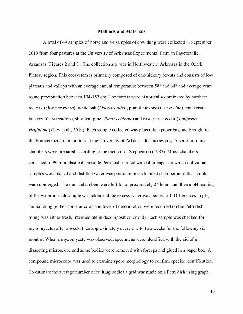
49
Methods and Materials
A total of 49 samples of horse and 44 samples of cow dung were collected in September
2019 from four pastures at the University of Arkansas Experimental Farm in Fayetteville,
Arkansas (Figures 2 and 3). The collection site was in Northwestern Arkansas in the Ozark
Plateau region. This ecosystem is primarily composed of oak-hickory forests and consists of low
plateaus and valleys with an average annual temperature between 58° and 64° and average year-
round precipitation between 104-152 cm. The forests were historically dominated by northern
red oak (Quercus rubra), white oak (Quercus alba), pignut hickory (Carya alba), mockernut
hickory (C. tomentosa), shortleaf pine (Pinus echinate) and eastern red cedar (Juniperus
virginiana) (Ley et al., 2019). Each sample collected was placed in a paper bag and brought to
the Eumycetozoan Laboratory at the University of Arkansas for processing. A series of moist
chambers were prepared according to the method of Stephenson (1985). Moist chambers
consisted of 90 mm plastic disposable Petri dishes lined with filter paper on which individual
samples were placed and distilled water was poured into each moist chamber until the sample
was submerged. The moist chambers were left for approximately 24 hours and then a pH reading
of the water in each sample was taken and the excess water was poured off. Differences in pH,
animal dung (either horse or cow) and level of deterioration were recorded on the Petri dish
(dung was either fresh, intermediate in decomposition or old). Each sample was checked for
myxomycetes after a week, then approximately every one to two weeks for the following six
months. When a myxomycete was observed, specimens were identified with the aid of a
dissecting microscope and some bodies were removed with forceps and glued in a paper box. A
compound microscope was used to examine spore morphology to confirm species identification.
To estimate the average number of fruiting bodies a grid was made on a Petri dish using graph

50
paper (See Figs. 3-4). Three additional sets of moist chamber cultures were also started in
December 2019 and January 2020. Twenty samples of weathered cow dung (at least 6 months
old) were collected from a pasture in West Virginia by Dr. Steve Stephenson; thirteen samples of
deer pellets from white-tailed deer (Odocoileus virginianus) were collected in Pennsylvania by
Dr. Autumn Coffey and 17 dung samples were collected form the North Dakota Zoo by Dr.
Francis Onduso. The latter included the dung of mule deer (Odocoileus hemionus), white-tailed
deer (Odocoileus virginianus), highland cattle (Bos taurus), bighorn sheep (Ovis canadensis),
reindeer (Rangifer tarandus) and camels (Camelus sp.). Species diversity (alpha-diversity) was
calculated using Shannon's diversity index H' = –∑ Pi log Pi, where Pi is the relative abundance
(the proportion of the total number of individuals or records of a species (Shannon and Weaver,
1963, Magurran, 1988).
Fig 1. Perichaena liceoides in a moist chamber culture of horse dung. Photo courtesy of Apulu
Ndotimi

51
Figure 2. Study site at the University of Arkansas Experimental Farm, Fayetteville, Arkansas.

52
Figure 3. Study site at the University of Arkansas Experimental Farm, Fayetteville, Arkansas.
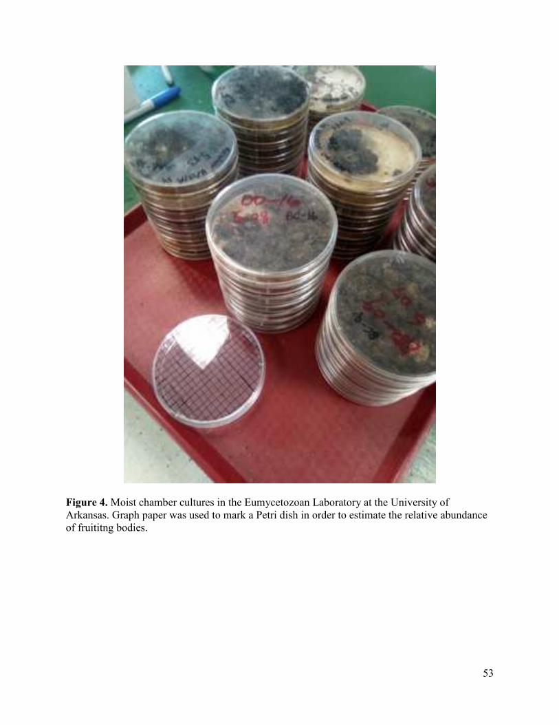
53
Figure 4. Moist chamber cultures in the Eumycetozoan Laboratory at the University of
Arkansas. Graph paper was used to mark a Petri dish in order to estimate the relative abundance
of fruititng bodies.

54
Figure 5. Estimating the number of fruiting bodies with the aid of a dissecting microscope.
Results
Twenty-seven of the 93 moist chambers prepared with samples from the University of
Arkansas Experimental Farm yielded myxomycetes a success rate of 29%. A total of four species
(Perichaena liceoides, Physarum sp., Perichaena depressa Libert, and Perichaena vermicularis
(Schw.) Rost.) were isolated, with Perichaena liceoides being by far the most common (See
Table 1). The other moist chamber series started in December 2019 and January 2020 did not

55
yield cultures, including the weathered cow dung samples. The moist chamber cultures from the
University of Arkansas Experimental farm did not yield cultures for the first two months, with
most appearing after three months. These observations are consistent with previous studies
indicating that coprophilous myxomycetes take at least three weeks to appear in moist chamber
cultures and many species will not yield cultures until the third month (Eliasson and Lundqvist,
1979). One species of coprophilous fungus, the Dung-Loving Birds’ Nest Fungi (Cyathus
stercoreus Schweinitz), was also observed.
Although there were slightly more samples of horse than cow dung (49 and 44,
respectively) there were more positive cultures in horse dung compared to cow dung. Eighteen
horse samples yielded specimens, a success rate of 37%. Only 8 cow dung samples produced
cultures, an 18% success rate (Figure 9). Cow dung had slightly higher diversity values (H' =
0.22) than horse dung (H' = 0.20) due to species richness values (3 for cow dung and 2 for horse
dung). Horse dung contained a higher average number of fruiting bodies than cow dung (Table
1).
Table 1. Species recorded by substrate.
Cow Dung Average Number of
Fruiting Bodies
Horse Dung Average Number of
Fruiting Bodies
Perichaena
liceoides
79 Perichaena liceoides 100
Physarum sp. 1 Perichaena
vermicularis
15
Perichaena
depressa
2

56
Table 2. Average number of fruiting bodies related to pH of the substrate
pH
Average
Number of
Fruiting
Bodies on
Cow Dung
pH
Average
Number of
Fruiting
Bodies on
Horse
Dung
Average 7.59 59.5 Average 7.23125 105.65
Max 8.08 320 Max 7.73 560
Min 6.69 1 Min 6.75 1
Richness 3 Richness 2
Table 3. Number of positive cultures related to the age of the substrate.
Substrate Age Horse Cow
Fresh 8 5
Intermediate 10 3
Weathered _ 0
Table 4. Values of species diversity.
Species diversity by substrate (H')
Horse Cow
0.20
0.22
Discussion
Eliasson and Lundqvist (1979) reported that Perichaena liceoides prefers the dung of
domesticated animals with cow dung being the most preferred. More positive cultures on horse
dung in this study may be due to the slightly lower pH in horse dung (Figure 8). The pH values
for this first set showed slightly higher averages for cow dung than for horse dung; the mean pH
for horse dung was 7.3 and the mean pH of cow dung samples was 7.70 (See Table 2). A
comparison of substrate age (either fresh, intermediate or weathered) indicated that older
substrate might be less productive. Both fresh and intermediate substrates collected from the

57
University of Arkansas Experimental Farm yielded cultures (see Table 3) and there were slightly
more fruiting bodies formed on horse dung that was in an intermediate stage of decomposition
compared to cow dung. The pH values of dung tend to be higher than most other substrates and
has been regarded in previous studies as a limiting factor to what species occur on dung
(Eliasson and Lundquist, 1979, Stephenson, 1989). The lack of success in the other moist
chamber cultures may have been that the samples from North Dakota came from caged animals
and the deer pellets and weathered cow dung samples were smaller sets of samples. Time may
also have been a factor in this study. Data collected from the University Experimental Farm did
not begin to yield cultures for several months, with many specimens not appearing until the third,
fourth and in some cases five months from when the moist chambers were started. Although
moist chambers for the other data sets were checked periodically for the first two months closure
of the University due to the historical COVID-19 pandemic hindered additional investigation.
Based on previously published data, deer pellets in temperate forests are not rich sources of
myxomycetes (Stephenson, 1989). Perhaps the abundance of alternate substrates in this sort of
habitat is a factor in low numbers of myxomycetes. In contrast, dung collected from the
University of Arkansas Experimental Farm was from open grassland with scattered trees. There
are some recent preliminary data that coprophilous myxomycetes may be abundant on dung in
grassland habitats and perhaps the habitat of animal vectors of coprophilous myxomycetes may
be one of the most important variables in determining species abundance and diversity of
coprophilous myxomycetes (Onduso et al 2019).

58
Figure 6. Average number of fruiting bodies on horse dung
0
100
200
300
400
500
6007
.12
7.7
7.0
1
7.3
3
7.5
8
7.2
3
6.7
5
7.7
1
7.5
8
7.4
6.8
8
7.2
8
7.3
1
6.9
1
7.0
6
7.4
2
7.1
7.1
3
7.3
4
7.2
5
7.4
6
7.1
4
6.9
3
7.0
3
Av
era
ge
Nu
mb
er
of
Fru
itin
g B
od
ies
pH
Average Number of Fruiting Bodies on
Horse Dung

59
Figure 7. Average number of fruiting bodies on cow dung
0
50
100
150
200
250
300
350
Av
era
ge
Nu
mb
er
of
Fru
itin
g B
od
ies
pH
Average Number of Fruiting Bodies on
Cow Dung

60
Figure 8. Average pH by substrate
0
2
4
6
8
10
12
Horse Dung pH Cow Dung pH
pH
Average pH by Substrate
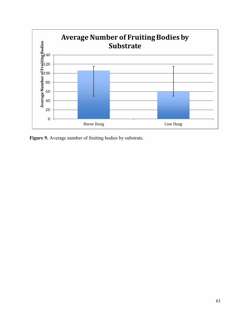
61
Figure 9. Average number of fruiting bodies by substrate.

62
Literature Cited
Adamontytė, G. et al. (2013) Myxomycetes in a forest affected by great cormorant colony: a case
study in Western Lithuania. Fungal diversity 59:131-146.
Adamontytė, G. (2003) Trichia papillata, a new coprophilous myxomycete species. Mycotaxon
7:379-384.
Blackwell, M. (1974) A new species of Licea (Myxomycetes). Proceedings of the Iowa
Academy of Science 81:6.
Blackwell, M., & Gilbertson, R. L. (1984). Distribution and sporulation phenology of
myxomycetes in the Sonoran desert of Arizona. Microbial Ecology, 10:369-377.
Cavender, J. C. (1973) Geographical Distribution of Acrasieae. Mycologia, 65:1044–1054.
Cox, J.J. (1981) Notes on coprophilous Myxomycetes from the western United States.
Mycologia 73: 741–747.
Eliasson, U. (1981) Patterns of occurrence of myxomycetes in a spruce forest in South Sweden.
Holarctic Ecology 4:20-31.
Eliasson, U. H. and Keller, H. W. (1999) Coprophilous myxomycetes: updated summary, key to
species, and taxonomic observations on Trichia brunnea, Arcyria elaterensis, and Arcyria
stipata. Karstenia 39:1-10.
Eliasson, U. (2013) Coprophilous myxomycetes: Recent advances and future research
directions. Fungal Diversity 59:85–90.
Hudson, H.J. (1986) Fungal biology. Edward Arnold, Baltimore.
Stephenson, S.L. (1985) Myxomyctes in the laboratory II: moist chamber cultures. American
Biology Teacher 47:487-489.
Ley, E. L. et al. (accessed 2020) Selecting plants for pollinators a regional guide for farmers,
land managers and gardeners in the Ozark broadleaf forest meadow province including parts of
Arkansas and Oklahoma. The Pollinator Partnership/North American Pollinator Protection
Campaign San Francisco, CA www.pollinator.org
Liu, Q. et al. (2015) Species diversity of myxomycetes associated with different terrestrial
ecosystems, substrata (microhabitats) and environmental factors. Mycological Progress 14:27.
Lundqvist, N. (1972) Nordic Sordariaceae s. lat. Symbolae Botanicae Upsalienses 20(1): 1-314.
Magurran, A. E. (1988) Ecological Diversity and its Measurement. Princeton University Press,
London.

63
Novozhilov, Y. K. & Schnittler, M. (2000) A new coprophilous species of Perichaena
(Myxomycetes) from the Russian Arctic (the Taimyr Peninsula and the Chukchi Peninsula).
Karstenia 40:117-122.
Novozhilov, Y. K., Mitchell, D. W., & Schnittler, M. (2003) Myxomycete biodiversity of the
Colorado Plateau. Mycological Progress 2:243-248.
Novozhilov, Y. K. et al. (2006) Myxomycete diversity and ecology in the arid regions of the
Lower Lolga River Basin (Russia). Fungal Diversity 23:193-241.
Olive, E. W. (1902) Monograph of the Acrasieae. Boston Society of Natural History. 1-513.
Olive, L. S. 1975. The Mycetozoans. Academic Press, New York.
Onduso, F. N., Tewari, S., & Tewari, L. (2019) Myxomycete diversity on bison and cow dung in
the Missouri River watershed of the Standing Rock Sioux Reservation, North Dakota. Journal of
Bacterial Mycology Open Access 4:75-79.
Raper, K. B. (1984). The Dictyostelids. Princeton University Press: Princeton, New Jersey.
Rollins, A. W., Landolt, J. C., & Stephenson, S. L. (2010) Dictyostelid cellular slime molds
associated with grasslands of the central and western United States. Mycologia 68:22-26.
Shannon, C. E. & Weaver, W. (1963) The Mathematical Theory of Communication. University
Illinois Press, Urbana.
Stephenson, S. L. & Landolt, L. C. (1992) Vertebrates as vectors of cellular slime moulds in
temperate forests. Mycological Research 96:670-672.
Stephenson, S.L. (1989) Distribution and ecology of myxomycetes in temperate forests. II.
patterns of occurrence on bark surface of living trees, leaf litter, and dung. Mycologia, 81:608-
621.
Stephenson, S.L. (2011) From morphological to molecular: studies of myxomycetes since the
publication of the Martin and Alexopoulos (1969) monograph. Fungal Diversity 50:21–34.
Schumacher, C.F. (1803) Enumeriatio plantarum in partibus Saellandiae septentrionalis et
orientalis. II. Hafniae.
Richardson, MJ. (2001) Diversity and occurrence of coprophilous fungi. Mycological Research
105:387-402.
Vlasenko, V. & Vlasenko, A. (2017). New data on the obligate coprophilous myxomycetes of
Siberia. News of Irkutsk State University. Series: Biology. Ecology. 21. 50-60.

64
Conclusions
This is the first published report that dictyostelid spores were isolated from reptiles and
indicates that these animals may aid in dispersal like birds have shown in previous studies
(Suthers, 1985). Swabs that were taken from reptiles in 2014 revealed four species of
dictyostelids: Dictyostelium mucoroides (3 isolates), D. discoideum (2), Polysphondylium
violaceum (2), and D. sphaerocephalum (1). Of the five samples collected at Lake Fayetteville,
four (excluding one Eastern Fence Lizard (Sceloporus undulates) yielded at least one species of
dictyostelid, a success rate of 80%. From the five samples collected at Lake Leatherwood two
had positive colonies, a success rate of 40%. Of these species isolated only one of them was
considered common from a previous survey of Arkansas dictyostelids. These findings were
surprising since the reptiles sampled were either primary or secondary carnivores and these
results may be related to the macro and microhabitats of these animals. Three-toed Box Turtles
(Terrapene Carolina triunguis) and Western Rat Snakes (Pantherophis obsoletus) are primarily
woodland species and may have picked up spores moving across the forest floor or from
climbing trees. Eastern Hognosed Snakes (Heterodon platirhinos) are also primarily found in dry
mixed deciduous forests or pine woods and feed almost exclusively on toads in the genus Bufo. It
is possible that this species could have acquired the spores of D. mucoroides either via contact
with the forest floor or by the ingestion of toads which feed primarily on insects. Northern Fence
Lizards (Sceloporus undulates) are commonly found at forest edges and are good climbers. In
contrast, the Eastern Collared Lizard (Crotaphytus collaris) is strictly a saxicolous (rock dweller)
species and is found in rocky, sandstone or limestone cedar glades rather than forested habitats.
The fact that Dictyostelium sphaerocephalum was only isolated from the Eastern Collared Lizard
(Crotaphytus collaris) may be due to its preference for drier less forested habitats. Previous

65
research indicates that D. sphaerocephalum is more common than other species of dictyostelids
in dry grassland habitats. Although more research is needed these preliminary results indicate
that animals such as reptiles can carry the spores of rare species of dictyostelids and may
possibly exert an impact on species distributions.
Of the moist chambers started with the dung of herbivorous mammals four species of
myxomycetes were isolated from horse and cow dung: Perichaena liceoides, Physarum sp.,
Perichaena depressa Libert, and Perichaena vermicularis (Schw.) Rost. Perichaena liceoides
was by far the most common species sampled. Horse dung samples contained a higher average
number of fruiting bodies then cow dung and this difference may have been due to the lower
average pH of horse dung. This differs from previous data indicating that Perichaena liceoides is
more common on cow then horse dung. A minimum of two months was required to yield any
fruiting bodies, with some not appearing until five months. These results are consistent with
previous research that coprophilous myxomycetes require a much longer than average growing
time and may be the reason that the other series of moist chambers started in December and
January did not yield any data (Eliasson, 2013). Deer pellets collected in the fall of 2019 did
appear to have fungal spores present in some of the samples although further identification was
not possible due to the closure of the University of Arkansas due to the 2020 COVID-19
pandemic. The role that animals play in the dispersal of myxomycetes, dictyostelids and higher
fungi is still poorly understood but may play significant ecological roles that effect the
distribution and genetics of these organisms. Further research may show new additional
associations as this study indicated from reptiles.