Photosystem I of Synechococcus elongatus at 4 Å resolution: comprehensive structure analysis

This article was downloaded by: [University of Alberta]On: 13 February 2013, At: 18:22Publisher: Taylor & FrancisInforma Ltd Registered in England and Wales Registered Number: 1072954 Registered office: Mortimer House,37-41 Mortimer Street, London W1T 3JH, UK
Geomicrobiology JournalPublication details, including instructions for authors and subscription information:http://www.tandfonline.com/loi/ugmb20
The Formation and Preservation of Synechococcuselongatus Cell Molds in Simulated Silica Sinter:Implications for the Identification of MicrofossilsFrançois Orange a , Stefan V. Lalonde b & Kurt O. Konhauser aa Department of Earth and Atmospheric Sciences, University of Alberta, Edmonton, Alberta,Canadab UMR 6538 Domaines Océaniques, Institut Universitaire Européen de la Mer, Université deBretagne Occidentale, Technopôle Brest-Iroise, Place Nicolas Copernic, Plouzané, FranceAccepted author version posted online: 30 May 2012.Version of record first published: 08Feb 2013.
To cite this article: François Orange , Stefan V. Lalonde & Kurt O. Konhauser (2013): The Formation and Preservationof Synechococcus elongatus Cell Molds in Simulated Silica Sinter: Implications for the Identification of Microfossils,Geomicrobiology Journal, 30:4, 327-336
To link to this article: http://dx.doi.org/10.1080/01490451.2012.688926
PLEASE SCROLL DOWN FOR ARTICLE
Full terms and conditions of use: http://www.tandfonline.com/page/terms-and-conditions
This article may be used for research, teaching, and private study purposes. Any substantial or systematicreproduction, redistribution, reselling, loan, sub-licensing, systematic supply, or distribution in any form toanyone is expressly forbidden.
The publisher does not give any warranty express or implied or make any representation that the contentswill be complete or accurate or up to date. The accuracy of any instructions, formulae, and drug doses shouldbe independently verified with primary sources. The publisher shall not be liable for any loss, actions, claims,proceedings, demand, or costs or damages whatsoever or howsoever caused arising directly or indirectly inconnection with or arising out of the use of this material.
Geomicrobiology Journal (2013) 30, 327–336Copyright C© Taylor & Francis Group, LLCISSN: 0149-0451 print / 1521-0529 onlineDOI: 10.1080/01490451.2012.688926
The Formation and Preservation of Synechococcus elongatusCell Molds in Simulated Silica Sinter: Implicationsfor the Identification of Microfossils
FRANCOIS ORANGE1∗, STEFAN V. LALONDE2, and KURT O. KONHAUSER1
1Department of Earth and Atmospheric Sciences, University of Alberta, Edmonton, Alberta, Canada2UMR 6538 Domaines Oceaniques, Institut Universitaire Europeen de la Mer, Universite de Bretagne Occidentale,Technopole Brest-Iroise, Place Nicolas Copernic, Plouzane, France
Received January 2012, Accepted April 2012
Siliceous sinters that precipitate around modern hot spring systems are able to fossilize the indigenous microbial communities, formingmolds that accurately outline the shape of the microorganisms. Over time, the biomass decays, and only silica molds or their infillmay remain as evidence of the former living cells. However, little is known regarding the fidelity of such silica molds in terms of sizeand morphology, and the preservation of critical parameters for the identification of ancient silicified microorganisms by silica moldsremains untested. Here we report experiments examining the formation of microbial molds of the cyanobacterium Synechococcuselongatus in silica gel. We demonstrate that post-depositional processes, primarily desiccation, are crucial for obtaining accurate androbust molds, and that initial desiccation acts to strengthen cell molds against further alteration. However, all silica gel treatmentssystematically created preservational biases (changes in size, additional structures) that may be misleading and may complicate theidentification of fossil microorganisms.
Keywords: early diagenesis, microfossils, silica gel, siliceous sinter, silicification, Synechococcus elongatus, taphonomy
Introduction
Siliceous sinters form in many hot springs and geysers follow-ing the precipitative loss of dissolved silica from geothermalwaters (Cady et al. 1995; Walter 1976a, 1976b; Walter et al.1972, 1976). Silica mineral precipitation may be the resultof several abiotic drivers, including cooling, pH change, orevaporation after subaerial exposure (Fournier 1985; Jonesand Renaut 1996; Jones et al. 1998, 2005; Renaut et al. 2002;Rimstidt and Cole 1983; White et al. 1956). Silicification canalso be promoted by reactive mineral and microbial cell sur-
The authors warmly thank Dr George W. Owttrim and DanaChamot (Department of Biological Sciences, University ofAlberta) for providing the cyanobacteria fresh cultures, andDe-Ann Rollings and George Braybrook (Scanning Electron Mi-croscope Laboratory, Department of Earth and Atmospheric Sci-ences, University of Alberta) for their help with the SEM. F.O.was funded by the European Science Foundation ArchEnvironExchange Grant #2723, and S.V.L. and K.O.K by the NaturalSciences and Engineering Research Council of Canada.∗Address correspondence to Francois Orange, Universidad dePuerto Rico, Facultad de Ciencias Naturales, Departamento deFisica, P.O. Box 70377, San Juan, PR 00936-8377, USA; Email:[email protected]
faces that serve as reactive substrata for heterogeneous nu-cleation (Benning et al. 2004a, 2004b; Konhauser et al. 2004;Lalonde et al. 2005, 2008a, 2008b; Phoenix et al. 2003; Yeeet al. 2003). In the case of the latter, the microbial communitiesoften become encrusted in amorphous silica. This fossiliza-tion process has been studied in depth during the last decadesthrough in situ studies (Cady and Farmer 1996; Handley et al.2005, 2008; Jones et al. 1998, 2001, 2003, 2004; Konhauser andFerris 1996; Konhauser et al. 2001; Schultze-Lam et al. 1995;Tobler et al. 2008; Walter et al. 1972, 1976) and fossilizationexperiments (Benning et al. 2004a, 2004b; Birnbaum et al.1989; Ferris et al. 1988; Francis et al. 1978; Lalonde et al.2005; Oehler 1976; Oehler and Schopf 1971; Orange et al.2009, 2011; Toporski et al. 2002; Westall 1997; Westall et al.1995). In particular, scanning electron microscopy (SEM) ob-servation of siliceous sinters has revealed a general chronologyof fossilization where silica particles on the cells progressivelygrow and merge, eventually forming a crust that completelyembeds the microbial community.
Silicification permits the excellent preservation of cells andmicrobial communities (e.g., Jones et al. 2001, 2008). The smallsize of initial silica precipitates (10s of nanometres in diameter)facilitates high fidelity molding of cell shape and preservationof details, such that septa separating two cells, internal cellularcomponents, and details of the cell wall may be conserved(Cady and Farmer 1996; Jones et al. 1998, 2003, 2005; Jones
Dow
nloa
ded
by [
Uni
vers
ity o
f A
lber
ta]
at 1
8:22
13
Febr
uary
201
3

328 Orange et al.
and Renaut 1996; Kyle et al. 2007; Renaut et al. 1998; Schultze-Lam et al. 1995; Tobler et al. 2008). By contrast, withoutfossilization, microorganisms typically degrade within a fewdays of death, leaving unidentifiable cell remnants (Bartley1996; Cassie and Cooper 1989; Francis et al. 1978; Oehler1976; Oehler and Schopf 1971; Orange et al. 2009; Phoenixand Konhauser 1999; Schultze-Lam et al. 1995; Walter et al.1976). The quality and preservation of silica cell molds appearsto depend on the rapidity of silica precipitation and completeentombment of microbial structures by silica.
Fossilized microorganisms, or their molds, may also be sub-ject to co- or post-depositional modification in shape, struc-ture or size, depending on the conditions of fossilization andcharacteristics of the microorganisms. Jones et al. (2001) sur-veyed the complexity of the microbial fossilization process innature, noting that microfossils from the same species mayappear morphologically quite different, yet at other times, dif-ferent species may appear very similar. This pattern has anumber of underlying reasons.
For instance, precipitation of silica on the outer surface ofthe cells may increase the size of the microfossil, making itappear as though the initial cell was larger than in actuality,whereas silica filling the void space left by the microorgan-ism can lead to the elimination of the cell contour, includingsepta separating two cells, unless the sheath, if present, hasbeen mineralized. Secondary minerals such as halite may alsofill void molds, possibly altering their quality. In a generalway, all of these processes may potentially modify or destroyinformation related to initial size and morphology of the mi-crofossils or of the mold, and thus constitute a preservationalbias. Therefore, if identification of microorganism taxa fromthe microbial molds is possible, it should be done with extremecare, and complete knowledge of the possible preservationalbiases.
The present study centers on an experimental approach tobetter characterize such potential preservational biases. Usingsimple silica gelation experiments and cultured cyanobacte-rial biomass, our objectives were to understand how (1) moldsdevelop during the formation of a siliceous matrix, (2) the ma-trix behaves when subjected to different stresses (evaporation,high temperature), and (3) these conditions impact initial andlong term silica mold fidelity.
Materials and Methods
Cell Growth
We used for this study the rod-shaped cyanobacterium, Syne-chococcus elongatus PCC 7942, whose genus includes microor-ganisms commonly found forming surface layers of microbialmats in hot spring systems (e.g., Ferris et al. 1996; Jones et al.1998; Walter et al. 1972). Pure cultures were grown underconstant illumination of ∼100 μE/m2/s in liquid BG-11 me-dia (Rippka et al. 1979) at 30◦C with shaking and filtered airbubbling. Cells were harvested by centrifugation (7500 rpm,15 min).
Silica Gelation Experiments
Concentrated sodium silicate solution (40◦–42◦ Be, soda wa-ter glass, 1.38–1.42 kg/L; 29.2% SiO2, 9.1% NaOH; FisherScientific) served as the silica source, diluted 10 times to aworking stock. Silica gel was obtained by adding 140 μL of12M HCl for each 5 mL of silicate stock solution. Gel formedwithin minutes after acid addition. For experiments with cells,100 μL of a S. elongatus cell pellet was added before gelation.The final gel thus contained ∼2.9% SiO2 and > 96% H2O.
The following experiments were performed with gels con-taining S. elongatus cells: (1) Immediately after the additionof acid, and before the formation of the gel, the cell-silica so-lution was spread at the bottom of a Petri dish, forming a∼1-mm-thick layer, and left to dry at room temperature, withfrequent sampling (24 h, 2 weeks, 2 months) and weighing. (2)A fresh cell-silica gel, as well as a cell-silica gel air-dried for2 months, were put in an oven at 200◦C to induce accelerateddrying and important stress, and sampled after 6 h, and then6 days. As a control experiment, a drop of silica gel was de-posited after HCl addition on a piece of Parafilm, and a TEMgrid (copper, grid square size = 60 × 60 μm) was put on thedrop to form a regular micrometer-scale pattern on the silicagel. The drop was either let to dry at room temperature or putin an oven at 200◦C for 6 h to study the compaction of silicaand morphological changes.
Electron Microscopy
Fresh gel samples, or samples at early stages of drying, wereprepared for SEM using the critical point drying method afterdehydration in ethanol with the procedure described in Or-ange et al. (2009). Other samples were observed directly afterair-drying, or after rinsing in distilled water to remove anyother precipitated salts (e.g., halite). Samples were placed onsilver-painted SEM sample stubs and gold coated. SEM ob-servations were performed at 5 kV using a JEOL 6301F FieldEmission Gun Scanning Electron Microscope (FEG-SEM) inthe Department of Earth and Atmospheric Sciences at theUniversity of Alberta.
Results and Discussion
S. elongatus cells are characterized by various lengths(Figure 1a), but little variation in width (0.8–1.0 μm;Figures 1a, 2b). Measurements of mold and cell widths weresubsequently used to characterize the evolution of gel orsinter volume through the different conditions tested.
Formation of the Silica Gel and Microbial Cell Molds
Silica gel formed within minutes after HCl addition due topH-induced oversaturation and rapid polymerization of silicacolloids (20–30 nm in diameter; Figure 1b). During its forma-tion the silica gel immediately immobilized the S. elongatuscells and enveloped them as a mold that preserved their sizeand morphology (Table 1; Figure 2b) with a sharp outline
Dow
nloa
ded
by [
Uni
vers
ity o
f A
lber
ta]
at 1
8:22
13
Febr
uary
201
3
Cyanobacteria Molds in Simulated Silica Sinter 329
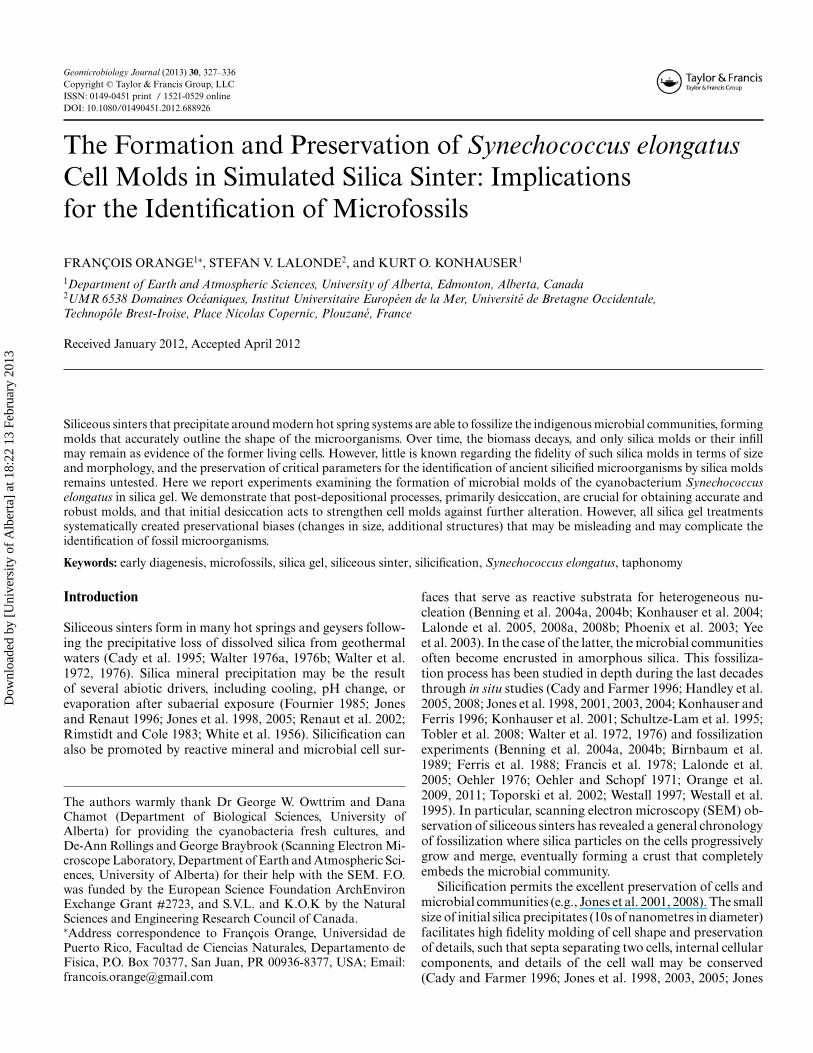
Fig. 1. Scanning electron microscope (SEM) micrographs showing the formation and air-drying of a silica gel mixed with Synechococ-cus elongatus cells. (a) fresh S. elongatus cells; (b) S. elongatus cell in a fresh silica gel, prepared with the critical point drying method;(c) S. elongatus cell and void mold in a silica gel air-dried for 24 h, prepared with the critical point drying method; (d) weight loss ofthe silica gel during air-drying; (e-h) S. elongatus cells (e, h) and void molds (f, g) in a silica gel air-dried for 2 weeks, prepared withthe critical point drying method. Note the granular crust surrounding the cell in plate h; (i-j) S. elongatus cells in a silica gel coveredby salt crystals air-dried for two months; and (k-l) void molds in a silica gel air-dried for 2 months, rinsed with distilled water. Allmicrographs were captured at 5 kV.
Dow
nloa
ded
by [
Uni
vers
ity o
f A
lber
ta]
at 1
8:22
13
Febr
uary
201
3
330 Orange et al.
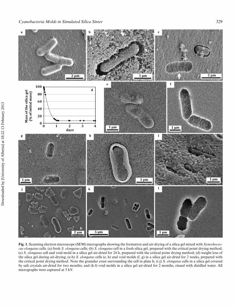
Table 1. Experimental conditions and mold width parameters
Number of Mean value Standard deviationSamples objects measured Range (μm) (μm) σ (μm)
TEM gridexperiments
TEM grid square 11 58.30–61.51 59.61 1.18Silica gel–Air-dried (6 days) 33 27.58–30.96 29.09 0.83Silica gel–Dried at 200◦C (6 h) 30 18.81–28.76 23.83 2.70
Wet gel experiments Synechococcus elongatus cells 18 0.77–0.99 0.87 0.07Fresh wet silica gel 13 0.77–1.02 0.87 0.06
Air-dryingexperiments
Silica gel–Air-dried (24 h) 12 0.86–1.12 0.95 0.11Silica gel–Air-dried (2 weeks) 31 0.42–1.13 0.81 0.18Silica gel–Air-dried (2 months) 28 0.69–1.23 0.91 0.10
High-temperaturedrying experiments
Silica gel–200◦C (6 h) 20 0.41–0.83 0.61 0.10Silica gel–200◦C (6 days) 21 0.38–1.08 0.68 0.21Air-dried gel (2 months)–200◦C
(6 h)21 0.71–1.05 0.87 0.10
Air-dried gel (2 months)–200◦C(6 days)
35 0.47–0.99 0.84 0.12
Fig. 2. Changes in size of Synechococcus elongatus cell molds in the silica gel. (a) Control; size of the square pattern drawn by a TEMgrid in silica gel after drying at room temperature and at 200◦C; (b) width of S. elongatus cell molds in fresh silica gel dried at roomtemperature for varying lengths of time; (c) width of S. elongatus cell molds in fresh silica gel dried at 200◦C for varying lengths oftime; and (d) width of S. elongatus cell molds in silica gel air-dried for 2 months and then dried at 200◦C for 6 h or 6 days. Meanvalues and standard deviations are shown (side bars). Measurements were made directly from SEM micrographs.
Dow
nloa
ded
by [
Uni
vers
ity o
f A
lber
ta]
at 1
8:22
13
Febr
uary
201
3
Cyanobacteria Molds in Simulated Silica Sinter 331
(Figure 1b). Clearly, the molds were formed by direct castingof the cells by the silica gel. Gaps observed between the geland cells (Figures 1b, 1c) are a consequence of the criticalpoint drying used for SEM preparation, which reduced thesize of the cells without affecting the silica gel. A repulsion ofsilica by cells can also produce such a gap, but would resultin molds with irregular outlines (Meunier et al. 2010; Rookeet al. 2008), which was not observed here. The silica particlesseen on cells (Figure 1b) may be the result of initial nucleationof silica directly on the cell wall, but may also be silica parti-cles sticking to the cell surface after having being remobilizedduring sample preparation.
Air-Drying of the Silica Gel
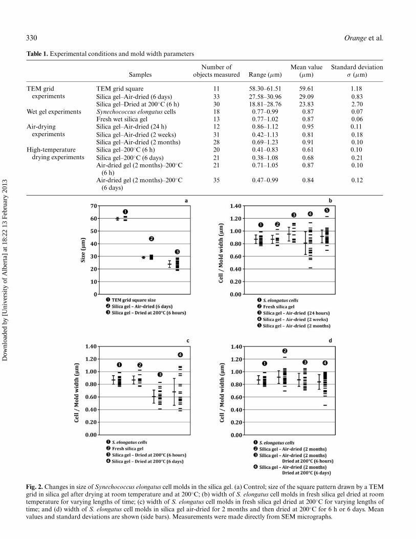
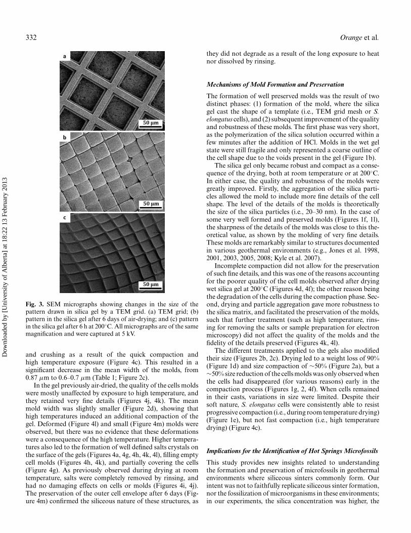
After exposure to air, the silica gels quickly dried and frag-mented, yielding pieces of dried silica that were coloured greenby the cells. The drying silica gel lost more than 90% of itsweight within a day of the dehydration process. This weightremained stable thereafter, with only 6.5% of its initial massremaining, and thus having lost almost all of its water (initialcontent >96%; Figure 1d). Drying was also accompanied bycompaction of the gel, which resulted in a ∼50% size decrease,as measured from TEM grid mold dimensions (from regular60 μm squares to 28–31 μm after air-drying; Figures 2a, 3a,3b, Table 1). This compaction was the result of the evaporationof most of the water contained in the gel and was accompaniedby the aggregation of the silica particles.
SEM observations reveal that the dried silica gel exhibits avery compact and homogeneous granular matrix, composedof closely packed silica particles (compare Figure 1b andFigure 1c). The size of these particles remained unchanged(20–30 nm) throughout drying, indicating that no further sil-ica precipitation occurred after the formation of the gel. De-hydration also led to the formation of salt crystals (mainlyNaCl, formed of Na from the sodium silicate solution and Clfrom the acid), which precipitated after the silica and partiallycovered the dried silica gel at the last stages of evaporation(Figures 1i, 1j). These salts crystals could be removed by rins-ing, which showed that they had no damaging effects to thesilica or the cells (Figures 1c, 1f, 1l).
During the entire drying experiment, S. elongatus cells re-mained well preserved, showing only a slight wrinkling of thecell surface due to dehydration and cell lysis (Figure 1i). Theyalso remained in direct contact with the silica (Figure 1e). Thecompaction led to finer and more detailed molds of cell shapesin the silica matrix, with very fine structures perfectly moldedby the silica (such as the septum between two dividing cells;Figure 1c, arrow; Figure 1l). The range in width of the moldsincreased during the drying (from 0.8–1.0 μm to 0.4–1.1 μmafter 2 weeks; Figure 2b), but the mean value remained closeto the initial value (Table 1), indicating no major size changesin the molds.
However, some void molds became significantly smaller(width 0.4–0.7 μm) (Figures 1g, 2b). As shown by salt crys-tals grown in these void molds, the cells of these molds hadlikely been removed early during the drying and compactionprocesses, which led to a reduced mold size. Some individ-
Table 2. Sample mass change (in percent) after air-drying or heattreatments of varying lengths of time
Percentage of mass ofsample remaining after
heat treatment
Samples 200◦C–6 h 200◦C–6 days
Fresh silica gel without cells 5.8% n.d.Fresh silica gel with
Synechococcus elongatus cells5.8% 5.7%
Silica gel air-dried during 2months
90.8% 91.8%
Each value is the result of at least two separate measurements.
ual or grouped cells were surrounded by a granular enve-lope that appeared to have formed at the cell/silica interface(Figures 1h, 1j, 1k). Its nature and origin remains unclear, butit may be the result of the formation of finer silica particleson the extracellular polymeric substances (EPS) covering, orsecreted, by the cells.
High-Temperature Drying of the Silica Gel
The high temperature experiments had two objectives. First,when applied to a fresh silica gel, it was to observe the effects ofaccelerated drying. Second, when applied to an already driedgel, it was to evaluate the effects of such an important stresson already well-formed molds.
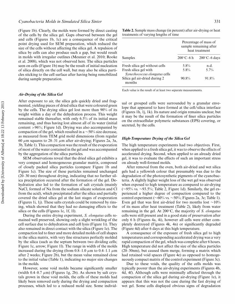
After removal from the oven, both air-dried and wet silicagels had a yellowish colour that presumably was due to thedegradation of the photosynthetic pigments of the cyanobac-teria. A slightly higher weight loss of the wet gel was observedwhen exposed to high temperature as compared to air-drying(∼95% vs. ∼93.5%; Table 2, Figure 1d). Similarly, the gel ex-perienced a higher degree of compaction compared to thecontrol experiment (∼60% vs. ∼50%; Figures 2a, 3c; Table 1).Even gel that was first air-dried for two months lost ∼10%of its mass after heat treatment (Table 2), likely from waterremaining in the gel. At 200◦C, the majority of S. elongatuscells were still present and in a good state of preservation afteronly 6 h (Figures 4a, 4i), however all cells were either com-pletely destroyed (Figures 4k, 4n) or significantly degraded(Figure 4d) after 6 days at this high temperature.
A consequence of the exposure of fresh silica gel to hightemperatures and corresponding accelerated drying was a veryrapid compaction of the gel, which was complete after 6 hours.High temperature did not affect the size of the silica particles(20–30nm), but caused them merge, forming a matrix whichhad retained void spaces (Figure 4e) as opposed to homoge-neously compact matrix of the control experiment (Figure 3c).
Due to these voids, the quality of the cells molds wastypically poorer than the air-drying experiments (Figures 4b,4d, 4f). Although cells were minimally affected through thecompaction of the silica gel during air-drying (Figure 1e), itappears that this was not the case during the fast drying ofwet gel. Some cells displayed obvious signs of degradation
Dow
nloa
ded
by [
Uni
vers
ity o
f A
lber
ta]
at 1
8:22
13
Febr
uary
201
3
332 Orange et al.
Fig. 3. SEM micrographs showing changes in the size of thepattern drawn in silica gel by a TEM grid. (a) TEM grid; (b)pattern in the silica gel after 6 days of air-drying; and (c) patternin the silica gel after 6 h at 200◦C. All micrographs are of the samemagnification and were captured at 5 kV.
and crushing as a result of the quick compaction andhigh temperature exposure (Figure 4c). This resulted in asignificant decrease in the mean width of the molds, from0.87 μm to 0.6–0.7 μm (Table 1; Figure 2c).
In the gel previously air-dried, the quality of the cells moldswere mostly unaffected by exposure to high temperature, andthey retained very fine details (Figures 4j, 4k). The meanmold width was slightly smaller (Figure 2d), showing thathigh temperatures induced an additional compaction of thegel. Deformed (Figure 4l) and small (Figure 4m) molds wereobserved, but there was no evidence that these deformationswere a consequence of the high temperature. Higher tempera-tures also led to the formation of well defined salts crystals onthe surface of the gels (Figures 4a, 4g, 4h, 4k, 4l), filling emptycell molds (Figures 4h, 4k), and partially covering the cells(Figure 4g). As previously observed during drying at roomtemperature, salts were completely removed by rinsing, andhad no damaging effects on cells or molds (Figures 4i, 4j).The preservation of the outer cell envelope after 6 days (Fig-ure 4m) confirmed the siliceous nature of these structures, as
they did not degrade as a result of the long exposure to heatnor dissolved by rinsing.
Mechanisms of Mold Formation and Preservation
The formation of well preserved molds was the result of twodistinct phases: (1) formation of the mold, where the silicagel cast the shape of a template (i.e., TEM grid mesh or S.elongatus cells), and (2) subsequent improvement of the qualityand robustness of these molds. The first phase was very short,as the polymerization of the silica solution occurred within afew minutes after the addition of HCl. Molds in the wet gelstate were still fragile and only represented a coarse outline ofthe cell shape due to the voids present in the gel (Figure 1b).
The silica gel only became robust and compact as a conse-quence of the drying, both at room temperature or at 200◦C.In either case, the quality and robustness of the molds weregreatly improved. Firstly, the aggregation of the silica parti-cles allowed the mold to include more fine details of the cellshape. The level of the details of the molds is theoreticallythe size of the silica particles (i.e., 20–30 nm). In the case ofsome very well formed and preserved molds (Figures 1f, 1l),the sharpness of the details of the molds was close to this the-oretical value, as shown by the molding of very fine details.These molds are remarkably similar to structures documentedin various geothermal environments (e.g., Jones et al. 1998,2001, 2003, 2005, 2008; Kyle et al. 2007).
Incomplete compaction did not allow for the preservationof such fine details, and this was one of the reasons accountingfor the poorer quality of the cell molds observed after dryingwet silica gel at 200◦C (Figures 4d, 4f); the other reason beingthe degradation of the cells during the compaction phase. Sec-ond, drying and particle aggregation gave more robustness tothe silica matrix, and facilitated the preservation of the molds,such that further treatment (such as high temperature, rins-ing for removing the salts or sample preparation for electronmicroscopy) did not affect the quality of the molds and thefidelity of the details preserved (Figures 4k, 4l).
The different treatments applied to the gels also modifiedtheir size (Figures 2b, 2c). Drying led to a weight loss of 90%(Figure 1d) and size compaction of ∼50% (Figure 2a), but a∼50% size reduction of the cells molds was only observed whenthe cells had disappeared (for various reasons) early in thecompaction process (Figures 1g, 2, 4f). When cells remainedin their casts, variations in size were limited. Despite theirsoft nature, S. elongatus cells were consistently able to resistprogressive compaction (i.e., during room temperature drying)(Figure 1e), but not fast compaction (i.e., high temperaturedrying) (Figure 4c).
Implications for the Identification of Hot Springs Microfossils
This study provides new insights related to understandingthe formation and preservation of microfossils in geothermalenvironments where siliceous sinters commonly form. Ourintent was not to faithfully replicate siliceous sinter formation,nor the fossilization of microorganisms in these environments;in our experiments, the silica concentration was higher, the
Dow
nloa
ded
by [
Uni
vers
ity o
f A
lber
ta]
at 1
8:22
13
Febr
uary
201
3

Cyanobacteria Molds in Simulated Silica Sinter 333
Fig. 4. SEM micrographs showing the results of drying experiments at 200◦C for silica gels (fresh and previously air-dried for 2 months)mixed with Synechococcus elongatus cells. Fresh silica gel: (a) silica gel covered with salts crystals (200◦C, 6 h); (b) S. elongatus cellmolds, with cellular remains (200◦C, 6 h, rinsed with distilled water); (c) S. elongatus cell with marks of crushing due to compactionand its mold (200◦C, 6 h, rinsed with distilled water); (d) silica gel with S. elongatus cell molds (6 days, 200◦C); (e) close-up on the silicamatrix (6 days, 200◦C); and (f) void S. elongatus cell molds (6 days, 200◦C, rinsed with distilled water). Previously air-dried silica gel:(g-h) S. elongatus cell and molds in the dried silica gel, partly covered or filled with salts crystals (6 h, 200◦C); (i-j) S. elongatus celland mold (6 h, 200◦C, rinsed with distilled water); (k-l) void molds, partly covered with salts crystals (6 h, 200◦C); and (m-n) voidmolds (6 days, 200◦C, rinsed with distilled water). All micrographs were captured at 5 kV.
Dow
nloa
ded
by [
Uni
vers
ity o
f A
lber
ta]
at 1
8:22
13
Febr
uary
201
3
334 Orange et al.
polymerization and encasing of the cells was faster and nonucleation of silica on microbial surfaces occurred.
Actually, our experimental setup was more similar to meth-ods of encapsulation, where microorganisms are encased in asilica gel for biomedical applications (e.g., Ferrer et al. 2006;Livage and Coradin 2006; Meunier et al. 2010; Roggendorfet al. 2001). Nonetheless, mold formation as simulated heredoes bear some similarities with the natural process, in that,following the nucleation of silica on microbial surfaces, the sil-ica particles aggregate and eventually merge, leading to a morerobust matrix, water expulsion, and a more accurate moldingof the cellular structures.
Importantly, despite not being a fossilization analogue, ourresults show a physical role for cells in their own preservationby silica encrustation. The final quality of the molds is notonly linked to compaction of the silica matrix, but also towhether cells had been potentially degraded by compaction(e.g. Figure 4c) or heat (e.g., Figure 4b). Air-dried cell moldexperiments showed minimal signs of cell degradation, alongwith molds preserved with the best quality (Figures 1e, 1f, 1i,1l). This appears to be similarly reflected in natural hot springsystems where numerous studies have demonstrated that veryprecise molds of cells in siliceous sinters are the result of a silicacrust developing on either live or minimally degraded cells(Cady and Farmer 1996; Jones and Renaut 1996; Konhauseret al. 2001; Jones et al. 1998, 2003, 2005, 2008; Kyle et al. 2007;Parenteau and Cady 2010; Renaut et al. 1998; Schultze-Lamet al. 1995; Tobler et al. 2008).
Any degradation of the cells during silica particle nucle-ation, growth, and aggregation could prevent the formationof the molds or lead to the formation of low-quality moldsof the cells. Such conditions lead to only a vague outline ofthe cell morphology in the resulting molds (e.g., Figure 4d).In addition, every treatment applied to the silica (i.e., drying,temperature) led to the modification of the size of the fossiltraces, and ultimately, loss of information about the originalsize of the microorganisms. The bright 150–300 nm mineralcrust that formed around some of the cells (Figures 1h, 1k,4m) may also be identified as a remnant of a cellular structure,thereby leading to inaccurate interpretations of the size andthe structure of the original microorganism. All of these fac-tors may contribute to preservational biases observed duringsiliceous sinter formation and fossilization processes (see Joneset al. 2001 for an extensive listing), and as such, they shouldbe considered to avoid being misled during the identificationof microfossils.
Only the already well formed and solid molds ultimatelyresisted additional treatments, exemplified by the good preser-vation of molds formed in an air-dried gel and then exposedto a 200◦C temperature (Figure 4k). In natural siliceous sin-ters, such molds would be subsequently exposed to variousdegrading processes during sinter growth and early diagene-sis, such as recrystallization, erosion, deformation, secondarymineral deposition, and later generations of silica precipita-tion. However, we demonstrate here that most crucial steps ofthe feature preservation process would have already occurred;early desiccation of the silica matrix appears important forboth the initial preservation of details and subsequent long-term resistance to further alteration. Traces of microorgan-
isms entombed under such conditions appear most likely tobe preserved.
Conclusions
This study provides new insight into the processes that leadto the preservation of very detailed molds of microorganismsin natural hot spring siliceous sinters through silica gelationexperiments simulating rapid cell entombment and early dia-genesis. We show that preservation is the result of two phases:(1) formation of the siliceous molds, as the result of the silicapolymerization and precipitation, and (2) a phase of strength-ening of the silica matrix and improvement of the quality ofthe molds, a consequence of silica particle aggregation andmatrix condensation associated with desiccation. These twophases are both essential for obtaining high fidelity, robustmolds. Original morphology and size of the microorganismsmay be preserved in the mold, but issues such as incompletesilica particle aggregation or the rapid degradation of cells athigh temperature have negative consequences on the qualityof the molds. Our experiments demonstrate that the transitionfrom a wet silica matrix to dry silica mass results in modifica-tion of the information preserved about the original size of themicroorganisms, and provides the first quantitative constraintson post-depositional size modification. As has been previouslyobserved in natural settings, this study confirms that preserva-tion biases are unavoidable during microfossils formation, andaccordingly, should be taken into account when consideringthe identification of microfossils.
References
Bartley JK. 1996. Actualistic taphonomy of cyanobacteria: implicationsfor the Precambrian fossil record. Palaios 11:571–586.
Benning LG, Phoenix VR, Yee N, Konhauser KO. 2004b. The dynam-ics of cyanobacterial silicification: an infrared micro-spectroscopicinvestigation. Geochim Cosmochim Acta 68:743–757.
Benning LG, Phoenix VR, Yee N, Tobin MJ. 2004a. Molecular charac-terization of cyanobacterial silicification using synchrotron infraredmicro-spectroscopy. Geochim Cosmochim Acta 68:729–741.
Birnbaum SJ, Wireman JW, Borowski R. 1989. Silica precipitation bythe anaerobic sulphate reducing bacterium Desulfovibrio desulfuri-cans: Effects upon cell morphology and implications for preserva-tion. In: Crick RE, editor. Origin, evolution, and modern aspects ofbiomineralization in plants and animals. Plenum Press, New York.p507–516.
Cady SL, Farmer JD. 1996. Fossilization processes in siliceous thermalsprings: trends in preservation along thermal gradients. In: BockGR, Goode JA, editors. Evolution of Hydrothermal Ecosystems onEarth (and Mars?). John Wiley and Sons, New York. p150–173.
Cady SL, Farmer JD, Des Marais DJ, Blake DF. 1995. Columnar andspicular geyserites from Yellowstone National Park, WY; scanningand transmission electron microscopy evidence for biogenicity (ab-stract). Geol Soc Am Abstracts Prog 27:A305.
Cassie V, Cooper RC. 1989. Algae of New Zealand thermal areas. Bib-liotheca Phycolog 78:1–159.
Ferrer ML, Garcia-Carvajal ZY, Yuste L, Rojo F, del Monte F. 2006.Bacteria viability in sol-gel materials revisited: cryo-SEM as a suit-able tool to study the structural integrity of encapsulated Bacteria.Chem Mater 18:1458–1463.
Dow
nloa
ded
by [
Uni
vers
ity o
f A
lber
ta]
at 1
8:22
13
Febr
uary
201
3
Cyanobacteria Molds in Simulated Silica Sinter 335
Ferris FG, Fyfe WS, Beveridge TJ. 1988. Metallic ion binding by Bacillussubtilis: Implications for the fossilization of microorganisms. Geol-ogy 16:149–152.
Ferris MJ, Ruff-Roberts AL, Kopczynski ED, Bateson MM, Ward DM.1996. Enrichment culture and microscopy conceal diverse ther-mophilic Synechococcus populations in a single hot spring microbialmat habitat. Appl Environ Microbiol 62:1045–1050.
Fournier RO. 1985. The behavior of silica in hydrothermal solutions.In: Berger BR, Bethke PM, editors. Geology and Geochemistryof Epithermal Systems: Society of Economic Geologists, Reviewsin Economic Geology, Vol. 2, Littleton, CO: Society of EconomicGeologists, p45–61.
Francis S, Margulis L, Barghoorn ES. 1978. On the experimental silicifi-cation of microorganisms. II. On the time of appearance of eukary-otic organisms in the fossil record. Precambrian Res 6:65–100.
Handley KM, Campbell KA, Mountain BW, Browne PRL. 2005.Abiotic-biotic controls on the origin and development of spicu-lar sinter: in situ growth experiments. Champagne Pool, Waiotapu,New Zealand. Geobiology 3:93–114.
Handley KM, Turner SJ, Campbell KA, Mountain BW. 2008. Silicifyingbiofilm exopolymers on a hot-spring microstromatolite: Templatingnanometer-thick laminae. Astrobiology 8:747–770.
Jones B, de Ronde CEJ, Renaut RW. 2008. Mineralized microbes fromGiggenbach submarine volcano. J Geophys Res 113:B08S05.
Jones B, Konhauser KO, Renaut RW, Wheeler R. 2004. Microbial silici-fication in Iodine Pool, Waimangu Geothermal area, North Island,New Zealand: Implications for recognition and identification of an-cient silicified microbes. J Geol Soc London 161:983–993.
Jones B, Renaut RW. 1996. Influence of thermophilic bacteria on calciteand silica precipitation in hot springs with water temperatures above90◦C: Evidence from Kenya and New Zealand. Can J Earth Sci33:72–83.
Jones B, Renaut RW, Konhauser KO. 2005. Genesis of large siliceousstromatolites at Frying Pan Lake, Waimangu geothermal field,North Island, New Zealand. Sedimentology 52:1229–1252.
Jones B, Renaut RW, Rosen MR. 1998. Microbial biofacies in hot-springsinters: A model based on Ohaaki Pool, North Island, New Zealand.J Sediment Res 68:413–434.
Jones B, Renaut RW, Rosen MR. 2001. Taphonomy of silicified filamen-tous microbes–Implications for identification. Palaios 16:580–592.
Jones B, Renaut RW, Rosen MR. 2003. Silicified microbes in a geysermound: the enigma of low-temperature cyanobacteria in a hightemperature setting. Palaios 18:87–109.
Konhauser KO, Ferris FG. 1996. Diversity of iron and silica precipitationby microbial mats in hydrothermal waters, Iceland: implications forPrecambrian iron formations. Geology 24:323–326.
Konhauser KO, Jones B, Phoenix VR, Ferris G, Renaut RW. 2004. Themicrobial role in hot spring silicification. Ambio 33:552–558.
Konhauser KO, Phoenix VR, Bottrell SH, Adams DG, Head IM. 2001.Microbial–silica interactions in Icelandic hot spring sinter: possibleanalogues for some Precambrian siliceous stromatolites. Sedimen-tology 48:415–433.
Kyle JE, Schroeder PA, Wiegel J. 2007. Microbial Silicification in Sintersfrom Two Terrestrial Hot Springs in the Uzon Caldera, Kamchatka,Russia. Geomicrobiol J, 24:627–641.
Lalonde SV, Konhauser KO, Reysenbach AL, Ferris FG. 2005. Theexperimental silicification of Aquificales and their role in hot springformation. Geobiology 3:41–52.
Lalonde SV, Smith DS, Owttrim GW, Konhauser KO. 2008a. Acid-base properties of cyanobacterial surfaces I: Influences of growthphase and nitrogen metabolism on surface reactivity. Geochim Cos-mochim Acta 72:1257–1268.
Lalonde SV, Smith DS, Owttrim GW, Konhauser KO. 2008b. Acid-base properties of cyanobacterial surfaces II: Silica as a chemicalstressor influencing cell surface reactivity. Geochim CosmochimActa 72:1269–1280.
Livage J, Coradin T. 2006. Living cells in oxide glasses. Rev MineralGeochem 64:315–332.
Meunier CF, Dandoy P, Su BL. 2010. Encapsulation of cells within silicamatrixes: towards a new advance in the conception of living hybridmaterials. J Coll Interf Sci 342:211–224.
Oehler JH. 1976. Experimental studies in Precambrian paleontology:structural and chemical changes in blue-green algae during simu-lated fossilization in synthetic chert. Geol Soc Am Bull 87:117–129.
Oehler JH, Schopf JW. 1971. Artificial microfossils: experimental stud-ies of permineralization of blue-green algae in silica. Science174:1229–1231.
Orange F, Disnar JR, Westall F, Prieur D, Baillif F. 2011. Metal bindingby the early Earth analogue microorganism, Archaea Methanocal-dococcus jannaschii and its effects on silicification. Palaeontology54:953–964.
Orange F, Westall F, Disnar JR, Prieur D, Bienvenu N, Le Romancer M,Defarge Ch. 2009. Experimental silicification of the extremophilicArchaea Pyrococcus abyssi and Methanocaldococcus jannaschii. Ap-plications in the search for evidence of life in early Earth and ex-traterrestrial rocks. Geobiology 7:403–418.
Parenteau MN, Cady SL. 2010. Microbial biosignatures in iron-mineralized phototrophic mats at Chocolate Posts hot springs, Yel-lowstone National Park, United States. Palaios 25:97–111.
Phoenix VR, Konhauser KO. 1999. Photosynthetic controls on the sili-cification of cyanobacteria. In: Armannsson H, editor. Geochem-istry of the Earth’s Surface. A.A. Balkema, Rotterdam, Netherlands.p275–278.
Phoenix VR, Konhauser KO, Ferris FG. 2003. Experimental study ofiron and silica immobilization by bacteria in mixed Fe-Si systems:Implications for microbial silicification in hot-springs. Can J EarthSci 40:1669–1678.
Renaut RW, Jones B, Tiercelin JJ. 1998. Rapid in situ silicification ofmicrobes at Loburu hot springs, Lake Bogoria, Kenya Rift Valley.Sedimentology 45:1083–1103.
Renaut RW, Jones B, Tiercelin JJ, Tarits C. 2002. Sublacustrine pre-cipitation of hydrothermal silica in rift lakes: Evidence from LakeBaringo, central Kenya Rift Valley. Sediment Geol 148:235–257.
Rimstidt JD, Cole DR. 1983. Geothermal mineralization I: The mecha-nism of formation of the Beowawe, Nevada, siliceous sinter deposit.Am J Sci 283:861–875.
Rippka R, Deruelles J, Waterbury JB, Herdman M, Stanier RY. 1979.Generic assignments, strain histories and properties of pure culturesof cyanobacteria. J Gen Microbiol 111:1–61.
Roggendorf H, Boschel D, Templer J. 2001. Structural evolution ofsodium silicate solutions dried to amorphous solids. J Non-CrystSolids 293–295:752–757.
Rooke JC, Leonard A, Sarmento H, Descyb JP, Si BL. 2008. Photosyn-thesis within porous silica gel: Viability and activity of encapsulatedcyanobacteria. J Mater Chem 18:2833–2841.
Schultze-Lam S, Ferris FG, Konhauser KO, Wiese RG. 1995. In situ sili-cification of an Icelandic microbial mat: implications for microfossilformation. Can J Earth Sci 32:2021–2026.
Tobler DJ, Stefansson A, Benning LG. 2008. In-situ grown silica sintersin Icelandic geothermal areas. Geobiology 6:481–502.
Toporski JKW, Steele A, Westall F, Thomas-Keprta KL, McKay DS.2002. The simulated silicification of bacteria—New clues to themodes and timing of bacterial preservation and implications for thesearch for extraterrestrial microfossils. Astrobiology 2:1–26.
Walter MR. 1976a. Hot-springs sediments in Yellowstone National Park.In: Walter MR, editor. Stromatolites: Developments in Sedimentol-ogy 20. Elsevier, Amsterdam. p489–98.
Walter MR. 1976b. Geyserites of Yellowstone National Park: an exam-ple of abiogenic “stromatolites.” In: Walter MR, editor. Stromato-lites: Developments in Sedimentology, Vol. 20. Elsevier, Amsterdam.p87–112.
Walter MR, Bauld J, Brock TD. 1972. Siliceous algal and bacterial stro-matolites in hot springs and geyser effluents of Yellowstone NationalPark. Science 178:402–405.
Walter MR, Bauld J, Brock TD. 1976. Microbiology and morphogen-esis of columnar stromatolites (Conophyton, Vacerrilla) from hot
Dow
nloa
ded
by [
Uni
vers
ity o
f A
lber
ta]
at 1
8:22
13
Febr
uary
201
3

336 Orange et al.
springs in Yellowstone National Park. In: Walter MR, editor. Stro-matolites: Developments in Sedimentology, Vol. 20. Elsevier, Ams-terdam. p273–310.
Westall F. 1997. The influence of cell wall composition on the fossilizationof bacteria and the implications for the search for early life forms.In: Cosmovici C, Bowyer S, Werthimer D, editors. Astronomicaland Biochemical Origins and the Search for Life in the Universe.Editori Compositrici, Bologna, Italy. p491–504.
Westall F, Boni L, Guerzoni E. 1995. The experimental silicification ofmicroorganisms. Palaeontology 38:495–528.
White DE, Brannock WW, Murata KJ. 1956. Silica in hot-spring waters.Geochim Cosmochim Acta 10:27–59.
Yee N, Phoenix VR, Konhauser KO, Benning LG, Ferris FG. 2003.The effect of cyanobacteria on silica precipitation at neutral pH:Implications for bacterial silicification in geothermal hot springs.Chem Geol 199:83–90.
Dow
nloa
ded
by [
Uni
vers
ity o
f A
lber
ta]
at 1
8:22
13
Febr
uary
201
3
Copyright © 2022 FDOKUMEN