First report of Pandora neoaphidis resting spore formation in vivo in aphid hosts
9
This article appeared in a journal published by Elsevier. The attached copy is furnished to the author for internal non-commercial research and education use, including for instruction at the authors institution and sharing with colleagues. Other uses, including reproduction and distribution, or selling or licensing copies, or posting to personal, institutional or third party websites are prohibited. In most cases authors are permitted to post their version of the article (e.g. in Word or Tex form) to their personal website or institutional repository. Authors requiring further information regarding Elsevier’s archiving and manuscript policies are encouraged to visit: http://www.elsevier.com/copyright
-
Upload
independent -
Category
Documents
-
view
0 -
download
0
Transcript of First report of Pandora neoaphidis resting spore formation in vivo in aphid hosts

This article appeared in a journal published by Elsevier. The attachedcopy is furnished to the author for internal non-commercial researchand education use, including for instruction at the authors institution
and sharing with colleagues.
Other uses, including reproduction and distribution, or selling orlicensing copies, or posting to personal, institutional or third party
websites are prohibited.
In most cases authors are permitted to post their version of thearticle (e.g. in Word or Tex form) to their personal website orinstitutional repository. Authors requiring further information
regarding Elsevier’s archiving and manuscript policies areencouraged to visit:
http://www.elsevier.com/copyright

Author's personal copy
First report of Pandora neoaphidis resting spore formationin vivo in aphid hosts
Ana Clara SCORSETTIa,*, Annette Bruun JENSENb, Claudia L�OPEZ LASTRAc,Richard A. HUMBERd
aInstituto Spegazzini, Facultad de Ciencias Naturales y Museo, UNLP, Calle 53 # 477, La Plata (1900), ArgentinabDepartment of Agriculture and Ecology, University of Copenhagen, Thorvaldsensvej 40, 1871 Frederiksberg C, DenmarkcCentro de Estudios Parasitol�ogicos y de Vectores (CEPAVE), CCT La Plata-Conicet-UNLP, Calle 2 # 584, La Plata (1900), ArgentinadRobert W. Holley Center for Agriculture and Health, USDA-ARS, 538 Tower Road, Ithaca, NY 14853, USA
a r t i c l e i n f o
Article history:
Received 17 December 2010
Received in revised form
4 November 2011
Accepted 7 November 2011
Available online 17 November 2011
Corresponding Editor:
Judith K. Pell
Keywords:
Entomopathogenic fungi
Entomophthorales
Pandora neoaphidis
Resting spores
Winter survival
a b s t r a c t
The entomopathogenic fungus Pandora neoaphidis is a recognized pathogen of aphids,
causes natural epizootics in aphid populations, and interacts and competes with aphid
predators and parasitoids. Survival of entomophthoralean fungi in periods of unsuitable
weather conditions or lack of appropriate host insects is accomplished mainly by thick-
walled resting spores (zygospores or azygospores). However, resting spores are not known
for some entomophthoralean species such as P. neoaphidis. Several hypotheses of P. neoa-
phidis winter survival can be found in the literature but so far these hypotheses do not in-
clude the presence of resting spores. Resting spores were found in an aphid population
where P. neoaphidis was the only entomophthoralean fungus observed during surveys con-
ducted in organic horticultural crops in greenhouses and open fields in Buenos Aires prov-
ince, Argentina. This study sought to use molecular methods to confirm that these resting
spores were, in fact, those of P. neoaphidis while further documenting and characterizing
these resting spores that were produced in vivo in aphid hosts. The double-walled resting
spores were characterized using light and transmission electron microscopy. The Argenti-
nean resting spores clustered together with P. neoaphidis isolates with bootstrap values
above 98 % in the small subunit ribosomal RNA (SSU rRNA) sequence analysis and with
bootstrap values above 99 % the Internal Transcribed Spacer (ITS) II region sequence anal-
ysis. This study is the first gene-based confirmation from either infected hosts or cultures
that P. neoaphidis is able to produce resting spores.
ª 2011 British Mycological Society. Published by Elsevier Ltd. All rights reserved.
Introduction
Theentomopathogenic fungus Pandora neoaphidis (Remaudi�ere
& Hennebert) Humber (Entomophthoromycotina: Entomophthor-
ales), is a recognized pathogen of aphids (Hemiptera:Aphididae).
This fungus has a worldwide distribution, and is well known
from Europe, Asia, Africa, South America, and Australia
(Wilding & Brady 1984; Glare & Milner 1991; Hatting et al.
2000; Scorsetti et al. 2007). Pandora neoaphidis is aphid-specific,
causes natural epizootics in aphid populations and interacts
and competes with aphid predators and parasitoids
(Baverstock et al. 2008a). Recently it has been demonstrated
that P. neoaphidis causes natural epizootics in different seasons
at different temperatures in South America and causes host
* Corresponding author. Tel./fax: þ54 0221 4219845.E-mail address: [email protected]
journa l homepage : www.e lsev ier . com/ loca te / funb io
f u n g a l b i o l o g y 1 1 6 ( 2 0 1 2 ) 1 9 6e2 0 3
1878-6146/$ e see front matter ª 2011 British Mycological Society. Published by Elsevier Ltd. All rights reserved.doi:10.1016/j.funbio.2011.11.002
Author's personal copy
mortality up to 56.6 %, thus confirming the capacity of this
fungus to be an effective biological control agent (Scorsetti
et al. 2010).
Survival of entomophthoralean fungi in periods of unsuit-
able weather conditions or lack of appropriate host insects is
accomplished mainly by thick-walled resting spores (zygo-
spores or azygospores). However, resting spores are not
known for all entomophthoralean species. Most species that
are only rarely collected and poorly known probably do, in
fact, form resting spores as often and in the same manner
as their better known relatives, but their resting spores remain
undetected from their few known specimens (Nielsen et al.
2007). However, for so common, widespread, and much stud-
ied a species as P. neoaphidis, the absence of information about
resting spores cannot be dismissed as either accidental or ar-
tifactual. It has long been believed that resting spore produc-
tion by P. neoaphidis either does not happen or must be
extraordinarily rare.
There are several hypotheses in the literature about winter
survival of P. neoaphidis. One is that the fungus survives by con-
tinued infection of anholocyclic populations of aphids. This
possibility is supported by the fact that the fungus is capable
of infecting aphids at temperatures as low as 5 �C (Wilding
1970). Baverstock et al. (2008b) have suggested that P. neoaphidis
could remain active throughout the year through a combina-
tion of continuous infection and as inoculum deposited on
the soil. There is evidence reported about the capability of
P. neoaphidis to survive for long periods outside the host in
soil as conidia or as hyphal bodies (Feng et al. 1992). Schofield
et al. (1995) showed that conidia remained infective for up to
32 d after incubation at 5 �C in 85%RH. Nielsen et al. (2003) sug-
gested that P. neoaphidismayoverwinter in the soil as ‘loricoco-
nidia’ (resistant structures formedby themarked thickeningof
a conidial wall; although theymay function in a manner simi-
lar to resting spores, the morphological, and developmental
differences between these spore types are discussed below).
In populations of aphids otherwise demonstrated sporulat-
ing infections of P. neoaphidis, Scorsetti et al. (2007) noted and
illustrated a few resting spore-like structures from Argenti-
nean aphid cadavers collected in the autumn (June 2003 and
May 2004) but the taxonomic identification of these resting
spores could not be confirmed by molecular or morphological
techniques. The linkage between entomophthoralean resting
spores and their conidial forms is not always straightforward
but congruence of sequences for several genes from both rest-
ing spores and conidial states effectively confirm the linkages
of resting spores with the corresponding conidial states that
are the basis for entomophthoralean taxonomy. Thomsen &
Jensen (2002) used a nested PCR techniques to link resting
spores with the Entomophthora conidial stages affecting the
same host species of flies and thus demonstrated that several
differing genotypes within the Entomophthora muscae species
complex could each complete their entire life cycle in a single
dipteran host species. Comparing sequences of the ribosomal
repeat (small subunit (SSU) and Internal Transcribed Spacer
(ITS)) is however a stronger method that also allows blast
search against the still growing collection of entomophthora-
lean sequences of these two regions in GenBank.
The objective of this study was to document the morphol-
ogy of resting spores found in field-infected aphidswhere only
P. neoaphidis infection was found in the aphid population and
to use sequences of the ribosomal repeat (SSU and ITS) to con-
firm the P. neoaphidis identity of these resting spores.
Materials and methods
Field survey
Surveys were conducted in organic horticultural crops in
greenhouses and open fields in Buenos Aires province, Argen-
tina, according to Scorsetti et al. (2007). Sampling was per-
formed weekly from April 2007 to April 2008. The climatic
conditions in this region are temperate with average temper-
ature of ca 18 �C.Lettuce (Lactuca sativa L.) was the main crop that was sam-
pled, but aphids were also collected from fields of tomato
(Lycopersicon esculentum Mill.), cabbage (Brassica oleracea L.),
pepper (Capsicum annuum L.), eggplant (Solanum melongena L.),
Swiss chard (Beta vulgaris var. cicla L.), and artichoke (Cynara
scolymus L.).
Laboratory
Field-collected dead and living aphids were placed individu-
ally in plastic cups with lids (150 cm3). Dead aphids with or
without externals signs of mycosis were collected with the
piece of plant substrate onto which they were attached by rhi-
zoids, and were then placed individually into sterilized plastic
containers (100 cm3) and transported to the laboratory for
analysis of fungal infection.
Dead aphids showing no external signs of mycoses were
put in 60 mm Petri dishes with a moistened filter paper and
maintained at 20 �C for 24e72 h to allow the complete devel-
opment of any fungi present in them. Healthy aphids were
preserved in 70 % ethanol as reference specimens for further
taxonomic identification.
The pathogen affecting the aphids collected during these
studies was confirmed to be Pandora neoaphidis by using stan-
dard taxonomic literature (Keller 1987, 1991; Humber 1989;
Ba1azy 1993).
Pandora neoaphidis-infected aphids were stored at 4 �C for
1 m in 60 mm Petri dishes with a moistened filter paper, and
then examined using a stereoscopic microscope every 72 h
for the possible development of environmentally resistant
resting structures.
Microscopic characteristics were described from aphids
with resting spores mounted in aceto-orcein (1 % w v�1) and
observed on an Olympus CH3 microscope. Fungal prepara-
tions were observed on an Olympus BX51 microscope and
photographed using an Olympus DP71 camera.
Semipermanent slides were made according to Humber
(1997) and deposited as herbariummaterial in the Mycological
Collection of the Institute of Botany Carlos Spegazzini (LPSC,
La Plata, Buenos Aires, Argentina).
The aphids containing resting spores were preserved indi-
vidually in Eppendorf microcentrifuge tubes containing 48 %
ethanol and kept at 4 �C in darkness until either extraction
of the DNA or fixation for transmission electron microscopy
(TEM).
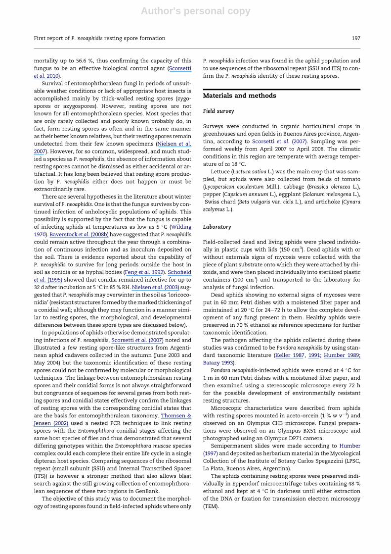
First report of P. neoaphidis resting spore formation 197

Author's personal copy
Electron microscopy
For TEM, thick-walled spores from a mechanically disrupted
infected aphid were fixed for 2 h in 3 % glutaraldehyde, post-
fixed for 1 h in 1 % osmium tetroxide, dehydrated in a graded
series of 30e100 % ethanol, cleared in propylene oxide, and
embedded in Epon 812. Thin (60 nm) sections were cut with
a diamond knife, stained with uranyl acetate and lead citrate,
and examined using a JEM 1200EX II (Jeol) TEM.
DNA extraction, PCR amplification, and sequencing
Two of the infected aphids bearing resting spores were used
for the molecular identification. Each of the aphids was
transferred into new Eppendorf microcentrifuge tubes, the
ethanol was allowed to evaporate, and the cadavers were
mechanically disrupted with a sterile plastic pistil in 100 ml
MiliQ water. The samples were washed twice by centrifuga-
tion for 2 min at 10 000�g. The supernatants were discarded
and 100 ml MilliQ water was added to each preparation. Five
microlitres of the washed spore suspension was added to
a glass slides coated with Gel Slick Solution (BioWhitataker
Molecular Application BMA; Rockland, ME, USA), and an-
other coated slide was put on top of the spores. The resting
spores were then disrupted mechanically by pressing the
slides hard with the thumbs with simultaneous circular
movements of the top slide. One microlitre of the crushed
spores was used as template for the PCR amplification. The
DNeasy� Plant Mini Kit (Qiagen GmbH, Hilden, Germany)
was used to extract the DNA from in vitro cultures of Pandora
nouryi ARSEF 362, P. nouryi ARSEF 366, and Pandora bullata
ARSEF 116, following the manufacturer’s instructions. Three
in vitro cultures were obtained from the United States De-
partment of Agriculture, Agricultural Research Service Col-
lection of Entomopathogenic Fungal Cultures (ARSEF) and
were grown in Graces liquid medium from where hyphal
mass was collected and freeze dried upon DNA extraction.
Universal fungal primers nu-SSU-0021-50 and nu-SSU-
1780-30 (Gargas & DePriest 1996) were used to amplify the
nuclear SSU ribosomal RNA (rRNA) gene with an initial dena-
turation for 3 min at 94 �C, followed by 35 cycles with dena-
turation for 1 min at 94 �C, annealing for 1 min at 60 �C,extension for 1.5 min at 72 �C, and a final extension for
7 min at 72 �C. Universal fungal primers ITS1 and ITS4
(White et al. 1990) were used to amplify the ITS regions of
ribosomal DNA with an initial denaturation for 3 min at
94 �C, followed by 35 cycles with denaturation for 1 min at
94 �C, annealing for 1 min at 55 �C, extension for 1.5 min at
72 �C, and a final extension for 7 min at 72 �C. Fungal univer-sal primers were used as a positive amplification control to
assure availability of DNA from the full range of entomoph-
thoralean species studied here.
The PCR reactions were carried out in 50 ml volumes each
with 200 mM of each dNTP, 1 mM of each primer, 1� Phusion
HF Buffer (with 1.5 mM MgCl2), 0.5 unit Phusion DNA poly-
merase (Finnzymes; Espoo, Finland), and 1 ml template
(DNA extractions from in vitro cultures or DNA from in vivo
material). Prior to sequencing, the PCR products were purified
with the illustra� DNA and Gel Band Purification Kit (GE
Healthcare; Buckinghamshire, UK). The purified PCR products
were sent to Eurofins MWG Operon (Ebersberg, Germany) for
sequencing in both directions. The following primers were
used for sequencing of the SSU: nu-SSU-0021-50 and nu-
SSU-1780-30 (Gargas & DePriest 1996) and nu-SSU-0553-30,nu-SSU-0573-50 and nu-SSU-1150-50 (White et al. 1990) and
for the ITS we used following primers for sequencing ITS1
Table 1 e List of the entomophthoralean species used inthis study, strain code, and GenBank accession numbers.
Species Strain code SSU ITS
Entomophaga aulicae FPMI 646 EAU35394
Entomophthora muscae F 1020 D29948
Erynia rhizospora ARSEF 1441 AF368514
Erynia sciarae ARSEF 1870 AF368515
Furia americana ARSEF 742 EF392554
Furia gastropachae ARSEF 5541 EF392562
Furia ithacensis ARSEF 1339 AF351134
Furia neopyralidarum ARSEF 1145 AF368518
Furia pieris ARSEF 781 AF368519
Pandora blunckii ARSEF 217 AF368520
Pandora bullata ARSEF 116 HQ677592 HQ677588
Pandora delphacis ARSEF 581 EF392551
Pandora delphacis ARSEF 459 AF368521
Pandora dipterigena ARSEF 397 AF368522
Pandora kondoiensis ARSEF 5707 AF543200
Pandora kondoiensis ARSEF 5708 AF543201
Pandora kondoiensis ARSEF 828 AF543199
Pandora kondoiensis ARSEF 825 AF351133
Pandora neoaphidis ARSEF 3240 EF392560
Pandora neoaphidis ARSEF 1607 EU267188
Pandora neoaphidis ARSEF 1609 AF543210
Pandora neoaphidis ARSEF 5374 AF543211
Pandora neoaphidis ARSEF 7937
MboIþEU267189 EU267189
Pandora neoaphidis ARSEF 7938
MboI�EU267191 EU267191
Pandora neoaphidis ARSEF 7938
MboIþEU267190 EU267190
Pandora neoaphidis ARSEF 7939 -1 EU267192 EU267192
Pandora neoaphidis ARSEF 7939 -3 EU267193 EU267193
Pandora neoaphidis ARSEF 835 AF543209
Pandora neoaphidis KVL 633 AF052405
Pandora neoaphidis NW 195 AF543204
Pandora neoaphidis NW 283 AF543205
Pandora neoaphidis NW 316 AF543206
Pandora neoaphidis NW 327 AF543207
Pandora neoaphidis NW 343 AF543202
Pandora neoaphidis NW 415 AF543208
Pandora neoaphidis Resting spores HQ677591 HQ677587
Pandora nouryi ARSEF 362 HQ677593 HQ677589
Pandora nouryi ARSEF 366 HQ677594 HQ677590
Strongwellsea castrans Resting spores AF052406
Zoophthora anglica ARSEF 396 AF368524
Zoophthora occidentalis ARSEF 3073 AF368525
Zoophthora radicans KVL 610 AF052404
Zoophthora radicans ARSEF 1699 DQ864988
Zoophthora radicans ARSEF 2411 EF151416
Zoophthora radicans ARSEF 6003 EF137934
Zoophthora radicans F 853 D61381
Zoophthora radicans NW 250 EF137938
Zoophthora radicans NW 323 EF137936
Zoophthora radicans NW 378 EF151414
Zoophthora radicans NW 386 EF151412
198 A. C. Scorsetti et al.

Author's personal copy
and ITS4 (White et al. 1990), Nu-5.8S-50 (Jensen & Eilenberg
2001) and Nu-5.8S-30 (Jensen et al. 2009).
In order to confirm the taxonomic affinity of the resting
sporesweblasted thesequences against theGenBankdatabase.
In addition the resting spore SSU rRNA sequence was aligned
withpublishedSSUrRNAsequences fromotherentomophthor-
alean species including three new SSU rRNA sequences e one
from P. bullata and two from P. nouryi isolates e were obtained
for the current study (Table 1).
The sequences were checked and aligned with Bioedit
v7.0.8.0. Parsimony and neighbour-joining analyses with 1000
bootstrap replications to determine the support for internal
branches were performed using MEGA 4 (Tamura et al. 2007).
In the SSU rRNA analysis Entomophthora muscae and Entomo-
phaga aulicae were used as outgroup taxa. In the ITS II analysis
all the Zoophthora radicans isolates were used as outgroups.
Results
Among the 562 Pandora-infectedaphidsthatwerestudied, sixca-
davers included structures with thick, double-layered walls that
appeared to standard entomophthoralean resting spores. These
six individuals were held at 4 �C for an additional 6 (n ¼ 4) or 9
(n ¼ 2) d to allow the full development and maturation of the
spores. All cadavers with the putative resting spores were col-
lected in mid-winter (August and September 2007) from Lactuca
sativa plants growing in open fields, and all of these hosts were
identified as Nasonovia ribisnigris (Mosley) (Hemiptera: Aphididae).
These six aphids were preserved in 48 % ethanol. Two of
them were processed for TEM, another two were used for
DNA extraction to determine the species identification bymo-
lecular analyses, and the remaining two are retained as
voucher specimens deposited as herbarium material in the
Mycological Collection of the Institute of Botany Carlos Spe-
gazzini (LPSC 47460, La Plata, Buenos Aires, Argentina).
The resting spores in these aphids averaged 31.4 � 2.4 mm
diam (24.8e37.2 mm; n¼ 300) (Fig 1A and B), andwere produced
both internally and externally on the surface of the aphid ca-
daver. Themacroscopic appearance of aphids filledwith these
structureswas dry and ‘sandy’. The resting sporeswere spher-
ical, globose, with surface slightly covered by rounded depres-
sions, varying from a pale yellow tint to deep amber (with the
colour apparently confined to the walls). The spores appeared
to be mature and dormant since there was a single consoli-
dated oil droplet filling most of the volume of individual
spores (Fig 1B).
Cross-sections of the resting spores observed with TEM
show two layers of the spore wall, a thin electron-dense
(dark) outer layer, and a much thicker electron-lucent (light)
inner layer (Fig 1C and D).
We obtained a sequence of the SSU rRNA gene from resting
spores of one aphid. The closest BLAST correspondences
(those with 99e100 % nucleotide identify) of the 1800 bp se-
quence from these resting spores were with eight different
Pandora neoaphidis sequences. The resting spore SSU rRNA se-
quence was aligned with other published entomophthoralean
SSU rRNA sequences but we only used the first 1100 positions
Fig 1 e Differential interference contrast micrograph (A), and phase contrast micrograph, (B) of Pandora neoaphidis mature
resting spores, with a central oil droplet (Od) filling most of the spore’s volume. Scale bar: 50 mm. Ultrastructure of double
spore wall of the resting spore, immature (C), andmature spore (D). Thin outer layer (w1), wide inner layer (w2), c: Cytoplasm.
X30000.
First report of P. neoaphidis resting spore formation 199
Author's personal copy
for the sequence analyses (the SSU alignment used can be
found as Supplement 1) since the SSU rRNA gene was only
partially sequenced for several of the other published ento-
mophthoralean species that we wanted to include.
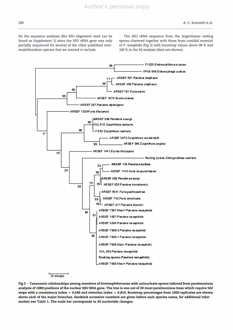
The SSU rRNA sequence from the Argentinean resting
spores clustered together with those from conidial material
of P. neoaphidis (Fig 2) with bootstrap values above 98 % and
100 % in the NJ analysis (data not shown).
Fig 2 e Taxonomic relationships amongmembers of Entomophthoraceaewith uninucleate spores inferred from parsimonious
analysis of 1080 positions of the nuclear SSU RNA gene. The tree is one out of 20 most parsimonious trees which require 562
steps with a consistency index [ 0.640 and retention index [ 0.810. Bootstrap percentages from 1000 replicates are shown
above each of the major branches. GenBank accession numbers are given before each species name, for additional infor-
mation see Table 1. The scale bar corresponds to 20 nucleotide changes.
200 A. C. Scorsetti et al.
Author's personal copy
The size of the amplified ITS region varied among the
different species included in this analysis from ca
1000e1500 bp (Table 1). Amplicons of the resting spores from
both aphids had a length of 1080 bp, as did those from all
the tested P. neoaphidis isolates. We obtained ITS sequences
from the resting spores of both aphids, and these resting spore
sequences were identical. When the sequences from the rest-
ing sporeswere blasted P. neoaphidis came outwith 99%nucle-
otide identitywith all the 18 deposited P. neoaphidis sequences.
Pandora kondoiensis followedwith only 87% nucleotide identity
of the part that could be aligned. The variation of the whole
ITS sequences within Entomophthoraceae and even within spe-
cies that produce uninucleate conidiawas too high formaking
unambiguous alignments. Most of the variation and also the
ITS size differences were found in the ITS I region, and there-
fore only the ITS II region (including the 5.8s rRNA gene
covering in all 585 positions including gaps) was used for the
sequence analyses corresponding to 490e1030 bp of the
GenBank sequence AF543202 of the P. neoaphidis isolate NW
343. Based on the SSU rRNA analysis we chose the Zoophthora
clade as outgroup (the ITS II alignment used can be found as
Supplement 2).
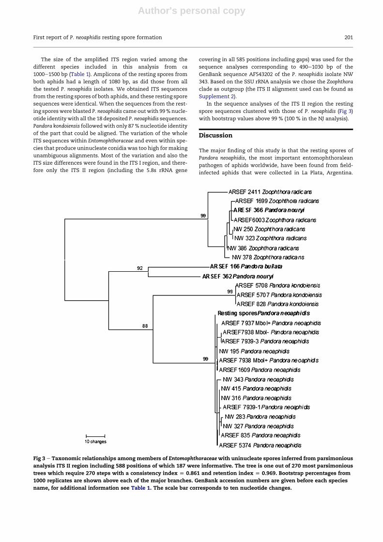
In the sequence analyses of the ITS II region the resting
spore sequences clustered with those of P. neoaphidis (Fig 3)
with bootstrap values above 99 % (100 % in the NJ analysis).
Discussion
The major finding of this study is that the resting spores of
Pandora neoaphidis, the most important entomophthoralean
pathogen of aphids worldwide, have been found from field-
infected aphids that were collected in La Plata, Argentina.
Fig 3 e Taxonomic relationships amongmembers of Entomophthoraceaewith uninucleate spores inferred from parsimonious
analysis ITS II region including 588 positions of which 187 were informative. The tree is one out of 270 most parsimonious
trees which require 270 steps with a consistency index [ 0.861 and retention index [ 0.969. Bootstrap percentages from
1000 replicates are shown above each of the major branches. GenBank accession numbers are given before each species
name, for additional information see Table 1. The scale bar corresponds to ten nucleotide changes.
First report of P. neoaphidis resting spore formation 201
Author's personal copy
Except for the report by Scorsetti et al. (2007) that is placed in
its appropriate perspective by the findings reported here, no
previous publication on the taxonomy or biology of this spe-
cies has provided any significant information about the mor-
phology e or even the existence e of resting spores for this
nearly ubiquitous species.
In the literature, the studies of overwintering survival by
P. neoaphidis focused on the conidial state (Brobyn et al. 1985;
Morgan 1994; Nielsen et al. 2003). Despite several hypotheses
about the morphological structures that might be responsible
for the winter survival of this fungal species, there were hints
in Scorsetti et al. (2007) that P. neoaphidis could produce resting
spores in field-infected aphids, but there were not enough
spores in those collections to allow gene-based confirmation
of their identification as P. neoaphidis. Uziel & Kenneth (1986)
suggested that a culture of P. neoaphidis produced what they
believed to be resting spores in completely uncontrolled labo-
ratory conditions, but the sizes of these structures (average di-
ameter of 74.2 mm, range 52e119 mm) were substantially larger
than any other resting spores reported within Entomophthor-
ales, and the nature of these structures remains uncertain.
In the present study we never found either hyphal bodies
or loricoconidia as environmentally resistant structures in
P. neoaphidis-infected aphids as had been reported by other au-
thors (Feng et al. 1992; Nielsen et al. 2003), but did find the pres-
ence of spores with the thick, double-layered walls that are
characteristic of all entomophthoralean resting spores as the
presumptive overwintering structures, and these thick-
walled spores were collected only during the mid-winter.
Apart from the molecular evidence presented here, the
morpho-developmental evidence also confirms that these
P. neoaphidis spores must be resting spores. Most of these
spores formed inside the aphid bodies although some were
superficial on the cadaver; their walls were distinctly
double-layered; and their shapes were spherical rather than
elongated (in no way similar to P. neoaphidis conidia). As
definedbyWeiser&Batko (1966), loricoconidia represent a sin-
gle-layered thickening of a conidial wall that necessarily re-
flect the shapes of the conidia from which they form, and
they can occur only where conidia are (or were) also present.
The conidia of P. neoaphidis form exclusively on the surface
of e and never inside e an infected host, and as in most ento-
mophthoralean entomopathogens, no conidia were formed
on the infected hosts where the resting spores formed.
The study site in Buenos Aires province is located in the
Pampeana phytogeographic region of Argentina (Cabrera &
Willink 1973), and it is important to note that this site has
a temperate climate with an average annual temperature of
18 �C and winter temperatures generally within the range of
3e8 �C. Zhou & Feng (2010) demonstrated that lower temper-
atures and longer daylight may result in more resting spore
production by Pandora nouryi in host cohorts at a given spore
concentration. The positive effect of lower temperature on
the resting spore formation agrees withmost previous reports
on the same phenomenon for other Entomophthorales
(Steinkraus & Kramer 1989; Feng et al. 1992; Pell et al. 2001).
Survival structures often also require periods of cold in order
to be able to germinate (Hajek 1997). The exact requirements
for dormancy and for initiation of germination and conidia-
tion are poorly understood for almost all entomophthoraleans
(Nielsen 2002), and they have remained completely unknown
for P. neoaphidis.
This study of Argentinean aphids presents the first genomi-
cally verified evidence that P. neoaphidis is, indeed, able to pro-
duce resting spores. To learn more about the environmental
(or other) conditions that allowed the production of this previ-
ouslyunobserved spore state should be ahighpriority. Progress
on these issueswill provide key insights about the field ecology
of this fungus and the biological mechanisms controlling the
initiations of epizootics of this aphid pathogen in Argentina
and throughout the world. More studies are needed to locate
further natural instances of these resting spores, to determine
what conditions might favour the breaking of the (presumed)
dormancy of fully mature spores, and whether there might be
any means to manipulate the formation and germination of
these resting spores to advance the practical application of P.
neoaphidis as a potent biological control agent against some of
the most significant insect pests of agricultural crops.
Acknowledgements
This study was partially supported by the National Research
Council of Argentina (CONICET) (PIP 0049), University of La
Plata (UNLP) and Danish National Research Foundation. We
also thank two anonymous reviewers for helpful comments
on the manuscript.
Supplementary material
Supplementary data related to this article can be found
online at doi:10.1016/j.funbio.2011.11.002.
r e f e r e n c e s
Ba1azy S, 1993. Entomophthorales. In: Flora of Poland (Flora Polska)Fungi (Mycota), vol. 24. Polish Academy of Science W. SzaferInstitute of Botany, Krakow.
Baverstock J, Baverstock KE, Clark SJ, Pell JK, 2008a. Transmissionof Pandora neoaphidis in the presence of co-occurring arthro-pods. Journal of Invertebrate Pathology 98: 356e359.
Baverstock J, Clark SJ, Pell JK, 2008b. Effect of seasonal abioticconditions and field margin habitat on the activity of Pandoraneoaphidis inoculum on soil. Journal of Invertebrate Pathology 97:282e290.
Brobyn P, Wilding N, Clark S, 1985. The persistence of infectivityof conidia of the aphid pathogen Erynia neoaphidis on leaves inthe field. Annual Applied Biology 107: 365e376.
Cabrera AL, Willink A, 1973. Biogeograf�ıa de Am�erica Latina. Secre-tar�ıa General de la Organizaci�on de los Estados Americanos,Washington, D.C.
Feng MG, Nowierski RM, Klein RE, Scharen AL, Sands DC, 1992.Spherical hyphal bodies of Pandora neoaphidis (Remaudi�ere &Hennebert) Humber (Zygomycetes: Entomophthorales) onAcyrthosiphon pisum (Harris) (Homoptera: Aphididae): a poten-tial overwintering form. Pan-Pacific Entomology 68: 100e104.
Gargas A, DePriest PT, 1996. A nomenclature for fungal PCRprimers with examples from intron-containing SSU rDNA.Mycologia 88: 745e748.
Glare TR, Milner RJ, 1991. Ecology of entomopathogenic fungi. In:Arora DK, Ajello L, Mukerji KG (eds), Handbook of Applied
202 A. C. Scorsetti et al.

Author's personal copy
Mycology. Humans, Animals, and Insects, vol. 2. Marcel Dekker,New York, pp. 547e612.
Hajek AE, 1997. Ecology of terrestrial fungal entomopathogens.Advances in Microbiological Letters 15: 193e249.
Hatting JL, Poprawski TJ, Miller RM, 2000. Prevalences of fungalpathogens and other natural enemies of cereal aphids (Ho-moptera: Aphididae) in wheat under dryland and irrigatedconditions in South Africa. BioControl 45: 179e199.
Humber RA, 1989. Synopsis of a revised classification for the En-tomophthorales (Zygomycotina). Mycotaxon 34: 441e460.
Humber RA, 1997. Fungi: identification. In: Lacey L (ed.), Manual ofTechniques in Insect Pathology. Academic Press, San Diego,pp. 153e185.
Jensen AB, Eilenberg J, 2001. Genetic variation within the insect-pathogenic genus Entomophthora, focusing on the E. muscaecomplex, using PCReRFLP of the ITS II and the LSU rDNA.Mycological Research 105: 307e312.
Jensen AB, Eilenberg J, Lopez Lastra C, 2009. Differential diver-gences of obligately insect-pathogenic Entomophthora speciesfrom fly and aphid hosts. FEMS Microbiology Letters 300:180e187.
Keller S, 1987. Arthropod-pathogenic Entomophthorales of Swit-zerland. I. Conidiobolus, Entomophaga and Entomophthora. Sydo-wia 40: 122e167.
Keller S, 1991. Arthropod-pathogenic Entomophthorales of Swit-zerland. II. Erynia, Eryniopsis, Neozygites, Zoophthora, andTarichium. Sydowia 43: 39e122.
Morgan LW, 1994. Survival, germination responses and infectivityof conidia of Erynia neoaphidis (Zygomycetes: Entomophthor-ales). PhD thesis, University of Wales, Cardiff, UK, pp. 187.
Nielsen C, 2002. Interactions between aphids and entomophthora-lean fungi. Characterisation, epizootiology and potential for mi-crobial control. Ph.D. thesis. Department of Ecology, The RoyalVeterinary and Agricultural University, Copenhagen, Denmark.
Nielsen C, Hajek AE, Humber RA, Bresciani J, Eilenberg J, 2003. Soilas an environment for survival of aphid-pathogenic Ento-mophthorales. Biological Control 28: 92e100.
Nielsen C, Jensen AB, Eilenberg J, 2007. Survival of entomoph-thoralean fungi infecting aphids and higher flies during un-favourable conditions and implications for conservationbiological control. In: Ekesi S, Maniania NK (eds), Use of Ento-mopathogenic Fungi in Biological Pest Management. ResearchSignpost, Trivandrum, India, pp. 13e38.
Pell JK, Eilenberg J, Hajek AE, Steinkraus DC, 2001. Biology, ecologyand pest management potential of Entomophthorales. In:Butt TM, Jackson CW, Magan N (eds), Fungi as Biocontrol Agents:progress, problems and potential. CABI Publications, Wallingford,Oxon, UK, pp. 71e153.
Schofield G, Pell JK, Harrington R, 16e21 July 1995. Overwinteringof the entomophthoralean fungus Erynia neoaphidis on foliageunder and laboratory conditions. In: Program and Abstracts.Ann. Mtg. Society for Invertebrate Pathology, Ithaca, NewYork, p. 54.
Scorsetti AC, Humber RA, Garcia JJ, Lopez Lastra CC, 2007. Naturaloccurrence of entomopathogenic fungi (Zygomycetes: Ento-mophthorales) of aphid (Hemiptera: Aphididae) pests of hor-ticultural crops in Argentina. BioControl 52: 641e655.
Scorsetti AC, Macia A, Steinkraus DC, Lopez Lastra CC, 2010.Prevalence of Pandora neoaphidis (Zygomycetes: Entomoph-thorales) infecting Nasonovia ribisnigri (Hemiptera: Aphididae)on lettuce crops in Argentina. Biological Control 52: 46e50.
Steinkraus DC, Kramer JP, 1989. Development of resting spores ofErynia aquatica (Zygomycetes: Entomopthoraceae) in Aedesaegypti (Diptera: Culicidae). Environmental Entomology 18:1147e1152.
Tamura K, Dudley J, Nei M, Kumar S, 2007. MEGA4: molecularEvolutionary Genetics Analysis (MEGA) software version 4.0.Molecular Biology and Evolution 24: 1596e1599.
Thomsen L, Jensen AB, 2002. Application of nested-PCR techniqueon resting spores from the Entomophthora muscae complex:implications for analyses of hostepathogen population inter-actions. Mycologia 94: 794e802.
Uziel A, Kenneth RG, 1986. In vitro resting-spore formation in Er-ynia neoaphidis. In: Samson RA, Vlak JM, Peters D (eds), Funda-mental and Applied Aspects of Invertebrate Pathology. Proc. Intern.Colloq. Insect Pathol.. Veldhoven, The Netherlands, p. 230.
Weiser J, Batko A, 1966. A new parasite of Culex pipiens L., Ento-mophthora destruens sp. nov. (Phycomycetes, Entomophthoraceae).Folia Parasitologica (Praha) 13: 144e149.
White TJ, Bruns TD, Lee SB, Taylor JW, 1990. Amplification anddirect sequencing of fungal ribosomal RNA genes for phylo-genetics. In: Innis MA, Gelfand DH, Sninsky JS, White TJ (eds),PCR Protocols: a guide to methods and applications. AcademicPress, San Diego, pp. 315e321.
Wilding N, 1970. The effect of temperature on the infectivity andincubation periods of the fungi Entomophthora aphidis and E.thaxteriana for the pea aphid Acyrthosiphon pisum. In: Proc. 4thInternat. Colloq. Insect Pathology. College Park, Maryland,pp. 84e88.
Wilding N, Brady BL, 1984. Descriptions of Pathogenic Fungi andBacteria. Set 82, Nos. 812, 814, 815, 817, 820. CommonwealthMycological Institute, UK.
Zhou X, Feng MG, 2010. Biotic and abiotic regulation of restingspore formation in vivo of obligate aphid pathogen Pandoranouryi: modeling analysis and biological implication. Journal ofInvertebrate Pathology 103: 83e88.
First report of P. neoaphidis resting spore formation 203