Asymmetric Synthesis of New β-Lactam Lipopeptides as Bacterial Signal Peptidase I Inhibitors
Upload
independentCategory
view
0download
0
507 Indian J. Dairy Sci. 66(6), 2013
INTRODUCTION
early all dairy farmers use antibiotics to treat
dairy cattle with respiratory conditions (97%)
and to a lesser extent mastitis (80%), metritis
(80%) and foot problems (83%). For mastitis,
penicillin (42%), ampicillin (26%) and tetracycline's
(18%) were commonly used antibiotics for treatment.
Penicillin (43%) and ceftiofur (41%) were the
antibiotics of choice for metritis or retained placentas
as well as foot problems (Zwald et al. 2004). The
current FDA approved antibiotic classes include β-
lactams, macrolides (erythromycin), coumarines
(novobiocin), and lincosamides (pirlimycin) (Pol and
Ruegg 2007). Of these four classes, β-lactams are
the most common antibiotics used for intramammary
treatment of mastitis on conventional dairy farms
(Pol and Ruegg 2007). The six main antimicrobials
that make up the β-lactam class are ampicillin,
amoxicillin, ceftiofur, cephapirin, cloxacillin, and
penicillin G (Holstege et al. 2002). The substantial
excretion of these residues in milk is attributed to
the indiscriminate use of antibiotics, lack of
medication records, use of unapproved drugs,
contaminated milking equipment, purchase of treated
cows, failure to observe withdrawal period in
lactation animals. Public concerns about drug use
in production, resistance to antibiotics and residues
in food as well as in the environment have generally
increased during the last decades (Refsdal 2000) and
awareness has risen especially in consumers of organic
products (Stolz et al., 2009; van Wijk-Jansen etal., 2009). These residues have been linked to allergic
reactions (in sensitive individuals), emergence of
resistant bacterial strains, and the impairment of
bacterial fermentation processes (Sierra et al. 2009).
Antibiotic residues must be monitored to prevent
contaminated milk from entering the human food
supply. The best way to monitor milk is through
the use of residue screening tests. These tests are
rapid, qualitative, and can detect a broad range of
antibiotic residues (Navrátilová 2008).
The analysis to detect antimicrobial residues in milk
is usually performed in two steps: first, a microbial
inhibition assay, enzymatic or receptor-based method
is used as a screening tool. Second, the samples
found positive are confirmed by a chemical method.
Spore Based Chromogenic Assay forDetection of βββββ-lactam Antibiotic in Milk
* Corresponding Author : Food Safety and Microbial Biosensors Lab, Dairy Microbiology Division, National Dairy Research Institute,Karnal, Haryana-132001. Email: [email protected]/ [email protected]
2013-081 Received:February 2013; Accepted:December 2013
Dormant spores of B. cereus 13061 used as a indicator strain for real time detection of βββββ-lactam antibiotic residues basedon βββββ-lactamase enzyme using nitrocefin as chromogenic substrate. Spore production of indicator strain was significantlyimproved by modifying the sporulation medium with an increase of 47. 86 % sporulation fold compared to conventionalmethod. A spore based chromogenic assay, where enzyme induction was optimized at 1.0 mL of milk sample, 500 μL ofspores suspension and 50 μL of nitrocefin after 20 min of incubation. The efficiency of the chromogenic assay was evaluatedthrough surveillance study on 94 samples of raw, pasteurized and dried milks samples in comparison with AOAC approvedCharm 6602 system and spore based assay. The presence of βββββ-lactam antibiotic was 8.51% at Codex MRL with falsepositive result only in 1.06 % of the samples. The assay can detect specific βββββ-lactam group of antibiotic within 15-20 min atregulatory codex limits with negligible sensitivity towards non βββββ-lactam groups. Output of the present study suggest thatdeveloped assay can be a viable option for dairy industry as a routine test for screening of βββββ-lactam groups in milk receivedat collection point, chilling centre, manufacturing unit, independent testing and R & D centre etc.
Keywords: βββββ-Lactam, Bacillus cereus ATCC 13061, Sporulation, Induction, nitrocefin Assay
Research Article
Kumar S., Raghu H. V., Kumar N .*, Singh N. A. and Malik R. K.
Food Safety and Microbial Biosensors Lab, Dairy Microbiology Division, National Dairy Research Institute, Karnal-132001
N
508
A confirmatory method has to detect which
molecule is present in the sample and quantitative
it. Liquid chromatography coupled with UV
detection (HPLC-UV) is the technique usually
adopted as a confirmatory method for antibiotic
residues (Ghidini et al. 2010). This technique has
some limitations: mainly it has a low sensitivity
and selectivity, therefore many purification steps
are needed (Faria Reyes et al. 2000). Sometimes, in
order to detect the analytes through a fluorescence
detector, a derivatization step is used to achieve
higher sensitivity (Marchetti et al. 2002). In any
case, the methods are quite time consuming and
unsuitable to process a great number of samples.
Spores based biosensors detection systems are cost
effective, rapid, and easy to perform and require
almost negligible infrastructural facilities. Using
dormant spores as a biosensor, an analytical system
for broad spectrum antibiotics (Kumar et al. 2006;
Kumar et al. 2012), Aflatoxin M1 (Kumar et al.2010; Singh et al. 2013) and bacterial contaminants
like Enterococci (Kaur, 2011; Deshmukh, 2013) in
milk has been developed & patented (Kumar et al.2012). Further, efforts were made for specific
detection of β-lactam antibiotics based on β-
lactamase enzymes which decolorize starch iodine
complex by hydrolyzing penicillin into penicilloic
acid. This reaction was exploited in detection of
β-lactamase activity in our laboratory using Bacillus
cereus 66 as biosensor (Kumar et al. 2009; Das etal. 2011) and further transformed into a kit
prototype for its commercial application (Gaare etal. 2012). Therefore, detecting β-lactamase (Buynak
2006) in biological samples by using chromogenic
substrate (such as the well known nitrocefin and
PADAC indicators) (O'Callaghan et al. 1972). The
spore based chromogenic assay is very specific and
sensitive to most of β-lactamse enzymes produced
by various group of bacteria. Therefore, the current
investigation has been designed to develop a
chromogenic assay for checking the presence of β-
lactam group in milk using nitrocefin as color
substrate/indicator and measurement of color
intensity developed in the test liquid in comparison
with a control taken as criteria for detection of β-
lactam antibiotic residues in milk at codex MRL.
MATERIALS AND METHODSMaterials
Fresh Benzyl penicillin (10, 00, 000 units) was
procured from CDH. 30 mg (50000 units) was
dissolved in 1000 μL of phosphate buffer. Dissolve
1 mg Nitrocefin in 100 μL dimethylsulfoxide (DMSO)
and vortex. Add 1.9 mL phosphate buffer (100
mM, pH 7) to produce 2 mL total volume. This
yields a working Nitrocefin solution of 500 mg.
mL-1 (approx. 1 mM). B. cereus ATCC 13061
procured from American type culture collection
(ATCC) was obtained as a freeze dried pellet.
Growth and sporulation
Single pure colony of indicator B. cereus ATCC
13061 cultures was grown on nutrient agar plates
followed by incubation at 35±2oC by using
microprocessor based incubator (Metrex scientific
instrument) for overnight. After overnight growth
it was transferred into 5 mL nutrient broth (NB)
(HiMedia Labs, Mumbai, India). Hundred mL of
NB was inoculated with indicator strain of B. cereus
ATCC 13061 at a rate of 1 mL. 100 mL-1 and
incubated at 35 ± 2oC for 24-48 hrs. The cell
suspension was centrifuged at 8944 g for 10 min
using eppendorf centrifuge 2801R, Germany and
pellet was washed, recentrifuged twice to eliminate
all cell bound enzymes with normal saline (0.85%
NaCl) (Kumar et al. 2012; Das et al. 2011; Gaare
et al. 2012; Singh et al. 2013). The cell pellet was
inoculated at the rate of 35±5 mL. 100 mL-1 in
optimized sporulation medium containing 0.15%
beef extract, 0.25% peptone, 0.025 % NaCl and
0.1% of tryptone followed by incubation at 35±2ºC
for 48 hrs. Total viable count (TVC) and spore
count (SC) (Downes and Ito 2001) was enumerated
after sporulation. After sporulation, spores were
checked for iodine decolouration time by using
iodometric assay developed by Das et al. (2011).
Optimization of spore production, spore suspension,
quantity of milk and nitrocefin solution was designed
using CRD (Critical random design) with six
replicates (n=6) (Snedecor and Cochran 1980).
Development of spore based chromogenic assayand kit prototype
The heat activated Spores B. cereus ATCC 13061
Kumar S. et al.
509 Indian J. Dairy Sci. 66(6), 2013
spores in the form of suspension (200-500 μL),
quantity of pre-treated milk sample (0.25-1.25 mL),
quantity of 1mM nitrocefin solution (25-50 μL)
were optimized for the development of SBCA and
incubate at 35±2ºC for different time periods (10-
35 min). The reduction in colour of the milk medium
from dark red to light red was taken as criteria for
enzyme induction in presence of β-lactam antibiotic
residues in milk. The SBCA was optimized and its
prototype was assembled into a commercial kit
prototype for its industrial application. The assay
was screened for limit of detection (LOD) against
4 different β-lactam antibiotic residues such as
penicillin, ampicillin, cefalexin and ceftriaxone for
assessing the performance of indicator strain B. cereusATCC 13061 by inoculating 1.00±0.25 μL of
reconstituted milk (10 gm of SMP in 9.0 mL of
distilled water at 40°C) spiked with β-lactam antibiotic
residues at EU/ Codex maximum residual limit
(MRL).
Evaluation of spore based chromogenic assay
Working efficacy of spore based chromogenic assay
(SBCA) was tested with reconstituted skimmed milk
spiked with different groups of antibiotics residues
namely penicillin, ampicillin, cefalexin, ceftriaxone,
tetracycline, streptomycin, kanamycin,
sulphamethazine, erythromycin, ofloxacin,
polymyxin B, neomycin, bacitracin &
chloramphenicol. The antibiotic free milk (25 nos)
collected from NDRI cattle yard spiked with benzyl
penicillin G (HiMedia Labs, Mumbai, India) at
>MRL in accordance with council regulation 37/
2010/EU (EC, 2010). Ceftriaxone belonging to
cephalosporin group was injected intravenously to
different farm animals (5 nos) & milk was drawn
at different intervals i.e. 12, 36, 48, 60 and 72 hrs
for checking its withdrawal period by using
developed assay. While antibiotic free milk served
as negative control. A total of 94 samples including
raw, pasteurised milk and dried milk products were
randomly collected from different sources for
analysis. The result of chromogenic assay results
were compared with microbial receptor based Charm
6602 analyzer (Charm Science Inc. USA) and spore
based assay (Kumar et al. 2012).
RESULTS AND DISCUSSIONSpore Production
In current investigation, due to modification of
conventional spore production process (Das et al.2011) with some modifications pertaining to
incubation time and temperature, composition of
sporulation medium, inoculum levels etc. A
significant increase in spore counts from 6.64 Log
CFU to 8.348 Log CFU with (35.48 to 53.70 folds)
meanwhile viable counts were decreased significantly
from 10.23 Log CFU to 8.57 Log CFU was observed
in modified sporulation medium. As a result the
germination and outgrowth period was found
decreased significantly from 6.81 hrs to 4.66±0.129
hrs. In our present study, enzyme action and iodine
decolourization step were combined and as a result
the iodometric assay time was reduced from 8.83
hrs to 5.128±0.311 hrs (Table 1). This reduction
in assay time was attributed to improved sporulation,
faster iodine decolourization (31 min compared to
94 min) in conventional iodometric assay. The iodine
decolourization time was 0.333±0.091 hrs. The work
pertaining to spore production in indicator strain
was initiated and the process was optimized by
Das et al. (2011). However, the level of conversion
of vegetative cells in spores was not significant. An
increase of 4.39 to 27.11 folds in spore count was
observed in sporulation medium. Induction of β-
lactamase enzyme in optimized medium with
supplementation of whey powder in germinant
mixture was achieved by Gaare et al. (2012). In
another study carried out by He et al. (2008) found
that spore production in B. cereus ATCC 13061,
B. cereus ATCC 10876, B. subtilis and B.
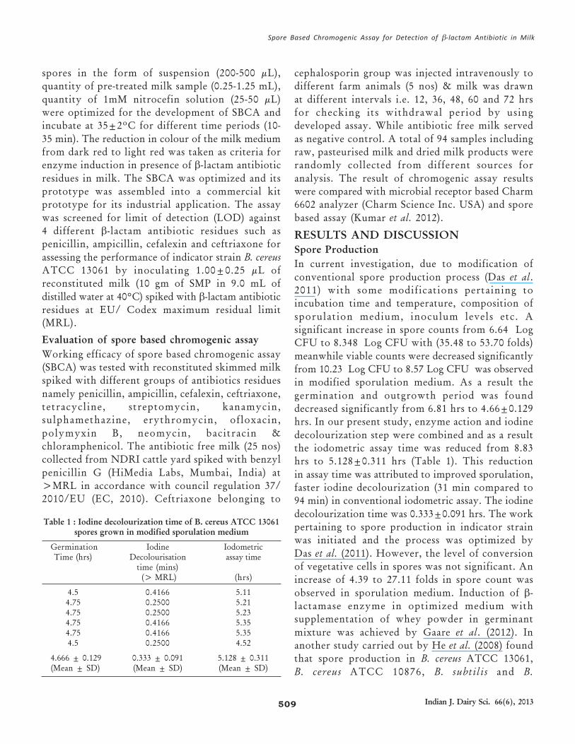
Table 1 : Iodine decolourization time of B. cereus ATCC 13061spores grown in modified sporulation medium
Germination Iodine IodometricTime (hrs) Decolourisation assay time
time (mins) (> MRL) (hrs)
4.5 0.4166 5.114.75 0.2500 5.214.75 0.2500 5.234.75 0.4166 5.354.75 0.4166 5.354.5 0.2500 4.52
4.666 ± 0.129 0.333 ± 0.091 5.128 ± 0.311(Mean ± SD) (Mean ± SD) (Mean ± SD)
Spore Based Chromogenic Assay for Detection of β-lactam Antibiotic in Milk
510
stearothermophilus by transferring vegetative cells
from refrigerated slant to tryptic soy agar (TSA),
followed by incubating at 37°C for 48 hrs.
Development of spore induction chromogenicassay and kit prototype
The cell suspension from the slant was inoculated
into optimized sporulation medium (35±5 mL. 100
mL-1) and incubated at 35±2oC for upto 48 hrs. A
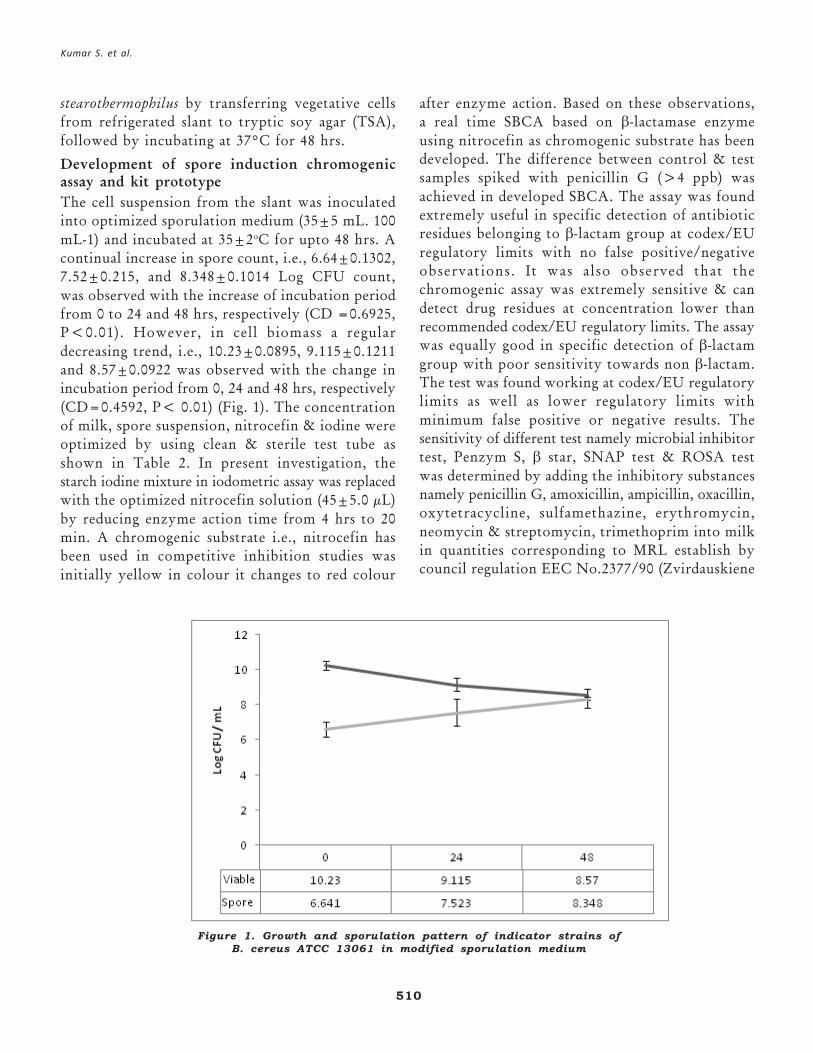
continual increase in spore count, i.e., 6.64±0.1302,
7.52±0.215, and 8.348±0.1014 Log CFU count,
was observed with the increase of incubation period
from 0 to 24 and 48 hrs, respectively (CD =0.6925,
P<0.01). However, in cell biomass a regular
decreasing trend, i.e., 10.23±0.0895, 9.115±0.1211
and 8.57±0.0922 was observed with the change in
incubation period from 0, 24 and 48 hrs, respectively
(CD=0.4592, P< 0.01) (Fig. 1). The concentration
of milk, spore suspension, nitrocefin & iodine were
optimized by using clean & sterile test tube as
shown in Table 2. In present investigation, the
starch iodine mixture in iodometric assay was replaced
with the optimized nitrocefin solution (45±5.0 μL)
by reducing enzyme action time from 4 hrs to 20
min. A chromogenic substrate i.e., nitrocefin has
been used in competitive inhibition studies was
initially yellow in colour it changes to red colour
after enzyme action. Based on these observations,
a real time SBCA based on β-lactamase enzyme
using nitrocefin as chromogenic substrate has been
developed. The difference between control & test
samples spiked with penicillin G (>4 ppb) was
achieved in developed SBCA. The assay was found
extremely useful in specific detection of antibiotic
residues belonging to β-lactam group at codex/EU
regulatory limits with no false positive/negative
observations. It was also observed that the
chromogenic assay was extremely sensitive & can
detect drug residues at concentration lower than
recommended codex/EU regulatory limits. The assay
was equally good in specific detection of β-lactam
group with poor sensitivity towards non β-lactam.
The test was found working at codex/EU regulatory
limits as well as lower regulatory limits with
minimum false positive or negative results. The
sensitivity of different test namely microbial inhibitor
test, Penzym S, β star, SNAP test & ROSA test
was determined by adding the inhibitory substances
namely penicillin G, amoxicillin, ampicillin, oxacillin,
oxytetracycline, sulfamethazine, erythromycin,
neomycin & streptomycin, trimethoprim into milk
in quantities corresponding to MRL establish by
council regulation EEC No.2377/90 (Zvirdauskiene
Figure 1. Growth and sporulation pattern of indicator strains ofB. cereus ATCC 13061 in modified sporulation medium
Kumar S. et al.
511 Indian J. Dairy Sci. 66(6), 2013
and Salomskieneet 2007). A test based on similar
principle using pure penicillinase enzyme was
developed for detection of β -lactam group in
biological fluid such as milk or urine (Klein 1986).
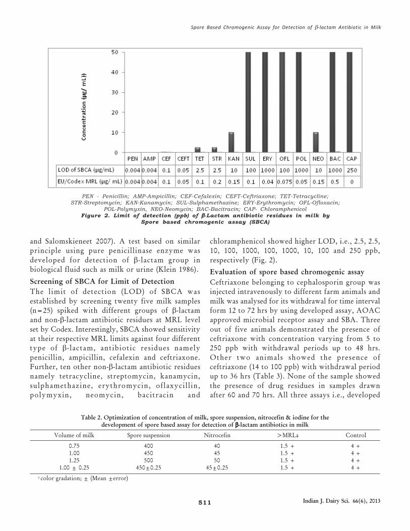
Screening of SBCA for Limit of Detection
The limit of detection (LOD) of SBCA was
established by screening twenty five milk samples
(n=25) spiked with different groups of β-lactam
and non-β-lactam antibiotic residues at MRL level
set by Codex. Interestingly, SBCA showed sensitivity
at their respective MRL limits against four different
type of β - lactam, antibiotic residues namely
penicillin, ampicillin, cefalexin and ceftriaxone.
Further, ten other non-β-lactam antibiotic residues
namely tetracycline, streptomycin, kanamycin,
sulphamethazine, erythromycin, oflaxycill in,
polymyxin, neomycin, bacitracin and
chloramphenicol showed higher LOD, i.e., 2.5, 2.5,
10, 100, 1000, 100, 1000, 10, 100 and 250 ppb,
respectively (Fig. 2).
Evaluation of spore based chromogenic assay
Ceftriaxone belonging to cephalosporin group was
injected intravenously to different farm animals and
milk was analysed for its withdrawal for time interval
form 12 to 72 hrs by using developed assay, AOAC
approved microbial receptor assay and SBA. Three
out of five animals demonstrated the presence of
ceftriaxone with concentration varying from 5 to
250 ppb with withdrawal periods up to 48 hrs.
Other two animals showed the presence of
ceftriaxone (14 to 100 ppb) with withdrawal period
up to 36 hrs (Table 3). None of the sample showed
the presence of drug residues in samples drawn
after 60 and 70 hrs. All three assays i.e., developed
PEN - Penicillin; AMP-Ampicillin; CEF-Cefalexin; CEFT-Ceftriaxone; TET-Tetracycline;STR-Streptomycin; KAN-Kanamycin; SUL-Sulphamethazine; ERY-Erythromycin; OFL-Ofloxacin;
POL-Polymyxin, NEO-Neomycin; BAC-Bacitracin; CAP- ChloramphenicolFigure 2. Limit of detection (ppb) of βββββ-Lactam antibiotic residues in milk by
Spore based chromogenic assay (SBCA)
Table 2. Optimization of concentration of milk, spore suspension, nitrocefin & iodine for thedevelopment of spore based assay for detection of βββββ-lactam antibiotics in milk
Volume of milk Spore suspension Nitrocefin >MRLa Control
0.75 400 40 1.5 + 4 +1.00 450 45 1.5 + 4 +1.25 500 50 1.5 + 4 +
1.00 ± 0.25 450±0.25 45±0.25 1.5 + 4 +
a color gradation; ± (Mean ±error)
Spore Based Chromogenic Assay for Detection of β-lactam Antibiotic in Milk
512
assay, microbial receptor assay, SBA could detect
the presence of ceftriaxone in positive milk sample.
The assay based on chromogenic principle developed
in current investigation was also validated by
comparing with AOAC approved system and SBA.
In overall 94 milk samples including 48 raw milk,
31 pasteurized milk and 15 dried milk samples
analyzed using developed assay, microbial receptor
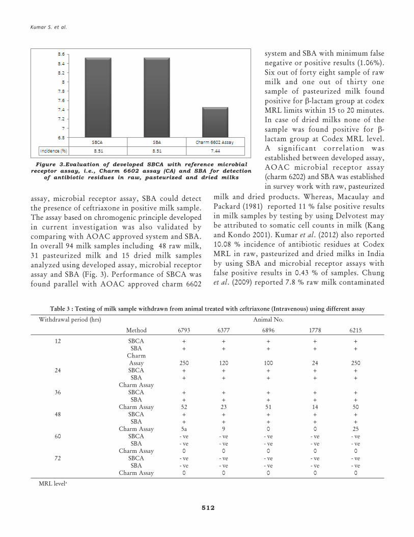
assay and SBA (Fig. 3). Performance of SBCA was
found parallel with AOAC approved charm 6602
milk and dried products. Whereas, Macaulay and
Packard (1981) reported 11 % false positive results
in milk samples by testing by using Delvotest may
be attributed to somatic cell counts in milk (Kang
and Kondo 2001). Kumar et al. (2012) also reported
10.08 % incidence of antibiotic residues at Codex
MRL in raw, pasteurized and dried milks in India
by using SBA and microbial receptor assays with
false positive results in 0.43 % of samples. Chung
et al. (2009) reported 7.8 % raw milk contaminated
Figure 3.Evaluation of developed SBCA with reference microbialreceptor assay, i.e., Charm 6602 assay (CA) and SBA for detection
of antibiotic residues in raw, pasteurized and dried milks
Table 3 : Testing of milk sample withdrawn from animal treated with ceftriaxone (Intravenous) using different assay
Withdrawal period (hrs) Animal No.
Method 6793 6377 6896 1778 6215
12 SBCA + + + + +SBA + + + + +
CharmAssay 250 120 100 24 250
24 SBCA + + + + +SBA + + + + +
Charm Assay36 SBCA + + + + +
SBA + + + + +Charm Assay 52 23 51 14 50
48 SBCA + + + + +SBA + + + + +
Charm Assay 5a 9 0 0 2560 SBCA - ve - ve - ve - ve - ve
SBA - ve - ve - ve - ve - veCharm Assay 0 0 0 0 0
72 SBCA - ve - ve - ve - ve - veSBA - ve - ve - ve - ve - ve
Charm Assay 0 0 0 0 0
MRL levela
system and SBA with minimum false
negative or positive results (1.06%).
Six out of forty eight sample of raw
milk and one out of thirty one
sample of pasteurized milk found
positive for β-lactam group at codex
MRL limits within 15 to 20 minutes.
In case of dried milks none of the
sample was found positive for β-
lactam group at Codex MRL level.
A significant correlation was
established between developed assay,
AOAC microbial receptor assay
(charm 6202) and SBA was established
in survey work with raw, pasteurized
Kumar S. et al.
513 Indian J. Dairy Sci. 66(6), 2013
with antibiotic residues collected from 13 cities of
Korea by using tetrazolium trichloride (TTC) and
STAR based protocols.
CONCLUSIONS
An innovative approach for detection of β-lactam
antibiotic residues in milk was attempted based on
induction and competitive enzyme action on β-lactam
antibiotic residues produced by B. cereus ATCC
13061. Because of modification sporulation medium
for sporulation of B. cereus there is significant decrease
in germination and outgrowth period. In our current
investigation, enzyme action and iodine
decolourization step were combined and as a result
the iodometric assay time was reduced. The starch
iodine mixture in iodometric assay was replaced with
nitrocefin as a chromogenic substrate by reducing
enzyme action time to 20 min with initial change of
colour from yellow to red has been developed for
its industrial application. The assay can detect specific
β-lactam antibiotic residues in milk within 20 min
at MRL level set by codex with negligible sensitivity
towards non β-lactam groups. The presence of
inhibitors other than antibiotic residues in milk did
not interfere with the working performance of the
developed assay. A significant correlation between
SBCA, SBA and AOAC approved microbial receptor
based assay (charm 6202) was established in survey
work with raw, pasteurized and dried milks with
minimum false positive/ negative results. The SBCA
can find immense application in dairy industry as
"ON FARM" milk screening test for β-lactam group
in milk at MRL codex limit.
ACKNOWLEDGEMENTS
The research work in this publication was carried
out under externally funded project awarded by
Ministry of Food Processing Industries (MoFPI),
Govt. of India with financial support to purchase
the desired equipments used in execution of proposed
research project. We acknowledge their immense
contribution in development of rapid bio-assay
prototype for its commercial application. The views
and conclusions contained in this document are those
of authors and should not be interpreted as
necessarily representing the official policies of MoFPI.
The Director NDRI is thankfully acknowledged
for Institute fellowship to Mr. Sunil Kumar.
REFERENCESBuynak, J. D. 2006 Understanding the longevity of the β-lactam
antibiotics and of antibiotic/β-lactamase inhibitor combinations.Biochem. Pharmacol. 71(7): 930-940.
Chung, H. H., Jung-Bin, L., Yun-Hee, C. and Kwang-Geun, L. 2009Analysis of sulfonamide and quinolone antibiotic residues inKorean milk using microbial assays and high performance liquidchromatography. Food Chem. 113:297-301.
Das, S., Kumar, N., Raghu, H. V., Haldar, L., Gaare, M., Singh, V.and Puniya, A. 2011 Microbial based assay for specific detection ofβ-lactam group of antibiotics in milk. J. Food Sci. Tech. doi:10.1007/s13197-011-0609-4.
Deshmukh S. (2013). Screening of Enterococci in milk usingdeveloped media and functionalized biochip. M. Tech thesissubmitted to National Dairy research Institute Karnal.
Downes, F. P. and Ito, K. 2001 Compendium of methods for themicrobiological examination of foods, 4th edn. In: AmericanPublic Health Association (Ed) Frances Pouch Downes Keith Ito,Washington DC.
EC (2010) Council regulation 37/2010/EU of 22nd December 2009on pharmacologically active substances and their classificationregarding maximum residues limits in foodstuffs of animal origin.Offic J Euro Union L15:1-72
Faria Reyes, J. F., Allara Cagnasso, M. G., Arenas De Moreno, L.,Marques Salas, E., Reyes, J. F. F. and Salas, E. M. 2000. Extractionand quantification of penicillin G in raw milk by highperformance liquid chromatography (HPLC). Revista Cientifica,Facultad de Ciencias Veterinarias, Universidad Del Zulia, 10:212-221.
Gaare, M., Kumar, N., Raghu, H. V., Khan, A. and Singh, V. K.2012. Specific detection of β-lactam antibiotics in milk by sporebased assay. Int. Res. J. Microbiol. 3(5):168-173.
Ghidini, S, Zanardi, E, Varisco, G and Chizzolini, R. 2003. Residuesof β-lactam antibiotics in bovine milk: confirmatory analysis byliquid chromatography tandem mass spectrometry after microbialassay screening Food Addit. Contam. 20 (6): 528-34.
He. L., Liu. Y., Lin. M., Mustapha. A., and Wang. Y. 2008. Detectingsingle Bacillus spores by surface enhanced Raman spectroscopy.Sens. & Instrumen. Food Qual. 2:247-253.
Holstege, D. M., Puschner, B., Whitehead, G. and Galey, F. D. 2002.Screening and Mass spectral confirmation of β-Lactam antibioticresidues in milk using LC-MS/MS. J. Agric. Food Chem.50(2):406-11.
Kang, J. H. and Kondo, F. 2001. Occurrence of false-positive resultsof inhibitor on milk samples using the Delvotest SP assay. J. Food.Prot. 64(8):1211-1215.
Kaur G. 2011. Spore germination: An innovative approach fordetecting enterococci in milk. M. Sc. thesis submitted to NationalDairy Research Institute Karnal.
Klein, J. H. 1986. Method of determining antibiotics in biologicalliquids. US patent 4568637.
Kumar, N., N.A. Singh, V.K. Singh, S. Bhand, and R.K. Malik.2010. "Development of Spore Inhibition Based-Enzyme SubstrateAssay (SIB-ESA) for Monitoring Aflatoxin M1 in Milk." IndianPatent Reg No. 3064/DEL/2010. Mumbai, Office of the
Spore Based Chromogenic Assay for Detection of β-lactam Antibiotic in Milk
514
Controller General of Patents, Designs & Trade Marks, Mumbai.Journal No. - 46/2012.
Kumar, N., Raghu, H. V., Kumar, A., Haldar, L., Khan, A., Rane, S.and Malik, R. K. 2012. Spore germination based assay formonitoring antibiotic residues in milk at dairy farm. World J.Microbiol. and Biotechnol. 28:2559-2566.
Kumar, N., S. Das, and G. Manju. 2009. "A Kit for Detection of b-Lactam Antibiotic Group in Milk Using Bacterial Spore asBiosensor." Indian Patent Reg No. 115/ DEL/ 2009. Office ofthe Controller General of Patents, Designs & Trade Marks,Mumbai. Journal No. 31/2010.
Kumar, N., S. Sawant, G.R. Patil, and R.K. Malik. 2006."Development of Analytical Process for Detection of AntibioticResidues in Milk Using Bacterial Spores as Biosensor." IndianPatent Reg No. 115/ DEL/ 2009. Office of the ControllerGeneral of Patents, Designs & Trade Marks, Mumbai. JournalNo. - 49/2011.
Macaulay, D. M. and Packard, V. S. 1981. Evaluation of methodsused to detect antibiotic residues in milk. J. Food Prot. 44:696-698
Marchetti, M., Schwaiger, I., and Schmid, E. R. 2002. Determinationof benzylpenicillin, oxacillin, cloxacillin, and dicloxacillin in cows'milk by ion-pair high-performance liquid chromatography afterpre-column derivatization. Fresenius J. Anal. Chem. 371: 64-67.
Navrátilová, P. 2008. Screening methods used for the detection ofveterinary drug residues in raw cow milk - a review. Czech J. FoodSci. 26:393-401.
O'Callaghan, C. H., Morris, A., Kirby, S. M. and Singler, A. H.1972. Novel method for detection of β-lactamase by using achromogenic cephalosporin substrate. Antimicrob. AgentsChemother. 1:283-288.
Pol, M. and Ruegg, P. L. 2007. Treatment practices and quantification
of antimicrobial drug usage in conventional and organic dairyfarms in Wisconsin. J. Dairy Sci. 90:249-261.
Refsdal, A. O. 2000. To treat or not to treat: a proper use ofhormones and antibiotics Anim. Reprod. Sci. 60(61): 109-119.
Sierra, D., Sánchez, A., Contreras, A., Luengo, C., Corrales, J. C.,Morales, C. T., de la Fe C., Guirao I. and Gonzalo C. 2009.Detection limits of four antimicrobial residue screening test for β-lactams in goat's milk. J. Dairy Sci. 92:3585-3591.
Singh N.A., Kumar N., Raghu H.V., Sharma P.K., Singh V.K.,Khan Alia, and Raghav N. (2013): Spore inhibition-based enzymesubstrate assay for monitoring of aflatoxin M1 in milk,Toxicological & Environmental Chemistry, DOI:10.1080/02772248.2013.807540
Snedecor GW, Cochran WG (1980) Statistical methods, 7th edn.The Iowa State University Press, Iowa.
Stolz, H., Bodini, A., Stolze, M., Hamm, U. and Richter, T. 2009.Lebensmittelqualität aus der Verbraucherperspektive - eineSynthese qualitativer Studien zur Wahrnehmung undBeurteilung verschiedener Qualitätskriterien bei Öko-ProduktenBer. Landwirtsch. 80:153-182.
van Wijk-Jansen, E., Ronteltap, A. and Jager, L. 2009. Het gezondevan biologisch voedsel: de beleving van consumenten [Thehealthy thing about organic products: What makes organic foodhealthy according to consumers?] LEI Wageningen UR, DenHaag.
Zvirdauskiene, R. and Salomskiene, J. 2007. An evaluation ofdifferent microbial and rapid tests for determining inhibitors inmilk. Food Con. 18(5):541-547.
Zwald, A. G., Ruegg, P. L., Kaneene, J. B., Warnick, L. D., Wells, S.J., Fossler, C., & Halbert, L. W. 2004. Management practices andreported antimicrobial usage on conventional and organic dairyfarms. Journal of Dairy Science, 87(1):191-201.
Kumar S. et al.
Copyright © 2022 FDOKUMEN