A STIM2 splice variant negatively regulates store-operated calcium entry

For Peer Review
Splice, insertion-deletion and nonsense mutations that
potentially perturb the Phenylalanine hydroxylase transcript are a major cause for Phenylketonuria in India
Journal: Journal of Cellular Biochemistry
Manuscript ID: JCB-13-0600.R1
Wiley - Manuscript type: Research Article
Date Submitted by the Author: n/a
Complete List of Authors: Bashyam, Murali; Centre for DNA Fingerprinting and Diagnostics,
Chaudhary, Ajay; Centre for DNA Fingerprinting and Diagnostics, Sinha, Manjari; Centre for DNA Fingerprinting and Diatnostics, Laboratory of Computational Biology Nagarajaram, Hampapatalu; Centre for DNA Fingerprinting and Diatnostics, Laboratory of Computational Biology ramadevi, akella; Centre for DNA Fingerprinting and Diagnostics, Diagnostics Division Ranganath, Prajnya; Centre for DNA Fingerprinting and Diagnostics, Diagnostics Division Dalal, Ashwin; Centre for DNA Fingerprinting and Diagnostics, Diagnostics Division Bashyam, Leena; University of Hyderabad, Genomics Facility
Gupta, Neerja; All India Institute of Medical Sciences, Kabra, Madhulika; All India Institute of Medical Sciences, Muranjan, Mamta; Seth GS Medical College and King Edward Memorial Hospital, Puri, Ratna; Sir Ganga Ram Hospital, Verma, Ishwar; Sir Ganga Ram Hospital, Nampoothiri, Sheela; Amrita Institute of Medical Sciences & Research Center, Kadandale, Jayarama; Centre for Human Genetics,
Keywords: Phenylketonuria, Phenylalanine hydroxylase, Splice site mutation, PAH transcript, Nonsense mediated decay
John Wiley & Sons, Inc.
Journal of Cellular Biochemistry

For Peer Review
1
Splice, insertion-deletion and nonsense mutations that potentially perturb the Phenylalanine
hydroxylase transcript are a major cause for Phenylketonuria in India
Murali D. Bashyam1*, Ajay K. Chaudhary1, Manjari Sinha2, Hampapathalu A. Nagarajaram2,
Radha RamaDevi3$, Prajnya Ranganath3, Ashwin Dalal3, Leena Bashyam4, Neerja Gupta5,
Madhulika Kabra5, Mamta Muranjan6, Ratna D. Puri7, Ishwar C. Verma7, Sheela Nampoothiri8,
Jayarama S. Kadandale9
1Laboratory of Molecular Oncology, 2Laboratory of Computational Biology, 3Diagnostics
Division, Centre for DNA Fingerprinting and Diagnostics, Hyderabad, India; 4Genomics facility,
School of Life Sciences, University of Hyderabad, Hyderabad, India; 5All India Institute of
Medical Sciences, New Delhi, India; 6Seth GS Medical College and King Edward Memorial
Hospital, Mumbai, India; 7Sir Ganga Ram Hospital, Delhi, India; 8Amrita Institute of Medical
Sciences & Research Center, Cochin, India; 9Centre for Human Genetics, Bengaluru, India.
$Present address: Sandor Proteomics Pvt. Ltd. Hyderabad, India
*Corresponding Author: Murali D. Bashyam, Laboratory of Molecular Oncology, Centre for
DNA Fingerprinting and Diagnostics, Nampally, Hyderabad 500001, India; Tel: 91-40-
24749383; Fax: 91-40-24749448; Email: [email protected]
Running Head: Unique PAH mutations cause Phenylketonuria in India
Page 1 of 39
John Wiley & Sons, Inc.
Journal of Cellular Biochemistry
123456789101112131415161718192021222324252627282930313233343536373839404142434445464748495051525354555657585960

For Peer Review
2
Key words:
1. Phenylketonuria
2. Phenylalanine hydroxylase
3. Splice site mutation
4. PAH transcript
5. Nonsense mediated decay
Number of figures: Three
Number of tables: Two
Contract grant sponsor: Department of Biotechnology, Government of India.
Contract grant no: nil
Page 2 of 39
John Wiley & Sons, Inc.
Journal of Cellular Biochemistry
123456789101112131415161718192021222324252627282930313233343536373839404142434445464748495051525354555657585960

For Peer Review
3
Abstract
Phenylketonuria (PKU) is an autosomal recessive metabolic disorder caused by mutational
inactivation of the Phenylalanine hydroxylase (PAH) gene. Missense mutations are the most
common PAH mutation type detected in PKU patients worldwide. We performed PAH mutation
analysis in twenty seven suspected Indian PKU families (including seven from our previous
study) followed by structure and function analysis of specific missense and splice/insertion-
deletion/nonsense mutations, respectively. Of the twenty seven families, disease causing
mutations were detected in twenty five. A total of twenty different mutations were identified of
which seven ‘unique’ mutations accounted for thirteen of twenty five mutation positive families.
The unique mutations detected exclusively in Indian PKU patients included three recurrent
mutations detected in three families each. The twenty mutations included only five missense
mutations in addition to five splice, four each nonsense and insertion-deletion mutations, a silent
variant in coding region and a 3’ UTR mutation. One deletion and two nonsense mutations were
characterized to confirm significant reduction in mutant transcript levels possibly through
activation of nonsense mediated decay. All missense mutations affected conserved amino acid
residues and sequence and structure analysis suggested significant perturbations in the enzyme
activity of respective mutant proteins. This is probably the first report of identification of a
significantly low proportion of missense PAH mutations from PKU families and together with
the presence of a high proportion of splice, insertion-deletion and nonsense mutations, points to a
unique PAH mutation profile in Indian PKU patients.
Page 3 of 39
John Wiley & Sons, Inc.
Journal of Cellular Biochemistry
123456789101112131415161718192021222324252627282930313233343536373839404142434445464748495051525354555657585960

For Peer Review
4
Introduction
Phenylketonuria (PKU; OMIM:261600) is an inborn error of amino acid metabolism inherited as
an autosomal recessive trait and is caused by mutations in the gene coding for the hepatic
enzyme phenylalanine hydroxylase (PAH). PAH is responsible for hydroxylation of
Phenylalanine to Tyrosine in the presence of Oxygen and the cofactor Tetrahydrobiopterin
(BH4). Insufficient/absent PAH enzyme activity results in mental retardation in affected children
due to accumulation of phenylalanine in the brain where it acts as a neurotoxin. A phenylalanine
restricted diet prevents brain damage [Blau et al. 2011] and therefore early detection of affected
individuals through mutation screening assumes significance. In addition, mutation analysis
facilitates genetic counseling of affected families.
More than 560 PKU causing mutations have been identified in the PAH gene from various
populations (PAH mutation database (PAHDB); www.pahdb.mcgill.ca). Majority of mutations
identified worldwide are rare or ‘private’; very few recurrent mutations have been reported
[Zschocke, 2003]. In our previous study [Bashyam et al. 2010], we identified four novel
mutations in seven suspected PKU families. In the current study, we have extended the analysis
to an additional twenty families. Our results suggest the occurrence of a unique PAH mutation
spectrum in Indian PKU patients.
Materials and methods
Patients, samples and mutation screening
The study was approved by the institute ethics committee and included twenty PKU families (in
addition to seven families analyzed in our previous study [Bashyam et al. 2010]). Eighteen of
twenty seven families were from the South Indian State of Andhra Pradesh while the rest
Page 4 of 39
John Wiley & Sons, Inc.
Journal of Cellular Biochemistry
123456789101112131415161718192021222324252627282930313233343536373839404142434445464748495051525354555657585960

For Peer Review
5
belonged to different geographical regions within India. Samples were collected from probands
for eighteen families (families 1-18; Table 1a) and from the parents for two families (families
19-20; Table 1a). Families 19 and 20 had a previous history of PKU and were therefore referred
for carrier testing; the proband samples were unfortunately unavailable. All patients exhibited
classical PKU symptoms which are described in Table 1a and document S1. Establishment of
disease, sample collection and the mutation screening procedure have been described earlier
[Bashyam et al. 2010]. Each mutation was confirmed through bi-directional DNA sequencing.
Interestingly, the proband from family 2 exhibited features of Metachromatic leucodystrophy (in
addition to classical PKU symptoms) and a mutation in the Arylsulfatase A gene (ARSA) was
also identified (Ranganath et al., personal communication).
Quantitation of PAH transcript
2ug of total RNA obtained from EBV transformed lymphoblasts was reverse transcribed using
oligo dT primer (GE Healthcare, USA) to generate cDNA as previously described [Bashyam et
al. 2010]. Appropriate dilutions of the cDNA were used for quantitative PCR performed on the
SDS 7500 (ABI, Foster City, CA, USA) using the SYBR Premix Ex Taq (Perfect Real Time kit;
Takara Bio Inc., Japan) in presence of primers PAH-RTF and PAH-RTR [Bashyam et al. 2010].
PCR and quantitation of relative PAH levels with respect to GAPDH were performed as
described before [Bashyam et al. 2010].
Protein sequence and structure analysis
Sequence and structure analysis of effect of missense mutations was performed as described
earlier [Bashyam et al., 2012; Bashyam et al. 2010]; details are given in methods S1.
Page 5 of 39
John Wiley & Sons, Inc.
Journal of Cellular Biochemistry
123456789101112131415161718192021222324252627282930313233343536373839404142434445464748495051525354555657585960

For Peer Review
6
Results
Splice, insertion-deletion and nonsense PAH mutations appear to be the major cause of PKU
in India
The study was carried out on twenty eight patients belonging to twenty seven families (inclusive
of our previous study [Bashyam et al. 2010]). Mutations were detected in all except two families
(Table 1a and figure 1 and S1). Of the total twenty mutations identified in our current and
previous studies, only five were missense including two novel mutations (figure 1 and Table 1a).
The novel missense mutations viz. p.G312R (c.934G>C) and p.E368G (c.1103A>G) affected
amino acid residues that exhibited significant conservation (figure 1). Absence of the novel
mutations in one hundred (two hundred chromosomes for c.1103A>G) and in fifty (one hundred
chromosomes for c.934G>C) normal individuals from the same local population was
independently confirmed using DNA sequencing (data not shown). The c.612T>C silent variant
is reported as an SNP in NCBI database
(http://www.ncbi.nlm.nih.gov/projects/SNP/snp_ref.cgi?rs=rs62514928) with unknown clinical
relevance and frequency, though it was not detected in sixty nine (one hundred and thirty eight
chromosomes) normal individuals in this study (data not shown).
An analysis of PAHDB reveals missense PAH mutations to be the most common form (341/567;
60%); therefore, the low frequency detected in the current study (5/20; 25%) (Table 1b) is highly
significant. None of the common missense mutations listed in PAHDB including p.R408W
(6.6%) and p.I65T (4%) were detected. On the other hand, the frequency of splice (5/20; 25%),
insertion/deletion (4/20; 20%) and nonsense (4/20; 20%) mutations detected in Indian PKU
patients (fig 2A and Table 1b) was significantly higher than the corresponding frequencies
reported in the PAHDB (11.5%, 15%, and 5% for splice, insertion-deletion, and nonsense
Page 6 of 39
John Wiley & Sons, Inc.
Journal of Cellular Biochemistry
123456789101112131415161718192021222324252627282930313233343536373839404142434445464748495051525354555657585960

For Peer Review
7
mutations, respectively). More importantly, twenty of twenty five (88%) mutation positive
families harbored potential PAH transcript perturbing mutations including splice, nonsense,
insertion-deletion and UTR mutations (Table 1b). In addition, thirteen of the twenty five families
harbored mutations which were exclusive for Indian PKU patients and were not detected in
patients from other countries (Table 1b), suggesting thereby the possible existence of a unique
PAH mutation spectrum among Indian PKU patients, as already suggested for other Mendelian
disorders (hypohidrotic ectodermal dysplasia [Bashyam et al. 2010] and familial hypertrophic
cardiomyopathy [Bashyam et al. 2012]). Among the unique mutations, c.1503A>G, c.1177insT
and c.168-2A>G were detected in three families each (Table 1b).
As expected, the carrier parents in families 19 and 20 harbored heterozygous mutations (Table
1a). Of the sixteen probands in whom mutations were identified, homozygous mutations were
detected in twelve, heterozygous mutations in two (families 6 and 16; Table 1a) and compound
heterozygous in two (families 9 and 13; Table 1a). Frequent occurrence of consanguineous
marriages is one reason for high frequency of homozygous mutations in Indian PKU patients. It
is possible that the second mutation in probands of families 6 and 16 could be located in regions
which were not screened (such as deep within introns). Similarly, mutations not detected in
families 17 and 18, could also be located in intronic regions not covered in the mutation
screening strategy. Exonic copy number gains and/or deletions, not screened in this study, could
be the other cause of disease in these affected families. All parent samples were confirmed to
harbor the respective mutation in heterozygous condition (Table 1a).
Page 7 of 39
John Wiley & Sons, Inc.
Journal of Cellular Biochemistry
123456789101112131415161718192021222324252627282930313233343536373839404142434445464748495051525354555657585960

For Peer Review
8
Functional characterization of select mutations reveals possible role of nonsense mediated
decay (NMD) induced degradation of mutant PAH transcript
Three nonsense (p.Y206X, p.R243X and p.R261X) and two deletion (c.358delT and
c.558_559delAT) mutations identified in this study are expected to result in the generation of
premature termination codon (PTC) situated 90, 117, 60, 123 and 114 bases upstream of the
subsequent exon-exon junctions, respectively. Therefore, the five mutations may trigger NMD
based degradation of the respective mutant transcript [Bashyam, 2009]. In order to test this
possibility, we isolated total RNA from EBV transformed lymphoblasts generated separately
from three probands (harboring c.358delT, p.Y206X and p.R243X mutations (figure 3A);
samples from other two probands were unavailable for transcript analysis) and quantified the
respective PAH transcript levels with respect to RNA isolated from normal lymphoblasts. All
three mutations resulted in significant reduction in the respective mutant transcript levels (figure
3B) indicating transcript degradation perhaps through nonsense mediated decay (NMD). The
mutations however did not result in an obvious change in transcript structure (such as exon
skipping due to the PTC through nonsense-associated altered splicing or (‘NAS’) [Wang et al.,
2002]), as confirmed by reverse transcription polymerase chain reaction analyses (data not
shown). In addition, the c.1177insT and c.976delT mutations (Table 1a) may not trigger NMD as
already described in our previous study [Bashyam et al. 2012].
Protein sequence and structure analysis reveals possible perturbations due to missense
mutations
Multiple sequence alignment (MSA) of PAH with its homologues revealed significant
conservation of the wild-type amino acid residues at the mutation sites (Figure 1 and S2) and
therefore, their substitutions by dissimilar residues are expected to hinder protein function. The
Page 8 of 39
John Wiley & Sons, Inc.
Journal of Cellular Biochemistry
123456789101112131415161718192021222324252627282930313233343536373839404142434445464748495051525354555657585960
For Peer Review
9
position-specific profile (Gribskov’s-scores) at the mutation sites (Table 2) confirmed this
inference. In addition, all five missense mutations were identified as ‘Disease’ causing mutations
by the web-server “Hansa” (hansa.cdfd.org.in:8080) [Acharya and Nagarajaram, 2012; Bashyam
et al., 2012; Bashyam et al. 2012] (Table 2).
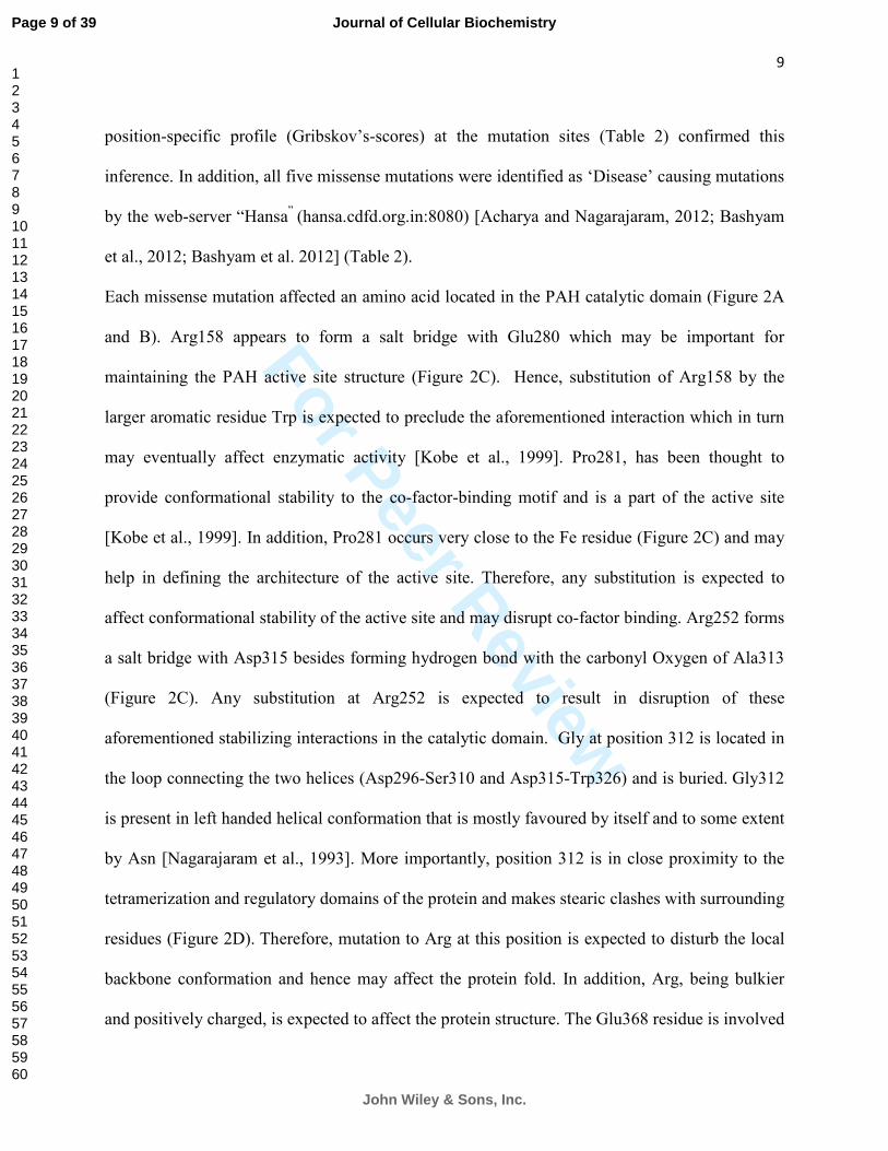
Each missense mutation affected an amino acid located in the PAH catalytic domain (Figure 2A
and B). Arg158 appears to form a salt bridge with Glu280 which may be important for
maintaining the PAH active site structure (Figure 2C). Hence, substitution of Arg158 by the
larger aromatic residue Trp is expected to preclude the aforementioned interaction which in turn
may eventually affect enzymatic activity [Kobe et al., 1999]. Pro281, has been thought to
provide conformational stability to the co-factor-binding motif and is a part of the active site
[Kobe et al., 1999]. In addition, Pro281 occurs very close to the Fe residue (Figure 2C) and may
help in defining the architecture of the active site. Therefore, any substitution is expected to
affect conformational stability of the active site and may disrupt co-factor binding. Arg252 forms
a salt bridge with Asp315 besides forming hydrogen bond with the carbonyl Oxygen of Ala313
(Figure 2C). Any substitution at Arg252 is expected to result in disruption of these
aforementioned stabilizing interactions in the catalytic domain. Gly at position 312 is located in
the loop connecting the two helices (Asp296-Ser310 and Asp315-Trp326) and is buried. Gly312
is present in left handed helical conformation that is mostly favoured by itself and to some extent
by Asn [Nagarajaram et al., 1993]. More importantly, position 312 is in close proximity to the
tetramerization and regulatory domains of the protein and makes stearic clashes with surrounding
residues (Figure 2D). Therefore, mutation to Arg at this position is expected to disturb the local
backbone conformation and hence may affect the protein fold. In addition, Arg, being bulkier
and positively charged, is expected to affect the protein structure. The Glu368 residue is involved
Page 9 of 39
John Wiley & Sons, Inc.
Journal of Cellular Biochemistry
123456789101112131415161718192021222324252627282930313233343536373839404142434445464748495051525354555657585960

For Peer Review
10
in formation of main-chain hydrogen bond with Lys371, Thr372 and Pro366. Furthermore,
analysis of the PAH tetrameric structure (2PAH [Fusetti et al., 1998]) revealed that Glu368 is in
close proximity with its counterpart from the other subunit (in the dimer/tetramer) (figure 2E).
From our structure analysis, we conclude that mutation to Glycine at this position may result in
increased conformational freedom and a substantial change in side chain volume in addition to
the loss of Glu-Glu contact at the dimer interface (figure 2E), which may in turn destabilize the
dimer/tetramer structure.
Discussion
In the current study, we have performed mutation analysis of twenty seven PKU families from
India including seven from our previous study [Bashyam et al. 2010]. A total of twenty different
mutations were detected in twenty five families; no mutation was detected in two. Therefore, the
mutation detection frequency was 25/27 (93%), similar to other reports [Groselj et al., 2012;
Stojiljkovic et al., 2006]. The c.168-2A>G, c.1177insT and c.1503A>G mutations appear to be
the most common PAH mutations causing PKU in the Indian population. This result assumes
significance, given the paucity of recurrent mutations detected from PKU patients worldwide.
In the previous study [Bashyam et al. 2010], we had provided the first evidence for a possible
role of NMD in PKU, based on analysis of the novel c.168-2A>G mutation. In the current study,
we have characterized two nonsense (p.Y206X and p.R243X) and one deletion (c.358delT) PAH
mutations (figure 3A) and detected a significant reduction in the levels of the respective mutant
transcripts suggesting possible NMD-induced transcript degradation in all three (figure 3B). Two
additional mutations detected in this study and one in our previous study (p.R176X) are also
expected to trigger NMD though we were unable to test the same due to lack of sample
availability. Our analyses therefore constitute the only evidence for role of NMD-based PAH
Page 10 of 39
John Wiley & Sons, Inc.
Journal of Cellular Biochemistry
123456789101112131415161718192021222324252627282930313233343536373839404142434445464748495051525354555657585960

For Peer Review
11
transcript degradation in PKU. In addition, this is probably the first report of characterization of
PAH nonsense mutations at the transcript level in PKU patients. A few earlier studies have
however reported alterations of the PAH transcript due to specific mutations [Dobrowolski et al.,
2010; Heintz et al., 2012; Stojiljkovic et al. 2010]. Given the low proportion of missense
mutations and the corresponding high frequency of splice, insertion-deletion and nonsense
mutations, alterations of the PAH transcript may be a common cause of PKU in the Indian
population, unlike other populations. Our study has underscored the importance of transcript
analysis for all truncation mutations for presence of possible transcript perturbations rather than
restricting the analyses to the truncated protein alone. The other significant feature of our study is
the demonstration of the importance of identification of disease causing mutations in carriers,
especially in families where PKU is established based on past history but proband sample is
unavailable. Knowledge of mutation in such families facilitates genetic counseling.
The nonsense and missense mutations detected in the present study (with the exception of the
novel p.E368G and p.G312R mutations) have been reported earlier from other populations. The
p.R261X mutation was identified in PKU patients from Brazil [Santana da Silva et al., 2003] and
Iran [Zare-Karizi et al. 2011]. The p.R243X mutation was detected in PKU patients from
Morocco and several European countries [Dahri et al. 2010] and was reported to be mildly
responsive to BH4 therapy [Perez-Duenas et al., 2004]. The p.Y206X mutation was reported
earlier from China [Song et al., 2005] and from other populations [Erlandsen and Stevens, 1999].
The p.R158W mutation is also reported from China [Takarada et al., 1993b] and Korea
[Takarada et al., 1993a]; similarly the p.R252Q has been reported from Taiwan [Chien et al.,
2004]. The p.P281L mutation is reported to be a common mutation in the Mediterranean
population [Perez et al., 1992] and has been suggested to induce skipping of exon 8, though the
Page 11 of 39
John Wiley & Sons, Inc.
Journal of Cellular Biochemistry
123456789101112131415161718192021222324252627282930313233343536373839404142434445464748495051525354555657585960

For Peer Review
12
mutation itself is located in exon7. However, the exon skipping effect is partial since both full
and partial length transcripts are detected in the affected lymphoblasts [Ellingsen et al., 1999];
the disease phenotype associated with this mutation therefore could be a net result of altered
protein sequence due to exon skipping and altered protein active site due to the amino acid
substitution (figure 2C). We detected a novel silent mutation c.612T>C in family 6. Though it is
reported as a variant with unknown clinical significance in the NCBI SNP database, it was not
detected in the normal population in our study. It is possible that this mutation may have an
effect on the PAH transcript. Of note, two mutations affecting the same codon viz. c.612T>G and
c.611A>G were reported earlier. The c.612T>G generates a PTC [Guldberg et al., 1993] whereas
the c.611A>G mutation was shown to generate a cryptic 5’ splice site leading to defective
splicing [Ellingsen et al., 1997].
Each of the five missense mutations detected in the present study affected conserved amino acid
residues (figure 1 and S2 and Table 2) and the perturbations are expected to destabilize enzyme
activity. Erlandsen and colleagues [Erlandsen and Stevens, 1999], using a PAH model
(constructed by superimposing the human PAH catalytic and tetramerization domains on the rat
PAH regulatory domain), reported Arg252 to form a salt bridge with Asp315 and a hydrogen
bond with Asp27side-chain. However these interactions are not seen in the human PAH crystal
structure [Fusetti et al., 1998] which is used in the present study and hence there is no
perceivable evidence that substitution of R252 by Gln would result in disruption of the above
mentioned stabilizing interactions.
Several observations made in the current study point to the possible existence of a unique PAH
mutation profile among Indian PKU patients. Firstly, a total of 7/20 (35%) mutations are
exclusive to the Indian population (Table 1b). Secondly, missense mutations occur at a
Page 12 of 39
John Wiley & Sons, Inc.
Journal of Cellular Biochemistry
123456789101112131415161718192021222324252627282930313233343536373839404142434445464748495051525354555657585960

For Peer Review
13
significantly lower frequency compared to other populations. In contrast, the frequencies of
splice, insertion-deletion and nonsense mutation types likely to perturb PAH transcript, are
significantly higher than that reported in the PAHDB. Therefore, mutations affecting PAH
transcript rather than PAH protein/enzyme activity appear to be the major cause of PKU in India
unlike other populations. Of note, the low missense mutation frequency reported in the current
study is similar to that reported from Iran [Zare-Karizi et al. 2011] but not from China [Song et
al., 2005] and Korea [Lee et al., 2004]. However, recurrent mutations reported from Indian PKU
patients are distinct from those identified from other Asian countries.
A few important clinical and socio-economic observations in the current study are worth
mentioning. Firstly, most patients presented with mental retardation and/or developmental delay.
One reason could be non-availability of special (phenylalanine-restricted) diet for the patent.
More importantly, we observed a significantly high frequency of consanguineous marriages
among PKU families. Fifteen of eighteen probands were associated with consanguinity resulting
in high frequency of homozygous mutations (Table 1a) as opposed to compound heterozygosity.
In fact, consanguinity appears to be a common factor in several autosomal recessive genetic
disorders in India [Bashyam et al., 2004; Kumar et al. 2013]. Therefore, it is important to educate
the patients and affected families (through genetic counseling) and the population in general
(through Government sponsored mass education programs) about this social problem.
In conclusion, the current study has revealed a significantly low frequency of missense mutations
causing PKU in the Indian population. In contrast, mutations affecting the PAH transcript appear
to be a common cause of PKU, as against other populations. The current study therefore has
important implications for PKU screening, patient management and genetic counseling in India
and underscores the importance of analyzing the PAH transcript.
Page 13 of 39
John Wiley & Sons, Inc.
Journal of Cellular Biochemistry
123456789101112131415161718192021222324252627282930313233343536373839404142434445464748495051525354555657585960

For Peer Review
14
Acknowledgements
We are extremely grateful to all patients, family members and control subjects for kindly
consenting to be a part of the study. Manjari, a registered PhD student of Manipal University, is
thankful to the Council of Scientific and Industrial Research, Government of India for junior and
senior research fellowships. All authors declare no conflict of interest.
Page 14 of 39
John Wiley & Sons, Inc.
Journal of Cellular Biochemistry
123456789101112131415161718192021222324252627282930313233343536373839404142434445464748495051525354555657585960

For Peer Review
15
References:
Acharya V, Nagarajaram HA (2012): Hansa: an automated method for discriminating disease
and neutral human nsSNPs. Hum Mutat 33:332-7.
Bashyam MD (2009): Nonsense-mediated decay: linking a basic cellular process to human
disease. Expert Rev Mol Diagn 9:299-303.
Bashyam MD, Bashyam L, Savithri GR, Gopikrishna M, Sangal V, Devi AR (2004): Molecular
genetic analyses of beta-thalassemia in South India reveals rare mutations in the beta-globin
gene. J Hum Genet 49:408-13.
Bashyam MD, Chaudhary AK, Reddy EC, Devi AR, Savithri GR, Ratheesh R, Bashyam L,
Mahesh E, Sen D, Puri R, Verma IC, Nampoothiri S, Vaidyanathan S, Chandrashekar MD,
Kantheti P (2010): Phenylalanine hydroxylase gene mutations in phenylketonuria patients from
India: identification of novel mutations that affect PAH RNA. Mol Genet Metab 100:96-9.
Bashyam MD, Chaudhary AK, Reddy EC, Reddy V, Acharya V, Nagarajaram HA, Devi AR,
Bashyam L, Dalal AB, Gupta N, Kabra M, Agarwal M, Phadke SR, Tainwala R, Kumar R,
Hariharan SV (2012): A founder ectodysplasin A receptor (EDAR) mutation results in a high
frequency of the autosomal recessive form of hypohidrotic ectodermal dysplasia in India. Br J
Dermatol 166:819-29.
Bashyam MD, Chaudhary AK, Sinha M, Nagarajaram HA, Devi AR, Bashyam L, Reddy EC,
Dalal A (2012): Molecular genetic analysis of MSUD from India reveals mutations causing
altered protein truncation affecting the C-termini of E1alpha and E1beta. J Cell Biochem
113:3122-32.
Bashyam MD, Purushotham G, Chaudhary AK, Rao KM, Acharya V, Mohammad TA,
Nagarajaram HA, Hariram V, Narasimhan C (2012): A low prevalence of MYH7/MYBPC3
Page 15 of 39
John Wiley & Sons, Inc.
Journal of Cellular Biochemistry
123456789101112131415161718192021222324252627282930313233343536373839404142434445464748495051525354555657585960

For Peer Review
16
mutations among familial hypertrophic cardiomyopathy patients in India. Mol Cell Biochem
360:373-82.
Blau N, MacDonald A, van Spronsen F (2011) There is no doubt that the early identification of
PKU and prompt and continuous intervention prevents mental retardation in most patients. Mol
Genet Metab 104 Suppl:S1.
Chien YH, Chiang SC, Huang A, Chou SP, Tseng SS, Huang YT, Hwu WL (2004): Mutation
spectrum in Taiwanese patients with phenylalanine hydroxylase deficiency and a founder effect
for the R241C mutation. Hum Mutat 23:206.
Dahri S, Desviat LR, Perez B, Leal F, Ugarte M, Chabraoui L (2010): Mutation analysis of
phenylketonuria patients from Morocco: high prevalence of mutation G352fsdelG and detection
of a novel mutation p.K85X. Clin Biochem 43:76-81.
Dobrowolski SF, Andersen HS, Doktor TK, Andresen BS (2010): The phenylalanine
hydroxylase c.30C>G synonymous variation (p.G10G) creates a common exonic splicing
silencer. Mol Genet Metab 100:316-23.
Ellingsen S, Knappskog PM, Apold J, Eiken HG (1999): Diverse PAH transcripts in
lymphocytes of PKU patients with putative nonsense (G272X, Y356X) and missense (P281L,
R408Q) mutations. FEBS Lett 457:505-8.
Ellingsen S, Knappskog PM, Eiken HG (1997): Phenylketonuria splice mutation (EXON6nt-
96A-->g) masquerading as missense mutation (Y204C). Hum Mutat 9:88-90.
Erlandsen H, Stevens RC (1999): The structural basis of phenylketonuria. Mol Genet Metab
68:103-25.
Fusetti F, Erlandsen H, Flatmark T, Stevens RC (1998): Structure of tetrameric human
phenylalanine hydroxylase and its implications for phenylketonuria. J Biol Chem 273:16962-7.
Page 16 of 39
John Wiley & Sons, Inc.
Journal of Cellular Biochemistry
123456789101112131415161718192021222324252627282930313233343536373839404142434445464748495051525354555657585960

For Peer Review
17
Groselj U, Tansek MZ, Kovac J, Hovnik T, Podkrajsek KT, Battelino T (2012): Five novel
mutations and two large deletions in a population analysis of the phenylalanine hydroxylase
gene. Mol Genet Metab 106:142-8.
Guldberg P, Romano V, Ceratto N, Bosco P, Ciuna M, Indelicato A, Mollica F, Meli C,
Giovannini M, Riva E, et al. (1993): Mutational spectrum of phenylalanine hydroxylase
deficiency in Sicily: implications for diagnosis of hyperphenylalaninemia in southern Europe.
Hum Mol Genet 2:1703-7.
Heintz C, Dobrowolski SF, Andersen HS, Demirkol M, Blau N, Andresen BS (2012): Splicing
of phenylalanine hydroxylase (PAH) exon 11 is vulnerable: Molecular pathology of mutations in
PAH exon 11. Mol Genet Metab 106:403-11.
Kobe B, Jennings IG, House CM, Michell BJ, Goodwill KE, Santarsiero BD, Stevens RC,
Cotton RG, Kemp BE (1999): Structural basis of autoregulation of phenylalanine hydroxylase.
Nat Struct Biol 6:442-8.
Kumar R, Bhave A, Bhargava R, Agarwal GG (2013): Prevalence and risk factors for
neurological disorders in children aged 6 months to 2 years in northern India. Dev Med Child
Neurol 55:348-56.
Lee DH, Koo SK, Lee KS, Yeon YJ, Oh HJ, Kim SW, Lee SJ, Kim SS, Lee JE, Jo I, Jung SC
(2004): The molecular basis of phenylketonuria in Koreans. J Hum Genet 49:617-21.
Nagarajaram HA, Sowdhamini R, Ramakrishnan C, Balaram P (1993): Termination of right
handed helices in proteins by residues in left handed helical conformations. FEBS Lett 321:79-
83.
Page 17 of 39
John Wiley & Sons, Inc.
Journal of Cellular Biochemistry
123456789101112131415161718192021222324252627282930313233343536373839404142434445464748495051525354555657585960

For Peer Review
18
Perez-Duenas B, Vilaseca MA, Mas A, Lambruschini N, Artuch R, Gomez L, Pineda J,
Gutierrez A, Mila M, Campistol J (2004): Tetrahydrobiopterin responsiveness in patients with
phenylketonuria. Clin Biochem 37:1083-90.
Perez B, Desviat LR, Die M, Ugarte M (1992): Mutation analysis of phenylketonuria in Spain:
prevalence of two Mediterranean mutations. Hum Genet 89:341-2.
Santana da Silva LC, Carvalho TS, da Silva FB, Morari L, Fachel AA, Pires R, Refosco LF,
Desnick RJ, Giugliani R, Saraiva Pereira ML (2003): Molecular characterization of
phenylketonuria in South Brazil. Mol Genet Metab 79:17-24.
Song F, Qu YJ, Zhang T, Jin YW, Wang H, Zheng XY (2005): Phenylketonuria mutations in
Northern China. Mol Genet Metab 86 Suppl 1:S107-18.
Stojiljkovic M, Jovanovic J, Djordjevic M, Grkovic S, Cvorkov Drazic M, Petrucev B, Tosic N,
Karan Djurasevic T, Stojanov L, Pavlovic S (2006): Molecular and phenotypic characteristics of
patients with phenylketonuria in Serbia and Montenegro. Clin Genet 70:151-5.
Stojiljkovic M, Zukic B, Tosic N, Karan-Djurasevic T, Spasovski V, Nikcevic G, Pavlovic S
(2010) Novel transcriptional regulatory element in the phenylalanine hydroxylase gene intron 8.
Mol Genet Metab 101:81-3.
Takarada Y, Kalanin J, Yamashita K, Ohtsuka N, Kagawa S, Matsuoka A (1993a):
Phenylketonuria mutant alleles in different populations: missense mutation in exon 7 of
phenylalanine hydroxylase gene. Clin Chem 39:2354-5.
Takarada Y, Yamashita K, Ohtsuka N, Kagawa S, Matsuoka A (1993b): Novel mutation in exon
7 of phenylalanine hydroxylase gene in a Chinese patient with phenylketonuria. Clin Chem
39:2357.
Page 18 of 39
John Wiley & Sons, Inc.
Journal of Cellular Biochemistry
123456789101112131415161718192021222324252627282930313233343536373839404142434445464748495051525354555657585960

For Peer Review
19
Wang J, Hamilton JI, Carter MS, Li S, Wilkinson MF (2002): Alternatively spliced TCR mRNA
induced by disruption of reading frame. Science 297:108-10.
Zare-Karizi S, Hosseini-Mazinani SM, Khazaei-Koohpar Z, Seifati SM, Shahsavan-Behboodi B,
Akbari MT, Koochmeshgi J (2011): Mutation spectrum of phenylketonuria in Iranian population.
Mol Genet Metab 102:29-32.
Zschocke J (2003): Phenylketonuria mutations in Europe. Hum Mutat 21:345-56.
Page 19 of 39
John Wiley & Sons, Inc.
Journal of Cellular Biochemistry
123456789101112131415161718192021222324252627282930313233343536373839404142434445464748495051525354555657585960

For Peer Review
20
Figure legends:
Figure 1. Identification and analysis of two novel PAH missense mutations (c.934G>C
(p.G312R); panel A and c.1103A>G (p.E368G); panel B). For each mutation,
electropherogram for the mutant sequence is on the left and for the normal sequence is on the
right; each mutation is indicated by an arrow in the electropherogram for the mutant sequence.
Multiple sequence alignment with PAH homologues from other species for the region
surrounding the affected amino acid residue for each mutation is also shown. The homologues
are as follows:
1. gi|4557819|ref|NP_000268.1| Homo sapiens; 2. gi|114646575|ref|XP_001156919.1| Pan
troglodytes; 3. gi|332241630|ref|XP_003269981.1| Nomascus leucogenys;
4. gi|355786459|gb|EHH66642.1| Macaca fascicularis; 5. gi|109098481|ref|XP_001094859.1|
Macaca mulatta; 6. gi|296212713|ref|XP_002752958.1| Callithrix jacchus;
7. gi|345781140|ref|XP_532671.3| Canis lupis familiaris; 8. gi|301759315|ref|XP_002915501.1|
Ailuropoda melanoleuca; 9. gi|149742972|ref|XP_001497778.1| Equus caballus;
10. gi|171543886|ref|NP_032803.2| Mus musculus; 11. gi|129975|sp|P04176.3| Rattus
norvegicus; 12. gi|354475067|ref|XP_003499751.1| Cricetulus griseus;
13. gi|291389824|ref|XP_002711342.1| Oryctolagus cunicu;
14. gi|344267654|ref|XP_003405681.1| Loxodonta Africana;
15. gi|114051455|ref|NP_001039523.1| Bos Taurus; 16. gi|348580699|ref|XP_003476116.1|
Cavia porcellus.
Page 20 of 39
John Wiley & Sons, Inc.
Journal of Cellular Biochemistry
123456789101112131415161718192021222324252627282930313233343536373839404142434445464748495051525354555657585960

For Peer Review
21
Figure 2. Analyses of PAH mutations identified from Indian PKU patients. Panel A (top)
shows the location of the mutations in the PAH gene. Mutations are colour coded: Red, splice;
blue, nonsense; green, in/del; orange, missense; black, silent and 3’UTR. Mutations identified
exclusively in Indian patients are denoted by ‘*’. A bar diagram (bottom) depicting three
important PAH domains is also shown. Panel B shows positions of all five PKU associated
missense mutations in the human PAH protein structure in orange ball and stick model; Fe is
depicted in magenta sphere. Panel C shows the structure context of the mutated R158, R252 and
P281 residues. Hydrogen bond is represented by dashed line. Panel D shows the position of
G312 in close proximity of the tetramerization and regulatory domains. Only neighboring
residues within 10 A0 of G312 are shown. Arg at 312 shows stearic clash with neighbouring
atoms. Panel E shows the structure context of the E368G missense mutation; PAH tetramer is
shown in surface model with Glu368 (from two chains) in red involved in dimer contacts. The
surface of the molecule is shown in yellow; helices are shown in blue and beta sheets in pink.
Inset shows surface changes when Glu368 mutates to Gly.
Figure 3. Characterization of nonsense and deletion mutations identified in this study.
Panel A indicates the position of each mutation with respect to the PAH amino acid sequence;
the premature termination codon generated due to each mutation is denoted in bold italic font.
For the c.358delT mutation both normal (top) and mutant (bottom) sequence is shown. The
deleted ‘T’ nucleotide is indicated by horizontal line. For each mutation the complete nucleotide
sequence of the corresponding exon (c.358delT, exon 4; p.Y206X, exon 6; p.R243X, exon 7) is
shown. Panel B shows the result of reverse transcription quantitative PCR carried out on RNA
isolated from each proband and from a normal sample.
Page 21 of 39
John Wiley & Sons, Inc.
Journal of Cellular Biochemistry
123456789101112131415161718192021222324252627282930313233343536373839404142434445464748495051525354555657585960

For Peer Review
22
Figure S1. Previously reported PAH mutations identified in this study. For each mutation,
electropherogram showing the mutant sequence is on the left and for the normal sequence is on
the right; each mutation is indicated by an arrow in the electropherogram for the mutant
sequence except for the c.358delT and c.558_559delAT mutations where the deleted nucleotides
are indicated by an arrow and by a horizontal line respectively in the electropherogram for the
normal sequence.
Figure S2. Multiple Sequence Alignment (MSA) of human PAH with homologues from
other species. The MSA was performed as described in the materials and methods section. Each
mutated residue is indicated by an arrow. The homologues are as follows:
gi|4557819|ref|NP_000268.1| Homo sapiens; gi|114646575|ref|XP_001156919.1| Pan
troglodytes; gi|332241630|ref|XP_003269981.1| Nomascus leucogenys;
gi|355786459|gb|EHH66642.1| Macaca fascicularis; gi|109098481|ref|XP_001094859.1|
Macaca mulatta; gi|296212713|ref|XP_002752958.1| Callithrix jacchus;
gi|345781140|ref|XP_532671.3| Canis lupis familiaris; gi|301759315|ref|XP_002915501.1|
Ailuropoda melanoleuca; gi|149742972|ref|XP_001497778.1| Equus caballus;
gi|171543886|ref|NP_032803.2| Mus musculus; gi|129975|sp|P04176.3| Rattus norvegicus;
gi|354475067|ref|XP_003499751.1| Cricetulus griseus; gi|291389824|ref|XP_002711342.1|
Oryctolagus cunicu; gi|344267654|ref|XP_003405681.1| Loxodonta Africana;
gi|114051455|ref|NP_001039523.1| Bos Taurus; gi|348580699|ref|XP_003476116.1| Cavia
porcellus.
Page 22 of 39
John Wiley & Sons, Inc.
Journal of Cellular Biochemistry
123456789101112131415161718192021222324252627282930313233343536373839404142434445464748495051525354555657585960

For Peer Review
23
Author’s contribution:
MDB designed the study, standardized experiments, analyzed the data and wrote the paper;
AKC, MS, LB and JSK performed the experiments and analyzed the data; HAN analyzed the
data and wrote the paper; RRD, PR, AD, NG, MK, MM, RDP, ICV and SN contributed
important samples and collected and analyzed data.
Page 23 of 39
John Wiley & Sons, Inc.
Journal of Cellular Biochemistry
123456789101112131415161718192021222324252627282930313233343536373839404142434445464748495051525354555657585960

For Peer Review
Figure 1. Identification and analysis of two novel PAH missense mutations (c.934G>C (p.G312R); panel A and c.1103A>G (p.E368G); panel B). For each mutation, electropherogram for the mutant sequence is on
the left and for the normal sequence is on the right; each mutation is indicated by an arrow in the
electropherogram for the mutant sequence. Multiple sequence alignment with PAH homologues from other species for the region surrounding the affected amino acid residue for each mutation is also shown. The
homologues are as follows: 1. gi|4557819|ref|NP_000268.1| Homo sapiens; 2. gi|114646575|ref|XP_001156919.1| Pan troglodytes; 3.
gi|332241630|ref|XP_003269981.1| Nomascus leucogenys; 4. gi|355786459|gb|EHH66642.1| Macaca fascicularis; 5. gi|109098481|ref|XP_001094859.1| Macaca
mulatta; 6. gi|296212713|ref|XP_002752958.1| Callithrix jacchus; 7. gi|345781140|ref|XP_532671.3| Canis lupis familiaris; 8. gi|301759315|ref|XP_002915501.1| Ailuropoda
melanoleuca; 9. gi|149742972|ref|XP_001497778.1| Equus caballus; 10. gi|171543886|ref|NP_032803.2| Mus musculus; 11. gi|129975|sp|P04176.3| Rattus norvegicus; 12.
gi|354475067|ref|XP_003499751.1| Cricetulus griseus;
13. gi|291389824|ref|XP_002711342.1| Oryctolagus cunicu; 14. gi|344267654|ref|XP_003405681.1| Loxodonta Africana;
15. gi|114051455|ref|NP_001039523.1| Bos Taurus; 16. gi|348580699|ref|XP_003476116.1| Cavia porcellus.
164x139mm (300 x 300 DPI)
Page 24 of 39
John Wiley & Sons, Inc.
Journal of Cellular Biochemistry
123456789101112131415161718192021222324252627282930313233343536373839404142434445464748495051525354555657585960

For Peer Review
Figure 2. Analyses of PAH mutations identified from Indian PKU patients. Panel A (top) shows the location of the mutations in the PAH gene. Mutations are colour coded: Red, splice; blue, nonsense; green, in/del;
orange, missense; black, silent and 3’UTR. Mutations identified exclusively in Indian patients are denoted by
‘*’. A bar diagram (bottom) depicting three important PAH domains is also shown. Panel B shows positions of all five PKU associated missense mutations in the human PAH protein structure in orange ball and stick model; Fe is depicted in magenta sphere. Panel C shows the structure context of the mutated R158, R252 and P281 residues. Hydrogen bond is represented by dashed line. Panel D shows the position of G312 in close proximity of the tetramerization and regulatory domains. Only neighboring residues within 10 A0 of G312 are shown. Arg at 312 shows stearic clash with neighbouring atoms. Panel E shows the structure context of the E368G missense mutation; PAH tetramer is shown in surface model with Glu368 (from two chains) in red involved in dimer contacts. The surface of the molecule is shown in yellow; helices are shown
in blue and beta sheets in pink. Inset shows surface changes when Glu368 mutates to Gly. 271x352mm (300 x 300 DPI)
Page 25 of 39
John Wiley & Sons, Inc.
Journal of Cellular Biochemistry
123456789101112131415161718192021222324252627282930313233343536373839404142434445464748495051525354555657585960

For Peer Review
Page 26 of 39
John Wiley & Sons, Inc.
Journal of Cellular Biochemistry
123456789101112131415161718192021222324252627282930313233343536373839404142434445464748495051525354555657585960

For Peer Review
Figure 3. Characterization of nonsense and deletion mutations identified in this study. Panel A indicates the position of each mutation with respect to the PAH amino acid sequence; the premature termination codon generated due to each mutation is denoted in bold italic font. For the c.358delT mutation both normal (top)
and mutant (bottom) sequence is shown. The deleted ‘T’ nucleotide is indicated by horizontal line. For each mutation the complete nucleotide sequence of the corresponding exon (c.358delT, exon 4; p.Y206X, exon 6; p.R243X, exon 7) is shown. Panel B shows the result of reverse transcription quantitative PCR carried out on
RNA isolated from each proband and from a normal sample. 248x447mm (300 x 300 DPI)
Page 27 of 39
John Wiley & Sons, Inc.
Journal of Cellular Biochemistry
123456789101112131415161718192021222324252627282930313233343536373839404142434445464748495051525354555657585960

For Peer Review
Table 1a. Clinical and molecular genetic analyses of Indian PKU families.
Family Patient Gender/Age Phe/Tyr levels
(µmol/l)a Mutationb Homozygous/
Heterozygous
Consanguinity
Family status
Mother Father
01 01 M/1 year,
8 months
1260/45 c.168-2A>G (IVS2-2A>G) Homozygous Present Carrier Carrier
02c 02 M/1 year 300/24 c.168-2A>G (IVS2-2A>G) Homozygous Present NA NA
03 03 F/7 years 1200/70 c.358delT Homozygous Present Carrier Carrier
04 04 M/2 years 1177/25 c.472C>T (p.R158W) Homozygous Absent Carrier NA
05 05 F/1 year,
2 months
1240/31 c.558_559delAT Homozygous Present Carrier Carrier
06 06 F/6 months 700/40 c.612T>C (p.Y204Y) Heterozygous Present Normal Carrier
07 07 M/2 years,
6 months
440/122 c.618C>A (p.Y206X) Homozygous Absent Carrier Carrier
08 08 F/2 years,
6 months
450/70 c.727C>T (p.R243X) Homozygous Present Carrier Carrier
08 09 (younger
sibling)
F/8 months NA/NA c.727C>T (p.R243X) Homozygous Present Carrier Carrier
09 10 M/2 years, 6 months
1519/42 c.755G>A (p.R252Q); c.1066-11G>A (IVS10-11G>A)
Heterozygous; Heterozygous
Absent Normal; Carrier
Carrier; Normal
10 11 F/1 year,
4 months
1100/27 c.781C>T (p.R261X) Homozygous Present Carrier Carrier
11 12 F/10 months 1900/70 c.781C>T (p.R261X) Homozygous Present NA NA
12 13 M/11 months 1243/34 c.842C>T (p.P281L) Homozygous Present NA NA
13 14 F/22 days 2129/66 c.913-7A>G (IVS8-7A>G);
c.934G>C (p.G312R)
Heterozygous; Heterozygous
Present Normal; Carrier
Carrier; Normal
14 15 M/10 months 506/32 c.1177insT Homozygous Present NA NA
15 16 M/2 years 1405/38 c.1177insT Homozygous Present NA NA
16 17 M/10 months NA/NA c.1503A>G Heterozygous Present NA NA
17 18 M/6 years, 6 months
1117/32 Mutation not detected - Present - -
18 19 M/10 months 1400/63 Mutation not detected - Present - -
19 Carrier parents
Father; Mother
- -
c.1103A>G (p.E368G);
c.1103A>G (p.E368G)
Heterozygous; Heterozygous
-
-
-
-
-
20 Carrier
parent
Father
-
c.1503A>G
Heterozygous
- -
-
Reference sequences: PAH - GenBank accession no. NM_000277.1, cDNA and amino acid nomenclature considers ‘A’ of translation initiation codon (ATG)
as the first nucleotide and ATG/methionine as the first codon/amino acid, respectively. aLevels were measured from blood samples using High Performance Liquid Chromatography. bNovel mutation is shown in bold face. cProband harbors Arylsulfatase A gene mutation in addition to the PAH mutation.
IVS, InterVening Sequence (intron); M, male; F, female; Phe, Phenylalanine; Tyr, Tyrosine; NA, not available.
Page 28 of 39
John Wiley & Sons, Inc.
Journal of Cellular Biochemistry
123456789101112131415161718192021222324252627282930313233343536373839404142434445464748495051525354555657585960

For Peer Review
Table 1b. List of PAH mutations identified in Indian PKU families.
Mutation Location Mutation type No. of families Source
c.60+5G>A (IVS1+5G>A) Intron 1 Splice 1 Previous studyb
c.168-2A>G (IVS2-2A>G)a Intron 2 Splice 3 Previousb and current study
c.358delT Exon 4 Deletion 1 Current study
c.472C>T (p.R158W) Exon 5 Missense 1 Current study
c.526C>T (p.R176X) Exon 6 Nonsense 1 Previous studyb
c.558_559delAT Exon 6 Deletion 1 Current study
c.612T>C (p.Y204Y)a Exon 6 Silent variant 1 Current study
c.618C>A (p.Y206X) Exon 6 Nonsense 1 Current study
c.727C>T (p.R243X) Exon 7 Nonsense 1 Current study
c.755G>A (p.R252Q) Exon 7 Missense 1 Current study
c.781C>T (p.R261X) Exon 7 Nonsense 2 Current study
c.842C>T (p.P281L) Exon 7 Missense 1 Current study
c.842+1G>A (IVS7+1G>A) Intron 7 Splice 1 Previous studyb
c.913-7A>G (IVS8-7A>G) Intron 8 Splice 1 Current study
c.934G>C (p.G312R)a Exon 9 Missense 1 Current study
c.976delTa Exon 10 Deletion 1 Previous studyb
c.1066-11G>A (IVS10-11G>A) Intron 10 Splice 1 Current study
c.1103A>G (p.E368G)a Exon 11 Missense 1 Current study
c.1177insTa Exon 11 Insertion 3 Previousb and current study
c.1503A>Ga 3’UTR 3’UTR 3 Previousb and current study amutations detected exclusively in Indian families. bBashyam et al., 2010.
IVS, InterVening Sequence (intron); UTR, untranslated region.
Page 29 of 39
John Wiley & Sons, Inc.
Journal of Cellular Biochemistry
123456789101112131415161718192021222324252627282930313233343536373839404142434445464748495051525354555657585960

For Peer Review
Table 2. Evaluation of missense mutations identified in PKU patients.
Mutationa Domain Structural explanation Gribskov’s
score
Predicted mutation
status by Hansa
p.R158W Biopterin_Ha Disruption of the shape of the active
site
+5.00 to 0.00 Disease
p.R252Q Biopterin_Ha Destabilization of interactions in the
catalytic domain
+5.00 to +1.00 Disease
p.P281L Biopterin_Ha Conformational destabilization in
the active site
+7.00 to -3.00 Disease
p.G312R Biopterin_Ha Unfavorable backbone conformation
leading to destabilization of protein structure
+6.00 to -2.00 Disease
p.E368G Biopterin_Ha Loss of dimer contacts and
destabilization of tetramer
+4.62 to -1.88
Disease
aNovel mutations are shown in bold face bBiopterin-dependent aromatic amino acid hydroxylase
Page 30 of 39
John Wiley & Sons, Inc.
Journal of Cellular Biochemistry
123456789101112131415161718192021222324252627282930313233343536373839404142434445464748495051525354555657585960

For Peer Review
Clinical features of PKU probands.
Family 1: A male child aged 1 year and 8 months, the third offspring of second degree
consanguineous parents, was referred with history of global developmental delay since early
infancy. The child attained head control by 7 months, rolling over by 9 months, sitting with
support by 1 year and bi-syllable speech by 19 months but was unable to sit without support or
stand with support. There was no history of seizures, hearing or visual deficits or any other focal
neurological deficits nor any adverse events in the antenatal or perinatal period. On examination,
there was microcephaly (head circumference 44 cm) and failure to thrive (weight 9 kgs). The
child’s scalp hair was hypopigmented. Tone was slightly reduced but deep tendon reflexes were
normal in all 4 limbs. Urine metabolic screen revealed a positive ferric chloride test. There was
history of similar profound developmental delay and intellectual disability in an older sister aged
9 years.
Family 2: A male child aged 1 year, the second offspring of third degree consanguineous
parents, was referred with history of right focal seizures since the second month of life and
significant global developmental delay which was noted by the parents since early infancy. Even
by age 1 year, the child had not attained social smile, head control, or ability to roll over or sit
with support. His hearing and visual perception were also poor. On examination; there was
microcephaly (head circumference 40 cm) and failure to thrive (weight 6.5 kgs). The child’s
scalp hair and irises were hypopigmented. Tone was increased and deep tendon reflexes were
exaggerated in all 4 limbs. There was no history of any other similarly affected family member.
Interestingly, the proband also exhibited classical symptoms of Metachromatic leucodystrophy.
Family 3: A female child aged 7 years, the only offspring of third degree consanguineous
parents, was referred with history of global developmental delay which was noted by the parents
Page 31 of 39
John Wiley & Sons, Inc.
Journal of Cellular Biochemistry
123456789101112131415161718192021222324252627282930313233343536373839404142434445464748495051525354555657585960

For Peer Review
since around 6 months of age. She started walking by 2 years of age, attained bisyllablic speech
by 3 years and a few meaningful words by 5 years. There was no history of seizures, hearing or
visual deficits or any other focal neurological deficits. There was no history of any other
similarly affected family member. On examination; Head circumference was at the 3rd centile -
49.5 cm; weight & height were normal. The child’s scalp hair and irises were hypopigmented.
Tone and deep tendon reflexes were normal in all 4 limbs. Cardiovascular system was normal
and per abdomen there was no organomegaly. Urine metabolic screen revealed a positive ferric
chloride test. Plasma amino acid HPLC showed significantly elevated phenylalanine levels.
Family 4: A male child aged 2 years, born to non-consanguineous parents, was referred with
history of global developmental delay and light colored hair. There was no history of any other
similarly affected family members.
Family 5: A female child aged 1 year and 2 months, born to third degree consanguineous
parents, was referred with global developmental delay and microcephaly. The child’s scalp hair
was hypopigmented. Her thyroid profile was normal. Plasma amino acid HPLC showed
significantly elevated phenylalanine levels.
Family 6: A female child aged 6 months, the first born to 1st cousin consanguineous parents
with refractory seizures, presented with classical PKU symptoms. Head circumference indicated
microcephaly. There was no following of objects, and social smile was not attained. EEG
showed generalized seizure disorder and brain MRI was normal except for microcephaly.
Family 7: A male child, first in birth order, aged 2 years and 6 months, born to non-
consanguineous parents by forceps delivery, was referred with history of delayed milestones,
motor and cognition. The child was sitting at 8 months and walking at 22 months, exhibited
speech delay, was hyperactive and possessed mildly hypopigmented hair.
Page 32 of 39
John Wiley & Sons, Inc.
Journal of Cellular Biochemistry
123456789101112131415161718192021222324252627282930313233343536373839404142434445464748495051525354555657585960

For Peer Review
Family 8: The proband, a female child aged 2 years and 6 months and the first offspring of third
degree consanguineous parents, was referred with history of global developmental delay which
was noted by the parents since early infancy. She had attained head control by 9 months, rolling
over by 1 year, sitting with support by 1 year and 6 months, standing/walking by 2 years and
occasional bi-syllable speech by 2 years. There was no history of seizures, hearing or visual
deficits, no adverse events in the antenatal or perinatal period and no history of acute worsening
episodes or stroke or regression. On examination, head circumference was 44 cm with lusterless
brown color hair. The proband’s karyotype was found to be 46, XX. Similar profound global
developmental delay and brownish colored hairs since birth were noticed in a younger sister aged
8 months.
Family 9: A male child aged 2 years and 6 months, born to non-consanguineous parents, was
referred with history of global developmental delay, behavioral problems and hyperactivity since
the age of 1 year.
Family 10: A female child aged 1 year and 4 months, the 4th offspring of third degree
consanguineous parents, was referred with history of global developmental delay, fair
complexioned skin and hypopigmented (golden brown) hair. There was no history of seizures.
On examination, there was microcephaly (head circumference 42 cm, < 2 SD) with no
neurological abnormality. Qualitative plasma and urine amino acid assays have shown spot of
elevated intensity in the phenylalanine region. Urine metabolic screen revealed a positive ferric
chloride test. There was family history of similar symptoms in an elder sister aged 12 years who
was diagnosed to have phenylketonuria and at present has severe intellectual disability.
Family 11: A female child aged 10 months, was born by In vitro fertilization to consanguineous
parent (uncle-niece marriage) after 12 years of infertility. The child presented with history of
Page 33 of 39
John Wiley & Sons, Inc.
Journal of Cellular Biochemistry
123456789101112131415161718192021222324252627282930313233343536373839404142434445464748495051525354555657585960

For Peer Review
developmental delay, myoclonic jerks, microcephaly and failure to thrive due to persistent
vomiting. Plasma amino acid HPLC showed very high elevated phenylalanine levels and
Phe/Tyr ratio was 27.
Family 12: A male child aged 11 months, the only offspring of third degree consanguineous
parents, was referred with history of developmental delay and light brown color hair. He had
attained social smile at 3 months, head control at 3 months, rolling over at 6 months, grasping
objects at 9 months but was unable to sit up. He suffered from stiffening of arms and legs with
flexion of the neck lasting for 5 seconds during sleep and sudden dropping of the neck when
awake. There was excessive weight gain, abnormal urine odor at 3 months of age and myoclonic
seizures at 5 months of age. The pregnancy, labor and neonatal period were uncomplicated.
Proband birth weight was 2.7 kg. On examination there was microcephaly (head circumference
44 cm) with weight of 13.1 kg (>97th centile WHO growth standards) and length of 73 cm (15-
50th centile WHO growth standards). There was hypotonia, normal deep tendon reflexes, flexor
plantar response and no involuntary movements.
Family 13: A female child 22 days old, born to 1st cousin consanguineous parents, was referred
based on newborn screening. Her plasma amino acid HPLC showed very high elevated
phenylalanine levels.
Family 14: The proband, a male child aged 10 months presented with history of global
developmental delay which was noted by the parents since early infancy. He was the third
offspring of second degree consanguineous parents. He had attained head control by 5-6 months,
rolling over by 5 months, and sitting with support by 9 months. He had not attained ability to sit
without support or stand with support. There was no history of seizures. He had bilateral hearing
deficit. There were no adverse events in the antenatal or perinatal period. There was history of
Page 34 of 39
John Wiley & Sons, Inc.
Journal of Cellular Biochemistry
123456789101112131415161718192021222324252627282930313233343536373839404142434445464748495051525354555657585960

For Peer Review
similar profound developmental delay and intellectual disability in an older brother aged 7 years.
On examination, there was sparse hipopigmented scalp hair. A Perianal rash was present. Tone
and deep tendon reflexes were normal in all four limbs. Cardiovascular system was normal and
per abdomen there was no organomegaly. MRI of the brain showed hypomyelination. Plasma
amino acid HPLC analysis showed significantly elevated phenylalanine levels.
Family 15: A male child aged 2 years born to 1st cousin parents was referred with
developmental delay and myoclonic seizures. At 2 months of age, he started getting seizures and
did not develop social smile at 5th month of life. Motor development was delayed and at 9
months there was no eye contact and the child was autistic. At 2 years, he was evaluated by the
geneticist and neurologist for developmental delay and seizures. EEG revealed generalized
seizure disorder, MRI brain showed leucodystrophy and leucomalacia. He presented with the
typical mousy odour of PKU with fair complexion and blond hair. Plasma amino acid analysis
showed high Phe/Tyr ratio. Urine metabolic screen revealed a positive ferric chloride test.
Family 16: A male child aged 10 months, the only offspring of third degree consanguineous
parents, was referred with history of delayed milestones. On examination; head circumference
was 40 cm. Proband had hypotonia, fair skin and silky blonde hair. There was no previous
history of any other similarly affected family member.
Family 17: A male child aged 6 years and 6 months, born to consanguineous parents was
referred with history of global developmental delay. There was no history of any other similarly
affected family member.
Family 18: A male child aged 10 months, born to 1st cousin consanguineous parents, was
referred with history of delayed motor mile stones and myoclonic seizures; social smile was
Page 35 of 39
John Wiley & Sons, Inc.
Journal of Cellular Biochemistry
123456789101112131415161718192021222324252627282930313233343536373839404142434445464748495051525354555657585960

For Peer Review
present. Child’s weight was 8.14 kgs (birth weight was 2.2 kgs) and the height was 75 cms.
Plasma amino acid HPLC analysis showed significantly high Phe/Tyr ratio.
Page 36 of 39
John Wiley & Sons, Inc.
Journal of Cellular Biochemistry
123456789101112131415161718192021222324252627282930313233343536373839404142434445464748495051525354555657585960

For Peer Review
Protein sequence and structure analysis
PAH homologues were identified using a BLAST[1] (http://blast.ncbi.nlm.nih.gov/Blast.cgi ) search
against the non-redundant database with its default parameters. Only the homologues corresponding to the
full length of PAH were used for multiple sequence alignment (MSA) using ClustalW-2.1[2] with default
parameters. The MSA was used as input for the PROPHECY program with BLOSUM62[3] available in
the EMBOSS[4] suite for calculating position-specific profile (also referred to as Gribskov’s[5] (G))
scores for the wild-type and the mutated residues at the mutation sites. We used the structure of the
human phenylalanine hydroxylase (PDB code 1PAH and 2PAH)[6, 7] for a detailed structure analysis of
mutations. The solvent accessible surface areas of the residues were calculated using PSA[8] program.
Any residue found loosing even 1% of its solvent accessible surface owing to dimer/tetramer formation
was considered as dimer/tetramer interface residue.
References
1 Altschul SF, Gish W, Miller W, Myers EW and Lipman DJ. Basic local alignment search tool. J Mol
Biol 1990;215:403-10.
2 Chenna R, Sugawara H, Koike T, Lopez R, Gibson TJ, Higgins DG et al. Multiple sequence
alignment with the Clustal series of programs. Nucleic Acids Res 2003;31:3497-500.
3 Henikoff S and Henikoff JG. Amino acid substitution matrices from protein blocks. Proc Natl
Acad Sci U S A 1992;89:10915-9.
4 Rice P, Longden I and Bleasby A. EMBOSS: the European Molecular Biology Open Software Suite.
Trends Genet 2000;16:276-7.
5 Gribskov M, McLachlan AD and Eisenberg D. Profile analysis: detection of distantly related
proteins. Proc Natl Acad Sci U S A 1987;84:4355-8.
Page 37 of 39
John Wiley & Sons, Inc.
Journal of Cellular Biochemistry
123456789101112131415161718192021222324252627282930313233343536373839404142434445464748495051525354555657585960

For Peer Review
6 Erlandsen H, Fusetti F, Martinez A, Hough E, Flatmark T and Stevens RC. Crystal structure of the
catalytic domain of human phenylalanine hydroxylase reveals the structural basis for
phenylketonuria. Nat Struct Biol 1997;4:995-1000.
7 Fusetti F, Erlandsen H, Flatmark T and Stevens RC. Structure of tetrameric human phenylalanine
hydroxylase and its implications for phenylketonuria. J Biol Chem 1998;273:16962-7.
8 Mizuguchi K, Deane CM, Blundell TL, Johnson MS and Overington JP. JOY: protein sequence-
structure representation and analysis. Bioinformatics 1998;14:617-23.
Page 38 of 39
John Wiley & Sons, Inc.
Journal of Cellular Biochemistry
123456789101112131415161718192021222324252627282930313233343536373839404142434445464748495051525354555657585960

For Peer Review
c.168-2A>G
c.472C>T
c.755G>A
c.842C>T
c.1503A>G
c.618C>A c.727C>T
c.1177insT
c.781C>T
c.1066-11G>A
c.558_559delAT
c.913-7A>G
c.612T>C
c.358delT
Page 39 of 39
John Wiley & Sons, Inc.
Journal of Cellular Biochemistry
123456789101112131415161718192021222324252627282930313233343536373839404142434445464748495051525354555657585960

For Peer Review
A
gi|4557819|ref|NP_000268.1 RFANQILSYGAELDADHPGFKDPVYRARRKQFADIAYNYRHGQPIPRVEY gi|114646575|ref|XP_001156919.1 RFANQILSYGAELDADHPGFKDPVYRARRKQFADIAYNYRHGQPIPRVEY
gi|332241630|ref|XP_003269981.1 RFANQILSYGAELDADHPGFKDPVYRARRKQFADIAYNYRHGQPIPRVEY gi|355786459|gb|EHH66642.1 RFANQILSYGAELDADHPGFKDPVYRARRKQFADTAYNYRHGQPIPRVEY
gi|109098481|ref|XP_001094859.1 RFANQILSYGAELDADHPGFKDPVYRARRKQFADTAYNYRHGQPIPRVEY gi|296212713|ref|XP_002752958.1 RFANQILSYGAELDADHPGFKDPVYRARRKQFADIAYNYRHGQPIPQVEY
gi|345781140|ref|XP_532671.3 RFANQILSYGAELDADHPGFKDPVYRARRKQFADIAYNYRHGQPIPQVEY
gi|301759315|ref|XP_002915501.1 RFANQILSYGAELDADHPGFKDPVYRARRKQFADIAYSYRHGQPIPQVEY gi|149742972|ref|XP_001497778.1 RFANQILSYGAELDADHPGFKDPVYRARRKYFADIAYNYQHGQPIPRVEY
gi|171543886|ref|NP_032803.2 RFANQILSYGAELDADHPGFKDPVYRARRKQFADIAYNYRHGQPIPRVEY gi|129975|sp|P04176.3 RFANQILSYGAELDADHPGFKDPVYRARRKQFADIAYNYRHGQPIPRVEY
gi|354475067|ref|XP_003499751.1 RFANQILSYGAELDADHPGFKDPVYRARRKQFADIAYNYRHGQPIPRVEY gi|291389824|ref|XP_002711342.1 RFANQILSYGAELDADHPGFKDPVYRARRKQFADIAYNYRHGQPIPRVEY
gi|344267654|ref|XP_003405681.1 RFANQILSYGAELDADHPGFTDPVYRERRKQFADIAYSYRHGQPIPRVEY gi|114051455|ref|NP_001039523.1 NFANQVLSYGAELDADHPGFKDPVYRARRKQFADIAYNYRHGQPIPRVEY
gi|348580699|ref|XP_003476116.1 RFANQILSYGAELDADHPGFKDPVYRARRKHFADIAYNYRHGQPIPRVEY .****:**************.***** *** *** **.*:******:***
B
gi|4557819|ref|NP_000268.1 FLQTCTGFRLRPVAGLLSSRDFLGGLAFRVFHCTQYIRHGSKPMYTPEPD gi|114646575|ref|XP_001156919.1 FLQTCTGFRLRPVAGLLSSRDFLGGLAFRVFHCTQYIRHGSKPMYTPEPD
gi|332241630|ref|XP_003269981.1 FLQTCTGFRLRPVAGLLSSRDFLGGLAFRVFHCTQYIRHGSKPMYTPEPD gi|355786459|gb|EHH66642.1 FLQTCTGFRLRPVAGLLSSRDFLGGLAFRVFHCTQYIRHGSKPMYTPEPD
gi|109098481|ref|XP_001094859.1 FLQTCTGFRLRPVAGLLSSRDFLGGLAFRVFHCTQYIRHGSKPMYTPEPD gi|296212713|ref|XP_002752958.1 FLQTCTGFRLRPVAGLLSSRDFLGGLAFRVFHCTQYIRHGSKPMYTPEPD
gi|345781140|ref|XP_532671.3 FLQTCTGFRLRPVAGLLSSRDFLGGLAFRVFHCTQYIRHGSKPMYTPEPD gi|301759315|ref|XP_002915501.1 FLQTCTGFRLRPVAGLLSSRDFLGGLAFRVFHCTQYIRHGSKPMYTPEPD
gi|149742972|ref|XP_001497778.1 FLQTCTGFRLRPVAGLLSSRDFLGGLAFRVFHCTQYIRHGSKPMYTPEPD gi|171543886|ref|NP_032803.2 FLQTCTGFRLRPVAGLLSSRDFLGGLAFRVFHCTQYIRHGSKPMYTPEPD
gi|129975|sp|P04176.3 FLQTCTGFRLRPVAGLLSSRDFLGGLAFRVFHCTQYIRHGSKPMYTPEPD gi|354475067|ref|XP_003499751.1 FLQTCTGFRLRPVAGLLSSRDFLGGLAFRVFHCTQYIRHGSKPMYTPEPD
gi|291389824|ref|XP_002711342.1 FLQTCTGFRLRPVAGLLSSRDFLGGLAFRVFHCTQYIRHGSKPMFTPEPD gi|344267654|ref|XP_003405681.1 FLQTCTGFRLRPVAGLLSSRDFLGGLAFRVFHCTQYIRHGSKPMYTPEPD
gi|114051455|ref|NP_001039523.1 FLQSCTGFRLRPVAGLLSSRDFLGGLAFRVFHCTQYIRHGSKPMYTPEPD gi|348580699|ref|XP_003476116.1 FLQTCTGFRLRPVAGLLSSRDFLGGLAFRVFHCTQYIRHGSKPMYTPEPD
***:****************************************:*****
R158
R252 P281
130 179
233 282
Page 40 of 39
John Wiley & Sons, Inc.
Journal of Cellular Biochemistry
123456789101112131415161718192021222324252627282930313233343536373839404142434445464748495051525354555657585960
Copyright © 2022 FDOKUMEN