Therapeutic Index of Gramicidin S is Strongly Modulated by d -Phenylalanine Analogues at the β-Turn
11
Therapeutic Index of Gramicidin S is Strongly Modulated by D-Phenylalanine Analogues at the -Turn Concepcio ´n Solanas, † Beatriz G. de la Torre, ‡ Marı ´a Ferna ´ndez-Reyes, § Clara M. Santiveri, | M. A ´ ngeles Jime ´nez, | Luis Rivas, § Ana I. Jime ´nez, † David Andreu,* ,‡ and Carlos Cativiela* ,† Departamento de Quı ´mica Orga ´nica, Instituto de Ciencia de Materiales de Arago ´n, UniVersidad de Zaragoza-CSIC, 50009 Zaragoza, Spain, Departament de Cie `ncies Experimentals i de la Salut, UniVersitat Pompeu Fabra, Dr. Aiguader 88, 08003 Barcelona, Spain, Centro de InVestigaciones Biolo ´gicas, CSIC, Ramiro de Maeztu 9, 28040 Madrid, Spain, and Instituto de Quı ´mica-Fı ´sica Rocasolano, CSIC, Serrano 119, 28006 Madrid, Spain ReceiVed July 17, 2008 Analogues of the cationic antimicrobial peptide gramicidin S (GS), cyclo(Val-Orn-Leu-D-Phe-Pro) 2 , with D-Phe residues replaced by different (restricted mobility, mostly) surrogates have been synthesized and used in SAR studies against several pathogenic bacteria. While all D-Phe substitutions are shown by NMR to preserve the overall -sheet conformation, they entail subtle structural alterations that lead to significant modifications in biological activity. In particular, the analogue incorporating D-Tic (1,2,3,4-tetrahydroiso- quinoline-3-carboxylic acid) shows a modest but significant increase in therapeutic index, mostly due to a sharp decrease in hemolytic effect. The fact that NMR data show a shortened distance between the D-Tic aromatic ring and the Orn δ-amino group may help explain the improved antibiotic profile of this analogue. Introduction A remarkable increase in bacterial resistance to classical antibiotics has in recent years emerged as a major global health concern and spurred intense efforts toward the development of new antimicrobial chemotherapy approaches. A number of peptides with antimicrobial activity have been identified in a wide variety of organisms 1,2 and recognized as promising candidates against multidrug resistant bacteria. An attractive feature of these peptides is that, by acting mostly through interaction with plasma membrane phospholipids, their develop- ing resistance is less likely than for other mechanisms of action because it would involve major changes in phospholipid composition that would in turn require substantial overhauling of membrane-based enzymatic and transport systems. Gramicidin S (GS a )[cyclo(Val-Orn-Leu-D-Phe-Pro) 2 ] (Figure 1), a cyclic decapeptide isolated from Bacillus breVis 3 and active against bacteria and fungi, 4,5 is one of the most widely studied antimicrobial peptides. GS adopts a full -sheet pleated structure 6-8 where the Val, Orn, and Leu residues align to form two antiparallel -strands while D-Phe and Pro form type II′ -turns. The structure is stabilized by four interstrand hydrogen bonds between the Leu and Val residues and displays C 2 symmetry. The -sheet places the hydrophobic Val and Leu side chains on a nonpolar face of the GS molecule and across from the cationic Orn side chains, thus creating an amphiphilic structure that is thought to be essential for bioactivity. 5,9 Although not fully unveiled, the mechanism of action of GS sets out with a peptide-membrane interaction, leading to the disruption of the lipid bilayer and concomitant enhancement of its permeability, 10 eventually resulting in cell death. Unfortu- nately, GS not only affects bacterial membranes but also mammalian cells such as erythrocytes. This hemolytic activity hampers its use as a systemic antibiotic and confines it to topical administration. 5 Significant efforts have been devoted to rationalize the biological behavior of GS with the ultimate goal of generating derivatives with an improved therapeutic index. In this respect, both the -strand 11-13 and -turn 14-23 regions of the molecule have been the subject of modification. SAR studies on these GS analogues have identified several factors underlying the biological properties of GS, cationic charge, 10,23 amphipathic character, 24,25 and -sheet structure, 23,24,26 are believed to be * To whom correspondence should be addressed. For D.A.: phone, +34- 933160868; fax, +34-933160901; e-mail, [email protected]. For C.C.: phone, +34-976761210; fax, +34-976761210; e-mail, [email protected]. † Universidad de Zaragoza-CSIC. ‡ Universitat Pompeu Fabra. § Centro de Investigaciones Biolo ´gicas, CSIC. | Instituto de Quı ´mica-Fı ´sica Rocasolano, CSIC. a Abbreviations: ATCC, American Type Culture Collection; Boc, tert- butoxycarbonyl; CECT, Spanish Type Culture Collection; COSY, correlated spectroscopy; DIEA, N,N-diisopropylethylamine; Dip, ,-diphenylalanine; DMF, N,N-dimethylformamide; DSS, 2,2-dimethyl-2-silapentane-5-sul- fonate; Flg, fluorenylglycine; For, formyl; GS, gramicidin S; HATU, 2-(7- aza-1H-benzotriazole-1-yl)-1,1,3,3-tetramethyluronium hexafluorophosphate; HBTU, 2-(1H-benzotriazole-1-yl)-1,1,3,3-tetramethyluronium hexafluoro- phosphate; HC 50 , 50% hemolytic concentration; HOBt, N-hydroxybenzo- triazole; Hpa, homophenylalanine; HSQC, heteronuclear single quantum coherence spectra; IM, inner membrane; LPS, lipopolysacharide; MALDI- TOF, matrix-assisted laser desorption ionization-time-of-flight; MIC 50 , minimal inhibitory concentration; 1-Nal, 1-naphthylalanine; 2-Nal, 2-naph- thylalanine; NOESY, nuclear Overhauser enhancement spectroscopy; OM, outer membrane; Orn, ornithine; Phg, phenylglycine; PXE, polymyxin E; rmsd, root-mean-square deviation; SAR, structure-activity relationship; TFA, trifluoroacetic acid; TFMSA, trifluoromethanesulfonic acid; TI, therapeutic index () HC 50 /MIC 50 ); Tic, 1,2,3,4-tetrahydroisoquinoline-3- carboxylic acid; TIS, triisopropylsilane; TOCSY, total correlated spectroscopy. Figure 1. -Sheet structure of gramicidin S. J. Med. Chem. 2009, 52, 664–674 664 10.1021/jm800886n CCC: $40.75 2009 American Chemical Society Published on Web 01/08/2009
Transcript of Therapeutic Index of Gramicidin S is Strongly Modulated by d -Phenylalanine Analogues at the β-Turn

Therapeutic Index of Gramicidin S is Strongly Modulated by D-Phenylalanine Analogues at the�-Turn
Concepcion Solanas,† Beatriz G. de la Torre,‡ Marıa Fernandez-Reyes,§ Clara M. Santiveri,| M. Angeles Jimenez,| Luis Rivas,§
Ana I. Jimenez,† David Andreu,*,‡ and Carlos Cativiela*,†
Departamento de Quımica Organica, Instituto de Ciencia de Materiales de Aragon, UniVersidad de Zaragoza-CSIC, 50009 Zaragoza, Spain,Departament de Ciencies Experimentals i de la Salut, UniVersitat Pompeu Fabra, Dr. Aiguader 88, 08003 Barcelona, Spain, Centro deInVestigaciones Biologicas, CSIC, Ramiro de Maeztu 9, 28040 Madrid, Spain, and Instituto de Quımica-Fısica Rocasolano, CSIC, Serrano 119,28006 Madrid, Spain
ReceiVed July 17, 2008
Analogues of the cationic antimicrobial peptide gramicidin S (GS), cyclo(Val-Orn-Leu-D-Phe-Pro)2, withD-Phe residues replaced by different (restricted mobility, mostly) surrogates have been synthesized andused in SAR studies against several pathogenic bacteria. While all D-Phe substitutions are shown by NMRto preserve the overall �-sheet conformation, they entail subtle structural alterations that lead to significantmodifications in biological activity. In particular, the analogue incorporating D-Tic (1,2,3,4-tetrahydroiso-quinoline-3-carboxylic acid) shows a modest but significant increase in therapeutic index, mostly due to asharp decrease in hemolytic effect. The fact that NMR data show a shortened distance between the D-Ticaromatic ring and the Orn δ-amino group may help explain the improved antibiotic profile of this analogue.
Introduction
A remarkable increase in bacterial resistance to classicalantibiotics has in recent years emerged as a major global healthconcern and spurred intense efforts toward the development ofnew antimicrobial chemotherapy approaches. A number ofpeptides with antimicrobial activity have been identified in awide variety of organisms1,2 and recognized as promisingcandidates against multidrug resistant bacteria. An attractivefeature of these peptides is that, by acting mostly throughinteraction with plasma membrane phospholipids, their develop-ing resistance is less likely than for other mechanisms of actionbecause it would involve major changes in phospholipidcomposition that would in turn require substantial overhaulingof membrane-based enzymatic and transport systems.
Gramicidin S (GSa) [cyclo(Val-Orn-Leu-D-Phe-Pro)2] (Figure1), a cyclic decapeptide isolated from Bacillus breVis3 and activeagainst bacteria and fungi,4,5 is one of the most widely studiedantimicrobial peptides. GS adopts a full �-sheet pleated
structure6-8 where the Val, Orn, and Leu residues align to formtwo antiparallel �-strands while D-Phe and Pro form type II′�-turns. The structure is stabilized by four interstrand hydrogenbonds between the Leu and Val residues and displays C2
symmetry. The �-sheet places the hydrophobic Val and Leuside chains on a nonpolar face of the GS molecule and acrossfrom the cationic Orn side chains, thus creating an amphiphilicstructure that is thought to be essential for bioactivity.5,9
Although not fully unveiled, the mechanism of action of GSsets out with a peptide-membrane interaction, leading to thedisruption of the lipid bilayer and concomitant enhancement ofits permeability,10 eventually resulting in cell death. Unfortu-nately, GS not only affects bacterial membranes but alsomammalian cells such as erythrocytes. This hemolytic activityhampers its use as a systemic antibiotic and confines it to topicaladministration.5
Significant efforts have been devoted to rationalize thebiological behavior of GS with the ultimate goal of generatingderivatives with an improved therapeutic index. In this respect,both the �-strand11-13 and �-turn14-23 regions of the moleculehave been the subject of modification. SAR studies on theseGS analogues have identified several factors underlying thebiological properties of GS, cationic charge,10,23 amphipathiccharacter,24,25 and �-sheet structure,23,24,26 are believed to be
* To whom correspondence should be addressed. For D.A.: phone, +34-933160868; fax, +34-933160901; e-mail, [email protected]. For C.C.:phone, +34-976761210; fax, +34-976761210; e-mail, [email protected].
† Universidad de Zaragoza-CSIC.‡ Universitat Pompeu Fabra.§ Centro de Investigaciones Biologicas, CSIC.| Instituto de Quımica-Fısica Rocasolano, CSIC.a Abbreviations: ATCC, American Type Culture Collection; Boc, tert-
butoxycarbonyl; CECT, Spanish Type Culture Collection; COSY, correlatedspectroscopy; DIEA, N,N-diisopropylethylamine; Dip, �,�-diphenylalanine;DMF, N,N-dimethylformamide; DSS, 2,2-dimethyl-2-silapentane-5-sul-fonate; Flg, fluorenylglycine; For, formyl; GS, gramicidin S; HATU, 2-(7-aza-1H-benzotriazole-1-yl)-1,1,3,3-tetramethyluronium hexafluorophosphate;HBTU, 2-(1H-benzotriazole-1-yl)-1,1,3,3-tetramethyluronium hexafluoro-phosphate; HC50, 50% hemolytic concentration; HOBt, N-hydroxybenzo-triazole; Hpa, homophenylalanine; HSQC, heteronuclear single quantumcoherence spectra; IM, inner membrane; LPS, lipopolysacharide; MALDI-TOF, matrix-assisted laser desorption ionization-time-of-flight; MIC50,minimal inhibitory concentration; 1-Nal, 1-naphthylalanine; 2-Nal, 2-naph-thylalanine; NOESY, nuclear Overhauser enhancement spectroscopy; OM,outer membrane; Orn, ornithine; Phg, phenylglycine; PXE, polymyxin E;rmsd, root-mean-square deviation; SAR, structure-activity relationship;TFA, trifluoroacetic acid; TFMSA, trifluoromethanesulfonic acid; TI,therapeutic index () HC50/MIC50); Tic, 1,2,3,4-tetrahydroisoquinoline-3-carboxylic acid; TIS, triisopropylsilane; TOCSY, total correlated spectroscopy.
Figure 1. �-Sheet structure of gramicidin S.
J. Med. Chem. 2009, 52, 664–674664
10.1021/jm800886n CCC: $40.75 2009 American Chemical SocietyPublished on Web 01/08/2009

essential for an interaction of the peptide with lipid bilayerswith some degree of specificity. In addition, global hydropho-bicity provided not only by the �-sheet27 but also by the�-turn14-16,23,28 appears to be strongly related to the biologicalproperties of GS. The �-turn region of GS can be successfullyreplaced by a variety of peptidomimetics, provided the sur-rogateshavehydrophobiccharacterandadequategeometry.5,15,29-31
This paper decribes a GS analogue series generated byreplacing the D-Phe residue in both �-turn regions by hithertounexplored, Phe-related residues. Mutations at these two posi-tions have received considerable attention14,23,32,33 as particularlypromising in the search for GS analogues with improvedpharmacological profiles. We have examined the antimicrobialand hemolytic activity of these analogues along with theirsecondary structures by NMR and found that, aside from the�-sheet conformation, side chain packing appears to play animportant role in biological activity. Thus, the analogue withthe best antimicrobial-to-hemolytic activity ratio (peptide 6,Figure 2) in the series displays rather distinctive packingbetween its cationic (Orn) and aromatic (D-Tic) sites.
Results
Peptide Design and Synthesis. Amino acid residues selectedas D-Phe replacements in GS are shown in Figure 2. All of themhave D configuration and include various modifications at thearomatic side chain such as insertion of a methylene group (Hpa,analogue 1) or further aromatic rings in diverse arrangements(analogues 2-5), including one (5) where the fluorenyl systemimposes coplanarity of the two phenyl rings, with presumableconformational restriction. In a final analogue (6), we exploredthe behavior of 1,2,3,4-tetrahydroisoquinoline-3-carboxylic acid(Tic) in which a methylene unit connects the aromatic substituentto the backbone.
Solid-phase Boc chemistry protocols18,22 were used to prepareGS and the six analogues in Figure 2. Noncommerciallyavailable Boc-D-Flg-OH was obtained in enantiomerically pureform following our previously reported methodology.34 Inaddition, the side chain of N-Boc ornithine was protected as aformamide as described by Kitagawa.35 For each peptide, thesynthetic route in Scheme 1 was followed. Once sequenceassembly was complete, the linear decapeptide was releasedfrom the polymeric support by TFMSA acidolysis and submittedto cyclization under high-dilution conditions. Deprotection ofthe Orn side chains by acid hydrolysis furnished the targetpeptides (GS and analogues 1-6), which were purified byreversed-phase HPLC in satisfactory yields and high purity. Allpeptides were further characterized by MS and NMR.
NMR Structural Studies. Peptides 1-6 were investigatedin aqueous solution by NMR in search for characteristic GStraits such as �-sheet conformation. Because GS and all
analogues each contain two Pro residues, minor NMR signalsfrom the cis conformation were observed, as expected in Pro-containing peptides. Still, the trans-trans rotamers wereconfirmed in all cases as the major species by the observationof Pro 13C�-13Cγ chemical shift differences (∆δPro ) δC� -δCγ, ppm) in the 5.0-5.9 ppm range characteristic of trans, vs∆δPro ∼ 10 ppm for cis Pro residues.36
The first evidence that the analogues adopt the same structureas GS comes from the similarity of their 1H, 13C, and 15Nchemical shifts with those of the parent peptide (SupportingInformation Table ST1). The significant differences (g0.2 ppm)observed for some protons are explained by anisotropy effectsfrom the aromatic ring currents. Analogues 5 and 6 are thosewith larger 1H chemical shift differences relative to GS, andthe only ones for which some 13C and 15N chemical shifts alsodeviate considerably from GS (g1.0 ppm; Supporting Informa-tion Table ST1).
Chemical shifts of CRH protons and CR carbons, given theirdependence on φ and ψ backbone torsion angles, are good indica-tors of secondary structure formation. For GS (Figure 1), ∆δCRH
conformational shifts (∆δCRH ) δCRHobserved - δCRH
random coil,ppm) expected for �-strand residues Val1/1′, Orn2/2′, and
Figure 2. Amino acids selected as D-Phe replacements for GS analogues 1-6.
Scheme 1. Synthetic Strategy for GS and Its Analoguesa
a Reagents and conditions: (a) standard solid-phase peptide synthesis (Bocchemistry); (b) TFA/TFMSA/TIS 10:1:1, rt, 90 min; (c) HBTU/HOBt/DIEA(3:3:5 equiv), DMF, rt, 1 h; (d) 20% HCl in MeOH, 37 °C, 21 h. D-Phe*stands for D-Phe or the corresponding substitute.
Gramicidin S Analogues Journal of Medicinal Chemistry, 2009, Vol. 52, No. 3 665

Leu3/3′ are positive (∆δCRH ) +0.40 ppm on average in�-sheets)37,38 and ∆δCR conformational shifts (∆δCR ) δCR
observed
- δCRrandom coil, ppm) negative (-1.6 ppm on average in
�-sheets)37. Turn residues were excluded from this analysis, asno references are available for random coil values of D-Phe*4/4′. The ∆δCRH and ∆δCR profiles of GS and peptides 1-4 and6 are very similar and fit the expected pattern, except for Val1/1′ (Figure 3), whose anomalous CRH chemical shifts can beexplained by the strong anisotropy of the aromatic ring, asreported for some �-hairpin peptides.39 The larger ∆δCRH and
very negative ∆δCR values displayed by Leu3/3′ in analogue 6are probably due to the cyclic nature of D-Tic4/4′, leading toeffects equivalent to those described for Pro-preceding resi-dues.40 In summary, these data confirm that peptides 1-4 and6 in aqueous solution adopt the same backbone conformationas GS. The resemblance is not so clear-cut in the case ofanalogue 5, for which ∆δCRH values for other residues than Val1/1′ also deviate from the expected behavior (Figure 3). Again,deviations are attributable to aromatic ring current effects, whichin this particular peptide might differ from the rest, as the phenyl
Figure 3. Histograms showing the ∆δCRH and ∆δCR profiles (∆δ ) δobserved - δrandom coil, ppm) for GS (black bars) and its analogues (patterned orcolored bars as indicated in the corresponding insets) in aqueous solution at pH 3.0 and 5 °C. Lys δrandom coil values40 were used to calculate the ∆δvalues for the Orn residues.
666 Journal of Medicinal Chemistry, 2009, Vol. 52, No. 3 Solanas et al.
rings in D-Flg (Figure 2) are necessarily coplanar. Furtherconfirmation about the �-sheet conformation of residues 1/1′to 3/3′ came from their large 3JCRH-NH coupling constants(8.2-10.0 Hz; Supporting Information Table ST2). Also, thesmall 3JCRH-NH coupling constants of residues 4/4′ corroboratetheir involvement in a turn.
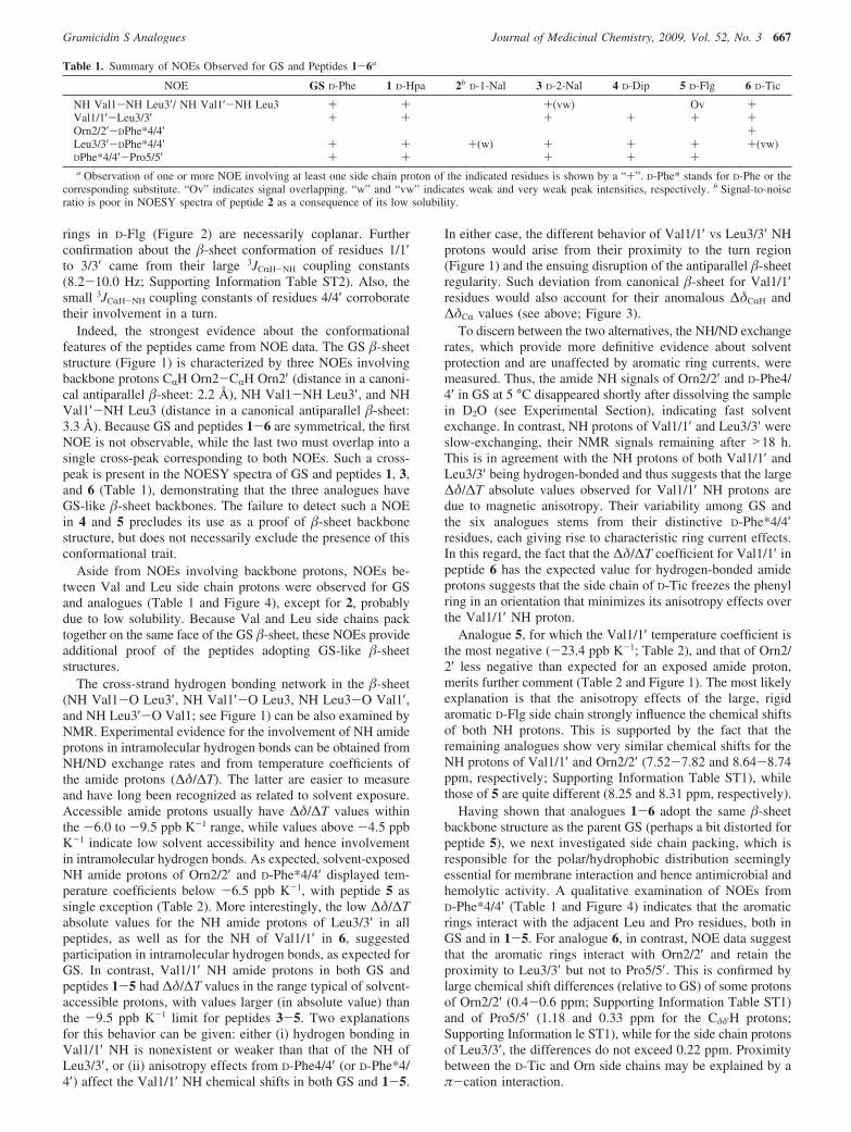
Indeed, the strongest evidence about the conformationalfeatures of the peptides came from NOE data. The GS �-sheetstructure (Figure 1) is characterized by three NOEs involvingbackbone protons CRH Orn2-CRH Orn2′ (distance in a canoni-cal antiparallel �-sheet: 2.2 Å), NH Val1-NH Leu3′, and NHVal1′-NH Leu3 (distance in a canonical antiparallel �-sheet:3.3 Å). Because GS and peptides 1-6 are symmetrical, the firstNOE is not observable, while the last two must overlap into asingle cross-peak corresponding to both NOEs. Such a cross-peak is present in the NOESY spectra of GS and peptides 1, 3,and 6 (Table 1), demonstrating that the three analogues haveGS-like �-sheet backbones. The failure to detect such a NOEin 4 and 5 precludes its use as a proof of �-sheet backbonestructure, but does not necessarily exclude the presence of thisconformational trait.
Aside from NOEs involving backbone protons, NOEs be-tween Val and Leu side chain protons were observed for GSand analogues (Table 1 and Figure 4), except for 2, probablydue to low solubility. Because Val and Leu side chains packtogether on the same face of the GS �-sheet, these NOEs provideadditional proof of the peptides adopting GS-like �-sheetstructures.
The cross-strand hydrogen bonding network in the �-sheet(NH Val1-O Leu3′, NH Val1′-O Leu3, NH Leu3-O Val1′,and NH Leu3′-O Val1; see Figure 1) can be also examined byNMR. Experimental evidence for the involvement of NH amideprotons in intramolecular hydrogen bonds can be obtained fromNH/ND exchange rates and from temperature coefficients ofthe amide protons (∆δ/∆T). The latter are easier to measureand have long been recognized as related to solvent exposure.Accessible amide protons usually have ∆δ/∆T values withinthe -6.0 to -9.5 ppb K-1 range, while values above -4.5 ppbK-1 indicate low solvent accessibility and hence involvementin intramolecular hydrogen bonds. As expected, solvent-exposedNH amide protons of Orn2/2′ and D-Phe*4/4′ displayed tem-perature coefficients below -6.5 ppb K-1, with peptide 5 assingle exception (Table 2). More interestingly, the low ∆δ/∆Tabsolute values for the NH amide protons of Leu3/3′ in allpeptides, as well as for the NH of Val1/1′ in 6, suggestedparticipation in intramolecular hydrogen bonds, as expected forGS. In contrast, Val1/1′ NH amide protons in both GS andpeptides 1-5 had ∆δ/∆T values in the range typical of solvent-accessible protons, with values larger (in absolute value) thanthe -9.5 ppb K-1 limit for peptides 3-5. Two explanationsfor this behavior can be given: either (i) hydrogen bonding inVal1/1′ NH is nonexistent or weaker than that of the NH ofLeu3/3′, or (ii) anisotropy effects from D-Phe4/4′ (or D-Phe*4/4′) affect the Val1/1′ NH chemical shifts in both GS and 1-5.
In either case, the different behavior of Val1/1′ vs Leu3/3′ NHprotons would arise from their proximity to the turn region(Figure 1) and the ensuing disruption of the antiparallel �-sheetregularity. Such deviation from canonical �-sheet for Val1/1′residues would also account for their anomalous ∆δCRH and∆δCR values (see above; Figure 3).
To discern between the two alternatives, the NH/ND exchangerates, which provide more definitive evidence about solventprotection and are unaffected by aromatic ring currents, weremeasured. Thus, the amide NH signals of Orn2/2′ and D-Phe4/4′ in GS at 5 °C disappeared shortly after dissolving the samplein D2O (see Experimental Section), indicating fast solventexchange. In contrast, NH protons of Val1/1′ and Leu3/3′ wereslow-exchanging, their NMR signals remaining after >18 h.This is in agreement with the NH protons of both Val1/1′ andLeu3/3′ being hydrogen-bonded and thus suggests that the large∆δ/∆T absolute values observed for Val1/1′ NH protons aredue to magnetic anisotropy. Their variability among GS andthe six analogues stems from their distinctive D-Phe*4/4′residues, each giving rise to characteristic ring current effects.In this regard, the fact that the ∆δ/∆T coefficient for Val1/1′ inpeptide 6 has the expected value for hydrogen-bonded amideprotons suggests that the side chain of D-Tic freezes the phenylring in an orientation that minimizes its anisotropy effects overthe Val1/1′ NH proton.
Analogue 5, for which the Val1/1′ temperature coefficient isthe most negative (-23.4 ppb K-1; Table 2), and that of Orn2/2′ less negative than expected for an exposed amide proton,merits further comment (Table 2 and Figure 1). The most likelyexplanation is that the anisotropy effects of the large, rigidaromatic D-Flg side chain strongly influence the chemical shiftsof both NH protons. This is supported by the fact that theremaining analogues show very similar chemical shifts for theNH protons of Val1/1′ and Orn2/2′ (7.52-7.82 and 8.64-8.74ppm, respectively; Supporting Information Table ST1), whilethose of 5 are quite different (8.25 and 8.31 ppm, respectively).
Having shown that analogues 1-6 adopt the same �-sheetbackbone structure as the parent GS (perhaps a bit distorted forpeptide 5), we next investigated side chain packing, which isresponsible for the polar/hydrophobic distribution seeminglyessential for membrane interaction and hence antimicrobial andhemolytic activity. A qualitative examination of NOEs fromD-Phe*4/4′ (Table 1 and Figure 4) indicates that the aromaticrings interact with the adjacent Leu and Pro residues, both inGS and in 1-5. For analogue 6, in contrast, NOE data suggestthat the aromatic rings interact with Orn2/2′ and retain theproximity to Leu3/3′ but not to Pro5/5′. This is confirmed bylarge chemical shift differences (relative to GS) of some protonsof Orn2/2′ (0.4-0.6 ppm; Supporting Information Table ST1)and of Pro5/5′ (1.18 and 0.33 ppm for the Cδδ′H protons;Supporting Information le ST1), while for the side chain protonsof Leu3/3′, the differences do not exceed 0.22 ppm. Proximitybetween the D-Tic and Orn side chains may be explained by aπ-cation interaction.
Table 1. Summary of NOEs Observed for GS and Peptides 1-6a
NOE GS D-Phe 1 D-Hpa 2b D-1-Nal 3 D-2-Nal 4 D-Dip 5 D-Flg 6 D-Tic
NH Val1-NH Leu3′/ NH Val1′-NH Leu3 + + +(vw) Ov +Val1/1′-Leu3/3′ + + + + + +Orn2/2′-DPhe*4/4′ +Leu3/3′-DPhe*4/4′ + + +(w) + + + +(vw)DPhe*4/4′-Pro5/5′ + + + + +a Observation of one or more NOE involving at least one side chain proton of the indicated residues is shown by a “+”. D-Phe* stands for D-Phe or the
corresponding substitute. “Ov” indicates signal overlapping. “w” and “vw” indicates weak and very weak peak intensities, respectively. b Signal-to-noiseratio is poor in NOESY spectra of peptide 2 as a consequence of its low solubility.
Gramicidin S Analogues Journal of Medicinal Chemistry, 2009, Vol. 52, No. 3 667

Figure 4. Selected NOESY spectral regions for peptides 1 (A) and 6 (B) in H2O/D2O 9:1 v/v at pH 3 and 5 °C. Relevant sequential and nonsequentialNOEs are boxed.
668 Journal of Medicinal Chemistry, 2009, Vol. 52, No. 3 Solanas et al.

To further visualize the side chain packing of peptides 1-6relative to GS, structure calculations were performed. Becauseof the symmetry of GS and analogues, every observed NOEhas four possible assignments. However, after our NMR analysisdemonstrated that the peptides adopt the GS �-sheet structure(Figure 1), some NOE ambiguities could be solved. Thus therestraints used for structure calculation combined (i) theoreticalrestraints for the backbone of strand residues Val1/1′, Orn2/2′,and Leu3/3′ derived from those expected for the GS �-sheet(Figure 1; more details in the Experimental Section and theSupporting Information) and (ii) experimental distance restraintsderived from the complete set of NOEs observed for eachpeptide. For every nonintraresidual NOE, all the existingpossibilities were taken into account, and the NOE assignmentscompatible with the GS �-sheet were identified by iterativestructure calculations. The final model structures obtained forGS and peptides 1-6 (Figures 5 and Supporting InformationFigure SF3) are well defined, particularly for 6 (see rmsd valuesin Supporting Information Table ST3). A general examinationof the structures corroborates the main conformational traitsdeduced from the qualitative analysis of NMR parameters.Concerning the orientation of the aromatic side chain at positions4/4′, the phenyl rings in 1 are quite disordered and explore amuch wider space than in GS, while the aromatic systems ofpeptides 4 and 5 adopt quite well-defined positions. Alsonoteworthy is the different relative orientation of the two phenylrings in D-Flg and D-Dip, in the latter case with the rings almostperpendicular to each other, and one in an orientation close tothat of the D-Phe ring in GS (Supporting Information FigureSF3). Even more remarkable are the differences between GSand analogue 6 (Figure 5) in the relative orientation of thearomatic rings and of the positively charged Orn side chains,with noticeable consequences on their respective biologicalactivities (see below).
Biological Activity. Microbicidal activities were determinedin solution because, in solid media, they tend to be undermea-sured, especially for Gram-negatives.4 Two Gram-positive(Staphylococcus aureus and Listeria monocytogenes) and aGram-negative (Acinetobacter baummanii ATCC 19606 and itsisogenic strain resistant to polymyxin E (PXE)) bacterial strainswere tested. Although GS is mostly active against Gram-positives, the occurrence of cutaneous infection by A. baumanniiand the increasing resistance to PXE, the last universal drugactive against these bacteria,41 prompted us to include both ofthem in the panel. Results are listed in Table 3.
Within the Gram-positive group, the tested set of analogueshad better and comparable activities, respectively, for Staphy-lococcus and Listeria than the parental GS. In contrast, againstthe Gram-negative A. baumannii, activity in the analogue seriestended to deteriorate relative to GS, more markedly so for theisogenic PXE-resistant strain. The sole exception was the D-Hpa-containing peptide 1, which performed slightly better than GS
against both PXE-sensitive and resistant strains, suggesting itis impervious to modifications in lipopolysacharide (LPS)structure.
The hemolytic effect of the peptides was evaluated on sheeperythrocytes (Table 3) as an indicator of toxicity againstmammalian cells. In this assay, five (1-5) out of six analoguesturned out to be more toxic than the parent GS. The findingthat the D-Flg analogue (5) was more cytotoxic and yet lesspotent than the parent antibiotic underscores the fact that subtlestructural changes in GS may translate into different abilitiesto damage prokaryotic and eukaryotic cells. An opposite, thoughsimilar and more promising, segregation of antibacterial andhemolytic activities was found for 6, which has GS-like MIC50
values for Gram-positives (Table 3) but is essentially harmlesstoward erythrocytes.
Beyond adequate intrinsic microbicidal activity, a convenientassessment of the clinical potential of an antibiotic is providedby its therapeutic index (TI, defined as HC50/MIC50, see Table3), where the cytotoxic and microbicidal effects are combined.As found in previous SAR studies on GS,23,25,42,43 improvedTIs are more often due to a decrease in cytotoxicity than to anincrease in antibiotic potency. In the present series, the above-mentioned (higher than for GS) toxicity values, coupled withthe minute, if any, improvements in microbicidal activity,resulted in TI values slightly lower or clearly worse than thatof the parent structure. The sole exception to this pattern wasthe D-Tic analogue (6), for which a modest but significantimprovement in TI was obtained (Table 3).
Discussion
The type II′ �-turn is a structural motif especially suited toinduce alignment of its two side sequence segments into anantiparallel �-sheet. The quintessential example of this situationis GS, whose type II′ �-turns have been a testing ground forextensive D-Phe-Pro replacements by a variety of peptidomi-metics, with variable success (see Introduction and refs 29-31,44-46). In comparison, fewer studies have addressed thesubstitution of either L-Pro17,47 or D-Phe33,46,48-50 by appropriatesurrogates. The goal of this work was to explore and structurallyinterpret the fine-tuning of GS antibiotic activity by replacementof D-Phe with a novel set of noncoded aromatic D-amino acidsalready tested as turn-promoters.
The activity of GS is assumed to be somehow related to thepermeabilization of bacterial membranes, although no unifiedopinion on the nature of such process exists.51 Hydrophobicityhas been cited as a relevant parameter,25,42,43 and inspection ofthe current set of D-Phe analogues (Figure 2) shows considerablevariation in the number and size of aromatic side chains, hencein overall hydrophobicity at this position. The presence of asizable hydrophobic domain is closely related to hemolyticactivity in antimicrobial peptides, as it seemingly promotes ahigher partition of the peptide into biological membranesregardless of their zwitterionic or anionic character.24,25,42
Accordingly, the hemolytic and Gram-positive microbicidalactivities run somewhat parallel, provided hydrophobicity doesnot drastically reduce peptide solubility in aqueous medium.52
For Gram-negatives, however, the situation changes in that anexternal barrier, i.e., the outer membrane (OM), must bedisrupted prior to the “lethal hit” i.e., permeabilization of theinner membrane (IM). In this scenario, while high hydrophobic-ity improves the affinity of the antibiotic peptide for LPS (themajor component of the OM), excess affinity may sequesterthe peptide into the OM, hence precluding its crucial interactionwith the IM target. Therefore, a rather subtle balance between
Table 2. Temperature Coefficientsa (∆δ/∆T, ppb ·K-1) for AmideProtons in GS and Its Analogues
peptide D-Phe*b Val1,1′(NH) Orn2,2′(NH) Leu3,3′(NH) D-Phe*4,4′(NH)
GS D-Phe -7.4 -7.1 -0.8 -8.61 D-Hpa -6.1 -7.8 -1.2 -11.02 D-1-Nal -8.0 -7.0 -1.5 -8.03 D-2-Nal -11.4 -6.6 0.2 -7.64 D-Dip -13.4 -7.3 -1.7 -10.15 D-Flg -23.4 -3.6 0.0 c6 D-Tic -1.3 -8.0 -3.9a Measured in H2O/D2O 9:1 (v/v) at pH 3.0. b D-Phe* stands for D-Phe
or the corresponding substitute. c Not determined; NH protons not observedat 25 °C probably because of fast exchange with water.
Gramicidin S Analogues Journal of Medicinal Chemistry, 2009, Vol. 52, No. 3 669

hydrophobicity and cationic character is desirable for activityagainst Gram-negatives.18 These considerations broadly agreewith the finding that peptides 1-5, more toxic than GS towarderythrocytes (Table 3), are also more hydrophobic than GS byan accepted criterion,23 namely RP-HPLC retention timesconsistently longer than those of the parent peptide (seeSupporting Information, Figure SF1). On the other hand, theD-Tic (6) analogue combines decreased cytotoxicity with higherhydrophobicity (longer HPLC retention time) than GS (Table3). Likewise, the similar HPLC retention times for peptides 1-4(10.3-10.6 min, Supporting Information Figure SF1) do notcorrelate well with their antimicrobial profiles. Thus, while 1improved on GS against all bacteria tested, Nal-containinganalogues 2 and 3, with presumably comparable hydrophobicity,were similar to GS against Gram-positives but practicallyinactive against Gram-negatives. In conclusion, it appears that,at least for this series, hydrophobicity has limited value inexplicating antibiotic performance.
NMR results shed additional light on the structure-activityanalysis of the peptides. In particular, they reveal a varyingdegree of conformational freedom in their side chains that canbe reasonably related to differences in antimicrobial activity.For instance, the aromatic ring of the D-Hpa-containing analogue(1), separated from the backbone by an extra methylene group,can plausibly explore a larger conformational space and
consequently a wider repertoire of membrane (either prokaryoticor eukaryotic) interaction modes than the D-Phe-containing GS,thus explaining its increased activity toward both bacterial andmammalian cells. In contrast, the naphthalene rings in 2 and 3have conformational freedoms similar in principle to the phenylgroup of GS but their bulkiness is likely to pose a limitation ontheir interaction with LPS and ensuing OM disruption in Gram-negative bacteria. For Gram-positives, on the other hand, nocomparable tightly packed external barrier exists, which wouldagree with both the preservation of antibiotic activity in bothanalogues as well as their hemolytic properties. Similar con-siderations on the orientation and/or rotational freedom of thearomatic rings can be applied to the contrasting activities of 4(D-Dip) and 5 (D-Flg). In the latter analogue, the two aromaticrings are fastened into a bulky planar tricylic system, whosepredictable sluggishness may help to explain its significantlyworse profile than 4.
By far the most striking result in the present series is theimproved therapeutic profile of the D-Tic analogue 6. With anapparent hydrophobicity (based on RP-HPLC retention time)slightly higher than GS, it is basically equipotent to GS againstGram-positives but has considerably lower toxicity, hence amuch better TI (Table 3). From the structural point of view, 6has two distinctive features over the other analogues in theseries: first, the severe conformational restriction on the (proline-
Figure 5. Model structures for GS (A) and peptide 6 (B) in two different views. Backbone atoms are shown in black. Side chains for aromaticD-Phe and D-Tic are colored in green, for Pro in cyan, for Orn in blue, and for Leu and Val in magenta.
Table 3. Cytotoxic (Hemolytic) and Antimicrobial Activity of GS and Analogues
erythrocytes S. aureus L. monocytogenes A. baumanni S A. baumannii Re
peptide D-Phe*a HC50b (µM) MIC50
c (µM) TId MIC50 (µM) TI MIC50 (µM) TI MIC50 (µM) TI
GS D-Phe 21.1 ((2.6) 7.9 ((0.8) 2.7 (1) 6.7 ((0.2) 3.1 (1) 10.1 ((0.4) 2.1 (1) 13.1 ((0.0) 1.6 (1)1 D-Hpa 7.0 ((0.3) 3.5 ((0.1) 2 (0.7) 2.4 ((0.0) 2.9 (0.9) 4.6 ((0.1) 1.5 (0.7) 6.2 ((0.1) 1.1 (0.7)2 D-1-Nal 5.0 ((0.1) 2.9 ((0.5) 1.7 (0.6) 8.6 ((0.6) 0.6 (0.2) 35.9 ((0.0) 0.1 (0.0) >40 >0.13 D-2-Nal 7.1 ((3.5) 4.6 ((0.6) 1.5 (0.6) 7.8 ((0.4) 0.9 (0.3) 39.6 ((0.0) 0.2 (0.1) >40 >0.24 D-Dip 8.6 ((1.1) 3.4 ((0.0) 2.5 (0.9) 7.7((0.6) 1.1 (0.4) 9.6 ((0.9) 0.9 (0.4) 17.8 ((0.0) 0.5 (0.3)5 D-Flg 6.2 ((0.3) 8 ((0.0) 0.8 (0.3) 9.2((1.4) 0.7 (0.2) 15.4 ((1.3) 0.4 (0.2) >40 >0.16 D-Tic >50 4.9 ((0.4) >10.2 (3.8) 8.3 ((0.5) >6 (2.0) >40 >ND >40 NDa D-Phe* stands for D-Phe or the corresponding substitute. b HC50: peptide concentration required for 50% lysis of sheep erythrocytes. c MIC50: peptide
concentration required for 50% inhibition of bacterial growth after 24 h, relative to a control culture. d TI (therapeutic index) ) HC50/MIC50. e An A.baumannii strain resistant to polymyxin E (colistin). ND, not determined. Standard deviations (SD) are shown in parentheses.
670 Journal of Medicinal Chemistry, 2009, Vol. 52, No. 3 Solanas et al.
like) imino acid D-Tic imposed by the cyclization between thephenyl group and the backbone and borne out by the observationof practically a single conformer by NMR. Interestingly, thehydrogen-bonding interaction between the δ-amino group ofOrn and the backbone carbonyl of D-Phe reported for GS andsome analogues53,54 would thus not be present in analogue 6.The absence of this favorable effect would most likely becompensated by the stabilizing contribution that the increasedrigidity of D-Tic -compared with D-Phe- imparts to the �-turnstructure. As a rigid side chain is conceivably more difficult toinsert into lipid bilayers than a flexible one (i.e., that of GS),this could account for the lack of hemolytic effect of 6 andthus its improved antibiotic profile. Second, in 6, the δ-NH3
+
groups of Orn are positioned close to the aromatic ring of D-Tic,possibly favored by a π-cation interaction. According to NMRand MD studies on the membrane-bound structure of GS,43,55,56
the peptide lies flat relative to the bilayer plane, near the interfacebetween polar and apolar regions, so that the δ-NH3
+ groupsof Orn can interact with phospholipid polar head groups. Now,if a similar contact model is assumed for analogue 6, theaforementioned π-cation interaction between D-Tic and Ornmust disappear. The thermodynamic penalty thus incurred wouldbe higher for the interaction with (zwitterionic) eukaryotic thanfor prokaryotic membranes, where the loss of the D-Tic-Orninteraction would be compensated by electrostatic attractionbetween Orn δ-NH3
+ and phosphate head groups. Again, thismay account for the higher relative loss in hemolytic than inbactericidal effect.
Conclusions
Analogues of the antimicrobial peptide GS with differentaromatic replacements at both D-Phe residues have beensynthesized in order to explore how the conformational freedomand the size of the aromatic moiety modulate both biologicaland structural properties. NMR studies confirmed that the overallmolecular shape of the GS framework is maintained in allanalogues (some distortion cannot be discarded for the D-Flgderivative), so that variations in antibiotic profile can be ascribedto the different D-Phe replacements. The results suggest thatboth bulkiness and orientation of the aromatic system at the4/4′ position are essential for biological activity, to the pointthat slight modifications in these parameters bring aboutsignificant changes in antibacterial activity as well as in thedesirable specificity for bacterial over mammalian cells. Aninteresting finding is that analogue 6, with D-Tic replacing D-Phe,retains antibiotic potency against Gram-positive bacteria but ishardly cytotoxic. Our NOE data clearly indicate that in thisanalogue the Orn side chain orients toward the D-Tic aromaticrings. A plausible explanation for this orientation is the existenceof a cation-π interaction between the δ-NH3
+ groups of theOrn side chain and the aromatic ring of D-Tic. In any event,and regardless of the nature of the interaction, the specificorientation of the D-Tic aromatic ring relative to the Orn sidechain allows explanation of the distinct biological behaviorexhibited by this peptide. While the increase in therapeutic indexrelative to GS is only 5-fold, it is nonetheless remarkablebecause, in contrast to other studies,25,42 it has been achievedwith minimal alteration of the GS structure, including chiralityand cycle size. Modifications of this type may be helpful inunderstanding the mechanism of action of GS and developingpeptide antibiotics with improved pharmaceutical applications.
Experimental Section
Chemicals and Instrumentation for Peptide Synthesis. Fluo-renylglycine was prepared as previously reported.34 N-Boc ornithine
was purchased from Bachem (Bubendorf, Switzerland) and its sidechain amino group was protected as a formamide by reaction withformic acid and 1,1′-oxalyldiimidazole.35 All other amino acidswere purchased from Senn Chemicals (Dielsdorf, Switzerland),NeoMPS (Strasbourg, France) or Fluka (Buchs, Switzerland).Chloromethylated Merrifield resin and TFMSA were from Fluka,HATU from GenScript (Piscataway, NJ), and HBTU from MatrixInnovation (Montreal, Quebec). Solvents and other chemicals werefrom SDS (Peypin, France). Mass determination by the MALDI-TOF technique was done in a Voyager DE-RP spectrometer(Applied Biosystems, Foster City, CA) using 2,5-dihydroxybenzoicacid as a matrix. Analytical RP-HPLC was performed on an LC-2010A workstation (Shimadzu Corporation, Kyoto, Japan) with aLuna C8 (3 µm, 50 mm × 4.6 mm) column (Phenomenex BV,Utrecht, The Netherlands) eluted with a linear 5-95% gradient ofCH3CN (+0.036% TFA, v/v) into H2O (+0.045% TFA, v/v) over15 min at 1 mL/min flow rate, with UV detection at 220 nm.Preparative RP-HPLC purification was done on a Phenomenex LunaC8 column (10 µm, 250 mm × 10 mm) running linear gradients ofCH3CN (+0.1% TFA, v/v) into H2O (+0.1% TFA, v/v) as indicatedfor each peptide, at a flow rate of 5 mL/min. The preparative systemincluded two Shimadzu LC-8A pumps, a Shimadzu SPD-10Adetector and a Foxy Jr. fraction collector (Teledyne Isco, Lincoln,NE).
General Procedure for Peptide Synthesis. All peptides weresynthesized manually by solid-phase methods on a Boc-Pro-Merrifield resin (0.1 mmol) using Boc chemistry. Boc-amino acids(0.3 mmol) were coupled by HBTU/DIEA (0.3 and 0.6 mmol,respectively) for 30-45 min in DMF. HATU was used instead ofHBTU for coupling of the 4/4′ residues. To avoid diketopiperazineformation, the third amino acid of each sequence was incorporatedby the in situ neutralization method.57 Coupling and deprotectionreactions were monitored by the nynhidrin58 or p-nitrophenylester59
colorimetric tests. The linear decapeptide was cleaved from the resinby treatment with TFA/TFMSA/TIS 10:1:1 (v/v/v) (4 mL) at roomtemperature for 90 min and then precipitated with cold tert-butylmethyl ether. The peptide was redissolved in glacial acetic acid,filtered off the resin, and lyophilized. The residue was dissolved inDMF to a final concentration of 2 mg/mL and stirred for 1 h atroom temperature in the presence of HBTU/HOBt/DIEA (3:3:5equiv). After solvent removal and lyophilization, the cyclizedpeptide was deformylated by treatment with 20% hydrochloric acidin methanol at 37 °C for 21 h. The solvent was evaporated underreduced pressure, and the residue was taken up in glacial aceticacid and lyophilized. Final purification was by preparative reversed-phase HPLC as indicated in each case. HPLC-homogeneousfractions were combined and lyophilized to give white powders ofg99% HPLC purity (see Supporting Information, Figure SF2).
cyclo(Val-Orn-Leu-D-Phe-Pro)2 (GS). RP-HPLC, linear 35-70%CH3CN gradient into H2O for 30 min (33 mg, 0.029 mmol, 25%overall yield). [M + H]+calcd ) 1141.7; [M + H]+obs ) 1141.1.
cyclo(Val-Orn-Leu-D-Hpa-Pro)2 (1). RP-HPLC, linear 45-75%CH3CN gradient into H2O for 30 min (51 mg, 0.044 mmol, 41%overall yield). [M + H]+ calcd ) 1169.7; [M + H]+obs ) 1169.5.
cyclo(Val-Orn-Leu-D-1-Nal-Pro)2 (2). RP-HPLC, linear 45-75%CH3CN gradient into H2O for 30 min (34 mg, 0.027 mmol, 26%overall yield). [M + H]+calcd ) 1241.7; [M + H]+obs ) 1241.8.
cyclo(Val-Orn-Leu-D-2-Nal-Pro)2 (3). RP-HPLC, linear 45-75%CH3CN gradient into H2O for 30 min (26 mg, 0.021 mmol, 20%overall yield). [M + H]+calcd ) 1241.7; [M + H]+obs ) 1241.6.
cyclo(Val-Orn-Leu-D-Dip-Pro)2 (4). RP-HPLC, linear 50-80%CH3CN gradient into H2O for 30 min (22 mg, 0.017 mmol, 15%overall yield). [M + H]+calcd ) 1293.8; [M + H]+obs ) 1293.6.
cyclo(Val-Orn-Leu-D-Flg-Pro)2 (5). RP-HPLC, linear 55-85%CH3CN gradient into H2O for 30 min (18 mg, 0.015 mmol, 14%overall yield). [M + H]+calcd ) 1289.7; [M + H]+obs ) 1289.8).
cyclo(Val-Orn-Leu-D-Tic-Pro)2 (6). RP-HPLC, linear 45-65%CH3CN gradient into H2O for 30 min (23 mg, 0.020 mmol, 19%overall yield). [M + H]+calcd ) 1165.7; [M + H]+obs ) 1165.8.
NMR Spectroscopy. Samples for NMR experiments wereprepared at 1-2 mM peptide concentration in 0.5 mL of H2O/D2O
Gramicidin S Analogues Journal of Medicinal Chemistry, 2009, Vol. 52, No. 3 671

9:1 (v/v) or pure D2O at pH 3.0. pH was measured with a glassmicroelectrode and was not corrected for isotope effects. NMRspectra were acquired on a Bruker AV 600 MHz spectrometerequipped with a z-gradient cryoprobe. A methanol sample was usedto calibrate the temperature of the NMR probe. One-dimensional(1D) and two-dimensional (2D) spectra were acquired by standardpulse sequences using presaturation of the water signal. Mixingtimes for 2D TOCSY and NOESY were 60 and 150 ms,respectively. The 1H-13C and 1H-15N HSQC spectra60 at natural13C and 15N abundance were recorded in D2O and H2O/D2O 9:1(v/v), respectively. Data were processed using TOPSPIN (BrukerBiospin, Rheinstetten, Germany) software. Sodium 2,2-dimethyl-2-silapentane-5-sulfonate (DSS) was used as an internal referencefor 1H chemical shifts. The 13C and 15N chemical shifts wereindirectly calibrated by multiplying the spectrometer frequency thatcorresponds to 0 ppm in the 1H spectrum, assigned to internal DSSreference, by 0.25144954 and 0.101329118, respectively.61
The 1H NMR signals of the peptides were assigned by sequentialassignment methods.62 The 13C and 15N resonances were thenassigned following the cross-correlations observed in the HSQCspectra between the proton and the heteronucleus to which it isbonded.
To measure the NH/ND exchange rates, a freeze-dried sampleof peptide GS was solved in pure D2O at pH 3.0, and after the10-15 min required to set up the NMR experiment, consecutive1D and 2D 20 ms-TOCSY spectra were recorded at 5 °C.
Structure Calculation. Structure calculations were performedby using the CYANA program63 and an annealing strategy. Sincethe nonproteinogenic amino acids were not included in the standardCYANA libraries, we built them using MOLMOL64 and manualoptimization (these libraries are available upon request from theauthors). For this purpose, X-ray data of compounds containingthese amino acids were retrieved from the Cambridge StructuralDatabase.65 Theoretical constraints for the GS �-sheet incorporatedfor structure calculation included φ and ψ angle restraints, lowerand upper-limit distance restraints for the four characteristic cross-strand hydrogen-bonds, and upper-limit distance restraints for thebackbone atoms of strand residues (Supporting Information TableST4). Experimental distance constraints were derived from 2DNOESY spectra recorded in H2O, also in D2O for analogue 6. TheNOE cross-peaks were integrated by using the automatic integrationsubroutine of the Sparky program (T. D. Goddard and S. G. Kneller,University of California at San Francisco) and then calibrated andconverted to upper-limit distance constraints with CYANA.63 Giventhe symmetrical nature of the peptides, for structure calculationsresidues were renumbered from 1 to 10 starting at Leu3 (Figure1). Lower and upper limit restraints required for peptide backbonecyclization were also introduced (see Supporting Information). Foreach peptide, a total of 50 conformers were generated, and the 20conformers with the lowest target function were analyzed. Modelstructures for GS and analogues 1-6 were examined with MOL-MOL. A side chain torsion angle was considered as well definedwhen its root-mean-square deviation between values in the 20 bestcalculated structures was less than (30°.
Antimicrobial Activity. Stocks of Staphylococcus aureus CECT240, Listeria monocytogenes CECT 4032, Acinetobacter baumanniiATCC 19606, and its isogenic colistin-resistant strain 19606R(obtained by continuous growing under increasing colistin concen-tration) were maintained at -80 °C in freezing medium (65%glycerol, 0.1 M MgSO4, 25 mM Tris-HCl, pH 8.0). Two days priorto the assay for microbicidal activity, they were thawed and grownin MBH medium (Mueller-Hinton II Broth Cation Adjusted(Becton-Dickinson, Cockeysville, MD) at 37 °C; for 19606R, 64µg/mL colistin sulfate (Sigma, Madrid, Spain) was included.41
Bacterial cells were harvested at exponential growth phase, washedtwice with phosphate buffered saline (PBS, 10 mM Na2HPO4, 1mM KH2PO4, 140 mM NaCl, 3 mM KCl, pH 7.0) and resuspendedin MBH, at 5 × 10⁵ CFU/mL. Aliquots (100 µL) from thissuspension were transferred into a polypropylene 96-well plate, andbacteria were allowed to proliferate for 24 h at 37 °C in the presenceof the corresponding analogue concentration. Afterward, growth
was measured by turbidimetry at 600 nm in a model 680 microplatereader (Bio-Rad Laboratories, Hercules. CA). MIC50 was definedas the lowest peptide concentration inhibiting bacterial growth by50%, relative to untreated control, and was calculated using theSigmaPlot (Systat Software, San Jose, CA) software, v. 9.0.
Hemolytic Activity Assay. Hemolytic activity of the peptideswas determined by triplicate. Defibrinated sheep blood (Biomedics,Madrid, Spain) was centrifuged and washed twice with Hank’smedium (136 mM NaCl; 4.2 mM Na2HPO4; 4.4 mM KH2PO4; 5.4mM KCl; 4.1 mM NaHCO3, pH 7.2), supplemented with 20 mMD-glucose (Hank’s-Glc). Erythrocytes were resuspended in the samebuffer at 2 × 10⁷ erythrocytes/mL, and 100 µL aliquots of thesuspension were incubated with the peptides (4 h, 37 °C).The remaining erythrocytes were harvested in a Micro 200microfuge (A. Hettich GmbH & Co KG, Germany) (14000 rpm, 5min, 4 °C), 80 µL of the supernatant were transferred into a 96-well culture microplate, and hemoglobin release was read at 550 nmin a Bio Rad 680 (Hercules, CA) microplate reader. The asymptoticordinate of the GS supernatant was taken as 100% hemolysis. HC50
values were calculated using SigmaPlot, version 9.0.
Acknowledgment. This work was supported by Ministeriode Educacion y Ciencia (BIO2005-07592-CO2-02 to D.A.,BFU2005-01855 and CTQ2008-0080 to M.A.J., CTQ2007-62245 to C.C.), Fondo de Investigaciones Sanitarias (PI061125and RD 06/0021/0006 to L.R., PI040885 to D.A.), by theregional governments of Aragon (research group E40), Cata-lunya (SGR2005-00494), and Madrid (S-BIO-0260/2006). Thisproject has been funded in whole or in part with Federal fundsfrom the National Cancer Institute, National Institutes of Health,under contract N01-CO-12400. C.S. and C.M.S. thank Minis-terio de Educacion y Ciencia and Consejo Superior de Inves-tigaciones Cientıficas-European Social Fund for an FPU andI3P fellowship, respectively. The content of this publication doesnot necessarily reflect the view of the policies of the Departmentof Health and Human Services, nor does mention of trade names,commercial products, or organization imply endorsement by theU.S. Government. This research was supported (in part) by theIntramural Research Program of the NIH, National CancerInstitute, Center for Cancer Research.
Supporting Information Available: NMR data, details onstructure calculations, and analytical data on GS and analogues 1-6.This material is available free of charge via the Internet at http://pubs.acs.org.
References(1) Hirsch, T.; Jacobsen, F.; Steinau, H. U.; Steinstraesser, L. Host defense
peptides and the new line of defence against multiresistant infections.Protein Pept. Lett. 2008, 15, 238–243.
(2) Parisien, A.; Allain, B.; Zhang, J.; Mandeville, R.; Lan, C. Q. Novelalternatives to antibiotics: bacteriophages, bacterial cell wall hydrolases,and antimicrobial peptides. J. Appl. Microbiol. 2008, 104, 1–13.
(3) Gause, G. F. Gramicidin S and its use in the treatment of infectedwounds. Nature 1944, 154, 703.
(4) Kondejewski, L. H.; Farmer, S. W.; Wishart, D. S.; Hancock, R. E.;Hodges, R. S. Gramicidin S is active against both gram-positive andgram-negative bacteria. Int. J. Pept. Protein. Res. 1996, 47, 460–466.
(5) Prenner, E. J.; Lewis, R. N.; McElhaney, R. N. The interaction of theantimicrobial peptide gramicidin S with lipid bilayer model andbiological membranes. Biochim. Biophys. Acta 1999, 1462, 201–221.
(6) Hull, S. E.; Karlsson, R.; Main, P.; Woolfson, M. M.; Dodson, E. J.The crystal structure of a hydrated gramicidin S-urea complex. Nature1978, 275, 206–207.
(7) Schmidt, G. M.; Hodgkin, D. C.; Oughton, B. M. A crystallographicstudy of some derivatives of gramicidin S. Biochem. J. 1957, 65, 744–750.
(8) Tishchenko, G. N.; Andrianov, V. I.; Vainstein, B. K.; Woolfson,M. M.; Dodson, E. Channels in the gramicidin S-with-urea structureand their possible relation to transmembrane ion transport. ActaCrystallogr., Sect. D: Biol. Crystallogr. 1997, 53, 151–159.
(9) Ovchinnikov, Y. A.; Ivanov, V. T. Conformational states and biologicalactivity of cyclic peptides. Tetrahedron 1975, 31, 2177–2209.
672 Journal of Medicinal Chemistry, 2009, Vol. 52, No. 3 Solanas et al.

(10) Katsu, T.; Kobayashi, H.; Fujita, Y. Mode of action of gramicidin Son Escherichia coli membrane. Biochim. Biophys. Acta 1986, 860,608–619.
(11) Afonin, S.; Glaser, R. W.; Berditchevskaia, M.; Wadhwani, P.; Guhrs,K. H.; Mollmann, U.; Perner, A.; Ulrich, A. S. 4-Fluorophenylglycineas a label for 19F NMR structure analysis of membrane-associatedpeptides. ChemBioChem 2003, 4, 1151–1163.
(12) Arai, T.; Imachi, T.; Kato, T.; Ogawa, H. I.; Fujimoto, T.; Nishino,N. Synthesis of [hexafluorovalyl1,1′]gramicidin S. Bull. Chem. Soc.Jpn. 1996, 69, 1383–1389.
(13) Waki, M.; Abe, O.; Okawa, R.; Kato, T.; Makisumi, S.; Izumiya, N.Studies of peptide antibiotics. XII. Syntheses of [2,2′-R,γ-diaminobu-tyric acid] and [2,2′-lysine]-gramicidin S. Bull. Chem. Soc. Jpn. 1967,40, 2904–2909.
(14) Aimoto, S. The synthesis of a heavy-atom derivative of gramicidin S(GS), [D-Phe(4-Br) 4,4′]-GS, by a novel method. Bull. Chem. Soc. Jpn.1988, 61, 2220–2222.
(15) Andreu, D.; Ruiz, S.; Carreno, C.; Alsina, J.; Albericio, F.; Jimenez,M. A.; de la Figuera, N.; Herranz, R.; Garcıa-Lopez, M. T.; Gonzalez-Muniz, R. IBTM-containing gramicidin S analogues: Evidence forIBTM as a suitable type II′ �-turn mimetic. J. Am. Chem. Soc. 1997,119, 10579–10586.
(16) Grotenbreg, G. M.; Buizert, A. E.; Llamas-Saiz, A. L.; Spalburg, E.;van Hooft, P. A.; de Neeling, A. J.; Noort, D.; van Raaij, M. J.; vander Marel, G. A.; Overkleeft, H. S.; Overhand, M. �-Turn modifiedgramicidin S analogues containing arylated sugar amino acids displayantimicrobial and hemolytic activity comparable to the natural product.J. Am. Chem. Soc. 2006, 128, 7559–7565.
(17) Kawai, M.; Yamamura, H.; Tanaka, R.; Umemoto, H.; Ohmizo, C.;Higuchi, S.; Katsu, T. Proline residue-modified polycationic analogsof gramicidin S with high antibacterial activity against both Gram-positive and Gram-negative bacteria and low hemolytic activity. J.Pept. Res. 2005, 65, 98–104.
(18) Lee, D. L.; Hodges, R. S. Structure-activity relationships of de novodesigned cyclic antimicrobial peptides based on gramicidin S. Biopoly-mers 2003, 71, 28–48.
(19) Ripka, W. C.; Delucca, G. V.; Bach, A. C.; Pottorf, R. S.; Blaney,J. M. Protein �-turn mimetics II: design, synthesis and evaluation inthe cyclic peptide gramicidin S. Tetrahedron 1993, 49, 3609–3628.
(20) Sato, K.; Kato, R.; Nagai, U. Studies on �-turn of peptides. XII.Synthetic conformation of weak activity of [D-Pro5,5′]-gramicidin Spredicted from �-turn preference of its partial sequence. Bull. Chem.Soc. Jpn. 1986, 59, 535–538.
(21) Tamaki, M.; Okitsu, T.; Araki, M.; Sakamoto, H.; Takimoto, M.;Muramatsu, I. Synthesis and properties of gramicidin S analogscontaining Pro-D-Phe sequence in place of D-Phe-Pro sequence in the�-turn part of the antibiotic. Bull. Chem. Soc. Jpn. 1985, 58, 531–535.
(22) Wishart, D. S.; Kondejewski, L. H.; Semchuk, P. D.; Sykes, B. D.;Hodges, R. S. A method for the facile solid-phase synthesis ofgramicidin S and its analogs. Lett. Pept. Sci. 1996, 3, 53–60.
(23) Yamada, K.; Shinoda, S. S.; Oku, H.; Komagoe, K.; Katsu, T.; Katakai,R. Synthesis of low-hemolytic antimicrobial dehydropeptides basedon gramicidin S. J. Med. Chem. 2006, 49, 7592–7595.
(24) Jelokhani-Niaraki, M.; Kondejewski, L. H.; Farmer, S. W.; Hancock,R. E.; Kay, C. M.; Hodges, R. S. Diastereoisomeric analogues ofgramicidin S: structure, biological activity and interaction with lipidbilayers. Biochem. J. 2000, 349, 747–755.
(25) Kondejewski, L. H.; Jelokhani-Niaraki, M.; Farmer, S. W.; Lix, B.;Kay, C. M.; Sykes, B. D.; Hancock, R. E.; Hodges, R. S. Dissociationof antimicrobial and hemolytic activities in cyclic peptide diastereomersby systematic alterations in amphipathicity. J. Biol. Chem. 1999, 274,13181–13192.
(26) Abraham, T.; Marwaha, S.; Kobewka, D. M.; Lewis, R. N.; Prenner,E. J.; Hodges, R. S.; McElhaney, R. N. The relationship between thebinding to and permeabilization of phospholipid bilayer membranesby GS14dK4, a designed analog of the antimicrobial peptide grami-cidin S. Biochim. Biophys. Acta 2007, 1768, 2089–2098.
(27) Abe, O.; Izumiya, N. Studies of peptide antibiotics. Analogs ofgramicidin S containing glycine or alanine in place of leucine. Bull.Chem. Soc. Jpn. 1970, 43, 1202–1207.
(28) Prenner, E. J.; Lewis, R. N.; Kondejewski, L. H.; Hodges, R. S.;McElhaney, R. N. Differential scanning calorimetric study of the effectof the antimicrobial peptide gramicidin S on the thermotropic phasebehavior of phosphatidylcholine, phosphatidylethanolamine and phos-phatidylglycerol lipid bilayer membranes. Biochim. Biophys. Acta1999, 1417, 211–223.
(29) Graciani, N. R.; Tsang, K. Y.; McCutchen, S. L.; Kelly, J. W. Aminoacids that specify structure through hydrophobic clustering andhistidine-aromatic interactions lead to biologically active peptidomi-metics. Bioorg. Med. Chem. 1994, 2, 999–1006.
(30) Kee, K. S.; Jois, S. D. Design of �-turn based therapeutic agents. Curr.Pharm. Des. 2003, 9, 1209–1224.
(31) Xiao, J.; Weisblum, B.; Wipf, P. Trisubstituted (E)-alkene dipeptideisosteres as beta-turn promoters in the gramicidin S cyclodecapeptidescaffold. Org. Lett. 2006, 8, 4731–4734.
(32) Grotenbreg, G. M.; Spalburg, E.; de Neeling, A. J.; van der Marel,G. A.; Overkleeft, H. S.; van Boom, J. H.; Overhand, M. Synthesisand biological evaluation of novel turn-modified gramicidin Sanalogues. Bioorg. Med. Chem. 2003, 11, 2835–2841.
(33) Shimohigashi, Y.; Kodama, H.; Imazu, S.; Horimoto, H.; Sakaguchi,K.; Waki, M.; Uchida, H.; Kondo, M.; Kato, T.; Izumiya, N. [4,4′-(Z)-dehydrophenylalanine]gramicidin S with stabilized bioactive con-formation and strong antimicrobial activity. FEBS Lett. 1987, 222,251–255.
(34) Royo, S.; Jimenez, A. I.; Cativiela, C. Synthesis of enantiomericallypure �,�-diphenylalanine (Dip) and fluorenylglycine (Flg). Tetrahe-dron: Asymmetry 2006, 17, 2393–2400.
(35) Kitagawa, T.; Arita, J.; Nogahata, A. Convenient one-pot method forformylation of amines and alcohols using formic acid and 1,1′-oxalyldiimidazole. Chem. Pharm. Bull. (Tokyo) 1994, 42, 1655–1657.
(36) Schubert, M.; Labudde, D.; Oschkinat, H.; Schmieder, P. A softwaretool for the prediction of Xaa-Pro peptide bond conformations inproteins based on 13C chemical shift statistics. J. Biomol. NMR 2002,24, 149–154.
(37) Santiveri, C. M.; Rico, M.; Jimenez, M. A. 13CR and 13C� chemicalshifts as a tool to delineate �-hairpin structures in peptides. J. Biomol.NMR 2001, 19, 331–345.
(38) Wishart, D. S.; Sykes, B. D.; Richards, F. M. Relationship betweennuclear magnetic resonance chemical shift and protein secondarystructure. J. Mol. Biol. 1991, 222, 311–333.
(39) Santiveri, C. M.; Rico, M.; Jimenez, M. A. Position effect of cross-strand side chain interactions on beta-hairpin formation. Protein Sci.2000, 9, 2151–2160.
(40) Wishart, D. S.; Bigam, C. G.; Holm, A.; Hodges, R. S.; Sykes, B. D.1H, 13C, and 15N random coil NMR chemical shifts of the commonamino acids. I. Investigations of nearest-neighbor effects. J. Biomol.NMR 1995, 5, 67–81.
(41) Saugar, J. M.; Rodrıguez-Hernandez, M. J.; de la Torre, B. G.; Pachon-Ibanez, M. E.; Fernandez-Reyes, M.; Andreu, D.; Pachon, J.; Rivas,L. Activity of cecropin A-melittin hybrid peptides against colistin-resistant clinical strains of Acinetobacter baumannii: molecular basisfor the differential mechanisms of action. Antimicrob. Agents Chemoth-er. 2006, 50, 1251–1256.
(42) Kondejewski, L. H.; Lee, D. L.; Jelokhani-Niaraki, M.; Farmer, S. W.;Hancock, R. E. W.; Hodges, R. S. Optimization of microbial specificityin cyclic peptides by modulation of hydrophobicity within a definedstructural framework. J. Biomol. Chem. 2002, 1, 67–74.
(43) McInnes, C.; Kondejewski, L. H.; Hodges, R. S.; Sykes, B. D.Development of the structural basis for antimicrobial and hemolyticactivities of peptides based on gramicidin S and design of novelanalogs using NMR spectroscopy. J. Biol. Chem. 2000, 275, 14287–14294.
(44) Bach, A. C.; Markwalder, J. A.; Ripka, W. C. Synthesis and NMRconformational analysis of a �-turn mimic incorporated into gramicidinSsA general approach to evaluate �-turn peptidomimetics. Int. J. Pept.Protein Res. 1991, 38, 314–323.
(45) Estiarte, M. A.; Rubiralta, M.; Dıez, A.; Thormann, M.; Giralt, E.Oxazolopiperidin-2-ones as type II′ �-turn mimetics: synthesis andconformational analysis. J. Org. Chem. 2000, 65, 6992–6999.
(46) Gibbs, A. C.; Bjorndahl, T. C.; Hodges, R. S.; Wishart, D. S. Probingthe structural determinants of type II′ �-turn formation in peptidesand proteins. J. Am. Chem. Soc. 2002, 124, 1203–1213.
(47) Matsuura, S.; Waki, M.; Izumiya, N. Studies of peptide antibiotics.Bull. Chem. Soc. Jpn. 1972, 45, 863–866.
(48) Higashijima, T.; Miyazawa, T.; Kawai, M.; Nagai, U. Gramicidin Sanalogs with a D-Ala, Gly, or L-Ala residue in place of the D-Pheresidue: molecular conformations and interactions with phospholipidmembrane. Biopolymers 1986, 25, 2295–2307.
(49) Aarstad, K.; Zimmer, T. L.; Laland, S. G. Replacement of phenyla-lanine in gramicidin S by other amino acids. FEBS Lett. 1979, 103,118–121.
(50) Ando, S.; Aoyagi, H.; Waki, M.; Kato, T.; Izumiya, N. Studies ofpeptide antibiotics. XLIII. Syntheses of gramicidin S analogs contain-ing D-serine or dehydroalanine in place of D-phenylalanine andasymmetric hydrogenation of the dehydroalanine residue. Int. J. Pept.Protein Res. 1983, 21, 313–321.
(51) Wu, M.; Maier, E.; Benz, R.; Hancock, R. E. Mechanism of interactionof different classes of cationic antimicrobial peptides with planarbilayers and with the cytoplasmic membrane of Escherichia coli.Biochemistry 1999, 38, 7235–7242.
(52) Lee, D. L.; Powers, J. P.; Pflegerl, K.; Vasil, M. L.; Hancock, R. E.;Hodges, R. S. Effects of single D-amino acid substitutions on disruptionof beta-sheet structure and hydrophobicity in cyclic 14-residueantimicrobial peptide analogs related to gramicidin S. J. Pept. Res.2004, 63, 69–84.
Gramicidin S Analogues Journal of Medicinal Chemistry, 2009, Vol. 52, No. 3 673

(53) Krauss, E. M.; Chan, S. I. Intramolecular hydrogen bonding ingramicidin S. 2. Ornithine. J. Am. Chem. Soc. 1982, 104, 6953–6961.
(54) Yamada, K.; Unno, M.; Kobayashi, K.; Oku, H.; Yamamura, H.; Araki,S.; Matsumoto, H.; Katakai, R.; Kawai, M. Stereochemistry ofprotected ornithine side chains of gramicidin S derivatives: X-raycrystal structure of the bis-Boc-tetra-N-methyl derivative of gramicidinS. J. Am. Chem. Soc. 2002, 124, 12684–12688.
(55) Salgado, J.; Grage, S. L.; Kondejewski, L. H.; Hodges, R. S.;McElhaney, R. N.; Ulrich, A. S. Membrane-bound structure andalignment of the antimicrobial beta-sheet peptide gramicidin S derivedfrom angular and distance constraints by solid state 19F-NMR.J. Biomol. NMR 2001, 21, 191–208.
(56) Mihailescu, D.; Smith, J. C. Atomic detail peptide-membraneinteractions: molecular dynamics simulation of gramicidin S in aDMPC bilayer. Biophys. J. 2000, 79, 1718–1730.
(57) Schnolzer, M.; Alewood, P.; Jones, A.; Alewood, D.; Kent, S. B. Insitu neutralization in Boc-chemistry solid phase peptide synthesis.Rapid, high yield assembly of difficult sequences. Int. J. Pept. ProteinRes. 1992, 40, 180–193.
(58) Kaiser, E.; Colescott, R. L.; Bossinger, C. D.; Cook, P. I. Color testfor detection of free terminal amino groups in solid-phase synthesisof peptides. Anal. Biochem. 1970, 34, 595–598.
(59) Madder, A.; Farcy, N.; Hosten, N. G. C.; De Muynck, H.; De Clercq,P. J.; Barry, J.; Davis, A. P. A novel sensitive colorimetric assay for
visual detection of solid phase bound amines. Eur. J. Org. Chem. 1999,2787–2791.
(60) Bax, A.; Lerner, L. Two-dimensional nuclear magnetic resonancespectroscopy. Science 1986, 232, 960–967.
(61) Markley, J. L.; Bax, A.; Arata, Y.; Hilbers, C. W.; Kaptein, R.; Sykes,B. D.; Wright, P. E.; Wuthrich, K. Recommendations for thepresentation of NMR structures of proteins and nucleic acidssIUPAC-IUBMB-IUPAB Inter-Union Task Group on the standardization ofdata bases of protein and nucleic acid structures determined by NMRspectroscopy. J. Biomol. NMR 1998, 12, 1–23.
(62) Wuthrich, K.; Billeter, M.; Braun, W. Polypeptide secondary structuredetermination by nuclear magnetic resonance observation of shortproton-proton distances. J. Mol. Biol. 1984, 180, 715–740.
(63) Guntert, P. Automated NMR structure calculation with CYANA.Methods Mol. Biol. 2004, 278, 353–378.
(64) Koradi, R.; Billeter, M.; Wuthrich, K. MOLMOL: a program fordisplay and analysis of macromolecular structures. J. Mol. Graph 1996,14, 29–32; 51-55.
(65) Allen, F. H. The Cambridge Structural Database: a quarter of a millioncrystal structures and rising. Acta Crystallogr., Sect. B: Struct. Sci.2002, 58, 380–388.
JM800886N
674 Journal of Medicinal Chemistry, 2009, Vol. 52, No. 3 Solanas et al.