Spinigerin induces apoptotic like cell death in a caspase independent manner in Leishmania donovani
11
Spinigerin induces apoptotic like cell death in a caspase independent manner in Leishmania donovani A.H. Sardar a , S. Das b , S. Agnihorti c , M. Kumar a , A.K. Ghosh a , K. Abhishek a , A. Kumar a , B. Purkait a , M.Y. Ansari d , P. Das a,⇑ a Division of Molecular Biology, Rajendra Memorial Research Institute of Medical Sciences (ICMR), Agamkuan, Patna 800007, India b Department of Microbiology, All India Institute of Medical Sciences, Phulwarishrif, Patna 801505 Bihar, India c Department of Biotechnology, National Institute of Pharmaceutical Education and Research (NIPER), Export Promotion Industrial Park (EPIP), Hajipur, Vaishali 844101, India d Department of Pharmacoinformatics, National Institute of Pharmaceutical Education and Research, Export Promotion Industrial Park (EPIP), Hajipur, Vaishali 844101, India highlights Annexin-V positive and TUNEL positive cells observed upon spinigerin exposure. ROS machinery was involved in spinigerin mediated apoptosis like death. Mitochondrial membrane potential was collapsed without caspase like activity. The infection rate was reduced by 20% upon exposure to 150 lM spinigerin. Computational analysis suggests spiningerin interacts with trypanothione reductase. graphical abstract article info Article history: Received 9 May 2013 Received in revised form 2 September 2013 Accepted 23 October 2013 Available online 1 November 2013 Keywords: AMP Spinigerin Apoptosis Annexin-V TUNEL abstract Antimicrobial peptides (AMPs) are multifunctional components of the innate immune system. Chemo- therapeutic agents used for treatment of visceral leishmaniasis (VL) are now threatened due to the emer- gence of acquired drug resistance and toxicity. AMPs are attractive alternative to conventional pharmaceuticals. In this study, first time we explored the antileishmanial activity of spinigerin originally derived from Pseudacanthotermes spiniger. Leishmania donovani promastigotes present apoptosis-like cell death upon exposure to spinigerin (IC 50 , 150 lM). The infection rate was reduced by 20% upon exposure to 150 lM spinigerin but no cytotoxicity on host macrophages was observed. Elevation of intracellular ROS level and down-regulation of two ROS detoxifying enzymes, ascorbate peroxidase (APx) and trypa- nothione reductase (TR) suggested essential role of ROS machinery during spinigerin mediated cell death. About 97% cell population was found to be Annexin-V positive; 44% cells being highly Annexin-V positive. Moreover, we observed morphological changes like cell rounding, nuclear condensation, oligonucleoso- mal DNA degradation and TUNEL positive cells without loss of membrane integrity upon spinigerin expo- sure, suggests apoptosis-like death. Interestingly, collapse in mitochondrial membrane potential and increased level of intracellular ROS and calcium were not associated with caspase like activity. Compu- tational analysis suggests spiningerin interacts with trypanothione reductase and thus probably inter- feres its function to detoxify the toxic ROS level. Therefore, spinigerin induces apoptosis-like cell death in L. donovani in a caspase-independent manner. The study elucidates the antileishmanial property of spinigerin that may be considered for future chemotherapeutic option alone or adjunct with other drug regimens for improved treatment of visceral leishmaniasis. Ó 2013 Elsevier Inc. All rights reserved. 0014-4894/$ - see front matter Ó 2013 Elsevier Inc. All rights reserved. http://dx.doi.org/10.1016/j.exppara.2013.10.011 ⇑ Corresponding author. Address: Rajendra Memorial Research Institute of Medical Sciences, Agamkuan, Patna 800007, Bihar, India. Fax: +91 0612 2634379 E-mail address: [email protected] (P. Das). Experimental Parasitology 135 (2013) 715–725 Contents lists available at ScienceDirect Experimental Parasitology journal homepage: www.elsevier.com/locate/yexpr
-
Upload
niperhajipur -
Category
Documents
-
view
1 -
download
0
Transcript of Spinigerin induces apoptotic like cell death in a caspase independent manner in Leishmania donovani

Experimental Parasitology 135 (2013) 715–725
Contents lists available at ScienceDirect
Experimental Parasitology
journal homepage: www.elsevier .com/locate /yexpr
Spinigerin induces apoptotic like cell death in a caspase independentmanner in Leishmania donovani
0014-4894/$ - see front matter � 2013 Elsevier Inc. All rights reserved.http://dx.doi.org/10.1016/j.exppara.2013.10.011
⇑ Corresponding author. Address: Rajendra Memorial Research Institute of Medical Sciences, Agamkuan, Patna 800007, Bihar, India. Fax: +91 0612 2634379E-mail address: [email protected] (P. Das).
A.H. Sardar a, S. Das b, S. Agnihorti c, M. Kumar a, A.K. Ghosh a, K. Abhishek a, A. Kumar a, B. Purkait a,M.Y. Ansari d, P. Das a,⇑a Division of Molecular Biology, Rajendra Memorial Research Institute of Medical Sciences (ICMR), Agamkuan, Patna 800007, Indiab Department of Microbiology, All India Institute of Medical Sciences, Phulwarishrif, Patna 801505 Bihar, Indiac Department of Biotechnology, National Institute of Pharmaceutical Education and Research (NIPER), Export Promotion Industrial Park (EPIP), Hajipur, Vaishali 844101, Indiad Department of Pharmacoinformatics, National Institute of Pharmaceutical Education and Research, Export Promotion Industrial Park (EPIP), Hajipur, Vaishali 844101, India
h i g h l i g h t s
� Annexin-V positive and TUNELpositive cells observed uponspinigerin exposure.� ROS machinery was involved in
spinigerin mediated apoptosis likedeath.� Mitochondrial membrane potential
was collapsed without caspase likeactivity.� The infection rate was reduced by 20%
upon exposure to 150 lM spinigerin.� Computational analysis suggests
spiningerin interacts withtrypanothione reductase.
g r a p h i c a l a b s t r a c t
a r t i c l e i n f o
Article history:Received 9 May 2013Received in revised form 2 September 2013Accepted 23 October 2013Available online 1 November 2013
Keywords:AMPSpinigerinApoptosisAnnexin-VTUNEL
a b s t r a c t
Antimicrobial peptides (AMPs) are multifunctional components of the innate immune system. Chemo-therapeutic agents used for treatment of visceral leishmaniasis (VL) are now threatened due to the emer-gence of acquired drug resistance and toxicity. AMPs are attractive alternative to conventionalpharmaceuticals. In this study, first time we explored the antileishmanial activity of spinigerin originallyderived from Pseudacanthotermes spiniger. Leishmania donovani promastigotes present apoptosis-like celldeath upon exposure to spinigerin (IC50, 150 lM). The infection rate was reduced by 20% upon exposureto 150 lM spinigerin but no cytotoxicity on host macrophages was observed. Elevation of intracellularROS level and down-regulation of two ROS detoxifying enzymes, ascorbate peroxidase (APx) and trypa-nothione reductase (TR) suggested essential role of ROS machinery during spinigerin mediated cell death.About 97% cell population was found to be Annexin-V positive; 44% cells being highly Annexin-V positive.Moreover, we observed morphological changes like cell rounding, nuclear condensation, oligonucleoso-mal DNA degradation and TUNEL positive cells without loss of membrane integrity upon spinigerin expo-sure, suggests apoptosis-like death. Interestingly, collapse in mitochondrial membrane potential andincreased level of intracellular ROS and calcium were not associated with caspase like activity. Compu-tational analysis suggests spiningerin interacts with trypanothione reductase and thus probably inter-feres its function to detoxify the toxic ROS level. Therefore, spinigerin induces apoptosis-like cell deathin L. donovani in a caspase-independent manner. The study elucidates the antileishmanial property ofspinigerin that may be considered for future chemotherapeutic option alone or adjunct with other drugregimens for improved treatment of visceral leishmaniasis.
� 2013 Elsevier Inc. All rights reserved.

716 A.H. Sardar et al. / Experimental Parasitology 135 (2013) 715–725
1. Introduction
Visceral leishmaniasis (VL) caused by Leishmania donovani is amajor public health problem in the Indian subcontinent and Africaand is classified as a neglected tropical disease. There are no li-censed vaccines, and chemotherapy is the mainstay to combatthe disease. The armamentarium of drugs currently approved islimited to amphotericin B and its various liposomal formulations,paromomycin, and pentavalent antimonials. Miltefosine, the onlywell tolerated oral drug approved for VL (Croft et al., 2006), cannotbe used in children and pregnant women, as gastrointestinal toxic-ity and teratogenicity were evident from clinical trials carried outin India (Jha et al., 1999). The other drugs and combination thera-pies also suffer from shortcomings, such as unacceptable adverseeffects, poor efficacy, limited accessibility due to high cost, andpoor compliance, as they require parenteral administration andlong treatment regimens (Nwaka and Hudson, 2006). Additionally,the other compounding factors of concern are the emergence ofdrug resistance, particularly toward pentavalent antimonials inthe Indian subcontinent (Croft et al., 2006), and resurgence of VLwith HIV as a co-infection (Savioli et al., 2006). These issuesemphasize an urgent need to carry out drug discovery programsthat would significantly accelerate and facilitate the identificationof alternative and safer chemotherapeutic agents againstleishmaniasis.
Antimicrobial peptides (AMPs) are an important component ofthe innate defense mechanism used by multicellular organismsto control the natural flora and combat a wide spectrum of patho-gens (Hancock, 2001; Sitaram and Nagaraj, 2002). Features thatmake AMPs attractive as alternatives to conventional antibioticsand pharmaceuticals include, rapidity of their action, their broadrange of targets, the low likelihood of resistance developmentand the ability to act in conjunction with existing regimens (Han-cock, 2001). The microbicidal interactions between the host andthe parasite occur in both the stages of life cycle. Parasites encoun-ter the immune system of both insects and mammalian hosts andinterface with a multitude of different anti-microbial peptides.
Several antimicrobial peptides possess an antiprotozoan modeof action that indicates parallels with their antibacterial, antiviralor antifungal modes of action. Magainin 2 was one of the first anti-microbial host defense peptides demonstrated to display antipro-tozoan activity, leading to swelling and eventual bursting ofParamecium caudatum (Zasloff, 2002). Peptides isolated from thegenera Phyllomedusa (dermaseptins and phylloseptin), Bombina(bombinins), Rana (temporins) and Xenopus (magainins) haveanti-leishmanial activity (Zasloff, 1987). The prototype peptidefor this group, dermaseptin-1, kills multiple Leishmania species atconcentrations from 2.3 to 12.5 lM (Mor and Nicolas, 1994; Her-nandez et al., 1992). DShypo-01, isolated from Phyllomedusa hypo-chondrialis, is even more potent, being active as low as 0.2 lM(Brand et al., 2006) and the 19 residue long phylloseptin-1 (at0.5 lM) from the related frog Phyllomedusa azurea is as active asantimonate against Leishmania amazonensis promastigotes(Kuckelhaus et al., 2009). The host defense peptides (HDPs) likeD-BMAP-28 and RI-BMAP-28 are found effective against the amas-tigote form of the parasite using a macrophage infection model(Lynn et al., 2011).
Spinigerin consists of 25 amino acids. It has been obtained fromtermite insect Pseudacanthotermes spiniger, having a slightly bentamphipathic helix structure which is similar to LL-37 and magai-nin-2 (Lamberty et al., 2001). It was also found active againstHIV at EC50 1.4 lM (Wang et al., 2010). It has net positive charge6 with 52% total hydrophobic residue and having antibacterial,antifungal and antiviral activity (Landon et al., 2006).
In this investigation we have attempted for the first time to ex-plore the antileishmanial activity of spinigerin. Upon treatment
with spinigerin L. donovani promastigotes undergo some morpho-logical and biochemical changes which leads to caspase-indepen-dent apoptosis-like cell death. This current study will be helpfulto understand the cell death mechanism induced by spinigerin. Fu-ture detailed investigations are needed to establish spinigerin asalternative and effective chemotherapeutic agent for better treat-ment of VL.
2. Materials and methods
2.1. Ethical statement
For animals use, procedures used were reviewed and approvedby the Animal Ethical Committee, Rajendra Memorial ResearchInstitute of Medical Sciences, Indian Council of Medical Research.The RMRI, ICMR follows ‘‘The Guide for the Care and Use of Labo-ratory Animals,’’ 8th edition by the Institute for Laboratory AnimalResearch.
2.2. Parasite culture, cell line and antimicrobial peptide
A cloned L. donovani AG83 (MHOM/IN/1983/AG83) cell line wasused in all experiments. The promastigotes were grown at 25 �C in25 cm2 flasks (Nunc) in fresh RPMI-1640 media (Sigma) supple-mented with 10% FBS (Gibco). Cultures were allowed to reach sta-tionary phase (5–6 days post inoculation), as determined bygrowth curve analysis (data not shown), prior to inoculation intofresh medium.
THP-1 cells were routinely cultured with RPMI-1640, supple-mented with 10% FBS (Gibco), and 2 mM L-glutamine andPen/Strep (Gibco, Grand Island, NY).
The antimicrobial peptide spinigerin was synthesized and puri-fied by HPLC and further analyzed in mass spectroscopy (pur-ity > 85%) from Thermo Fisher Sceintific, USA. The amino acidsequence of spinigerin is ‘‘HVDKKVADKVLLLKQLRIMRLLTRL’’.
2.3. In vitro and ex vivo cell viability assay
Leishmania promastigotes (1 � 106 cells/ml) were cultured in 24well plates and treated with increasing concentrations of spiniger-in and miltefosine. Cells were harvested at 3 h intervals and a MTTassay was performed according to the manufacturer’s protocol (In-Vitro Toxicology Assay kit, MTT Based, Sigma, USA). The percentcell viability was determined by comparing to untreated L. dono-vani cultures. Each measurement was performed in triplicate anddata are expressed as the mean.
For amastigote cell viability assay, peritoneal macrophageswere isolated from 4 to 6 week old BALB/c mice by peritoneal la-vage in accordance RMRIMS animal house facility. After isolation,macrophages were washed once with 5 ml complete RPMI mediumsupplemented with 10% FBS. 2 � 105 peritoneal macrophages wereseeded into wells of a 24 well plate (Nunc) in a 500 ll volume ofcomplete RPMI medium and allowed to adhere to the cover slipsovernight at 37 �C in 5% CO2. Infections were performed routinelyat a ratio of 10 parasites per macrophage. Briefly, macrophageswere infected with day 4 stationary phase promastigotes for 4 h,washed 3 times with RPMI medium, and subsequently culturedin complete RPMI medium for 48 h. Cells were incubated withincreasing concentrations of spinigerin for 24 h. Subsequently,macrophages were fixed by incubation in 10% formaldehyde/PBSat room temperature for 10 min, permeabilized in 100% methanolfor 20 min and stained with giemsa (Invitrogen, USA). The slideswere viewed under a phase-contrast microscope (Olympus BX41) and parasite burden per macrophage was quantified as anaverage of 100 macrophages, and expressed as amastigotes per
A.H. Sardar et al. / Experimental Parasitology 135 (2013) 715–725 717
100 macrophages. The percentage of peritoneal macrophage cellviability was also studied by MTT assay to observe the toxicity ef-fect of spinigerin.
2.4. Cellular morphology analysis of spinigerin treated and untreatedL. donovani promastigotes under microscopy
Changes in the cellular morphology for untreated and spinigerintreated L. donovani cells (1 � 106 cells/ml) incubated for 3–9 hwere examined under a phase-contrast microscope. Cellular mor-phology was also observed for promastigotes treated with150 lM spinigerin for 9 h after pretreatment with EGTA andE-64d (data not shown).
2.5. Assay of membrane integrity
To determine the membrane integrity, untreated and 150 lMspinigerin treated promastigotes (1 � 106 cells/ml) were incubatedfor 3–9 h and membrane integrity was assayed. The release of lac-tate dehydrogenase from the cells with damaged membrane wasmeasured by CytoTox assay kit (Promega) following the manufac-turer’s instructions. 1% Triton X-100 treated promastigotes for15 min was taken as positive control where 100% cell lyses oc-curred. Each measurement was performed in triplicate and dataexpressed as mean ± S.D.
2.6. Detection of accumulation of ROS
Intracellular ROS levels were determined using 20,70-dichlorodi-hydrofluorescein diacetate (H2DCFDA) (Sigma), which when oxi-dized becomes the highly fluorescent compound dichlorofluorescein (DCF). Levels of ROS in spinigerin treated promastig-otes were monitored at various time intervals by incubating ali-quots of parasite culture with 0.4 mM H2DCFDA for 15 min in thedark. The cells were washed with PBS (pH 7.2) and lysed with lysisbuffer (1% SDS & 1% Triton X-100 in 10 mM Tris) and the fluores-cence intensity of the supernatant was measured using spectroflu-orimeter (LS55, Perkin Elmer) with excitation wavelength at504 nm and emission wavelength at 529 nm. The reagent blankwas prepared with 0.4 mM H2DCFDA in lysis buffer. Each measure-ment was performed in triplicate and data expressed asmean ± S.D.
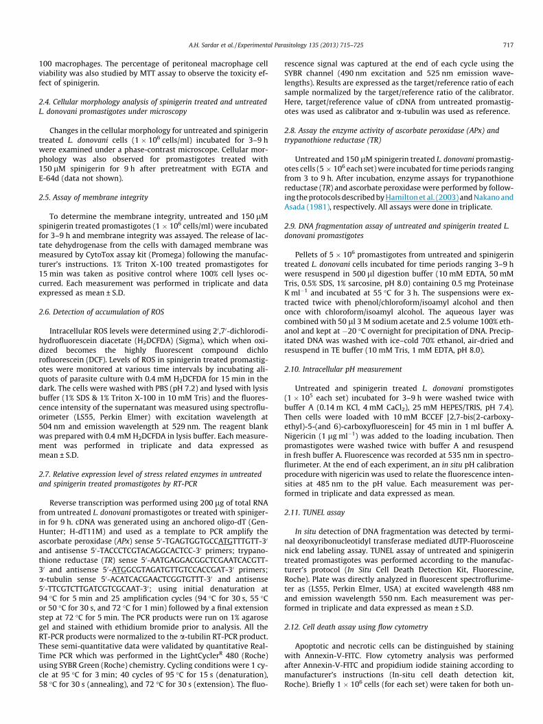
2.7. Relative expression level of stress related enzymes in untreatedand spinigerin treated promastigotes by RT-PCR
Reverse transcription was performed using 200 lg of total RNAfrom untreated L. donovani promastigotes or treated with spiniger-in for 9 h. cDNA was generated using an anchored oligo-dT (Gen-Hunter; H-dT11M) and used as a template to PCR amplify theascorbate peroxidase (APx) sense 50-TGAGTGGTGCCATGTTTGTT-30
and antisense 50-TACCCTCGTACAGGCACTCC-30 primers; trypano-thione reductase (TR) sense 50-AATGAGGACGGCTCGAATCACGTT-30 and antisense 50-ATGGCGTAGATGTTGTCCACCGAT-30 primers;a-tubulin sense 50-ACATCACGAACTCGGTGTTT-30 and antisense50-TTCGTCTTGATCGTCGCAAT-30; using initial denaturation at94 �C for 5 min and 25 amplification cycles (94 �C for 30 s, 55 �Cor 50 �C for 30 s, and 72 �C for 1 min) followed by a final extensionstep at 72 �C for 5 min. The PCR products were run on 1% agarosegel and stained with ethidium bromide prior to analysis. All theRT-PCR products were normalized to the a-tubilin RT-PCR product.These semi-quantitative data were validated by quantitative Real-Time PCR which was performed in the LightCyclerR 480 (Roche)using SYBR Green (Roche) chemistry. Cycling conditions were 1 cy-cle at 95 �C for 3 min; 40 cycles of 95 �C for 15 s (denaturation),58 �C for 30 s (annealing), and 72 �C for 30 s (extension). The fluo-
rescence signal was captured at the end of each cycle using theSYBR channel (490 nm excitation and 525 nm emission wave-lengths). Results are expressed as the target/reference ratio of eachsample normalized by the target/reference ratio of the calibrator.Here, target/reference value of cDNA from untreated promastig-otes was used as calibrator and a-tubulin was used as reference.
2.8. Assay the enzyme activity of ascorbate peroxidase (APx) andtrypanothione reductase (TR)
Untreated and 150 lM spinigerin treated L. donovani promastig-otes cells (5 � 106 each set) were incubated for time periods rangingfrom 3 to 9 h. After incubation, enzyme assays for trypanothionereductase (TR) and ascorbate peroxidase were performed by follow-ing the protocols described by Hamilton et al. (2003) and Nakano andAsada (1981), respectively. All assays were done in triplicate.
2.9. DNA fragmentation assay of untreated and spinigerin treated L.donovani promastigotes
Pellets of 5 � 106 promastigotes from untreated and spinigerintreated L. donovani cells incubated for time periods ranging 3–9 hwere resuspend in 500 ll digestion buffer (10 mM EDTA, 50 mMTris, 0.5% SDS, 1% sarcosine, pH 8.0) containing 0.5 mg ProteinaseK ml�1 and incubated at 55 �C for 3 h. The suspensions were ex-tracted twice with phenol/chloroform/isoamyl alcohol and thenonce with chloroform/isoamyl alcohol. The aqueous layer wascombined with 50 ll 3 M sodium acetate and 2.5 volume 100% eth-anol and kept at �20 �C overnight for precipitation of DNA. Precip-itated DNA was washed with ice–cold 70% ethanol, air-dried andresuspend in TE buffer (10 mM Tris, 1 mM EDTA, pH 8.0).
2.10. Intracellular pH measurement
Untreated and spinigerin treated L. donovani promstigotes(1 � 105 each set) incubated for 3–9 h were washed twice withbuffer A (0.14 m KCl, 4 mM CaCl2), 25 mM HEPES/TRIS, pH 7.4).Then cells were loaded with 10 mM BCCEF [2,7-bis(2-carboxy-ethyl)-5-(and 6)-carboxyfluorescein] for 45 min in 1 ml buffer A.Nigericin (1 lg ml�1) was added to the loading incubation. Thenpromastigotes were washed twice with buffer A and resuspendin fresh buffer A. Fluorescence was recorded at 535 nm in spectro-flurimeter. At the end of each experiment, an in situ pH calibrationprocedure with nigericin was used to relate the fluorescence inten-sities at 485 nm to the pH value. Each measurement was per-formed in triplicate and data expressed as mean.
2.11. TUNEL assay
In situ detection of DNA fragmentation was detected by termi-nal deoxyribonucleotidyl transferase mediated dUTP-Fluorosceinenick end labeling assay. TUNEL assay of untreated and spinigerintreated promastigotes was performed according to the manufac-turer’s protocol (In Situ Cell Death Detection Kit, Fluorescine,Roche). Plate was directly analyzed in fluorescent spectroflurime-ter as (LS55, Perkin Elmer, USA) at excited wavelength 488 nmand emission wavelength 550 nm. Each measurement was per-formed in triplicate and data expressed as mean ± S.D.
2.12. Cell death assay using flow cytometry
Apoptotic and necrotic cells can be distinguished by stainingwith Annexin-V-FITC. Flow cytometry analysis was performedafter Annexin-V-FITC and propidium iodide staining according tomanufacturer’s instructions (In-situ cell death detection kit,Roche). Briefly 1 � 106 cells (for each set) were taken for both un-

718 A.H. Sardar et al. / Experimental Parasitology 135 (2013) 715–725
treated and spinigerin treated promastigote culture. Cells werecentrifuged and washed twice with PBS and resuspend in 100 llof Annexin-V-FLUOS labeling solution. Required volume of Annex-in V and propedium iodide were added to solution and incubatedfor 15 min. Then analyzed immediately by flow cytometry at exci-tation wavelength 488 (PI) & 488–550 (Annexin V) and emissionwavelength 518 (PI) & 617 (Annexin V).
2.13. Caspase assays
The fluorogenic homogeneous caspase assay determines thepresence of any of the active caspases including 2, 3, 6, 7, 8, 9and 10. To determine the caspase activity of L. donovani promastig-otes following exposure to 150 lM spinigerin for 3, 6 and 9 h, afluorogenic homogeneous caspase assay was performed using theHomogeneous Caspase Assay kit (Roche) according to manufac-turer’s instructions. Briefly, after treatment with spinigerin, thecaspase substrate, prediluted in incubation buffer, was added andincubated for 2 h at 37 �C. The fluorometric measurement was re-corded with a spectrofluorimeter (LS 55, Perkin Elmer, USA) withexcitation at 504 nm and emission at 529 nm. The amount ofdeveloped flurochrome is proportional to the concentration of acti-vated caspases. For a positive control, the human macrophage cellline THP-1, treated with actinomycin-D for 24 h and L. donovanipromastigotes, treated with miltefosine for 9 h were analyzed sim-ilarly. Caspase-12 activity was measured fluorometrically usingcaspase-12 assay kit (Biovision). The assay is based on detectionof cleavage of substrate ATAD-AFC (7-amino-4-trifluoromethylcoumarin) to free AFC, which was measured by a micro plate read-er (emission, 400 nm; excitation, 505 nm). Caspase-12 inhibitor, Z-ATAD-FMK was added in control reactions before spinigerin addi-tion and incubated for 30 min. EGTA was used as extracellularCa2+ chelator. Each measurement was performed in triplicate anddata expressed as mean ± S.D.
2.14. Ca2+ analysis
Intracellular Ca2+ concentration was measured by using thefluorescent probe Fura-2-AM. Untreated and 150 lM spinigerintreated promastigotes for 9 h were harvested and washed twicewith wash buffer (0.116 M NaCl, 5.4 mm KCl, 0.08 mM MgSO4,5.5 mM D-glucose and 0.05 M HEPES, pH 7.0). Cells were resuspendin the loading buffer containing previously used wash buffer with1.5% sucrose and 6 M of Fura-2-AM for 1 h with mild shaking at37 �C. The fluorescence intensity was recorded using spetroflurom-eter (excitation at 340 nm; emission at 510 nm). Each measure-ment was performed in triplicate and data expressed as mean.
2.15. DWm determination
Mitochondrial membrane potential (DWm) was measuredusing the JC-1 probe as described previously (Banki et al., 1999;Bermudez et al., 1997) with slight modifications. Accumulation ofJC-1 (a cationic mitochondrial vital dye) in mitochondria is directlyproportional with membrane potential. Therefore, the fluorescenceof JC-1 can be used as an indicator of the relative mitochondrial en-ergy state. The dye exists as a monomer at lower concentrations(emission, 530 nm, and green fluorescence) and forms J aggregatesat higher concentrations (emission, 590 nm, and red fluorescence).Briefly, spinigerin treated promastigotes were collected and incu-bated for 7 min with 10 lM JC-1 at 37 �C and were subsequentlywashed and resuspend in medium. The suspensions were mea-sured for fluorescence at two different wavelengths as mentionedabove. The ratio of the reading at 590 nm and that at 530 nmwas considered the relative DWm value. Antimycin-A (0.05 lM,1 h), an inhibitor of mitochondrial respiratory complex III was used
as positive control to induce apoptosis. Cyclosporin-A was used asa blocker of permeability transition pore (PTP). Each measurementwas performed in triplicate and data expressed as mean ± S.D.
2.16. Measurement of total fluorescent lipid peroxidation product
The L. donovani promastigotes (107) treated and untreated withspinigerin at the IC50 dose for 3, 6, and 9 h were pelleted down andwashed twice with PBS. The pellet was suspended in 2 ml of 15%sodium dodecyl sulfate (SDS) in PBS solution. The fluorescenceintensities of the total fluorescent lipid peroxidation products ob-tained from the SDS–promastigote interaction were estimated(Shimasaki, 1994) with excitation at 360 nm and emission at430 nm. A parallel set of treated groups was incubated with butyl-ated hydroxytoluene (BHT; 20 mM), a specific inhibitor of lipidperoxidation. Each measurement was performed in triplicate anddata expressed as mean ± S.D.
2.17. THP-1 cell viability assay
THP-1 cells were seeded into 10 ml volumes at 1 � 105 cells/ml.Cells were incubated in presence of 150 lM and 200 lM spinigerinfor 24 h. The THP-1 cell viability was assessed by MTT assay as de-scribed above. The percent macrophage cell viability was deter-mined by comparing with untreated THP-1 cultures. Eachmeasurement was performed in triplicate and data are expressedas the mean ± S.D.
3. Results
3.1. Leishmanicidal activity of spinigerin
The percentage of metabolically active cells upon treatment withspinigerin was studied by MTT assay to determine the IC50 value ofspinigerin. We observed the reduction of metabolically active cellsupon increasing the concentrations of spinigerin and incubationtime. About 55–60% cell viability was observed when the Leishmaniapromastigotes were incubated 150 lM of spinigerin for 9 h, whereas85% and 45% cell viability was observed at 100 lM and 200 lM ofspinigerin at 9 h, respectively (Fig. 1B). This result suggests thatdue to spinigerin exposure the promastigote cell viability was re-duced in a concentration dependent manner. After treatment of bothperitoneal macrophages and THP-1 cell line with increasing concen-trations of spinigerin for 9 h, <5% and <2% cell death was observedrespectively at 150 lM spinigerin whereas about 22% and 18% celldeath was observed respectively at 200 lM spinigerin (Fig. 1A andC). As there was about 50–60% cell death in promastigotes culturebut negligible cytotoxic effect on murine peritoneal macrophage aswell as human macrophage cell line, THP-1 culture, the IC50 valueof spinigerin was determined as 150 lM at 9 h.
Simultaneously the leishmanicidal activity of miltefosine wasalso performed by incubating promastigotes with increasing con-centrations of it. About 50% cell viability was observed at 5 lMat 9 h and at higher concentrations (100 lM) cell viability was de-creased to <2% at 9 h (Fig. 1D) which suggests IC50 value of miltefo-sine is about 5 lM and higher concentrations around 100 lM isvery toxic for promastigotes.
3.2. Spinigerin induces morphological changes in L. donovanipromastigotes without loss of membrane integrity
Phase contrast microscopy of untreated and spinigerin treatedpromastigotes was performed to observe the morphologicalchanges, characteristics of apoptosis like death. We observed that

Fig. 1. Cell viability assay to determine the IC50 value of spinigerin and miltefosine and membrane integrity assay: (A) MTT assay for evaluation of % cell viability of murineperitoneal macrophage (PEC) after treatment with 150 lM and 200 lM of spinigerin for 24 h. (B) MTT assay for evaluation of % cell viability after treatment with increasingconcentrations (5–200 lM) of spinigerin for 6–48 h. 50–55% cell viability was observed at 150 lM, whereas 85% and 45% cell viability was observed at 100 lM and 200 lM,respectively. (C) MTT assay for evaluation of % cell viability of human macrophage THP-1 cell line after treatment with 150 lM and 200 lM of spinigerin for 24 h. (D) MTTassay for evaluation of % cell viability after treatment with increasing concentrations (5–100 lM) of miltefosine for 6–48 h. 50–60% cell viability was observed at 5 lM,whereas 12% and 2% cell viability observed at 50 lM and 100 lM. (E) Measurement of released lactate dehydrogenase to determine the membrane integrity of L. donovanipromastigotes after treatment with 150 lM spinigerin for 3, 6 and 9 h. All the experiments were performed in triplicate and data are expressed as the mean. Vertical lines ontop of the bars represent s.e. values.
A.H. Sardar et al. / Experimental Parasitology 135 (2013) 715–725 719
after treatment with spinigerin for 9 h at its IC50 value, the cells be-come rounded and the length of the flagella also shortened (Fig. 2A).
To determine the membrane integrity of untreated and spininger-in treated promastigotes throughout the whole course of incubationwith spinigerin, the release of lactate dehydrogenase from the cellswith damaged membrane was measured by CytoTox assay kit (Pro-mega). In positive control cells lysed with 1% Triton X-100, where al-most 100% cell lysis was expected. As the membrane is rupturedtotally in positive control, the enzyme activity was very high. Butwe observed very low enzyme activity in both untreated and treatedpromastigotes and there were no significant difference in membraneintegrity between untreated and treated promastigotes (Fig. 1E).
3.3. L. donovani promastigotes show PS externalization upontreatment with spinigerin
Apoptotic like cell death was further confirmed by PS external-ization study in FACS. Flow cytometry analysis revealed that the per-centage of apoptotic cells following spinigerin treatment increasedin a time dependent manner. The percentage of Annexin-V positivecells was observed about 80% at 3 h when promastigotes treatedwith spinigerin at its IC50 value. After 6 h and 9 h induction withspinigerin the percentage of Annexin-V positive increased to 97%and 98.4%. In the Annexin-V positive populations there were twodistinct cell populations were found. The left square contained lowAnnexin-V cells and the right square contained high Annexin-V po-sitive cells. This Annexin-V low and high population was dividedaccording to their mean fluroscent intensity (MFI) value. The propi-dium iodide (PI) positive cells in untreated, spinigerin treated cellswere found to be less than <2% of total population (Fig. 2B).
3.4. Ascorbate peroxidase (APx) and trypanothione reductase (TR) aredown-regulated in response to ROS generated during spinigerinexposure
Intracellular accumulation of ROS in both spinigerin treated anduntreated promastigotes at regular intervals of 3 h up to 9 h weremeasured using H2DCFDA. We observed increased ROS level withtime of exposure, with 2-, 2.96- and 5.24-fold at 3, 6 and 9 h,respectively compared to untreated control (Fig. 3A). But in pres-ence of N-acetyl cysteine (NAC), a known quencher of ROS, the in-creased level of ROS was decreased to the level of untreatedcontrol. The basal level of ROS indicates, a very low amount of freeradicals were present in untreated sample which is negligible.
The relative expression level of two stress-regulatory enzymes(known to be up-regulated in response to ROS) was studied byboth semi-quantitative and real-time PCR. The mRNA level of try-panothione reductase (TR) and ascorbate peroxidase (APx) was de-creased with increase in time of spinigerin exposure (Fig. 3B). Inreal-time PCR study the mRNA level of APx and TR were found tobe down regulated by 3.4- and 2.5-fold, respectively at 9 h com-pared to untreated control (Fig. 3C). Our enzymatic activity assayshowed that with increasing exposure time the activity was re-duced which suggested that the activity of these two enzymes(APx and TR) were reduced due to spinigerin exposure (Table 1).
3.5. Caspase independent apoptosis like cell death upon treatmentwith spinigerin
Homogeneous caspase assay was performed to study theinvolvement of any of the active caspase molecule like 2, 3, 6, 7,

Fig. 2. Analysis of morphological changes and Annexin-V assay of L. donovani promastigotes after spinigerin exposure: (A) Phase contrast microphotograph of L. donovanipromastigotes; (a) Untreated promastigotes, (b) promastigotes treated with 150 lM spinigerin for 3 h, (c) promastigotes treated with 150 lM spinigerin for 6 h, (d)promastigotes treated with 150 lM spinigerin for 9 h. (B) Flow cytomerty analysis of Annexin-V assay. (a and b) Untreated promastigotes, (c and d) promastigotes treatedwith 150 lM spinigerin for 3 h, 80% cells were observed low Annexin-V positive and about 3% were high Annexin-V positive, (e and f) promastigotes treated with 150 lMspinigerin for 6 h, 97% cells were observed Annexin-V positive, out of which 21% were high Annexin-V positive, (g and h) promastigotes treated with 150 lM spinigerin for9 h, 98.4% cells were observed Annexin-V positive, out of which about 44% were high Annexin-V positive.
Fig. 3. Measurement of ROS and involvement of ROS detoxifying enzyme in apoptosis like cell death: (A) Measurement of intracellular accumulation of ROS after treatmentwith 150 lM spinigerin for 3, 6 and 9 h. N-acetyl cysteine (NAC) was used as quencher of ROS. (B) Semi-quantitative RT-PCR image of trypanothione reductase (TR), ascorbateperoxidase (APx) and a-tubulin. (C) Real-Time PCR analysis for TR, APx and a-tubulin. The APx and TR mRNA level decreased about 3.4-fold and 2.5-fold at 9 h, respectively. Allthe experiments were performed in triplicate and data are expressed as the mean. Vertical lines on top of the bars represent s.e. values and ⁄ denotes significant changes withP < 0.005.
720 A.H. Sardar et al. / Experimental Parasitology 135 (2013) 715–725
8, 9 and 10 in this apoptotic like death. We did not found any in-crease in caspase activity except caspase-12 activity upon treat-ment with 150 lM spinigerin for 3, 6 and 9 h compared to
untreated control. The human macrophage cell line, THP-1 treatedwith actinomycin D and L. donovani promastigotes treated withmiltefosine (5 lM) (Paris and Loiseau, 2004) were considered as
A.H. Sardar et al. / Experimental Parasitology 135 (2013) 715–725 721
positive control, where very high caspase activity was observed(Fig. 4A). Interestingly we have found about 5-fold increase in cas-pase-12 activity compared to untreated control (Fig. 5B). But whenpromastogotes were pre-incubated with caspase-12-specific inhib-itor, extracellular Ca2+ chelator, the increased caspase-12 activitywas found to be reduced.
3.6. Spinigerin exposure causes increased [Ca2+]i accumulation andlipid peroxidation in L. donovani promastigotes
Free radical production is associated with changes in Ca2+
homeostasis (Lipton and Nicotera, 1998). Our spectroflurometryresults showed the intracellular level of Ca2+ increased about1.5-fold in spinigerin treated promastigotes compared to untreatedcontrol promastigotes. This increased level of [Ca2+]i was decreasedto normal level in presence of extracellular Ca2+ chelator EGTA(Fig. 4B).
In other side we observed with increasing time the lipid perox-idation was increased and reached to 10-fold higher values at 9 hcompared to untreated control, when promastigotes were treatedwith spinigerin at its IC50 value (Fig. 4C). In presence of butylatedhydroxytoluene (BHT), a specific inhibitor of lipid peroxidation,the increased level of lipid peroxidation decreased to normal asin untreated control.
3.7. Spinigerin exposure causes disruption of mitochondrial membranepotential (DWm) and acidification of intracellular environment
Changes in mitochondrial membrane potential (DWm) wereinvestigated. Interestingly after treatment with 150 lM spinigerin,we found inverse relation of time with mitochondrial membranepotential that decreased gradually and reached 5-fold lower com-pared to untreated control at 9 h (Fig. 4D). In presence of antimy-cin-A, inhibitor of mitochondrial respiratory complex III, themitochondrial membrane potential (DWm) was reduced drasti-cally but when pre-incubated with cyclosporine-A, a blocker ofpermeability transition pore (PTP) the membrane potential was re-stored (Fig. 4D). This result clearly demonstrated that interferencewith respiratory chain function in L. donovani leads to alterationsof mitochondrial membrane potential (DWm) in promastigoteswhich validates previous reports (Mehta and Shaha, 2004).
Previous investigations showed that the intracellular pH level isdecreased due to combined effect of free radical, increased level ofintracellular Ca2+ ion and decreased level of mitochondrial mem-brane potential (Demaurex et al., 2003). We observed that withexposure to spinigerin, the intracellular pH was changed to 5.97at 9 h with increasing exposure time, whereas in untreated controlthe intracellular pH value found to be almost constant i.e. pH-7.6(Fig. 4E). Therefore, our result clearly demonstrated that the intra-cellular environment of promastigotes was changed to acidic dueto spinigerin exposure.
3.8. Oligosomal DNA degradation of L. donovani promastigotes uponexposure to spinigerin
Oligosomal DNA degradation in spinigerin treated promastigoteat 9 h in agarose gel was observed. But in case of 3 h and 6 h
Table 1Activities of trypanothione reductase (TR) and ascorbate peroxidase (APx) fo
Spinigerin exposure (150 lM) Ascorbate peroxidase (APx) (u
Untreated 6.2 ± 0.33 h 5.7 ± 0.49 h 4.2 ± 0.712 h 2.1 ± 0.8
incubation of promastigotes with spinigerin very less DNA degra-dation was observed. In presence of EGTA and E-64d the DNA frag-mentation was almost abolished (Fig. 5A).
Further confirmation of the DNA degradation, TUNEL assay wascarried out (Gavrieli et al., 1992). Quantitative spectrofluorimetrydata reveals 63% TUNEL positive promastigotes when incubatedwith spinigerin for 9 h. In case of untreated control very less about<0.1% TUNEL positive cells were found. In presence of EGTA andE-64d the DNA very less about <0.1% TUNEL positive cells wereobserved like untreated control (Fig. 5C).
3.9. Cell viability of L. donovani amastigotes were reduced due tospiningerin exposure
As the amastigote stage is the disease-causing stage of the par-asite (Croft et al., 2006), it is essential that the peptides are also ac-tive for the killing of the amastigote stage. To assess theantiamastigote activity of the spinigerin, peritoneal-derived mousemacrophages were infected with the L. donovani AG83 strain andsubsequently treated with the increasing concentrations of spinig-erin; 100, 150 and 200 lM for 24 h. L. donovani amastigote burdensin the infected macrophages were inhibited by 13%, 20% and 40%,respectively. These data demonstrated that the amastigote cell via-bility was reduced due to spinigerin exposure in a concentrationdependent manner (Fig. 6A and B).
4. Discussion
Antimicrobial peptides (AMPs) are structurally diverse highlycationic proteins between 10 and 50 amino acids and are compo-nents of the innate immune systems of organisms within all king-doms. AMPs have several functions and are known to interact withand disrupt microbial surface membranes leading to cell death(Hancock and Diamond, 2000; Izadpanah and Gallo, 2005). Theamphipathicity of AMPs enable them to interact with negatively-charged microbial membranes. Direct destabilization of surface-membranes by AMPs occurs through a variety of mechanismsand can initiate microbial death by triggering induction of auto-phagic, necrotic or apoptotic-cell death (Brogden, 2005). AMPscan interact in different ways and can kill the microorganism, theycan directly attach with the cell membrane of the microorganismand form pores or interact with intracellular components likeDNA, nucleus, mitochondria and malfunction or inhibit differentcellular process like transcription, translation and protein synthe-sis (Kim, 2005; Marr et al., 2012).
Despite being structurally diverse, AMPs contain relatively highpercentages of basic amino acids and form either predominately a-helical structures or b-pleated sheets. Certain subsets of AMPs(defensins and some cathelicidins) are cysteine-rich that allowextensive intra-disulfide bonding which is important for activity(Ganz and Lehrer, 1994). Spinigerin is a natural peptide secretedfrom the insect gut epithelium cells. Previous studies establishedits antimicrobial property mainly against bacteria and virus (Lam-berty et al., 2001; Wang et al., 2010). But the antileishmanial prop-erty of this antimicrobial peptide is not studied well.
In this study we have investigated the antileishmanial activityof spinigerin and mode of cell death due to spinigerin exposure.
r treated and spinigerin treated L. donovani promastigotes.
nits mg�1) Trypanothione reductase (TR) (units mg�1)
7.6 ± 0.35.9 ± 0.33.8 ± 0.52.8 ± 0.1

Fig. 4. Biochemical assay for apoptosis like death of Leishmania donovani promastigotes after spinigerin exposure: (A) Measurement of caspase activity after treatment with150 lM spinigerin for 3, 6 and 9 h. (B) Measurement of intracellular Ca2+ after treatment with 150 lM spinigerin for 3, 6 and 9 h. (C) Measurement of lipid peroxidation aftertreatment with 150 lM spinigerin for 3, 6 and 9 h. Butylated hydroxytoluene (BHT; 20 mM), a specific inhibitor of lipid peroxidation. (D) Measurement of change inmitochondrial membrane potential (DWm) after treatment with 150 lM spinigerin for 3, 6 and 9 h. Antimycin-A, an inhibitor of mitochondrial respiratory complex III wasused as positive control to induce apoptosis. Cyclosporin-A was used as a blocker of permeability transition pore (PTP). (E) Measurement of change in intracellular pH aftertreatment with 150 lM spinigerin for 3, 6 and 9 h. All the experiments were performed in triplicate and data are expressed as the mean. Vertical lines on top of the barsrepresent s.e. values and * denotes significant changes with P < 0.005.
Fig. 5. Spinigerin induces DNA fragmentation in a Ca2+ and calpain-protease dependent manner: (A) Agarose gel image of oligoneucleosomal DNA degradation of L. donovanipromastigotes after treatment with 150 lM spinigerin for 3, 6 and 9 h. (B) Measurement of caspase-12 activity in presence and absence of different inhibitors after treatmentof L. donovani promastigotes with 150 lM spinigerin for 9 h. Caspase-12 specific inhibitor (C-12 inhibitor) was used to test the specificity of the caspase-12 activity. EGTA wasused as extracellular Ca2+ chelator. (C) Measurement of TUNEL positive cells Of L. donovani promastigotes after treatment with 150 lM spinigerin for 3, 6 and 9 h. EGTA andE64d were used as extracellular Ca2+ chelator and E-64d, a cell permeable variant of E-64 can efficiently block calpain-like cysteine protease mediated apoptosis-like death.Each measurement was performed in triplicate and data are expressed as the mean. Vertical lines on top of the bars represent s.e. values and * denotes significant changeswith P < 0.005.
722 A.H. Sardar et al. / Experimental Parasitology 135 (2013) 715–725

Fig. 6. Cell viability of L. donovani amastigotes after treatment with spinigerin: (A) Phase contrast microphotograph after Geimsa staining of peritoneal-derived mousemacrophages infected with L. donovani amastigotes. Peritoneal-derived mouse macrophages were infected with L. donovani promastigotes and subsequently treated withincreasing concentrations of spinigerin for 24 h. The arrow indicted the representative L. donovani amastigotes. (a) Treated with 100 lM spinigerin, (b) treated with 150 lMspinigerin, (c) treated with 200 lM spinigerin, (d) untreated. (B) Column graph showing parasite burden in infected murine macrophage after treatment with spinigerin.L. donovani amastigote burdens in the infected macrophage were reduced about 13%, 20% and 40% at 100 lM and 150 lM and 200 lM, respectively. Each measurement wasperformed in triplicate and data are expressed as the mean. Vertical lines on top of the bars represent s.e. values and * denotes significant changes with P < 0.005.
A.H. Sardar et al. / Experimental Parasitology 135 (2013) 715–725 723
The IC50 of spinigerin was determined as 150 lM at 9 h which ishigher when compared to miltefosine (5 lM) (Fig. 1B and D). Pre-vious reports showed the IC50 value of miltefosine is 3.3–4.6 lMwhich is very close to our observation (Dorlo et al., 2012). Interest-ingly about <5% and <2% metabolically inactive cells were ob-served, after incubation of murine peritoneal macrophage andhuman macrophage cell line THP-1 with spinigerin at its IC50 valuefor 24 h, which suggests that at its IC50 value spinigerin has notoxic effect on human macrophages at least for 24 h (Fig. 1A andC). Our observation is validated by earlier report which showedthat exposure to spinigerin has no lytic activity against mamma-lian erythrocytes (Landon et al., 2006). 200 lM of spinigerinshowed about 18% reduction at 24 h in THP-1 cell viability whichsuggest that 200 lM has some cytotoxic effect on human macro-phage cell (Fig. 1C).
The morphological changes like rounding of cell, shortening ofthe flagella without loss of membrane integrity are well known/conceive phenomenon of apoptosis-like cell death in case ofLeishmana sp. (Shaha, 2006). Analysis with phase-contrast micro-scope, upon spinigerin exposure at its IC50 value at 9 h observedsimilar changes suggest the cell death of L. donovani promastigotesmay be apoptosis like death (Fig. 2A and Fig. 1E).
Externalization of phosphotidyl serine (PS) in the outer leaflet ofthe membrane bi-layer is an established feature of apoptosis-likedeath. Previous reports suggested that in early stages of eukaryoticapoptosis, cells externalize PS while maintaining membrane integ-rity (Shaha, 2006). In similar experiment we observed about 80%cells were Annexin V positive at 3 h when exposed to150 lMspinigerin which reached 98% at 9 h. Further analysis of data,two distinct populations observed one low Annexin V (55%) andanother high Annexin V (Fig. 2B). This suggests that apoptosis
divided into two stages: the commitment stage and the executionstage as suggested in previous investigation (Dumont et al., 2000).Cells can be rescued by repair mechanisms if they are not yet com-mitted to apoptosis, but once they are committed to apoptosis theycould not be rescued and will have to die. So, the present data sug-gests that during early phase of spinigerin exposure though 80%cells shows apoptosis signal 50% cells go to the late apoptotic stageor commitment stage for apoptosis-like death at 9 h.
ROS and RNS are usually generated when any foreign moleculeenters into cell as a part of innate immune defense mechanism aspresent study too (Fonseca-Silva et al., 2011). Addition of spinig-erin into L. donovani promastigote culture, ROS level was elevatedin time dependent manner suggesting with increasing time ROSlevel was increased. Further in presence of NAC (ROS quencher)the increased level of ROS decreased to the level of untreated con-trol (Fig. 3A). This result suggests that cell death due to spinigerinexposure is ROS mediated. To validate the ROS mediated celldeath the involvement of two major enzymes (known to be playrole in ROS scavenging in case of L. donovani), trypanothionereductase (TR) and ascorbate peroxidase (APx) were studied(Tovar et al., 1998; Pal et al., 2010; Sardar et al., 2013). The mRNAlevel of TR and APx was found to decrease about 3.8 and 2.9-fold,respectively through in real time PCR analysis (Fig. 3B and C).Similarly we observed the reduced activity of these two enzymestrypanothione reductase (TR) and ascorbate peroxidase (APx)after exposure to 150 lM spinigerin (Table 1).These rela-tive expression data and enzymatic activity assay data suggestthat upon prolonged exposure to the endogenous ROS generateddue to spinigerin exposure is not efficiently detoxified and be-comes extremely toxic for the parasite, resulting in decreasedviability.

724 A.H. Sardar et al. / Experimental Parasitology 135 (2013) 715–725
Increase in [Ca2+]I due to elevation of ROS level is another com-mon features of apoptosis-like death (Gatti et al., 1998). Mamma-lian caspase-12 undergoes activation in a calcium-dependentmanner (Nakagawa et al., 2000). We also observed increased[Ca2+]I level as well as caspase-12 activity after incubation withspinigerin (Fig. 4B). In presence of extracellular Ca2+ chelator,EGTA, the increased level of Ca2+ and casepase-12 activity de-creased drastically, which suggests the elevation of [Ca2+]I wasmainly contributed by Ca2+ influx machinery from external sourceand internal source contributed very minimum.
Nuclear condensation and segmentation are generally attrib-uted to caspase activity which leads to cleavage of substrates suchas DNase, lamines (Gatti et al., 1998). Surprisingly, our homoge-neous caspase assay showed no caspase activity except caspase-12 due to spinigerin exposure (Fig. 4A). E-64d, a cell permeablevariant of E-64 can efficiently block calpain-like cysteine proteasemediated apoptosis-like death. The abolition of DNA ladderingwith the pretreatment with E-64d suggests the involvement of cal-pain like cysteine protease in this apoptosis like death (Fig. 5A andB).
We also observed that due to spinigerin treatment the mito-chondrial membrane potential was collapsed at 9 h and simulta-neously the peroxidation of lipid was elevated which againconfirmed the apoptosis-like death (Fig. 4C and D) (Shimasaki,1994). Endoplasmic reticulum (ER) and glycosome are present inL. donovani. Therefore it may be suggested that after loss of mem-brane potential of ER and glycosome, protons are released into thecytosol, which leads to acidification (Demaurex et al., 2003). Wealso observed acidification by lowering of intracellular pH due tospinigerin exposure (Fig. 4E).
Oligonucleosomal DNA degradation is another feature of apop-tosis-like death (Hengartner, 2000). The gel image of DNA ladder-ing clearly showed oligosomal DNA degradation of L. donovani aftertreatment with spinigerin at 9 h. In presence of Ca2+ chelator, EGTAthe DNA laddering was completely abolished which further sup-ported that the involvement of Ca2+ influx machinery in this apop-tosis-like cell death (Fig 5B). Previous investigation showed theinvolvement cysteine protease and surface mettloprotease inapoptosis-like cell death in case of Leishmania sp. (Mottram et al.,2004 and Kulkarni et al., 2006). Our result showed that in presenceof calpain like cysteine protease inhibitor E-64d the DNA ladderingalso abolished (Fig. 5A). This data suggested that calpain like cys-teine protease was involved in this apoptosis-like cell death. TUN-EL assay shows the double stranded DNA breakage which is auniversal phenomenon of apoptosis (Hengartner, 2000). The spet-rofluorimetry results showed 63% TUNEL positive promastigoteswhen incubated with spinigerin for 9 h but in prsensce of EGTAand E-64d about <10% TUNEL positives cells were observed(Fig. 5A and C). This TUNEL assay result further confirmed theinvolvement of Ca2+ influx machinery and calpain like cysteineprotease in this apoptosis-like cell death.
It is always necessary to evaluate the antileishmanial activity ofany probable chemotherapeutic agents on amastigote stage be-cause it will be the ultimate target to control the disease in human.The treatment of 150 lM of spinigerin to amastigotes, 20% reduc-tion in amstigote cell viability was observed which suggests thatspinigerin is active both against promastigotes as well as amstig-otes (Fig. 6A and B).
Trypanothione reductase (TR) is an important enzyme involvedin detoxification of elevated ROS level in case of L. donovani (Tovaret al., 1998; Sardar et al., 2013). Both the semi-quantitative andreal-time PCR analysis showed that the mRNA expression of trypa-nothione reductase is reduced in response to ROS elevation afterspinigerin exposure (Fig. 3B–D). Insufficient amount of this en-zyme inside the cell may cause decrease in cell viability due to dis-ability of the cell to detoxify the elevated ROS. A computational
analysis was performed to observe is there any interaction be-tween spinigerin with trypanothione reductase. The analysis sug-gests spinigerin interacts with trypanothione reductase thusprobably interfere its functions which are responsible for the via-bility of parasites (data not shown). Similarly proper functioningof HIV-1 protease is very much important for the intracellular via-bility of HIV-1 virus (Wang et al., 2010). As previous study sug-gested that spinigerin has anti-HIV activity, computationalanalysis of HIV-1 protease with spinigerin was also performedand analysis shown spinigerin interacts with HIV-1 protease (datanot shown).
5. Conclusion
This investigation is the first report of antileishmanial activityof the cationic antimicrobial peptide spinigerin. Our results clearlyshowed that the spinigerin induces apoptosis like cell death in acaspase independent manner in L. donovani promastigotes. So,spinigerin may be considered for developing future chemothera-peutic agents for the treatment of VL. Additionally the antiviralactivity of spinigerin against HIV increases its possibility for devel-oping future attractive chemotherapeutic agents for treating criti-cal case of Leishmania-HIV co-infection. To establish spinigerin asalternative and safe chemotherapeutic agent (alone or in combina-tion) further detailed investigations is necessary.
Competing interests
The authors have declared that no competing interests exist.
Acknowledgments
This work has been funded jointly by the Indian Council of Med-ical Research (ICMR), Ministry of Health and Family Welfare, Indiaand National Institute of Pharmaceutical Education and Research(NIPER), Union ministry of Chemicals and Fertilizers, India.
References
Banki, K., Hutter, E., Gonchoroff, N.J., Perl, A., 1999. Elevation of mitochondrialtransmembrane potential and reactive oxygen intermediate levels are earlyevents and occur independently from activation of caspases in Fas signaling. J.Immunol. 162, 1466–1479.
Bermudez, R., Dagger, F., D’Aquino, J.A., Benaim, G., Dawidowicz, K., 1997.Characterization of mitochondrial electron-transfer in Leishmania mexicana.Mol. Biochem. Parasitol. 90, 43–54.
Brand, G.D., Leite, J.R., de Sa, Mandel, Mesquita, S.M., Silva, D.A., Prates, L.P., et al.,2006. Novel dermaseptins from Phyllomedusa hypochondrialis (Amphibia).Biochem. Biophys. Res. Commun. 347, 739–746.
Brogden, K.A., 2005. Antimicrobial peptides: pore formers or metabolic inhibitors inbacteria. Nat. Rev. Microbiol. 3, 238–250.
Croft, S.L., Seifert, K., Yardley, V., 2006. Current scenario of drug development forleishmaniasis. Indian J. Med. Res. 123, 399–410.
Demaurex, N., Frieden, M., Arnaudeau, S., 2003. ER calcium and ER chaperones: newplayers in apoptosis? In: Eggleton, P., Michalak, M. (Eds.), Calreticulam, seconded. Eurekah, Austin, TX, pp. 134–142.
Dorlo, T.P.C., Balasegaram, M., Beijnen, J.H., Vries, P.J.D., 2012. Miltefosine: a reviewof its pharmacology and therapeutic efficacy in the treatment of leishmaniasis.J. Antimicrob. Chemother. 67, 2576–2597.
Dumont, C.L., Durrbach, A., Bide‘ re, N., Rouleau, M., Kroemer, G., Bernard, G., Hirsch,F., Charpentier, B., Susin, S.A., Senik, A., 2000. Caspase-independentcommitment phase to apoptosis in activated blood T lymphocytes:reversibility at low apoptotic insult. Blood 96, 1030–1038.
Fonseca-Silva, F., Inacio, J.D., Canto-Cavalheiro, M.M., Almeida-Amaral, E.E., 2011.Reactive oxygen species production and mitochondrial dysfunction contributeto quercetin induced death in Leishmania amazonensis. PLoS One 8 (6(2)),e14666.
Ganz, T., Lehrer, R.I., 1994. Defensins. Curr. Opin. Immunol. 6, 584–589.Gatti, R., Belletti, S., Oriandini, G., Bussolati, O., Dall’Asta, V., Gazzola, G.C., 1998.
Comparison of annexin V and Calcein-AM as early vital markers of apoptosis inadherent cells by confocal laser microscopy. J. Histochem. Cytochem. 46, 895–900.

A.H. Sardar et al. / Experimental Parasitology 135 (2013) 715–725 725
Gavrieli, Y., Sherman, Y., Ben-Sasson, S.A., 1992. Identification of programmed celldeath in situ via specific labeling of nuclear DNA fragmentation. J. Cell Biol. 119(3), 493–501.
Hamilton, C.J., Saravanamuthu, A., Eggleston, I.M., Fairlamb, A.H., 2003. Ellman’s-reagent-mediated regeneration of trypanothione in situ: substrate-economicalmicroplate and time-dependent inhibition assays for trypanothione reductase.Biochem. J. 369, 529–537.
Hancock, R.E.W., 2001. Cationic peptides: effectors in innate immunity and novelantimicrobials. Lancet Infect. Dis. 1, 156–164.
Hancock, R.E., Diamond, G., 2000. The role of cationic antimicrobial peptides ininnate host defences. Trends Microbiol. 8, 402–410.
Hengartner, M.O., 2000. The biochemistry of apoptosis. Nature 407, 770–776.Hernandez, C., Mor, A., Dagger, F., Nicolas, P., Hernandez, A., Dunia, I., 1992.
Functional and structural damage in Leishmania mexicana exposed to thecationic peptide dermaseptin. Eur. J. Cell Biol. 59, 414–424.
Izadpanah, A., Gallo, R.L., 2005. Antimicrobial peptides. J. Am. Acad. Dermatol. 52,381–390.
Jha, T.K. et al., 1999. Miltefosine, an oral agent, for the treatment of Indian visceralleishmaniasis. N. Engl. J. Med. 341, 1795–1800.
Kim, A.B., 2005. Antimicrobial peptides: pore formers or metabolic inhibitors inbacteria. Nature 3, 25.
Kuckelhaus, S.A., Leite, J.R., Muniz, J., Sampaio, M.I., Bloch, R.N., Tosta, C.E., 2009.Antiplasmodial and antileishmanial activities of phylloseptin-1, anantimicrobial peptide from the skin secretion of Phyllomedusa azurea(Amphibia). Exp. Parasitol. 123, 11–16.
Kulkarni, M.M., McMaster, W.R., Kamysz, E., Kamysz, W., Engman, D.M., McGwire,B.S., 2006. The major surface-metalloprotease of the parasitic protozoan,Leishmania, protects against antimicrobial peptide-induced apoptotic killing.Mol. Microbiol. 62 (5), 1484–1497.
Lamberty, M., Zachary, D., Lanot, R., Bordereau, C., Robert, A., Hoffmann, J.A., Bulet,P., 2001. Insect immunity. Constitutive expression of a cysteine-rich antifungaland a linear antibacterial peptide in a termite insect. J. Biol. Chem. 276, 4085–4092.
Landon, C., Meudal, H., Boulanger, N., Bulet, P., Vovelle, F., 2006. Solution structuresof stomoxyn and spinigerin, two insect antimicrobial peptides with an a-helicalconformation. Biopolymers 81, 92–103.
Lipton, S.A., Nicotera, P., 1998. Calcium, free radicals and excitotoxins in neuralapoptosis. Cell Calcium 23, 165–171.
Lynn, M.A., Kindrachuk, J., Marr, A.K., Jenssen, H., Pante, N., 2011. Effect of BMAP-28antimicrobial peptides on Leishmania major promastigote and amastigotegrowth: role of Leishmanolysin in parasite survival. PLoS Negl. Trop. Dis. 5(5), e1141. http://dx.doi.org/10.1371/journal.pntd.0001141.
Marr, A.K., McGwire, B.S., McMaster, W.R., 2012. Modes of action of Leishmanicidalantimicrobial peptides. Future Microbiol. 7 (9), 1047–1059.
Mehta, A., Shaha, C., 2004. Pentamidine cytotoxicity inhibition results in increasedchain inhibition: complex II promastigotes in response to respiratory apoptoticdeath in Leishmania donovani. J. Biol. Chem. 279, 11798–11813.
Mor, A., Nicolas, P., 1994. Isolation and structure of novel defensive peptides fromfrog skin. Eur. J. Biochem. 219, 145–154.
Mottram, J.C., Coombs, G.H., Alexander, J., 2004. Cysteine peptidases as virulencefactors of Leishmania. Curr. Opin. Microbiol. 7, 375–381.
Nakagawa, T., Zhu, H., Morishima, N.L.E., Xu, J., Yankner, B.A., Yuan, J., 2000.Caspase-12 mediates endoplasmicreticulum-specific apoptosis and cytotoxicityby amyloid-b. Nature 403, 98–103.
Nakano, Y., Asada, K., 1981. Hydrogen peroxide is scavenged by ascorbate-specificperoxidase in spinach chloroplasts. Plant Cell Physiol. 22 (5), 867–880.
Nwaka, S., Hudson, A., 2006. Innovative lead discovery strategies for tropicaldiseases. Nat. Rev. Drug Discovery 5, 941–955.
Pal, S., Dolai, S., Yadav, R.K., Adak, S., 2010. Ascorbate Peroxidase from Leishmania majorcontrols the virulence of infective stage of promastigotes by regulating oxidativestress. PLoS One 5, e11271. http://dx.doi.org/10.1371/journal.pone.0011271.
Paris, C., Loiseau, P.M., Bories, C., Bréard, J., 2004. Miltefosine induces apoptosis-likedeath in Leishmania donovani promastigotes 10.1128/AAC. Antimicrob. AgentsChemother. 48 (3), 852–859.
Sardar, A.H., Kumar, S., Kumar, A., Purkait, B., Das, S., Sen, A., Kumar, M., Sinha, K.K.,Singh, D., Equbal, A., Ali, V., Das, P., 2013. Proteome changes associated withLeishmania donovani promastigote adaptation to oxidative and nitrosativestresses. J. Proteomics 81, 185–199.
Savioli, L. et al., 2006. Response from Savioli and colleagues from the Department ofNeglected Tropical Diseases, World Health Organization. PLoS Med. 3, e283.
Shaha, C., 2006. Apoptosis in Leishmania species & its relevance to diseasepathogenesis. Indian J. Med. Res. 123, 233–244.
Shimasaki, H., 1994. Assay of fluorescent lipid peroxidation products. MethodsEnzymol. 233, 338–346.
Sitaram, N., Nagaraj, R., 2002. Host-defense antimicrobial peptides: importance ofstructure for activity. Curr. Pharm. Des. 8, 727–742.
Tovar, J., Cunningham, M.L., Smith, A.C., Croft, S.L., Fairlamb, A.H., 1998. Down-regulation of Leishmania donovani trypanothione reductase by heterologousexpression of a trans-dominant mutant homologue: effect on parasiteintracellular survival. Proc. Natl. Acad. Sci. 95, 5311–5316.
Wang, G., Watson, K.M., Peterkofsky, A., Robert, W.B., 2010. Identification of novelhuman immunodeficiency virus type 1-inhibitory peptides based on theantimicrobial peptide database. Antimicrob. Agents Chemother. 66, 1343–1346.
Zasloff, M., 1987. Magainins, a class of antimicrobial peptides from Xenopus skin:isolation, characterization of two active forms, and partial cDNA sequence of aprecursor. Proc. Natl. Acad. Sci. USA 84, 5449–5453.
Zasloff, M., 2002. Antimicrobial peptides of multicellular organisms. Nature 415,389–395.