Sperm chromatin integrity in DDT-exposed young men living in a malaria area in the Limpopo Province,...
17
Sperm chromatin integrity in DDT-exposed young men living in a malaria area in the Limpopo Province, South Africa C. de Jager 1,5 , N.H. Aneck-Hahn 1,2 , M.S. Bornman 2 , P. Farias 3 , G. Leter 4 , P. Eleuteri 4 , M. Rescia 4 and M. Spanò 4 1 Environmental and Occupational Health, School of Health Systems & Public Health, University of Pretoria, PO Box 667, Pretoria 0001, South Africa 2 Andrology, Department of Urology, University of Pretoria, PO Box 667, Pretoria 0001, South Africa 3 Instituto Nacional de Salud Publica, Avenida Universidad 655, Col. Sta. Maria Ahuacatitlan, Cuernavaca, Morelos 62508, Mexico 4 Section of Toxicology and Biomedical Sciences, BAS-BIOTEC-MED, ENEA Casaccia Research Center, 00123 Rome, Italy 5 Correspondence address. Tel: +27-12-354-2072; Fax: +27-12-354-2071; E-mail: [email protected] Abstract BACKGROUND: There is mounting evidence that deteriorated semen quality may be associated with increased serum concentration of 1,1,1-trichloro-2,2-bis(chlorodiphenyl)ethane (DDT) and its metabolites. The problem is exacerbated in situations where DDT is the only resource available to control malaria mosquitoes and DDT metabolite plasma concentration can reach 1000-fold the level found in other populations. There are limited and contradictory epidemiological data on whether DDT/dichlorodiphenyl-dichloroethylene (DDE) can also damage sperm DNA. Therefore, there is a need to investigate the possible adverse effects on human sperm genetic integrity in a sufficiently large study population with adequate exposure contrasts, especially in the high exposure range. METHODS: We conducted a cross-sectional study, recruiting 209 young males from three communities in an endemic malaria area where DDT is sprayed annually. Blood plasma p,p'-DDT and its metabolite p,p'-DDE levels were measured and expressed as lipid adjusted p,p'-DDT and p,p'-DDE values. The sperm chromatin structure assay and Aniline Blue test were used to assess sperm DNA/chromatin integrity. RESULTS: The lipid adjusted p,p'-DDT mean (±SD) and median concentrations were 109.2 (±106.6) and 83.9 μg/g, respectively; and the lipid adjusted p,p'-DDE mean (±SD) and median concentrations were 246.2 (±218.5) and 177.8 μg/g, respectively. The results point to a weak association between DDT/DDE plasma concentration and the incidence of sperm with chromatin defects. CONCLUSIONS: The results suggest that non-occupational environmental DDT exposure may have a negative impact on sperm chromatin integrity in young South African males. Key words: DNA damage/SCSA/Aniline Blue/DDT/spermatozoa The Stockholm Convention, a global multilateral agreement aiming at protecting human and environmental health from the effects of exposure to dangerous and ubiquitous chemical compounds, has initially targeted a number of chemicals known as persistent organic pollutants (POPs) POPs are noted for their toxicity, persistence and bioaccumulative characteristics. The chemicals to be eliminated with priority include several industrial chemical products and by-products including polychlorinated biphenyls, dioxins, furans and also DDT (1,1,1-trichloro-2,2- bis(chlorodiphenyl)ethane). However, exemptions are available for countries that are still using DDT to combat malaria, which causes millions of deaths every year, particularly in Africa. Although South Africa ratified the Stockholm Convention on 4 September 2002 (Bouwman, 2004), it is one of the countries that has applied for exemption as far as using DDT for malaria vector control. In Africa, indoor residual spraying of DDT has become part of the national Roll Back Malaria strategic plan in
Transcript of Sperm chromatin integrity in DDT-exposed young men living in a malaria area in the Limpopo Province,...

Sperm chromatin integrity in DDT-exposed young men living in a malaria area in the Limpopo Province, South Africa
C. de Jager1,5, N.H. Aneck-Hahn1,2, M.S. Bornman2, P. Farias3, G. Leter4, P. Eleuteri4, M. Rescia4 and M. Spanò4
1 Environmental and Occupational Health, School of Health Systems & Public Health, University of Pretoria, PO Box 667, Pretoria 0001, South Africa 2 Andrology, Department of Urology, University of Pretoria, PO Box 667, Pretoria 0001, South Africa 3 Instituto Nacional de Salud Publica, Avenida Universidad 655, Col. Sta. Maria Ahuacatitlan, Cuernavaca, Morelos 62508, Mexico 4 Section of Toxicology and Biomedical Sciences, BAS-BIOTEC-MED, ENEA Casaccia Research Center, 00123 Rome, Italy
5 Correspondence address. Tel: +27-12-354-2072; Fax: +27-12-354-2071; E-mail: [email protected]
Abstract
BACKGROUND: There is mounting evidence that deteriorated semen quality may be associated with increased serum concentration of 1,1,1-trichloro-2,2-bis(chlorodiphenyl)ethane (DDT) and its metabolites. The problem is exacerbated in situations where DDT is the only resource available to control malaria mosquitoes and DDT metabolite plasma concentration can reach 1000-fold the level found in other populations. There are limited and contradictory epidemiological data on whether DDT/dichlorodiphenyl-dichloroethylene (DDE) can also damage sperm DNA. Therefore, there is a need to investigate the possible adverse effects on human sperm genetic integrity in a sufficiently large study population with adequate exposure contrasts, especially in the high exposure range.
METHODS: We conducted a cross-sectional study, recruiting 209 young males from three communities in an endemic malaria area where DDT is sprayed annually. Blood plasma p,p'-DDT and its metabolite p,p'-DDE levels were measured and expressed as lipid adjusted p,p'-DDT and p,p'-DDE values. The sperm chromatin structure assay and Aniline Blue test were used to assess sperm DNA/chromatin integrity.
RESULTS: The lipid adjusted p,p'-DDT mean (±SD) and median concentrations were 109.2 (±106.6) and 83.9 µg/g, respectively; and the lipid adjusted p,p'-DDE mean (±SD) and median concentrations were 246.2 (±218.5) and 177.8 µg/g, respectively. The results point to a weak association between
DDT/DDE plasma concentration and the incidence of sperm with chromatin defects.
CONCLUSIONS: The results suggest that non-occupational environmental DDT exposure may have a negative impact on sperm chromatin integrity in young South African males.
Key words: DNA damage/SCSA/Aniline Blue/DDT/spermatozoa
The Stockholm Convention, a global multilateral agreement aiming at protecting human and environmental health from the effects of exposure to dangerous and ubiquitous chemical compounds,
has initially targeted a number of chemicals known as persistent organic pollutants (POPs) POPs are noted for their toxicity, persistence and bioaccumulative characteristics. The chemicals to be eliminated with priority include several industrial chemical products and by-products including polychlorinated biphenyls, dioxins, furans and also DDT (1,1,1-trichloro-2,2-bis(chlorodiphenyl)ethane). However, exemptions are available for countries that are still using DDT to combat malaria, which causes millions of deaths every year, particularly in Africa. Although South Africa ratified the Stockholm Convention on 4 September 2002 (Bouwman, 2004), it is one of the countries that has applied for exemption as far as using DDT for malaria vector control. In Africa, indoor residual spraying of DDT has become part of the national Roll Back Malaria strategic plan in

several countries (Hougard et al., 2002; Rogan and Chen, 2005). In South Africa, DDT is sprayed in the low-altitude parts of Limpopo Province, Mpumalanga Province and KwaZulu Natal. Currently of the approximately 40 million people in South Africa, 10% (4 million) live in a malaria-risk area (Rogan and Chen, 2005). This puts the inhabitants of the rural communities in these areas at risk of being exposed to high concentrations DDT and DDE (dichlorodiphenyl-dichloroethylene). The exposure occurs
through inhalation (indoor air spraying of dwellings and outdoors), dermal contact (soil and house dust) and ingestion of contaminated foods and water.
A number of reports have indicated that organochlorine pesticides, including DDT and its metabolites, are not only toxic but may act as endocrine disruptors (Turusov et al., 2002). Endocrine disrupting chemicals can be defined as compounds that influence normal hormone function generally causing adverse effects (Godduhn and Duffy, 2003). Technical grade DDT is a mixture of p,p'-DDT ( 85%), o,p'-DDT ( 15%) and o,o'-DDT (trace amounts) with both p,p'-DDT and o,p'-DDT having estrogenic activity. p,p'-Dichlorodiphenyl-dichloroethylene (p,p'-DDE), the main metabolite of p,p'-DDT, is a widespread, persistent environmental contaminant (Turusov et al., 2002). The p,p'-DDE isomer is anti-androgenic by inhibitive binding to androgen receptors and has been shown to inhibit the action of testosterone (Kelce et al., 1995; Danzo, 1997; Bhatia et al., 2005). The hypothesis has been advanced that p,p'-DDE may interact in an additive or multiplicative way with other endocrine
disruptive environmental pollutants (Turusov et al., 2002). Serum levels of p,p'-DDE are an integrated measure of internal dose, reflecting exposure from all sources over the previous years (Hauser et al., 2003a). Reproductive disorders were among the first adverse impacts linked to organochlorine exposure (Beard, 2005).
Reproductive abnormalities attributed to DDT/DDE exposure have been reported in a variety of wildlife animals (Colborn et al., 1993; Fry, 1995; Guillette et al., 1995; Guillette and Gunderson, 2001; Edwards et al., 2006; Guillette and Moore, 2006) and in laboratory rats (Kilian et al., 2007). Other evidence of hormone disrupting effects of DDT and its metabolites has included reproductive defects and eggshell thinning in avian species (Fry, 1995; Snedeker, 2001). DDT or p,p'-DDE may alter sex hormone metabolism by reducing available testosterone to tissues (Guillette et al., 1995) or by other not yet sufficiently explored mechanisms (such as the prostaglandin pathway) (Guillette, 2006).
The link between human reproductive health and DDT exposure has recently started to be addressed by adequate epidemiological efforts on adequately contrasted populations (Bonde et al., 2008). Sperm motility, morphology, count and semen volume were significantly negatively affected by DDT/DDE in a South African population of 311 young males living in a malaria area in the Limpopo Province (Aneck-Hahn et al., 2007). In other studies DDT exposure also adversely impacted sperm quality, mainly through decreased motility (Dalvie et al., 2004a; de Jager et al., 2006; Toft et al., 2006). Alterations in the levels of sex hormones have also been reported (Dalvie et al., 2004b; Asawasinsopon et al., 2006; Giwercman et al., 2006; Rylander et al., 2006a). Therefore, it seems that DDT can negatively affect, in a variety of situations, several biomarkers of male reproductive integrity, especially when high contamination levels are considered.
As sperm DNA integrity represents an essential requirement for the accurate transmission of genetic information (Erenpreiss et al., 2006), studies have also been set up to investigate possible adverse effects of DDT/DDE also on human sperm genetic integrity. The limited epidemiological available data did not find any significant association between DDT/DDE exposure and human sperm DNA damage using the Comet assay (Hauser et al., 2003b), the Sperm Chromatin Structure Assay (SCSA) (Rignell-Hydbom et al., 2005; Spanò et al., 2005a), and TUNEL (Terminal deoxunucleotidyl transferase-driven dUTP Nick End Labelling) assay (Stronati et al., 2006). However, all these studies are characterized by a relatively low level of exposure to DDT, as detected by the plasma analysis of DDE, and where the main route of exposure to DDT was through diet. Interestingly, defects in sperm chromatin condensation as assayed by the Aniline Blue (AB) technique have been observed in another cohort of 116 occupationally non-exposed young Mexican males living in endemic malaria areas and characterized by DDT plasma levels 100 times higher than those found in the US and European studies (de Jager et al., 2006). The AB technique is considered an indirect assay to detect sperm chromatin defects as it stains basic proteins loosely associated with DNA and is unable to bind to the chromatin of normal sperm, which is very densely packaged (Auger et al., 1990).

In the present study, we have applied the SCSA method (Evenson et al., 2002; Perreault et al., 2003), which measures by flow cytometry (FCM) the susceptibility of sperm nuclear DNA to denaturation in situ followed by Acridine Orange (AO) staining (Evenson et al., 1980). The SCSA method is considered an indirect indicator of DNA damage because it measures the amount of single-stranded
DNA after treatments that normally do not denature sperm DNA. SCSA can detect two independent abnormal traits of the mature male gamete: i.e. sperm with altered chromatin condensation, which is indicated by high DNA stainability (HDS); and sperm with increased DNA damage, evidenced by the DNA fragmentation index (DFI). Increased fractions of sperm with abnormal chromatin, expressed in terms of DFI and HDS, have been associated with decreased natural fecundability (Evenson et al., 1999; Spanò et al., 2000) and reduced assisted pregnancy rates (Spanò et al., 2005b; Bungum et al., 2007). The SCSA technique is now included in a variety of large-scale epidemiological studies to detect more subtle male reproductive health effects of environmental toxicants (Evenson and Wixon, 2005) and has already been deployed to study possible DDT-induced sperm DNA damage (Rignell-Hydbom et al., 2005; Spanò et al., 2005a).
This study investigated whether non-occupational exposure to DDT could be associated with sperm DNA/chromatin defects as assayed by the SCSA and the AB in young healthy men in a rural area of Limpopo Province, South Africa, where DDT is still seasonally sprayed. The purpose of the study was to gather information on possible adverse effects on human sperm genetic integrity of DDT at high exposure levels.
Materials and Methods
Study design and population In a cross-sectional study design, the participants recruited were volunteer, non-occupationally exposed Venda men between 18 and 44 years old (mean ± SD was 22.9 ± 5) and had been living in the communities for at least a year. Participants were excluded if they presented with a history of diagnosed infertility, testicular trauma, orchitis, urinary infection, sexually transmitted diseases, use of hormonal medication, exposure to known gonadotoxins or had neuropsychiatric disorders.
Study area Limpopo Province is situated in the north-eastern corner of South Africa and is divided into six districts, with the study area lying within the Vhembe district. After consultation with the regional Department of Health and Social Development, three rural communities, Dididi, Tshiulungoma and Tshikhudini, near Thoyohandou were selected from the malaria endemic area. The housing in these communities consists of traditional mud dwellings with thatched (grass) roofs or brick and cement houses. DDT is sprayed inside onto unpainted brick, cement and mud walls annually, but not on the painted walls.
Recruitment and sampling The Ethics Committee of the Faculty of Health Sciences, University of Pretoria (Reference no: 43/2003) and the Limpopo Provincial Government's Department of Health approved the research protocol (07 November 2002). An initial visit to the proposed study area took place in October 2003 and a village representative was selected from each community to assist with the recruitment of participants and was trained to assist with the study questionnaire.
The Tshilidzini Hospital and Thoyohandou Health Care Centre were used as a central laboratory point. Samples were collected between November 2003 and July 2005. The participants produced
semen samples in specially provided rooms adjacent to the onsite laboratory. In addition to the semen sample, blood samples were collected and all participants signed an informed consent form and completed a questionnaire. The questionnaire included questions on their general history, pesticide exposure, diet and fertility history. During this period 362 participants were recruited, of whom 51 were unable to produce a semen sample or did not meet the inclusion criteria.
Exposure assessment Blood samples were collected from each participant (Aneck-Hahn et al., 2007). The samples were centrifuged at 1400 x g for 10 min at room temperature. Plasma was stored at –20°C on site and then

transferred to a –70°C freezer until analyzed. The Agricultural Research Council, Onderstepoort Veterinary Institute, Residue Laboratory in Pretoria, South Africa, determined DDT and its metabolites, using a Shimadzu System consisting of a GC 2010 gas chromatograph and GC-MS QP2010 mass spectrometer. Organochlorine pesticides were extracted from plasma samples and sample clean up was performed on a C18 cartridge and the analyses were eluted with methyl tert-butyl ether. The GCMS 006 analytical method (Onderstepoort Veterinary Institute) based on Pitarch et al. (2003) was used. A fortified/internal standard calibration curve for each batch of 50 samples was prepared. A correlation coefficient for the fortified calibration curve of 0.98 was required. Quantification levels of 5 ppb (parts-per-billion) were reached. Concentrations of DDT compounds in the plasma were expressed on a lipid-adjusted basis (µg/g). The detection limit for p,p'-DDT and p,p'-DDE was 0.02 µg/g lipid adjusted. Total cholesterol and triglycerides were determined by enzymatic methods and the total plasma lipid concentration was calculated according to the formula proposed by Rylander et al. (2006b).
Semen analyses Semen samples were obtained from 209 participants after the prescribed 3-day period of sexual abstinence. Semen specimens were produced by masturbation directly into a sterile wide mouthed
container. The semen sample was then incubated at 37°C until liquefied. Trained researchers performed semen analyses and additional andrological tests according to the standards and
procedures of the World Health Organization (WHO, 1999) and quality control (QC) procedures were adhered to.
After liquefaction the following seminal physical characteristics were assessed; appearance, liquefaction, viscosity, ejaculate volume and semen pH (Mortimer, 1994). Sperm concentration was
determined using a haemocytometer (WHO, 1999). Sperm motility was assessed manually on a wet preparation using the WHO (1999) motility classification. This uses the class a–d sperm progression rating (a= rapid progressive motility; d= immotile sperm) (WHO, 1999). The viable sperm were assessed using the eosin–nigrosin method (Mortimer, 1994). The presence of leukocytes, erythrocytes, bacteria and agglutinates were also noted. The presence of immunoglobulin on the sperm surface was assessed using the IgG test (SperMar test) on all fresh samples with motility <40%. Sperm morphology slides were stained using the Papanicolau method and scored according the WHO (1999). The morphology assessment was performed by the same technologist in the Andrology laboratory at the University of Pretoria. This laboratory follows strict QC and quality assurance procedures and participates in an external QC programme with the European Society for Human Reproduction and Embryology (Aneck-Hahn et al., 2007).
Sperm chromatin structure assay Two aliquots of 0.2 ml of the semen sample were immediately flash frozen after liquefaction and stored at –80°C. At the end of sample collection, the coded frozen semen samples were shipped on dry ice to the FCM facility of the Section of Toxicology and Biomedical Sciences (ENEA Casaccia, Rome, Italy) for SCSA analysis. On the day of analysis, the samples were thawed in a 37°C water bath and analysed immediately. The SCSA was applied following the procedure described elsewhere
(Evenson et al., 2002; Spanò et al., 2005a). Briefly, 1–2 x 10⁶ cells were treated with an acid-detergent solution (0.08 N HCl, 0.15 mol/l NaCl, 0.1% Triton-X 100; pH 1.2) for 30 s and then stained with 6 mg/l of purified AO (Molecular Probes, Eugene, OR, USA) in a phosphate-citrate buffer, pH 6.0.
Cells were analysed by a FACScan (Becton Dickinson, San José, USA) equipped with an air-cooled argon ion laser and standard optical filters to collect green and red fluorescence. Measurements
began 3 min after AO staining and stopped when a total of 10 000 events had been accumulated for each sample, keeping the flow rate at 200 cells/s. Off-line analysis of the flow cytometric data was carried out by using dedicated software (SCSASoft; SCSA Diagnostics, Brookings, SD, USA). The percentage of abnormal sperm with detectable DFI (%DFI) was calculated from the DFI frequency histogram obtained from the ratio between the red and total (red plus green) fluorescence intensity (Evenson et al., 2002). High DNA stainability (%HDS) was calculated based on the percentage of sperm with high levels of green fluorescence (Evenson et al., 2002). For the flow cytometer set-up and calibrations, a reference semen sample retrieved from our laboratory repository was used. Calibration aliquots were thawed and measured at each start-up of the flow cytometer and after every 10 samples to ensure standardization and stability of the instrument from sample to sample and from day to day. Inter-day SCSA variability, expressed as coefficients of variation of the %DFI and %HDS
measurements, was 10.7 and 10.4%, respectively.
The AB sperm chromatin condensation test The AB staining method was also used to detect sperm chromatin condensation. A basic semen analysis (WHO, 1999) was done on the 209 samples collected; however, of these only 131 had sufficient ejaculate volume for AB assessment. One hundred sperm per sample were categorized and scored. Spermatozoa stained with AB were considered positive and categorized into 2+ (staining light
blue) and 3+ (staining dark blue) grades (Terquem and Dadoune, 1983; Henkel et al., 2001). A normal ejaculate should contain at least 75% AB negative sperm (Henkel, 2007). The normal value is <25% 2+ and 3+ AB positive sperm. Slides were stained and evaluated according to standard methods and criteria (Foresta et al., 1992; Henkel et al., 2001).
Statistical analysis The sperm chromatin anomalies, mirrored by the SCSA variables %DFI and %HDS and AB-derived parameters, were analysed as a function of current lipid-adjusted serum levels of both p,p'-DDT and p,p'-DDE. Besides analysing outcome variables on a continuous scale, we dichotomized the %DFI, %HDS and AB values into high and low levels by a threshold value. In the case of %DFI this value corresponded to 30%, approximately the level above which it is strongly related to reduced fecundability in the general population and in assisted reproduction (Evenson et al., 1999; Spanò et al., 2000; Evenson et al., 2002; Evenson and Wixon, 2006; Bungum et al., 2007). HDS was dichotomized at 15% and AB at 2+ and 3+ below 25% versus 2+ and 3+ equal or more than 25%. p,p'-DDT and p,p'-DDE plasma levels were also introduced as continuous or as categorical variables in the analyses. These analyses focused upon exposure–response relations and identification of possible thresholds of exposure below or above which effects were diminished. In order to accomplish this objective, p,p'-DDT and p,p'-DDE concentrations were divided into quartiles.
An exploratory data analysis was conducted to detect missing or outlier values. Tabulation and graphical representation was done to describe the distribution of each variable. Wilcoxon rank sum tests and Spearman non-parametric correlation and were used to determine median differences and associations between SCSA parameters and AB parameters (as dependent variables) and p,p'-DDT and p,p'-DDE levels and conventional semen characteristics (as independent variables).
Linear and logistic regression analyses were conducted to explore associations between the different sperm chromatin integrity outcomes (as continuous and dichotomous dependent variables) and the independent variables, to determine the risk factors and to identify confounding factors. When introduced as continuous variables, we used the log transformation of the %DFI and %HDS and square rooted transformation of AB in order to normalize their skewed distribution. We started by introducing in the models the few determinants of sperm chromatin anomalies that are well established in the literature, namely period of sexual abstinence and age (Spanò et al., 1998, 2005a; Evenson et al., 2000; Richthoff et al., 2002; Giwercman et al., 2003; Wyrobek et al., 2006). Next, a saturated multivariate regression model was produced for each sperm chromatin damage variable and all independent variables showing a P-value 0.15 in the bivariate analyses. A stepwise elimination process was used to exclude any variable with a P-value >0.05 or with no influence (not altering the other coefficients or doing so by 10% or less) in the model until a final model was obtained.
All analyses were carried out by SAS software (SAS, version 9.1; SAS Institute Inc., Cary, NC, USA) and Stata Statistical Software (Release 8, 2003; StataCorp LP, TX, USA).
Results
The men included in this study ranged from 18 to 44 years of age (mean ± SD = 23 ± 5). Almost 40% of these men reported smoking (on average 4 cigarettes/day). Table I shows the semen parameters and the complete SCSA results (N = 209) along with the AB results of 131 men.
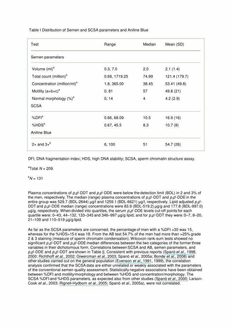
Table I Distribution of Semen and SCSA parameters and Aniline Blue
Test Range Median Mean (SD)
Semen parameters
Volume (ml)a 0.3, 7.0 2.0 2.1 (1.4)
Total count (million)a 0.69, 1719.25 74.99 121.4 (179.7)
Concentration (million/ml)a 1.8, 365.00 38.45 53.41 (49.8)
Motility (a+b+c)a 0, 81 57 49.8 (21)
Normal morphology (%)a 0, 14 4 4.2 (2.9)
SCSA
%DFIa 0.66, 68.09 10.5 16.9 (16)
%HDSa 0.67, 45.5 8.3 10.7 (8)
Aniline Blue
2+ and 3+b 6, 100 51 54.7 (26)
DFI, DNA fragmentation index; HDS, high DNA stability; SCSA, sperm chromatin structure assay.
aTotal N = 209.
bN = 131
Plasma concentrations of p,p'-DDT and p,p'-DDE were below the detection limit (BDL) in 2 and 3% of the men, respectively. The median (range) plasma concentrations of p,p'-DDT and p,p'-DDE in the entire group was 529.7 (BDL-2644) µg/l and 1259.1 (BDL-6621) µg/l, respectively. Lipid adjusted p,p'-DDT and p,p'-DDE median (range) concentrations were 83.9 (BDL-519.2) µg/g and 177.8 (BDL-997.0) µg/g, respectively. When divided into quartiles, the serum p,p'-DDE levels cut-off points for each quartile were: 0–43, 44–132, 133–345 and 346–997 µg/g lipid; and for p,p'-DDT they were: 0–7, 8–20, 21–109 and 110–519 µg/g lipid.
As far as the SCSA parameters are concerned, the percentage of men with a %DFI >30 was 15, whereas for the %HDS>15 it was 18. From the AB test 54.7% of the men had more than >25% grade 2 & 3 staining (measure of sperm chromatin condensation). Wilcoxon rank-sum tests showed no significant p,p'-DDT and p,p'-DDE median differences between the two categories of the former three variables in their dichotomous form. Correlations between SCSA and AB, semen parameters, and p,p'-DDE and p,p'-DDT are shown in Table II. Consistent with previous reports (Spanò et al., 1998, 2000; Richthoff et al., 2002; Giwercman et al., 2003; Spanò et al., 2005a; Bonde et al., 2008) and other studies carried out on the general population (Evenson et al., 1991, 1999), the correlation analysis confirmed that the SCSA data are either unrelated or weakly associated with the parameters
of the conventional semen quality assessment. Statistically negative associations have been obtained between %DFI and motility/morphology and between %HDS and concentration/morphology. The SCSA %DFI and %HDS parameters, as expected also from other studies (Spanò et al., 2000; Larson-Cook et al., 2003; Rignell-Hydbom et al., 2005; Spanò et al., 2005a), were not correlated.

Table II Spearman rank correlations between SCSA and AB, semen parameters, and DDE and DDT
%DFI %HDS %DFI +%HDS (t)
ABa
Semen parameter
Volume (ml) 0.054 (0.440) 0.13 (0.061) 0.10 (0.144) 0.045 (0.609)
Total count (million) 0.04 (0.612) –0.06 (0.421) 0.005 (0.940) –0.138 (0.119)
Concentration (million/ml) 0.02 (0.757) –0.17 (0.016) –0.05 (0.510) –0.190 (0.030)
Motility (a+b+c) –0.19 (0.008) –0.13 (0.066) –0.21 (0.002) –0.113 (0.199)
Normal morphology (%) –0.22 (0.002) –0.15 (0.038) –0.23 (<0.001) 0.117 (0.193)
SCSA
%DFI 1.0 (<0.001) –0.12 (0.090) 0.82 (<0.001) 0.182 (0.070)
%HDS –0.12 (0.090) 1.0 (<0.001) 0.31 (<0.001) 0.039 (0.701)
DDE and DDT/total lipids (µg/g)
p,p'-DDE 0.150 (0.031) –0.117 (0.094) 0.044 (0.529) 0.150 (0.091)
p,p'-DDT 0.131 (0.061) –0.136 (0.052) 0.011 (0.876) 0.149 (0.094)
AB, aniline blue; DFI, DNA fragmentation index; HDS, high DNA stability; SCSA, sperm chromatin structure assay. Bold values indicate statistically significant P<0.05.
aN = 131 men.
The relation between %DFI and lipid adjusted p,p'-DDE plasma concentration, divided into quartiles (0–43, 44–132, 133–345 and >346 µg/g lipids) is reported in Fig. 1. Simple linear regression results showed an increasing trend in the log transformed %DFI coefficients as p,p'-DDE categories
increased (Q2r = 0.23, Q3r = 0.37, Q4r = 0.41). This trend was significant when the first p,p'-DDE quartile was compared with the third and fourth quartiles (both P < 0.05), but not when it was compared with the second (P = 0.22).

Figure 1 The relation between %DFI and lipid adjusted p,p'-DDE plasma concentration, divided into quartiles.
A scatter plot reporting the transformed log values of %DFI as a function of the log lipid adjusted p,p'-DDE plasma concentration is shown in Fig. 2, where r = 0.12 and P = 0.03. However, there was only a weak and marginally statistically significant positive association emerging from the regression analysis between the log transformed values of %DFI and the non-transformed lipid adjusted p,p'-DDE plasma concentration: r = 0.0005, P = 0.075. In a final multivariate regression model, the log transformed
%DFI was associated with both age and p,p'-DDE plasma concentrations. As age increases, %DFI decreases (r = –0.037, P = 0.002) and as DDE increases, so does %DFI (r = 0.0006, P = 0.059).
Figure 2 The transformed log values of %DFI as a function of the log lipid adjusted p,p'-DDE plasma concentration.

No associations were observed between serum p,p'-DDE levels and %HDS. Even though there was a weak and marginally significant negative correlation between p,p'-DDT and %HDS (Spearman's rho = – 0.14 and P = 0.052), the simple linear regression P-value was 0.22. The same lack of association also emerged when the total structural chromatin damage (%DFI and %HDS) was considered. The outcome measures were adjusted for effects of study group, age and period of sexual abstinence but none of a variety of other potential confounders qualified for inclusion in the final models.
No association was seen between square root transformed AB grade 2 + 3 and lipid adjusted either p,p'-DDE or p,p'-DDT. Likewise, when AB grade 2 + 3 was dichotomized, logistic regressions results
showed an odds ratio of 1.00 with a P > 0.15 for both p,p'-DDE and p,p'-DDT. However, sperm concentration (square root transformed) in 130 men was negatively and significantly associated with
AB (� = –0.02 and 95% CI = –0.04, –0.001).
As seen in Table II, %HDS showed a negative and significant correlation to sperm concentration (–0.17 P = 0.016). Percentage DFI was negatively correlated with both total sperm motility (–0.19 P = 0.008) and percentage normal morphology (–0.22 P = 0.002). The latter was also negatively correlated with %HDS (–0.15 P = 0.038). There was a weak and marginally significant correlation between AB and %DFI (0.182 P = 0.070) (Table II).
Discussion
The main finding of the study was the weak, but positive, significant association between %DFI and p,p'-DDE levels (either in univariate or in the multivariate analysis). It is the first time that such an association has emerged in a large-scale epidemiological study, probably because, for the first time, sperm DNA/chromatin defects have been correlated with DDT/DDE serum level in a very high exposure range. This concentration range has never previously been explored in relation to sperm genetic integrity. Previous negative studies included populations where contact with the pesticide occurred mainly through diet. Hauser et al. (2003b) studied 212 infertility patients in the US, and reported a mean DDE serum level of 254 ng/g of lipid. Rignell-Hydbom et al. (2005) studied 176 Swedish fishermen and reported a median DDE serum level of 240 ng/g of lipid. Within the framework of an EU project (INUENDO), Spanò et al. (2005a) studied 707 men (514 Europeans and 193 Greenland Inuits) and Stronati et al. (2006) studied 652 men (452 Europeans and 200 Greenland Inuits) and reported a median DDE serum concentration of 560 ng/g of lipid. The present study is characterized by a mean p,p'-DDE level of 21 5470 ng/g of lipid (90 230 ng/g for DDT), 385 times higher than that of the INUENDO study. This value is also five times higher than that found in a study of a population of Mexican men exposed to DDT sprayed in the environment (45 000 ng/g lipid; de Jager et al., 2006). Of relevance in this last study, impaired sperm chromatin condensation (as evaluated by the AB assay) was observed in 46.6% of participants, and the most severe category of incomplete DNA condensation was also positively correlated with DDE concentration. In this study, we report 54.7% of the young men had impaired sperm chromatin condensation, as measured by the AB assay.
Here the SCSA %DFI and %HDS parameters were not correlated, a finding in agreement with other studies (Spanò et al., 2000, 2005a; Larson-Cook et al., 2003; Rignell-Hydbom et al., 2005). Thus, the men recruited in this study demonstrate two different aspects of sperm abnormalities; one linked to the occurrence of DNA damage, and the other related to the maturation process of DNA packaging around the protamine core leading to chromatin condensation alteration (Evenson et al., 2002). In this study, an inverse association between age and %DFI emerged. This discrepancy with the published literature may be a chance finding. Due to the very narrow age range of this population, which is quite young with only 10% of men older than 30 years (max 44 years), the age spectrum is probably not adequately represented.
No associations were found between serum levels of DDT/DDE and the %HDS. We have also mentioned that the %DFI level and, to a lesser extent, the %HDS fraction can be related to the fertility
potential (Evenson et al., 1999, 2002; Spanò et al., 2000; Evenson and Wixon, 2006; Bungum et al., 2007). In the general population the probability of fathering a child (fecundability) starts to decrease when %DFI > 20% (Spanò et al., 2000).

DDT is known to impair semen quality (Ayotte et al., 2001; Hauser et al., 2003a; Rignell-Hydbom et al., 2004; Dalvie et al., 2004a, b; de Jager et al., 2006; Aneck-Hahn et al., 2007), namely lowering
semen volume, sperm count, motility and viability. Aneck-Hahn et al. (2007), in addition to the positive associations between DDE serum levels and oligozoo- and asthenozoospermia, found that the incidence of teratozoospermia (normal sperm <15%) was high (almost 99%) in individuals with increased serum DDE levels. The cohort considered in this study includes a large subgroup of the population described by Aneck-Hahn et al. (2007), where the results imply that non-occupational exposure to DDT is associated with impaired semen parameters in men. Notably, 15% of men are characterized by a %DFI value 30%, whereas for 18% of them the %HDS value was 15%. In the INUENDO study, the only other comparable survey where SCSA has been used to detect sperm chromatin damage as a function of DDE dose levels (Spanò et al., 2005a), these values were 7.1% for %DFI and 22.6% for %HDS, respectively. Therefore, it appears that in our study the incidence of men characterized by an abnormally elevated incidence of sperm with DNA breaks is quite high. However, the mean values of %DFI are not so different between the two studies, being 16.9 ± 16% (range 0.7–68.1%) in this study and 13.3 ± 9.9% (range 1.3–88.2%) in the INUENDO study. The same applies for %HDS, being 10.7 ± 8.0% (range 0.7–45.5%) in this study and 11.5 ± 7.4% (range 1.6–62.3%). Notably, in the INUENDO study, the results obtained with the SCSA have been confirmed and reinforced by the results obtained on the same cohort using a different sperm DNA fragmentation assay like the TUNEL assay (Stronati et al., 2006). Furthermore, when stratifying p,p'-DDE blood levels into high and low (cut-off at 569 ng/g lipid), and looking at the androgen receptor gene polymorphism associated with the CAG repeat length, %DFI was 40% higher in the high p,p'-DDE exposure group for CAG 21 (Giwercman et al., 2007).
This is the first study to address sperm chromatin integrity in young men living in a currently DDT-sprayed area with high levels of DDT and DDE. In this study p,p'-DDT exposure was associated with an, albeit moderate, increased occurrence of sperm with chromatin defects. No other variables evaluated provided additional information as to the possible pathophysiological pathways relating to our findings. Human ejaculates always exhibit a certain fraction of sperm cells with abnormal DNA and reduced chromatin integrity (Evenson et al., 1991; Spanò et al., 1998; Sakkas et al., 2003a). The origin of these abnormal cells is not fully understood (Aitken and De Iuliis, 2007; Varghese et al., 2008) but it is believed that they can arise intratesticularly as the results of defective apoptotic mechanisms and/or derailments during the cellular and nuclear remodelling occurring during the latest stages of spermiogenesis (unrepaired nicks operated by the DNA topoisomerase II and/or altered histone-to-protamine substitution) (Agarwal and Said, 2003; Sakkas et al., 2003b; Marcon and Boissonneault, 2004). It also seems likely that p,p'-DDT and p,p'-DDE may affect seminal vesicle function, which is androgen-dependent. This possibility is supported by the low mean ejaculate volume (1.96 ± 0.33 ml) in DDT exposed men (Aneck-Hahn et al., 2007). Toxic chemicals or their metabolites can act directly on accessory glands like the seminal vesicles thereby altering the quality and/or quantity of the secretions that may affect ejaculate volume (Pant et al., 2004). Another possible source is an excessive load of reactive oxygen species overwhelming the natural antioxidative defenses present in the ejaculated semen (Ford, 2004; Agarwal et al., 2008). A two-step hypothesis
has recently been put forward where defective spermatogenesis can lead to decreased sperm production and impaired chromatin remodelling with lower levels of nuclear protamination. In this
condition, chromatin is more vulnerable and DNA becomes susceptible to various stressors, in particular oxidative stress (Aitken et al., 2009). Despite the certainty that androgen action is essential for the regulation of spermatogenesis in adult mammals, little is known about the mechanisms via which this regulation is exerted and by which DDT and its metabolites could affect these pathways. We can speculate that DDT and DDE, being estrogenic and anti-androgenic compounds, could inhibit androgens from binding to their receptors, eventually leading to lower testosterone levels. However, binding to the specific receptors is only one of the several, and still unknown, ways a chemical can act as an endocrine disrupter (Tabb and Blumberg, 2006.
As demonstrated from the low level of variability of the measurements using the reference sample and from the QC exercise, SCSA was confirmed to be a sound and reliable technique to assay sperm
chromatin integrity (Evenson et al., 1991; Giwercman et al., 1999). AO (SCSA) can be used to distinguish between spermatozoa with native DNA and single-stranded DNA as a marker for abnormal
chromatin condensation.
The SCSA ascertains the susceptibility of sperm nuclear DNA to denaturation in situ by measuring the amount of single stranded DNA after a mild low pH treatment that normally do not denature sperm

DNA. As AO is a metachromatic dye which, under blue light, fluoresces green when intercalated into double-stranded native DNA and red when complexed with single-stranded denatured DNA, the relative amount of red versus green fluorescence is measured by FCM on a per sperm basis, in large numbers of sperm in only a few minutes. Each sperm is classified as normal or abnormal based on the amount of single stranded DNA it contains, and the percentage of abnormal cells is calculated for each sample. SCSA seems particularly fit for epidemiological surveys, because only a small (0.1 ml) volume of semen is needed for the analysis and it can be frozen, stored, shipped and assayed at the end of the sampling period, thus minimising inter-assay variation (Perreault et al., 2003). The SCSA has already successfully been applied to medium- to large-scale epidemiological studies to investigate possible effects of suspected reprotoxicants (Larsen et al., 1998; Kolstad et al., 1999; Perreault et al.,
2000; Selevan et al., 2000; Bonde et al., 2002; Sanchez-Peña et al., 2004; Jönsson et al., 2005; Rignell-Hydbom et al., 2005; Rubes et al., 2005; Spanò et al., 2005a) and the present study represents by far the largest survey ever attempted in Africa in the field of reproductive molecular epidemiology, demonstrating the feasibility and robustness of the approach also in the case of samples collected in remote geographical areas.
AB staining of the sperm nucleus reveals disturbed chromatin condensation by increased intensity staining of the sperm head. In a normal ejaculate, approximately 25% of the sperm would be classified as Grades 2 and 3 according to AB staining for chromatin condensation (Foresta et al., 1992). A high percentage of chromatin defects of sperm nuclei (25% Grades 2 and 3) relate to the nucleoprotein content of DNA (incomplete or defective sperm DNA condensation) and, therefore, will have a negative effect on sperm function. The combined effect on Grades 2 and 3 staining was calculated to be >25% in 54.7% of the cases (n = 131) in this study population. Furthermore, those sperm that would theoretically be most severely affected with impaired condensation (Grade 2 and 3) correlated significantly positively with %DFI in the SCSA. In addition the AB (and %HDS) were significantly
negatively correlated with sperm concentration. This might indicate a subtle effect, one that, if it persisted, would in time have a negative impact on fertility, as AB staining provides an indication of male fertility potential (Foresta et al., 1992; de Jager et al., 2006).
In this study both the SCSA and AB assay were successfully applied to investigate possible sperm DNA/chromatin damage in general and to correlate the parameters derived from both assays. In the past the SCSA was used in a European study (INUENDO) (Spanò et al., 2005a) and the AB assay in a study in Mexico (de Jager et al., 2006). There are several other studies, carried out using either SCSA or other sperm DNA integrity assays, demonstrating that chemicals belonging to the class of (non-persistent) pesticides whose exposure can be correlated with a higher incidence of sperm with defective DNA (Bian et al., 2004; Sanchez-Peña et al., 2004; Xia et al., 2005; Meeker et al., 2004; Pérez-Herrera et al., 2008).
In conclusion, our results suggest that non-occupational environmental DDT exposure might have a negative impact on the sperm chromatin integrity of South African young males and additional factors
(e.g. genetic background, lifestyle habits, characterization of actual POP mixture and their xenohormonal activities) need to be investigated in the future.
Funding
The National Research Foundation (NRF) and the Medical Research Council (MRC), South Africa partly funded this study.
Acknowledgements
Thanks to Ms G. Schulenburg, Ms C. Van Zijl and Prof. R. Delport for technical assistance on the project and to Mr R. Mudzielwana from the Malaria Control Programme of the Department of Health
and Social Development, Limpopo Provincial Government, for his assistance in facilitating communication between the Community leaders, field workers and the project team.

Conflict of interest: The authors declare no financial or personal conflicts of interest that might affect any aspect of this study.
References
Agarwal A, Said TM. Role of sperm chromatin abnormalities and DNA damage in male infertility. Hum Reprod Update (2003) 9:331–345.
Agarwal A, Makker K, Sharma R. Clinical relevance of oxidative stress in male factor infertility: an update. Am J Reprod Immunol (2008) 59:2–11.
Aitken RJ, De Iuliis GN. Origins and consequences of DNA damage in male germ cells. Reprod Biomed Online (2007) 14:727–733.
Aitken RJ, De Iuliis GN, McLachlan RI. Biological and clinical significance of DNA damage in the male germ line. Int J Androl (2009) 32:46–56.
Aneck-Hahn NH, Schulenburg GW, Bornman MS, Farias P, de Jager C. Impaired semen quality associated with environmental DDT exposure in young men living in a malaria area in the Limpopo Province, South Africa. J Androl (2007) 28:423–434.
Asawasinsopon R, Prapamontol T, Prakobvitayakit O, Vaneesorn A, Mangklabruks A, Hock B. Plasma levels of DDT and their association with reproductive hormones in adult men from northern Thailand. Sci Total Environ (2006) 355:98–105.
Auger J, Mesbah M, Huber C, Dadoune JP. Aniline blue staining as a marker of sperm chromatin defects associated with different semen characteristics discriminates between proven fertile and suspected infertile men. Int J Androl (1990) 13:452–462.
Ayotte P, Giroux S, Dewailly E, Hernandez Avilla M, Farias P, Danis R, Villanueva Diaz C. DDT spraying for malaria control and reproductive function in Mexican men. Epidemiology (2001) 12:366–367.
Beard J. DDT and human health. Sci Total Environ (2005) 355:78–89.
Bhatia R, Shiau R, Petreas M, Weinraub JM, Farhang L, Eskenazi B. Organochlorine pesticides and male genital anomalies in the child health and development studies. Environ Health Perspect (2005) 113:220–224.
Bian Q, Xu LC, Wang SL, Xia YK, Tan LF, Chen JF, Song L, Chang HC, Wang XR. Study on the relation between occupational fenvalerate exposure and spermatozoa DNA damage of pesticide factory workers. Occup Environ Med (2004) 61:999–1005.
Bonde JP, Joffe M, Apostoli P, Dale A, Kiss P, Spano M, Caruso F, Giwercman A, Bisanti L, Porru S, et al. Sperm count and chromatin structure in men exposed to inorganic lead: lowest adverse effect levels. Occup Environ Med (2002) 59:234–242.
Bonde JP, Toft G, Rylander L, Rignell-Hydbom A, Giwercman A, Spano M, Manicardi GC, Bizzaro D, Ludwicki JK, Zvyezday V, et al. Fertility and markers of male reproductive function in Inuit and European populations spanning large contrasts in blood levels of persistent organochlorines. Environ Health Perspect (2008) 116:269–277.
Bouwman H. South Africa and the Stockholm convention on persistent organic pollutants. S Afr J Sci (2004) 100:323–328.

Bungum M, Humaidan P, Axmon A, Spano M, Bungum L, Erenpreiss J, Giwercman A. Sperm DNA integrity assessment in prediction of assisted reproduction technology outcome. Hum Reprod (2007) 22:174–179.
Colborn T, Vom Saal FS, Soto AM. Developmental effects of endocrine-disrupting chemicals in wildlife and humans. Environ Health Perspect (1993) 101:378–384.
Dalvie MA, Myers JE, Thompson ML, Robins TG, Dyer S, Riebow J, Molekwa J, Jeebhay M, Millar R, Kruger P. The long-term effects of DDT exposure on semen, fertility, and sexual function of malaria vector-control workers in Limpopo Province, South Africa. Environ Res (2004) a 96:1–8.
Dalvie MA, Myers JE, Lou Thompson M, Dyer S, Robins TG, Omar S, Riebow J, Molekwa J, Kruger P, Millar R. The hormonal effects of long-term DDT exposure on malaria vector-control workers in Limpopo Province, South Africa. Environ Res (2004) b 96:9–19.
Danzo BJ. Environmental xenobiotics may disrupt normal endocrine function by interfering with the binding of physiological ligands to steroid receptors and binding proteins. Environ Health Perspect (1997) 105:294–301.
de Jager C, Farias P, Barraza-Villarreal A, Avila MH, Ayotte P, Dewailly E, Dombrowski C, Rousseau F, Sanchez VD, Bailey JL. Reduced seminal parameters associated with environmental DDT exposure and p,p'-DDE concentrations in men in Chiapas, Mexico: a cross-sectional study. J Androl (2006) 27:16–27.
Edwards TM, Moore BC, Guillette LJ Jr. Reproductive dysgenesis in wildlife: a comparative view. Int J Androl (2006) 29:109–121.
Erenpreiss J, Spano M, Erenpreisa J, Bungum M, Giwercman A. Sperm chromatin structure and male fertility: biological and clinical aspects. Asian J Androl (2006) 8:11–29.
Evenson DP, Wixon R. Environmental toxicants cause sperm DNA fragmentation as detected by the Sperm Chromatin Structure Assay (SCSA). Toxicol Appl Pharmacol (2005) 207:532–537.
Evenson DP, Wixon R. Meta-analysis of sperm DNA fragmentation using the sperm chromatin structure assay. Reprod Biomed Online (2006) 12:466–472.
Evenson DP, Darzynkiewicz Z, Melamed MR. Relation of mammalian sperm chromatin heterogeneity to fertility. Science (1980) 210:1131–1133.
Evenson DP, Jost LK, Baer R, Turner T, Schrader S. Individuality of DNA denaturation patterns in human sperm as measured by the sperm chromatin structure assay. Reprod Toxicol (1991) 5:115–125.
Evenson DP, Jost LK, Marshall D, Zinaman MJ, Clegg E, Purvis K, de Angelis P, Clausen OP. Utility of the sperm chromatin structure assay as a diagnostic and prognostic tool in the human fertility clinic. Hum Reprod (1999) 14:1039–1049.
Evenson DP, Jost LK, Corzett M, Balhorn R. Characteristics of human sperm chromatin structure following an episode of influenza and high fever: a case study. J Androl (2000) 21:739–746.
Evenson DP, Larson KL, Jost LK. Sperm chromatin structure assay: its clinical use for detecting sperm DNA fragmentation in male infertility and comparisons with other techniques. J Androl (2002) 23:25–43.
Ford WC. Regulation of sperm function by reactive oxygen species. Hum Reprod Update (2004) 10:387–399.
Foresta C, Zorzi M, Rossato M, Varotto A. Sperm nuclear instability and staining with aniline blue: abnormal persistence of histones in spermatozoa in infertile men. Int J Androl (1992) 15:330–337.
Fry DM. Reproductive effects in birds exposed to pesticides and industrial chemicals. Environ Health Perspect (1995) 103:165–171.
Giwercman A, Spanò M, Lahdetie J, Bonde JP. Quality assurance of semen analysis in multicenter studies. Asclepios. Scand J Work Environ Health (1999) 25:23–25.
Giwercman A, Richthoff J, Hjollund H, Bonde JP, Jepson K, Frohm B, Spanò M. Correlation between sperm motility and sperm chromatin structure assay parameters. Fertil Steril (2003) 80:1404–1412.
Giwercman AH, Rignell-Hydbom A, Toft G, Rylander L, Hagmar L, Lindh C, Pedersen HS, Ludwicki JK, Lesovoy V, Shvets M, et al. Reproductive hormone levels in men exposed to persistent organohalogen pollutants: a study of Inuit and three European cohorts. Environ Health Perspect (2006) 114:1348–1353.
Giwercman A, Rylander L, Rignell-Hydbom A, Jonsson BA, Pedersen HS, Ludwicki JK, Lesovoy V, Zvyezday V, Spano M, Manicardi GC, et al. INUENDO. Androgen receptor gene CAG repeat length as a modifier of the association between persistent organohalogen pollutant exposure markers and semen characteristics. Pharmacogenet Genomics (2007) 17:391–401.
Godduhn A, Duffy LK. Multi-generation health risks of persistent organic pollution in the far north: use of the precautionary approach in the Stockholm Convention. Environ Sci Policy (2003) 6:341–353.
Guillette LJ Jr. Endocrine disrupting contaminants–beyond the dogma. Environ Health Perspect (2006) 114:9–12.
Guillette LJ Jr, Gunderson MP. Alterations in development of reproductive and endocrine systems of wildlife populations exposed to endocrine-disrupting contaminants. Reproduction (2001) 122:857–864.
Guillette LJ Jr, Moore BC. Environmental contaminants, fertility, and multioocytic follicles: a lesson from wildlife? Semin Reprod Med (2006) 24:134–141.
Guillette LJ Jr, Gross TS, Gross DA, Rooney AA, Percival HF. Gonadal steroidogenesis in vitro from juvenile alligators obtained from contaminated or control lakes. Environ Health Perspect (1995) 103:31–36.
Hauser R, Chen ZY, Pothier L, Ryan L, Altshul L. The relationship between human semen parameters and environmental exposure to polychlorinated biphenyls and p,p'-DDE. Environ Health Perspect (2003) a 111:1505–1511.
Hauser R, Singh NP, Chen Z, Pothier L, Altshul L. Lack of an association between environmental exposure to polychlorinated biphenyls and p,p'-DDE and DNA damage in human sperm measured using the neutral comet assay. Hum Reprod (2003) b 18:2525–2533.
Henkel R. Detection of DNA damage in sperm. In: Male Infertility Diagnosis and Treatment—Oehninger SC, Kruger TF, eds. (2007) UK: Informa Healthcare, Informa Ltd. 132–232.
Henkel R, Menkveld R, Kleinhappl M, Schill WB. Seasonal changes in human sperm chromatin condensation. J Assist Reprod Genet (2001) 18:371–377.
Hougard JM, Fontenille D, Chandre F, Darriet F, Carnevale P, Guillet P. Combating malaria vectors in Africa: current directions of research. Trends Parasitol (2002) 18:283–286.
United Nations Environment Programme, Division of Technology, Industry and Economics, Chemicals Branch.
Stockholm Convention on persistent organic pollutants.
Jönsson BA, Richthoff J, Rylander L, Giwercman A, Hagmar L. Urinary phthalate metabolites and biomarkers of reproductive function in young men. Epidemiology (2005) 16:487–493.
Kelce WR, Stone CR, Laws SC, Gray LE, Kemppainen JA, Wilson EM. Persistent DDT metabolite p,p'-DDE is a potent androgen receptor antagonist. Nature (1995) 375:581–585.
Kilian E, Delport R, Bornman MS, de Jager C. Simultanious exposure to low concentrations of DDT, deltamethrin, phytoestrogens and nonylphenol harms reproductive parameters in male rats. Andrologia (2007) 39:128–135.
Kolstad HA, Bonde JP, Spanò M, Giwercman A, Zschiesche W, Kaae D, Larsen SB, Roeleveld N. Change in semen quality and sperm chromatin structure following occupational styrene exposure. Asclepios. Int Arch Occup Environ Health (1999) 72:135–141.
Larsen SB, Giwercman A, Spanò M, Bonde JP. A longitudinal study of semen quality in pesticide spraying Danish farmers. The ASCLEPIOS Study Group. Reprod Toxicol (1998) 12:581–589.
Larson-Cook K, Brannian JD, Hansen KA, Kasperson K, Aamold ET, Evenson DP. Relationship between the outcomes of assisted reproductive techniques and sperm DNA fragmentation as measured by the sperm chromatin structure assay. Fertil Steril (2003) 80:895–902.
Marcon L, Boissonneault G. Transient DNA strand breaks during mouse and human spermiogenesis new insights in stage specificity and link to chromatin remodeling. Biol Reprod (2004) 70:910–918.
Meeker JD, Singh NP, Ryan L, Duty SM, Barr DB, Herrick RF, Bennett DH, Hauser R. Urinary levels of insecticide metabolites and DNA damage in human sperm. Hum Reprod (2004) 19:2573–2580.
Mortimer D. Practical Laboratory Andrology (1994) New York: Oxford University Press.
Pant N, Mathur N, Banerjee AK, Srivastava SP, Saxena DK. Correlation of chlorinated pesticides concentration in semen with seminal vesicle and prostatic markers. Reprod Toxicol (2004) 19:209–214.
Pérez-Herrera N, Polanco-Minaya H, Salazar-Arredondo E, Solís-Heredia MJ, Hernández-Ochoa I, Rojas-García E, Alvarado-Mejía J, Borja-Aburto VH, Quintanilla-Vega B. PON1Q192R genetic polymorphism modifies organophosphorous pesticide effects on semen quality and DNA integrity in agricultural workers from southern Mexico. Toxicol Appl Pharmacol (2008) 230:261–268.
Perreault SD, Rubes J, Robbins WA, Evenson DP, Selevan SG. Evaluation of aneuploidy and DNA damage in human spermatozoa: applications in field studies. Andrologia (2000) 32:247–254.
Perreault SD, Aitken RJ, Baker HW, Evenson DP, Huszar G, Irvine DS, Morris ID, Morris RA, Robbins WA, Sakkas D, et al. Integrating new tests of sperm genetic integrity into semen analysis: breakout group discussion. Adv Exp Med Biol (2003) 518:253–268.
Pitarch E, Serrano R, Lopez FJ, Hernandez F. Rapid multiresidue determination of organochlorine and organophosphorus compounds in human serum by solid phase extraction and gas chromatography coupled to tandem mass spectrometry. Anal Bioanal Chem (2003) 376:189–197.
Richthoff J, Spanò M, Giwercman YL, Frohm B, Jepson K, Malm J, Elzanaty S, Stridsberg M, Giwercman A. The impact of testicular and accessory sex gland function on sperm chromatin integrity as assessed by the sperm chromatin structure assay (SCSA). Hum Reprod (2002) 17:3162–3169.

Rignell-Hydbom A, Rylander L, Giwercman A, Jönsson BAG, Nilsson-Ehle P, Hagmar L. Exposure to CB-153 and p,p'-DDE and male reproductive function. Hum Reprod (2004) 19:2066–2075.
Rignell-Hydbom A, Rylander L, Giwercman A, Jönsson BA, Lindh C, Eleuteri P, Rescia M, Leter G, Cordelli E, Spano M, et al. Exposure to PCBs and p,p'-DDE and human sperm chromatin integrity. Environ Health Perspect (2005) 113:175–179.
Rogan WJ, Chen A. Health risks and benefits of bis(4-chlorophenyl)-1,1,1- trichloroethane (DDT). Lancet (2005) 366:763–773.
Rubes J, Selevan SG, Evenson DP, Zudova D, Vozdova M, Zudova Z, Robbins WA, Perreault SD. Episodic air pollution is associated with increased DNA fragmentation in human sperm without other changes in semen quality. Hum Reprod (2005) 20:2776–2783.
Rylander L, Wallin E, Jonssson BA, Stridsberg M, Erfurth EM, Hagmar L. Associations between CB-153 and p,p'-DDE and hormone levels in serum in middle-aged and elderly men. Chemosphere (2006) a 65:375–381.
Rylander L, Nilsson-Ehle P, Hagmar L. A simplified precise method for adjusting serum levels of persistent organohalogen pollutants to total serum lipids. Chemosphere (2006) b 62:333–336.
Sakkas D, Seli E, Bizzaro D, Tarozzi N, Manicardi GC. Abnormal spermatozoa in the ejaculate: abortive apoptosis and faulty nuclear remodeling during spermatogenesis. Reprod Biomed Online (2003) a 7:428–432.
Sakkas D, Manicardi GC, Bizzaro D. Sperm nuclear DNA damage in the human. Adv Exp Med Biol (2003) b 518:73–84.
Sanchez-Peña LC, Reyes BE, Lopez-Carrillo L, Recio R, Moran-Martinez J, Cebrian ME, Quintanilla-Vega B. Organophosphorous pesticide exposure alters sperm chromatin structure in Mexican agricultural workers. Toxicol Appl Pharmacol (2004) 196:108–113.
Selevan SG, Borkovec L, Slott VL, Zudova Z, Rubes J, Evenson DP, Perreault SD. Semen quality and reproductive health of young Czech men exposed to seasonal air pollution. Environ Health Perspect (2000) 108:887–894.
Snedeker SM. Pesticides and breast cancer risk: A review of DDT, DDE and dieldrin. Environ Health Perspect (2001) 109:35–47.
Spanò M, Kolstad AH, Larsen SB, Cordelli E, Leter G, Giwercman A, Bonde JP. The applicability of the flow cytometric sperm chromatin structure assay in epidemiological studies. Asclepios. Hum Reprod (1998) 13:2495–2505.
Spanò M, Bonde JP, Hjollund HI, Kolstad HA, Cordelli E, Leter G. Sperm chromatin damage impairs human fertility. The Danish first pregnancy planner study team. Fertil Steril (2000) 73:43–50.
Spanò M, Toft G, Hagmar L, Eleuteri P, Rescia M, Rignell-Hydbom A, Tyrkiel E, Zvyezday V, Bonde JP. INUENDO. Exposure to PCB and p, p’-DDE in European and Inuit populations: impact on human sperm chromatin integrity. Hum Reprod (2005) a 20:3488–3499.
Spanò M, Seli E, Bizzaro D, Manicardi GC, Sakkas D. The significance of sperm nuclear DNA strand breaks on reproductive outcome. Curr Opin Obstet Gynecol (2005) b 17:255–260.

Stronati A, Manicardi GC, Cecati M, Bordicchia M, Ferrante L, Spanò M, Toft G, Bonde JP, Jönsson BA, Rignell-Hydbom A, et al. Relationships between sperm DNA fragmentation, sperm apoptotic markers and serum levels of CB-153 and p,p'-DDE in European and Inuit populations. Reproduction (2006) 132:949–958.
Tabb MM, Blumberg B. New modes of action for endocrine-disrupting chemicals. Mol Endocrinol (2006) 20:475–482.
Terquem A, Dadoune JP. Aniline blue staining of human spermatozoon chromatin. Evaluation of nuclear maturation. In: The Sperm Cell—Andre J, ed. (1983) The Hague: Martinus Nijhoff Publishers. 249–252.
Toft G, Rignell-Hydbom A, Tyrkiel E, Shvets M, Giwercman A, Lindh CH, Pedersen HS, Ludwicki JK, Lesovoy V, Hagmar L, et al. Semen quality and exposure to Persistent Organochlorine Pollutants. Epidemiology (2006) 17:450–458.
Turusov V, Rakitsky V, Tomatis L. Dichlorodiphenyltrichloroethane (DDT): ubiquity, persistence and risks. Res Rev (2002) 110:125–128.
Varghese AC, Du Plessis SS, Agarwal A. Male gamete surivival at stake: causes and solutions. Reprod Biomed Online (2008) 17:866–880.
World Health Organization. WHO Laboratory Manual for the Examination of Human Semen and Sperm-Cervical Mucus Interaction (1999) 4th edn. New York: Cambridge University Press.
Wyrobek AJ, Eskenazi B, Young S, Arnheim N, Tiemann-Boege I, Jabs EW, Glaser RL, Pearson FS, Evenson D. Advancing age has differential effects on DNA damage, chromatin integrity, gene mutations, and aneuploidies in sperm. Proc Natl Acad Sci USA (2006) 103:9601–9606.
Xia Y, Cheng S, Bian Q, Xu L, Collins MD, Chang HC, Song L, Liu J, Wang S, Wang X. Genotoxic effects on spermatozoa of carbaryl-exposed workers. Toxicol Sci (2005) 85:615–623.