Spectrum of chromosomal aberrations in peripheral lymphocytes of hospital workers occupationally...
12
© Kamla-Raj 2010 Int J Hum Genet, 10(1-3): 147-158 (2010) Spectrum of Chromosomal Aberrations in Peripheral Blood Lymphocytes of Gastrointestinal Tract (GIT) and Breast Cancer Patients Kamlesh Guleria and Vasudha Sambyal Department of Human Genetics, Guru Nanak Dev University, Amritsar 143 005, Punjab, India KEYWORDS Peripheral Blood Lymphocytes. Gastrointestinal Cancer. Breast Cancer. Chromosomal Aberrations ABSTRACT The aim of present study was to assess the spectrum of chromosomal aberrations in peripheral blood lymphocytes of sporadic Gastrointestinal tract (GIT) and Breast cancer patients. Ninety eight patients (56 GIT cancer and 42 breast cancer) and seventy seven unrelated healthy set of control individuals were investigated in the present study. Lymphocytes were cultured using standard protocol. In each case, 100 metaphases were screened for numerical as well as structural aberrations. Higher frequency of aberrant metaphases with chromosomal aberrations including gaps, breaks, terminal deletions, acentric fragments, double minutes, acrocentric associations, premature chromatid separations, pulverisations, polyploidy, loss and gain of chromosomes, ring chromosome and marker chromosomes were observed in cancer patients as compared to controls. A non-random involvement in aberrations of chromosomes harbouring genes implicated in tumorigenesis was observed in GIT as well as in breast cancer patients. Aberrations in peripheral blood lymphocytes (PBLs) can indicate the constitutional anomalies and understanding of molecular basis of chromosomal instability (CIN) phenotype can help in earlier diagnosis or prognosis. INTRODUCTION Genomic instability is a prerequisite for the onset of cancer. The majority of cancer cells are aneuploid, representing dynamic karyotypic changes, including gain or loss of whole chromo- somes, chromosomal rearrangements and amplification or deletion of genetic material. There are two hypotheses namely the chromo- somal instability hypothesis and the gene muta- tion hypothesis that differ in the type of genomic alterations necessary for a normal cell to become a cancerous cell (Marx 2002). Chromosomal instability (CIN) in cancer is driven by proceeding mutation in growth controlling oncogenes and tumor suppressor genes. Analysis of aneuploid cancer cells in vitro reveal that chromosome losses and gains occur at >10 -2 per chromosome per cycle, which is 10-100 times greater than in karyotypically stable diploid cancers of the same histological type (Lengauer et al. 1997). The elevated rate of chromosome missegregation in aneuploid tumors cells with CIN causes pheno- typic changes that contribute to tumor cell evolution and pose therapeutic challenges (Gao et al. 2007). Defects in both bipolar spindle assembly and the spindle assembly checkpoint have been identified in some CIN tumor cell lines (Cahill et al. 1998; Lingle et al. 2002). The mecha- nism how the tumor cells acquire extra chromo- somes and maintain during cell division is not clear. The frequency of chromosomal aberrations (CAs) in human peripheral blood lymphocytes (PBLs) has routinely been used as a tool for the identification of occupational and environmental hazards (Carrano and Natarajan 1988; Rossner et al. 1995; Waters et al. 1999; Albertini et al. 2000; Sram and Binkova 2000; Bonassi et al. 2005). The association between CAs in peripheral lympho- cytes and increased risk for cancer has been observed in a Nordic cohort (Hagmar et al. 1994, 1998, 2004), in an Italian cohort (Bonassi et al. 1995) and in a nested case-control study carried out in Taiwan (Liou et al. 1999). The association between frequency of CAs and risk of cancer was not modified by sex, age, cigarette smoking, occu- pational exposure, or time since the cytogenetic assay performed (Bonassi et al. 2005). A positive association between the frequency of CAs in PBLs and the risk of cancer at different sites has been supported by numerous clinical observations, in particular, of patients suffering Address for correspondence: Dr. Vasudha Sambyal, Reader, Department of Human Genetics, Guru Nanak Dev University, Amritsar143 005, Punjab, India Telephone: 0183-2258802-07 Ext. 3445 Fax: 0183-2258819 E-mail: [email protected]
Transcript of Spectrum of chromosomal aberrations in peripheral lymphocytes of hospital workers occupationally...

© Kamla-Raj 2010 Int J Hum Genet, 10(1-3): 147-158 (2010)
Spectrum of Chromosomal Aberrations in Peripheral BloodLymphocytes of Gastrointestinal Tract (GIT) and
Breast Cancer Patients
Kamlesh Guleria and Vasudha Sambyal
Department of Human Genetics, Guru Nanak Dev University, Amritsar 143 005, Punjab, India
KEYWORDS Peripheral Blood Lymphocytes. Gastrointestinal Cancer. Breast Cancer. Chromosomal Aberrations
ABSTRACT The aim of present study was to assess the spectrum of chromosomal aberrations in peripheral bloodlymphocytes of sporadic Gastrointestinal tract (GIT) and Breast cancer patients. Ninety eight patients (56 GITcancer and 42 breast cancer) and seventy seven unrelated healthy set of control individuals were investigated in thepresent study. Lymphocytes were cultured using standard protocol. In each case, 100 metaphases were screened fornumerical as well as structural aberrations. Higher frequency of aberrant metaphases with chromosomal aberrationsincluding gaps, breaks, terminal deletions, acentric fragments, double minutes, acrocentric associations, prematurechromatid separations, pulverisations, polyploidy, loss and gain of chromosomes, ring chromosome and markerchromosomes were observed in cancer patients as compared to controls. A non-random involvement in aberrationsof chromosomes harbouring genes implicated in tumorigenesis was observed in GIT as well as in breast cancer patients.Aberrations in peripheral blood lymphocytes (PBLs) can indicate the constitutional anomalies and understanding ofmolecular basis of chromosomal instability (CIN) phenotype can help in earlier diagnosis or prognosis.
INTRODUCTION
Genomic instability is a prerequisite for theonset of cancer. The majority of cancer cells areaneuploid, representing dynamic karyotypicchanges, including gain or loss of whole chromo-somes, chromosomal rearrangements andamplification or deletion of genetic material.There are two hypotheses namely the chromo-somal instability hypothesis and the gene muta-tion hypothesis that differ in the type of genomicalterations necessary for a normal cell to becomea cancerous cell (Marx 2002). Chromosomalinstability (CIN) in cancer is driven by proceedingmutation in growth controlling oncogenes andtumor suppressor genes. Analysis of aneuploidcancer cells in vitro reveal that chromosomelosses and gains occur at >10-2 per chromosomeper cycle, which is 10-100 times greater than inkaryotypically stable diploid cancers of the samehistological type (Lengauer et al. 1997). Theelevated rate of chromosome missegregation inaneuploid tumors cells with CIN causes pheno-
typic changes that contribute to tumor cellevolution and pose therapeutic challenges (Gaoet al. 2007). Defects in both bipolar spindleassembly and the spindle assembly checkpointhave been identified in some CIN tumor cell lines(Cahill et al. 1998; Lingle et al. 2002). The mecha-nism how the tumor cells acquire extra chromo-somes and maintain during cell division is notclear.
The frequency of chromosomal aberrations(CAs) in human peripheral blood lymphocytes(PBLs) has routinely been used as a tool for theidentification of occupational and environmentalhazards (Carrano and Natarajan 1988; Rossner etal. 1995; Waters et al. 1999; Albertini et al. 2000;Sram and Binkova 2000; Bonassi et al. 2005). Theassociation between CAs in peripheral lympho-cytes and increased risk for cancer has beenobserved in a Nordic cohort (Hagmar et al. 1994,1998, 2004), in an Italian cohort (Bonassi et al.1995) and in a nested case-control study carriedout in Taiwan (Liou et al. 1999). The associationbetween frequency of CAs and risk of cancer wasnot modified by sex, age, cigarette smoking, occu-pational exposure, or time since the cytogeneticassay performed (Bonassi et al. 2005).
A positive association between the frequencyof CAs in PBLs and the risk of cancer at differentsites has been supported by numerous clinicalobservations, in particular, of patients suffering
Address for correspondence:Dr. Vasudha Sambyal,Reader, Department of Human Genetics, Guru NanakDev University, Amritsar143 005, Punjab, IndiaTelephone: 0183-2258802-07 Ext. 3445Fax: 0183-2258819E-mail: [email protected]

148 KAMLESH GULERIA AND VASUDHA SAMBYAL
from hereditary chromosome breakage syndro-mes (Mathur et al. 2000) and several other pre-cancerous conditions such as preleukemic statesof adult T-cell leukemia (Nishino 1988), dysplasticnevus syndrome (Caporaso et al. 1987), or nevoidbasal-cell syndrome (Shafei-Benaissa et al. 1998).Different case-control studies have reported asignificant increase in the frequency of aberrantcells in PBLs of cancer patients (Barrios et al.1988; Abarbanel et al. 1991; Barrios et al. 1991;Barletta et al. 1993; Gebhart et al. 1993; Dave etal. 1995; Dhillon et al. 1996; Patel et al. 1997;Dhillon and Dhillon 1998; Trivedi et al. 1998; Royet al. 2000, 2001), but these studies have beensubjected to criticism because of small samplesize.
The aim of present study was to assess thespectrum of chromosomal aberrations inperipheral blood lymphocytes of Gastrointestinaltract (GIT) and Breast cancer patients and theirdiagnostic or prognostic utility. Frequency ofsporadic GIT and Breast cancer is higher in areasadjoining Amritsar city of Punjab, India and iscontinuously increasing.
MATERIAL AND METHODS
The study was carried out under theguidelines of ethical committee constituted byGuru Nanak Dev University, Amritsar and tenetsof declaration of Helsinki. Ninety eight patients(56 GIT cancer and 42 breast cancer) from SriGuru Ram Das Rotary Cancer Hospital Amritsarand Government Medical College Amritsar,Punjab were investigated in the present study.The age of patients ranged from 25 to 95 years.Blood samples of 77 age and sex matchedunrelated healthy control individuals were alsoinvestigated in this study. Relevant informationincluding age, gender, occupation, personal andfamily medical history, habitat, habits and dietwere recorded on a pre-tested structured ques-tionnaire. Lymphocytes were cultured usingstandard protocol (Moorhead et al. 1960) withfew modifications. GTG banding was done usingBenn and Perle (1986) technique. In each case,100 metaphases were examined for numerical aswell as structural aberrations. Chromosomes wereidentified and classified according to Inter-
Table 1: Epidemiological profile of cancer patients and unrelated healthy control individuals
Gastrointestinal cancers
Breast Oesophageal Gastric Intestinal* Others** Controls
No. of subjects 42 34 3 13 6 77Age in years
(Range) 28-90 45-95 58-70 32-55 30-60 28-80Gender
Males 1 17 2 7 - 28Females 41 17 1 6 6 49
OccupationFarmer 1 8 - 1 - 8Labourer 1 8 - 2 - -Shopkeeper - 1 - 2 - 3Govt. Employee - - 1 3 - 21Housewives 37 13 1 5 6 34Teacher 3 2 - - - 5Sweeper - 1 1 - - 1Factory worker - 1 - - - -Research student - - - - - 5
HabitatRural 21 27 - 7 4 32Sub urban 5 1 - - 1 5Urban 16 6 3 6 1 40
HabitsSmoker - 3 2 1 - -Alcoholic - 12 1 4 1 12Drugs 2 - - 1 - -Non-vegetarian 8 10 2 2 2 12
*Cecum, Colon, Colorectal, Rectal**Pancreatic, Gall bladder
SPECTRUM OF CHROMOSOMAL ABERRATIONS IN GIT AND BREAST CANCER 149
national system for human cytogeneticnomenclature (ISCN 2005).
RESULTS
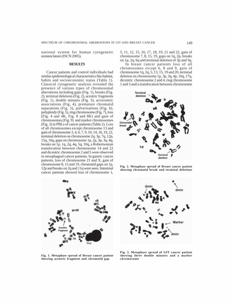
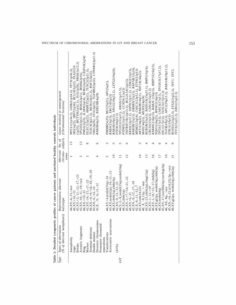
Cancer patients and control individuals hadsimilar epidemiological characteristics like habitat,habits and socioeconomic status (Table 1).Classical cytogenetic analysis revealed thepresence of various types of chromosomalaberrations including gaps (Fig. 1), breaks (Fig.2), terminal deletions (Fig. 2), acentric fragments(Fig. 1), double minutes (Fig. 3), acrocentricassociations (Fig. 4), premature chromatidseparations (Fig. 5), pulverisations (Fig. 6),polyploidy (Fig. 5), ring chromosome (Fig. 7), loss(Fig. 4 and 4K, Fig. 8 and 8K) and gain ofchromosomes (Fig. 9) and marker chromosomes(Fig. 3) in PBLs of cancer patients (Table 2). Lossof all chromosomes except chromosome 13 andgain of chromosome 3, 4, 6, 7, 9, 10, 14, 16, 19, 22,terminal deletion on chromosome 2q, 3p, 7q, 12p,15q, 16q, gaps on chromosome 1p, 2p, 3p, 3q, 4q,breaks on 1p, 1q, 2q, 4q, 5q, 10q, a Robertsoniantranslocation between chromosome 14 and 22and dicentric chromosome 2 and 5 were observedin oesophageal cancer patients. In gastric cancerpatients, loss of chromosome 21 and X, gain ofchromosome 8, 15 and 19, chromatid gaps on 1p,12p and breaks on 2q and 11q were seen. Intestinalcancer patients showed loss of chromosome 1,
5, 11, 12, 15, 16, 17, 18, 19, 21 and 22, gain ofchromosome 7, 8, 15, 19, gaps on 1q, 2p, breakson 1p, 2q, 6q and terminal deletion of 3p and 3q.
In breast cancer patients loss of allchromosomes except 6, 8 and 9, gain ofchromosome 1q, 2q, 5, 13, 15, 19 and 20, terminaldeletion on chromosome 1p, 3p, 3q, 4p, 16q, 17q,dicentric chromosome 2 and 4, ring chromosome1 and 3 and a translocation between chromosome
Fig. 3. Metaphase spread of GIT cancer patientshowing three double minutes and a markerchromosome
Fig. 2. Metaphase spread of Breast cancer patientshowing chromatid break and terminal deletions
Fig. 1. Metaphase spread of Breast cancer patientshowing acentric fragment and chromatid gap

150 KAMLESH GULERIA AND VASUDHA SAMBYAL
Fig. 4. Metaphase spread of GIT cancer patientshowing D, D & G and D & G acrocentricassociations and loss of chromosomes
Fig. 4 K. Karyotype of the metaphase spread showing D, D & G and D & G acrocentric associations andloss of chromosomes
Fig. 5. Polyploid metaphase showing prematurechromatid separations
SPECTRUM OF CHROMOSOMAL ABERRATIONS IN GIT AND BREAST CANCER 151
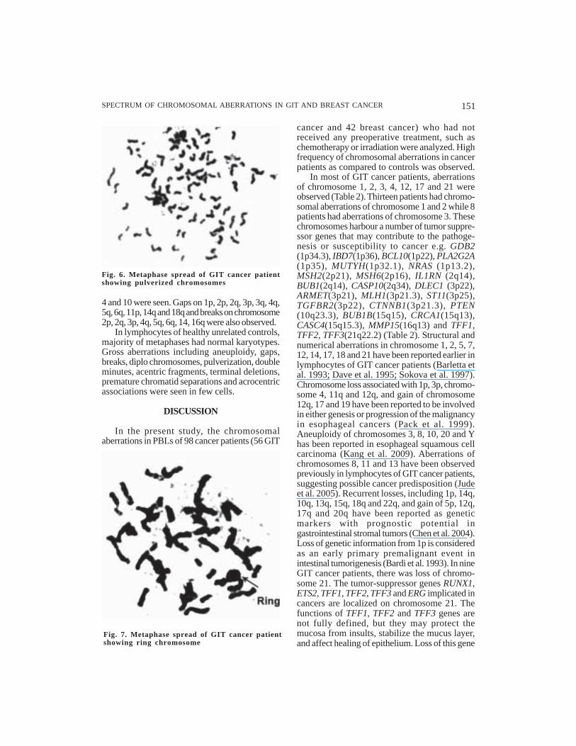
Fig. 6. Metaphase spread of GIT cancer patientshowing pulverized chromosomes
Fig. 7. Metaphase spread of GIT cancer patientshowing ring chromosome
cancer and 42 breast cancer) who had notreceived any preoperative treatment, such aschemotherapy or irradiation were analyzed. Highfrequency of chromosomal aberrations in cancerpatients as compared to controls was observed.
In most of GIT cancer patients, aberrationsof chromosome 1, 2, 3, 4, 12, 17 and 21 wereobserved (Table 2). Thirteen patients had chromo-somal aberrations of chromosome 1 and 2 while 8patients had aberrations of chromosome 3. Thesechromosomes harbour a number of tumor suppre-ssor genes that may contribute to the pathoge-nesis or susceptibility to cancer e.g. GDB2(1p34.3), IBD7(1p36), BCL10(1p22), PLA2G2A(1p35), MUTYH(1p32.1), NRAS (1p13.2),MSH2(2p21), MSH6(2p16), IL1RN (2q14),BUB1(2q14), CASP10(2q34), DLEC1 (3p22),ARMET(3p21), MLH1(3p21.3), ST11(3p25),TGFBR2(3p22), CTNNB1(3p21.3), PTEN(10q23.3), BUB1B(15q15), CRCA1(15q13),CASC4(15q15.3), MMP15(16q13) and TFF1,TFF2, TFF3(21q22.2) (Table 2). Structural andnumerical aberrations in chromosome 1, 2, 5, 7,12, 14, 17, 18 and 21 have been reported earlier inlymphocytes of GIT cancer patients (Barletta etal. 1993; Dave et al. 1995; Sokova et al. 1997).Chromosome loss associated with 1p, 3p, chromo-some 4, 11q and 12q, and gain of chromosome12q, 17 and 19 have been reported to be involvedin either genesis or progression of the malignancyin esophageal cancers (Pack et al. 1999).Aneuploidy of chromosomes 3, 8, 10, 20 and Yhas been reported in esophageal squamous cellcarcinoma (Kang et al. 2009). Aberrations ofchromosomes 8, 11 and 13 have been observedpreviously in lymphocytes of GIT cancer patients,suggesting possible cancer predisposition (Judeet al. 2005). Recurrent losses, including 1p, 14q,10q, 13q, 15q, 18q and 22q, and gain of 5p, 12q,17q and 20q have been reported as geneticmarkers with prognostic potential ingastrointestinal stromal tumors (Chen et al. 2004).Loss of genetic information from 1p is consideredas an early primary premalignant event inintestinal tumorigenesis (Bardi et al. 1993). In nineGIT cancer patients, there was loss of chromo-some 21. The tumor-suppressor genes RUNX1,ETS2, TFF1, TFF2, TFF3 and ERG implicated incancers are localized on chromosome 21. Thefunctions of TFF1, TFF2 and TFF3 genes arenot fully defined, but they may protect themucosa from insults, stabilize the mucus layer,and affect healing of epithelium. Loss of this gene
4 and 10 were seen. Gaps on 1p, 2p, 2q, 3p, 3q, 4q,5q, 6q, 11p, 14q and 18q and breaks on chromosome2p, 2q, 3p, 4q, 5q, 6q, 14, 16q were also observed.
In lymphocytes of healthy unrelated controls,majority of metaphases had normal karyotypes.Gross aberrations including aneuploidy, gaps,breaks, diplo chromosomes, pulverization, doubleminutes, acentric fragments, terminal deletions,premature chromatid separations and acrocentricassociations were seen in few cells.
DISCUSSION
In the present study, the chromosomalaberrations in PBLs of 98 cancer patients (56 GIT

152 KAMLESH GULERIA AND VASUDHA SAMBYAL
Fig. 8 K. Karyotype of the metaphase spread showing loss of chromosomes
Fig. 9. Metaphase spread of Breast cancer patientshowing gain of chromosome (47,XX,+3)
cluster has been previously reported in humanGIT tumors (Katoh 2003).
Aberrations of chromosome 1, 2, 3, 13, 19 and22 were seen in most of the breast cancer patients.Seven breast cancer patients had abnormalitiesof chromosome 1, ten had of chromosome 2 andeleven patients had aberrations of chromosome3. These chromosomes harbour many putativeoncogenes that play an important role inpathogenesis e.g. BRCD2(1p36), RAD54L(1p32),
Fig. 8. Metaphase spread of Breast cancer patientshowing loss of chromosomes
SPECTRUM OF CHROMOSOMAL ABERRATIONS IN GIT AND BREAST CANCER 153
Type
Type
s of
abe
rrat
ions
Rep
rese
ntat
ive
aber
rant
Abe
rran
tN
o. o
fG
enes
inv
olve
d in
tum
orig
enes
is(%
of
aber
rant
met
apha
ses)
kary
otyp
ech
rom
o-
subj
ects
(Chr
omos
omal
loc
atio
n)so
me
Ane
uplo
idy
44,X
Y,-
4,-1
5,+
ace
11
3M
YC
L1
(1p3
4.2)
, G
DB
2(1
p34.
3),
TP
73(1
p36.
3),
Gap
s46
,XY
,+10
,-12
SKI(
1q22
), I
BD
7(1p
36),
BC
L10
(1p2
2),
PL
A2G
2AB
reak
s4
5,X
Y,+
3,-
11,-
12
,-1
7,+
22
(1p3
5),
MU
TY
H(1
p32.
1),
DJ1
(1p3
6),
NR
AS(
1p13
.2)
Ace
ntri
c fr
agm
ents
43,X
Y,-
10,-
11,-
19,+
ace
21
3T
P5
3I3
(2p2
3.3)
, M
SH
2(2
p21)
, M
SH
6(2p
16),
Rin
g46
,XX
,+19
,-21
PM
S1(2
q33)
, IL
1RN
(2q1
4),
BU
B1(
2q14
), C
ASP
10(2
q34)
Mar
ker
42
,XY
,-1
4,-
15
,-1
7,-
22
38
DL
EC
1(3p
22),
AR
ME
T(3
p21)
, T
US
C2(
3p21
.3),
Ter
min
al d
elet
ions
46,X
X,+
4,+
9,+
16,-
18,-
19,-
20T
US
C4(
3p21
.3),
ML
H1
(3p2
1.3)
, R
AF
1(3p
25),
Dou
ble
min
utes
43
,XX
,-6
,-1
4,-
16
VH
L(3
p26)
, ST
11(3
p25)
, T
GF
BR
2(3p
22),
CT
NN
B1(
3p21
.3)
Dic
entr
ic c
hrom
osom
es42
,X,
-Y,
-6,-
11,-
124
6R
AB
28
(4q1
5),
RA
B2
3B
(4q2
8)P
rem
atur
e ch
rom
atid
sepa
rati
ons
Tra
nslo
cati
ons
46,X
Y,+
4,te
rdel
(15q
),-1
65
3H
MM
R(5
q33)
, M
CC
(5q2
1),
AP
C(5
q21)
,A
croc
entr
ic a
ssoc
iati
ons
43,X
Y,t
erde
l(2p
),-1
0,-1
1,-1
2R
AB
3C(5
q13)
, X
RC
C4(
5q13
)46
,XY
,cht
b(1q
),ch
tb(3
q)1
05
RA
B18
(10p
12),
SN
CG
(10q
23.2
), L
ZT
52(1
0q24
),(4
1%)
42
,XY
,-9
,-11
,-1
2,-
21
PT
EN
(10q
23.3
)G
IT44
,Y,-
5,-X
,ter
del(
15q)
,ter
del(
16q)
11
5T
P5
3I1
1(1
1p
11),
HR
AS
(11
p1
5.5
),46
,XY
,cht
b(1p
)P
TP
RT
(11p
11.2
), C
CN
D1
(11q
13)
45,X
Y,-
1,+
7,+
14,-
21,-
221
26
TN
FR
SF
1A
(12p
13.2
), B
CL
2L
14
(12p
13),
46,X
Y,+
6,-2
1K
RA
S(1
2p12
.1),
SSP
N(1
2p11
.2),
CD
K2(
12q1
3),
42
,XX
,-9
,-1
2,-
17
,-1
8R
AB
5B
(12q
13),
ER
BB
3(1
2q13
), R
AP
1B
(12q
14),
42
,X,-
6,-
11
,-1
2,-
7M
DM
2(1
2q14
), C
ASC
1(12
p12.
1),
ET
V6(
12p1
3)4
5,X
Y,r
ob
(14
;22
)1
43
AK
T1
(14q
32),
RA
B2B
(14q
11),
HIF
1A(1
4q12
),46
, X
, -X
,11q
-, +
mar
MA
X(1
4q23
), M
LH
3(14
p24)
45,X
Y,-
21,c
htb(
2q),
chtg
(12p
)1
54
BU
B1B
(15q
15),
RA
D5
1A
(15q
15.1
), B
MF
(15q
14),
45
,XY
,-1
,-1
2,+
19
CR
CA
1(15
q13)
, C
ASC
4(1
5q15
.3)
46,X
Y,-
1,+
8,+
15,-
17,c
htb(
6q)
16
4R
AB
40C
(16p
13),
RA
B26
(16
q13)
, M
MP
15(1
6q13
),46
,XY
,g(2
p),
terd
el(3
q),c
htb(
2q)
NQ
O1
(16q
22),
MY
H11
(16p
13)
46,X
Y,t
erde
l(6q
)1
76
TP
53(1
7p13
), C
AS
C3
(17q
11),
TP
53I1
3(1
7q11
.2),
45,X
Y,-
17,i
soch
tb(2
q),i
soch
tg(3
q)H
IC1
(17p
13),
TU
SC
5(17
p13.
3)46
, X
X,
8q-,
+ac
e1
94
TG
FB
(19q
13),
AP
C2
(19p
13.3
), R
AB
11B
(19p
13.2
),45
,XX
,-14
,-22
,t(1
4;22
),3p
-,+
ace
ST
K11
(19
p1
3)
46,X
Y,g
(2p)
, te
rdel
(3q)
,cht
b(2q
)2
19
RU
NX
1(21
q22.
3),
ET
S2(2
1q22
.2),
TF
F1,
TF
F2,
TF
F3(
21q2
2.2)
, E
RG
(21q
22.3
)
Tab
le 2
: D
etai
led
cyt
ogen
etic
pro
file
s of
can
cer
pat
ien
ts a
nd
un
rela
ted
hea
lth
y co
ntr
ols
ind
ivid
ual
s

154 KAMLESH GULERIA AND VASUDHA SAMBYAL
Type
Type
s of
abe
rrat
ions
Rep
rese
ntat
ive
aber
rant
Abe
rran
tN
o. o
fG
enes
inv
olve
d in
tum
orig
enes
is(%
of
aber
rant
met
apha
ses)
kary
otyp
ech
rom
o-
subj
ects
(Chr
omos
omal
loc
atio
n)so
me
Ane
uplo
idy
42
,XX
,-3
,-4
,-2
1,-
22
17
MY
CL
1(1p
34.2
), T
P73
(1p3
6.3)
, SK
I(1q
22),
BR
CD
2(1p
36),
Gap
s4
3,X
X,-
13
,-1
9,-
22
RA
D5
4L
(1p3
2),
DJ1
(1p3
6),
NR
AS(
1p13
.2)
Bre
aks
44,X
X,-
20,-
222
10
RE
L(2
p13)
, T
P5
3I3
(2p2
3.3)
, M
SH2(
2p21
), M
SH6
(2p1
6),
Ace
ntri
c fr
agm
ents
36,X
,-X
,-1,
-5,-
10,
-11,
-12,
PM
S1(2
q33)
, C
ASP
8(2
q33)
, B
AR
D1(
2q35
)-1
3,-1
5,-1
6,-1
8,-2
2,+
mar
31
1B
AP
1(3
p21)
, A
RM
ET
(3p2
1),
TU
SC
2(3p
21.3
), T
US
C4
(3p2
1.3)
,R
ing
42
,XX
,-1
,-2
,-4
,-1
8R
AF
1(3p
25),
VH
L(3
p26)
, ST
11(3
p25)
, P
IK3C
A(3
q26.
3),
Mar
ker
42
,XX
,-3
,-4
,-2
1,-
22
TG
FB
R2
(3p2
2),
CT
NN
B1
(3p2
1.3)
Ter
min
al d
elet
ions
42
,XX
,-1
3,-
14
,-2
0,-
22
45
RA
B2
8(4
q15)
, R
AB
23
B(4
q28)
Dou
ble
min
utes
46,X
,-X
,+5
54
HM
MR
(5q3
3),
RA
B3C
(5q1
3),
XR
CC
4(5q
13)
Dic
entr
ic c
hrom
osom
es47
,XX
,+13
11
5T
P5
3I1
1(1
1p11
), H
RA
S(1
1p15
.5),
SL
C2
2A
1L
(11p
15.5
),P
rem
atur
e ch
rom
atid
TS
G1
01
(11p
15.1
), A
TM
(11q
22.3
), B
RC
ATA
(11q
23)
sepa
rati
ons
Tra
nslo
cati
ons
47,X
X,+
3A
croc
entr
ic a
ssoc
iati
ons
43
,XX
,-3
,-4
,-1
61
36
GE
R(1
3q14
), E
PST
I1(1
3q13
), B
RC
A2(
13q1
2.3)
, B
Bre
ast
46,X
X,t
(4q;
10q)
,10q
-R
CA
3(1
3q21
), T
NF
SF11
(13q
14),
TP
TI(
13q1
2)(4
4%)
49,X
X,-
2,-7
,+16
,+20
,1
43
AK
T1
(14q
32),
RA
B2B
(14q
11),
HIF
1A(1
4q12
),+
mar
1,m
ar2,
mar
3M
AX
(14q
23),
BR
MS1
L(1
4q13
.2)
46,
XX
,r(1
),r(
3)1
64
PAB
L2
(16q
12),
RA
B4
0C
(16p
13),
RA
B2
6(1
6q13
),M
MP
15(1
6q13
), N
QO
1(16
q22)
44
,XY
,-1
9,-
20
/43
,XY
,-1
3,
17
3B
RC
A1
(17q
21),
PP
MID
(17q
22),
TP
53(
17p1
3),
-17
,-2
0/4
3,Y
,-X
,-4
,-2
0B
RIP
1(1
7q22
), E
RB
B2(
17q2
1.1)
, C
AS
C3(
17q1
1),
TP
53I1
3(1
7q11
.2),
TU
SC
5(17
p13.
3),
BC
PR
(17p
13.3
),A
XIN
2(1
7q24
), T
OP
2A
(17q
21)
19
6T
GF
B(1
9q13
), R
AB
11B
(19p
13.2
), S
TK
11(1
9p13
)2
21
1C
HE
K2(
22q1
2),
BC
RL
(22q
11),
CR
KL
(22q
11.2
),M
AF
F(2
2q13
.1),
ST
13(2
2q13
.2),
GST
T1(
22q1
1)A
neup
loid
y, G
aps,
Bre
aks
46,X
Y/4
6,X
Y,c
htg(
1),+
ace
Dip
lo c
hrom
osom
e46
,XY
/46,
XY
,ter
del(
2)P
ulve
riza
tion
46,X
Y/4
6,X
Y,c
htb(
2)D
oubl
e m
inut
es46
,XX
/44,
XX
,-3,
-12
Con
trol
sA
cent
ric
frag
men
ts46
,XX
/45,
XX
,-19
Ter
min
al d
elet
ions
46,X
Y/4
5,X
Y,-
2P
rem
atur
e ch
rom
atid
46,X
Y/4
4,X
Y,-
8,-1
1, -
17,+
18
sepa
rati
ons
Acr
ocen
tric
ass
ocia
tion
s46
,XY
(8.5
7%
)46
,XX
Tab
le 2
: C
ontd
....

SPECTRUM OF CHROMOSOMAL ABERRATIONS IN GIT AND BREAST CANCER 155
BAP1(3p21), PIK3CA(3q26.3), EPSTI1(13q13),BRCA2(13q12.3), BRCA3(13q21), RAB11B(19p13.2), STK11(19p13), CHEK2(22q12) andBCRL(22q11) (Table 2). Significantly higherfrequency of aberrant metaphases in PBLs ofbreast cancer patients as compared to controlshas also been reported earlier (Barrios et al. 1991;Cecener et al. 1998). Non-random involvement ofchromosomes 1, 3, 11, 13, 16 and 17 (Pathak 1986)and of chromosome 5, 12, 16 and 17 have beenreported in breast cancer patients (Trivedi et al.1998). In PBLs of benign breast cancer patientsincreased frequency of aneusomy of chromosome1 as compared to controls has been observed(Verdoodt et al. 1994). Aneusomy of chromosome1, 11 and 17 has been reported in Japanese breastcancer patients (Takehisa et al. 2007). Gaps andbreaks have also been reported in peripheral bloodleucocytes of breast cancer patients (Ochi et al.1988). Loss of chromosomes 1, 3 and r(11) hasalso been reported in PBLs of breast cancer patient(Mirfakhraie et al. 2002). In PBLs of breast cancerpatients, frequent involvement of chromosomes1, 2 and B, D and E group chromosomes has alsobeen reported (Patel et al. 1997; Roy et al 2000). Ina male breast cancer patient there was a loss ofchromosome 19 and 20. Loss of chromosome 19has also been reported in a male breast cancerpatient (Udayakumar and Bhargava 1994).Comparative Genomic Hybridization (CGH)analysis revealed gain of +1q, +8q, +17q and lossof -13q in Iranian breast carcinomas patients(Ghaffari et al. 2008). CGH analysis also observedfrequent losses at 7q11, 14q24.3-q31 and 17q22-q24 in lymph node metastasis patients and lossesat 5p15, 12q24 and 17q22-q24 in distant metastasisbreast cancer patients (Friedrich et al. 2008). In 11breast cancer patients, there was loss of chromo-some 22. The genes, CHEK2 (22q12), BCRL(22q11), CRKL (22q11.21), GSTT1 (22q11) andMAFF (22q13.1) implicated in cancers are localizedon chromosome 22. CHEK2 is a putative tumorsuppressor gene and encodes a protein involvedin cell cycle checkpoint regulation. Mutations inCHEK2 are associated with a two-fold increase inbreast cancer risk (Meijers-Heijbour et al. 2002;Shaag et al. 2005).
Double minutes and premature chromatidseparations were seen in GIT as well as in breastcancer patients. Premature chromosomal conden-sation and double minutes have been reportedin the lymphocytes and tumor tissue of the breast
cancer patients (Udayakumar and Bhargava 1994,1995). In present study acentric fragments wereobserved in both categories of cancer patients.The losses of chromosome or chromosomesegments harbour tumor suppressor genes anddominantly acting growth regulatory genes.Polyploidy has also been seen in PBLs of cancerpatients. Polyploidy is an indicator of fastgrowing tumors. Ploidy status is associated withthe advancing stage of tumor but not statisticallyassociated with the differentiation of tumor (Blantet al. 2001). Higher frequency of satellite associa-tions were seen in cancer patients as comparedto controls. Acrocentric associations are consi-dered an indicator of acrocentic chromosomesto be involved in Robertsonion translocation.
In the present study, trisomies of chromosome3, 4, 6, 7, 8, 9, 10, 14, 15, 16, 19 and 22 in GITcancers patients and of chromosome 1, 2, 5, 13,15, 19 and 20 in breast cancer patients were seen.The associations between several genes on thesame chromosome may represent a generalmechanism by which trisomies affect develop-ment and cancer. Elevated and significant variableexpression of multiple genes on trisomicchromosomes has been reported (Taub et al.1999; Hertzberg et al. 2007). Aneusomies ofspecific chromosomes as observed in cancerpatients in the present study, indicate that thesechromosomes may contain gene (s) that areimportant for neoplastic progression when theirdosage is imbalanced. Aneuploidy is not only avery early event but also increases with aggre-ssiveness of the tumor and is proportional to thedegree of malignancy (Han et al. 1996; Sugai etal. 1999; Reid et al. 2000; Doak et al. 2003; Williamset al. 2005). From animal model studies it has beenconcluded that aneoploidy reduces cellularfitness by repressing cell proliferation, alters theirproperties and influences their immortalizingcapabilities (Baker et al. 2004; Weaver et al. 2007).
In the present study, cancer patients andcontrol individuals had similar epidemiologicalcharacteristics like habitat, habits and socio-economic status (Table 1). However, the cancerpatients had higher frequency of chromosomalaberrations as compared to controls. Highfrequency of aberrations in PBLs of cancerpatients similar to those seen in tumor tissueindicated that defective genetic mechanismsexpressed in tumor tissue are also manifested insimilar manner in circulating lymphocytes of

156 KAMLESH GULERIA AND VASUDHA SAMBYAL
patients. Aberrations of chromosome 1, 2, 3, 4, 5,11, 14, 16, 17 and 19 were observed in both GITand breast cancer patients in current study (Table2). The involvement of these chromosomes /chromosomal regions implicated in tumorigenesishas already been reported in tumor tissue. Thesechromosomes harbour genes involved intumorigenesis including many low penetrancegenes which may also contribute to the cancerpathogenesis in the studied patients. Lowpenetrance gene products affect the pathwayslike detoxification of environmental carcinogenssteroid hormone metabolism, DNA damage repairand immune surveillance involved in carcinoge-nesis. Recurrent chromosomal aberrations insolid tumors can reveal the genetic pathwaysinvolved in the evolution of malignancy and insome cases predict biological behaviour. How-ever, the role of individual’s genetic backgroundin shaping karyotypes of sporadic tumors isunknown. Aberrations in PBLs indicate theconstitutional anomalies and understanding ofmolecular basis of CIN phenotype can help inearlier diagnosis or prognosis. A part of studyhas already been published (Guleria and Sambyal2003; Guleria et al. 2005; Kaur and Sambyal 2008;Kaur et al. 2009).
REFERENCES
Abarbanel J, Shabtai F, Kyzer S, Chaimof C 1991.Cytogenetic studies in patients with gastric cancer.World J Surg, 15: 778-782.
Albertini RJ, Anderson D, Douglas GR, Hagmar L,Hemminki K, Merlo F, Natarajan AT, Norppa H,Shuker DE, Tice R, Waters MD, Aitio A 2000. IPCSguidelines for the monitoring of genotoxic effectsof carcinogens in humans. International Programmeon Chemical Safety. Mutat Res, 463: 111-172.
Baker DJ, Jeganathan KB, Cameron JD, Thompson M,Juneja S, Kopecka A, Kumar R, Jenkins RB, de GroenPC, Roche P, van Deursen JM 2004. BubR1insufficiency causes early onset of aging-associatedphenotypes and infertility in mice. Nat Genet, 36:744-749.
Bardi G, Pandis N, Fenger C, Kronborg O, Bomme L,Heim S 1993. Deletion of 1p36 as a primarychromosomal aberration in intestinal tumorigenesis.Cancer Res, 53: 1895-1898.
Barletta C, Scillato F, Sega FM, Mannella E 1993. Geneticalteration in gastrointestinal cancer: a molecularand cytogenetic study. Anticancer Res, 13: 2325-2329.
Barrios L, Caballín MR, Miro R, Fuster C, Berrozpe G,Subias A, Batlle X, Egozcue J 1988. Chromosomeabnormalities in peripheral blood lymphocytes fromuntreated Hodgkin’s patients. A possible evidence
for chromosome instability. Hum Genet, 78: 320-324.
Barrios L, Caballin MR, Miro R, Fuster C, Guedea F,Subias A, Egozene J 1991. Chromosomal instabilityin breast cancer patients. Hum Genet, 88: 39-41.
Benn PA, Perle MA 1986. Chromosome staining andbanding techniques. In: DE Rooney, BH Czepul-kowski (Eds.): Human Cytogenetics, A PracticalApproach. Oxford: IRL Press Ltd, England, P. 54
Blant SA, Ballini JP, Caron CT, Fontolliet C, Monnier P,Laurini NR 2001. Evolution of DNA ploidy duringsquamous cell carcinogenesis in the esophagus. DisEsophagus, 14: 178-184.
Bonassi S, Abbondandolo A, Camurri L, Dal Pra L, DeFerrari M, Degrassi F, Forni A, Lamberti L, LandoC, Padovani P, Sbrana I, Vecchio D, Puntoni R 1995.Are chromosome aberrations in circulatinglymphocytes predictive of a future cancer onset inhumans? Preliminary results of an Italian cohortstudy. Cancer Genet Cytogenet, 79: 133-135.
Bonassi, S, Ugolini D, Kirsch-Volders M, Stromberg U,Vermeulen R, Tucker J D 2005. Human populationstudies with cytogenetic biomarkers: review of theliterature and future prospectives. Environ MolMutagen, 45: 258-270.
Cahill DP, Lengauer C, Yu J, Riggins GJ, Willson JK,Markowitz SD, Kinzler KW, Vogelstein B 1998.Mutations of mitotic checkpoint genes in humancancers. Nature, 392: 300-303.
Caporaso N, Greene MH, Tsai S, Pickle LW, Mulvihill JJ1987. Cytogenetics and dysplastic nevus syndrome:is dysplastic nevus syndrome a chromosomeinstability disorder? Cancer Genet Cytogenet, 24:299-314.
Carrano AV and Natarajan AT 1988. Internationalconsiderations for population monitoring usingcytogenetic techniques. Commission for Protectionagainst Environmental Mutagens and Carcinogens.Mutat Res, 204: 379-406.
Ceçener G, Egeli U, Tasdelen I, Tunca B, Duman H, KizilA 1998. Common fragile site expression and geneticpredisposition to breast cancer. Teratog CarcinogMutagen, 18: 279-291.
Chen Y, Tzengb C, Lioub C, Changb M, Lib C, Linb C2004. Biological significance of chromosomalimbalance aberrations in gastrointestinal stromaltumors. J Biomed Sci, 11: 65-71.
Dave BJ, Hopwood VL, Hughes JI, Mellilo D, JacksonGL, Pathak S 1995. Nonrandom chromosomalabnormalities in lymphocyte cultures of individualswith colorectal polyps and of asymptomaticrelatives of patients with colorectal cancer orpolyps. Int J Radiat Biol, 68: 429-435.
Dhillon VS, Dhillon IK 1998. Chromosome aberrationsand sister chromatid exchange studies in patientswith prostate cancer: Possible evidence ofchromosome instability. Cancer Genet Cytogenet,100: 143-147.
Dhillon VS, Kler RS, Dhillon IK 1996. Choromosomeinstability and sister chromatid exchange (SCE)studies in patients with carcinoma of cervix uteri.Cancer Genet Cytogenet, 86: 54-57.
Doak SH, Jenkins GJ, Parry EM, D’Souza FR, GriffithsAP, Toffazal N, Shah V, Baxter JN, Parry JM 2003.Chromosome 4 hyperploidy represents an early
SPECTRUM OF CHROMOSOMAL ABERRATIONS IN GIT AND BREAST CANCER 157
genetic aberration in premalignant Barrett’soesophagus. Gut, 52: 623-628.
Friedrich K, Weber T, Scheithauer J, Meyer W, HaroskeG, Kunze KD, Baretton G 2008. Chromosomalgenotype in breast cancer progression: Comparisonof primary and secondary manifestations. Cell Oncol,30: 39-50.
Gao C, Furge K, Koeman J, Dykema K, Su Y, Cutler ML,Werts A, Haak P, Vande Woude GF 2007. Chro-mosome instability, chromosome transcriptome,and clonal evolution of tumor cell populations. ProcNatl Acad Sci USA, 104: 8995-9000.
Gebhart E, Romahn R, Schneider A, Hoffmann M, RauD, Tittelbach H 1993. Cytogenetic studies in lym-phocytes of patients with rectal cancer. EnvironHealth Perspect, 101: 169-175.
Ghaffari SR, Sabokbar T, Pour PN, Dastan J, MehrkhaniF, Shoraka S, Mohagheghi MA, Tirgari F, Mosavi-Jarrahi A 2008. Comparative Genomic Hybridiza-tion (CGH) Analysis of Chromosomal Aberrationsin Iranian Patients with Invasive Ductal CarcinomaBreast Cancer. Asian Pac J Cancer Prev, 9: 66-70.
Guleria K, Sambyal V 2003. Chromosomal instability inperipheral blood leucocytes of oesophageal cancerpatients. Int J Hum Genet, 3: 179-186.
Guleria K, Singh HP, Singh J, Kaur H, Sambyal V 2005.Non-random chromosomal aberrations in peripheralblood leucocytes of gastrointestinal tract and breastcancer patients. Int J Hum Genet, 5: 205-211.
Hagmar L, Bonassi S, Stromberg U, Micoczy Z, LandoC, Hansteen IL, Huici Montagud A, Knudsen L,Norppa H, Reuterwall C, Tinnemberg H, BroggerA, Forni A, Hogsted B, Lambert B, Mitelman F,Nordenson I, Salomaa S, Skerfving S 1998. Cancerpredictive value of cytogenetic markers used inoccupational health surveillance programs: a reportfrom an ongoing study by the European Study Groupon Cytogenetic Biomarkers and Health. Mutat Res,405: 171-178.
Hagmar L, Brogger A, Hansteen IL, Heim S, Hogstedt B,Knudsen L, Lambert B, Linnainmaa K, MitelmanF, Nordenson I, Reuterwall C, Salomaa S, SkerfvingS, Sorsa M 1994. Cancer risk in humans predictedby increased levels of chromosome aberrations inlymphocytes: Nordic Study Group on the HealthRisk of Chromosome Damage. Cancer Res, 54:2919-2922.
Hagmar L, Stromberg U, Bonassi S, Hansteen I, KnudsenL, Lindholm C, Norppa H 2004. Impact of typesof lymphocyte chromosomal aberrations on humancancer risk results from Nordic and Italian cohorts.Cancer Res, 64: 2258-2263.
Han K, Oh EJ, Kim YS, Kim YG, Lee KY, Kang CS, KimBK, Kim WI, Shim SI, Kim SM 1996. Chromosomalnumerical aberrations in gastric carcinoma: analysisof eighteen cases using in situ hybridization. CancerGenet Cytogenet, 92: 122-129.
Hertzberg L, Betts DR, Raimondi SC, Schafer BW,Notterman DA, Domany E, Izraeli S 2007. Predic-tion of chromosomal aneuploidy from gene expre-ssion data. Genes Chromosomes Cancer, 46: 75-86.
ISCN 2005. Recommendations of the InternationalStanding Committee on Human CytogeneticNomenclature. Lisa G Shaffer, Niels Tommerup(Eds.). Basel: Karger.
Jude ALC, Sasikala K, Chandrasekar TS, Kumar AR, SudhaS, Devi MV and Balachander N 2005. Cytogeneticfinding in cancerous and non-cancerous lesions ofthe digestive system. Int J Hum Genet, 5: 199-203.
Kang W, Yao HQ, Fang LL, Cai Y, Han YL, Xu X, ZhangY, Jia XM, Wang MR 2009. Aneuploid analysis ofchromosomes 3, 8, 10, 20 and Y in esophagealsquamous cell carcinoma. Yi Chuan, 31: 255-260.
Katoh M 2003. Trefoil factors and human gastric cancer(review). Int J Mol Med, 12: 3-9.
Kaur H, Monga GK, Setia N, Sudan M, Uppal MS, Yamini,Batra APS, Sambyal V 2009. Chromosomalinstability in the lymphocytes of breast cancerpatients. Indian J Hum Genet, 15: 13-18.
Kaur P, Sambyal V 2008. Lymphocytic chromosomalinstability in sporadic gastrointestinal tract (GIT)cancer patients and their first-degree relatives. IntJ Hum Genet, 8: 335-342.
Langauer C, Kinzler KW, Vogelstein B 1997. Geneticinstability in colorectal cancers. Nature, 386: 623-627.
Lingle WL, Barrett SL, Negron VC, D’Assoro AB,Boeneman K, Liu W, Whitehead CM, Reynolds C,Salisbury JL 2002. Centrosome amplification driveschromosomal instability in breast tumordevelopment. Proc Natl Acad Sci USA, 99: 1978-1983.
Liou SH, Lung JC, Chen YH, Yang T, Hsieh LL, ChenCJ, Wu TN 1999. Increased chromosome-typeaberration frequencies as biomarkers of cancer riskin a Blackfoot endemic area. Cancer Res, 59: 1481-1484.
Marx J 2002. Debate surges over the origins of genomicdefects in cancer. Science, 297: 544-546.
Mathur R, Chowdhury MR, Singh G 2000. Recentadvances in chromosome breakage syndromes andtheir diagnosis. Indian Pediatr, 37: 615-825.
Meijers-Heijboer H, van den Ouweland A, Klijn J,Wasielewski M, de Snoo A, Oldenburg R, HollestelleA, Houben M, Crepin E, van Veghel-Plandsoen M,Elstrodt F, van Duijn C, Bartels C, Meijers C, SchutteM, McGuffog L, Thompson D, Easton D, Sodha N,Seal S, Barfoot R, Mangion J, Chang-Claude J, EcclesD, Eeles R, Evans DG, Houlston R, Murday V, NarodS, Peretz T, Peto J, Phelan C, Zhang HX, Szabo C,Devilee P, Goldgar D, Futreal PA, Nathanson KL,Weber B, Rahman N, Stratton MR; CHEK2-BreastCancer Consortium. 2002. Low-penetrance suscep-tibility to breast cancer due to CHEK2(*)1100delCin non carriers of BRCA1 or BRCA2 mutations. NatGenet, 31: 55-59.
Mirfakhraie R, Atri M, Mehdipour P 2002. Cytogeneticabnormalities in the lymphocytes of a female patientwith primary breast carcinoma. Cancer GenetCytogenet, 132: 169-170.
Moorhead PS, Nowell PC, Mellman WJ, Battips DM,Hungerford DA 1960. Chromosome preparationsof leukocytes cultured from human peripheral blood.Exp Cell Res, 20: 613-616.
Nishino K 1988. Chromosome instability in preleukemicstates of adult T-cell leukemia (pre-ATL). CancerGenet Cytogenet, 30: 191-200.
Ochi H, Watanabe S, Furuya T, Tsugane S 1988.Chromosome fragility of lymphocytes from breast
158 KAMLESH GULERIA AND VASUDHA SAMBYAL
cancer patients in relation to epidemiologic data.Jpn J Cancer Res, 79: 1024-1030.
Pack SD, Karkera JD, Zhuang Z, Pak ED, Balan KV,Hwu P, Park WS, Pham T, Ault DO, Glaser M,Liotta L, Detera-Wadleigh SD, Wadleigh RG 1999.Molecular cytogenetic fingerprinting of esophagealsquamous cell carcinoma by comparative genomichybridization reveals a consistent pattern of chro-mosomal alterations. Genes Chrom Cancer, 106:11-17.
Patel RK, Trivedi AH, Arora DC, Bhatavdekar JM, PatelDD 1997. DNA repair proficiency in breast cancerpatients and their first-degree relatives. Int J Cancer,73: 20-24.
Pathak S 1986. Specific chromosome anomalies inhuman cancer. Cancer Bull, 38: 129-134.
Reid BJ, Levine DS, Longton G, Blount PL, RabinovitchPS 2000. Predictors of progression to cancer inBarrett’s esophagus: baseline histology and flowcytometry identify low- and high-risk patientsubsets. Am J Gastroenterol, 95: 1669-1676.
Rossner P, Cerna M, Bavorova H, Pastorkova A,Ocadlikova D 1995. Monitoring of human exposureto occupational genotoxicants. Cent Eur J PublicHealth, 3: 219-223.
Roy SK, Trivedi AH, Bakshi SR, Patel RK, Shukla PH,Patel SJ, Bhatavdekar JM, Patel DD, Shah PM2000. Spontaneous chromosomal instability inbreast cancer families. Cancer Genet Cytogenet,118: 52-56.
Roy SK, Trividi AH, Bakshi SR, Patel SJ, Shukla PS,Shah AD, Majithiya DB, Patel DD, Shah PM 2001.A study of chromosome aneuploidy in hereditarybreast cancer patients and their healthy bloodrelatives. J Exp Clin Cancer Res, 20: 103-109.
Shaag A, Walsh T, Renbaum P, Kirchhoff T, Nafa K,Shiovitz S, Mandell JB, Welcsh P, Lee MK, Ellis N,Offit K, Levy-Lahad E, King MC 2005. Functionaland genomic approaches reveal an ancient CHEK2allele associated with breast cancer in the AshkenaziJewish population. Hum Mol Genet, 14: 555-563.
Shafei-Benaissa E, Savage JR, Babin P, Larregue M,Papworth D, Tanzer J, Bonnetblanc JM, Huret JL1998. The naevoid basal-cell carcinoma syndrome(Gorlin syndrome) is a chromosomal instabilitysyndrome. Mutat Res, 397: 287-292.
Sokova OI, Krichenko OP, Kulagina OE, KonstantinovaLN, Chebotarev AN, Fleishman EV 1997. Karyo-typic anomalies and chromosomal sites of increasedfragility in colorectal cancer. Genetika, 33: 1297-1302.
Sram RJ, Binkova B 2000. Molecular epidemiologystudies on occupational and environmental exposureto mutagens and carcinogens, 1997-1999. EnvironHealth Perspect, 108: 57-70.
Sugai T, Nakamura S, Uesugi N, Habano W, Yoshida T,Tazawa H, Orii S, Suto T, Itoh C 1999. Role ofDNA aneuploidy, over expression of p53 geneproduct, and cellular proliferation in the progressionof gastric cancer. Cytometry, 15; 38: 111-117.
Takehisa M, Sasa M, Bando Y, Hirose T, Morimoto T,Nagao T, Tangoku A 2007. Chromosomal aneusomy(chr 1, 11, 17) detected by fluorescence in situ hybri-dization may be a prognostic factor in breast cancer.Anticancer Res, 27: 1073-1078.
Taub JW, Huang X, Matherly LH, Stout ML, Buck SA,Massey GV, Becton DL, Chang MN, Weinstein HJ,Ravindranath Y 1999. Expression of chromosome21-localized genes in acute myeloid leukemia:Differences between Down syndrome and non-Downsyndrome blast cells and relationship to in vitrosensitivity to cytosine arabinoside and daunorubicin.Blood, 94: 1393-1400.
Trivedi AH, Roy SK, Bhachech SH, Patel RK, Dalal AA,Bhatavdekar JM, Patel DD 1998. Cytogeneticevaluation of 20 sporadic breast cancer patientsand their first degree relatives. Breast Cancer ResTreat, 48: 187-190.
Udayakumar AM, Bhargava MK 1995. Double minutesand premature chromosome condensation in bloodlymphocytes of four breast cancer patients. AntiCancer Res, 15: 1577-1580.
Udayakumar AM, Bhargava MK 1994. Chromosomalaberrations in peripheral blood lymphocytes ofbreast cancer patients prior to any therapy. AnnGenet, 37:192-195.
Verdoodt B, Castelain P, Bourgain C, Kirsch-Volders M1994. Aneuploidy for chromosome 1 and over allDNA content in benign and malignant breast disease.Cancer Genet Cytogenet, 78: 53-63.
Waters MD, Stack HF, Jackson MA 1999. Genetictoxicology data in the evaluation of potential humanenvironmental carcinogens. Mutat Res, 437: 21-49.
Weaver BA, Silk AD, Montagna C, Verdier-Pinard P,Cleveland DW 2007. Aneuploidy acts bothoncogenically and as a tumor suppressor. CancerCell, 11: 25–36.
Williams L, Jenkins GJ, Doak SH, Fowler P, Parry EM,Brown TH, Griffiths AP, Williams JG, Parry JM2005. Fluorescence in situ hybridisation analysis ofchromosomal aberrations in gastric tissue: thepotential involvement of Helicobacter pylori. Br JCancer, 92: 1759-1766.