Specific interaction of hepatitis C virus glycoproteins with mannan binding lectin inhibits virus...
11
RESEARCH ARTICLE Speci fi c interaction of hepatitis C virus glycoproteins with mannan binding lectin inhibits virus entry Kristelle S. Brown 1 , Michael J. Keogh 1 , Ania M. Owsianka 2 , Richard Adair 2 , Arvind H. Patel 2,4 , James N. Arnold 3 , Jonathan K. Ball 1 , Robert B. Sim 3 , Alexander W. Tarr 1 ✉ , Timothy P. Hickling 1,5 1 Institute of Infection, Immunity and Inflammation, School of Molecular Medical Sciences, The University of Nottingham, Queen’s Medical Centre, Nottingham, NG7 2UH, UK 2 The Medical Research Council - University of Glasgow Centre for Virus Research, University of Glasgow, Church Street, Glasgow, G11 5JR, UK 3 MRC Immunochemistry Unit, Department of Biochemistry, University of Oxford, OX1 3QU, UK 4 Present address: Vitrology Biotech, 5 South Avenue, Clydebank Business Park, Glasgow, G81 2LG, UK 5 Present address: Pfizer Technologies Ltd., Ramsgate Road, Sandwich, Kent, CT13 9NJ, UK ✉ Correspondence: [email protected] Received June 9, 2010 Accepted June 24, 2010 ABSTRACT Mannan-binding lectin (MBL) is a soluble innate immune protein that binds to glycosylated targets. MBL acts as an opsonin and activates complement, contributing to the destruction and clearance of infecting microorganisms. Hepatitis C virus (HCV) encodes two envelope glycopro- teins E1 and E2, expressed as non-covalent E1/E2 heterodimers in the viral envelope. E1 and E2 are potential ligands for MBL. Here we describe an analysis of the interaction between HCV and MBL using recombi- nant soluble E2 ectodomain fragment, the full-length E1/ E2 heterodimer, expressed in vitro, and assess the effect of this interaction on virus entry. A binding assay using antibody capture of full length E1/E2 heterodimers was used to demonstrate calcium dependent, saturating binding of MBL to HCV glycoproteins. Competition with various saccharides further confirmed that the interac- tion was via the lectin domain of MBL. MBL binds to E1/ E2 representing a broad range of virus genotypes. MBL was shown to neutralize the entry into Huh-7 cells of HCV pseudoparticles (HCVpp) bearing E1/E2 from a wide range of genotypes. HCVpp were neutralized to varying degrees. MBL was also shown to neutralize an authentic cell culture infectious virus, strain JFH-1 (HCVcc). Furthermore, binding of MBL to E1/E2 was able to activate the complement system via MBL-associated serine protease 2. In conclusion, MBL interacts directly with HCV glycoproteins, which are present on the surface of the virion, resulting in neutralization of HCV particles. KEYWORDS hepatitis C virus, neutralization, mannose binding lectin, chronic viral infection INTRODUCTION Hepatitis C virus (HCV) is the causal agent of hepatitis C, an infection causing more than 170 million chronic infections worldwide (Anon, 1999). HCV infection has been proven to cause liver fibrosis, cirrhosis and liver cancer, as well as digestive tract hemorrhage. Approximately 1.4 million deaths in 2001 were caused by chronic liver disease, of which 280,000 were attributed to HCV infection (Kim, 2002). It is estimated that some 20% of infected patients are able to spontaneously clear the virus (Zhang et al., 2006). A further 50%–60% of patients have a sustained response following antiviral treatment. The response to treatment is dependent on the genotype of the infecting virus (Pawlotsky, 2006). This might be a result of differing efficacy of a sustained immune response against these viruses. Indeed, results of animal and clinical studies highlight a potential role for neutralizing antibody responses in both acute and chronic infection (Farci et al., 1994; Ishii et al., 1998; Steinmann et al., 2004; Pestka et al., 2007). Furthermore, the strength and quality of T cell responses have been associated with viral clearance or 664 © Higher Education Press and Springer-Verlag Berlin Heidelberg 2010 Protein Cell 2010, 1(7): 664–674 DOI 10.1007/s13238-010-0088-9 Protein & Cell
-
Upload
nottingham -
Category
Documents
-
view
2 -
download
0
Transcript of Specific interaction of hepatitis C virus glycoproteins with mannan binding lectin inhibits virus...
RESEARCH ARTICLE
Specific interaction of hepatitis C virusglycoproteins with mannan binding lectininhibits virus entry
Kristelle S. Brown1, Michael J. Keogh1, Ania M. Owsianka2, Richard Adair2, Arvind H. Patel2,4,James N. Arnold3, Jonathan K. Ball1, Robert B. Sim3, Alexander W. Tarr1✉, Timothy P. Hickling1,5
1 Institute of Infection, Immunity and Inflammation, School of Molecular Medical Sciences, The University of Nottingham,Queen’s Medical Centre, Nottingham, NG7 2UH, UK
2 The Medical Research Council - University of Glasgow Centre for Virus Research, University of Glasgow, Church Street,Glasgow, G11 5JR, UK
3 MRC Immunochemistry Unit, Department of Biochemistry, University of Oxford, OX1 3QU, UK4 Present address: Vitrology Biotech, 5 South Avenue, Clydebank Business Park, Glasgow, G81 2LG, UK5 Present address: Pfizer Technologies Ltd., Ramsgate Road, Sandwich, Kent, CT13 9NJ, UK✉ Correspondence: [email protected] June 9, 2010 Accepted June 24, 2010
ABSTRACT
Mannan-binding lectin (MBL) is a soluble innate immuneprotein that binds to glycosylated targets. MBL acts as anopsonin and activates complement, contributing to thedestruction and clearance of infecting microorganisms.Hepatitis C virus (HCV) encodes two envelope glycopro-teins E1 and E2, expressed as non-covalent E1/E2heterodimers in the viral envelope. E1 and E2 arepotential ligands for MBL. Here we describe an analysisof the interaction between HCV and MBL using recombi-nant soluble E2 ectodomain fragment, the full-length E1/E2 heterodimer, expressed in vitro, and assess the effectof this interaction on virus entry. A binding assay usingantibody capture of full length E1/E2 heterodimers wasused to demonstrate calcium dependent, saturatingbinding of MBL to HCV glycoproteins. Competition withvarious saccharides further confirmed that the interac-tion was via the lectin domain of MBL. MBL binds to E1/E2 representing a broad range of virus genotypes. MBLwas shown to neutralize the entry into Huh-7 cells of HCVpseudoparticles (HCVpp) bearing E1/E2 from a widerange of genotypes. HCVpp were neutralized to varyingdegrees. MBL was also shown to neutralize an authenticcell culture infectious virus, strain JFH-1 (HCVcc).Furthermore, binding of MBL to E1/E2 was able toactivate the complement system via MBL-associatedserine protease 2. In conclusion, MBL interacts directly
with HCV glycoproteins, which are present on thesurface of the virion, resulting in neutralization ofHCV particles.
KEYWORDS hepatitis C virus, neutralization, mannosebinding lectin, chronic viral infection
INTRODUCTION
Hepatitis C virus (HCV) is the causal agent of hepatitis C, aninfection causing more than 170million chronic infectionsworldwide (Anon, 1999). HCV infection has been proven tocause liver fibrosis, cirrhosis and liver cancer, as well asdigestive tract hemorrhage. Approximately 1.4 million deathsin 2001 were caused by chronic liver disease, of which280,000 were attributed to HCV infection (Kim, 2002). It isestimated that some 20% of infected patients are able tospontaneously clear the virus (Zhang et al., 2006). A further50%–60% of patients have a sustained response followingantiviral treatment. The response to treatment is dependenton the genotype of the infecting virus (Pawlotsky, 2006). Thismight be a result of differing efficacy of a sustained immuneresponse against these viruses. Indeed, results of animal andclinical studies highlight a potential role for neutralizingantibody responses in both acute and chronic infection(Farci et al., 1994; Ishii et al., 1998; Steinmann et al., 2004;Pestka et al., 2007). Furthermore, the strength and quality ofTcell responses have been associated with viral clearance or
664 © Higher Education Press and Springer-Verlag Berlin Heidelberg 2010
Protein Cell 2010, 1(7): 664–674DOI 10.1007/s13238-010-0088-9
Protein & Cell

persistence (Gerlach et al., 1999; Thimme et al., 2001). Therecurrently is no effective vaccine and a substantial proportionof treated patients do not achieve sustained response totreatment.
Although adaptive immunity is directly correlated with viralclearance, the capacity for the innate immune system toprime adaptive responses also contributes to the quality of theimmune response (Pulendran and Ahmed, 2006; Sanders etal., 2006). Further evidence is gathering as to the importanceof the innate immune system in combating HCV disease, forexample, NK cells (Takehara and Hayashi, 2005; Golden-Mason et al., 2008) and intracellular innate immunity (Meyeret al., 2002). One innate immune factor linked with HCVdisease progression and response to treatment is mannanbinding lectin (MBL) (Matsushita et al., 1998a, b; Sasaki et al.,2000). MBL is an acute-phase reactant protein secreted fromthe liver into the blood (Thiel et al., 1992; Bouwman et al.,2005). It is a soluble pattern recognition receptor (PRR),which is able to recognize carbohydrate patterns associatedwith infectious non-self surfaces. MBL exists in complexeswith MBL-associated serine proteases (MASPs) (Tan et al.,1996; Thiel et al., 1997), which activate the complementcascade to limit the spread of infection. MBL is a C-type lectinthat contains the EPN motif, which confers specificity formannose-type sugars (Weis et al., 1992). MBL polymorph-isms resulting in a deficiency of MBL have been linked with anincreased risk of bacterial infections (Kilpatrick et al., 2003).More recently MBL deficiency has been identified as animportant factor in the defense against hepatitis B virusinfection (Chong et al., 2005). MBL has previously beenshown to interact with a range of viruses, including HIV (Yinget al., 2004; Ji et al., 2005a), influenza (Anders et al., 1990;Malhotra et al., 1994) and Ebola (Ji et al., 2005b) viruses.Previous studies have identified MBL polymorphisms asfactors that influence the course of HCV infection (Matsushitaet al., 1998b; Sasaki et al., 2000; Koutsounaki et al., 2008).MBL has also been implicated in the pathogenesis of HCVinfection (Endo et al., 2001; Brown et al., 2007a). Otherstudies have found no correlation between low serum levelsof MBL and susceptibility to hepatitis C virus (Kilpatrick et al.,2003; Vallinoto et al., 2009).
To date there have been no reports of the biochemicalinteraction between MBL and HCV. Here we show for the firsttime that MBL can interact directly with the E1 and E2glycoproteins of HCV, and that this interaction results inneutralization of entry in the HCVpp and HCVcc models ofinfection, as well as activation of the lectin-dependentcomplement fixation pathway.
RESULTS
Although MBL has been linked with disease progression andresponse to treatment, its interaction with hepatitis C virusstructural proteins has not yet been demonstrated. Our study
aimed to investigate the binding of MBL to HCV glycoproteins,and subsequently find whether this interaction inhibits virusentry.
Binding of MBL to HCV sE2 and E1/E2
To determine the binding of MBL to the glycoproteins of HCV,an immunosorbent assay was employed with a fixedconcentration of target molecules and a dilution series ofpurified MBL. MBL was shown to bind to HCV H77c sE2 in adose-dependent, saturable manner (Fig. 1A). The sE2 boundless MBL than the equivalent mass of pure mannan. Bindingwas demonstrated to be dependent on the presence ofcalcium ions, and was completely abrogated by EDTA, achelator of calcium ions. Denaturing the sE2 had only a minoreffect on binding of MBL, suggesting that the tertiary structureof the virus glycoprotein is not required for the presence ofMBL-specific molecular patterns. There was no binding toBSA under these conditions.
Figure 1. Binding of MBL to HCV glycoproteins, purifiedsE2661 and E1/E2. (A) Purified human MBL was applied towells coated with 20 μg/mL mannan (■), BSA (▲), native HCVsE2661 in the presence of 5 mMCaCl2 (●) or 5 mM EDTA (□), ordenatured HCV sE2661 (○). (B) MBL was applied to wells withE1/E2 captured from HEK 293FT cell lysate by monoclonalantibody ALP98 in the presence of 5mM CaCl2 (●) or 5 mM
EDTA (○). Control lysates containing no E1/E2 were included(▲). In each case, MBL binding was detected by ELISA.
© Higher Education Press and Springer-Verlag Berlin Heidelberg 2010 665
Mannan binding lectin inhibition of hepatitis C virus Protein & Cell
MBL binding to HCV glycoproteins was further investigatedwith E1/E2 heterodimers. Specificity in this assay wasachieved by capturing E1/E2 from cell lysates via themonoclonal antibody ALP98, directed to a linear epitopebetween amino acids 644–651 (referenced to the polyproteinof strain H77c) in the E2 protein. To ensure that this antibodydid not interfere with the binding of MBL, a comparison wasmade with another anti-E2 monoclonal antibody AP33,directed to a distinct epitope between amino acids 413–420of E2 (Tarr et al., 2006). Capture of E1/E2 via either of theseantibodies allowed subsequent detection of MBL binding,although a stronger signal was obtained when capturing theE1/E2 complex with ALP98 (data not shown). MBL bound toE1/E2 from cell lysates in a dose-dependent manner. Thisbinding was also Ca2+-dependent, as shown by abrogation ofbinding in the presence of EDTA (Fig. 1B). A negative controllysate from untransfected cells was used to show thespecificity of this interaction.
Saccharide competition of MBL binding to HCV E1/E2
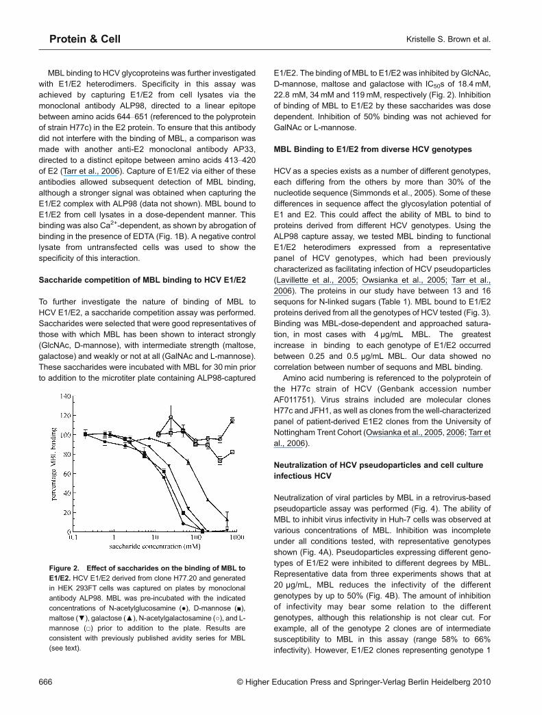
To further investigate the nature of binding of MBL toHCV E1/E2, a saccharide competition assay was performed.Saccharides were selected that were good representatives ofthose with which MBL has been shown to interact strongly(GlcNAc, D-mannose), with intermediate strength (maltose,galactose) and weakly or not at all (GalNAc and L-mannose).These saccharides were incubated with MBL for 30min priorto addition to the microtiter plate containing ALP98-captured
E1/E2. The binding of MBL to E1/E2 was inhibited by GlcNAc,D-mannose, maltose and galactose with IC50s of 18.4 mM,22.8 mM, 34mM and 119mM, respectively (Fig. 2). Inhibitionof binding of MBL to E1/E2 by these saccharides was dosedependent. Inhibition of 50% binding was not achieved forGalNAc or L-mannose.
MBL Binding to E1/E2 from diverse HCV genotypes
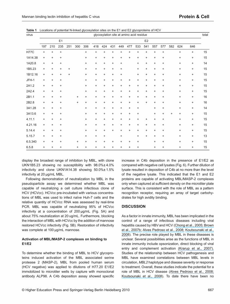
HCV as a species exists as a number of different genotypes,each differing from the others by more than 30% of thenucleotide sequence (Simmonds et al., 2005). Some of thesedifferences in sequence affect the glycosylation potential ofE1 and E2. This could affect the ability of MBL to bind toproteins derived from different HCV genotypes. Using theALP98 capture assay, we tested MBL binding to functionalE1/E2 heterodimers expressed from a representativepanel of HCV genotypes, which had been previouslycharacterized as facilitating infection of HCV pseudoparticles(Lavillette et al., 2005; Owsianka et al., 2005; Tarr et al.,2006). The proteins in our study have between 13 and 16sequons for N-linked sugars (Table 1). MBL bound to E1/E2proteins derived from all the genotypes of HCV tested (Fig. 3).Binding was MBL-dose-dependent and approached satura-tion, in most cases with 4 μg/mL MBL. The greatestincrease in binding to each genotype of E1/E2 occurredbetween 0.25 and 0.5 µg/mL MBL. Our data showed nocorrelation between number of sequons and MBL binding.
Amino acid numbering is referenced to the polyprotein ofthe H77c strain of HCV (Genbank accession numberAF011751). Virus strains included are molecular clonesH77c and JFH1, as well as clones from the well-characterizedpanel of patient-derived E1E2 clones from the University ofNottingham Trent Cohort (Owsianka et al., 2005, 2006; Tarr etal., 2006).
Neutralization of HCV pseudoparticles and cell cultureinfectious HCV
Neutralization of viral particles by MBL in a retrovirus-basedpseudoparticle assay was performed (Fig. 4). The ability ofMBL to inhibit virus infectivity in Huh-7 cells was observed atvarious concentrations of MBL. Inhibition was incompleteunder all conditions tested, with representative genotypesshown (Fig. 4A). Pseudoparticles expressing different geno-types of E1/E2 were inhibited to different degrees by MBL.Representative data from three experiments shows that at20 μg/mL, MBL reduces the infectivity of the differentgenotypes by up to 50% (Fig. 4B). The amount of inhibitionof infectivity may bear some relation to the differentgenotypes, although this relationship is not clear cut. Forexample, all of the genotype 2 clones are of intermediatesusceptibility to MBL in this assay (range 58% to 66%infectivity). However, E1/E2 clones representing genotype 1
Protein & Cell
Figure 2. Effect of saccharides on the binding of MBL toE1/E2. HCV E1/E2 derived from clone H77.20 and generatedin HEK 293FT cells was captured on plates by monoclonalantibody ALP98. MBL was pre-incubated with the indicated
concentrations of N-acetylglucosamine (●), D-mannose (■),maltose (▼), galactose (▲), N-acetylgalactosamine (○), and L-mannose (□) prior to addition to the plate. Results are
consistent with previously published avidity series for MBL(see text).
666 © Higher Education Press and Springer-Verlag Berlin Heidelberg 2010
Kristelle S. Brown et al.
display the broadest range of inhibition by MBL, with cloneUKN1B5.23 showing no susceptibility with 98.0%± 4.0%infectivity and clone UKN1A14.38 showing 50.0%± 1.5%infectivity at 20 μg/mL MBL.
Following demonstration of neutralization by MBL in thepseudoparticle assay we determined whether MBL wascapable of neutralizing a cell culture infectious clone ofHCV (HCVcc). HCVcc pre-incubated with various concentra-tions of MBL was used to infect naïve Huh-7 cells and therelative quantity of HCVcc RNA was assessed by real-timePCR. MBL was capable of neutralizing 95% of HCVccinfectivity at a concentration of 200 µg/mL (Fig. 5A) andabout 75% neutralization at 20 ug/mL. Furthermore, blockingthe interaction of MBL with HCVcc by the addition of mannoserestored HCVcc infectivity (Fig. 5B). Restoration of infectivitywas complete at 100 µg/mL mannose.
Activation of MBL/MASP-2 complexes on binding toE1/E2
To determine whether the binding of MBL to HCV glycopro-teins induced activation of the MBL associated serineprotease 2 (MASP-2), MBL from pooled human serum(HCV negative) was applied to dilutions of H77.20 E1/E2immobilized to microtiter wells by capture with monoclonalantibody ALP98. A C4b deposition assay showed specific
increase in C4b deposition in the presence of E1/E2 ascompared with negative cell lysates (Fig. 6). Further dilution oflysate resulted in deposition of C4b at no more than the levelof the negative lysate. This indicated that the E1 and E2proteins are capable of activating MBL/MASP-2 complexesonly when captured at sufficient density on the microtiter platesurface. This is consistent with the role of MBL as a patternrecognition receptor, requiring an array of target carbohy-drates for high avidity binding.
DISCUSSION
As a factor in innate immunity, MBL has been implicated in thecontrol of a range of infectious diseases including viralhepatitis caused by HBVand HCV (Chong et al., 2005; Brownet al., 2007b; Alves Pedroso et al., 2008; Koutsounaki et al.,2008). The precise role played by MBL in these diseases isunclear. Several possibilities arise as the functions of MBL ininnate immunity include opsonization, direct blocking of viralentry and complement activation (Krarup et al., 2007).Studies of the relationship between HCV pathogenesis andMBL have examined correlations between MBL levels incirculation,MBL2 haplotype and disease severity or responseto treatment. Overall, these studies indicate the potential for arole of MBL in HCV disease (Alves Pedroso et al., 2008;Koutsounaki et al., 2008). To date there have been no
Table 1 Locations of potential N-linked glycosylation sites on the E1 and E2 glycoproteins of HCV
virus glycosylation site at amino acid residue total
E1 E2
197 210 235 251 300 306 418 424 431 449 477 533 541 557 577 582 624 646
H77C + + + + + + + + + + + + + + + 15
1A14.38 + + + + + + + + + + + + + + + 15
1A20.8 + + + + + + + + + + + + + + 14
1B5.23 + + + + + + + + + + + + + + + 15
1B12.16 + + + + + + + + + + + + + + + 15
JFH-1 + + + + + + + + + + + + + + + 15
2A1.2 + + + + + + + + + + + + + + + 15
2A2.4 + + + + + + + + + + + + + + + 15
2B1.1 + + + + + + + + + + + + + + + + 16
2B2.8 + + + + + + + + + + + + + + + + 16
3A1.28 + + + + + + + + + + + + + + 14
3A13.6 + + + + + + + + + + + + + + + 15
4.11.1 + + + + + + + + + + + + + + + 15
4.21.16 + + + + + + + + + + + + + + + 15
5.14.4 + + + + + + + + + + + + + + + 15
5.15.7 + + + + + + + + + + + + + 13
6.5.340 + + + + + + + + + + + + + + + 15
6.5.8 + + + + + + + + + + + + + + + 15
© Higher Education Press and Springer-Verlag Berlin Heidelberg 2010 667
Mannan binding lectin inhibition of hepatitis C virus Protein & Cell

published studies on the biochemical interaction of MBL withHCV and only one measuring MBL function in relation todisease (Brown et al., 2007a). Here we demonstrate MBLbinding to glycoproteins derived from diverse genotypes ofHCV and show that such binding is sufficient to inhibit virionsfrom infecting cells.
The binding of MBL to target surfaces relies on theinteraction of its lectin domains with an array of carbohydratesacross different N-linked glycans. The conformation of lectindomains in native MBL favors high avidity binding only to sucharrays as are found on microbial surfaces and not on hostglycoproteins (Hart et al., 2003). MBL has a preference for
Protein & Cell
Figure 3. Analysis of the binding of MBL to HCV E1/E2 from a diverse range of viral genotypes. Lysates of HEK 293FTcellscontaining viral glycoproteins derived from genotypes 1a, 1b (A), 2a, 2b (B), 3a, 3b, 4 (C), 5 and 6 (D) or negative lysates (○) werecaptured in wells by monoclonal antibody ALP98 or AP33 as appropriate. Clones were selected on the basis that they formedfunctional glycoproteins as determined by the pseudotype assay described previously (Lavillette et al., 2005; Owsianka et al., 2005;
Tarr et al., 2006). (A) Genotype 1 clones were H77.20 (n), UKN1A14.38 (●), UKN1A20.8 (▼), UKN1B5.23 (▲), and UKN1B12.16(◆). (B) Genotype 2 clones were JFH-1 (n), UKN2A1.2 (●), UKN2A2.4 (▼), UKN2B1.1 (▲) and UKN2B2.8 (◆). (C) Genotype 3 and4 clones were UKN3A1.28 (n), UKN3A13.6 (●), UKN4.11.1 (▼) and UKN4.21.1 (▲). (D) Genotype 5 and 6 clones were UKN5.14.4
(n), UKN5.15.7 (●), UKN6.5.340 (▼) and UKN6.5.8 (▲). MBL was applied as indicated in a twofold dilution series and was detectedby ELISA.
Figure 4. Neutralization of HCV pseudoparticles (HCVpp) derived from diverse genotypes by MBL. (A) HCVpp derived fromclones H77 (n), JFH-1 (▼), UKN 1B5.23 (▲) and UKN 4.21.16 ( ◆) were pre-incubated with MBL at the indicated concentrations
prior to infection of Huh-7 cells. The neutralization activity of MBL is expressed as percentage of inhibition of the infectious titers. (B) Acomparison of the sensitivity of HCVpp, displaying glycoproteins derived from different genotypes, to MBL at 20 μg/mL.
668 © Higher Education Press and Springer-Verlag Berlin Heidelberg 2010
Kristelle S. Brown et al.

binding to terminal mannose and N-acetyl glucosamineresidues. The precise avidity of MBL binding to a targettherefore depends upon both the carbohydrate structurespresent and their spatial orientation. We have demonstratedthat the glycoproteins derived from different HCV genotypesall bind MBL, but to differing extents. Differences in bindingoccurred between glycoproteins derived from two viruses ofthe same genotype that differed in the number of N-linkedglycan sequons present. This was highlighted by the twogenotype 3 isolates and the two genotype 5 isolates (Fig. 3).However, there was no consistent pattern to the relative levelsof binding based on the number of glycosylation sequons.Production of glycoproteins with individual glycan knockoutmutations would permit characterization of binding in com-parison to wild-type proteins. This may provide furtherevidence for binding of MBL to differentially glycosylated
glycoproteins. Differences in the number of potential glyco-sylation sites occupied by glycans have been linked to HCVcell entry and the accessibility of neutralizing antibodies toepitopes on E2 (Goffard and Dubuisson, 2003; Helle et al.,2007). MBL may preferentially bind glycoproteins where thenumber, or positions, of glycans present inhibit antibodyneutralization.
Binding of MBL to HCV glycoproteins, and subsequentinhibition of infectivity in in vitro models demonstrates thatessential interactions in the entry steps of the virus areblocked byMBL. This inhibitory effect has also been observedfor the bacterial lectin cyanovirin-N (Helle et al., 2006), whichblocks the interaction with the CD81 entry receptor. It remainsto be determined which interactions are specifically blockedby MBL. Our current data demonstrates that MBL at a levelwithin the range observed in HCV patients in vivo (Brown etal., 2007a) is capable of blocking infectivity in all thegenotypes tested to varying degrees, and as such musttarget a conserved step in the entry process. It was previouslyshown that neutralization of influenza A by MBL was relatedto the number of potential glycosylation sites present (Read-ing et al., 1997). A clearly defined relationship betweennumbers of N-linked glycan sequons and MBL neutralizationdoes not exist for the HCV pseudoparticles tested here.However, there does seem to be a trend toward those morehighly glycosylated on E1 being more susceptible to MBLneutralization. The lack of a clear association betweennumber of potential glycosylation sites and neutralizationpotentially indicates a difference between MBL binding torecombinant glycoproteins as opposed to virus particles.There is likely to be a more subtle effect of overall spatialarrangement of glycans on the surface of the virion asopposed to soluble proteins. Furthermore, it is possible thattarget glycans are presented in different arrays on the surfaceof HCVpp and HCVcc. For JFH-1 envelope sequences, MBLwas able to neutralize HCVcc better than HCVpp. Thisdifference has been observed for neutralizing monoclonalantibodies (Owsianka et al., 2008). Subtle differencesbetween proteins in the context of sE2, recombinant E1/E2,
Figure 5. Inhibition of HCVcc by MBL. (A) Purified MBL was incubated with HCVcc virions before adding to target cells. Inhibition
was determined for serial dilutions of MBL as determined by the reduction in relative quantity (RQ) of HCVcc RNA. (B) Using20 μg/mL MBL to neutralize entry of HCVcc, infectivity was restored with the addition of mannose, in a dose-dependent manner.
Figure 6. Activation of MBL/MASP-2 complexes wasdetermined by the level of C4b deposited after binding toE1/E2 (●) or negative lysate (○). MBL/MASP-2 complex
activity is expressed in arbitrary units based on C4b depositionas measured by ELISA.
© Higher Education Press and Springer-Verlag Berlin Heidelberg 2010 669
Mannan binding lectin inhibition of hepatitis C virus Protein & Cell

virus like particles or pseudoparticles have previously beendemonstrated (Owsianka et al., 2001; Clayton et al., 2002).
All potential glycosylation sequons in the envelopeglycoproteins of HCV have been described as being occupiedby glycans, except for a C-terminal position in E1 which isfollowed by a proline residue (Goffard and Dubuisson, 2003).These glycans display microheterogeneity, with the majoritybeing occupied by high mannose, and a minority havingcomplex oligosaccharides (Iacob et al., 2008). However,these studies only examined individual strains of HCV.Comparisons between different genotypes with equal num-bers of N-linked glycan sequons, as used in the present study,are difficult to interpret as there is limited information on whichof the sequons in each strain are modified, and to what extent.The fine structure of these proteins is likely to be subtlydifferent. Studies to determine precise structures of glycanspresent on both E1 and E2, together with the spatialarrangements of these glycans, would aid understanding ofthe role of MBL in binding to HCV. It would be informative tolook at different combinations of glycosylation in the context ofthe same polypeptide backbone, to determine the specificglycans required for MBL binding. It also remains to bedetermined if the different glycosylation states of the glycansmodulates MBL binding. For influenza, glycans attachedneuraminidase (Malhotra et al., 1994) or to the globular headof HA conferred sensitivity to collectins (Reading et al., 1997).
We have demonstrated here that binding of MBL to HCVglycoproteins is sufficient to activate the complement systemvia deposition of C4b. Complement was previously shown toenhance antibody neutralization of HCVpp (Meyer et al.,2002), suggesting that MBL-mediated deposition of comple-ment could be involved in clearance of viral particles. C4activity has also been shown to correlate with successfulresponse to therapy (Dumestre-Perard et al., 2002). Thisindicates that the role of MBL in HCV disease may includeblocking viral entry and activation of the complement system.Complement has been implicated in clearance of HIV virions(Aasa-Chapman et al., 2005). It was also recently demon-strated that L-ficolin, a protein structurally and functionallyrelated to MBL, can activate complement on binding to HCV(Liu et al., 2009). This conserved function of collagenous,complement-activating proteins is likely to be an importantelement of the immune response to viral infections.
Previous studies concerning MBL and HCV have focusedon clinical data. They have linked MBL polymorphisms todisease severity or potential response to treatment, associat-ing mutant alleles with more aggressive disease or a poorresponse to therapy (Matsushita et al., 1998a, b; Sasaki et al.,2000; Alves Pedroso et al., 2008; Koutsounaki et al., 2008).MBL polymorphisms affect circulating levels of the proteinand the number of lectin domains per molecule. Some studiesexamined the relationship of serum levels of MBL were withdisease outcomes (Kilpatrick et al., 2003; Brown et al., 2007a;
Alves Pedroso et al., 2008; Koutsounaki et al., 2008). Thesestudies implicate MBL in the pathogenesis of chronic HCVdisease. However, these findings might be indicative of otherunderlying factors influencing HCV disease. Polymorphismsof MBL have been linked with susceptibility to hepatocellularcarcinoma or cirrhosis in individuals infected with hepatitis Bvirus (Chong et al., 2005). This may be linked to an inability toclear virus as MBL activates complement on binding to HBVsurface antigen. Further studies are required to confirm therole of MBL in HCV disease.
Taken together, our results support the idea that MBL mayplay a role in HCV disease, through blocking entry of virionsinto host cells and activating the complement system to clearviral particles and recruit other elements of the immunesystem. The precise nature of this activity is likely to beinfluenced by the extent of glycosylation of the viralglycoproteins and the spatial arrangement of the N-linkedglycans; factors which differ across and within genotypes.Further studies are warranted to investigate the result of MBLbinding to viral particles in terms of complement activationand opsonization of viral particles.
MATERIALS AND METHODS
Cell culture
Human hepatoma cells Huh-7 (Nakabayashi et al., 1982) and human
epithelial kidney (HEK) 293FT cells (Invitrogen) were grown inDulbecco’ s modified Eagle’s medium, (Invitrogen) supplementedwith 10% fetal calf serum, 5% non-essential amino acids, and 2mM
glutamine (Sigma).
Viral glycoproteins
HCV E2 was produced and purified as previously described, withsome modifications (Yamada et al., 2005). Briefly, HEK 293FTcells inOptiMEM (Invitrogen) were transfected with plasmid expressing
soluble E2 fused to 6 × his tag (sE2661 or sE2) using Lipofectamine2000 (Invitrogen). Supernatant was harvested after 72 h and 20mMiodoacetamide was added. Supernatants were centrifuged to remove
debris, adjusted to 300mM NaCl and loaded onto a HisTrap column(1mL: GE Healthcare). HCV sE2 was eluted with 150mM imidazolefollowing a 50mM imidazole wash. Fractions were concentrated on a
Vivaspin column (Vivascience). Concentrated sE2 was subjected tosize exclusion chromatography on a TSK 3000SWXL column (7.5 ×300mm: TosoHaas). Fractions of monomeric sE2 were retained forbinding studies.
HCV E1/E2 heterodimer was produced in HEK 293FT cells aspreviously described (Owsianka et al., 2005). E1/E2 clones arenamed here using the nomenclature of the well-defined UKN
glycoprotein panel (Lavillette et al., 2005; Owsianka et al., 2005;Tarr et al., 2006). Transiently transfected cells were harvested after72 h by lysis in 50mMTris-HCl, 150mMNaCl, 20mM Iodoacetamide,
1% NP-40, pH 7.6. A sample of cell lysate was analyzed for E1/E2content. The remainder of the cell lysate was stored frozen at −80°Cuntil assayed.
Protein & Cell
670 © Higher Education Press and Springer-Verlag Berlin Heidelberg 2010
Kristelle S. Brown et al.

Antibodies
Polyclonal anti-human MBL was raised in rabbits against recombi-
nant MBL and was adsorbed to remove anti-mannan antibodiesas described previously (Arnold et al., 2004). Mouse monoclonalanti-HCV E2 antibodies ALP98 and AP33 were raised againstrecombinant soluble E2 as previously described (Clayton et al.,
2002).
Mannan binding lectin
Human MBL-MASPs complexes were purified from 1 L of pooledcitrated plasma (HD Supplies), free from anti-HCVantibodies, using amethod adapted from Tan et al. (Tan et al., 1996). Briefly, serum was
generated from the plasma, then PEG 3350 (Sigma) added to a finalconcentration of 7% (w/v) and the mixture stirred overnight at 4°C.Precipitated proteins were dissolved in ice-cold TBS-TCa2+ Buffer(50mM Tris-HCl, 1 M NaCl, 20mM CaCl2, 0.05% Tween, pH 7.8) and
incubated with 25mL mannan-agarose (Sigma) for 2 h. Bound MBLwas eluted with TBS-EDTA Buffer (50mM Tris-HCl, 1 M NaCl, 10mMEDTA, pH 7.6). Contaminating IgG and IgM were removed using a
HiTrap Protein G HP column (GE Healthcare) and anti-human IgMagarose (Sigma) respectively. MBL purity was assessed by SDS-PAGE and quantified by ELISA as previously described (Arnold et al.,
2004). MASPS are not separated from MBL by these purificationprocedures. MBL without MASPs was obtained by dialyzing thepreparation into gel filtration running buffer (0.1 M sodium acetate,
0.2M NaCl, 5 mM EDTA, pH 5.0) and running on a Superose 6 gelfiltration column (GE Healthcare). The fractions judged to containMBL, were analyzed by SDS-PAGE and pooled. The resulting MBLwas pure and was used as a standard to quantify MBL by ELISA
(Arnold et al., 2004).
Detection of MBL binding to HCV sE2 by enzyme-linked
immunosorbent assay (ELISA)
ELISA plates (Maxisorp, Nunc) were coated with 50ml HCV sE2,mannan or BSA (all at 20 µg/mL) in coating buffer (0.1M NaHCO3 pH9.5) for 1 h at 37°C. After washing 3 times with phosphate bufferedsaline (PBS) 0.05% (v/v) Tween-20 (PBS-T) wells were incubated
with 200 µL blocking buffer (PBS-T containing 5% (w/v) milk powder)for 1 h at 37°C. The plates were washed 3 times with wash buffer(10 mM HEPES, 1 M NaCl, 5 mM CaCl2, 0.05% (v/v) Tween-20, pH
7.6). MBL was diluted in wash buffer or EDTA Buffer (10mM HEPES,1M NaCl, 5 mM EDTA, 0.05% (v/v) Tween-20, pH 7.6) in a 3-foldserial dilution (3 mg/mL to 12 ng/mL) and 50 μL was added to the
appropriate wells. Plates were incubated with MBL overnight at 4°C.Washing was repeated and 50 µL of anti-rMBL, diluted 1/700 in washbuffer, was added per well for incubation at 37°C for 1 h. Wells were
washed and incubated with 50 µL of 1.3 µg/mL alkaline phosphatase(AP)-conjugated monoclonal anti-rabbit IgG (Sigma) in washbuffer. Following 3 final washes, 50 µL of pNPP (ρ-NitrophenylPhosphate) substrate (Sigma) was added to each well. The OD at
405 nm was determined after 30min incubation at roomtemperature.
Detection of MBL binding to HCV E1/E2 by capture ELISA
Each well of a 96 well microtiter plate (Nunc) was coated with 50 µL of
10 µg/mL mouse monoclonal antibody ALP98 in coating buffer andincubated for 1 h at 37°C. Blocking was then performed with 200 µLper well of PBS-Tovernight at 4°C. Three washes per well were then
performed with wash buffer. Next, 50 µL of H77c (47) E1/E2 lysateswere added to each well, at 1/10 dilution in PBS-T, then incubated for1 h at 37°C. Wells were washed again three times with wash buffer.
Two fold serial dilutions of MBL were made in wash buffer, or EDTAbuffer to give final concentrations of MBL from 4 μg/mL to 31 ng/mL.MBL dilutions were added in triplicate to each appropriate well and
incubated at 37°C for 1 h (50 μL/well). Bound MBL was detected asdescribed for the sE2 assay.
Binding of MBL to representative clones of each HCV genotype ofE1/E2 was performed by the above method. Equivalent concentra-
tions of each E1/E2 were calculated experimentally as previouslydescribed (Clayton et al., 2002). Briefly, proteins were captured withGalanthus nivalis agglutinin (GNA; Sigma) and bound proteins
detected with ALP98 followed by anti-mouse IgG AP conjugate andpNPP substrate. The dilution of lysate at which equivalent ODs wereobtained was subsequently used in the MBL assay.
Saccharide competition assay
To test for the inhibition of binding by saccharides, E1/E2 glycopro-teins were captured on microtiter plates by monoclonal antibody
ALP98 as described above. MBL (1 μg/mL) was incubated withvarious concentrations of each saccharide for 30min at 20°C beforeaddition to the plate. Bound MBL was detected as above. IC50s were
determined from inhibition curves.
HCVpp infection and neutralization assays
Full-length E1/E2 (representing amino acid residues 170 to 746 of theHCV open reading frame referenced to strain H77c (Yanagi et al.,1997)) clones were generated and their nucleotide sequence
determined as previously described (Lavillette et al., 2005). HCVppwere produced essentially as previously described (Bartosch et al.,2003) by cotransfection of plasmids expressing the full-length E1/E2,murine leukemia virus (MLV) Gag-Pol and the MLV transfer vector
carrying either the green fluorescent protein (GFP) or the fireflyluciferase gene under the control of the human cytomegaloviruspromoter. MBL-mediated neutralization of HCVpp was carried out in a
similar manner to that previously described for antibody neutralization(Owsianka et al., 2005). MBL was pre-incubated with HCVpp for30 min at 37°C in 3-fold serial dilutions from 180 μg/mL to 0.25 μg/mL
and then the particles were used to infect Huh-7 cells to observeinhibition of infectivity. Infectivity was expressed as percentage ofreporter gene activity compared with uninhibited control.
Production of cell culture infectious HCV (HCVcc) and
neutralization assays
HCVcc was generated essentially as described by Wakita et al.(2005). Briefly, the plasmid pJFH1-pUC carrying the full-length cDNA
of the genotype 2a HCV strain JFH-1 was linearized with XbaI andtreated with mung bean nuclease. The linearized construct was thenused as a template to generate viral genomic RNA by in vitro
transcription. Approximately 10 µg of in vitro synthesized RNA waselectroporated into Huh-7 cells in a 0.4 cm Gene Pulser cuvette (Bio-Rad) and pulsed once at 960 µF and 270V using the GenePulser
© Higher Education Press and Springer-Verlag Berlin Heidelberg 2010 671
Mannan binding lectin inhibition of hepatitis C virus Protein & Cell

Xcell (Bio-Rad) electroporator. The transfected cells were immedi-ately mixed with cell medium and seeded into a tissue culture dish orflask. Following incubation at 37°C for 4 d, the medium containing the
infectious virus progeny was clarified by brief centrifugation to removecell debris, filtered through 0.45 µm pore-sized membrane, and usedto infect naïve Huh-7 cells.
To perform MBL-mediated neutralization of HCVcc, the filteredmedium containing HCVcc was mixed with appropriate amounts ofMBL and incubated for 1 h at 37°C prior to application to the naïve
Huh-7 cells. Following incubation at 37°C for 4 d, cells wereinvestigated for HCVcc RNA by quantitative RT-PCR (qRT-PCR).Total RNA was isolated using an RNeasy mini kit (Qiagen). For Real-Time analysis of total RNA from infected cells, a relative quantification
(RQ) reaction was performed, where each sample was normalized toan endogenous control gene (GAPDH). First, cDNA was generatedby reverse transcription of the total RNA using TaqMan Reverse
Transcription Reagents (Applied Biosystems), with random primers.Each cDNA was analyzed in triplicate in a singleplex reaction todetect either HCV target cDNA with a Fluorescein-CE Phosphor-
amidite (6-FAM) labeled probe (250 nM) and HCV specific primers(900 nM each), or GAPDH with a pre-validated endogenous controlVIC-probe/primer mix (Applied Biosystems). The HCV probesequence was 6-FAM-AAAGGCCTTGTGGTACTG-MGB (synthe-
sized by Applied Biosystems), and primer sequences were: 5'-TCTGCGGAACCGGTGAGTAC-3', forward, and 5'- GCACTCG-CAAGCACCCTATC-3', reverse (Sigma). Real-time reactions were
run using TaqMan Fast Universal PCRMaster Mix, without ampEraseUNG, (Applied Biosystems) under Fast Universal conditions on a7500 Fast Real-Time PCR machine and data analyzed using 7500
Fast System Software (SDS v1.3.1) (Applied Biosystems).The specificity of MBL-mediated inhibition of virus infection was
confirmed by pre-incubating 20 µg/mLMBL with 10-fold dilution series
of mannose (Sigma) starting from 0 to 100 μg/mL at 37°C for 45min.This mix was then incubated with the virus for a further 45min at 37°Cbefore infecting the cells and the levels of infectivity were determinedby measuring viral RNA by qRT-PCR.
Activation of MBL/MASP-2 complex on binding to E1/E2
H77 E1/E2 was captured in the ALP98-coated wells of a Microfluor 2
white microtiter plate (Thermo Labsystems) as described above.Wells were blocked with 5% skimmed milk powder in PBS-T.Following three washes with PBS-T, wells were incubated with
serum diluted 1:1 in 40mM HEPES, 2 M NaCl, 10mM CaCl2, pH 7.4.Plates were incubated for 1 h on ice. MBL/MASP-2 complex activitywas assayed as previously described (Mayilyan et al., 2006) with
modifications. Wells were washed twice at 37°C with 20mM HEPES,1M NaCl, 5 mM CaCl2, 0.1% (v/v) Tween-20, pH 7.4 and twice with20mM HEPES, 140mM NaCl, 5 mM CaCl2, pH 7.4 (wash buffer).The use of 1M NaCl prevents C1q binding to the antibody on the
plate. The wells were incubated with 4 μg/mL purified humancomplement component C4 diluted in wash buffer then washed 3times in the same buffer. Rabbit anti-C4 (Sigma) diluted 1:1000 in
wash buffer was applied to the wells and incubated for 1 h at 20°C.After three washes, alkaline-phosphatase conjugated goat anti-rabbitIgG (Sigma), diluted 1:1000, was added to the wells. After three
washes with wash buffer, wells were developed with 200 µL of1 mM 4-methylumbelliferyl-phosphate (Calbiochem), in 5 mM 2-(N-cyclohexylamino) ethanesulfonic acid (CHES), 1 mM MgCl2, pH 9.8.
The fluorescent product was measured by excitation at 355 nm andemission at 460 nm using a microtiter plate reader (Fluoroskan,ThermoLife Sciences). MBL/MASP-2 complex activity was calculated
from the initial slope of the activity curve and expressed as arbitraryunits per minute. All samples were tested in triplicate.
ACKNOWLEDGEMENTS
We would like to thank Takaji Wakita, Jens Bukh, Mark Harris andFrancois-Loic Cosset for provision of reagents. Funding was providedby The Royal Society, The University of Nottingham, and the Medical
Research Council (UK).
ABBREVIATIONS
CHES, 2-(N-cyclohexylamino) ethanesulfonic acid; EDTA,
ethylenediaminetetraacetic acid; E1/E2, HCV E1 and E2 glycopro-teins; GNA, galanthus nivalis agglutinin; HCV, hepatitis C virus;HCVcc, cell cultured HCV; HCVpp, HCV pseudoparticle; HEPES, 4-
(2-hydroxyethyl)-1-piperazineethanesulfonic acid; MASP-2, MBL-associated serine protease 2; MBL, mannan binding lectin; MLV,murine leukaemia virus; PBS-T, phosphate buffered saline with
Tween-20; 6-FAM, fluorescein-CE phosphoramidite
REFERENCES
Aasa-Chapman, M.M., Holuigue, S., Aubin, K., Wong, M., Jones, N.
A., Cornforth, D., Pellegrino, P., Newton, P., Williams, I., Borrow, P.,et al. (2005). Detection of antibody-dependent complement-mediated inactivation of both autologous and heterologous virus
in primary human immunodeficiency virus type 1 infection. J Virol79, 2823–2830.
Alves Pedroso, M.L., Boldt, A.B., Pereira-Ferrari, L., Steffensen, R.,
Strauss, E., Jensenius, J.C., Ioshii, S.O., and Messias-Reason, I.(2008). Mannan-binding lectin MBL2 gene polymorphism inchronic hepatitis C: association with the severity of liver fibrosis
and response to interferon therapy. Clin Exp Immunol 152,258–264.
Anders, E.M., Hartley, C.A., and Jackson, D.C. (1990). Bovine and
mouse serum beta inhibitors of influenza A viruses are mannose-binding lectins. Proc Natl Acad Sci U S A 87, 4485–4489.
Anon. (1999). Global surveillance and control of hepatitis C. Report of
a WHO Consultation organized in collaboration with the ViralHepatitis Prevention Board, Antwerp, Belgium. J Viral Hepat 6,35–47.
Arnold, J.N., Radcliffe, C.M., Wormald, M.R., Royle, L., Harvey, D.J.,Crispin, M., Dwek, R.A., Sim, R.B., and Rudd, P.M. (2004). Theglycosylation of human serum IgD and IgE and the accessibility of
identified oligomannose structures for interaction with mannan-binding lectin. J Immunol 173, 6831–6840.
Bartosch, B., Dubuisson, J., and Cosset, F.L. (2003). Infectioushepatitis C virus pseudo-particles containing functional E1-E2envelope protein complexes. J Exp Med 197, 633–642.
Bouwman, L.H., Roos, A., Terpstra, O.T., de Knijff, P., van Hoek, B.,Verspaget, H.W., Berger, S.P., Daha, M.R., Frölich, M., van derSlik, A.R., et al. (2005). Mannose binding lectin gene polymorph-
isms confer a major risk for severe infections after livertransplantation. Gastroenterology 129, 408–414.
Brown, K.S., Keogh, M.J., Tagiuri, N., Grainge, M.J., Presanis, J.S.,
Protein & Cell
672 © Higher Education Press and Springer-Verlag Berlin Heidelberg 2010
Kristelle S. Brown et al.

Ryder, S.D., Irving, W.L., Ball, J.K., Sim, R.B., and Hickling, T.P.(2007a). Severe fibrosis in hepatitis C virus-infected patients isassociated with increased activity of the mannan-binding lectin
(MBL)/MBL-associated serine protease 1 (MASP-1) complex. ClinExp Immunol 147, 90–98.
Brown, K.S., Ryder, S.D., Irving, W.L., Sim, R.B., and Hickling, T.P.
(2007b). Mannan binding lectin and viral hepatitis. Immunol Lett108, 34–44.
Chong, W.P., To, Y.F., Ip, W.K., Yuen, M.F., Poon, T.P., Wong, W.H.,Lai, C.L., and Lau, Y.L. (2005). Mannose-binding lectin in chronichepatitis B virus infection. Hepatology 42, 1037–1045.
Clayton, R.F., Owsianka, A., Aitken, J., Graham, S., Bhella, D., andPatel, A.H. (2002). Analysis of antigenicity and topology of E2glycoprotein present on recombinant hepatitis C virus-like parti-
cles. J Virol 76, 7672–7682.
Dumestre-Perard, C., Ponard, D., Drouet, C., Leroy, V., Zarski, J.P.,Dutertre, N., and Colomb, M.G. (2002). Complement C4 monitor-
ing in the follow-up of chronic hepatitis C treatment. Clin ExpImmunol 127, 131–136.
Endo, M., Ohsawa, I., Ohi, H., Fujita, T., Matsushita, M., and Fujita, T.
(2001). Mannose-binding lectin contributes to glomerulonephritisinduced by hepatitis C virus infection. Nephron 87, 374–375.
Farci, P., Alter, H.J., Wong, D.C., Miller, R.H., Govindarajan, S., Engle,R., Shapiro, M., and Purcell, R.H. (1994). Prevention of hepatitis Cvirus infection in chimpanzees after antibody-mediated in vitroneutralization. Proc Natl Acad Sci U S A 91, 7792–7796.
Gerlach, J.T., Diepolder, H.M., Jung, M.C., Gruener, N.H., Schraut, W.W., Zachoval, R., Hoffmann, R., Schirren, C.A., Santantonio, T.,
and Pape, G.R. (1999). Recurrence of hepatitis C virus after loss ofvirus-specific CD4(+) T-cell response in acute hepatitis C.Gastroenterology 117, 933–941.
Goffard, A., and Dubuisson, J. (2003). Glycosylation of hepatitis Cvirus envelope proteins. Biochimie 85, 295–301.
Golden-Mason, L., Madrigal-Estebas, L., McGrath, E., Conroy, M.J.,
Ryan, E.J., Hegarty, J.E., O’Farrelly, C., and Doherty, D.G. (2008).Altered natural killer cell subset distributions in resolved andpersistent hepatitis C virus infection following single source
exposure. Gut 57, 1121–1128.
Hart, M.L., Saifuddin, M., and Spear, G.T. (2003). Glycosylationinhibitors and neuraminidase enhance human immunodeficiency
virus type 1 binding and neutralization by mannose-binding lectin.J Gen Virol 84, 353–360.
Helle, F., Goffard, A., Morel, V., Duverlie, G., McKeating, J., Keck, Z.Y., Foung, S., Penin, F., Dubuisson, J., and Voisset, C. (2007). Theneutralizing activity of anti-hepatitis C virus antibodies is modu-lated by specific glycans on the E2 envelope protein. J Virol 81,
8101–8111.
Helle, F., Wychowski, C., Vu-Dac, N., Gustafson, K.R., Voisset, C.,
and Dubuisson, J. (2006). Cyanovirin-N inhibits hepatitis C virusentry by binding to envelope protein glycans. J Biol Chem 281,25177–25183.
Iacob, R.E., Perdivara, I., Przybylski, M., and Tomer, K.B. (2008).Mass spectrometric characterization of glycosylation of hepatitisC virus E2 envelope glycoprotein reveals extended microheter-
ogeneity of N-glycans. J Am Soc Mass Spectrom 19, 428–444.
Ishii, K., Rosa, D., Watanabe, Y., Katayama, T., Harada, H., Wyatt, C.,Kiyosawa, K., Aizaki, H., Matsuura, Y., Houghton, M., et al. (1998).
High titers of antibodies inhibiting the binding of envelope to human
cells correlate with natural resolution of chronic hepatitis C.Hepatology 28, 1117–1120.
Ji, X., Gewurz, H., and Spear, G.T. (2005a). Mannose binding lectin(MBL) and HIV. Mol Immunol 42, 145–152.
Ji, X., Olinger, G.G., Aris, S., Chen, Y., Gewurz, H., and Spear, G.T.
(2005b). Mannose-binding lectin binds to Ebola and Marburgenvelope glycoproteins, resulting in blocking of virus interactionwith DC-SIGN and complement-mediated virus neutralization. J
Gen Virol 86, 2535–2542.
Kilpatrick, D.C., Delahooke, T.E., Koch, C., Turner, M.L., and Hayes,P.C. (2003). Mannan-binding lectin and hepatitis C infection. Clin
Exp Immunol 132, 92–95.
Kim, W.R. (2002). The burden of hepatitis C in the United States.Hepatology 36, S30–S34.
Koutsounaki, E., Goulielmos, G.N., Koulentaki, M., Choulaki, C.,Kouroumalis, E., and Galanakis, E. (2008). Mannose-binding lectin
MBL2 gene polymorphisms and outcome of hepatitis C virus-infected patients. J Clin Immunol 28, 495–500.
Krarup, A., Wallis, R., Presanis, J.S., Gál, P., Sim, R.B., and Sommer,
P. (2007). Simultaneous activation of complement and coagulationby MBL-associated serine protease 2. PLoS ONE 2, e623.
Lavillette, D., Tarr, A.W., Voisset, C., Donot, P., Bartosch, B., Bain, C.,
Patel, A.H., Dubuisson, J., Ball, J.K., and Cosset, F.L. (2005).Characterization of host-range and cell entry properties of themajor genotypes and subtypes of hepatitis C virus. Hepatology 41,
265–274.
Liu, J., Ali, M.A., Shi, Y., Zhao, Y., Luo, F., Yu, J., Xiang, T., Tang, J., Li,D., Hu, Q., et al. (2009). Specifically binding of L-ficolin to N-
glycans of HCV envelope glycoproteins E1 and E2 leads tocomplement activation. Cell Mol Immunol 6, 235–244.
Malhotra, R., Haurum, J.S., Thiel, S., and Sim, R.B. (1994). Binding ofhuman collectins (SP-A and MBP) to influenza virus. Biochem J304, 455–461.
Matsushita, M., Hijikata, M., Matsushita, M., Ohta, Y., and Mishiro, S.(1998b). Association of mannose-binding lectin gene haplotypeLXPA and LYPB with interferon-resistant hepatitis C virus infection
in Japanese patients. J Hepatol 29, 695–700.
Matsushita, M., Hijikata, M., Ohta, Y., Iwata, K., Matsumoto, M.,Nakao, K., Kanai, K., Yoshida, N., Baba, K., and Mishiro, S.
(1998a). Hepatitis C virus infection and mutations of mannose-binding lectin gene MBL. Arch Virol 143, 645–651.
Mayilyan, K.R., Presanis, J.S., Arnold, J.N., Hajela, K., and Sim, R.B.
(2006). Heterogeneity of MBL-MASP complexes. Mol Immunol 43,1286–1292.
Meyer, K., Basu, A., Przysiecki, C.T., Lagging, L.M., Di Bisceglie, A.M., Conley, A.J., and Ray, R. (2002). Complement-mediatedenhancement of antibody function for neutralization of pseudotypevirus containing hepatitis C virus E2 chimeric glycoprotein. J Virol
76, 2150–2158.
Nakabayashi, H., Taketa, K., Miyano, K., Yamane, T., and Sato, J.
(1982). Growth of human hepatoma cells lines with differentiatedfunctions in chemically defined medium. Cancer Res 42,3858–3863.
Owsianka, A., Clayton, R.F., Loomis-Price, L.D., McKeating, J.A., andPatel, A.H. (2001). Functional analysis of hepatitis C virus E2glycoproteins and virus-like particles reveals structural dissimila-
rities between different forms of E2. J Gen Virol 82, 1877–1883.
Owsianka, A., Tarr, A.W., Juttla, V.S., Lavillette, D., Bartosch, B.,
© Higher Education Press and Springer-Verlag Berlin Heidelberg 2010 673
Mannan binding lectin inhibition of hepatitis C virus Protein & Cell

Cosset, F.L., Ball, J.K., and Patel, A.H. (2005). Monoclonalantibody AP33 defines a broadly neutralizing epitope on thehepatitis C virus E2 envelope glycoprotein. J Virol 79,
11095–11104.
Owsianka, A.M., Tarr, A.W., Keck, Z.Y., Li, T.K., Witteveldt, J., Adair,R., Foung, S.K., Ball, J.K., and Patel, A.H. (2008). Broadly
neutralizing human monoclonal antibodies to the hepatitis Cvirus E2 glycoprotein. J Gen Virol 89, 653–659.
Pawlotsky, J.M. (2006). Therapy of hepatitis C: from empiricism toeradication. Hepatology 43, S207–S220.
Pestka, J.M., Zeisel, M.B., Bläser, E., Schürmann, P., Bartosch, B.,
Cosset, F.L., Patel, A.H., Meisel, H., Baumert, J., Viazov, S., et al.(2007). Rapid induction of virus-neutralizing antibodies and viralclearance in a single-source outbreak of hepatitis C. Proc Natl
Acad Sci U S A 104, 6025–6030.
Pulendran, B., and Ahmed, R. (2006). Translating innate immunityinto immunological memory: implications for vaccine development.
Cell 124, 849–863.
Reading, P.C., Morey, L.S., Crouch, E.C., and Anders, E.M. (1997).Collectin-mediated antiviral host defense of the lung: evidence
from influenza virus infection of mice. J Virol 71, 8204–8212.
Sanders, C.J., Yu, Y., Moore, D.A. 3rd, Williams, I.R., and Gewirtz, A.
T. (2006). Humoral immune response to flagellin requires T cellsand activation of innate immunity. J Immunol 177, 2810–2818.
Sasaki, K., Tsutsumi, A., Wakamiya, N., Ohtani, K., Suzuki, Y.,
Watanabe, Y., Nakayama, N., and Koike, T., and the K. Sasaki, A.Tsutsumi, N. Wakamiya. (2000). Mannose-binding lectin poly-morphisms in patients with hepatitis C virus infection. Scand J
Gastroenterol 35, 960–965.
Simmonds, P., Bukh, J., Combet, C., Deléage, G., Enomoto, N.,Feinstone, S., Halfon, P., Inchauspé, G., Kuiken, C., Maertens, G.,
et al. (2005). Consensus proposals for a unified system ofnomenclature of hepatitis C virus genotypes. Hepatology 42,962–973.
Steinmann, D., Barth, H., Gissler, B., Schürmann, P., Adah, M.I.,Gerlach, J.T., Pape, G.R., Depla, E., Jacobs, D., Maertens, G., etal. (2004). Inhibition of hepatitis C virus-like particle binding to
target cells by antiviral antibodies in acute and chronic hepatitis C.J Virol 78, 9030–9040.
Takehara, T., and Hayashi, N. (2005). Natural killer cells in hepatitis C
virus infection: from innate immunity to adaptive immunity. ClinGastroenterol Hepatol 3, S78–S81.
Tan, S.M., Chung, M.C., Kon, O.L., Thiel, S., Lee, S.H., and Lu, J.(1996). Improvements on the purification of mannan-binding lectinand demonstration of its Ca(2 +)-independent association with aC1s-like serine protease. Biochem J 319, 329–332.
Tarr, A.W., Owsianka, A.M., Timms, J.M., McClure, C.P., Brown, R.J.,
Hickling, T.P., Pietschmann, T., Bartenschlager, R., Patel, A.H.,and Ball, J.K. (2006). Characterization of the hepatitis C virus E2epitope defined by the broadly neutralizing monoclonal antibody
AP33. Hepatology 43, 592–601.
Thiel, S., Holmskov, U., Hviid, L., Laursen, S.B., and Jensenius, J.C.(1992). The concentration of the C-type lectin, mannan-binding
protein, in human plasma increases during an acute phaseresponse. Clin Exp Immunol 90, 31–35.
Thiel, S., Vorup-Jensen, T., Stover, C.M., Schwaeble, W., Laursen, S.B., Poulsen, K., Willis, A.C., Eggleton, P., Hansen, S., Holmskov,U., et al. (1997). A second serine protease associated withmannan-binding lectin that activates complement. Nature 386,
506–510.
Thimme, R., Oldach, D., Chang, K.M., Steiger, C., Ray, S.C., and
Chisari, F.V. (2001). Determinants of viral clearance and persis-tence during acute hepatitis C virus infection. J Exp Med 194,1395–1406.
Vallinoto, A.C., da Silva, R.F., Hermes, R.B., Amaral, I.S., Miranda, E.C., Barbosa, M.S., Moia, L.J., Conde, S.R., Soares, M.C., Lemos,J.A., et al. (2009). Mannose-binding lectin gene polymorphisms
are not associated with susceptibility to hepatitis C virus infection inthe Brazilian Amazon region. Hum Immunol 70, 754–757.
Wakita, T., Pietschmann, T., Kato, T., Date, T., Miyamoto, M., Zhao, Z.,
Murthy, K., Habermann, A., Kräusslich, H.G., Mizokami, M., et al.(2005). Production of infectious hepatitis C virus in tissue culturefrom a cloned viral genome. Nat Med 11, 791–796.
Weis, W.I., Drickamer, K., and Hendrickson, W.A. (1992). Structure ofa C-type mannose-binding protein complexed with an oligosac-charide. Nature 360, 127–134.
Yamada, E., Montoya, M., Schuettler, C.G., Hickling, T.P., Tarr, A.W.,Vitelli, A., Dubuisson, J., Patel, A.H., Ball, J.K., and Borrow, P.(2005). Analysis of the binding of hepatitis C virus genotype 1a and
1b E2 glycoproteins to peripheral blood mononuclear cell subsets.J Gen Virol 86, 2507–2512.
Yanagi, M., Purcell, R.H., Emerson, S.U., and Bukh, J. (1997).Transcripts from a single full-length cDNA clone of hepatitis C virusare infectious when directly transfected into the liver of achimpanzee. Proc Natl Acad Sci U S A 94, 8738–8743.
Ying, H., Ji, X., Hart, M.L., Gupta, K., Saifuddin, M., Zariffard, M.R.,and Spear, G.T. (2004). Interaction of mannose-binding lectin with
HIV type 1 is sufficient for virus opsonization but not neutralization.AIDS Res Hum Retroviruses 20, 327–335.
Zhang, M., Rosenberg, P.S., Brown, D.L., Preiss, L., Konkle, B.A.,
Eyster, M.E., and Goedert, J.J., and the Second MulticenterHemophilia Cohort Study. (2006). Correlates of spontaneousclearance of hepatitis C virus among people with hemophilia.
Blood 107, 892–897.
Protein & Cell
674 © Higher Education Press and Springer-Verlag Berlin Heidelberg 2010
Kristelle S. Brown et al.