DFT Mechanistic Proposal of the Ruthenium Porphyrin-Catalyzed Allylic Amination by Organic Azides
Upload
independentCategory
view
0download
0
Molecules 2012, 17, 10586-10613; doi:10.3390/molecules170910586
molecules ISSN 1420-3049
www.mdpi.com/journal/molecules
Review
Specific Binding of Anionic Porphyrin and Phthalocyanine to the G-Quadruplex with a Variety of in Vitro and in Vivo Applications
Hidenobu Yaku 1,2,3,*, Takashi Murashima 2,3, Daisuke Miyoshi 2,3 and Naoki Sugimoto 2,3
1 Advanced Technology Research Laboratories, Panasonic Corporation, 3-4 Hikaridai, Seika-cho,
Soraku-gun, Kyoto 619-0237, Japan 2 Frontier Institute for Biomolecular Engineering Research (FIBER), Konan University,
7-1-20 Minatojima-minamimachi, Chuo-ku, Kobe 650-0047, Japan;
E-Mails: [email protected] (T.M.); [email protected] (D.M.);
[email protected] (N.S.) 3 Faculty of Frontiers of Innovative Research in Science and Technology (FIRST), Konan University,
7-1-20 Minatojima-minamimachi, Chuo-ku, Kobe 650-0047, Japan
* Author to whom correspondence should be addressed; E-Mail: [email protected];
Tel.: +81-774-98-2580; Fax: +81-774-98-2585.
Received: 31 July 2012; in revised form: 27 August 2012 / Accepted: 29 August 2012 /
Published: 5 September 2012
Abstract: The G-quadruplex, a four-stranded DNA structure with stacked guanine tetrads
(G-quartets), has recently been attracting attention because of its critical roles in vitro and
in vivo. In particular, the G-quadruplex functions as ligands for metal ions and aptamers for
various molecules. Interestingly, the G-quadruplex can show peroxidase-like activity with
an anionic porphyrin, iron (III) protoporphyrin IX (hemin). Importantly, hemin binds to
G-quadruplexes with high selectivity over single-stranded DNA (ssDNA) and
double-stranded DNA (dsDNA), which is attributable to an electrostatic repulsion of
phosphate groups in ssDNA and dsDNA. The G-quadruplex and hemin-G-quadruplex
complex allow development of sensing techniques to detect DNA, metal ions and proteins.
In addition to hemin, anionic phthalocyanines also bind to the G-quadruplex formed by
human telomere DNA, specifically over ssDNA and dsDNA. Since the binding of anionic
phthalocyanines to the G-quadruplex causes an inhibition of telomerase activity, which
plays a role in the immortal growth of cancer cells, anionic phthalocyanines are promising
as novel anticancer drug candidates. This review focuses on the specific binding of hemin
OPEN ACCESS
Molecules 2012, 17 10587
and anionic phthalocyanines to G-quadruplexes and the applications in vitro and in vivo of
this binding property.
Keywords: anionic phthalocyanine; DNAzyme; G-quadruplex; hemin; peroxidase; telomerase
1. Introduction
The fabrication of functional nanomaterials, which includes functional dyes (porphyrins and
phthalocyanines) [1–3], carbon nanomaterials (fullerenes, carbon nanotubes, or graphenes) [4–6] and
nanoparticles (gold nanoparticles, magnetic nanoparticles, or quantum dots) [6–9], by biomolecules
such as nucleic acids, proteins and lipids has recently been opening up an entire new and exciting
research area. Among the biomolecules used, G-quadruplex DNA has been attracting attention because
of its property of exhibiting peroxidase-like activity with iron (III) protoporphyrin IX, also known as
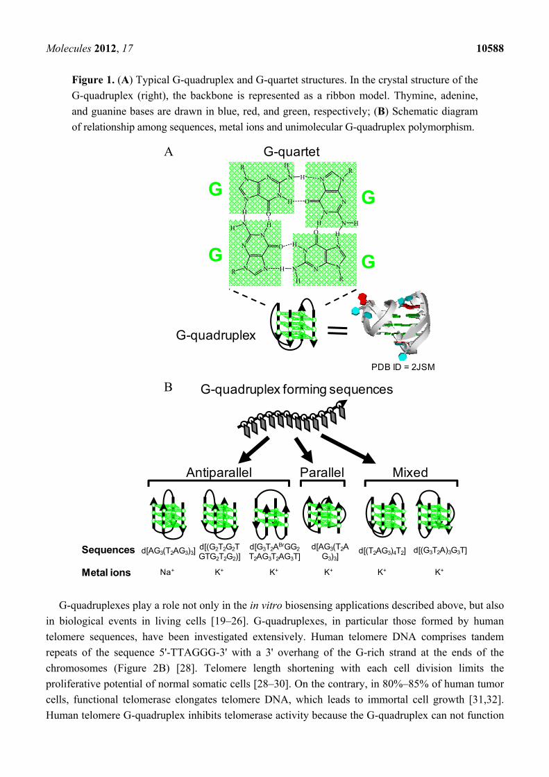
hemin. The G-quadruplex is a four-stranded DNA structure with stacked guanine tetrads, G-quartets,
which are held together via eight Hoogsteen hydrogen bonds (Figure 1A). Since the prediction of the
existence of the G-quadruplex [10], G-quadruplexes formed by different guanine-rich (G-rich)
sequences are found not only in genomic DNA but are also generated during in vitro screening for
aptamers targeting molecules such as proteins [11–14]. Structural studies have demonstrated that the
G-rich sequences can fold to form various types of G-quadruplex conformations depending on the
sequences and the experimental conditions (e.g., coexisting metal ion, metal ion concentration and
degree of molecular crowding) (Figure 1B) [11–14]. These findings combined with the peroxidase-like
activity of the hemin-G-quadruplex complex have stimulated development of various applications,
mainly detection techniques targeting DNA, proteins and metal ions [15–18]. The detection is
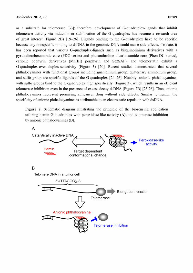
composed of the following two steps. The first involves DNA conformational change to a catalytically
active form of G-quadruplex from inactive DNA such as single-stranded DNA (ssDNA) and
double-stranded DNA (dsDNA), depending on the target molecule. The second step is analysis of the
peroxidase-like activity of the G-quadruplex with hemin (Figure 2A). The key point is the specific
binding of hemin to the G-quadruplex because the system can not work if hemin binds to ssDNA and
dsDNA nonspecifically and the complexes then exert peroxidase-like activity. Cationic -conjugated
molecules such as cationic porphyrins usually bind to G-quadruplexes via - interaction with
G-quartets and via electrostatic interaction with anionic phosphate groups on G-quadruplexes [19–26].
However, the specificity of G-quadruplexes is usually low because the electrostatic interaction also
causes non-specific binding to dsDNA and ssDNA [20,22,23,25,26]. In contrast, the specificity of
hemin binding to G-quadruplexes is significantly high. This specific binding is attributable to an
electrostatic repulsion between the carboxyl groups of hemin and the phosphate groups of DNA,
leading to inhibition of hemin binding to ssDNA and dsDNA. In order to overcome the electrostatic
repulsion, the large -planar of hemin can form - stacking interactions with G-quartets of
G-quadruplexes despite the electrostatic repulsion [27].
Molecules 2012, 17 10588
Figure 1. (A) Typical G-quadruplex and G-quartet structures. In the crystal structure of the
G-quadruplex (right), the backbone is represented as a ribbon model. Thymine, adenine,
and guanine bases are drawn in blue, red, and green, respectively; (B) Schematic diagram
of relationship among sequences, metal ions and unimolecular G-quadruplex polymorphism.
G-quadruplex forming sequences
d[(G3T2A)3G3T]d[AG3(T2AG3)3]
K+
d[(T2AG3)4T2]
K+ K+
d[AG3(T2AG3)3] d[(G2T2G2TGTG2T2G2)]
Na+ K+
d[G3T2ABrGG2
T2AG3T2AG3T]
K+
Sequences
Metal ions
MixedParallelAntiparallel
A G-quartet
G G
GG
G-quadruplex
PDB ID = 2JSM
B
G-quadruplexes play a role not only in the in vitro biosensing applications described above, but also
in biological events in living cells [19–26]. G-quadruplexes, in particular those formed by human
telomere sequences, have been investigated extensively. Human telomere DNA comprises tandem
repeats of the sequence 5'-TTAGGG-3' with a 3' overhang of the G-rich strand at the ends of the
chromosomes (Figure 2B) [28]. Telomere length shortening with each cell division limits the
proliferative potential of normal somatic cells [28–30]. On the contrary, in 80%–85% of human tumor
cells, functional telomerase elongates telomere DNA, which leads to immortal cell growth [31,32].
Human telomere G-quadruplex inhibits telomerase activity because the G-quadruplex can not function
N
N
N
N
O
H
N
H
N N
N
NO
H N
H
H
N
N
N
N
O
H
N
H
NN
N
N O
HNH
H
H
H
R
RR
R
Molecules 2012, 17 10589
as a substrate for telomerase [33]; therefore, development of G-quadruplex-ligands that inhibit
telomerase activity via induction or stabilization of the G-quadruplex has become a research area
of great interest (Figure 2B) [19–26]. Ligands binding to the G-quadruplex have to be specific
because any nonspecific binding to dsDNA in the genomic DNA could cause side effects. To date, it
has been reported that various G-quadruplex-ligands such as bisquinolinium derivatives with a
pyridodicarboxaminde core (PDC series) and phenanthroline dicarboxamide core (Phen-DC series),
cationic pophyrin derivatives (Mn(III) porphyrin and Se2SAP), and telomestatin exhibit a
G-quadruplex-over duplex-selectivity (Figure 3) [20]. Recent studies demonstrated that several
phthalocyanines with functional groups including guanidinium group, quaternary ammonium group,
and sulfo group are specific ligands of the G-quadruplex [24–26]. Notably, anionic phthalocyanines
with sulfo groups bind to the G-quadruplex high specifically (Figure 3), which results in an efficient
telomerase inhibition even in the presence of excess decoy dsDNA (Figure 2B) [25,26]. Thus, anionic
phthalocyanines represent promising anticancer drug without side effects. Similar to hemin, the
specificity of anionic phthalocyanines is attributable to an electrostatic repulsion with dsDNA.
Figure 2. Schematic diagram illustrating the principle of the biosensing application
utilizing hemin-G-quadruplex with peroxidase-like activity (A), and telomerase inhibition
by anionic phthalocyanines (B).
A
B
Hemin
Catalytically inactive DNA Peroxidase-like
activity
Target dependent conformational change
Elongation reaction
Telomerase
5’-(TTAGGG)n-3’
Telomere DNA in a tumor cell
Telomerase inhibition
Anionic phthalocyanine
Molecules 2012, 17 10590
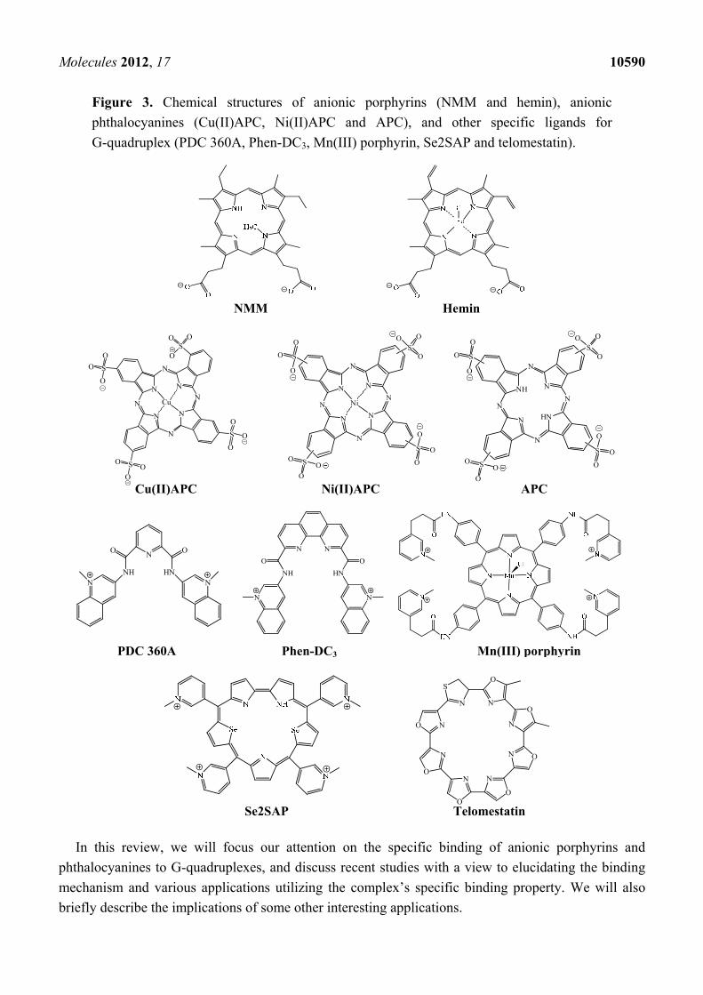
Figure 3. Chemical structures of anionic porphyrins (NMM and hemin), anionic
phthalocyanines (Cu(II)APC, Ni(II)APC and APC), and other specific ligands for
G-quadruplex (PDC 360A, Phen-DC3, Mn(III) porphyrin, Se2SAP and telomestatin).
In this review, we will focus our attention on the specific binding of anionic porphyrins and
phthalocyanines to G-quadruplexes, and discuss recent studies with a view to elucidating the binding
mechanism and various applications utilizing the complex’s specific binding property. We will also
briefly describe the implications of some other interesting applications.
N
OS
N
O N N
O
O
N N O
N
OO
N
N
HNNH
OO
NN
NN
NH HN
OO
NN
PDC 360A Phen-DC3 Mn(III) porphyrin
N
NN
N
N
N
N
N
SO O
O
SO
O
O
SOO
O
S
O
O
O
Cu
N
NN
N
N
N
N
N
SO
O
O
Ni
S
O
O
O
S O
O
O
S
O
OO
NH
HNN
N
N
N
N
N
SO
O
O
S
O
O
O
S O
O
O
S
O
OO
Cu(II)APC Ni(II)APC APC
NMM Hemin
Se2SAP Telomestatin
Molecules 2012, 17 10591
2. Hemin-G-quadruplex Interaction for in Vitro Applications
Hemin, which is an anionic porphyrin with two carboxyl groups (Figure 3), is used as a cofactor for
a variety of enzymes such as catalases, peroxidases and monooxygenases in living cells. In the late
1990s, it was discovered that some G-quadruplexes bind to hemin and the complexes show peroxidase-
like activity [27]. Importantly, no DNA structures than other the G-quadruplex has such properties.
Moreover, the peroxidase-like activity depends on G-quadruplex polymorphisms [27,34]. Thus, the
studies on the relationships among DNA structure, binding property of hemin and the peroxidase-like
activity have been conducted extensively. Furthermore, many applications utilizing the hemin-G-
quadruplex peroxidase activity have been reported recently. Here, we discuss recent advances based on
these studies.
2.1. Mechanism of Induction of Peroxidase-Like Activity
In 1996 Sen’s group conducted an interesting investigation to find DNA aptamers for
N-methylmesoporphyrin IX (NMM) (Figure 3) using an in vitro selection (Systematic Evolution of
Ligands by Exponential enrichment, SELEX) method [35]. Surprisingly, the authors found that some
G-rich DNAs including PS2 and PS5 bind to NMM despite the anionic properties of NMM (Table 1) [35].
More importantly, they showed that the G-rich DNAs are more than just NMM aptamers. NMM is a
stable transition-state analogue for porphyrin-metallation reactions, implying that the NMM aptamers
catalyze porphyrin metallation. In fact, it was demonstrated that PS2- and PS5-related DNAs have the
ability to catalyze metallation [36,37]. Furthermore, since Sen’s group found that hemin, which is an
anionic porphyrin utilized as a cofactor of peroxidase in nature, also binds to the NMM aptamers, the
authors investigated the peroxidase-like activity of hemin-PS2.M and -PS5.M (Table 1) [27]. The
investigation demonstrated that the observed catalytic velocity (Vobs) of PS2.M-hemin was 250 times
greater than that of hemin alone when 2,2'-azino-bis(3-ethylbenzothiazoline-6-sulphonic acid) (ABTS)
was used as a substrate in the presence of H2O2 (Figure 4) [27]. Interestingly, K+ was fundamental for
the activity of PS2.M-hemin although Na+ and Mg2+ inhibited the activity [27]. The result implies that
the catalytically-active form of PS2.M is a specific G-quadruplex structure that is stabilized by K+. The
characteristic ability of hemin to bind specifically to the G-quadruplex is a key point for many
applications described below.
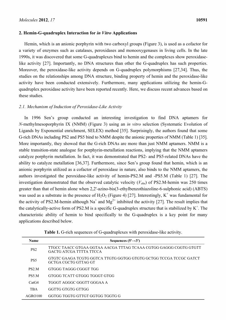
Table 1. G-rich sequences of G-quadruplexes with peroxidase-like activity.
Name Sequences (5'→3')
PS2 TTGCC TAACC GTGAA GGTAA AACGA TTTAG TCAAA CGTGG GAGGG CGGTG GTGTT GACTG ATCGA TTTTA TTCCA
PS5 GTGTC GAAGA TCGTG GGTCA TTGTG GGTGG GTGTG GCTGG TCCGA TCCGC GATCT GCTGA CGCTG GTTAG GT
PS2.M GTGGG TAGGG CGGGT TGG
PS5.M GTGGG TCATT GTGGG TGGGT GTGG
CatG4 TGGGT AGGGC GGGTT GGGAA A
TBA GGTTG GTGTG GTTGG
AGRO100 GGTGG TGGTG GTTGT GGTGG TGGTG G
Molecules 2012, 17 10592
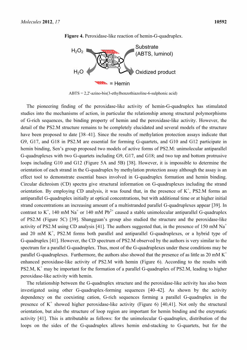
Figure 4. Peroxidase-like reaction of hemin-G-quadruplex.
ABTS = 2,2'-azino-bis(3-ethylbenzothiazoline-6-sulphonic acid)
= Hemin
Oxidized product
Substrate (ABTS, luminol)
H2O2
H2O
The pioneering finding of the peroxidase-like activity of hemin-G-quadruplex has stimulated
studies into the mechanisms of action, in particular the relationship among structural polymorphisms
of G-rich sequences, the binding property of hemin and the peroxidase-like activity. However, the
detail of the PS2.M structure remains to be completely elucidated and several models of the structure
have been proposed to date [38–41]. Since the results of methylation protection assays indicate that
G9, G17, and G18 in PS2.M are essential for forming G-quartets, and G10 and G12 participate in
hemin binding, Sen’s group proposed two models of active forms of PS2.M: unimolecular antiparallel
G-quadruplexes with two G-quartets including G9, G17, and G18; and two top and bottom protrusive
loops including G10 and G12 (Figure 5A and 5B) [38]. However, it is impossible to determine the
orientation of each strand in the G-quadruplex by methylation protection assay although the assay is an
effect tool to demonstrate essential bases involved in G-quadruplex formation and hemin binding.
Circular dichroism (CD) spectra give structural information on G-quadruplexes including the strand
orientation. By employing CD analysis, it was found that, in the presence of K+, PS2.M forms an
antiparallel G-quadruplex initially at optical concentrations, but with additional time or at higher initial
strand concentrations an increasing amount of a multistranded parallel G-quadruplexes appear [39]. In
contrast to K+, 140 mM Na+ or 140 mM Pb2+ caused a stable unimolecular antiparallel G-quadruplex
of PS2.M (Figure 5C) [39]. Shangguan’s group also studied the structure and the peroxidase-like
activity of PS2.M using CD analysis [41]. The authors suggested that, in the presence of 150 mM Na+
and 20 mM K+, PS2.M forms both parallel and antiparallel G-quadruplexes, or a hybrid type of
G-quadruplex [41]. However, the CD spectrum of PS2.M observed by the authors is very similar to the
spectrum for a parallel G-quadruplex. Thus, most of the G-quadruplexes under these conditions may be
parallel G-quadruplexes. Furthermore, the authors also showed that the presence of as little as 20 mM K+
enhanced peroxidase-like activity of PS2.M with hemin (Figure 6). According to the results with
PS2.M, K+ may be important for the formation of a parallel G-quadruplex of PS2.M, leading to higher
peroxidase-like activity with hemin.
The relationship between the G-quadruplex structure and the peroxidase-like activity has also been
investigated using other G-quadruplex-forming sequences [40–42]. As shown by the activity
dependency on the coexisting cation, G-rich sequences forming a parallel G-quadruplex in the
presence of K+ showed higher peroxidase-like activity (Figure 6) [40,41]. Not only the structural
orientation, but also the structure of loop region are important for hemin binding and the enzymatic
activity [41]. This is attributable as follows: for the unimolecular G-quadruplex, distribution of the
loops on the sides of the G-quadruplex allows hemin end-stacking to G-quartets, but for the
Molecules 2012, 17 10593
antiparallel G-quadruplex protrusion of the loops over the top and bottom G-quartets inhibits
end-stacking. For the multistranded parallel G-quadruplexes formed by T4GnT4 (n = 6 or 8), T4 termini
of the strands hinders the end-stacking of hemin [41]. Systematic studies on the structure-function
relationship of d(G2Tn)3G2 and d(G3Tn)3G2 (n = 1 − 4) also demonstrated that the parallel
G-quadruplexes in the presence of 100 mM K+ have high peroxidase-like activity although the
antiparallel G-quadruplexes in the presence of 100 mM Na+ have little activity [42].
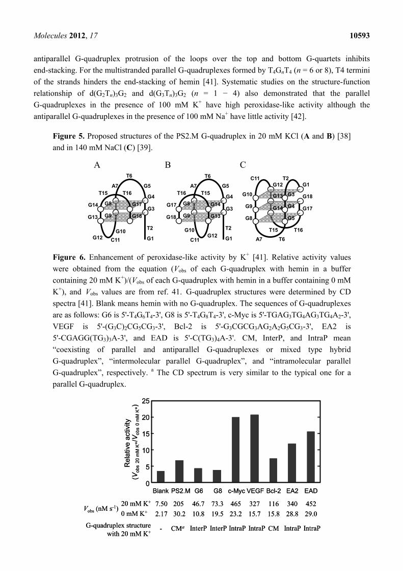
Figure 5. Proposed structures of the PS2.M G-quadruplex in 20 mM KCl (A and B) [38]
and in 140 mM NaCl (C) [39].
A B C
G1
G17
T2
G3
G4
G5
T6
A7
G8
G9
G10
C11G12
G13
G14
T15 T16
G18
G1
G14
T2
G3
G4
G5
T6
A7
G8
G9
G10
C11G12
G18
G17
T16 T15
G13
G1
G14
T2
G3
G4
G5
T6A7
G8
G9
G10
C11G12
G18
G17
T16 T15
G13
Figure 6. Enhancement of peroxidase-like activity by K+ [41]. Relative activity values
were obtained from the equation (Vobs of each G-quadruplex with hemin in a buffer
containing 20 mM K+)/(Vobs of each G-quadruplex with hemin in a buffer containing 0 mM
K+), and Vobs values are from ref. 41. G-quadruplex structures were determined by CD
spectra [41]. Blank means hemin with no G-quadruplex. The sequences of G-quadruplexes
are as follows: G6 is 5'-T4G6T4-3', G8 is 5'-T4G8T4-3', c-Myc is 5'-TGAG3TG4AG3TG4A2-3',
VEGF is 5'-(G3C)2CG5CG3-3', Bcl-2 is 5'-G3CGCG3AG2A2G5CG3-3', EA2 is
5'-CGAGG(TG3)3A-3', and EAD is 5'-C(TG3)4A-3'. CM, InterP, and IntraP mean
“coexisting of parallel and antiparallel G-quadruplexes or mixed type hybrid
G-quadruplex”, “intermolecular parallel G-quadruplex”, and “intramolecular parallel
G-quadruplex”, respectively. a The CD spectrum is very similar to the typical one for a
parallel G-quadruplex.
25
20
15
10
5
0Blank
7.50
2.17
PS2.M
205
30.2
G6
46.7
10.8
G8
73.3
19.5
c-Myc
465
23.2
VEGF
327
15.7
Bcl-2
116
15.8
EA2
340
28.8
EAD
452
29.0Vobs (nM s-1)
20 mM K+
0 mM K+
Rel
ativ
e a
ctiv
ity
(Vob
s20
mM
K+/V
obs
0 m
MK
+)
G-quadruplex structurewith 20 mM K+
- CMa InterP InterP IntraP IntraP IntraP IntraPCM
Molecules 2012, 17 10594
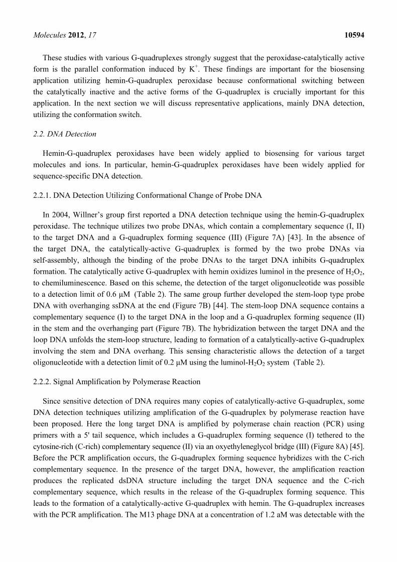
These studies with various G-quadruplexes strongly suggest that the peroxidase-catalytically active
form is the parallel conformation induced by K+. These findings are important for the biosensing
application utilizing hemin-G-quadruplex peroxidase because conformational switching between
the catalytically inactive and the active forms of the G-quadruplex is crucially important for this
application. In the next section we will discuss representative applications, mainly DNA detection,
utilizing the conformation switch.
2.2. DNA Detection
Hemin-G-quadruplex peroxidases have been widely applied to biosensing for various target
molecules and ions. In particular, hemin-G-quadruplex peroxidases have been widely applied for
sequence-specific DNA detection.
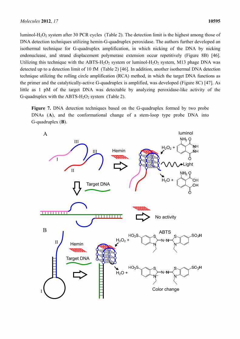
2.2.1. DNA Detection Utilizing Conformational Change of Probe DNA
In 2004, Willner’s group first reported a DNA detection technique using the hemin-G-quadruplex
peroxidase. The technique utilizes two probe DNAs, which contain a complementary sequence (I, II)
to the target DNA and a G-quadruplex forming sequence (III) (Figure 7A) [43]. In the absence of
the target DNA, the catalytically-active G-quadruplex is formed by the two probe DNAs via
self-assembly, although the binding of the probe DNAs to the target DNA inhibits G-quadruplex
formation. The catalytically active G-quadruplex with hemin oxidizes luminol in the presence of H2O2,
to chemiluminescence. Based on this scheme, the detection of the target oligonucleotide was possible
to a detection limit of 0.6 μM (Table 2). The same group further developed the stem-loop type probe
DNA with overhanging ssDNA at the end (Figure 7B) [44]. The stem-loop DNA sequence contains a
complementary sequence (I) to the target DNA in the loop and a G-quadruplex forming sequence (II)
in the stem and the overhanging part (Figure 7B). The hybridization between the target DNA and the
loop DNA unfolds the stem-loop structure, leading to formation of a catalytically-active G-quadruplex
involving the stem and DNA overhang. This sensing characteristic allows the detection of a target
oligonucleotide with a detection limit of 0.2 μM using the luminol-H2O2 system (Table 2).
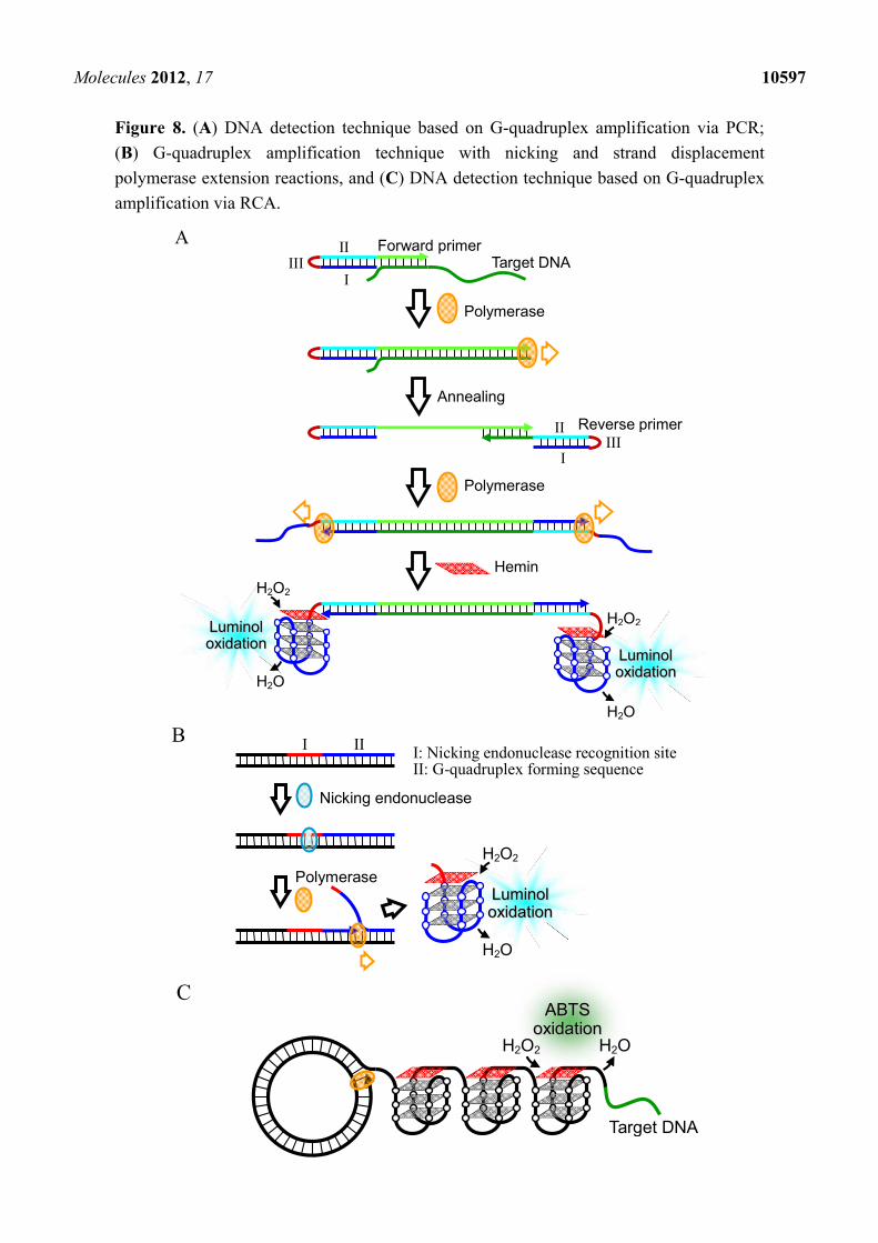
2.2.2. Signal Amplification by Polymerase Reaction
Since sensitive detection of DNA requires many copies of catalytically-active G-quadruplex, some
DNA detection techniques utilizing amplification of the G-quadruplex by polymerase reaction have
been proposed. Here the long target DNA is amplified by polymerase chain reaction (PCR) using
primers with a 5' tail sequence, which includes a G-quadruplex forming sequence (I) tethered to the
cytosine-rich (C-rich) complementary sequence (II) via an oxyethyleneglycol bridge (III) (Figure 8A) [45].
Before the PCR amplification occurs, the G-quadruplex forming sequence hybridizes with the C-rich
complementary sequence. In the presence of the target DNA, however, the amplification reaction
produces the replicated dsDNA structure including the target DNA sequence and the C-rich
complementary sequence, which results in the release of the G-quadruplex forming sequence. This
leads to the formation of a catalytically-active G-quadruplex with hemin. The G-quadruplex increases
with the PCR amplification. The M13 phage DNA at a concentration of 1.2 aM was detectable with the
Molecules 2012, 17 10595
luminol-H2O2 system after 30 PCR cycles (Table 2). The detection limit is the highest among those of
DNA detection techniques utilizing hemin-G-quadruplex peroxidase. The authors further developed an
isothermal technique for G-quadruplex amplification, in which nicking of the DNA by nicking
endonuclease, and strand displacement polymerase extension occur repetitively (Figure 8B) [46].
Utilizing this technique with the ABTS-H2O2 system or luminol-H2O2 system, M13 phage DNA was
detected up to a detection limit of 10 fM (Table 2) [46]. In addition, another isothermal DNA detection
technique utilizing the rolling circle amplification (RCA) method, in which the target DNA functions as
the primer and the catalytically-active G-quadruplex is amplified, was developed (Figure 8C) [47]. As
little as 1 pM of the target DNA was detectable by analyzing peroxidase-like activity of the
G-quadruplex with the ABTS-H2O2 system (Table 2).
Figure 7. DNA detection techniques based on the G-quadruplex formed by two probe
DNAs (A), and the conformational change of a stem-loop type probe DNA into
G-quadruplex (B).
A
Hemin
Target DNA
No activity
H2O2 +
H2O +
I
II
III
III
luminol
Light
Hemin
Target DNA
H2O2 +
H2O +
I
II
ABTS
Color change
B
Molecules 2012, 17 10596
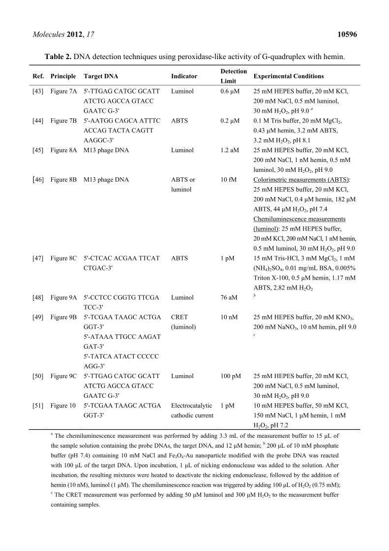
Table 2. DNA detection techniques using peroxidase-like activity of G-quadruplex with hemin.
Ref. Principle Target DNA Indicator Detection
Limit Experimental Conditions
[43] Figure 7A 5'-TTGAG CATGC GCATT
ATCTG AGCCA GTACC
GAATC G-3'
Luminol 0.6 μM 25 mM HEPES buffer, 20 mM KCl,
200 mM NaCl, 0.5 mM luminol,
30 mM H2O2, pH 9.0 a
[44] Figure 7B 5'-AATGG CAGCA ATTTC
ACCAG TACTA CAGTT
AAGGC-3'
ABTS 0.2 μM 0.1 M Tris buffer, 20 mM MgCl2,
0.43 μM hemin, 3.2 mM ABTS,
3.2 mM H2O2, pH 8.1
[45] Figure 8A M13 phage DNA Luminol 1.2 aM 25 mM HEPES buffer, 20 mM KCl,
200 mM NaCl, 1 nM hemin, 0.5 mM
luminol, 30 mM H2O2, pH 9.0
[46] Figure 8B M13 phage DNA ABTS or
luminol
10 fM Colorimetric measurements (ABTS):
25 mM HEPES buffer, 20 mM KCl,
200 mM NaCl, 0.4 μM hemin, 182 μM
ABTS, 44 μM H2O2, pH 7.4
Chemiluminescence measurements
(luminol): 25 mM HEPES buffer,
20 mM KCl, 200 mM NaCl, 1 nM hemin,
0.5 mM luminol, 30 mM H2O2, pH 9.0
[47] Figure 8C 5'-CTCAC ACGAA TTCAT
CTGAC-3'
ABTS 1 pM 15 mM Tris-HCl, 3 mM MgCl2, 1 mM
(NH4)2SO4, 0.01 mg/mL BSA, 0.005%
Triton X-100, 0.5 μM hemin, 1.17 mM
ABTS, 2.82 mM H2O2
[48] Figure 9A 5'-CCTCC CGGTG TTCGA
TCC-3'
Luminol 76 aM b
[49] Figure 9B 5'-TCGAA TAAGC ACTGA
GGT-3'
5'-ATAAA TTGCC AAGAT
GAT-3'
5'-TATCA ATACT CCCCC
AGG-3'
CRET
(luminol)
10 nM 25 mM HEPES buffer, 20 mM KNO3,
200 mM NaNO3, 10 nM hemin, pH 9.0 c
[50] Figure 9C 5'-TTGAG CATGC GCATT
ATCTG AGCCA GTACC
GAATC G-3'
Luminol 100 pM 25 mM HEPES buffer, 20 mM KCl,
200 mM NaCl, 0.5 mM luminol,
30 mM H2O2, pH 9.0
[51] Figure 10 5'-TCGAA TAAGC ACTGA
GGT-3'
Electrocatalytic
cathodic current
1 pM 10 mM HEPES buffer, 50 mM KCl,
150 mM NaCl, 1 μM hemin, 1 mM
H2O2, pH 7.2 a The chemiluminescence measurement was performed by adding 3.3 mL of the measurement buffer to 15 μL of
the sample solution containing the probe DNAs, the target DNA, and 12 μM hemin; b 200 μL of 10 mM phosphate
buffer (pH 7.4) containing 10 mM NaCl and Fe3O4-Au nanoparticle modified with the probe DNA was reacted
with 100 μL of the target DNA. Upon incubation, 1 μL of nicking endonuclease was added to the solution. After
incubation, the resulting mixtures were heated to deactivate the nicking endonuclease, followed by the addition of
hemin (10 nM), luminol (1 μM). The chemiluminescence reaction was triggered by adding 100 μL of H2O2 (0.75 mM); c The CRET measurement was performed by adding 50 μM luminol and 300 μM H2O2 to the measurement buffer
containing samples.
Molecules 2012, 17 10597
Figure 8. (A) DNA detection technique based on G-quadruplex amplification via PCR;
(B) G-quadruplex amplification technique with nicking and strand displacement
polymerase extension reactions, and (C) DNA detection technique based on G-quadruplex
amplification via RCA.
A Target DNA
Annealing
Polymerase
Polymerase
H2O2
H2O
H2O2
H2O
LLuummiinnooll ooxxiiddaattiioonn
I III
II
IIII
II
Forward primer
Reverse primer
Hemin
LLuummiinnooll ooxxiiddaattiioonn
Nicking endonuclease
Polymerase
I: Nicking endonuclease recognition site II: G-quadruplex forming sequence
H2O2
H2O
LLuummiinnooll ooxxiiddaattiioonn
I IIB
C
AABBTTSS ooxxiiddaattiioonn
H2O2 H2O
Target DNA
Molecules 2012, 17 10598
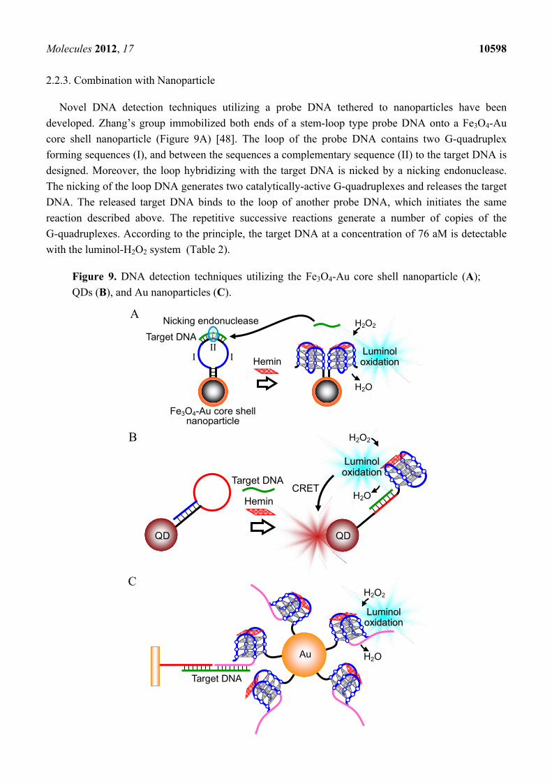
2.2.3. Combination with Nanoparticle
Novel DNA detection techniques utilizing a probe DNA tethered to nanoparticles have been
developed. Zhang’s group immobilized both ends of a stem-loop type probe DNA onto a Fe3O4-Au
core shell nanoparticle (Figure 9A) [48]. The loop of the probe DNA contains two G-quadruplex
forming sequences (I), and between the sequences a complementary sequence (II) to the target DNA is
designed. Moreover, the loop hybridizing with the target DNA is nicked by a nicking endonuclease.
The nicking of the loop DNA generates two catalytically-active G-quadruplexes and releases the target
DNA. The released target DNA binds to the loop of another probe DNA, which initiates the same
reaction described above. The repetitive successive reactions generate a number of copies of the
G-quadruplexes. According to the principle, the target DNA at a concentration of 76 aM is detectable
with the luminol-H2O2 system (Table 2).
Figure 9. DNA detection techniques utilizing the Fe3O4-Au core shell nanoparticle (A);
QDs (B), and Au nanoparticles (C).
A
Hemin
Target DNA
Nicking endonuclease
II I I
Fe3O4-Au core shellnanoparticle
H2O2
H2O
LLuummiinnooll ooxxiiddaattiioonn
H2O2
H2O
LLuummiinnooll ooxxiiddaattiioonn
CRETHemin
QD QD
Target DNA
B
H2O2
H2O
LLuummiinnooll ooxxiiddaattiioonn
Target DNA
Au
C
Molecules 2012, 17 10599
Chemiluminescence resonance energy transfer (CRET) of the chemiluminescence from oxidized
luminol to quantum dots (QDs) was further combined for DNA detection (Figure 9B) [49]. The probe
DNA used to demonstrate this principle is a stem-loop type DNA, which is immobilized to CdSe/ZnS
QDs. The hybridization of the target DNA with the loop region unfolds the stem-loop structure, which
results in formation of the catalytically-active G-quadruplex. Luminol chemiluminescence produced by
the G-quadruplex peroxidase-like activity excites the CdSe/ZnS ODs, leading to a CRET signal.
Designing of probe DNA sequences and regulating the emission wavelength of CdSe/ZnS QDs
depending on the target DNA sequence allows the detection of different target DNAs simultaneously
with different CRET signals (Table 2). Willner’s group also modified the Au nanoparticle with a
probe DNA, which contains a complementary sequence to the target DNA and a catalytically-active
G-quadruplex (Figure 9C) [50]. The functionalized Au nanoparticles are gathered to a gold substrate
via the target DNA. The detection limit for analyzing the activity of the G-quadruplex on the gold
substrate with the luminol-H2O2 system is 100 pM (Table 2).
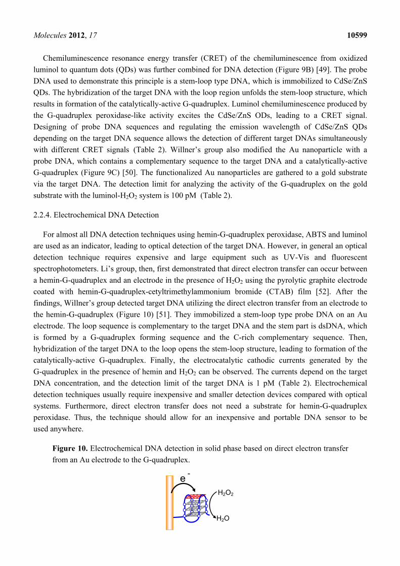
2.2.4. Electrochemical DNA Detection
For almost all DNA detection techniques using hemin-G-quadruplex peroxidase, ABTS and luminol
are used as an indicator, leading to optical detection of the target DNA. However, in general an optical
detection technique requires expensive and large equipment such as UV-Vis and fluorescent
spectrophotometers. Li’s group, then, first demonstrated that direct electron transfer can occur between
a hemin-G-quadruplex and an electrode in the presence of H2O2 using the pyrolytic graphite electrode
coated with hemin-G-quadruplex-cetyltrimethylammonium bromide (CTAB) film [52]. After the
findings, Willner’s group detected target DNA utilizing the direct electron transfer from an electrode to
the hemin-G-quadruplex (Figure 10) [51]. They immobilized a stem-loop type probe DNA on an Au
electrode. The loop sequence is complementary to the target DNA and the stem part is dsDNA, which
is formed by a G-quadruplex forming sequence and the C-rich complementary sequence. Then,
hybridization of the target DNA to the loop opens the stem-loop structure, leading to formation of the
catalytically-active G-quadruplex. Finally, the electrocatalytic cathodic currents generated by the
G-quadruplex in the presence of hemin and H2O2 can be observed. The currents depend on the target
DNA concentration, and the detection limit of the target DNA is 1 pM (Table 2). Electrochemical
detection techniques usually require inexpensive and smaller detection devices compared with optical
systems. Furthermore, direct electron transfer does not need a substrate for hemin-G-quadruplex
peroxidase. Thus, the technique should allow for an inexpensive and portable DNA sensor to be
used anywhere.
Figure 10. Electrochemical DNA detection in solid phase based on direct electron transfer
from an Au electrode to the G-quadruplex.
H2O2
H2O
e -
Molecules 2012, 17 10600
2.3. Biosensors for Various Targets
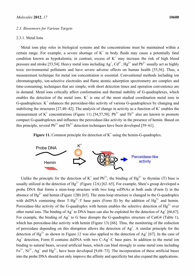
2.3.1. Metal Ions
Metal ions play roles in biological systems and the concentrations must be maintained within a
certain range. For example, a severe shortage of K+ in body fluids may cause a potentially fatal
condition known as hypokalemia; in contrast, excess of K+ may increase the risk of high blood
pressure and stroke [53,54]. Heavy metal ions including Ag+, Cd2+, Hg2+ and Pb2+ usually act as highly
toxic environmental pollutants and have severe adverse effects on human health [55,56]. Thus, a
measurement technique for metal ion concentration is essential. Conventional methods including ion
chromatography, ion-selective electrodes and flame atomic adsorption spectrometry are complex and
time-consuming; techniques that are simple, with short detection times and operation convenience are
in demand. Metal ions critically affect conformation and thermal stability of G-quadruplexes, which
enables the detection of the metal ions. K+ is one of the most studied coordination metal ions in
G-quadruplexes. K+ enhances the peroxidase-like activity of various G-quadruplexes by changing and
stabilizing the structures [27,40–42]. The analysis of change in activity as a function of K+ enables the
measurement of K+ concentrations (Figure 11) [54,57,58]. Pb2+ and Tb3+ also are known to promote
compact G-quadruplexes and influence the peroxidase-like activity in the presence of hemin. Based on
this principle, several Pb2+ and Tb3+ detection techniques have been developed [59–61].
Figure 11. Common principle for detection of K+ using the hemin-G-quadruplex.
Hemin
Probe DNA
Peroxidase-like activity
K+
Unlike the principle for the detection of K+ and Pb2+, the binding of Hg2+ to thymine (T) base is
usually utilized in the detection of Hg2+ (Figure 12A) [62–65]. For example, Shen’s group developed a
probe DNA that forms a stem-loop structure with two long ssDNAs at both ends (Form I) in the
absence of Hg2+ and hemin (Figure 12B) [65]. The stem-loop structure is changed to the G-quadruplex
with dsDNA containing three T-Hg2+-T base pairs (Form II) by the addition of Hg2+ and hemin.
Peroxidase-like activity of the G-quadruplex with hemin enables the selective detection of Hg2+ over
other metal ions. The binding of Ag+ to DNA bases can also be exploited for the detection of Ag+ [66,67].
For example, the binding of Ag+ to G base disrupts the G-quadruplex structure of CatG4 (Table 1),
which has peroxidase-like activity with hemin (Figure 13) [66]. Thus, the monitoring of the reduction
of peroxidase depending on this disruption allows the detection of Ag+. A similar principle for the
detection of Hg2+ as shown in Figure 12 was also applied to the detection of Ag+ [67]. In the case of
Ag+ detection, Form II contains dsDNA with two C-Ag+-C base pairs. In addition to the metal ion
binding to natural bases, several artificial bases, which can bind strongly to some metal ions including
Fe3+, Ni2+, Ag+ and Hg2+, have been developed [9,68–73]. The incorporation of these artificial bases
into the probe DNA should not only improve the affinity and specificity but also expand the applications.
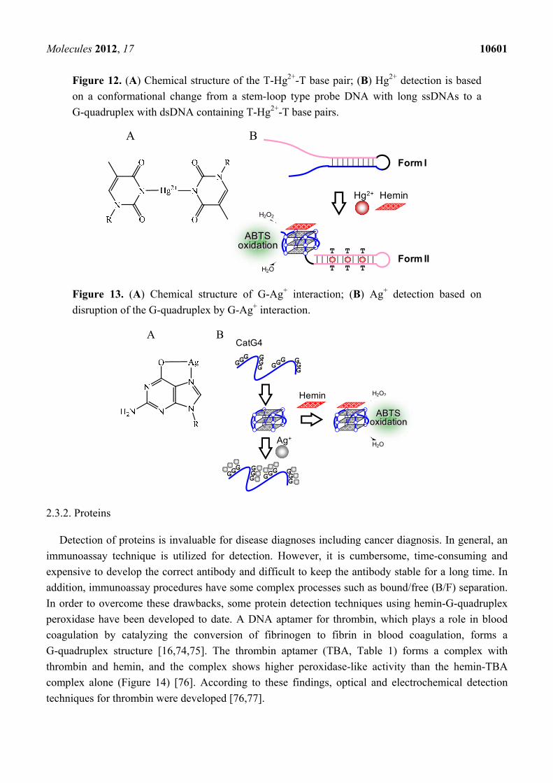
Molecules 2012, 17 10601
Figure 12. (A) Chemical structure of the T-Hg2+-T base pair; (B) Hg2+ detection is based
on a conformational change from a stem-loop type probe DNA with long ssDNAs to a
G-quadruplex with dsDNA containing T-Hg2+-T base pairs.
A B
H2O2
H2O
ABTSoxidation
T T T
T T T
Hg2+ Hemin
Form I
Form II
Figure 13. (A) Chemical structure of G-Ag+ interaction; (B) Ag+ detection based on
disruption of the G-quadruplex by G-Ag+ interaction.
H2O2
H2O
ABTSoxidation
GGG G
GG GG
G GGG
GGG G
GG GG
G GGG
Hemin
Ag+
CatG4 A B
2.3.2. Proteins
Detection of proteins is invaluable for disease diagnoses including cancer diagnosis. In general, an
immunoassay technique is utilized for detection. However, it is cumbersome, time-consuming and
expensive to develop the correct antibody and difficult to keep the antibody stable for a long time. In
addition, immunoassay procedures have some complex processes such as bound/free (B/F) separation.
In order to overcome these drawbacks, some protein detection techniques using hemin-G-quadruplex
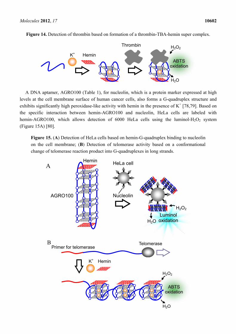
peroxidase have been developed to date. A DNA aptamer for thrombin, which plays a role in blood
coagulation by catalyzing the conversion of fibrinogen to fibrin in blood coagulation, forms a
G-quadruplex structure [16,74,75]. The thrombin aptamer (TBA, Table 1) forms a complex with
thrombin and hemin, and the complex shows higher peroxidase-like activity than the hemin-TBA
complex alone (Figure 14) [76]. According to these findings, optical and electrochemical detection
techniques for thrombin were developed [76,77].
Molecules 2012, 17 10602
Figure 14. Detection of thrombin based on formation of a thrombin-TBA-hemin super complex.
AABBTTSS ooxxiiddaattiioonn
H2O2
H2O
K+
Hemin
Thrombin
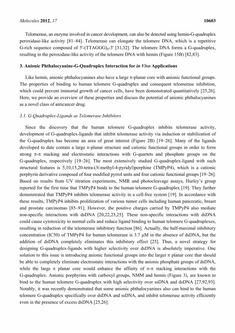
A DNA aptamer, AGRO100 (Table 1), for nucleolin, which is a protein marker expressed at high
levels at the cell membrane surface of human cancer cells, also forms a G-quadruplex structure and
exhibits significantly high peroxidase-like activity with hemin in the presence of K+ [78,79]. Based on
the specific interaction between hemin-AGRO100 and nucleolin, HeLa cells are labeled with
hemin-AGRO100, which allows detection of 6000 HeLa cells using the luminol-H2O2 system
(Figure 15A) [80].
Figure 15. (A) Detection of HeLa cells based on hemin-G-quadruplex binding to nucleolin
on the cell membrane; (B) Detection of telomerase activity based on a conformational
change of telomerase reaction product into G-quadruplexes in long strands.
HeLa cell
Nucleolin AGRO100
Hemin
LLuummiinnooll ooxxiiddaattiioonn
H2O2
H2O
A
AABBTTSS ooxxiiddaattiioonn
H2O2
H2O
Hemin
K+
Telomerase Primer for telomerase
B
Molecules 2012, 17 10603
Telomerase, an enzyme involved in cancer development, can also be detected using hemin-G-quadruplex
peroxidase-like activity [81–84]. Telomerase can elongate the telomere DNA, which is a repetitive
G-rich sequence composed of 5'-(TTAGGG)n-3' [31,32]. The telomere DNA forms a G-quadruplex,
resulting in the peroxidase-like activity of the telomere DNA with hemin (Figure 15B) [82,83].
3. Anionic Phthalocyanine-G-Quadruplex Interaction for in Vivo Applications
Like hemin, anionic phthalocyanines also have a large -planar core with anionic functional groups.
The properties of binding to human telomere G-quadruplex and consequent telomerase inhibition,
which could prevent immortal growth of cancer cells, have been demonstrated quantitatively [25,26].
Here, we provide an overview of these properties and discuss the potential of anionic phthalocyanines
as a novel class of anticancer drug.
3.1. G-Quadruplex-Ligands as Telomerase Inhibitors
Since the discovery that the human telomere G-quadruplex inhibits telomerase activity,
development of G-quadruplex-ligands that inhibit telomerase activity via induction or stabilization of
the G-quadruplex has become an area of great interest (Figure 2B) [19–26]. Many of the ligands
developed to date contain a large -planar structure and cationic functional groups in order to form
strong - stacking and electrostatic interactions with G-quartets and phosphate groups on the
G-quadruplex, respectively [19–26]. The most extensively studied G-quadruplex-ligand with such
structural features is 5,10,15,20-tetra-(N-methyl-4-pyridyl)porphine (TMPyP4), which is a cationic
porphyrin derivative composed of four modified pyrrol units and four cationic functional groups [19–26].
Based on results from UV titration experiments, NMR and photocleavage assays, Hurley’s group
reported for the first time that TMPyP4 binds to the human telomere G-quadruplex [19]. They further
demonstrated that TMPyP4 inhibits telomerase activity in a cell-free system [19]. In accordance with
these results, TMPyP4 inhibits proliferation of various tumor cells including human pancreatic, breast
and prostate carcinomas [85–91]. However, the positive charges carried by TMPyP4 also mediate
non-specific interactions with dsDNA [20,22,23,25]. These non-specific interactions with dsDNA
could cause cytotoxicity to normal cells and reduce ligand binding to human telomere G-quadruplexes,
resulting in reduction of the telomerase inhibitory function [86]. Actually, the half-maximal inhibitory
concentration (IC50) of TMPyP4 for human telomerase is 3.7 μM in the absence of dsDNA, but the
addition of dsDNA completely eliminates this inhibitory effect [25]. Thus, a novel strategy for
designing G-quadruplex-ligands with higher selectivity over dsDNA is absolutely imperative. One
solution to this issue is introducing anionic functional groups into the larger planar core that should
be able to completely eliminate electrostatic interactions with the anionic phosphate groups of dsDNA,
while the large planar core would enhance the affinity of - stacking interactions with the
G-quadruplex. Anionic porphyrins with carboxyl groups, NMM and hemin (Figure 3), are known to
bind to the human telomere G-quadruplex with high selectivity over ssDNA and dsDNA [27,92,93].
Notably, it was recently demonstrated that some anionic phthalocyanines also can bind to the human
telomere G-quadruplex specifically over dsDNA and ssDNA, and inhibit telomerase activity efficiently
even in the presence of excess dsDNA [25,26].
Molecules 2012, 17 10604
3.2. Selective Binding of Anionic Phthalocyanines to the Human Telomere G-quadruplex
Incorporation of anionic functional groups into phthalocyanines is a promising strategy for
eliminating non-specific binding to dsDNA. The capacity of an anionic copper phthalocyanine
containing four sodium salt forms of sulfo groups (Cu(II)APC, Figure 3) to bind to the human
telomere G-quadruplex and its capacity to inhibit telomerase activity have been investigated [25,26].
Based on visible absorbance titration experiments, Cu(II)APC bound to a human telomere
G-quadruplex (5'-GGG(TTAGGG)3-3') with a dissociation constant (Kd) of 42 μM in the presence of
100 mM KCl. On the contrary, little or no absorbance change was observed for ssDNA (5'-T21-3') or
dsDNA (5'-AGAAGAGAAAGA-3'/5'-TCTTTCTCTTCT-3') (Table 3). These results indicate that
Cu(II)APC can bind to the G-quadruplex with high selectivity over ssDNA and dsDNA. Notably, the
Kd of Cu(II)APC for the G-quadruplex in the presence of excess decoy dsDNA, lambda DNA
(a condition intended to mimic conditions in cell nuclei) was almost the same as that in the absence of
lambda DNA (Table 3). Furthermore, to investigate the effect of a coordinating metal on anionic
phthalocyanine binding to the G-quadruplex, binding assays were performed using nickel anionic
phthalocyanine containing four sodium salt forms of sulfo groups (Ni(II)APC, Figure 3). Ni(II)APC
also bound to the G-quadruplex and the Kd values in the absence and presence of lambda DNA were
almost identical (Table 3).
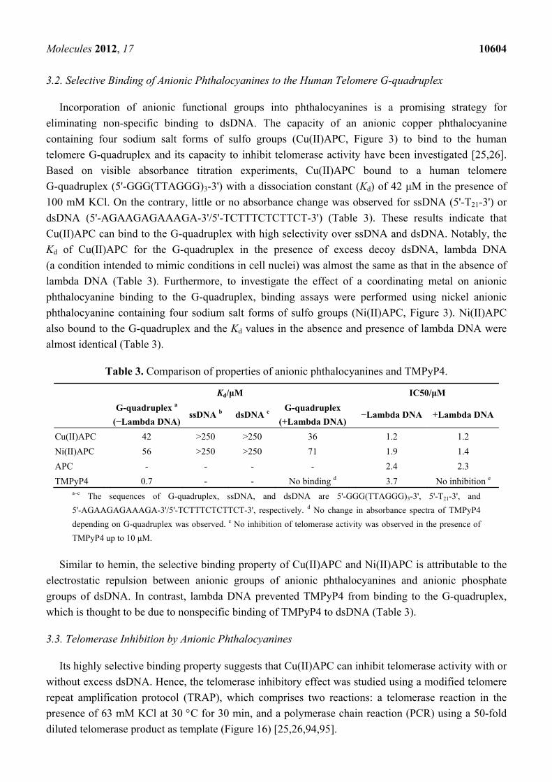
Table 3. Comparison of properties of anionic phthalocyanines and TMPyP4.
Kd/μM IC50/μM
G-quadruplex a
(−Lambda DNA) ssDNA b dsDNA c
G-quadruplex
(+Lambda DNA)−Lambda DNA +Lambda DNA
Cu(II)APC 42 >250 >250 36 1.2 1.2
Ni(II)APC 56 >250 >250 71 1.9 1.4
APC - - - - 2.4 2.3
TMPyP4 0.7 - - No binding d 3.7 No inhibition e a–c The sequences of G-quadruplex, ssDNA, and dsDNA are 5'-GGG(TTAGGG)3-3', 5'-T21-3', and
5'-AGAAGAGAAAGA-3'/5'-TCTTTCTCTTCT-3', respectively. d No change in absorbance spectra of TMPyP4
depending on G-quadruplex was observed. e No inhibition of telomerase activity was observed in the presence of
TMPyP4 up to 10 μM.
Similar to hemin, the selective binding property of Cu(II)APC and Ni(II)APC is attributable to the
electrostatic repulsion between anionic groups of anionic phthalocyanines and anionic phosphate
groups of dsDNA. In contrast, lambda DNA prevented TMPyP4 from binding to the G-quadruplex,
which is thought to be due to nonspecific binding of TMPyP4 to dsDNA (Table 3).
3.3. Telomerase Inhibition by Anionic Phthalocyanines
Its highly selective binding property suggests that Cu(II)APC can inhibit telomerase activity with or
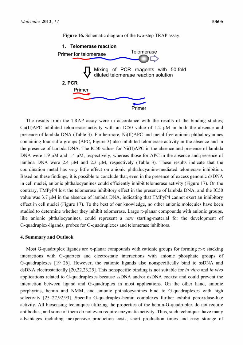
without excess dsDNA. Hence, the telomerase inhibitory effect was studied using a modified telomere
repeat amplification protocol (TRAP), which comprises two reactions: a telomerase reaction in the
presence of 63 mM KCl at 30 C for 30 min, and a polymerase chain reaction (PCR) using a 50-fold
diluted telomerase product as template (Figure 16) [25,26,94,95].
Molecules 2012, 17 10605
Figure 16. Schematic diagram of the two-step TRAP assay.
Primer for telomerase
Mixing of PCR reagents with 50-fold diluted telomerase reaction solution
Primer
Primer
1. Telomerase reaction
2. PCR
Telomerase
The results from the TRAP assay were in accordance with the results of the binding studies;
Cu(II)APC inhibited telomerase activity with an IC50 value of 1.2 μM in both the absence and
presence of lambda DNA (Table 3). Furthermore, Ni(II)APC and metal-free anionic phthalocyanines
containing four sulfo groups (APC, Figure 3) also inhibited telomerase activity in the absence and in
the presence of lambda DNA. The IC50 values for Ni(II)APC in the absence and presence of lambda
DNA were 1.9 μM and 1.4 μM, respectively, whereas those for APC in the absence and presence of
lambda DNA were 2.4 μM and 2.3 μM, respectively (Table 3). These results indicate that the
coordination metal has very little effect on anionic phthalocyanine-mediated telomerase inhibition.
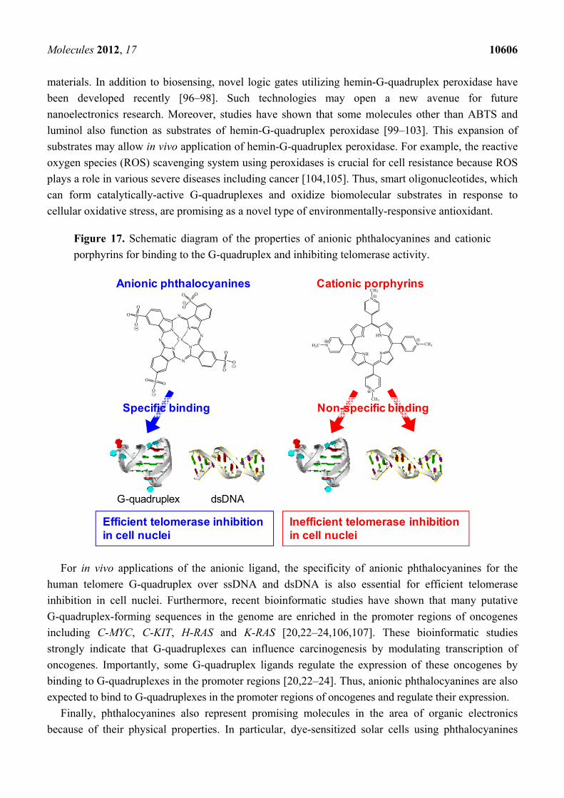
Based on these findings, it is possible to conclude that, even in the presence of excess genomic dsDNA
in cell nuclei, anionic phthalocyanines could efficiently inhibit telomerase activity (Figure 17). On the
contrary, TMPyP4 lost the telomerase inhibitory effect in the presence of lambda DNA, and the IC50
value was 3.7 μM in the absence of lambda DNA, indicating that TMPyP4 cannot exert an inhibitory
effect in cell nuclei (Figure 17). To the best of our knowledge, no other anionic molecules have been
studied to determine whether they inhibit telomerase. Large -planar compounds with anionic groups,
like anionic phthalocyanines, could represent a new starting-material for the development of
G-quadruplex-ligands, probes for G-quadruplexes and telomerase inhibitors.
4. Summary and Outlook
Most G-quadruplex ligands are -planar compounds with cationic groups for forming - stacking
interactions with G-quartets and electrostatic interactions with anionic phosphate groups of
G-quadruplexes [19–26]. However, the cationic ligands also nonspecifically bind to ssDNA and
dsDNA electrostatically [20,22,23,25]. This nonspecific binding is not suitable for in vitro and in vivo
applications related to G-quadruplexes because ssDNA and/or dsDNA coexist and could prevent the
interaction between ligand and G-quadruplex in most applications. On the other hand, anionic
porphyrins, hemin and NMM, and anionic phthalocyanines bind to G-quadruplexes with high
selectivity [25–27,92,93]. Specific G-quadruplex-hemin complexes further exhibit peroxidase-like
activity. All biosensing techniques utilizing the properties of the hemin-G-quadruplex do not require
antibodies, and some of them do not even require enzymatic activity. Thus, such techniques have many
advantages including inexpensive production costs, short production times and easy storage of
Molecules 2012, 17 10606
materials. In addition to biosensing, novel logic gates utilizing hemin-G-quadruplex peroxidase have
been developed recently [96–98]. Such technologies may open a new avenue for future
nanoelectronics research. Moreover, studies have shown that some molecules other than ABTS and
luminol also function as substrates of hemin-G-quadruplex peroxidase [99–103]. This expansion of
substrates may allow in vivo application of hemin-G-quadruplex peroxidase. For example, the reactive
oxygen species (ROS) scavenging system using peroxidases is crucial for cell resistance because ROS
plays a role in various severe diseases including cancer [104,105]. Thus, smart oligonucleotides, which
can form catalytically-active G-quadruplexes and oxidize biomolecular substrates in response to
cellular oxidative stress, are promising as a novel type of environmentally-responsive antioxidant.
Figure 17. Schematic diagram of the properties of anionic phthalocyanines and cationic
porphyrins for binding to the G-quadruplex and inhibiting telomerase activity.
Anionic phthalocyanines Cationic porphyrins
Specific binding Non-specific binding
Efficient telomerase inhibitionin cell nuclei
Inefficient telomerase inhibitionin cell nuclei
G-quadruplex dsDNA
For in vivo applications of the anionic ligand, the specificity of anionic phthalocyanines for the
human telomere G-quadruplex over ssDNA and dsDNA is also essential for efficient telomerase
inhibition in cell nuclei. Furthermore, recent bioinformatic studies have shown that many putative
G-quadruplex-forming sequences in the genome are enriched in the promoter regions of oncogenes
including C-MYC, C-KIT, H-RAS and K-RAS [20,22–24,106,107]. These bioinformatic studies
strongly indicate that G-quadruplexes can influence carcinogenesis by modulating transcription of
oncogenes. Importantly, some G-quadruplex ligands regulate the expression of these oncogenes by
binding to G-quadruplexes in the promoter regions [20,22–24]. Thus, anionic phthalocyanines are also
expected to bind to G-quadruplexes in the promoter regions of oncogenes and regulate their expression.
Finally, phthalocyanines also represent promising molecules in the area of organic electronics
because of their physical properties. In particular, dye-sensitized solar cells using phthalocyanines
N
HNN
NH
N
N
N
N
H3C
CH3
CH3
CH3
N
NN
N
N
N
N
N
SO O
O
SO
O
O
SOO
O
S
O
O
O
Cu
Molecules 2012, 17 10607
have been attracting increasing attention [108,109]. However, it is difficult to control association state,
orientation and localization of phthalocyanines in the functional films on the devices because of their
aggregation property. Programmable DNA nanostructure technology may be a promising approach to
solving this problem [9]. A recent study has shown that G-quadruplexes can be fabricated on DNA
nanostructure scaffolds [110]. Therefore, the desired dispersion and localization of anionic
phthalocyanines could be realized because of the specific binding to the G-quadruplexes on the
scaffold. Thus, together, anionic phthalocyanines and G-quadruplexes will contribute not only to the
biomedical field but also to the organic electronics field.
Acknowledgments
This work was supported in part by Grants-in-Aid for Scientific Research, Scientific Research on
Innovative Areas “Nanomedicine Molecular Science” (No. 2306), and the “MEXT-Supported Program
for the Strategic Research Foundation at Private Universities” (2009–2014) from the Ministry of
Education, Culture, Sports, Science and Technology, Japan, and the Hirao Taro Foundation of the
Konan University Association for Academic Research.
References
1. Endo, M.; Shiroyama, T.; Fujitsuka, M.; Majima, T. Four-way-branched DNA-porphyrin
conjugates for construction of four double-helix-DNA assembled structures. J. Org. Chem. 2005,
70, 7468–7472.
2. Liu, X.J.; Qi, C.; Bing, T.; Cheng, X.H.; Shangguan, D.H. Highly selective phthalocyanine-
thymine conjugate sensor for Hg2+ based on target induced aggregation. Anal. Chem. 2009, 81,
3699–3704.
3. Hammer, R.P.; Owens, C.V.; Hwang, S.H.; Sayes, C.M.; Soper, S.A. Asymmetrical,
water-soluble phthalocyanine dyes for covalent labeling of oligonucleotides. Bioconjug. Chem.
2002, 13, 1244–1252.
4. Bianco, A.; Da Ros, T.; Prato, M.; Toniolo, C. Fullerene-based amino acids and peptides. J. Pept. Sci.
2001, 7, 208–219.
5. Yang, W.R.; Thordarson, P.; Gooding, J.J.; Ringer, S.P.; Braet, F. Carbon nanotubes for
biological and biomedical applications. Nanotechnology 2007, 18, 412001–412012.
6. Wang, H.; Yang, R.H.; Yang, L.; Tan, W.H. Nucleic acid conjugated nanomaterials for enhanced
molecular recognition. ACS Nano 2009, 3, 2451–2460.
7. Zhang, S.G. Fabrication of novel biomaterials through molecular self-assembly. Nat. Biotechnol.
2003, 21, 1171–1178.
8. Wang, Z.D.; Lu, Y. Functional DNA directed assembly of nanomaterials for biosensing.
J. Mater. Chem. 2009, 19, 1788–1798.
9. Endo, M.; Sugiyama, H. Chemical approaches to DNA nanotechnology. ChemBioChem 2009,
10, 2420–2443.
10. Gellert, M.; Lipsett, M.N.; Davies, D.R. Helix formation by guanylic acid. Proc. Natl. Acad. Sci.
USA 1962, 48, 2013–2018.
11. Keniry, M.A. Quadruplex structures in nucleic acids. Biopolymers 2001, 56, 123–146.
Molecules 2012, 17 10608
12. Davis, J.T. G-quartets 40 years later: From 5'-GMP to molecular biology and supramolecular
chemistry. Angew. Chem. Int. Ed. Engl. 2004, 43, 668–698.
13. Dai, J.X.; Carver, M.; Yang, D.Z. Polymorphism of human telomeric quadruplex structures.
Biochimie 2008, 90, 1172–1183.
14. Kaushik, M.; Kaushik, S.; Bansal, A.; Saxena, S.; Kukreti, S. Structural diversity and specific
recognition of four stranded G-quadruplex DNA. Curr. Mol. Med. 2011, 11, 744–769.
15. Willner, I.; Shlyahovsky, B.; Zayats, M.; Willner, B. DNAzymes for sensing, nanobiotechnology
and logic gate applications. Chem. Soc. Rev. 2008, 37, 1153–1165.
16. Liu, J.W.; Cao, Z.H.; Lu, Y. Functional nucleic acid sensors. Chem. Rev. 2009, 109, 1948–1998.
17. Kosman, J.; Juskowiak, B. Peroxidase-mimicking DNAzymes for biosensing applications:
A review. Anal. Chim. Acta 2011, 707, 7–17.
18. Sen, D.; Poon, L.C.H. RNA and DNA complexes with hemin Fe(III) heme are efficient
peroxidases and peroxygenases: How do they do it and what does it mean? Crit. Rev. Biochem. Mol.
2011, 46, 478–492.
19. Wheelhouse, R.T.; Sun, D.K.; Han, H.Y.; Han, F.X.G.; Hurley, L.H. Cationic porphyrins as
telomerase inhibitors: The interaction of tetra-(N-methyl-4-pyridyl)porphine with quadruplex
DNA. J. Am. Chem. Soc. 1998, 120, 3261–3262.
20. Monchaud, D.; Teulade-Fichou, M.P. A hitchhiker’s guide to G-quadruplex ligands.
Org. Biomol. Chem. 2008, 6, 627–636.
21. De Cian, A.; Lacroix, L.; Douarre, C.; Temime-Smaali, N.; Trentesaux, C.; Riou, J.F.; Mergny, J.L.
Targeting telomeres and telomerase. Biochimie 2008, 90, 131–155.
22. Luedtke, N.W. Targeting G-quadruplex DNA with small molecules. Chimia 2009, 63, 134–139.
23. Neidle, S. Human telomeric G-quadruplex: The current status of telomeric G-quadruplexes as
therapeutic targets in human cancer. FEBS J. 2010, 277, 1118–1125.
24. Ou, T.M.; Lu, Y.J.; Tan, J.H.; Huang, Z.S.; Wong, K.Y.; Gu, L.Q. G-quadruplexes: Targets in
anticancer drug design. ChemMedChem 2008, 3, 690–713.
25. Yaku, H.; Murashima, T.; Miyoshi, D.; Sugimoto, N. Anionic phthalocyanines targeting
G-quadruplexes and inhibiting telomerase activity in the presence of excessive DNA duplexes.
Chem. Commun. 2010, 46, 5740–5742.
26. Yaku, H.; Fujimoto, T.; Murashima, T.; Miyoshi, D.; Sugimoto, N. Phthalocyanines: A new class
of G-quadruplex-ligands with many potential applications. Chem. Commun. 2012, 48, 6203–6216.
27. Travascio, P.; Li, Y.F.; Sen, D. DNA-enhanced peroxidase activity of a DNA aptamer-hemin
complex. Chem. Biol. 1998, 5, 505–517.
28. Moyzis, R.K.; Buckingham, J.M.; Cram, L.S.; Dani, M.; Deaven, L.L.; Jones, M.D.; Meyne, J.;
Ratliff, R.L.; Wu, J.R. A highly conserved repetitive DNA-sequence, (TTAGGG)n, present at the
telomeres of human-chromosomes. Proc. Natl. Acad. Sci. USA 1988, 85, 6622–6626.
29. Harley, C.B.; Futcher, A.B.; Greider, C.W. Telomeres shorten during ageing of human
fibroblasts. Nature 1990, 345, 458–460.
30. Harley, C.B.; Vaziri, H.; Counter, C.M.; Allsopp, R.C. The telomere hypothesis of cellular aging.
Exp. Gerontol. 1992, 27, 375–382.
31. Greider, C.W.; Blackburn, E.H. Identification of a specific telomere terminal transferase-activity
in tetrahymena extracts. Cell 1985, 43, 405–413.
Molecules 2012, 17 10609
32. Morin, G.B. The human telomere terminal transferase enzyme is a ribonucleoprotein that
synthesizes TTAGGG repeats. Cell 1989, 59, 521–529.
33. Zahler, A.M.; Williamson, J.R.; Cech, T.R.; Prescott, D.M. Inhibition of telomerase by G-quartet
DNA structures. Nature 1991, 350, 718–720.
34. Travascio, P.; Bennet, A.J.; Wang, D.Y.; Sen, D. A ribozyme and a catalytic DNA with
peroxidase activity: Active sites versus cofactor-binding sites. Chem. Biol. 1999, 6, 779–787.
35. Li, Y.F.; Geyer, C.R.; Sen, D. Recognition of anionic porphyrins by DNA aptamers.
Biochemistry 1996, 35, 6911–6922.
36. Li, Y.F.; Sen, D. A catalytic DNA for porphyrin metallation. Nat. Struct. Biol. 1996, 3, 743–747.
37. Li, Y.F.; Sen, D. Toward an efficient DNAzyme. Biochemistry 1997, 36, 5589–5599.
38. Lee, H.W.; Chinnapen, D.J.F.; Sen, D. Structure-function investigation of a deoxyribozyme with
dual chelatase and peroxidase activities. Pure Appl. Chem. 2004, 76, 1537–1545.
39. Majhi, P.R.; Shafer, R.H. Characterization of an unusual folding pattern in a catalytically active
guanine quadruplex structure. Biopolymers 2006, 82, 558–569.
40. Kong, D.M.; Cai, L.L.; Guo, J.H.; Wu, J.; Shen, H.X. Characterization of the G-quadruplex
structure of a catalytic DNA with peroxidase activity. Biopolymers 2009, 91, 331–339.
41. Cheng, X.H.; Liu, X.J.; Bing, T.; Cao, Z.H.; Shangguan, D.H. General peroxidase activity of
G-quadruplex-hemin complexes and its application in ligand screening. Biochemistry 2009, 48,
7817–7823.
42. Kong, D.M.; Yang, W.; Wu, J.; Li, C.X.; Shen, H.X. Structure-function study of peroxidase-like
G-quadruplex-hemin complexes. Analyst 2010, 135, 321–326.
43. Yi, X.; Pavlov, V.; Gill, R.; Bourenko, T.; Willner, I. Lighting up biochemiluminescence by the
surface self-assembly of DNA-hemin complexes. ChemBioChem 2004, 5, 374–379.
44. Xiao, Y.; Pavlov, V.; Niazov, T.; Dishon, A.; Kotler, M.; Willner, I. Catalytic beacons for the
detection of DNA and telomerase activity. J. Am. Chem. Soc. 2004, 126, 7430–7431.
45. Cheglakov, Z.; Weizmann, Y.; Beissenhirtz, M.K.; Willner, I. Ultrasensitive detection of DNA
by the PCR-induced generation of DNAzymes: The DNAzyme primer approach. Chem. Commun.
2006, 3205–3207.
46. Weizmann, Y.; Beissenhirtz, M.K.; Cheglakov, Z.; Nowarski, R.; Kotler, M.; Willner, I. A virus
spotlighted by an autonomous DNA machine. Angew. Chem. Int. Ed. Engl. 2006, 45, 7384–7388.
47. Tian, Y.; He, Y.; Mao, C.D. Cascade signal amplification for DNA detection. ChemBioChem
2006, 7, 1862–1864.
48. Bi, S.; Zhang, J.L.; Zhang, S.S. Ultrasensitive and selective DNA detection based on nicking
endonuclease assisted signal amplification and its application in cancer cell detection.
Chem. Commun. 2010, 46, 5509–5511.
49. Freeman, R.; Liu, X.Q.; Winner, I. Chemiluminescent and chemiluminescence resonance energy
transfer (CRET) detection of DNA, metal ions, and aptamer-substrate complexes using hemin/
G-quadruplexes and CdSe/ZnS quantum dots. J. Am. Chem. Soc. 2011, 133, 11597–11604.
50. Niazov, T.; Pavlov, V.; Xiao, Y.; Gill, R.; Willner, I. DNAzyme-functionalized Au nanoparticles
for the amplified detection of DNA or telomerase activity. Nano Lett. 2004, 4, 1683–1687.
51. Pelossof, G.; Tel-Vered, R.; Elbaz, J.; Willner, I. Amplified biosensing using the horseradish
peroxidase-mimicking DNAzyme as an electrocatalyst. Anal. Chem. 2010, 82, 4396–4402.
Molecules 2012, 17 10610
52. Yang, Q.L.; Nie, Y.J.; Zhu, X.L.; Liu, X.J.; Li, G.X. Study on the electrocatalytic activity of
human telomere G-quadruplex-hemin complex and its interaction with small molecular ligands.
Electrochim. Acta 2009, 55, 276–280.
53. Teixeira, M.F.S.; Freitas, B.H.; Seraphim, P.M.; Salmazo, L.O.; Nobre, M.A.; Lanfredi, S.
Development of an electrochemical sensor for potassium ions based on KSr2Nb5O15 modified
electrode. Proced. Chem. 2009, 1, 293–296.
54. Fan, X.Y.; Li, H.T.; Zhao, J.; Lin, F.; Zhang, L.; Zhang, Y.; Yao, S. A novel label-free
fluorescent sensor for the detection of potassium ion based on DNAzyme. Talanta 2012, 89, 57–62.
55. Ratte, H.T. Bioaccumulation and toxicity of silver compounds: A review. Environ. Toxicol. Chem.
1999, 18, 89–108.
56. Jarup, L. Hazards of heavy metal contamination. Br. Med. Bull. 2003, 68, 167–182.
57. Li, T.; Wang, E.; Dong, S.J. G-quadruplex-based DNAzyme as a sensing platform for
ultrasensitive colorimetric potassium detection. Chem. Commun. 2009, 580–582.
58. Yang, X.; Li, T.; Li, B.L.; Wang, E.K. Potassium-sensitive G-quadruplex DNA for sensitive
visible potassium detection. Analyst 2010, 135, 71–75.
59. Li, T.; Wang, E.; Dong, S. Lead(II)-induced allosteric G-quadruplex DNAzyme as a colorimetric
and chemiluminescence sensor for highly sensitive and selective Pb2+ detection. Anal. Chem.
2010, 82, 1515–1520.
60. Li, C.L.; Liu, K.T.; Lin, Y.W.; Chang, H.T. Fluorescence detection of lead(II) ions through their
induced catalytic activity of DNAzymes. Anal. Chem. 2011, 83, 225–230.
61. Zhang, J.; Gao, Q.L.; Chen, P.P.; Chen, J.H.; Chen, G.N.; Fu, F.F. A novel Tb3+-promoted
G-quadruplex-hemin DNAzyme for the development of label-free visual biosensors.
Biosens. Bioelectron. 2011, 26, 4053–4057.
62. Li, T.; Dong, S.J.; Wang, E. Label-free colorimetric detection of aqueous mercury ion (Hg2+)
using Hg2+-modulated G-quadruplex-cased DNAzymes. Anal. Chem. 2009, 81, 2144–2149.
63. Kong, D.M.; Wu, J.; Wang, N.; Yang, W.; Shen, H.X. Peroxidase activity-structure relationship
of the intermolecular four-stranded G-quadruplex-hemin complexes and their application in Hg2+
ion detection. Talanta 2009, 80, 459–465.
64. Li, T.; Li, B.L.; Wang, E.K.; Dong, S.J. G-quadruplex-based DNAzyme for sensitive mercury
detection with the naked eye. Chem. Commun. 2009, 3551–3553.
65. Jia, S.M.; Liu, X.F.; Li, P.; Kong, D.M.; Shen, H.X. G-quadruplex DNAzyme-based Hg2+ and
cysteine sensors utilizing Hg2+-mediated oligonucleotide switching. Biosens. Bioelectron. 2011,
27, 148–152.
66. Zhou, X.H.; Kong, D.M.; Shen, H.X. Ag+ and cysteine quantitation based on G-quadruplex-
hemin DNAzymes disruption by Ag+. Anal. Chem. 2010, 82, 789–793.
67. Zhou, X.H.; Kong, D.M.; Shen, H.X. G-quadruplex-hemin DNAzyme-amplified colorimetric
detection of Ag+ ion. Anal. Chim. Acta 2010, 678, 124–127.
68. Tanaka, K.; Tengeiji, A.; Kato, T.; Toyama, N.; Shiro, M.; Shionoya, M. Efficient incorporation
of a copper hydroxypyridone base pair in DNA. J. Am. Chem. Soc. 2002, 124, 12494–12498.
69. Tanaka, K.; Yamada, Y.; Shionoya, M. Formation of silver(I)-mediated DNA duplex and triplex
through an alternative base pair of pyridine nucleobases. J. Am. Chem. Soc. 2002, 124, 8802–8803.
Molecules 2012, 17 10611
70. Tanaka, K.; Tengeiji, A.; Kato, T.; Toyama, N.; Shionoya, M. A discrete self-assembled metal
array in artificial DNA. Science 2003, 299, 1212–1213.
71. Tanaka, K.; Clever, G.H.; Takezawa, Y.; Yamada, Y.; Kaul, C.; Shionoya, M.; Carell, T.
Programmable self-assembly of metal ions inside artificial DNA duplexes. Nat. Nanotechnol.
2006, 1, 190–194.
72. Muller, J. Chemistry: Metals line up for DNA. Nature 2006, 444, 698–698.
73. Takezawa, Y.; Maeda, W.; Tanaka, K.; Shionoya, M. Discrete Self-Assembly of iron(III) ions
inside triple-stranded artificial DNA. Angew. Chem. Int. Ed. Engl. 2009, 48, 1081–1084.
74. Bock, L.C.; Griffin, L.C.; Latham, J.A.; Vermaas, E.H.; Toole, J.J. Selection of single-stranded-
DNA molecules that bind and inhibit human thrombin. Nature 1992, 355, 564–566.
75. Macaya, R.F.; Schultze, P.; Smith, F.W.; Roe, J.A.; Feigon, J. Thrombin-binding DNA aptamer
forms a unimolecular quadruplex structure in solution. Proc. Natl. Acad. Sci. USA 1993, 90,
3745–3749.
76. Li, T.; Wang, E.K.; Dong, S.J. G-quadruplex-based DNAzyme for facile colorimetric detection
of thrombin. Chem. Commun. 2008, 3654–3656.
77. Shen, B.J.; Wang, Q.; Zhu, D.; Luo, J.J.; Cheng, G.F.; He, P.A.; Fang, Y.Z. G-quadruplex-based
DNAzymes aptasensor for the amplified electrochemical detection of thrombin. Electroanalysis
2010, 22, 2985–2990.
78. Girvan, A.C.; Teng, Y.; Casson, L.K.; Thomas, S.D.; Jüliger, S.; Ball, M.W.; Klein, J.B.;
Pierce, W.M., Jr.; Barve, S.S.; Bates, P.J. AGRO100 inhibits activation of nuclear factor-kappa B
(NF-kappa B) by forming a complex with NF-kappa B essential modulator (NEMO) and
nucleolin. Mol. Cancer Ther. 2006, 5, 1790–1799.
79. Ireson, C.R.; Kelland, L.R. Discovery and development of anticancer aptamers. Mol. Cancer Ther.
2006, 5, 2957–2962.
80. Li, T.; Shi, L.L.; Wang, E.K.; Dong, S.J. Multifunctional G-quadruplex aptamers and their
application to protein detection. Chem.-Eur. J. 2009, 15, 1036–1042.
81. He, Y.; Tian, Y.; Mao, C.D. Human telomeric DNA sequences have a peroxidase apoenzyme
activity. Mol. Biosyst. 2009, 5, 238–240.
82. Freeman, R.; Sharon, E.; Teller, C.; Henning, A.; Tzfati, Y.; Willner, I. DNAzyme-like activity
of hemin-telomeric G-quadruplexes for the optical analysis of telomerase and its inhibitors.
ChemBioChem 2010, 11, 2362–2367.
83. Stefan, L.; Denat, F.; Monchaud, D. Deciphering the DNAzyme activity of multimeric
quadruplexes: Insights into their actual role in the telomerase activity evaluation assay.
J. Am. Chem. Soc. 2011, 133, 20405–20415.
84. Kosman, J.; Juskowiak, B. Optimization study of the catalytic activity of DNAzymes based on
telomeric G-quadruplexes. Cent. Eur. J. Chem. 2012, 10, 368–372.
85. Izbicka, E.; Wheelhouse, R.T.; Raymond, E.; Davidson, K.K.; Lawrence, R.A.; Sun, D.;
Windle, B.E.; Hurley, L.H.; Von Hoff, D.D. Effects of cationic porphyrins as G-quadruplex
interactive agents in human tumor cells. Cancer Res. 1999, 59, 639–644.
86. Rha, S.Y.; Izbicka, E.; Lawrence, R.A.; Davidson, K.K.; Sun, D.; Moyer, M.P.; Roodman, G.D.;
Hurley, L.H.; Von Hoff, D.D. Effect of telomere and telomerase interactive agents on human
tumor and normal cell lines. Clin. Cancer Res. 2000, 6, 987–993.
Molecules 2012, 17 10612
87. Kim, M.Y.; Gleason-Guzman, M.; Izbicka, E.; Nishioka, D.; Hurley, L.H. The different
biological effects of telomestatin and TMPyP4 can be attributed to their selectivity for interaction
with intramolecular or intermolecular G-quadruplex structures. Cancer Res. 2003, 63, 3247–3256.
88. Liu, W.; Sun, D.; Hurley, L.H. Binding of G-quadruplex-interactive agents to distinct
G-quadruplexes induces different biological effects in MiaPaCa cells. Nucleosides Nucleotides
Nucleic Acids 2005, 24, 1801–1815.
89. Mikami-Terao, Y.; Akiyama, M.; Yuza, Y.; Yanagisawa, T.; Yamada, O.; Yamada, H. Antitumor
activity of G-quadruplex-interactive agent TMPyP4 in K562 leukemic cells. Cancer Lett. 2008, 261,
226–234.
90. Mikami-Terao, Y.; Akiyama, M.; Yuza, Y.; Yanagisawa, T.; Yamada, O.; Kawano, T.; Agawa, M.;
Ida, H.; Yamada, H. Antitumor activity of TMPyP4 interacting G-quadruplex in retinoblastoma
cell lines. Exp. Eye Res. 2009, 89, 200–208.
91. Gunaratnam, M.; Swank, S.; Haider, S.M.; Galesa, K.; Reszka, A.P.; Beltran, M.; Cuenca, F.;
Fletcher, J.A.; Neidle, S. Targeting human gastrointestinal stromal tumor cells with a
quadruplex-binding small molecule. J. Med. Chem. 2009, 52, 3774–3783.
92. Arthanari, H.; Basu, S.; Kawano, T.L.; Bolton, P.H. Fluorescent dyes specific for quadruplex
DNA. Nucleic Acids Res. 1998, 26, 3724–3728.
93. Ren, J.S.; Chaires, J.B. Sequence and structural selectivity of nucleic acid binding ligands.
Biochemistry 1999, 38, 16067–16075.
94. Kim, N.W.; Piatyszek, M.A.; Prowse, K.R.; Harley, C.B.; West, M.D.; Ho, P.L.; Coviello, G.M.;
Weinrich, S.L.; Shay, J.W. Specific association of human telomerase activity with immortal cells
and cancer. Science 1994, 266, 2011–2015.
95. Yu, H.Q.; Zhang, D.H.; Gu, X.B.; Miyoshi, D.; Sugimoto, N. Regulation of telomerase activity
by the thermodynamic stability of a DNA·RNA hybrid. Angew. Chem. Int. Ed. Engl. 2008, 47,
9034–9038.
96. Shlyahovsky, B.; Li, Y.; Lioubashevski, O.; Elbaz, J.; Willner, I. Logic gates and antisense DNA
devices operating on a translator nucleic acid scaffold. ACS Nano 2009, 3, 1831–1843.
97. Li, T.; Wang, E.K.; Dong, S.J. Potassium-lead-switched G-quadruplexes: A new class of DNA
logic gates. J. Am. Chem. Soc. 2009, 131, 15082–15083.
98. Li, T.; Ackermann, D.; Hall, A.M.; Famulok, M. Input-dependent induction of oligonucleotide
structural motifs for performing molecular logic. J. Am. Chem. Soc. 2012, 134, 3508–3516.
99. Rojas, A.M.; Gonzalez, P.A.; Antipov, E.; Klibanov, A.M. Specificity of a DNA-based
(DNAzyme) peroxidative biocatalyst. Biotechnol. Lett. 2007, 29, 227–232.
100. Golub, E.; Freeman, R.; Willner, I. A hemin/G-quadruplex acts as an NADH oxidase and NADH
peroxidase mimicking DNAzyme. Angew. Chem. Int. Ed. Engl. 2011, 50, 11710–11714.
101. Nakayama, S.; Wang, J.X.; Sintim, H.O. DNA-based peroxidation catalyst—What is the exact
role of topology on catalysis and is there a special binding site for catalysis? Chem.-Eur. J. 2011,
17, 5691–5698.
102. Yang, X.J.; Fang, C.L.; Mei, H.C.; Chang, T.J.; Cao, Z.H.; Shangguan, D.H. Characterization of
G-quadruplex/hemin peroxidase: Substrate specificity and inactivation kinetics. Chem.-Eur. J.
2011, 17, 14475–14484.
Molecules 2012, 17 10613
103. Poon, L.C.H.; Methot, S.P.; Morabi-Pazooki, W.; Pio, P.; Bennet, A.J.; Sen, D. Guanine-rich
RNAs and DNAs that bind heme robustly catalyze oxygen transfer reactions. J. Am. Chem. Soc.
2011, 133, 1877–1884.
104. Mates, J.M. Effects of antioxidant enzymes in the molecular control of reactive oxygen species
toxicology. Toxicology 2000, 153, 83–104.
105. D'Autreaux, B.; Toledano, M.B. ROS as signalling molecules: Mechanisms that generate
specificity in ROS homeostasis. Nat. Rev. Mol. Cell Biol. 2007, 8, 813–824.
106. Huppert, J.L.; Balasubramanian, S. Prevalence of quadruplexes in the human genome.
Nucleic Acids Res. 2005, 33, 2908–2916.
107. Todd, A.K.; Johnston, M.; Neidle, S. Highly prevalent putative quadruplex sequence motifs in
human DNA. Nucleic Acids Res. 2005, 33, 2901–2907.
108. Schumann, S.; Hatton, R.A.; Jones, T.S. Organic photovoltaic: Devices based on water-soluble
copper phthalocyanine. J. Phys. Chem. C 2011, 115, 4916–4921.
109. Ryan, J.W.; Anaya-Plaza, E.; Escosura A.D.; Torres, T.; Palomares, E. Small molecule solar
cells based on a series of water-soluble zinc phthalocyanine donors. Chem. Commun. 2012, 48,
6094–6096.
110. Liu, Y.; Lin, C.X.; Li, H.Y.; Yan, H. Aptamer-directed self-assembly of protein arrays on a DNA
nanostructure. Angew. Chem. Int. Ed. Engl. 2005, 44, 4333–4338.
© 2012 by the authors; licensee MDPI, Basel, Switzerland. This article is an open access article
distributed under the terms and conditions of the Creative Commons Attribution license
(http://creativecommons.org/licenses/by/3.0/).
Copyright © 2022 FDOKUMEN