Spectroscopic studies of novel porphyrin-copper(II) and zinc(II) complexes that share the...
12
Transition Metal Chemistry 27: 906917, 2002. @ 2002 KIulver Academic Publishers. Printed in the Nefherlands Spectroscopic studies of novel porphyrin-copper(I1) and zinc(I1) complexes that share the pinch- porphyrin family structure of iron(II1) complex models of peroxidases JosC Luis Garate-Morales and Yasmi Reyes-Ortega* Centro de Quimica, Instituto de Ciencias, Universidad Autdnoma de Puebla. 14 Sur 6301. Col. San Manuel. Puebla, Pue. 72570, MPxico Cecilio Alvarez-Toledano Instituto de Quimica, Universidad Nacional Autdnoma de MPxico. Circuito exterior SIN, Ciudad Universitaria. MPxico, D.F. 04510, Mkxico RenC GutiCrrez-Perez Facultad de Ciencias Quimicas, Universidad Autdnoma de Puebla. Ave. Sun Claudio SIN, Ciudad Universitaria. Puebla, Pue. 72570, MPxico Daniel Ramirez-Rosales and Rafael Zamorano-Ulloa Escuela Superior de Fisica y Matemciticas, Instituto PolitPcnico Nacional. Ave Instituto PolitPcnico Nacional SIN, Ed$ 9, Unidad Profesional Adolfo Ldpez Mateos, Sun Pedro Zacatenco, MPxico, D.F. 07738, MPxico Eduardo Basurto-Uribe Departamento de Ciencias Bcisicas, Universidad Autdnoma Metropolilana-Azcapotzalco, Ave Sun Pablo 180, MPxico, D.F. 02200, MPxico Julio Hernindez-Diaz and Rosalinda Contreras Departamento de Quimica, Centro de Investigaciones y de Estudios Avanzados del IPN. A.P. 14-740, MPxico, D.F. 07000, MPxico Received 26 October 2001; accepted 26 February 2002 Abstract Six novel pinch-porphyrin complexes [(picdien)(protoporphyrinate dimethyl ester)]copper(II) (7), [(picdien)(meso- porphyrinate dimethyl ester)]copper(II) (8) and [(picdien)(deuteroporphyrinate dimethyl ester)]copper(II) (9), [(picdien)(protoporphyrinate dimethyl ester)]zinc(II) (13), [(picdien)(mesoporphyrinate dimethyl ester)]zinc(II) (14) and [(picdien)(deuteroporphyrinate dimethyl ester)]zinc(II) (15), were prepared from the corresponding free copper(I1)-porphyrins (44), and zinc(I1)-porphyrins (10-12) and picdien (N-(3H-imidazol-4-ylmethy1)-K-{2- [(3H-imidazol-4-ylmethyl)-amino]-ethyl)-ethane-2,3-diamine). Spectroscopic studies show that complexes (7- 9) and (13-15) have the pinch-porphyrin type structure previously found in iron(II1) complex models of peroxidases. Complexes (7-9), were characterized by u.v.-vis., m.c.d., and e.s.r. spectroscopy. Em. spectra of the copper parent compounds (4-6) at ca. 10-~-10-~ M concentrations were typical of copper(I1)-dimers. The addition of the picdien ligand broke up the dimers as detected by e.s.r. Compounds (7- 9) are predominantly monomeric at ca. M concentration. The presence of picdien in (7-9) distorts the porphyrin internal portion of the plane so as to make these four internal nitrogen atoms, coordinated to copper(II), e.s.r.-distinguishable. MO and ligand field theories were used to characterize and to evaluate the directional covalence parameters of compounds (7- 9). A non-fully axial, out-of-the-porphyrin-plane bonding was found for (7-9), similar to the bonding of the pinch-porphyrins-iron(II1). However the in-plane distortion produced by the presence of the picdien ligand on copper(I1) is significantly larger than in pinch-porphyrin- iron(II1). The n.m.r. data show that the porphyrin-zinc(I1) is the less strained and has the weakest bonded structure. The coordination number of the pinch-porphyrin with iron(III), copper(I1) and zinc(II), is in all cases six. Introduction * Author for correspondence Syntheses, modeling and spectroscopic studies of metal- coordinated porphyrins have contributed to our know- ledge of the essential factors that characterize active sites
Transcript of Spectroscopic studies of novel porphyrin-copper(II) and zinc(II) complexes that share the...

Transition Metal Chemistry 27: 906917, 2002. @ 2002 KIulver Academic Publishers. Printed in the Nefherlands
Spectroscopic studies of novel porphyrin-copper(I1) and zinc(I1) complexes that share the pinch-porphyrin family structure of iron(II1) complex models of peroxidases
JosC Luis Garate-Morales and Yasmi Reyes-Ortega* Centro de Quimica, Instituto de Ciencias, Universidad Autdnoma de Puebla. 14 Sur 6301. Col. San Manuel. Puebla, Pue. 72570, MPxico
Cecilio Alvarez-Toledano Instituto de Quimica, Universidad Nacional Autdnoma de MPxico. Circuito exterior SIN, Ciudad Universitaria. MPxico, D.F. 04510, Mkxico
RenC GutiCrrez-Perez Facultad de Ciencias Quimicas, Universidad Autdnoma de Puebla. Ave. Sun Claudio SIN, Ciudad Universitaria. Puebla, Pue. 72570, MPxico
Daniel Ramirez-Rosales and Rafael Zamorano-Ulloa Escuela Superior de Fisica y Matemciticas, Instituto PolitPcnico Nacional. Ave Instituto PolitPcnico Nacional SIN, Ed$ 9, Unidad Profesional Adolfo Ldpez Mateos, Sun Pedro Zacatenco, MPxico, D.F. 07738, MPxico
Eduardo Basurto-Uribe Departamento de Ciencias Bcisicas, Universidad Autdnoma Metropolilana-Azcapotzalco, Ave Sun Pablo 180, MPxico, D.F. 02200, MPxico
Julio Hernindez-Diaz and Rosalinda Contreras Departamento de Quimica, Centro de Investigaciones y de Estudios Avanzados del IPN. A.P. 14-740, MPxico, D.F. 07000, MPxico
Received 26 October 2001; accepted 26 February 2002
Abstract
Six novel pinch-porphyrin complexes [(picdien)(protoporphyrinate dimethyl ester)]copper(II) ( 7 ) , [(picdien)(meso- porphyrinate dimethyl ester)]copper(II) ( 8 ) and [(picdien)(deuteroporphyrinate dimethyl ester)]copper(II) (9), [(picdien)(protoporphyrinate dimethyl ester)]zinc(II) (13), [(picdien)(mesoporphyrinate dimethyl ester)]zinc(II) (14) and [(picdien)(deuteroporphyrinate dimethyl ester)]zinc(II) (15), were prepared from the corresponding free copper(I1)-porphyrins ( 4 4 ) , and zinc(I1)-porphyrins (10-12) and picdien (N-(3H-imidazol-4-ylmethy1)-K-{2- [(3H-imidazol-4-ylmethyl)-amino]-ethyl)-ethane-2,3-diamine).
Spectroscopic studies show that complexes (7-9) and (13-15) have the pinch-porphyrin type structure previously found in iron(II1) complex models of peroxidases. Complexes (7-9), were characterized by u.v.-vis., m.c.d., and e.s.r. spectroscopy. E m . spectra of the copper parent compounds (4-6) at ca. 10 -~ -10 -~ M concentrations were typical of copper(I1)-dimers. The addition of the picdien ligand broke up the dimers as detected by e.s.r. Compounds (7-9) are predominantly monomeric at ca. M concentration. The presence of picdien in (7-9) distorts the porphyrin internal portion of the plane so as to make these four internal nitrogen atoms, coordinated to copper(II), e.s.r.-distinguishable. MO and ligand field theories were used to characterize and to evaluate the directional covalence parameters of compounds (7-9). A non-fully axial, out-of-the-porphyrin-plane bonding was found for (7-9), similar to the bonding of the pinch-porphyrins-iron(II1). However the in-plane distortion produced by the presence of the picdien ligand on copper(I1) is significantly larger than in pinch-porphyrin- iron(II1). The n.m.r. data show that the porphyrin-zinc(I1) is the less strained and has the weakest bonded structure. The coordination number of the pinch-porphyrin with iron(III), copper(I1) and zinc(II), is in all cases six.
Introduction
* Author for correspondence
Syntheses, modeling and spectroscopic studies of metal- coordinated porphyrins have contributed to our know- ledge of the essential factors that characterize active sites

in metallo-proteins [14]. At least five metallo-protein models have been reported. They show diverse magnetic and electronic structures, which determine their elec- tronic and magnetic behavior and functions, and allow understanding of the function of native proteins [5].
The substitution of iron by copper has been used as a tool to study enzymes. It provides information about the structure, its integrity, stability, spectroscopic behavior, and biochemical activity [6-231.
Our group reported new models of peroxidase en- zymes, namely pinch-porphyrins (Scheme l), which proved to be among the most successful spectroscopic and functional models [24]. They are dimethyl esters of proto-, meso-, and deuterioporphyrin iron(III), which bear an axially coordinated picdien ligand. This ligand consists of two pyridyl groups ortho-linked by a nitrogen- containing hydrocarbon chain [24, 251.
We decided to investigate how copper (paramagnetic S = 112) and zinc atoms (diamagnetic S = 0) affect the local and overall structure, symmetry and stability of these pinch-porphyrins in order to contrast the structural parameters among them with those of the pinch-por- phyrin-iron(II1) models. We report herein the synthesis and spectroscopic studies of new pinch-porphyrins of copper(II), (7-9), andzinc(II), (13-15) (Scheme 1) which complements our knowledge of iron pinch-porphyrins.
Experimental
Titration and other spectrophotometric measurements were performed with u.v.-vis./n.i.r. Shimadzu 3100 and Beckman DU 7500 spectrophotometers, using a quartz cell with a l-cm optical path. A 19G800 nm interval was used for the measurements. M.c.d. spectra were recorded using a JASCO circular dichroism polarimeter model J- 500A. A magnetic field of 0.4 T parallel to the light beam was applied by a Jasco optical electromagnet. M.c.d.
spectra were recorded for an interval of 300-600 nm, with a sensibility of 2 mdeg cm-' and a scale of 10 nm cm-' was used. All spectral measurements, u.v.-vis. and m.c.d., were performed at 25 OC, and the DMSO and CHC13 solutions of (1-6) complexes were ca. mM .
'H-n.m.r. spectra were recorded with a JEOL Eclipse 270 MHz spectrometer, using TMS as reference and a window of + 50 to -30 p.p.m. Solutions of unmetallated porphyrins (1-3) were ca. 3 x M and ca. 15 x
M at 25 "C, in DMSO-d6 and CDC13. E.s.r. measurements were made with a JEOL JES-
RE3X spectrometer at liquid-N2 temperature. Micro- wave X-band (ca. 9.8 GHz) was used. These spectra were recorded in a field width of G500 mT and the concentration of the metallo-porphyrin compounds ranged from lo-' to M . Simulations of the e.s.r. spectra were carried out, in a first step, by the SPRIT computational program [26]. In a second stage, the first simulations were refined using the computational pro- gram QPOW [27].
Spectrophotometric and titration measurements were made in CHC4 for (21, ( 3 ) , (5 ) , (6), (a), ( 9 ) , ( l l ) , (12) , (14) and (15) (meso- and deuteroporphyrin families) and in DMSO for ( I ) , ( 4 ) , (7), (10) and (13) (protoporphyrin). Copper and zinc porphyrins were prepared as described in previous studies [28]. Solution concentrations of ( I ) in DMSO and (2) and (3) in CHC13 were determined by spectrophotometric methods. Compound N-(3H-imidazol-4-ylmethy1)-N- {2-[(3H-imidazol-4-ylmethyl)-amino]-ethyl}-ethane-2,3- diarnine (picdien) was prepared by the method of Ahmed et al. [25].
Synthesis of porphyrin-copper(ZZ) and zinc(ZZ)-picdien complexes
All complexes were prepared .by the Reyes-Ortega method (Scheme 1) [24]. (1) The complexed protopor-
Pr = proptonate methyl ester l V
R = -CH=CH2 protoporphyrin-IX (1) metalled porphyrlns
R = -CH&H3 mesoporphyrin-lX (2) M"' = CU" (3-6) R = -H deuteroporphyrin-lX (3) M"* = ( 1 0- 12)
pinch-porphyrlns MW = CU" (7-9) M"+ = ~ n " ( 1 3- 15)
Scheme I .

phyrin-copper(I1) (4) was dissolved in DMSO, meso- porphyrin-copper(I1) (5) and deuteroporhyrin-cop- per(11) (6) in CHC13 at 25 OC and then they were monitored by u.v.-vis. and e.s.r spectra. To these solutions was added a picdien solution in DMSO for (4) or CHC13 solutions for (5) and (6) until reaching 1 equivalent. Then, an excess of the picdien ligand was added to make sure that the new species did not change in composition. U.v.-vis. spectra did not change once the ligand was in excess (up to 100% excess). (2) Solutions of (4-6) in CHC13 at 25 OC were treated with the picdien ligand in 1:l stoichiometric relation. The mixtures were stirred for 6 h, then observed by u.v.-vis. and e.s.r. spectroscopies. (3) The reaction mixtures were boiled under reflux for 6 h and the u.v.-vis. and e.s.r. spectra recorded after this time. Spectra of the complexes prepared by experiments (1-3) were the same. This fact provides evidence for the same com- plexes (7-9) was formed no matter which route was followed.
The 'H-n.m.r. spectra of (1-15) were recorded in CDC13, with exception of (I), (4), (7), (10) and (13) that were in DMSO-d6. To the solutions of (4-6) and (10-12) were added picdien 0.2 equivalents in solution in five consecutive steps and for each step a 'H-n.m.r. spectrum was recorded.
Assignments of the u.v.-vis. transitions for (4-9) are: Bl, t A, (A++) -- 17,200-17,853 cm-' (580-560nm) B,, t B3, (A,,,) -- 19,200-18,800 cm-' (523-530 nm) B1, t B2, (A,,) -- 25,100-24,570 cm-l (398-407 nm) Spectroscopic data 'H-n.m.r/[~, p.p.m.1: compound
(1): 4 CH3-heme groups [3.48, 3.48, 3.54, 3.551; CH=CH2(2)(2) [8.10]; C H = C C ( ) ( ) [6.09]; C H = a - - (3)(2) [6.26]; m=CH2(2)(4) [8.14]; CH=%(2)(4) [6.09]; CH==(3)(4) [6.26]; %CH2C02CH3(6) [4.27]; C H 2 s C 0 2 C H 3 ( 6 ) [3.16]; CH2CH2CO2c3(6) [3.59]; CXJCH2CO2CH3(7) [4.27]; CH2B2C02CH3(7) [3.1]; CH2CH2C02E3(7) [3.59]; Ha, H,g, H,, Ha [9.96, 9.95, 9.83, 9.821; compound (2): 4 CH3-heme [3.56, 3.57, 3.60, 3.59];m2<H3(2)(2,4) [4.02]; CH2-m(2)- (2,4) [1.80]; =CH3(2)(2,4) [4.02]; CH2-B3(2)(2,4)- [l .80]; %CH2C02CH3(6) [4.37]; CH2=C02CH3(6) [3.23]; CH2CH2CO2m3(6) [3.60]; CH2CH2C02CH3(7) [4.37]; CH2%C02CH3(7) [3.23]; CH2CH2C02B3(7) [3.59]; Ha, Hp, H,, Ha [4H, 10.031; compound (3): 4 CH3-heme groups [3.60, 3.57, 3.68, 3.701; H(2) [9.06]; H(4) [9.04]; CIJ2CH2CO2CH3(6) [4.38]; C H 2 E 2 C 0 2 - CH3(6) [3.24]; CH2CH2CO2E3(6) [3.59]; E 2 C H 2 - C02CH3(7) [4.38]; CH2=C02CH3(7) [3.24]; CH2CH2C02E3(7) [3.59]; H,, H,g, H,, Ha [10.06, 10.06, 10.06, 9.991; compound (5): 4 CH3-heme groups [4, 3.651; B2-CH3(2)(2,4) [6.48]; CH2<A3(2)(2,4) [1.82]; %-CH3(2)(2,4) [6.48]; m2-CH3(2)(2,4) [1.82]; E2CH2CO2CH3(6) [6.48]; CH&C02CH3(6) [3.23]; CH2CH2C02=3(6) [3.65]; E2CH2CO2CH3(7) [6.48]; CH2G2C02CH3(7) [3.23]; CH2CH2CO2B3(7) [3.65]; 'H,, Hp, 'H,, * H ~ [4, 11.23 p.p.m]; compound (6): 4 CH3-heme groups [4, 3.651; H(2) [8.37]; H(4) [8.37]; %CH2C02CH3(6) [6.40]; C H 2 w 2 C H 3 ( 6 )
[3.24]; CH2CH2C02a3(6) [3.65]; =CH2C02CH3(7) [6.40]; CH&C02CH3(7) [3.24]; CH2CH2C02E3(7) [3.65]; compound (7): 4 CH3-heme groups [4, 3.251; CH=CH2(2)(2) [8.68]; CH=CC(2)(2) [6.39]; CH=C&- - (3)(2) [6.39]; =%H2(2)(4) [8.68]; CH=B2(2)(4) [6.39]; CH<C(3)(4) [6.39]; m2CH2CO2CH3(6) [4.56]; CH2g2C02CH3(6) [2.52]; CH2CH2CO2S3(6) [3.25]; B2CH2C02CH3(7) [4.56]; CH2s?O2CH3(7) [2.52]; CH2CH2C02B3(7) [3.25]; 'H,, Hp, H,, * H ~ [4, 10.071; compound (8): 4 CH3-heme [4, 3.651; CHI- CH3(2)(2,4) [6.45]; CH2-E3(2)(2,4) [1.82]; CH,-CH3- (2)(2,4) [6.45]; m2<H3(2)(2,4) [1.82]; =CHI- C02CH3(6) [6.45]; CH2%C02CH3(6) [3.62]; CH2CH2- CO2%(6) [3.65]; =CH2C02CH3(7) [6.45]; C H 2 B - C02CH3(7) [3.62]; CH2CH2CO2=3(7) [3.65]; Ha, H,g, H,, Ha [4, 11 SO]; compound (9): 4 CH3-heme groups [4, 3.901; H(2) [8.45]; H(4) [8.45]; =CH2C02CH3(6) [6.39]; CH2=C02CH3(6) [3.13]; CH2CH2C02C&(6) [3.64]; E2CH2CO2CH3(7) [6.39]; CH2SFO2CH3(7) [3.13]; CH2CH2CO2m3(7) [3.64]; *H,, Hp, H,, * ~ 6 [4, 11.191; compound (10): 4 CH3-heme groups [3.60, 3.60, 3.63, 3.751; B=CH2(2)(2) [8.53]; C H = 9 ( 2 ) ( 2 ) [6.17]; CH=%(3)(2) [6.17]; g=CH2(2)(4) [8.53]; CH= CH2(2)(4) [6.44]; CH=E2(3)(4) [6.44]; =CH2- - C02CH3(6) [4.32]; CH2=CO2CH3(6) [3.31]; CH2- CH2C02E3(6) [3.8]; %CH2C02CH3(7) [4.32]; CH2=CO2CH3(7) [3.31]; CH2CH2C02CH3(7) [3.8]; Ha, Hp, H,, Ha [10.27, 10.18, 10.16, 10.151; compound (11): 4 CH3-heme groups [3.64, 3.57, 4.55, 4.301; m-CH3(2) (2,4) [3.86]; CH2-B3(2)(2,4) [1.76]; CH2-CH3(2)(2,4) [3.86]; CH2-m3(2)(2,4) [1.76]; - CH2CH2C02CH3(6) [4.21]; CH2=C02CH3(6) [3.14]; - CH2CH2C02E3(6) [3.67]; =CH2C02CH3(7) [4.21]; CH2%CO2CH3(7) [3.14]; CH2CH2CO2E3(7) [3.64]; Ha, H,g, H,, Ha [9.55, 9.50, 9.49, 9.631; compound (12): 4 CH3-heme groups [3.62, 3.34, 3.60, 3.651; H(2) [8.69]; H(4) [8.69]; =CH2CO2CH3(6) [4.17]; C H 2 m - C02CH3(6) [3.08]; CH2CH2C02m3(6) [3.47]; %- CH2C02 CH3(7) [4.17]; CH2CH2C02CH3(7) [3.08]; CH2CH2C02B3(7) [3.50]; ' H F H ~ , 'H,, *H* [9.51, 9.34, 9.51, 9.301; compound (13): 4 CH3-heme groups [3.58, 3.58, 3.61, 3.741; CHXH2(2)(2) [8.51]; CH= CH2(2)(2) [6.lS]; C H = C C ( ~ ) ) [6.15]; ==CH2(2)(4) - [8.51]; C H = B (2)(4) [6.42]; CH=%(3)(4) [6.42]; CH2CH2C02CH3(6) [4.31]; CH2B2C02CH3(6) [2.96]; - CH2CH2CO2g3(6) [3.75]; g2CH2C02CH3(7) [4.31]; CH2BC02CH3(7) [2.96]; CH2CH2C02E3(7) [3.75]; Ha, HD, H,, Ha [10.13, 10.13, 10.51, 10.111; compound (14): 4 CH3-heme groups [3.35, 3.35, 3.65, 3.651; CHI- CH3(2)(2,4) [3.97]; CH2<C3(2)(2,4) [I .77]; %- CH3(2)(2,4) [3.97]; CH2-w3(2)(2,4) [1.77]; CH2CH2CO2CH3(6) [4.32]; C H 2 B C 0 2 C H 3 ( 6 ) [3.17]; - CH2CH2CO2g3(6) [3.35]; %CH2C02CH3(7) [4.32]; CH2=C02CH3(7) [3.17]; CH2CH2C02=3(7) [3.35]; H,, H,g, H,, Ha [9.82, 9.82, 9.97, 9.831; compound (15) : 4 CH3-heme groups [3.35, 3.35, 3.65, 3.651; H(2) [9.01]; H(4) [9.01]; H(4) [9.02]; m2CH2C02CH3(6) [4.35]; CH2BC02CH3(6) [3.22]; CH2CH2CO2CFJ(6) [3.55]; CH2CH2C02CH3(7) [4.35]; CH2%C02CH3(7) [3.22]; -

CH2CH2Cq2m3(7) [3.55]; H,, Hp, H,, Hg [9.91, 9.91, 9.99, 9.871. Conc. ca. 1.5 x M .
Results and discussion
U.v.-vis. spectroscopy
U.v.-vis. spectra of (7-9) and (13-15) are typical for porphyrin complexes with copper(I1) and zinc(I1) ions, respectively [I I, 12, 29-35]. The presence of an intense Soret band [1,,,/Abs. for (7-9) are 398-407 nm/1.54- 1.7 1, for (13-15) are 400-403 nml1.24-1.331 and two wide and weak Q-bands [1,,,/Abs. of Q, for (7-9) are 523-534 nm/O.O7-O. 1, for (13-15) are 53CL548 nm/ 0.06-0.1; of Q2 for (7-9) are 57&662 nm/0.11-0.15, for (13-15) are 530-548 nml0.07-0.091 indicate that the complexes have a symmetry LD4h [18, 24, 29-35].
The data of (7-9) are more intense than those of the parent compounds ( 4 4 ) , indicating that the amount of ligand field distortion in the ligand field and excited states increases [35]. The same symmetry is revealed by the u.v.-vis. spectra of the pinch-porphyrin-iron(II1) already reported [24].
Magnetic circular dicroism spectroscopy
Magnetic circular dicroism (M.c.~.) spectroscopy has been used on complexes (4-9) [36]. Figure 1 presents the spectra of complexes (4) and (7); these are representative of complexes (4-9). Table 1 summarizes the m.c.d. data.
The m.c.d. spectra of the pinch-copper(I1) complexes (7-9) display two negative weak and broad bands, Q, in the 512-566 nm range. The band broadening is attrib- uted to the presence of the ligand which increases the number of available molecular transitions, now includ- ing n-orbitals of the pyridyl groups, and several tran- sitions d -+ d [35, 371.
These m.c.d. spectra of (4-9) are type B negative, characteristic of <D4h symmetry where the degeneracy in the molecular level has not been completely removed
Tahle I . MCD spectral data for complexes (4-9)
Fig.]. M.c.d. spectra of (a) ( 6 ) ; and (b) ( 9 ) complexes. [elM = molar ellipiicityjgrad cm2 dmol-' T - I .
[36]. All spectra have an intense negative N band due to a c.t. transition in an energy interval 30CL340 nm [37].
' H - n.m.r. studies
The proximity among two, three or more porphyrin- copper(I1) rings of ( 4 4 ) leads to a magnetic exchange interaction between Cu-Cu ions which can be ferro- magnetic [37-421. Such an interaction has been observed directly in the 'H-n.m.r. spectra of copper porphyrins, as shown by a shift of the signals to higher frequencies (ca. 40 p.p.m.) [39-44].
Highly efficient relaxation mechanisms by spin-spin interaction, generates broad signals [39,44]. Addition of the ligand to ( 4 4 ) decreases the ferromagnetic ex- change interaction by segregating the porphyrins. This effect improves the readability of the spectra giving sharper signals and significant shifting to lower frequen- cies (Figure 2).
To be able to assign the 'H-n.m.r. spectral signals of the pinch-porphyrins (7-9), increasing amounts of the picdien ligand were added to ( 4 4 ) up to 1 equivalent. The assignment is also based on comparison of the
Complex I,. [ ~ I M h r c t [ ~ I M A Q ~ L ~ I M 1, [ ~ I M
( 4 ) 332.0 +0.28 379 +1.4, - - - - 406 -2.9
(7) 340.6 f0.29 387 + 1.5. - - - -
407 -1.3 (5 ) 304 +1.7, 378 +1.6, 512 -3.7 566 -3.2
326 -2.5 394 -5.0 ( 8 ) 308 f 1 . 0 , 378 + I S , 514 -8.7 566 -7.3
328 -7.1 402 -9.3 f 6 ) 300 -2.1, 378 -1.7 516 -10.0 560 -8.0
326 -8.6 398 -12.5 (9 ) 300 -1.4, 379 f 1 . 8 , 516 -17.3 558 -12.6
309 -11.3 394 26.5
A = long wave (nm); [elM x = molar ellipticity (grad cm2 dmol-I T-I).

(4) + 1 eq. of picdien = (
(4) +O.6 eq. of picdien I
Fig. 2. 'H-n.m.r. spectra of (a) unmetalled protoporphyrin dimethyl ester ( I ) ; (b) (protoporphyrinate dimethyl ester)copper(ll) (4); (c)-(e) progressive axial picdien ligand titration for obtaining pinch-porphyrin [(picdien)(protoporphyrinate dimethyl ester)] copper(I1) (11).
'H-n.m.r. spectra of the zinc(I1) and copper(I1) com- porphyrins (1-3) change significantly upon metal com- plexes. The 'H-n.m.r. spectra of the unmetallated plexation [copper(II), Figure 2(a), (b) or zinc(II), Fig-

( I I ) + 1 eq. de picdien = (14)
( I I) + 0.6 eq. de picdien
I
(1 1) + 0.2 eq. de picdien
Fig. 3. 'H-n.m.r. spectra of (a) unmetalled mesoporphyrin dimethyl ester (2); (b) (mesoporphyrinate dimethyl ester)copper(ll) (5); (cHe) progressive axial picdien ligand litration for obtaining pinch-porphyrin [@icdien)(mesoporphyrinate dimethyl ester)] copper(l1) (14).
ure 3(a), (b)] and again a transformation is observed by addition of the ligand [(Figure 2(ck(e) and Figure 3(c)- (e))l.
Figure 2(a) shows the ' ~ - n . m . r . / D M ~ o - d ~ spectrum of (1) and Figure 2(b) the spectrum of (4 ) , Figures 2(c)-(e) show the effect of addition of the ligand. Figure 3(at(e) show the ' ~ - n . m . r . / ~ ~ C l ~ spectra of (2, l l ) , their titrations with the picdien ligand and their transformation into (14). Tables 2 and 3 summarize the
relevant chemical shift changes (A6 of meso and methyl- heme protons) of the (1-3) ring signals by their metallation to give ( 4 4 ) and (10-12) and by the ligand co-ordination in (7-9) and (13-15).
The A6H.meso and AacH,-heme values inform us about the groups that are affected by the ligand coordination which changes the symmetry and the ring currents.
Table 2 shows for pinch-protoporphyrin of copper(I1) (7-9), and zinc(I1) ( l 3 ) , [(Aa7-', A6,3-,]
PIVCSB5
Pencil

912
Table 2. The mesoproton chemical shift of copper(I1) and zinc(I1)
Compound A6-H, A6-Hg A6-H, A6-H6
The values given are: (1) mesoproton chemical shift (A6) of copper(I1) (7-9) and zinc(I1) (13-15) pinch-porphyrins minus the porphyrin free base (1-3); (2) the A6 of meso protons of copper(I1) ( 4 4 ) and zinc(I1) (10-12) porphyrins minus porphyrins free base (1-3); (3) the A6 of chemical shifts of meso protons of copper(I1) (7-9) pinch-porphyrins minus copper(I1) (4-6) porphyrins; and (4) the A6 of chemical shifts of meso protons of zinc(I1) (13-15) pinch-porphyrins minus zinc(I1) (10- 12) porphyrins; Chemical shifts (Ad) that do not appear in this table is because they could not be obtained; all values are in p.p.m.
Table 3. The methyl-heme and methoxy propionic acid proton chemical shifts (A6) of copper(I1) and zinc(I1)
Compound A6-CH3(I) A6-CH3(3) A6-CH3(5) A6-CH3(8) A6- OCH,
( 4 1 ) - - - - 0.34 (7-1) -0.23 -0.23 -0.29 -0.25 -0.34 (5-2) 0.09 0.08 0.05 0.06 0.06 (8-2) 0.09 0.08 0.05 0.06 0.06 (6-3) 0.05 0.08 0.03 0.05 0.05 (9-3) 0.30 0.33 0.22 0.20 0.06 (9-6) 0.25 0.25 0.25 0.25 0.01 (10-1) 0.12 0.12 0.18 0.20 0.21 (13-1) 0.10 0.10 0.07 0.19 0.16 (13-10) -0.02 -0.02 -0.02 -0.01 -0.05 (11-2) 0.08 0.10 -0.95 0.71 0.04 (14-2) -0.21 -0.22 0.05 0.06 -0.35 (14-11) -0.19 -0.32 -0.90 -0.65 -0.29 ( 2 - 0.02 -0.23 -0.08 -0.05 -0.12 (15-3) 0.08 -0.1 1 -0.09 0.01 -0.04 (15-2) 0.06 0.12 -0.01 0.06 -0.03
The values given are: (1) methyl-heme and methoxy propionic acid proton chemical shift (A6) of copper(I1) (4-6) and zinc(I1) (10-12) porphyrins minus porphyrins free base (1-3); (2) the A6 of meso protons of copper(I1) (7-9) and zinc(I1) (13-15) pinch-porphyrins minus porphyrins free base (1-3); (3) the A6 of meso protons of copper(I1) (7-9) minus copper(I1) (4-6) porphyrins; and (4) the A6 of chemical shifts of meso protons of zinc(I1) (13-15) pinch-porphyrins minus zinc(I1) (10-12) porphyrins; all values are in p.p.m.
the a-electron delocalization of the proton methine groups produced by ligand coordination. The values indicate that this is an effect of deshielding the methine protons. In compounds (7-9) and (13) , this effect is similar between the pairs of protons H,, Hp and H,, Hb. The peripheral 2,4-substituents (vinyl groups) in proto-
porphyrin are electron-withdrawing groups which de- shield the porphyrin ring [44]. The latter implies that the copper atom acts as a R-donor to the pyridine rings. The structures of copper(I1) pinch-porphyrins are stabilized by a Jahn-Teller tetragonal distortion which diminishes the electron repulsion between the electron density of pyridine rings and the d-electron of the copper ion [42]. However, for the zinc pinch mesoporphyrin (14) (Adlk2) and deuteroporhyrin (15) the ligand shields the meso-H (Table 2). This effect is higher for (15) which suggests an additional donor effect from their 2,4-substituents [ethyl groups for (14) and hydro- gen groups for (15) ] are doing it. The n.m.r. data inform about the deformation of the porphyrin plane when the ligand is coordinated at copper(I1) and zinc(I1) ions in pinch compounds.
Table 3 shows that the ligand in copper(I1)-proto- porphyrin ( 7 ) produces a shielding effect (Ah7-,) on the methyl-heme groups. This suggests that the new pinch structure has the methyl-heme groups less in the plane of the porphyrin than has its precursor ( 4 ) , and its a-spin delocalization diminishes. For pinch-protoporphyrin- zinc(I1) (13) (Adl3-,) the opposite behavior is observed, the methyl-heme protons being more in the porphyrin plane showing a higher a-spin delocalization. This suggests that the porphyrin is more planar for (13) than for ( 7 ) . The discussed data contrast with those of the pinch-porphyrin-iron(II1) reported; the chemical shifts for the methyl-heme groups are at higher frequen- cies (>39 p.p.m.) which shows a predominant a-spin. delocalization and the position of methyl groups in the porphyrin plane [24].
The other two pinch-porphyrin-copper(I1) complexes ( 8 ) and ( 9 ) exhibit the methyl-heme groups at higher frequencies than is the case for their free precursors (23 and ( 3 ) . The deshielding effect indicates that the electron density of the methyl groups is attracted by the copper(I1) ion via a-spin delocalization. The pinch- derivatives (14) and (15) show shielding and deshiel- ding on the methyl-heme groups and a higher magnetic anisotropy than for ( 8 ) and ( 9 ) . It is important to note that the pyridyl groups of the ligand separate the porphyrin ring into two parts. For (14) the 1-methyl and 3-methyl groups are out of the porphyrin plane, whereas 5-methyl and 8-methyl groups are in plane. For (15) a deformation puts out of plane the 3-methyl and 5-methyl groups and the I-methyl and 8-methyl groups in plane.
Values of A6 (chemical shifts for the coordinated ligand minus chemical shifts for the free picdien ligand) for pinch-porphyrins (7-9) and (13-15) are recorded in Table 4 (Scheme 2). Values of A6 > 0 are found for the protons of the picdien chain which are closer to the edge of the porphyrin ring; values of A6 < 0 correspond to the protons of the picdien chain which are located over the plane of the ring. E.g. He (A6 = -0.41 p.p.m., lower frequency) and Hf (A6 = 0.40 p.p.m., higher frequency) in the coordinated ligand to copper(I1)-mesoporphyrin ( 8 ) show a opposite shift. This suggests that the He

91 3
Table 4. Chemical shifts and A6 of picdien coordinated to copper(I1) (7-9) and zinc(I1) (13-15) pinch-porphyrins minus free picdien protons
picdien = I,9-Bis(2-pyn'dyi)-2,5,&triazanonane
Scheme 2.
proton is located above the plane of the porphyrin, whereas the Hf proton is closer to the edge of the ring, probably due to Jahn-Teller tetragonal distortion of the octahedral d9 non-linear complexes of copper(I1) with polydentate ligands [42, 45, 461.
For pinch-mesoporphyrin-zinc(I1) (14) both He and Hf protons on the picdien ligand to show shifts to lower frequencies, suggesting that both kinds of protons are above the ring plane or further apart from the edge of the porphyrin ring. This overall behavior indicates a less strained structure and axially weaker bonded structure which allows this position to the He and Hf protons of the picdien ligand.
E.s.r. spectroscopy
E.s.r. studies of paramagnetic pinch-porphyrin-cop- per(I1) (7-9) were made in order to observe the effects of the presence of the pyridyl group and the superhy- perfine interaction effects of the pyrrolic nitrogen atoms in the coordination sphere of the copper(I1) ion. Clear e.s.r. differences appear in the spectra of the picdien complexes (7-9) with respect to ( 4 4 ) .
Figure 4 shows the e.s.r. spectra for complexes (4) and (7) in glass form. Figure 5 depicts the e.s.r. spectra for complexes (5) and (a), which are very similar to those of (6) and (9). Table 5 summarizes the values of the e m . parameters obtained by optimal computational simulation of each spectrum (4-9).
The spectra of the parent compounds ( 4 4 ) did not show any change or splitting within the interval con- centrations ca. 10-~-10-~ M. This means that in spite of progressive dilution of the samples, the dimeric species remained intact. Principal features of the spectra of (4- 6) are typical of copper(I1)-dimers and, in addition, an e.s.r. signal was registered for each compound at half
1629 Gauss
Fig. 4. E.s.r. spectra of (a) (4), inset shows the dimer typical signal at half field; (b) (7), inset shows the hyperfine interactions.
field (H = 1620 G , due to AM, = &2 transitions) shown in the inset of Figures 4 and 5 [43, 45481.
One equivalent of picdien ligand was added to MeOH solutions (ca. M) of ( 4 4 ) [(I)-DMSO, (5)-, and (6)-CHC13] and the e.s.r. spectra were obtained which showed an incipient hyperfine interaction [monomeric copper(I1) species formation] for the new complexes (7- 9). The intensity of these hyperfine lines increases gradually as the dilution reaches lo-' M, at which concentration dimeric copper(I1) species have been reported to exist [43, 46-48]. These facts indicate that, at a concentration of ca. M , the picdien ligand blocks the axial co-ordination sites and that monomeric porphyrins are present. Hence, ca. molar picdien resulted in a more efficient dimer breaking as compared to the dimer breaking action of pyridine [47].
The e.s.r. spectrum of (7) shows a typical axial profile (gx = g,, = 2.035, g, = 2.250) [49] with hyperfine struc- ture, whereas the spectrum of the parent compound (4) shows a broad singlet, complexes (8) and (9) show axial spectra with a small rhombicity (g, -- g, # g,; it was necessary the use three g-values for best reproduc-

Fig. 5. E.s.r. spectra of (a) (5), inset shows the dimer typical signal at half field; (b) (8), inset shows the hyperfine interactions.
ing the spectra) and exhibited hyperfine and superhy- perfine interactions [49-551. On the other hand, parent compounds (5 ) and (6) showed a partial central and weak superhyperfine splitting. The number of principal lines in the e.s.r. spectra of (5) and (8) is the same. A principal line is defined here as seen in the whole spectrum before amplification. These lines in complex (8) form two groups centered at g~ = 2.048 and g, = 1.971 with a separation between consecutive lines of 14 G for group A and 19 G for group B [Figure 6(b)].
Table 5. E.s.r. data for copper(l1) pinch-porphyrins (4-9)
This fact indicates immediately that the nitrogen atoms involved in coordination are non-equivalent. Further zooming of lines of the e x . spectra of (8) and (9), indicate irregular distances with three-line (9.2 G) and two-line (7.7 G) splitting of the principal lines. There- fore, the non-uniform superhyperfine splitting of the four in-plane nitrogen atoms, clearly indicates that the symmetry of these systems is 5Dzh [49-551.
The e.s.r. spectra simulations were performed in order to obtain the A- and g-optimized values for compounds (4-9) [51] and then to calculate the covalence para- meters a, a', p, 6' and 6" [49-551. Simulations of the e.s.r. spectra of (4-9) were determined allowing 510% vari- ations of the values of the hyperfine coupling constants and Ag 5 0.3. These standard deviations were taken into account in the covalence-parameter calculations, and the significance of these covalence parameters was determined within these limiting values [26, 271.
E.s.r. and u.v.-vis. measurements, in combination with ligand field theory, have been applied to a wide range of copper(I1) complexes of varying symmetry in order to perform a fruitful analysis of the covalent character of the coordinating bonds among the cop- per(I1) ion and the ligands [17, 51-55].
On the basis of the 5Dzh symmetry of (4-9) deduced from u.v.-vis., m.c.d. and e.s.r., the molecular orbital wave functions were used as described in the literature [49-551.
The a, a', P, 6' and 6" wave function coefficients indicate the degree of directional bonding among the atomic orbitals of the metal and the molecular-orbital ligands that fulfil the symmetry requirements [49-551. Thus, one completely ionic bond would have its bonding parameter (bp), bp = 0 and a completely covalent bond would have bp = (112) [50-541. The associated calcula- tion procedure has been described in detail by several authors [49-551. The equations are solved for a, a', P, 6' and 6" in terms of the experimentally obtained spectro- scopic parameters g,, g,, g,, A,, A,, A , (e.s.r.) and Ax2+, AXz, Ayz (u.v.-vis.). With these numerical values, the molecular coefficients were calculated and are summarized in Table 6 for complexes (4-9).
For (7) the value has a significant increase of 7.5% relative to its precursor (4). This a-value shows that the picdien coordination to the porphyrin-copper(I1) reduces the covalency of the unpaired electron in the xy-plane. One can note that the 'H-n.m.r. spectrum in this case showed a shift of u-electrons from the meso protons
Complex g.~ g~ g~ A, x lo2 (cm-') A,, x lo2 (cm-') A= x lo2 (cm-') Spectrum
(4) 2.035 2.035 2.250 30.0 30.0 140.0 Isotropic (7) 2.036 2.036 2.290 30.0 30.0 160.0 Ax~a l (5) 1.84 2.009 2.070 33.0 33.0 150.0 Rhombic (8) 1.87 2.015 2.070 33.0 30.0 130.0 Rhombic (6) 1.87 2.032 2.070 26.0 26.0 140.0 Rhombic (9) 0.87 2.020 2.080 26.0 26.0 130.0 Rhombic
towards the metal, while the unpaired electron of copper(I1) is shared with the axial ligand. Parameter 6' (yz-plane) for (7) diminished by 0.05 units from the value for (4) , indicates a slight covalence increase among the d,,-orbital and the porphyrin-pz-orbitals with the n-orbitals of the pyridyl ligand. Here again, the 'H-n.m.r. spectrum indicates that the copper atom acts as a n donor towards the pyridine rings. For (7) and (4) the electron density belonging to the dz-$-orbital of copper(I1) ion, remains localized within it, ) = 1.
For (8) and (9 ) a diminished, whereas a' increased by 9.2% for (8) and 11.2% for (9) , respectively. The value of jl increases 22.5% in (9) respect to the corresponding parameter of its parent compound (6) . This indicates that the covalency in the xy-plane through the d , ~ $ -
(a) orbitals diminishes but, through the d,-orbitals, in- creases significantly when the picdien axial ligand coordinates to form (9). In addition, according to Gersmann and Swalen [49] the coordination in this case can be carried out through the d,-orbital instead of the d X ~ ~ 9 , due to the 45" orbital rotation that is executed in going to Dlh symmetry. For (9) the 6' value implies an increase of covalency among the copper(I1)-dy,-orbitals and the deuteroporphyrin pz-orbitals with the n-orbitals of the pyridyl axial picdien.
Thus, for the three picdien-porphyrin-copper(I1) com- plexes, delocalization of the unpaired electron through the d,, orbital is altered upon bonding of the picdien but the copper(I1)-d,,-orbital is unaffected (6" = 1). Hence, the more sensitive d-orbitals of copper(I1) to the (pyridyne) coordination of the picdien ligand are d,, and d , ~ in (7-9) and, in addition, d, for complex (7).
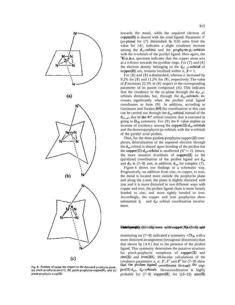
Figure 6 shows our findings in a schematic way. Progressively, on addition from zinc, to copper, to iron, the metal is located more outside the porphyrin plane and along the z-axis; the plane is slightly distorted with zinc and it is more distorted in two different ways with copper and iron; the picdien ligand chain is more loosely bonded to zinc, and more tightly bonded to iron. Accordingly, the copper and iron porphyrins show substantial d,, and d,z orbital coordination involve- ment. /Rt pp Pinch-porphyrin with Conclusions zinc(I1) (13-15) complexes were with synthesized. copper(I1) Spectroscopic (7-9) and
monitoring on (7-9) indicated a symmetry <Dlh with a more distorted arrangement (tetragonal distortion) than
8 8 that shown by ( 4 4 ) due to the presence of the picdien
ligand. This symmetry determines the putative structure for pinch-porphyrin complexes of copper(I1) and
(c) zinc(I1) and iron(II1). Molecular calculations of the covalence parameters a, a', /?', 6" and 6" for (7-9) show
Fig. 6. Position of metal ion respect to the distorted porphyrin-plane: that the picdien ligand the (a) pinch-porphyrin-zinc(11); (b) pinch-porphyrin-copper(l~); and (c) per(II)-dyz, d,z-orbitals. Hexacoordination is highly pinch-porphyrin-iron(II1). probable for (7-9) copper(II), for (13-15) zinc(I1)

Table 6 . Molecular coefficients for pinch-porphyrin-copper(I1) complexes (4-9)
Compound go A. x lo2 (cm-I) a a' p 6'
Standard deviations calculated for each covalence parameter in parentheses.
and for iron(II1) pinch-porphyrins. The metal ion is in a distorted local environment, but the nature of the distortions are different for each metal. For copper(I1) it is mainly governed by a Jahn-Teller distortion, whereas for iron(II1) the distortion is driven by the axial displacement out of the plane of the porphyrin ring of the iron(II1). All the above structural effects are shown schematically in Figure 6.
Acknowledgements
18. M.W. Renner, K.M. Barkigia and J. Fajer, Inorg. Chim. Acta, 263, 181 (1997).
19. M.F. Perutz, Scientific American, 239, 92 (1978). 20. M.M. Maltempo, T.H. Moss and M.A. Cusanovich, Biochim.
Biophys. Acta, 342, 290 (1974). 21. M.H. Goff and M.A. Phillippi, J. Am. Chem. Soc., 105, 7567
(1983). 22. G.N. La Mar, M.A. Viscio, K.M. Smith, W.S. Caughey and M.L.
Smith, J. Am. Chem. Soc., 100, 8085 (1978). 23. D. Mandon, R. Weiss, K. Jayaraj, A. Gold, J. Terner, E. Bill and
A.X. Trautwein, Inorg. Chem., 31, 4402 (1992). 24. Y. Reyes-Ortega, C. Alvarez-Toledano, D. Ramirez-Rosales, A.
Sinchez-Sandoval, E. Gonzilez-Vergara and R. Zamorano-Ulloa, J. Chem. Soc., Dalton Trans., 667 (1998).
Thanks are due to Dr ~ ~ d ~ t ~ Hernhndez Arana for the 25. E. Ahmed, C. Chartterjee, C.J. Coosksey, M.L. Tobe and G.J. Williams, Chem. Soc. Dallon Trans., 64 (1989).
m.c'd' measurements made in the Depart- 26. E. Basurto-Uribe and R. Zamorano-Ulloa, Reporte Interno, ment of Universidad Autcinoma Metropolitana Campus E S F M / F O ~ / ~ ~ , ESFM-IPN, Mexico, 1993. Iztapalapa, MCxico, D.F
References
1. L. Stryer, Biochemistry, Freeman and Co, 3rd edit., USA, 1988, p. 518.
2. J.C. Kendrew, P.E. Dickerson, B.E. Strandberg, R.G. Hart, D.R. Davis, D.C. Phillips and V.C. Shore, Nature, 185, 422 (1960).
3. D. Ramirez-Rosales, Masters Thesis (Physics), Escuela Superior de Fisica y Matemiticas-Instituto Politknico National, Mkxico, D.F., 1995.
4. H.B. Gray and W.R. Ellisin, Jr., in I. Bertini, H.B. Gray, S.J. Lippard and J.S. Valentine, (eds), Bioinorganic Chemistry; Uni- versity Science Book, USA, 1994, p. 3 15.
5. B.C. Saunders, Holmes-Sidle A.G. and B.P. Stark, Peroxidase, Butterworths, EUA, 1964, p. 1.
6. C. Meredith, L. Findlay, L.C. Dickinson and J.C.W. Chien, J. Am. Chem. Soc., 99, 5 I68 (1977).
7. J.T. Groves, R.C. Haushalter, M. Nakamura, T.E. Nemo and B.J. Evans, J. Am. Chem. Soc., 103, 2884 (1981).
8. T.G. Traylor, W.A. Lee and D.V. Stynes, J. Am. Chem. Soc., 106, 239 (1984).
9. C.A. Reed and F. Guiset, J. Am. Chem. Soc., 118, 3281 (1996). 10. Y. Nonomura, N. Yoshioka and H. Inoue, Inorg. Chim. Acta, 224,
I81 (1994). 1 I . M.W. Renner, K.M. Barkigia, Y. Zhang, C.J. Medforth, K.M.
Smith and J. Fajer, J. Am. Chem. Soc., 116, 8582 (1994). 12. F. D'Souza, G.R. Deviprasad and M.E. Zandler, J. Chem. Soc.,
Dalton Trans., 3699 (1997). 13. R.P. Houser and W.B. Tolman, Inorg. Chem., 34, 1632 (1995). 14. B.S. Erler, W.F. Scholz, Y.J. Lee, W.R. Scheidt and C.A. Reed,
J . Am. Chem. Soc., 109, 2644 (1987). 15. H. Fuji, Inorg. Chem., 32, 875 (1993). 16. M. Ravikanth, D. Reddy, A. Misra and T.K. Chandrashekar,
J . Chem. Soc., Dalton Trans., 1137 (1993). 17. H. Yokoi and M. Iwaizumi, Bull. Chem. Jpn., 53, 1489 (1980).
27. M.J. Nilges, Ph.D. Thesis, University of Illinois, Urbana, Illinois, (1979), (ii) R.L. Belford, M.J. Nilges, EPR Symposium, 21st Rocky Mountain Conference, Denver Colorado (1979). (iii) A.M. Mau- rice, Ph.D. Thesis, University of Illinois, Urbana, Illinois.
28. A.D. Falk, The Porphyrins and Metalloporphyrins, Elsevier, New York, 1960, p. 798.
29. M. Zerner and M. Gouterman, Theor. Chim. Acta, 4, 44 (1966). 30. M. Zerner, M. Gouterman and H. Kobayashi, Theor. Chim. Acta,
6, 363 (1966). 31: J.W. Owens and O'Connor, Ch. Coord. Chem. Rev., 84, 1 (1998). 32. M. Gouterman, in D. Dolphin (ed), The Porphyrins, Academic
Press, London, 1978, vol. 4, p. 61. 33. F. Adar, in D. Dolphin (ed), The Porphyrins, Academic Press, New
York, 1978, vol. 4, p. 167. 34. H. Alpsoh, D.W. Corwin, W.E. Baker and G.G. Kleinsphen,
J . Am. Chem. Soc., 85, 3621 (1963). 35. E.I. Solomon, M.L. Kirk, D.R. Garnelis and S. Pulver, Melhods
Enzymol., 246, 71 (1995). 36. (a) J.C. Sutherland, Methods in Enzymology, 36, Academic Press.
Inc., EUA, p. 110 (1995); (b) D.M. Dooley and J.H. Dawson, Coord. Chem. Rev., 60, 1 (1994); (c) J.H. Dawson, S. Khodayan, Ch. Zhuorg and M. Sono, J. Inorg. Biochem., 45, 179 (1992).
37. W.R. Browett and M.J. Stillman, Inorg. Chim. Acla, 49, 69 (1981). 38. M.R. Cheesman, W.G. Zumft and A.J. Thomson, Biochem., 37,
3994 (1998). 39. G.M. Godziela and H.M. Goff, J. Am. Chem. Soc., 108, 2237
(1986). 40. A.M. Walker, in K.M. Kadish, K.M. Smith and R. Guilard (eds),
The Porphyrin Handbook Academic Press, USA, 2000, vol. 5 (n.m.r. and e.p.r.) p. 167.
41. P.F. Knowles, D.H. Marsh and W.E. Rattle, Magnetic Resonance of Biomolecules, Wiley, USA, 1976, p. 328.
42. R.S. Drago, Physicnl Methods in Chemistry, Saunders College Publishing, EUA 1977, p. 246.
43. L. Banci, Inorg. Chem., 24, 782 (1985). 44. R.J. Kurland, R.G. Little, D.G. Davis and Ch. Ho, Biochenl., 12,
2237 (1971). 45. F.E. Mabbs, Chem. Soc. Rev., 313 (1993).
46. P.R. Athappan and G. Rajagopal, Polyhedron, 15, 527 (1996). 47. A. McCragh, C.B. Storm and W.S. Koski, J. Am. Chem. Soc.: 87,
1470 (1965). 48. J.A. De Bolfo, T.D. Smith, J.F. Boas and J.R. Pilbrow, J. Chem.
Soc., Dalton Trans. 1523 (1975). 49. H.R. Gersmann and J.D. Swalen, J. Chem Phys., 36, 3221 (1962). 50. I. Zink and R.S. Drago, J. Am. Chem Sor., 93, 4550 (1972). 51 A.H. Maki and B.R. McGarvey, J. Chem. Phys., 29, 35 (1958).
917
52. D. Kivelson and R. Neiman, J. Chem. Phys., 35, 149 (1961). 53. H.A. Kuska. M.T. Rogers and R.E. Drullinger, J. Phys. Chem., 71
(1967). 54. E.M. Roberts and W.S. Koski, J. Am. Chem. Soc., 82, 3006 (1960). 55. K-E. Falk, E. Ivanova, B. Roos and T. Vanngbrd, 1nor.g. Chem., 9,
556 (1970).
TMCH 5236