Air-stable, heme-like water-soluble iron(II) porphyrin: in situ preparation and characterization
10
ORIGINAL PAPER Air-stable, heme-like water-soluble iron(II) porphyrin: in situ preparation and characterization Ro ´bert Husza ´nk Gyo ¨rgy Lendvay Otto ´ Horva ´th Received: 23 June 2006 / Accepted: 29 January 2007 / Published online: 28 February 2007 ȑ SBIC 2007 Abstract Preparation of the water-soluble, kinetically labile, high-spin iron(II) tetrakis(4-sulfonatophenyl)por- phyrin, Fe(II)TPPS 4– , has been realized in neutral or weakly acidic solutions containing acetate buffer. The buffer played a double role in these systems: it was used for both adjusting pH and, via formation of an acetato com- plex, trapping trace amounts of iron(III) ions, which would convert the iron(II) porphyrins to the corresponding ir- on(III) species. Fe(II)TPPS 4– proved to be stable in these solutions even after saturation with air or oxygen. In the absence of acetate ions, however, iron(II) ions play a cat- alytic role in the formation of iron(III) porphyrins. While the kinetically inert iron(III) porphyrin, Fe(III)TPPS 3– , is a regular one with no emission and photoredox properties, the corresponding iron(II) porphyrin displays photoinduced features which are typical of sitting-atop complexes (red- shifted Soret absorption and blueshifted emission and Q absorption bands, photoinduced porphyrin ligand-to-metal charge transfer, LMCT, reaction). In the photolysis of Fe(II)TPPS 4– the LMCT process is followed by detach- ment of the reduced metal center and an irreversible ring- opening of the porphyrin ligand, resulting in the degrada- tion of the complex. Possible oxygen-binding ability of Fe(II)TPPS 4– (as a heme model) has been studied as well. Density functional theory calculations revealed that in solutions with high acetate concentration there is very little chance for iron(II) porpyrin to bind and release O 2 , devi- ating from heme in a hydrophobic microenvironment in hemoglobin. In the presence of an iron(III)-trapping addi- tive that is much less strongly coordinated to the iron(II) center than the acetate ion, Fe(II)TPPS 4– may function as a heme model. Keywords Iron porphyrins Heme-model Sitting atop Catalysis Photochemistry Introduction Heme proteins (containing iron porphyrin derivatives in their active sites) play an essential role in living organisms in oxygen transport and storage (hemoglobin, myoglobin) and in electron transfer processes (cytochrome c, cyto- chrome oxidase) [1, 2]. Hemoglobin contained within the red blood cells holds four ferrous porphyrin type groups which provide the active site for oxygen binding and are responsible for the red color of blood. Simple compounds that would bind oxygen reversibly for many cycles could also be of value as emergency blood substitutes or for fractionation of oxygen from air. There have been many attempts to model the function of hemoglobin and myoglobin (the oxygen–carbon dioxide cycle) by use of some kind of oxygen carriers as blood substitutes. In the last few decades, considerable efforts were made to find compounds that are able to mimic the hemoglobin function. Two classes of blood substi- tutes—namely, perfluorocarbon-type derivatives [3–8] and different human hemoglobin and bovine hemoglobin R. Husza ´nk G. Lendvay O. Horva ´th (&) Department of General and Inorganic Chemistry, University of Pannonia, P.O. Box 158, Veszpre ´m 8201, Hungary e-mail: [email protected] G. Lendvay Institute of Structural Chemistry, Hungarian Academy of Sciences, P.O. Box 17, Budapest 1525, Hungary 123 J Biol Inorg Chem (2007) 12:681–690 DOI 10.1007/s00775-007-0217-y
-
Upload
independent -
Category
Documents
-
view
3 -
download
0
Transcript of Air-stable, heme-like water-soluble iron(II) porphyrin: in situ preparation and characterization
ORIGINAL PAPER
Air-stable, heme-like water-soluble iron(II) porphyrin:in situ preparation and characterization
Robert Huszank Æ Gyorgy Lendvay Æ Otto Horvath
Received: 23 June 2006 / Accepted: 29 January 2007 / Published online: 28 February 2007
� SBIC 2007
Abstract Preparation of the water-soluble, kinetically
labile, high-spin iron(II) tetrakis(4-sulfonatophenyl)por-
phyrin, Fe(II)TPPS4–, has been realized in neutral or
weakly acidic solutions containing acetate buffer. The
buffer played a double role in these systems: it was used for
both adjusting pH and, via formation of an acetato com-
plex, trapping trace amounts of iron(III) ions, which would
convert the iron(II) porphyrins to the corresponding ir-
on(III) species. Fe(II)TPPS4– proved to be stable in these
solutions even after saturation with air or oxygen. In the
absence of acetate ions, however, iron(II) ions play a cat-
alytic role in the formation of iron(III) porphyrins. While
the kinetically inert iron(III) porphyrin, Fe(III)TPPS3–, is a
regular one with no emission and photoredox properties,
the corresponding iron(II) porphyrin displays photoinduced
features which are typical of sitting-atop complexes (red-
shifted Soret absorption and blueshifted emission and Q
absorption bands, photoinduced porphyrin ligand-to-metal
charge transfer, LMCT, reaction). In the photolysis of
Fe(II)TPPS4– the LMCT process is followed by detach-
ment of the reduced metal center and an irreversible ring-
opening of the porphyrin ligand, resulting in the degrada-
tion of the complex. Possible oxygen-binding ability of
Fe(II)TPPS4– (as a heme model) has been studied as well.
Density functional theory calculations revealed that in
solutions with high acetate concentration there is very little
chance for iron(II) porpyrin to bind and release O2, devi-
ating from heme in a hydrophobic microenvironment in
hemoglobin. In the presence of an iron(III)-trapping addi-
tive that is much less strongly coordinated to the iron(II)
center than the acetate ion, Fe(II)TPPS4– may function as a
heme model.
Keywords Iron porphyrins � Heme-model �Sitting atop � Catalysis � Photochemistry
Introduction
Heme proteins (containing iron porphyrin derivatives in
their active sites) play an essential role in living organisms
in oxygen transport and storage (hemoglobin, myoglobin)
and in electron transfer processes (cytochrome c, cyto-
chrome oxidase) [1, 2]. Hemoglobin contained within the
red blood cells holds four ferrous porphyrin type groups
which provide the active site for oxygen binding and are
responsible for the red color of blood. Simple compounds
that would bind oxygen reversibly for many cycles could
also be of value as emergency blood substitutes or for
fractionation of oxygen from air.
There have been many attempts to model the function of
hemoglobin and myoglobin (the oxygen–carbon dioxide
cycle) by use of some kind of oxygen carriers as blood
substitutes. In the last few decades, considerable efforts
were made to find compounds that are able to mimic the
hemoglobin function. Two classes of blood substi-
tutes—namely, perfluorocarbon-type derivatives [3–8] and
different human hemoglobin and bovine hemoglobin
R. Huszank � G. Lendvay � O. Horvath (&)
Department of General and Inorganic Chemistry,
University of Pannonia,
P.O. Box 158, Veszprem 8201, Hungary
e-mail: [email protected]
G. Lendvay
Institute of Structural Chemistry,
Hungarian Academy of Sciences,
P.O. Box 17, Budapest 1525, Hungary
123
J Biol Inorg Chem (2007) 12:681–690
DOI 10.1007/s00775-007-0217-y

preparations [9–14]—have been tested in clinical therapy.
In spite of partial success, all of these compounds have
numerous disadvantages and show many side effects, so
they have not become widespread in medical practice. As
an alternative, preparation of the heme group [hydrophobic
or hydrophilic iron(II) porphyrin] synthetically as a possi-
ble blood substitute has also been investigated intensively
[15–22]. These studies were focused almost exclusively on
a nonaqueous environment (probably because of the
hydrophobic nature of the heme group), while the desired
simple oxygen carrier should be able to function in aqueous
media under physiological conditions.
Molecular oxygen has a triplet, paramagnetic ground
state and high electronegativity. These features make it
one of the most powerful oxidizing agents. Stable yet
reversible coordination of this strong oxidant to the ir-
on(II) central atom is rather difficult, because heme
compounds mostly react irreversibly with dioxygen. Al-
most all synthetically prepared iron(II) porphyrin com-
plexes in organic solvents were found to be very unstable
in air and are rapidly oxidized to ferric porphyrin, which is
biologically inactive [23]. This process is an irreversible
overall redox reaction:
4FeIIPþ O2 ! 2 FeIIIP� O� FeIIIP� �
: ð1Þ
The initial binding of molecular oxygen is followed by a
bimolecular redox process, which leads to the formation of
a dioxygen-bridged dimer complex. This l-peroxo dimer
can be converted via the formation of an oxo-ferryl inter-
mediate to the l-oxo dimer. This bridged species can fur-
ther decompose to iron(III) porphyrin.
In fact, the initial dimerization process takes place very
efficiently in organic media where the complexes are
not ionic, i.e., have no charge. The same reaction, how-
ever, is probably less favorable in aqueous—especially
dilute—solution, because the solubility of the complex is
generally ensured by ionic substituents on the porphyrin
ring, and then both iron porphyrin units have large positive
or negative charge and repel each other. Nevertheless, the
presence of water (even a trace amount) causes autoxida-
tion in most iron(II) porphyrins coordinating dioxygen
[24]. In aqueous systems, even denatured hemoglobin
proved to be extremely oxygen sensitive after losing the
hydrophobic microenvironment ensured by the globin
moiety for the iron(II) center. This phenomenon has been
attributed to the nucleophilic attack of water molecules at
the iron–oxygen coordinative bond, excluding the O2•–
species formed in inner-sphere electron transfer from the
iron(II) center. Accordingly, iron(II) porphyrins in aqueous
media have been mostly studied in supramolecular systems
of biological origin [24]. Quite recent investigations indi-
cated that a possible way to prepare iron(II) porphyrins in
aqueous systems is in situ reduction of a water-soluble
iron(III) porphyrin encapsulated in an O-methylated
b-cyclodextrin dimer ensuring a hydrophobic microenvi-
ronment for the metal center [25]. This system proved to be
relatively stable against autoxidation in the presence of
dioxygen. However, preparation of air-stable iron(II) por-
phyrin in a real hydrophilic environment required a dif-
ferent strategy [26, 27]. In addition, the conversion of
ferrous porphyrin to ferric porphyrin in aqueous solution
has not yet been explained.
Besides the significant biological function of ferrous
porphyrin derivatives, the description of thermal and pho-
toinduced properties of water-soluble complexes would
also be important in bioinorganic chemistry. Nowadays,
interest in metalloporphyrins keeps increasing, owing to
their importance in many fields, such as biochemistry,
medical research and catalysis. The photophysical and
photochemical properties of excited metalloporphyrins
have been utilized, e.g., in optical sensors [28–30], artificial
photosynthetic systems (light-harvesting dendrimers) [31]
and photodynamic therapy [32–34].
The main subject of this paper is a report on the prep-
aration of iron(II) tetrakis(4-sulfonatophenyl)porphyrin,
Fe(II)TPPS4–, in aqueous solution so that the complex
formed is stable and is not oxidized in the presence of
oxygen. Production of Fe(II)TPPS4– was realized by the
complexation reaction of free-base porphyrin (H2TPPS4–)
and iron(II) ions in the presence of iron(III)-trapping ace-
tate buffer. In the following, after summarizing the tech-
niques used, we present the preparation method, then the
equilibrium, photophysical and photochemical properties
of the complex, according to which the complex can be
classified as a sitting-atop metal porphyrin. Finally, we
discuss whether this complex can serve as a simple heme
model with respect to reaction with dissolved molecular
oxygen.
Materials and methods
Materials
All the solutions studied were made by using ion-
exchanged then distilled Milli-Q water. H2TPPS4– and
FeSO4�7H2O were purchased from Aldrich. The iron(III)
porphyrin [iron(III)meso-tetrakis(4-sulfonatophenyl)por-
phyrin] was from Frontier Scientific Europe. In order to
hinder the formation of iron(III) ions, argon was bubbled
and metallic iron was added into the iron(II) sulfate solu-
tion. The pH values of 6 and 7 were obtained by acetate
buffers made from CH3COONa and CH3COOH in the
appropriate ratio. The pH was measured with a pH meter
using a glass electrode. For the exclusion of oxygen,
682 J Biol Inorg Chem (2007) 12:681–690
123
99.996% argon gas and the Schlenck technique were used
where necessary.
[Fe(II)TPPS4–, 4Na+]
The water-soluble porphyrin was synthesized in the equi-
librium reaction of 1.5 · 10–4 M Fe(II) and 3 · 10–6 or
5 · 10–6 M free-base porphyrin in aqueous, argon-satu-
rated solution at pH 6 and 7. The solutions also contained
0.3 M acetate buffer to adjust the pH and to trap trace
amounts of iron(III) ions, and, in addition, 1 M NaCl at pH
7 to increase the ionic strength. In the first step of the
process, the iron(II) and the acetate solutions were mixed
and then immediately degassed with argon gas (for about
30 min). At pH 7, NaCl was also added in the first step.
Subsequently, the mixture was kept under argon for 4 h.
Then porphyrin was added, also under oxygen-free condi-
tions. The system was allowed to react for at least 2 days at
room temperature. The spectral change of the starting
system (containing H2TPPS4–) indicates the formation of
the complex (details in ‘‘Formation of Fe(III)TPPS3– and
Fe(II)TPPS4– in water’’).
Apparatus
All the absorption and emission spectra were recorded with
a Specord S100 UV–vis one-way spectrophotometer and
with a PerkinElmer LS50 spectrofluorimeter, respectively.
Nanosecond emission lifetimes were measured using
PicoQuant time-correlated single photon counting appara-
tus. For the assessments and data fitting, deconvolution
software was used (FluoFit from PicoQuant). For contin-
uous photolysis at different wavelengths an AMKO LTI
system (consisting of a 150-W high pressure Xe–Hg arc
lamp and a monochromator) was utilized. The light
intensity was determined with a thermopile calibrated by
ferrioxalate actinometry. Quantum yield measurements
were carried out with samples of nearly 100% light
absorption. The concentration of the known porphyrin
species in the solutions was determined by using their
molar absorption coefficients, utilizing a least-squares fit-
ting method on the 350–500-nm region of the spectra.
Theoretical methods
Density functional theory (DFT) was used for the theoret-
ical determination of the structures and energies of iron(II)
porphyrin and its derivatives containing other ligands in the
singlet, triplet and quintet states. We used the B3LYP
[35–37] combination of functionals as coded in Gaussian98
[38], in conjunction with the LANL2DZ [39–42] basis set.
This combination proved to be adequate in numerous
related applications in the literature [43]. We made tests
using other basis sets, including 3–21G* and 6–31G*, and
found that the computed structures remain essentially the
same. Where comparison was possible, our results agree
well with those of earlier calculations [43, 44]. The
molecular geometries presented are true minima as checked
by calculation of vibrational frequencies. For the compari-
son of the energies of complexes with various spin multi-
plicity, the DSCF method was used; i.e., the structures and
energies were calculated using the restricted self-consistent-
field (SCF) procedure (RB3LYP) for the singlet species and
the unrestricted method (UB3LYP) for the open-shell
molecules, and the energies were directly compared. The
convergence of both the SCF iteration and the geometry
optimization was very slow, so level shifting, enforcing and
relaxing symmetry and change of occupation of orbitals
were necessary to satisfy tight convergence criteria.
Results
Formation of Fe(III)TPPS3– and Fe(II)TPPS4– in water
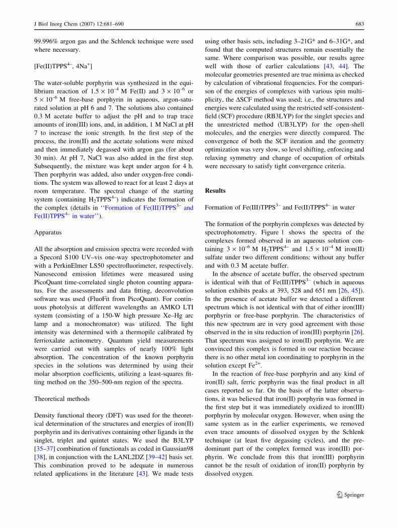
The formation of the porphyrin complexes was detected by
spectrophotometry. Figure 1 shows the spectra of the
complexes formed observed in an aqueous solution con-
taining 3 · 10–6 M H2TPPS4– and 1.5 · 10–4 M iron(II)
sulfate under two different conditions: without any buffer
and with 0.3 M acetate buffer.
In the absence of acetate buffer, the observed spectrum
is identical with that of Fe(III)TPPS3– (which in aqueous
solution exhibits peaks at 393, 528 and 651 nm [26, 45]).
In the presence of acetate buffer we detected a different
spectrum which is not identical with that of either iron(III)
porphyrin or free-base porphyrin. The characteristics of
this new spectrum are in very good agreement with those
observed in the in situ reduction of iron(III) porphyrin [26].
That spectrum was assigned to iron(II) porphyrin. We are
convinced this complex is formed in our reaction because
there is no other metal ion coordinating to porphyrin in the
solution except Fe2+.
In the reaction of free-base porphyrin and any kind of
iron(II) salt, ferric porphyrin was the final product in all
cases reported so far. On the basis of the latter observa-
tions, it was believed that iron(II) porphyrin was formed in
the first step but it was immediately oxidized to iron(III)
porphyrin by molecular oxygen. However, when using the
same system as in the earlier experiments, we removed
even trace amounts of dissolved oxygen by the Schlenk
technique (at least five degassing cycles), and the pre-
dominant part of the complex formed was iron(III) por-
phyrin. We conclude from this that iron(III) porphyrin
cannot be the result of oxidation of iron(II) porphyrin by
dissolved oxygen.
J Biol Inorg Chem (2007) 12:681–690 683
123
Since we found that in all preparation procedures of
iron(III) porphyrins that have been reported in the literature
the starting materials were iron(II) compounds and free-
base porphyrin [26, 46, 47], we checked whether Fe3+ ions
react with free-base porphyrin at all. We did not observe
complexation between ferric ions and free-base porphyrin
even when they were allowed to react for 1 week; hence, a
reaction different from the reaction in Eq. 1, suggested in
the literature, must be responsible for the formation of
iron(III) porphyrin.
We think that Fe2+ ions catalyze the reaction of free-
base porphyrin and Fe3+ ions, trace amounts of which are
always present in iron(II) solutions. It is known that for-
mation of kinetically inert metalloporphyrins can be cata-
lyzed by metal ions such as Hg(II), Cd(II), Pb(II) and
Cu((II), via formation of kinetically labile sitting-atop
complexes [48]. In the case of the relatively large ionic
radius, the metal center cannot fit into the core of the
porphyrin; hence, it is localized out of the ligand plane,
distorting it. Such a distortion makes the porphyrin co-
ordinatively more active. In our case, iron(II) porphyrin is
formed in the first step and two new coordination positions
arise on the other side of the porphyrin plane (on two
diagonally situated nitrogen atoms) and become accessible
for another metal ion, e.g., iron(III). Following the coor-
dination of iron(III), it excludes the iron(II) ion bound more
weakly on the other side of the porphyrin, and occupies the
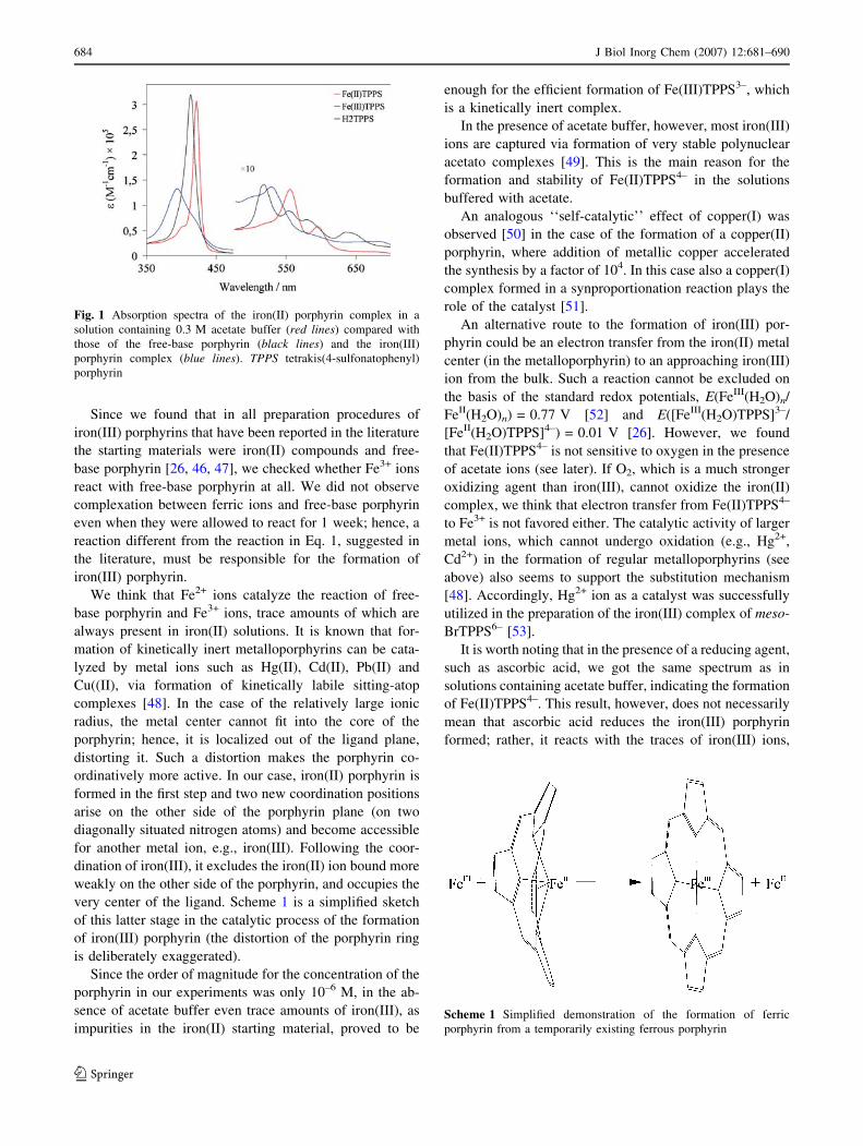
very center of the ligand. Scheme 1 is a simplified sketch
of this latter stage in the catalytic process of the formation
of iron(III) porphyrin (the distortion of the porphyrin ring
is deliberately exaggerated).
Since the order of magnitude for the concentration of the
porphyrin in our experiments was only 10–6 M, in the ab-
sence of acetate buffer even trace amounts of iron(III), as
impurities in the iron(II) starting material, proved to be
enough for the efficient formation of Fe(III)TPPS3–, which
is a kinetically inert complex.
In the presence of acetate buffer, however, most iron(III)
ions are captured via formation of very stable polynuclear
acetato complexes [49]. This is the main reason for the
formation and stability of Fe(II)TPPS4– in the solutions
buffered with acetate.
An analogous ‘‘self-catalytic’’ effect of copper(I) was
observed [50] in the case of the formation of a copper(II)
porphyrin, where addition of metallic copper accelerated
the synthesis by a factor of 104. In this case also a copper(I)
complex formed in a synproportionation reaction plays the
role of the catalyst [51].
An alternative route to the formation of iron(III) por-
phyrin could be an electron transfer from the iron(II) metal
center (in the metalloporphyrin) to an approaching iron(III)
ion from the bulk. Such a reaction cannot be excluded on
the basis of the standard redox potentials, E(FeIII(H2O)n/
FeII(H2O)n) = 0.77 V [52] and E([FeIII(H2O)TPPS]3–/
[FeII(H2O)TPPS]4–) = 0.01 V [26]. However, we found
that Fe(II)TPPS4– is not sensitive to oxygen in the presence
of acetate ions (see later). If O2, which is a much stronger
oxidizing agent than iron(III), cannot oxidize the iron(II)
complex, we think that electron transfer from Fe(II)TPPS4–
to Fe3+ is not favored either. The catalytic activity of larger
metal ions, which cannot undergo oxidation (e.g., Hg2+,
Cd2+) in the formation of regular metalloporphyrins (see
above) also seems to support the substitution mechanism
[48]. Accordingly, Hg2+ ion as a catalyst was successfully
utilized in the preparation of the iron(III) complex of meso-
BrTPPS6– [53].
It is worth noting that in the presence of a reducing agent,
such as ascorbic acid, we got the same spectrum as in
solutions containing acetate buffer, indicating the formation
of Fe(II)TPPS4–. This result, however, does not necessarily
mean that ascorbic acid reduces the iron(III) porphyrin
formed; rather, it reacts with the traces of iron(III) ions,
Scheme 1 Simplified demonstration of the formation of ferric
porphyrin from a temporarily existing ferrous porphyrin
Fig. 1 Absorption spectra of the iron(II) porphyrin complex in a
solution containing 0.3 M acetate buffer (red lines) compared with
those of the free-base porphyrin (black lines) and the iron(III)
porphyrin complex (blue lines). TPPS tetrakis(4-sulfonatophenyl)
porphyrin
684 J Biol Inorg Chem (2007) 12:681–690
123
forming a complex or reducing it to iron(II), and blocks
them in a similar way to acetate ions. It should also be
mentioned that in situ formation of iron(II) porphyrin was
observed in the presence of nitrogen monoxide [54].
The absorption spectrum of Fe(II)TPPS4– in aqueous
solution containing 0.3 M acetate buffer exhibits strong
peaks at 421, 557 and 598 nm (for more detailed charac-
terization see ‘‘Photophysical studies’’). This compound
proved to be kinetically labile. Its formation reaction is
described by Eq. 2. The apparent equilibrium constant (K¢)for this reaction can be expressed by the actual concen-
trations, according to the law of mass action (Eq. 3). Since
constant pH was applied in all solutions studied, the effect
of H+ concentration is not taken into account in this case.
Fe2þ þ H2TPPS4�$K Fe(II)TPPS4� þ 2Hþ ð2Þ
K 0 ¼ K
[Hþ]2¼ [Fe(II)TPPS4�]
[Fe2þ][H2TPPS4�]ð3Þ
a0 ¼[H2TPPS4�]
[H2TPPS4�]þ [Fe(II)TPPS4�]¼ 1
1þ K 0[Fe2þ]
ð4Þ
Thus, the partial molar ratio of the free-base porphyrin (a0)
can be given as a function of Fe2+ concentration (Eq. 4).
Hence, 1/a0 is in linear correlation with the Fe2+ concen-
tration, and the slope of this straight line is K¢. Accord-
ingly, K¢ can be estimated from the spectra of solutions
containing constant porphyrin concentrations and various
concentrations of iron(II), evaluating the absorbances in the
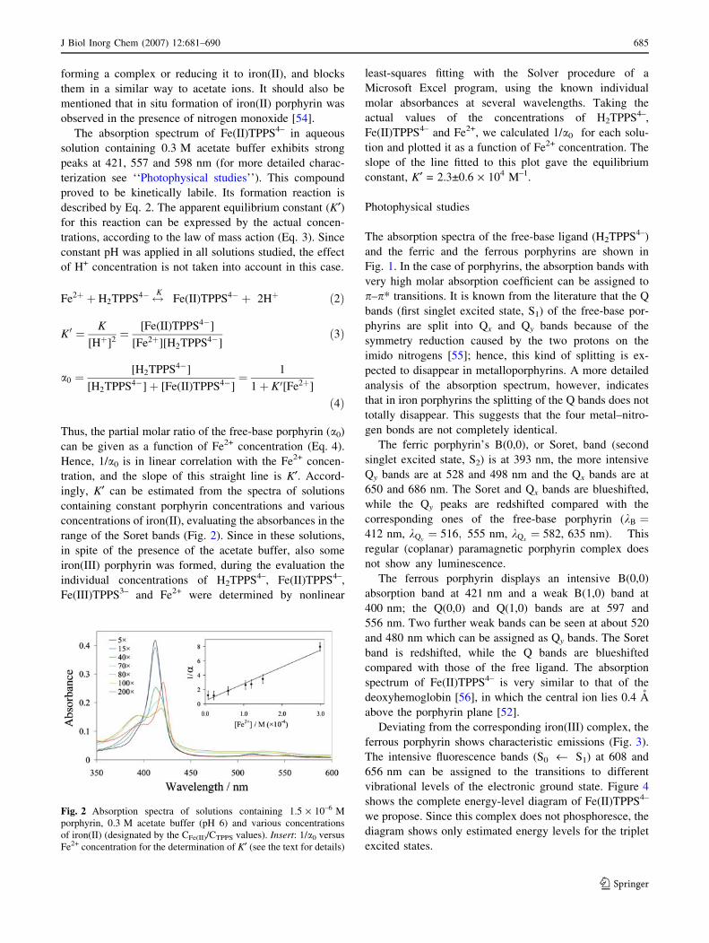
range of the Soret bands (Fig. 2). Since in these solutions,
in spite of the presence of the acetate buffer, also some
iron(III) porphyrin was formed, during the evaluation the
individual concentrations of H2TPPS4–, Fe(II)TPPS4–,
Fe(III)TPPS3– and Fe2+ were determined by nonlinear
least-squares fitting with the Solver procedure of a
Microsoft Excel program, using the known individual
molar absorbances at several wavelengths. Taking the
actual values of the concentrations of H2TPPS4–,
Fe(II)TPPS4– and Fe2+, we calculated 1/a0 for each solu-
tion and plotted it as a function of Fe2+ concentration. The
slope of the line fitted to this plot gave the equilibrium
constant, K¢ = 2.3±0.6 · 104 M–1.
Photophysical studies
The absorption spectra of the free-base ligand (H2TPPS4–)
and the ferric and the ferrous porphyrins are shown in
Fig. 1. In the case of porphyrins, the absorption bands with
very high molar absorption coefficient can be assigned to
p–p* transitions. It is known from the literature that the Q
bands (first singlet excited state, S1) of the free-base por-
phyrins are split into Qx and Qy bands because of the
symmetry reduction caused by the two protons on the
imido nitrogens [55]; hence, this kind of splitting is ex-
pected to disappear in metalloporphyrins. A more detailed
analysis of the absorption spectrum, however, indicates
that in iron porphyrins the splitting of the Q bands does not
totally disappear. This suggests that the four metal–nitro-
gen bonds are not completely identical.
The ferric porphyrin’s B(0,0), or Soret, band (second
singlet excited state, S2) is at 393 nm, the more intensive
Qy bands are at 528 and 498 nm and the Qx bands are at
650 and 686 nm. The Soret and Qx bands are blueshifted,
while the Qy peaks are redshifted compared with the
corresponding ones of the free-base porphyrin (kB ¼412 nm, kQy
¼ 516; 555 nm, kQx¼ 582, 635 nm): This
regular (coplanar) paramagnetic porphyrin complex does
not show any luminescence.
The ferrous porphyrin displays an intensive B(0,0)
absorption band at 421 nm and a weak B(1,0) band at
400 nm; the Q(0,0) and Q(1,0) bands are at 597 and
556 nm. Two further weak bands can be seen at about 520
and 480 nm which can be assigned as Qy bands. The Soret
band is redshifted, while the Q bands are blueshifted
compared with those of the free ligand. The absorption
spectrum of Fe(II)TPPS4– is very similar to that of the
deoxyhemoglobin [56], in which the central ion lies 0.4 A
above the porphyrin plane [52].
Deviating from the corresponding iron(III) complex, the
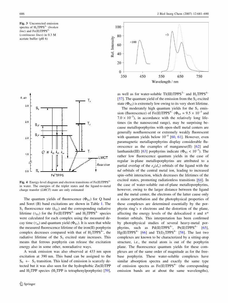
ferrous porphyrin shows characteristic emissions (Fig. 3).
The intensive fluorescence bands (S0 ‹ S1) at 608 and
656 nm can be assigned to the transitions to different
vibrational levels of the electronic ground state. Figure 4
shows the complete energy-level diagram of Fe(II)TPPS4–
we propose. Since this complex does not phosphoresce, the
diagram shows only estimated energy levels for the triplet
excited states.
Fig. 2 Absorption spectra of solutions containing 1.5 · 10–6 M
porphyrin, 0.3 M acetate buffer (pH 6) and various concentrations
of iron(II) (designated by the CFe(II)/CTPPS values). Insert: 1/a0 versus
Fe2+ concentration for the determination of K¢ (see the text for details)
J Biol Inorg Chem (2007) 12:681–690 685
123
The quantum yields of fluorescence (FS1) for Q band
and Soret (B) band excitations are shown in Table 1. The
S1 fluorescence rate (kS1) and the corresponding radiative
lifetime (sS1) for the Fe(II)TPPS4– and H2TPPS4– species
were calculated for each complex using the measured de-
cay time (sm) and quantum yield (FS1). It is seen that while
the measured fluorescence lifetime of the iron(II) porphyrin
complex decreases compared with that of H2TPPS4–, the
radiative lifetime of the S1 excited state increases. This
means that ferrous porphyrin can release the excitation
energy also in some other, nonradiative ways.
A weak emission was also observed at 433 nm upon
excitation at 390 nm. This band can be assigned to the
S0 ‹ S2 transition. This kind of emission is scarcely de-
tected but it was also seen for the hydrophobic Zn(II)TPP
and H2TPP species (H2TPP is tetraphenylporphyrin) [59],
as well as for water-soluble Tl(III)TPPS3– and H2TPPS4–
[57]. The quantum yield of the emission from the S2 excited
state (FS2) is extremely low owing to its very short lifetime.
The moderately high quantum yields for the S1 emis-
sion (fluorescence) of Fe(II)TPPS4– (FS1 = 9.5 · 10–3 and
7.0 · 10–3), in accordance with the relatively long life-
times (in the nanosecond range), may be surprising be-
cause metalloporphyrins with open-shell metal centers are
generally nonfluorescent or extremely weakly fluorescent
with quantum yields below 10–4 [60, 61]. However, even
paramagnetic metallaporphyrins display considerable flu-
orescence as the examples of manganese(II) [62] and
lanthanide(III) [63] porphyrins indicate (FS1 < 10–3). The
rather low fluorescence quantum yields in the case of
regular in-plane metalloporphyrins are attributed to a
partial overlap of the eg(dp) orbitals of the ligand with the
nd orbitals of the central metal ion, leading to increased
spin–orbit interaction, which decreases the lifetimes of the
excited states, promoting radiationless transitions [64]. In
the case of water-soluble out-of-plane metalloporphyrins,
however, owing to the larger distance between the ligand
and the metal center, the electrons of the latter cause only
a minor perturbation and the photophysical properties of
these complexes are determined essentially by the por-
phyrin ring’s p electrons and the distortion of the plane,
affecting the energy levels of the delocalized p and p*
frontier orbitals. This interpretation has been confirmed
by photophysical studies of several heavy-metal por-
phyrins, such as Pd(II)TPPS4–, Pt(II)TPPS4– [65],
Hg(II)TPPS4– [66] and Tl(I)2TPPS4– [58]. The last two
complexes are known to be characterized by a sitting-atop
structure, i.e., the metal atom is out of the porphyrin
plane. The fluorescence quantum yields for these com-
plexes are of the same order of magnitude as for the free-
base porphyrin. These water-soluble complexes have
similar absorption spectra and exactly the same type
of emission spectra as Fe(II)TPPS4– (the corresponding
emission bands are at about the same wavelengths),
Fig. 3 Uncorrected emission
spectra of H2TPPS4– (brokenline) and Fe(II)TPPS4–
(continuous lines) in 0.3 M
acetate buffer (pH 6)
Fig. 4 Energy-level diagram and electron transitions of Fe(II)TPPS4–
in water. The energies of the triplet states and the ligand-to-metal
charge transfer (LMCT) state are only estimated
686 J Biol Inorg Chem (2007) 12:681–690
123
indicating the similar structure of the latter. In accordance
with the interpretation above, for Tl(III)TPPS3– [57] the
fluorescence quantum yield is 2 orders of magnitude less
than for the previous complexes. In this case, as a con-
sequence of the smaller size of the metal center, it is
close to in-plane position, resulting in stronger spin-orbit
coupling.
Photolysis of Fe(II)TPPS4–
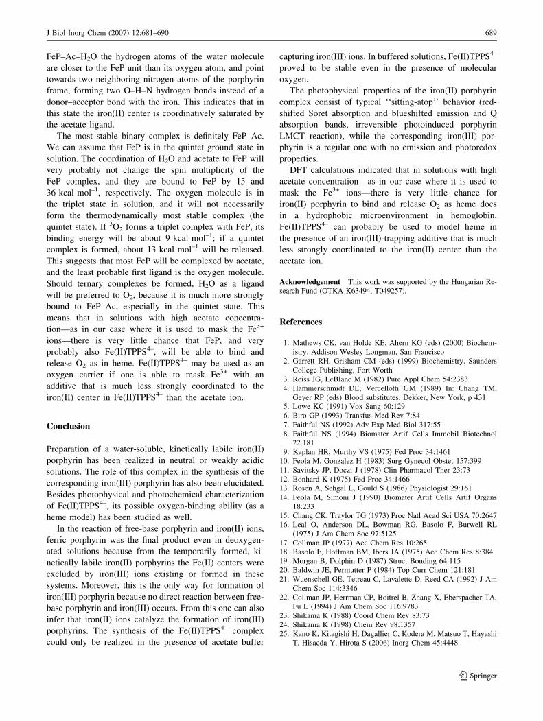
Irradiation of the iron(II) porphyrin at both the Soret band
(421 nm) and a Q band (556 nm) resulted in the degrada-
tion of the complex itself (Fig. 5), accompanied by the
formation of ring-opened tetrapyrrole derivatives (bile-type
pigments) as indicated by the new bands at 390 and
450 nm [67]. The same types of products were obtained in
photolyses of other sitting-atop porphyrin complexes of
Tl(I), Tl(III) and Hg(II) [58, 66, 68] as well as in the case
of photobleaching of H2TPPS4– [69]. The irradiated ferrous
porphyrin was transformed almost completely during a 40-
min illumination period.
The quantum yields for the degradation are summarized
in Table 1. The photochemistry of this complex is char-
acterized by a porphyrin ligand-to-metal charge transfer
(LMCT) reaction, similarly to the photoinduced reactions
in the case of sitting-atop metalloporphyrins with a metal
center other than iron(II) (Tl+, Hg2+) [57, 66]. Irradiation of
H2TPPS4– under the same conditions results in degrada-
tion, the quantum yield of which is 2 orders of magnitude
lower than that observed for the ferrous porphyrin
(Table 1). In the absence of a metal center, no LMCT
process can occur. In the case of the sitting-atop com-
plexes, the LMCT process takes place in an indirect way,
because appropriate absorption bands do not appear in the
absorption spectra. Thus, the excited electron must come
from one of the orbitals of the porphyrin ligand to the iron
center. Since the rate of the phototransformation does not
decrease significantly in the presence of an efficient triplet
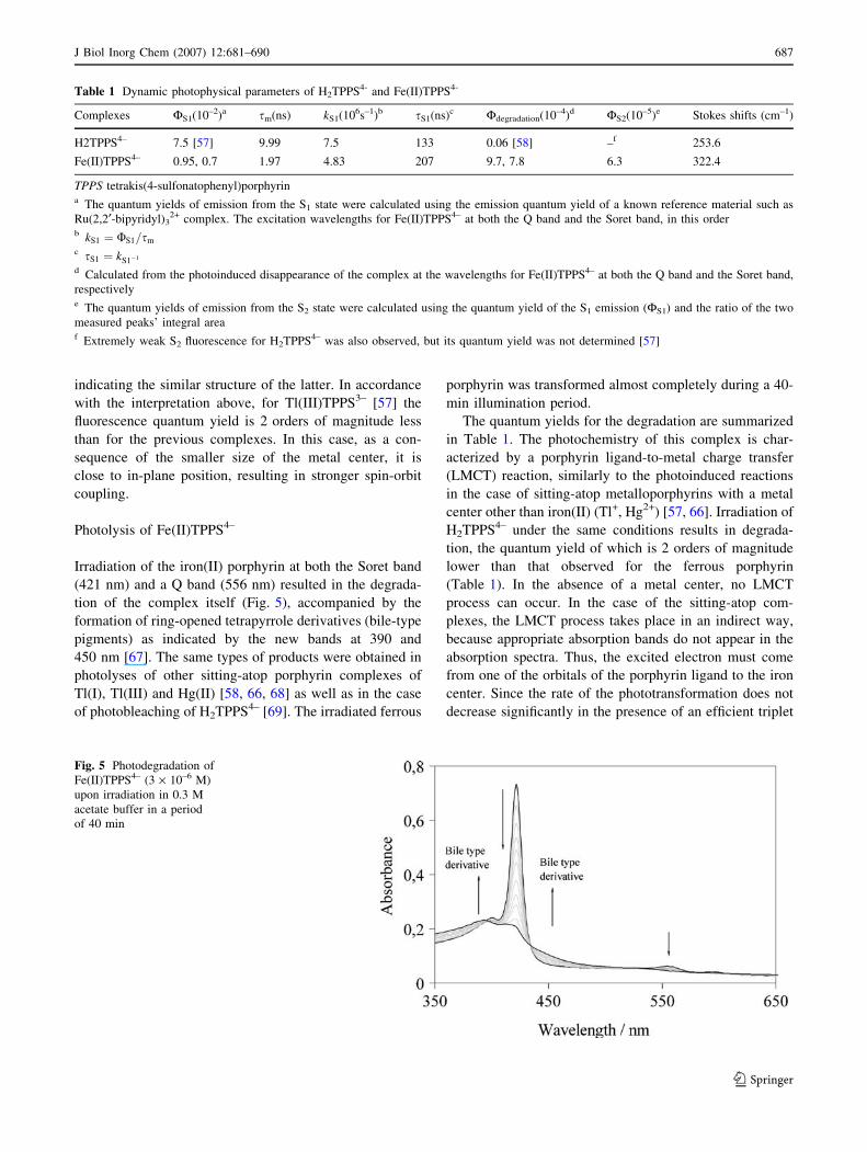
Table 1 Dynamic photophysical parameters of H2TPPS4- and Fe(II)TPPS4-
Complexes FS1(10–2)a sm(ns) kS1(106s–1)b sS1(ns)c Fdegradation(10–4)d FS2(10–5)e Stokes shifts (cm–1)
H2TPPS4– 7.5 [57] 9.99 7.5 133 0.06 [58] –f 253.6
Fe(II)TPPS4– 0.95, 0.7 1.97 4.83 207 9.7, 7.8 6.3 322.4
TPPS tetrakis(4-sulfonatophenyl)porphyrina The quantum yields of emission from the S1 state were calculated using the emission quantum yield of a known reference material such as
Ru(2,2¢-bipyridyl)32+ complex. The excitation wavelengths for Fe(II)TPPS4– at both the Q band and the Soret band, in this order
b kS1 ¼ US1=sm
c sS1 ¼ kS1�1
d Calculated from the photoinduced disappearance of the complex at the wavelengths for Fe(II)TPPS4– at both the Q band and the Soret band,
respectivelye The quantum yields of emission from the S2 state were calculated using the quantum yield of the S1 emission (FS1) and the ratio of the two
measured peaks’ integral areaf Extremely weak S2 fluorescence for H2TPPS4– was also observed, but its quantum yield was not determined [57]
Fig. 5 Photodegradation of
Fe(II)TPPS4– (3 · 10–6 M)
upon irradiation in 0.3 M
acetate buffer in a period
of 40 min
J Biol Inorg Chem (2007) 12:681–690 687
123

quencher (dissolved molecular oxygen), the LMCT process
must occur through the S1 and S2 singlet excited states
(Fig. 4).
In contrast, irradiation of the iron(III) porphyrin, under
the same conditions, caused no permanent chemical change
in the system, even if a similar LMCT reaction may take
place in the primary photochemical step. The reason for
this is that the regular in-plane metalloporphyrins can be
reversibly oxidized [70], because the temporarily formed pcation is very stable and the electron promoted to the metal
center has enough time to be transferred back to the por-
phyrin ligand. In contrast, in the case of sitting-atop por-
phyrins, after the LMCT reaction, the reduced metal ion,
[Fe(I) in our case] can be easily detached because of the
kinetic lability of this complex, and, thus, the electron
cannot be transferred back.
Scheme 2 demonstrates the assumed key step of the
photoinduced reaction of the iron(II) porphyrin.
Fe(II)TPPS4– as a possible heme model
For Fe(II)TPPS4– to be useful as a water-soluble heme
analog, it should be able to bind and release molecular
oxygen. To see if this is possible, after the Fe(II)TPPS4–
complex had been formed, the solution was saturated with
oxygen. The ferrous species proved to be stable against
oxidation; no conversion to the ferric form was observed
even over weeks. Besides, after the saturation of the
solution with oxygen, the spectra of the iron(II) porphyrin
complex did not change. This means that either molecular
oxygen does not coordinate to the ferrous porphyrin under
such conditions, or this coordination does not cause spec-
tral changes.
A possible reason why O2 is not coordinated to
Fe(II)TPPS4– is that the latter is more strongly coordinated
by some other ligands present in the solution used for the
synthesis of Fe(II)TPPS4–, so molecular oxygen cannot be
connected to the iron ion. In order to see how strongly the
most abundant ligands in the solution are bound to
Fe(II)TPPS4–, we performed DFT calculations using ir-
on(II) porphyrin as a model compound, denoted in the
following as FeP. We explored in some cases how much
the energetics is influenced by the presence of the four
phenyl groups of TPP, and found that the changes are small
and we can rely on the data obtained with the bare por-
phyrin ring. We investigated the binding energy of H2O, O2
and CH3COO– (denoted as Ac) to FeP in the singlet, triplet
and quintet states. As the iron(II) porphyrin molecule is
assumed to have two unoccupied coordination sites, not
only its 1:1 complexes with each ligand were studied, but
also all possible combinations of them. We found that for
all complexes except FeP and FeP–Ac–O2 the quintet state
is the ground state. Selected binding energies are listed in
Table 2.
In most cases the binding energies in ‘‘binary’’ FeP–L
complexes are found to change monotonously with the spin
multiplicity of the system. The acetate ion was found to
form the strongest bond with the iron(II) center (its binding
energy is 45–36 kcal mol–1 to the singlet and the quintet
iron porphyrin, respectively). The binding energy of the
first H2O ligand is much smaller, between 27 and
15 kcal mol–1, depending on the multiplicity. The binding
energy of 3O2 to FeP is around 10 kcal mol–1. The second
ligand is always connected to the opposite side of the
porphyrin plane compared with the first ligand, and is
generally much less strongly connected to the iron(II)
center than in the absence of the other ligand. For example,
the binding energy of the acetate ion in FeP–H2O.Ac is
about 13 kcal mol–1 smaller than in FeP–Ac, in all three
electronic states. In the binary FeP–H2O complex in all
electronic states and in the singlet and triplet states of the
ternary FeP–Ac–H2O complex, the H2O molecule, as ex-
pected, is connected to the iron through one of the lone
pairs of the oxygen atom. In contrast, in the quintet state of
Scheme 2 Simplified demonstration of the mechanism for the photoinduced degradation of iron(II) porphyrin. For the sake of simplicity, the
ionic substituents are designated by dashed lines)
Table 2 The dissociation energy (kcal mol–1) obtained in density
functional theory calculations for binary complexes of iron(II) por-
phyrin (FeP) with H2O, CH3COO– (Ac) and 3O2 as well as the energy
needed to remove a H2O or 3O2 ligand from their ternary complexes
containing Ac. See the text for details of the calculations
FeP–H2O FeP–Ac FeP–O2 FeP–Ac–H2O FeP–Ac–O2
S 26.6 45.3 11.4 13.7 –9.2
T 19.7 31.5 8.6 8.9 7.2
Q 14.9 36.3 12.4 11.2 0.1
688 J Biol Inorg Chem (2007) 12:681–690
123
FeP–Ac–H2O the hydrogen atoms of the water molecule
are closer to the FeP unit than its oxygen atom, and point
towards two neighboring nitrogen atoms of the porphyrin
frame, forming two O–H–N hydrogen bonds instead of a
donor–acceptor bond with the iron. This indicates that in
this state the iron(II) center is coordinatively saturated by
the acetate ligand.
The most stable binary complex is definitely FeP–Ac.
We can assume that FeP is in the quintet ground state in
solution. The coordination of H2O and acetate to FeP will
very probably not change the spin multiplicity of the
FeP complex, and they are bound to FeP by 15 and
36 kcal mol–1, respectively. The oxygen molecule is in
the triplet state in solution, and it will not necessarily
form the thermodynamically most stable complex (the
quintet state). If 3O2 forms a triplet complex with FeP, its
binding energy will be about 9 kcal mol–1; if a quintet
complex is formed, about 13 kcal mol–1 will be released.
This suggests that most FeP will be complexed by acetate,
and the least probable first ligand is the oxygen molecule.
Should ternary complexes be formed, H2O as a ligand
will be preferred to O2, because it is much more strongly
bound to FeP–Ac, especially in the quintet state. This
means that in solutions with high acetate concentra-
tion—as in our case where it is used to mask the Fe3+
ions—there is very little chance that FeP, and very
probably also Fe(II)TPPS4–, will be able to bind and
release O2 as in heme. Fe(II)TPPS4– may be used as an
oxygen carrier if one is able to mask Fe3+ with an
additive that is much less strongly coordinated to the
iron(II) center in Fe(II)TPPS4– than the acetate ion.
Conclusion
Preparation of a water-soluble, kinetically labile iron(II)
porphyrin has been realized in neutral or weakly acidic
solutions. The role of this complex in the synthesis of the
corresponding iron(III) porphyrin has also been elucidated.
Besides photophysical and photochemical characterization
of Fe(II)TPPS4–, its possible oxygen-binding ability (as a
heme model) has been studied as well.
In the reaction of free-base porphyrin and iron(II) ions,
ferric porphyrin was the final product even in deoxygen-
ated solutions because from the temporarily formed, ki-
netically labile iron(II) porphyrins the Fe(II) centers were
excluded by iron(III) ions existing or formed in these
systems. Moreover, this is the only way for formation of
iron(III) porphyrin because no direct reaction between free-
base porphyrin and iron(III) occurs. From this one can also
infer that iron(II) ions catalyze the formation of iron(III)
porphyrins. The synthesis of the Fe(II)TPPS4– complex
could only be realized in the presence of acetate buffer
capturing iron(III) ions. In buffered solutions, Fe(II)TPPS4–
proved to be stable even in the presence of molecular
oxygen.
The photophysical properties of the iron(II) porphyrin
complex consist of typical ‘‘sitting-atop’’ behavior (red-
shifted Soret absorption and blueshifted emission and Q
absorption bands, irreversible photoinduced porphyrin
LMCT reaction), while the corresponding iron(III) por-
phyrin is a regular one with no emission and photoredox
properties.
DFT calculations indicated that in solutions with high
acetate concentration—as in our case where it is used to
mask the Fe3+ ions—there is very little chance for
iron(II) porphyrin to bind and release O2 as heme does
in a hydrophobic microenvironment in hemoglobin.
Fe(II)TPPS4– can probably be used to model heme in
the presence of an iron(III)-trapping additive that is much
less strongly coordinated to the iron(II) center than the
acetate ion.
Acknowledgement This work was supported by the Hungarian Re-
search Fund (OTKA K63494, T049257).
References
1. Mathews CK, van Holde KE, Ahern KG (eds) (2000) Biochem-
istry. Addison Wesley Longman, San Francisco
2. Garrett RH, Grisham CM (eds) (1999) Biochemistry. Saunders
College Publishing, Fort Worth
3. Reiss JG, LeBlanc M (1982) Pure Appl Chem 54:2383
4. Hammerschmidt DE, Vercellotti GM (1989) In: Chang TM,
Geyer RP (eds) Blood substitutes. Dekker, New York, p 431
5. Lowe KC (1991) Vox Sang 60:129
6. Biro GP (1993) Transfus Med Rev 7:84
7. Faithful NS (1992) Adv Exp Med Biol 317:55
8. Faithful NS (1994) Biomater Artif Cells Immobil Biotechnol
22:181
9. Kaplan HR, Murthy VS (1975) Fed Proc 34:1461
10. Feola M, Gonzalez H (1983) Surg Gynecol Obstet 157:399
11. Savitsky JP, Doczi J (1978) Clin Pharmacol Ther 23:73
12. Bonhard K (1975) Fed Proc 34:1466
13. Rosen A, Sehgal L, Gould S (1986) Physiologist 29:161
14. Feola M, Simoni J (1990) Biomater Artif Cells Artif Organs
18:233
15. Chang CK, Traylor TG (1973) Proc Natl Acad Sci USA 70:2647
16. Leal O, Anderson DL, Bowman RG, Basolo F, Burwell RL
(1975) J Am Chem Soc 97:5125
17. Collman JP (1977) Acc Chem Res 10:265
18. Basolo F, Hoffman BM, Ibers JA (1975) Acc Chem Res 8:384
19. Morgan B, Dolphin D (1987) Struct Bonding 64:115
20. Baldwin JE, Permutter P (1984) Top Curr Chem 121:181
21. Wuenschell GE, Tetreau C, Lavalette D, Reed CA (1992) J Am
Chem Soc 114:3346
22. Collman JP, Herrman CP, Boitrel B, Zhang X, Eberspacher TA,
Fu L (1994) J Am Chem Soc 116:9783
23. Shikama K (1988) Coord Chem Rev 83:73
24. Shikama K (1998) Chem Rev 98:1357
25. Kano K, Kitagishi H, Dagallier C, Kodera M, Matsuo T, Hayashi
T, Hisaeda Y, Hirota S (2006) Inorg Chem 45:4448
J Biol Inorg Chem (2007) 12:681–690 689
123
26. Barley MH, Takeuchi KJ, Meyer TJ (1986) J Am Chem Soc
108:5876
27. Huszank R, Horvath O (2005) J Chem Soc Chem Commun 224
28. Mitchell-Koch JT, Pietrzak M, Malinowska E, Meyerhoff ME
(2006) Electroanalysis 18:551
29. Amao Y, Komori T, Tabuchi Y, Yamashita Y, Kimura K (2005)
Sensor Lett 3:168
30. Balaji T, Sasidharan M, Matsunaga H (2005) Analyst 130:1162
31. Harriman A, Sauvage JP (1996) Chem Soc Rev 25:41
32. Scalise I, Durantini EN (2004) J Photochem Photobiol A 162:105
33. Nyman ES, Hynninen PH (2004) J Photochem Photobiol B 73:1
34. Chandrashekar TK, Ganesan S (2003) Photochem Photobiol
78:487
35. Becke AD (1993) J Chem Phys 98:5648
36. Lee C, Yang W, Parr RG (1988) Phys Rev B 37:785
37. Becke AD (1988) Phys Rev A 38:3098
38. Frisch MJ, Trucks GW, Schlegel HB, Scuseria GE, Robb MA,
Cheeseman JR, Zakrzewski VG, Montgomery JA, Stratmann RE,
Burant JC, Dapprich S, Millam JM, Daniels AD, Kudin KN,
Strain MC, Farkas O, Tomasi J, Barone V, Cossi M, Cammi R,
Mennucci B, Pomelli C, Adamo C, Clifford S, Ochterski J, Pet-
ersson GA, Ayala PY, Cui Q, Morokuma K, Rega N, Salvador P,
Dannenberg JJ, Malick DK, Rabuck AD, Raghavachari K,
Foresman JB, Cioslowski J, Ortiz JV, Baboul AG, Stefanov BB,
Liu G, Liashenko A, Piskorz P, Komaromi I, Gomperts R, Martin
RL, Fox DJ, Keith T, Al-Laham MA, Peng CY, Nanayakkara A,
Challacombe M, Gill PMW, Johnson B, Chen W, Wong MW,
Andres JL, Gonzalez C, Head-Gordon M, Replogle ES, Pople JA
(2002) Gaussian 98, revision A.11.4. Gaussian, Pittsburgh
39. Dunning TH, Hay PJ (1976) In: Schaefer HF III (ed) Modern
theoretical chemistry, vol 3. Plenum, New York, p 1
40. Hay PJ, Wadt WR (1985) J Chem Phys 82:270
41. Wadt WR, Hay PJ (1985) J Chem Phys 82:284
42. Hay PJ, Wadt WR (1985) J Chem Phys 82:299
43. Dunietz BD, Dreuw A, Head-Gordon M (2003) J Phys Chem B
107:5623
44. Rovira C, Kunc K, Hutter J, Ballone P, Parrinello M (1997)
J Phys Chem A 101:8914
45. Hoshino M, Ozawa K, Seki H, Ford PC (1993) J Am Chem Soc
115:9568
46. Fleischer EB, Palmer JM, Srivastava TS, Chatterjee A (1971)
J Am Chem Soc 93:3162
47. Taniguchi VT (1978) PhD thesis, University of California, Irvine
48. Tabata M, Tanaka M (1985) J Chem Soc Chem Commun 42
49. Ciavatta L, Nunziata G, Sillen LG (1969) Acta Chem Scand
42:1637
50. Tabata M, Babasaki M (1992) Inorg Chem 2:5268
51. Tabata M, Miyata W, Nahar N (1995) Inorg Chem 34:6492
52. Shriver DF, Atkins PW (eds) (2002) Inorganic chemistry. Oxford
University Press, Oxford, p 651
53. Xu X, Zhang H, Zhang C, Cheng J (1991) Anal Chem 63:2532
54. Fernandez BO, Lorkovic IM, Ford PC (2003) Inorg Chem 42:2
55. Rest A (1982) In: Coyle JD, Hill RR, Roberts DR (eds) Light,
chemical change and life. The Open University Press, Walton
Hall
56. Weissbluth M (1967) Struct Bonding 2:1
57. Valicsek Z, Horvath O (2007) J Photochem Photobiol A 186:1
58. Valicsek Z, Horvath O, Stevenson KL (2004) Photochem Pho-
tobiol Sci 3:669
59. Yu HZ, Baskin JS, Zewail AH (2002) J Phys Chem A 106:9845
60. Antipas A, Buchler JW, Gouterman M, Smith PD (1977) J Am
Chem Soc 100:3015
61. Harriman A (1980) J Chem Soc Faraday Trans I 77:369
62. Harriman A (1980) J Chem Soc Faraday Trans I 76:1978
63. Tsvirk MP, Stelmakh GF, Pyatosin VE Solovyov KN, Kachura
FF, Piskarsas AS, Gadonas RA (1986) Chem Phys 106:467
64. Kalyanasundaram K (1992) Photochemistry of porphyrin com-
plexes. Academic, London
65. Kubat P, Mosinger J (1996) J Photochem Photobiol A 96:93
66. Horvath O, Valicsek Z, Vogler A (2004) Inorg Chem Commun
7:854
67. Bensasson RV, Land EJ, Truscott TG (eds) (1983) Flash pho-
tolysis and pulse radiolysis. Pergamon, Oxford, p 47
68. Smith KM, Lai J-J (1980) Tetrahedron Lett 21:433
69. Rotomskis R, Streckyte G, Bagdonas S (1997) J Photochem
Photobiol B 39:167
70. Felton RH (1978) In: Dolphin D (ed) The porphyrins. Academic,
New York, p 53
690 J Biol Inorg Chem (2007) 12:681–690
123