MeCP2/H3meK9 are involved in IL6 gene silencing in pancreatic adenocarcinoma cell lines
Upload
independentCategory
view
0download
0
Serum Soluble Interleukin 6 (IL-6) Receptor and IL-6/Soluble IL-6Receptor Complex in Systemic Juvenile Rheumatoid ArthritisFabrizio De Benedetti, Margherita Massa, Patrizia Pignatti, Salvatore Albani, Daniela Novick,* and Alberto MartiniClinica Pediatrica, Universita degli Studi di Pavia, Instituto di Ricovero e Cura a Carattere Scientifico Policlinico San Matteo,27100 Pavia, Italy; and *Department ofMolecular Genetics and Virology, Weizmann Institute, 76100 Rehovot, Israel
Abstract
By using a sandwich ELISA, soluble human IL-6 receptor(sIL-6 R) levels were measured in the sera of 20 healthy chil-dren and of 25 patients with systemic juvenile rheumatoid ar-thritis (JRA). In patients with systemic JRA, serum sIL-6 Rlevels (114.6±37.7 ng/ml) were significantly lower (P < 0.01)than those of healthy children (161.2±45.5 ng/ml). SerumsIL-6 R levels were negatively correlated (r = -0.610, P< 0.001) with serum IL-6 levels measured with the B9 cells.The serum IL-6/sIL-6 R complex was detected using anELISA based on a monoclonal antibody to IL-6 for capture andon a monoclonal antibody to human sIL-6 R for detection.Healthy controls had little, if any, detectable serum IL-6/sIL-6 R complex (OD 0.024±0.027), while the majority of patientswith systemic JRA presented measurable serum IL-6 /sIL-6 Rcomplex (OD 0.492±0.546). IL-6 levels estimated in the cir-culating IL-6/sIL-6 R complexes were in the range of nano-grams per milliliter and 20-fold higher than those measuredby the B9 cells. Since serum C-reactive protein concentrationswere much more correlated with serum levels of IL-6 / sIL-6 Rcomplexes (r = 0.713, r2 = 0.51, P < 0.0001) than with theserum IL-6 levels measured with the B9 cells (r = 0.435, r2= 0.19, P = 0.05), the large quantities of serum IL-6 present inIL-6/sIL-6 R complexes appear to be biologically relevant invivo, at least as far as the induction by IL-6 of acute phaseprotein production. (J. Clin. Invest. 1994.93:2114-2119.) Keywords: interleukin 6 * interleukin 6 receptor * C-reactive protein* juvenile rheumatoid arthritis
Introduction
IL-6 has multiple biological effects on immune and inflamma-tory responses (for review see reference 1). It has been shownto induce fever and hepatocyte acute phase response, to en-hance proliferation and maturation ofB cells as well as immu-noglobulin production, and to activate T lymphocyte function.Elevated levels of IL-6 have been found in peripheral blood orother biological fluids in a variety of diseases, including bacte-rial and viral infections, neoplasia, trauma, and chronic inflam-matory diseases (2).
The receptor mediating the biological activities of IL-6 hasbeen identified as two different membrane glycoproteins, an
Address correspondence to Alberto Martini, M.D., Clinica Pediatrica,IRCCS San Matteo, P. le Golgi 2, 27100 Pavia, Italy.
Receivedfor publication 16 September 1993.
80-kD protein, referred to as the ligand-binding protein (IL-6receptor [IL-6 R]),' and a 130-kD protein, referred to as thesignal transducing protein (3, 4). cDNA encoding both mole-cules has been cloned and expressed and its biochemical proper-ties were analyzed (3-5). In particular, a genetically engi-neered truncated form of the 80-kD IL-6 R, lacking the trans-membrane and the intracytoplasmatic domains, has beenexpressed successfully and has been shown to be able to asso-ciate with the 130-kD protein in the presence of IL-6 and tomediate IL-6 functions (6). Moreover, a soluble form ofIL-6 R(sIL-6 R) has been purified as a 50-kD protein from humanurine (7) and has been shown to be present in human serumand to bind recombinant human IL-6 with a binding affinitysimilar to that of the entire recombinant IL-6 R molecule (8).
Juvenile rheumatoid arthritis (JRA) is a clinically heteroge-nous condition currently divided into different clinkal formsbased on symptoms at onset (9). The systemic form is charac-terized by chronic arthritis associated with high spiking fever,other systemic features, including evanescent cutaneous rash,hepatosplenomegaly, lymphoadenomegaly, and serositis, andwith prominent laboratory evidence ofinflammation (9). Theother JRA onset forms are characterized mainly by chronicjoint inflammation and are subdivided according to the num-ber ofjoints involved during the first 6 mo of disease in pau-ciarticular (four or less joints involved) or polyarticular JRA(five or more joints involved) (9). We have shown previouslythat serum and synovial fluid IL-6 levels are elevated in pa-tients with JRA during active disease and normalize duringremission (10, 11). In patients with systemic JRA, serum IL-6levels are much higher than those present in patients with poly-articular or pauciarticular JRA (10, 11) or in patients withadult rheumatoid arthritis (12, 13). In addition, in patientswith systemic JRA, synovial fluid IL-6 levels are significantlyhigher than those present in patients with polyarticular or pau-ciarticular JRA or in patients with adult rheumatoid arthritis(De Benedetti, F., V. Gerloni, M. Massa, P. Pignatti, R. Capo-rali, C. M. Montecucco, K. Matsushima, F. Fantini, and A.Martini, manuscript in preparation). Moreover, in systemicJRA, serum IL-6 levels are correlated with the extent and sever-ity ofjoint involvement and with platelet counts (10). Takentogether, these data suggest that IL-6 plays an important role inthe pathogenesis of systemic JRA. To better understand thebiology of IL-6 and its relation to the sIL-6 R in systemic JRA,in this study we measured serum sIL-6 R levels, evaluated theirrelation with IL-6 levels, and developed an assay system for thedetection of IL-6/sIL-6 R complexes in sera of patients withsystemic JRA.
1. Abbreviations used in this paper: AP, alkaline phosphatase; CRP,C-reactive protein; HGF, hybridoma growth factor; IL-6 R, IL-6 recep-tor; JRA, juvenile rheumatoid arthritis; sIL-6 R, soluble IL-6 R.
2114 De Benedetti, Massa, Pignatti, Albani, Novick, and Martini
J. Clin. Invest.© The American Society for Clinical Investigation, Inc.0021-9738/94/05/2114/06 $2.00Volume 93, May 1994, 2114-2119

Methods
Patients. 60 children (mean age 7.2 yr, range 2-17 yr) who fulfilled theAmerican College of Rheumatology's (formerly the American Rheu-matism Association) proposed criteria for the diagnosis of JRA (9)were included in the study. 25 had systemic, 15 polyarticular, and 20pauciarticular JRA. HLA B27 or rheumatoid factor-positive (by Latexagglutination test) patients were excluded from the study. All patientswere studied during active disease as defined by the presence ofsynovi-tis on examination. Six patients were receiving no treatment. Theothers were treated with nonsteroidal antiinflammatory drugs, asso-
ciated in approximately halfofthem with methotrexate (single weeklydose) and/or prednisone (on an alternate-day regimen in the majorityof cases). During the course of this study, some patients were testedmore than once during different phases of the disease or during differ-ent treatments. Sera obtained from 20 healthy children (mean age 8.1yr, range 3-18 yr), hospitalized for bone marrow donation or for minorsurgical procedures, were used as controls. Permission for drawing ofextra blood during routine venipuncture was obtained from parents ofall children.
Blood was allowed to clot for 2 h at room temperature and centri-fuged at 1,500 g for 10 min. Sera were stored at -70'C until used. 12synovial fluid samples, 3 from patients with systemic JRA and 9 frompatients with pauciarticular JRA, obtained before intraarticular steroidinjection were also studied. Synovial fluids were centrifuged at l0,OOOgfor 10 min and stored at -70'C until use.
Assay procedurefor sIL-6 R. sIL-6 R levels were measured using a
sandwich ELISA, based on the use ofmonoclonal antibodies 34.14 andalkaline phosphatase (AP)-conjugated 22.3. These two monoclonalantibodies were selected for the ELISA because previous studiesshowed that they bind to different epitopes on the sIL-6 R molecule( 14). Polystyrene plates (Costar Corp., Cambridge, MA) were coatedovernight at 4°C with monoclonal antibody 34.14 (20 ,g/ml in PBS),and nonspecific binding sites were blocked by further incubation withPBS/2% BSA for 1 h at 37°C. Serum and synovial fluid samples were
added at a 1:100 dilution in PBS/ 1% BSA, and the plates were incu-bated for 3 h at 37°C. Natural sIL-6 R, purified to homogeneity fromhuman urine as described previously ( 15), was used as a standard.After washing, plates were incubated for 2 h at room temperature withAP-conjugated monoclonal antibody 22.3 (0.5 ug/ml in PBS/1%BSA). The substratep-nitrophenyl phosphate was added, and the opti-cal density read at 405 nM. The detection limit of the ELISA is 40pg/ml of purified human sIL-6 R.
Detection ofIL-6/sIL-6R complex. To detect the presence ofIL-6/sIL-6 R complex, we constructed an ELISA based on the use of amonoclonal antibody to human IL-6 (34.1 ) (kindly provided by Inter-pharm Laboratories, Nes-Ziona, Israel) ( 16) for capture and a mono-clonal antibody to the sIL-6 R (AP-conjugated 22.3) for detection.Monoclonal antibody 22.3 was chosen because it was shown not toinhibit the binding ofradiolabeled human recombinant IL-6 to humannatural sIL-6 R ( 14). Polystyrene plates were coated overnight at roomtemperature with monoclonal antibody 34.1 to IL-6 at a concentrationof 50 tg/ml in PBS or with PBS alone. After blocking the nonspecificbinding sites with a 2-h incubation at 37°C with PBS/2% BSA, eachtest serum, diluted 1:1 in PBS, was added to wells coated with monoclo-nal antibody 34.1 to IL-6 and to wells coated with PBS alone, and theplates were incubated for 2 h at 37°C. Plates were not washed, and theAP-conjugated monoclonal antibody 22.3 to the sIL-6 R was added tothe wells containing test samples at a final concentration of 1.3 ,g/ml.The plates were incubated for 1 h at 37°C with gentle swirling. Thisapproach was chosen because preliminary experiments with patients'sera and with normal sera in the presence of exogenous IL-6 showedthat washing the plates followed by a 1-h incubation with AP-conju-gated 22.3 in PBS/ 1% BSA resulted in a marked decrease in the opticaldensity, conceivably because of dissociation of the IL-6/sIL-6 R com-
plex. After washing the plates, the assay was developed as described forthe sIL-6 R ELISA. The background optical density using PBS/2%BSA as a test sample was always below 0.190. The results are expressed
as specific optical density obtained by subtracting the nonspecific opti-cal density found in wells coated with PBS alone to the optical densityobserved in wells coated with the monoclonal antibody 34.1 to humanIL-6. To compare the levels of IL-6 present in the IL-6/sIL-6 R com-plex with the amount ofIL-6 measured by the hybridoma growth factor(HGF) assay (see below), the amount of IL-6 was extrapolated from astandard curve obtained by adding increasing concentrations ofhumanrecombinant IL-6 derived from Chinese hamster ovary cells (GenzymeCorp., Cambridge, MA) to a reference serum from a healthy adultcontrol. This serum had 100 ng/ml of sIL-6 R and contained neitherdetectable IL-6 in the HGF assay nor detectable IL-6/sIL-6 R complex(specific OD 0.000). To obtain acceptable resolution over a wide rangeof IL-6 concentrations, optical density at 405 nM was read after differ-ent times of incubation with the substrate p-nitrophenyl phosphate(see Results and Fig. 5).
Assayfor IL-6. IL-6 levels were measured using the hybridoma cellline B9 (kindly provided by Dr. L. Aarden, Netherland Red Cross,Amsterdam), as described previously (10). Chinese hamster ovarycell-derived recombinant human IL-6 (Genzyme Corp.) was used toculture B9 cells and as a standard in the assay.L-6 levels were ex-pressed, unless otherwise indicated, in HGF units-(HGF U), where 1HGFU is the amount ofIL-6 required to achieve half-maximal prolifer-ation of B9 cells. The conversion from HGF U/ml to pg/ml was per-formed using the amount of human recombinant IL-6 required toachieve half-maximal proliferation of B9 cells in each assay, that is,- 4.5 pg/ml ofhuman recombinant IL-6.
Statistical analysis. Data were analyzed using the Mann-WhitneyU test for unpaired samples and the Spearman correlation coefficient.
Results
sIL-6 R levels in patients with JRA. Patients with systemic JRApresented serum sIL-6 R levels (114.6±37.7 ng/ml) signifi-cantly lower (P < 0.01 ) than those found in healthy children( 161.2±45.5 ng/ml), while patients with polyarticular or pau-ciarticular JRA had serum IL-6 R levels (176.7±66.9 and165.9±51.5 ng/ml, respectively) comparable with those ofhealthy children (Fig. 1). To rule out the possibility that thelow serum sIL-6 R levels in patients with systemic JRA couldbe due to the binding of endogenous IL-6 present in patients'sera to the sIL-6 R, thus masking the epitope recognized by themonoclonal antibodies used in the ELISA, we tested the effectof preincubation of normal sera :with exogenous IL-6. As
li
E 300-CM
250-n-i
LUJJ 200 -
-J
r(D 150 -
-J100-
w 500n
St.
CTRL SYST POLY PAUCI
JRA
Figure 1. Serum sIL-6 R levels in healthy controls (CTRL) and inpatients with systemic (SYST), polyarticular (POLY), or pauciarti-cular (PAUCI) JRA. The horizontal continuous line represents themean of each group.
IL-6/Soluble IL-6 Receptor Complex in Juvenile Rheumatoid Arthritis 2115
-4:"
4,,0I
0
t0
0 0 0

shown in Fig. 2 for two representative sera, the addition of 20ng/ml of human recombinant IL-6 did not have an effect onthe measurement of the sIL-6 R in the assay system.
When patients with JRA were divided according to theirtreatment, no differences in serum sIL-6 R levels were observed(data not shown), thus suggesting that nonsteroidal antiin-flammatory drugs, prednisone or methotrexate, do not have adirect effect on the release of sIL-6 R in vivo. No significantcorrelation ofserum sIL-6 R levels with erythrocyte sedimenta-tion rate values or with C-reactive protein (CRP) concentra-tions was found in the three JRA onset types (data not shown).
In patients with systemic JRA, serum sIL-6 R levels werenegatively correlated (r = -0.610, P < 0.001 ) with serum IL-6levels measured with the B9 cells (Fig. 3). To further evaluatethe relationship between serum IL-6 and sIL-6 R levels, wemeasured IL-6 and sIL-6 R levels in serum samples obtainedduring the' febrile peak from two patients with systemic JRA.As shown in Fig. 4 for one representative patient, the increasein serum IL-6 levels was associated with a decrease in sIL-6 R,whose levels were inversely related to body temperature.
sIL-6 R was also found in synovial fluid samples at nano-gram per milliliter concentrations with no evident differenceamong patients with systemic or pauciarticular JRA. As re-ported previously ( 10, 1 1), while IL-6 levels were much higherin synovial fluids than in the corresponding serum, this was nottrue for sIL-6 R, suggesting that, unlike IL-6, sIL-6 R is notpreferentially produced at, and thus released from, sites of in-flammation (Table I).
Presence of circulating IL-6/sIL-6 R complex in patientswith systemic JRA. To identify the circulating IL-6/sIL-6 Rcomplex, we constructed an ELISA based on the use ofa mono-clonal antibody to human IL-6 (34.1 ) for capture and a mono-clonal antibody to the sIL-6 R (AP-conjugated 22.3) for detec-tion ofthe IL-6/sIL-6 R complex. To control for possible non-specific reactivity secondary to the binding of the serum sIL-6R to plastic, all sera tested were incubated in wells coated withPBS alone and in wells coated with the monoclonal antibody34.1 to IL-6. A low background was found with no significantdifferences in the background optical density obtained in wellscoated with PBS alone between sera from patients with sys-temic JRA (n = 25) and sera from healthy controls (n = 12)(0.030±0.031 and 0.021±0.020, respectively; P = 0.79). The
0I.-
-JLi
-J
(0
-j
Li(/)
100
50
* .0 0
* 0
* 00 * * *
. 0
80 110 140 170 200
SERUM sIL-6 R LEVELS (ng/ml)
Figure 3. Relationship between serum IL-6 and serum sIL-6 R levelsin patients with systemic JRA. Serum IL-6 levels were measured usingan HGF assay as described in Methods. The dotted line representsthe detection limit of the HGF assay. r = -0.618; P < 0.001.
addition of increasing concentrations of human recombinantIL-6 to normal sera resulted in a dose-dependent increase inspecific optical density, demonstrating that this assay system isable to detect the IL-6/sIL-6 R complex formed by the bindingof exogenous IL-6 to its soluble receptor in human serum.Dose-response curves obtained at different times ofincubationwith the substrate for the reference serum are shown in Fig. 5.
As shown in Fig. 6, healthy children had little ifany detect-able IL-6/sIL-6 R complex in their serum (specific OD0.024±0.027), while the majority of sera from patients withsystemic JRA had detectable levels of IL-6/sIL-6 R complex(specific OD 0.492±0.546) with a highly significant differencewith sera from healthy controls (P < 0.0001). Comparison ofIL-6 levels measured by the HGF assay and those estimated inthe IL-6/sIL-6 R complex, as measured by the ELISA, in thesame 24 serum samples, lead us to unexpected results. Asshown in Table II, in patients with systemic JRA, serum IL-6levels estimated in the IL-6/sIL-6 R complex were in the rangeof nanograms per milliliter and were much higher than thosemeasured by the HGF assay (mean ratio IL-6 in IL-6/sIL-6 Rcomplex/IL-6 measured by the HGF assay: 22.4). Of interest,when the same comparison was made for IL-6 levels present insynovial fluid samples, such a difference was not observed (Ta-ble II). Moreover, comparison ofthe correlation between IL-6levels estimated by the two assay systems in sera or in synovialfluids showed that results by the two assay systems are strictlycorrelated in synovial fluid samples (r2 = 0.78) but are much
SERUM 1
SERUM 2
0.0 0.5 t.oO.D-
Figure 2. Effect of preincubation of normal sera with recombinanthuman IL-6 (20 ng/ml) on the detection of sIL-6 R. Normal serawere incubated at 37°C for 45 min in the absence or in the presenceof20 ng/ml ofrecombinant human IL-6 and then tested in the ELISAfor sIL-6 R as described in Methods. Results from two representativesera are shown. Background, white box; no IL-6, dotted box; IL-6 (20ng/ml), striped box.
E
Coca:
ni
100-
90-
80-
70-
60-
50-
40
400-
t-
\ 300-
C,I
*s 200-J
D 100-
0- 18 20 22 24
HOURS
-39
C.)_
-38 O
0.-
00ui
-37 I-
ao
1362 _
Figure 4. Serum IL-6 and sIL-6 R levels during the febrile peak inrepresentative patients with systemic JRA. Body temperature, closedsquares; serum IL-6, closed triangles; serum IL-6 R, closed circles.
2116 De Benedetti, Massa, Pignatti, Albani, Novick, and Martini
--------------
00
40-
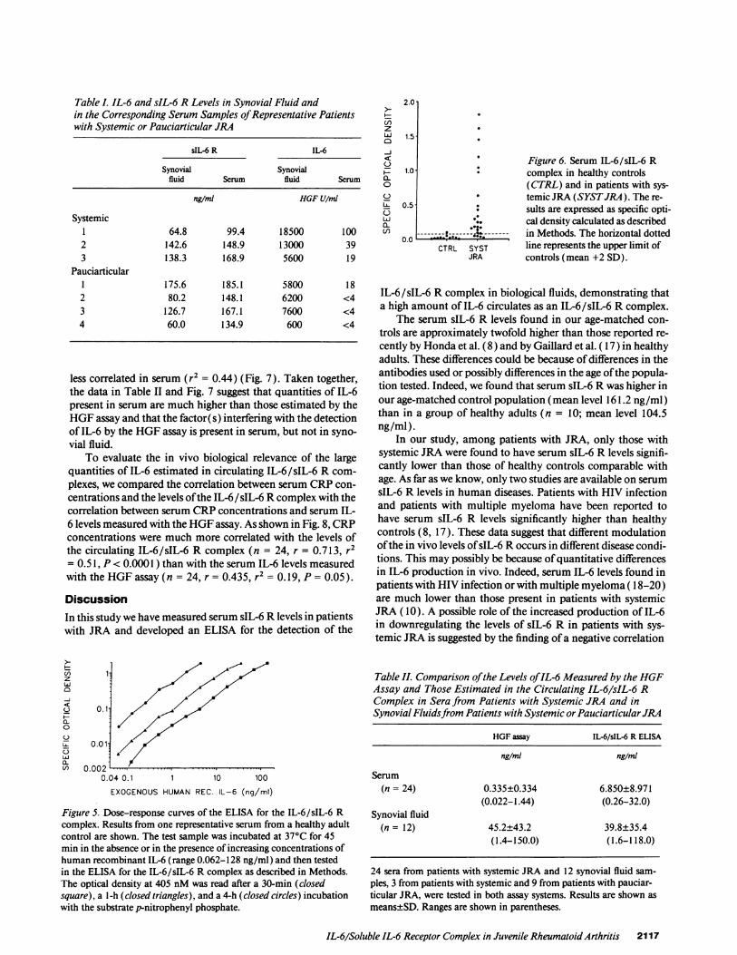
Table I. IL-6 and sIL-6 R Levels in Synovial Fluid andin the Corresponding Serum Samples ofRepresentative Patientswith Systemic or Pauciarticular JRA
sIL-6 R IL-6
Synovial Synovialfluid Serum fluid Serum
ng/ml HGF U/ml
Systemic1 64.8 99.4 18500 1002 142.6 148.9 13000 393 138.3 168.9 5600 19
Pauciarticular1 175.6 185.1 5800 182 80.2 148.1 6200 <43 126.7 167.1 7600 <44 60.0 134.9 600 <4
less correlated in serum (r2 = 0.44) (Fig. 7). Taken together,the data in Table II and Fig. 7 suggest that quantities of IL-6present in serum are much higher than those estimated by theHGF assay and that the factor(s) interfering with the detectionof IL-6 by the HGF assay is present in serum, but not in syno-vial fluid.
To evaluate the in vivo biological relevance of the largequantities of IL-6 estimated in circulating IL-6/sIL-6 R com-plexes, we compared the correlation between serum CRP con-centrations and the levels ofthe IL-6/sIL-6 R complex with thecorrelation between serum CRP concentrations and serum IL-6 levels measured with the HGF assay. As shown in Fig. 8, CRPconcentrations were much more correlated with the levels ofthe circulating IL-6/sIL-6 R complex (n = 24, r = 0.713, r2= 0.5 1, P < 0.0001 ) than with the serum IL-6 levels measuredwith the HGF assay (n = 24, r = 0.435, r2 = 0.19, P = 0.05).
DiscussionIn this study we have measured serum sIL-6 R levels in patientswith JRA and developed an ELISA for the detection of the
Ut) 1.0
z
0.011
0~
UJ0A. 00
0.0020.04 0.1 1 10 100
EXOGENOUS HUMAN REC. IL-6 (ng/ml)
Figure 5. Dose-response curves of the ELISA for the IL-6/sIL-6 Rcomplex. Results from one representative serum from a healthy adultcontrol are shown. The test sample was incubated at 37°C for 45min in the absence or in the presence of increasing concentrations ofhuman recombinant IL-6 (range 0.062-128 ng/ml) and then testedin the ELISA for the IL-6/sIL-6 R complex as described in Methods.The optical density at 405 nM was read after a 30-min (closedsquare), a 1-h (closed triangles), and a 4-h (closed circles) incubationwith the substrate p-nitrophenyl phosphate.
U)zwL0-J
0
C-)CLA
0-C/)
2.0
1.5
1.0
0.5-
0.0
Figure 6. Serum IL-6/sIL-6 Rcomplex in healthy controls(CTRL) and in patients with sys-
* temic JRA (SYSTJRA). The re-: sults are expressed as specific opti-
cal density calculated as described. s...----.k------- in Methods. The horizontal dottedCTRL SYST line represents the upper limit of
JRA controls (mean +2 SD).
IL-6/sIL-6 R complex in biological fluids, demonstrating thata high amount of IL-6 circulates as an IL-6/sIL-6 R complex.
The serum sIL-6 R levels found in our age-matched con-trols are approximately twofold higher than those reported re-cently by Honda et al. (8) and by Gaillard et al. ( 17) in healthyadults. These differences could be because of differences in theantibodies used or possibly differences in the age ofthe popula-tion tested. Indeed, we found that serum sIL-6 R was higher inour age-matched control population (mean level 161.2 ng/ml)than in a group of healthy adults (n = 10; mean level 104.5ng/ml).
In our study, among patients with JRA, only those withsystemic JRA were found to have serum sIL-6 R levels signifi-cantly lower than those of healthy controls comparable withage. As far as we know, only two studies are available on serumsIL-6 R levels in human diseases. Patients with HIV infectionand patients with multiple myeloma have been reported tohave serum sIL-6 R levels significantly higher than healthycontrols (8, 17). These data suggest that different modulationofthe in vivo levels ofsIL-6 R occurs in different disease condi-tions. This may possibly be because of quantitative differencesin IL-6 production in vivo. Indeed, serum IL-6 levels found inpatients with HIV infection orwith multiple myeloma ( 18-20)are much lower than those present in patients with systemicJRA (10). A possible role of the increased production of IL-6in downregulating the levels of sIL-6 R in patients with sys-temic JRA is suggested by the finding of a negative correlation
Table II. Comparison ofthe Levels ofIL-6 Measured by the HGFAssay and Those Estimated in the Circulating IL-6/sIL-6 RComplex in Sera from Patients with Systemic JRA and inSynovial Fluidsfrom Patients with Systemic orPauciarticularJRA
HGF assay IL-6/sIL-6 R ELISA
ng/ml ng/ml
Serum(n = 24) 0.335±0.334 6.850±8.971
(0.022-1.44) (0.26-32.0)Synovial fluid
(n = 12) 45.2±43.2 39.8±35.4(1.4-150.0) (1.6-118.0)
24 sera from patients with systemic JRA and 12 synovial fluid sam-ples, 3 from patients with systemic and 9 from patients with pauciar-ticular JRA, were tested in both assay systems. Results are shown asmeans±SD. Ranges are shown in parentheses.
IL-6/Soluble IL-6 Receptor Complex in Juvenile Rheumatoid Arthritis 2117

Ar=0.665R2 =0.44
0.1
***.0
0 * * *
3 10 100
SERUM IL-6 (HGF U/ml)
Br=0.881R2 =0.78
0.1
0.0100
0
300 1000
0
10000
SYNOVIAL FLUID IL-6 (HGF U/mi)
Figure 7. Comparison ofthe correlation between IL-6 levels measuredby the HGF assay with the levels ofIL-6/sIL-6 R complex in serum(A) and in synovial fluid (B).
ever, preincubation of control sera with increasing concentra-tions of exogenous human recombinant IL-6 resulted in adose-dependent increase in optical density, demonstrating thatthis assay system is able to detect the IL-6/sIL-6 R complexformed by the binding of IL-6 to its soluble receptor present inhuman serum. This also confirms the findings of Honda et al.(8) and of Gaillard et al. ( 17) that circulating soluble IL-6 R isable to bind IL-6. Almost all sera ofpatients with systemic JRAdisplayed high optical density in this assay system, demonstrat-ing the presence of elevated serum IL-6/sIL-6 R complex lev-els. These results suggest strongly that IL-6, or at least part of it,circulates bound to its soluble receptor in patients with sys-temic JRA. To the best ofour knowledge, this is the firstdemon-stration of the presence of circulating cytokine/soluble cyto-kine receptor complexes in human diseases. Our data are con-sistent with the following hypothesis on the biology of IL-6:IL-6 produced at sites of inflammation (e.g., joints) or pro-duced in the peripheral blood in response to stimuli released byinflammatory sites (e.g., IL-l or TNF-a) binds the serum sIL-6R and it is vehiculated in the blood as an IL-6/sIL-6 R com-plex. This binding may protect IL-6 from protease degradation,in a manner similar to what has been described for IL-4 and thesoluble IL-4 receptor (24), and/or stabilize IL-6 bioactivity, ashas been demonstrated for TNF-a and its soluble receptors(25). The IL-6/sIL-6 R complex will thus associate with the1 30-kD signal transducing protein on the surface oftarget cellsand mediate IL-6 bioactivities.
Comparison of serum IL-6 levels measured by the HGFassay and those estimated in circulating IL-6/sIL-6 R com-
between serum sIL-6 R levels with serum IL-6 levels, measuredwith the HGF assay, and by the decrease in serum sIL-6 Rlevels that was associated with the increase in serum IL-6 levelsduring the febrile peak. The mechanisms of the production ofsIL-6 R have not yet been clarified (21, 22). Nevertheless, ourdata suggest that two possible mechanisms, not necessarily mu-tually exclusive, may account for the decrease in the serum
sIL-6 R levels in patients with systemic JRA: (a) since IL-6 hasbeen reported to inhibit the expression of the IL-6 R gene both
in vivo and in vitro (23), a direct effect of increased in vivoIL-6 production on the expression ofthe IL-6 R gene is conceiv-able; and (b) the decrease in serum sIL-6 R levels may be a
consequence ofa consumption ofcirculating sIL-6 R due to theformation of IL-6/sIL-6 R complexes in the presence of IL-6and of the subsequent binding and internalization of the com-plex by target cells expressing the 130-kD signal transducingprotein.
This second hypothesis is based on the assumption thatcirculating IL-6/sIL-6 R complexes are present in patients withsystemic JRA. Since the serum soluble form of IL-6 R and thegenetically engineered truncated form of IL-6 R have beenshown to bind IL-6 with a binding affinity similar to the entire80-kD molecule (6, 8) and given the serum concentrations ofthe sIL-6 R found in this study, it is conceivable to hypothesizethat IL-6 circulates bound to its soluble receptor. To verify thishypothesis, we constructed an ELISA based on the use of a
monoclonal antibody to IL-6 for capture and a monoclonalantibody to sIL-6 R for revealing the presence ofthe IL-6/sIL-6 R complex. Using this approach we found no evidence ofcirculating IL-6/sIL-6 R complex in healthy controls. How-
r=0.435R2=0. 19p=0.05
E
Dl
en
D,
100
10
A
00
* .0
* 0
0
.0 *
0
0.6 10SERUM CRP (mg%)
a
C:co
-J
en
r=0.713R2=0.51p<0.0001
1.0
0.1
0.0
0.6 1
B
.
0-@00
0000
* *.
*
*
10SERUM CRP (mgX)
Figure 8. Correlation of serum CRP concentrations with serum IL-6levels measured by the HGF assay (A) or with circulating IL-6/sIL-6R complex levels (B) in patients with systemic JRA.
2118 De Benedetti, Massa, Pignatti, Albani, Novick, and Martini
a0
Ix
-J
W
(D
-J
D
en
Co
o
J(ON
CD
-J
0
-JLa.-J
0z(n
.
1

plexes led us to the unexpected finding that, in patients withsystemic JRA, IL-6 was present in nanogram per milliliteramounts in the circulating IL-6/sIL-6 R complexes comparedwith the few hundreds ofpicograms per milliliter measured bythe HGF assay. Therefore, our data suggest that in systemicJRA, but also possibly in other disease conditions, IL-6 circu-lates in much higher amounts than those measured by theHGF assay. The possibility that the HGF assay detects only aportion of IL-6 present in human serum is also supported bythe consistent finding that IL-6 levels measured with the hepa-tocyte-stimulating factor assay have been found to be muchhigher than those measured by the HGF assay in a variety ofdisease states (26-29). More recently, May et al. (30) havedemonstrated that the bulk ofIL-6 present in peripheral bloodcirculates in two high molecular weight complexes (100-150and 400-500 kD) and that this IL-6 is not detected by the HGFassay (30). Of note, among the proteins present in the 100-150-kD complex, a 50-kD material was present, consistentwith the molecular weight of the human natural sIL-6 R.Taken together, the discrepancies between serum IL-6 levelsmeasured by the HGF assay and by the hepatocyte-stimulatingfactor assay (26-29), our results, and those ofMay et al. (30)raise the issue of the biological role of the large quantities ofIL-6 present in the peripheral blood. In our study, the strictcorrelation ofserum IL-6 levels measured in the IL-6/sIL-6 Rcomplex ELISA with CRP concentrations in patients with sys-temic JRA suggests that the high amounts ofIL-6 present in thecirculating IL-6/sIL-6 R complex are biologically relevant invivo, at least as far as the induction by IL-6 of acute phaseprotein production.
In conclusion, in this study we reported a decrease in sIL-6R levels in patients with systemic JRA, a disease characterizedby a marked increase in serum IL-6 levels. We developed anassay system for detection and measurement of circulating IL-6/sIL-6 R complexes and demonstrated that IL-6 circulates asan IL-6/sIL-6 R complex. We have also found that large quan-tities ofIL-6, not detected by the HGF assay, are present in theperipheral blood ofpatients with systemicJRA as IL-6/ sIL-6 Rcomplexes, whose in vivo biological relevance is suggested bytheir strict correlation with serum CRP concentrations.
Acknowledgments
This work was supported by the Istituto di Ricovero e Cura a CarattereScientifico Policlinico San Matteo, Pavia, Italy and by the Istituto diRicerca Cesare Serono, Ardea, Italy.
References1. Van Snick, J. 1990. Interleukin 6: an overview. Annu. Rev. Immunol.
8:253-278.2. Hirano, T., S. Akira, T. Taga, and T. Kishimoto. 1990. Biological and
clinical aspects of interleukin 6. Immunol. Today. 1 1:443-449.3. Yamasaki, T., T. Taga, Y. Hirata, H. Yawata, Y. Kawanishi, B. Seed, T.
Taniguchi, T. Hirano, and T. Kishimoto. 1988. Cloning and expression of thehuman interleukin-6 (BSF-2/IFN#2) receptor. Science (Wash. DC). 241:825-828.
4. Taga, T., M. Hibi, T. Hirata, K. Yamasaki, K. Yasukawa, T. Matsuda, T.Hirano, and T. Kishimoto. 1989. Interleukin 6 triggers the association of itsreceptor with a possible signal transducer, gp 130. Cell. 58:573-579.
5. Yawata, H., K. Yasukawa, S. Natsuka, M. Murakami, K Yamasaki, M.Hibi, T. Taga, and T. Kishimoto. 1993. Structure-function analysis ofthe humanIL-6 receptor dissociation ofamino acid residues required for IL-6 binding andfor IL-6 signal transduction through gpl30. EMBO (Eur. Mol. Biol. Organ) J.12:1705-1712.
6. Yasukawa, K., T. Saito, T. Fukunaga, Y. Sekimori, Y. Kishihara, H. Fukui,Y. Osugi, T. Matsuda, H. Yawata, T. Hirano, et al. 1990. Purification and charac-terization of soluble human IL-6 receptor expressed in CHO cell. J. Biochem.108:673-681.
7. Novick, D., H. Engelmann, D. Wallach, and M. Rubinstein. 1989. Solublecytokine receptors are present in normal human urine. J. Exp. Med. 170:1409-1414.
8. Honda, M., S. Yamammoto, M. Cheng, K. Yasukawa, H. Suzuki, T. Saito,Y. Osugi, T. Tokunaga, and T. Kishimoto. 1992. Human soluble IL-6 receptorits detection and enhanced release by HIV infection. J. Immunol. 148:2175-2180.
9. Brewer, E. J., Jr., J. Bass, J. Baum, J. T. Cassidy, C. Fink, J. Jacobs, V.Hanson, J. E. Levinson, J. Shaller, and J. S. Stillman. 1977. Current proposedrevision ofJRA criteria. Arthritis Rheum. 20(Suppl.):195-199.
10. De Benedetti, F., M. Massa, P. Robbioni, A. Ravelli, G. R. Burgio, and A.Martini. 1991. Correlation of serum interleukin 6 levels with joint involvementand thrombocytosis in systemic juvenile rheumatoid arthritis. Arthritis Rheum.34:1158-1163.
11. De Benedetti, F., P. Robbioni, M. Massa, S. Viola, S. Albani, and A.Martini. 1992. Serum interleukin 6 and joint involvement in polyarticular andpauciarticular juvenile chronic arthritis. Clin. Exp. Rheumatol. 10:493-498.
12. Hirano, T., T. Matsuda, M. Turner, N. Miyasaka, G. Buchan, B. Tang, K.Sato, M. Shimizu, R. Maini, M. Feldmann, and T. Kishimoto. 1988. Excessiveproduction of interleukin 6/B cell stimulatory factor-2 in rheumatoid arthritis.Eur. J. Immunol. 18:1797-1801.
13. Houssiau, F. A., J. P. Devogelaer, J. Van Damme, C. Nagant de Deux-chaisnes, and J. Van Snick. 1988. Interleukin 6 in synovial fluid and serum ofpatients with rheumatoid arthritis and other inflammatory arthritis. ArthritisRheum. 31:784-788.
14. Novick, D., H. Engelmann, D. Wallach, M. Revel, 0. Leitner, and M.Rubinstein. 1991. Monoclonal antibodies to the soluble human IL-6 receptoraffinity purification, ELISA, and inhibition of ligand binding. Hybridoma.10:137-145.
15. Novick, D., H. Engelmann, D. Wallach, 0. Leitner, M. Revel, and M.Rubinstein. 1990. Purification ofsoluble cytokine receptors from normal humanurine by ligand-affinity and immunoaffinity chromatography. J. Chromatogr.5 10:33 1-337.
16. Novick, D., Z. Eshhar, M. Revel, and Y. Mory. 1989. Monoclonal antibod-ies for affinity purification of IL-6/IFN-#2 and for neutralization ofHGF activ-ity. Hybridoma. 8:561-567.
17. Gaillard, J. P., R. Bataille, H. Brailly, C. Zuber, K. Yasukawa, M. Attal, N.Maruo, T. Taga, T. Kishimoto, and B. Klein. 1993. Increased and highly stablelevels of functional interleukin 6 receptor in sera of patients with monoclonalgammopathy. Eur. J. Immunol. 23:820-824.
18. Honda, M., K. Kitamura, Y. Mizutani, M. Oishi, M. Arai, T. Okura, K.Igarahi, K. Yasukawa, T. Hirano, T. Kishimoto, et al. 1990. Quantitative analysisofserum IL-6 and its correlation with increased levels ofserum IL-2R in HIV-in-duced diseases. J. Immunol. 145:4059-4064.
19. Bataille, R., M. Jourdan, X.-G. Zhang, and B. Klein. 1989. Serum levels ofinterleukin 6, a potent myeloma cell growth factor, as a reflect ofdisease severityin plasma cell dyscrasias. J. Clin. Inv. 84:2008-2011.
20. Ludwig, H., D. Nachbaur, E. Fritz, M. Krainer, and H. Huber. 1991.Interleukin 6 is a prognostic factor in multiple myeloma. Blood. 77:2794-2795.
21. Lust, J. A., K. A. Donovan, M. P. Kline, R. R. Greipp, R. A. Kyle, andN. J. Maihle. 1992. Isolation ofan mRNA encoding a soluble form ofthe humaninterleukin-6 receptor. Cytokine. 4:96-100.
22. Mullberg, J., H. Schooltink, T. Stoyan, M. Gunther, L. Graeve, G. Buse,A. Mackiewicz, P. C. Heinrich, and S. Rose-John. 1993. The soluble interleukin 6receptor is generated by shedding. Eur. J. Immunol. 23:473-480.
23. Portier, M., D. Lees, E. Caron, M. Jourdan, J. M. Boiron, R. Bataille, andB. Klein. 1992. Up-regulation of interleukin (IL)-6 receptor gene expression invitro and in vivo in IL-6 deprived myeloma cells. FEBS (Fed. Eur. Biochem.Soc.) Lett. 302:35-38.
24. Fernandez-Botran, R., and E. S. Vitetta. 1991. Evidence that naturalmurine soluble interleukin 4 receptors may act as transport proteins. J. Exp.Med. 174:673-681.
25. Aderka, D., H. Engelmann, Y. Maor, C. Brakebusch, and D. Wallach.1992. Stabilization ofthe bioactivity oftumor necrosis factor by its soluble recep-tors. J. Exp. Med. 175:323-329.
26. Sehgal, P. B., G. Grieninger, and G. Tosato, editors. 1989. Regulation ofthe acute phase and immune responses: interleukin 6. Ann. NY Acad. Sci.557:583 pp.
27. Fong, Y., L. L. Moldawer, M. Marano, H. Wei, S. B. Tatter, R. H. Clarick,U. Santhanam, D. Sherris, L. T. May, P. B. Sehgal, and S. F. Lowry. 1989.Endotoxemia elicits increased circulating ,2-IFN/IL-6 in man. J. Immunol.142:2321-2324.
28. Helfgott, D. C., S. B. Tatter, U. Santhanam, R. H. Clarick, N. Bhardwaj,L. T. May, and P. B. Sehgal. 1989. Multiple forms ofIFN-,B2/IL-6 in serum andbody fluids during acute bacterial infection. J. Immunol. 142:948-953.
29. Jablons, D. M., J. J. Mule, J. K. McIntosh, P. B. Sehgal, L. T. May, C. M.Huang, S. A. Rosemberg, and M. T. Lotze. 1989. IL-6/IFN-j#2 as a circulatinghormone. Induction by cytokine administration in humans. J. Immunol.142:1542-1547.
30. May, L. T., H. Viguet, J. S. Kenney, N. Ida, A. C. Allison, and P. B. Sehgal.1992. High levels of"complexed" interleukin 6 in human blood. J. Biol. Chem.267:19698-19704.
IL-6/Soluble IL-6 Receptor Complex in Juvenile RheumatoidArthritis 2119
Copyright © 2022 FDOKUMEN