SOX6 controls dorsal progenitor identity and interneuron diversity during neocortical development
12
1238 VOLUME 12 | NUMBER 10 | OCTOBER 2009 NATURE NEUROSCIENCE ARTICLES 1 Massachusetts General Hospital–Harvard Medical School Center for Nervous System Repair, Departments of Neurosurgery and Neurology, Program in Neuroscience, Harvard Medical School, Boston, Massachusetts, USA. 2 Nayef Al-Rodhan Laboratories, Massachusetts General Hospital, Boston, Massachusetts, USA. 3 Department of Stem Cell and Regenerative Biology, and Harvard Stem Cell Institute, Harvard University, Cambridge, Massachusetts, USA. 4 Department of Molecular and Cellular Biology, Harvard University, Cambridge, Massachusetts, USA. 5 Present address: Department of Basic Neurosciences and Clinic of Neurology, University of Geneva, Switzerland. Correspondence should be addressed to J.D.M. ([email protected]). Received 26 May; accepted 22 July; published online 5 August 2009; doi:10.1038/nn.2387 at which they are born 3,6–9 . In the pallium, distinct excitatory projection neuron subtypes are born sequentially under the control of temporally coordinated programs that guide their subtype specification and differentiation 3 . Simultaneously, inhibitory cortical interneurons, which constitute approximately 25% of all cortical neurons, are primarily born in the subpallial medial (MGE) and caudal ganglionic eminences (CGE) 2 . Acquisition of distinct interneuron subtype identities, distinguishable by molecular, morphological and electrophysiological phenotypes, depends on both the place and time of birth in the MGE and CGE 2,6–12 . Differentiating interneurons then migrate tangentially toward and then radially into the cortex to populate their final laminar destinations alongside concurrently born pallium-derived excitatory projection neurons 2,13 . Because cortical interneurons are implicated in several developmental disorders 14 , including epilepsy 15 , autism 16 and schizophrenia 17 , understanding the molecular controls over their subtype diversity might clarify some causes of and potential therapeutic approaches to these important disorders. Although major progress has been made in understanding the regulation of broad aspects of neuronal heterogeneity during development 1 , only recently have specific controls over excitatory 3,18–26 and inhibitory 27–31 cortical neuron subtype differentiation been characterized. We recently reported that SOX5 postmitotically controls the sequential generation of distinct pallium-derived excitatory corticofugal projection neuron populations, regulating their subtype diversity 22,26 . Motivated by the complementary and largely redundant functions of SOX5 and SOX6 in other systems 32,33 , we hypothesized that Two broad functional classes of cortical neurons, excitatory projection neurons and inhibitory interneurons, arise from spatially and molecularly segregated pallial (dorsal) and subpallial (ventral) proliferative ventricular zones of the telencephalon, respectively 1–3 . Parcellation of these proliferative regions into molecularly segregated domains separated at the pallial-subpallial boundary (PSB) is critical for the generation of these distinct classes of neurons. In these broad excitatory and inhibitory neuronal classes, tremendous subtype diversity arises largely from the dynamic temporal expression of progenitor and postmitotic transcriptional regulators. Both of these developmental mechanisms (inter- and intra-domain segregation of molecular regulators) combine to give rise to the extraordinary neuronal diversity of the adult mammalian brain. The parcellation of the proliferative neuroepithelium at the PSB is defined and maintained by the interactions of several critical early patterning transcription factors, exemplified by the repressive interaction of pallium-expressed Neurogenin2 (Ngn2, also known as Neurog2) on the generally subpallium-expressed Mash1 (also known as Ascl1) 1 . Accordingly, loss of Ngn2 function results in dorsal expansion of Mash1 expression and a consequent ventralization of pallial progenitors, which aberrantly give rise to subpallial-like neurons 4,5 . The dynamic interaction between this pair of transcription factors exemplifies the delicate balance of molecular regulators that are required to establish and maintain the PSB. Throughout corticogenesis, these pallial and subpallial progenitors give rise to neurons whose fates depend largely on the location and time SOX6 controls dorsal progenitor identity and interneuron diversity during neocortical development Eiman Azim 1–3 , Denis Jabaudon 1–3,5 , Ryann M Fame 1–4 & Jeffrey D Macklis 1–3 The neuronal diversity of the CNS emerges largely from controlled spatial and temporal segregation of cell type-specific molecular regulators. We found that the transcription factor SOX6 controls the molecular segregation of dorsal (pallial) from ventral (subpallial) telencephalic progenitors and the differentiation of cortical interneurons, regulating forebrain progenitor and interneuron heterogeneity. During corticogenesis in mice, SOX6 and SOX5 were largely mutually exclusively expressed in pallial and subpallial progenitors, respectively, and remained mutually exclusive in a reverse pattern in postmitotic neuronal progeny. Loss of SOX6 from pallial progenitors caused their inappropriate expression of normally subpallium-restricted developmental controls, conferring mixed dorsal-ventral identity. In postmitotic cortical interneurons, loss of SOX6 disrupted the differentiation and diversity of cortical interneuron subtypes, analogous to SOX5 control over cortical projection neuron development. These data indicate that SOX6 is a central regulator of both progenitor and cortical interneuron diversity during neocortical development. © 2009 Nature America, Inc. All rights reserved.
Transcript of SOX6 controls dorsal progenitor identity and interneuron diversity during neocortical development

1238 volume 12 | number 10 | oCTober 2009 nature neuroscience
a r t i c l e s
1Massachusetts General Hospital–Harvard Medical School Center for Nervous System Repair, Departments of Neurosurgery and Neurology, Program in Neuroscience, Harvard Medical School, Boston, Massachusetts, USA. 2Nayef Al-Rodhan Laboratories, Massachusetts General Hospital, Boston, Massachusetts, USA. 3Department of Stem Cell and Regenerative Biology, and Harvard Stem Cell Institute, Harvard University, Cambridge, Massachusetts, USA. 4Department of Molecular and Cellular Biology, Harvard University, Cambridge, Massachusetts, USA. 5Present address: Department of Basic Neurosciences and Clinic of Neurology, University of Geneva, Switzerland. Correspondence should be addressed to J.D.M. ([email protected]).
Received 26 May; accepted 22 July; published online 5 August 2009; doi:10.1038/nn.2387
at which they are born3,6–9. In the pallium, distinct excitatory projection neuron subtypes are born sequentially under the control of temporally coordinated programs that guide their subtype specification and differentiation3. Simultaneously, inhibitory cortical interneurons, which constitute approximately 25% of all cortical neurons, are primarily born in the subpallial medial (MGE) and caudal ganglionic eminences (CGE)2. Acquisition of distinct interneuron subtype identities, distinguishable by molecular, morphological and electrophysiological phenotypes, depends on both the place and time of birth in the MGE and CGE2,6–12. Differentiating interneurons then migrate tangentially toward and then radially into the cortex to populate their final laminar destinations alongside concurrently born pallium-derived excitatory projection neurons2,13. Because cortical interneurons are implicated in several developmental disorders14, including epilepsy15, autism16 and schizophrenia17, understanding the molecular controls over their subtype diversity might clarify some causes of and potential therapeutic approaches to these important disorders.
Although major progress has been made in understanding the regulation of broad aspects of neuronal heterogeneity during development1, only recently have specific controls over excitatory3,18–26 and inhibitory27–31 cortical neuron subtype differentiation been characterized. We recently reported that SOX5 postmitotically controls the sequential generation of distinct pallium-derived excitatory corticofugal projection neuron populations, regulating their subtype diversity22,26. Motivated by the complementary and largely redundant functions of SOX5 and SOX6 in other systems32,33, we hypothesized that
Two broad functional classes of cortical neurons, excitatory projection neurons and inhibitory interneurons, arise from spatially and molecularly segregated pallial (dorsal) and subpallial (ventral) proliferative ventricular zones of the telencephalon, respectively1–3. Parcellation of these proliferative regions into molecularly segregated domains separated at the pallial- subpallial boundary (PSB) is critical for the generation of these distinct classes of neurons. In these broad excitatory and inhibitory neuronal classes, tremendous subtype diversity arises largely from the dynamic temporal expression of progenitor and postmitotic transcriptional regulators. Both of these developmental mechanisms (inter- and intra-domain segregation of molecular regulators) combine to give rise to the extraordinary neuronal diversity of the adult mammalian brain.
The parcellation of the proliferative neuroepithelium at the PSB is defined and maintained by the interactions of several critical early patterning transcription factors, exemplified by the repressive interaction of pallium-expressed Neurogenin2 (Ngn2, also known as Neurog2) on the generally subpallium-expressed Mash1 (also known as Ascl1)1. Accordingly, loss of Ngn2 function results in dorsal expansion of Mash1 expression and a consequent ventralization of pallial progenitors, which aberrantly give rise to subpallial-like neurons4,5. The dynamic interaction between this pair of transcription factors exemplifies the delicate balance of molecular regulators that are required to establish and maintain the PSB.
Throughout corticogenesis, these pallial and subpallial progenitors give rise to neurons whose fates depend largely on the location and time
SOX6 controls dorsal progenitor identity and interneuron diversity during neocortical developmentEiman Azim1–3, Denis Jabaudon1–3,5, Ryann M Fame1–4 & Jeffrey D Macklis1–3
The neuronal diversity of the CNS emerges largely from controlled spatial and temporal segregation of cell type-specific molecular regulators. We found that the transcription factor SOX6 controls the molecular segregation of dorsal (pallial) from ventral (subpallial) telencephalic progenitors and the differentiation of cortical interneurons, regulating forebrain progenitor and interneuron heterogeneity. During corticogenesis in mice, SOX6 and SOX5 were largely mutually exclusively expressed in pallial and subpallial progenitors, respectively, and remained mutually exclusive in a reverse pattern in postmitotic neuronal progeny. Loss of SOX6 from pallial progenitors caused their inappropriate expression of normally subpallium-restricted developmental controls, conferring mixed dorsal-ventral identity. In postmitotic cortical interneurons, loss of SOX6 disrupted the differentiation and diversity of cortical interneuron subtypes, analogous to SOX5 control over cortical projection neuron development. These data indicate that SOX6 is a central regulator of both progenitor and cortical interneuron diversity during neocortical development.
©20
09 N
atu
re A
mer
ica,
Inc.
All
rig
hts
res
erve
d.
nature neuroscience volume 12 | number 10 | oCTober 2009 1239
a r t i c l e s
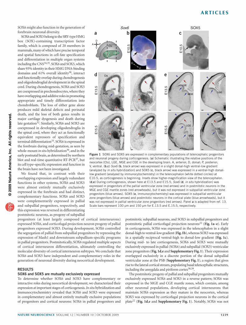
postmitotic subpallial neurons, and SOX5 in subpallial progenitors and postmitotic pallial corticofugal projection neurons22 (Fig. 1a–e). Early in corticogenesis, SOX6 was expressed in the telencephalon in a slight dorsal-high to ventral-low gradient (Fig. 1b), whereas SOX5 was expressed in a spatially reciprocal ventral-high to dorsal-low gradient (Fig. 1c). During mid- to late corticogenesis, SOX6 and SOX5 were mutually exclusively expressed in pallial (SOX6) and subpallial (SOX5) ventricular zone progenitors (Fig. 1d,e and Supplementary Fig. 1). Their expression overlapped exclusively in a discrete portion of the dorsal subpallial ventricular zone at the PSB (Supplementary Fig. 1), a region that gives rise to the lateral cortical stream, populating basal telencephalic structures, including the amygdala and piriform cortex38,39.
The postmitotic progeny of pallial and subpallial progenitors mutually exclusively expressed SOX6 and SOX5 in a reverse pattern; SOX6 was expressed in the MGE and CGE mantle zones, which contain, among other neuronal populations, developing cortical interneurons that maintain SOX6 expression as they mature in the neocortex, whereas SOX5 was expressed by corticofugal projection neurons in the cortical plate22 (Fig. 1d,e and Supplementary Fig. 1). Notably, SOX6 was not
SOX6 might also function in the generation of forebrain neuronal diversity.
SOX6 and SOX5 belong to the SRY-type HMG box (SOX)-containing transcription factor family, which is composed of 20 members in mammals, many of which have precise temporal and spatial functions in cell-fate specification and differentiation in multiple organ systems including the CNS34,35. SOX6 and SOX5, which share 93% identity in their HMG DNA-binding domains and 61% overall identity36, interact and functionally overlap during chondrogenesis and oligodendroglial development in the spinal cord. During chondrogenesis, SOX6 and SOX5 are coexpressed in prechondrocytes, where they have overlapping and additive roles in promoting appropriate and timely differentiation into chondroblasts. The loss of either gene alone produces mild skeletal defects and perinatal death, and the loss of both genes results in major cartilage dysgenesis and death during late gestation32. Similarly, SOX6 and SOX5 are coexpressed in developing oligodendroglia in the spinal cord, where they act as functionally equivalent repressors of specification and terminal differentiation33. SOX6 is expressed in the forebrain during mid-gestation, as seen by whole-mount in situ hybridization36, and in the early postnatal brain, as determined by northern blot and real-time quantitative RT-PCR37, but its cell type–specific expression and function in the brain have not been investigated.
We found that, in contrast with their overlapping expression and largely redundant functions in other systems, SOX6 and SOX5 were almost entirely mutually exclusively expressed in the forebrain and had distinct, complementary functions. SOX6 and SOX5 were complementarily expressed in pallial and subpallial progenitors, respectively, and this expression was reversed in differentiating postmitotic neurons, as progeny of subpallial progenitors (at least largely composed of cortical interneurons) expressed SOX6, and corticofugal projection neuron progeny of pallial progenitors expressed SOX5. During development, SOX6 controlled the segregation of pallial from subpallial progenitors by repressing the expression of Mash1 and downstream subpallium-specific programs in pallial progenitors. Postmitotically, SOX6 regulated multiple aspects of cortical interneuron differentiation, ultimately controlling the molecular diversity of cortical interneuron subtypes. We conclude that SOX6 and SOX5 have independent and complementary roles in the generation of neuronal diversity during neocortical development.
RESULTSSOX6 and SOX5 are mutually exclusively expressedTo determine whether SOX6 and SOX5 have complementary or interactive roles during neocortical development, we characterized their expression at important stages of corticogenesis. In situ hybridization and immunocytochemistry revealed that SOX6 and SOX5 were expressed in complementary and almost entirely mutually exclusive populations of progenitors and cortical neurons: SOX6 in pallial progenitors and
Figure 1 SOX6 and SOX5 are expressed in complementary populations of telencephalic progenitors and neuronal progeny during corticogenesis. (a) Schematic illustrating the relative positions of the neocortex (Ctx), LGE, MGE and CGE in the developing brain. A, anterior; D, dorsal; P, posterior; V, ventral. (b,c) Sox6 (b, black arrow) was expressed in a slight dorsal-high ventral-low gradient (analyzed by in situ hybridization) and SOX5 (c, black arrow) was expressed in a ventral-high dorsal-low gradient (analyzed by immunocytochemistry) in the telencephalon (white dotted circles) at E10.5, as corticogenesis is beginning. Insets show higher-magnification view of the telencephalon. (d,e) During corticogenesis, shown here at E13.5 and E15.5, Sox6 (d, in situ hybridization) was expressed in progenitors of the pallial ventricular zone (red arrows) and in postmitotic neurons in the MGE and CGE mantle zones (red arrowheads), but it was not expressed in subpallial ventricular zone progenitors (blue arrows). SOX5 (e, immunocytochemistry) was expressed in subpallial ventricular zone progenitors (blue arrows) and postmitotic neurons in the cortical plate (blue arrowheads), but it was not expressed in pallial ventricular zone progenitors (red arrows). Panel a is adapted from ref. 13. Scale bars represent 100 µm and 150 µm for E.13.5 and E.15.5, respectively.
a
d e
b cSox6 SOX5
E10
.5
E13
.5
E13
.5
E15
.5
E15
.5
E10
.5
D
VPA
LGE CGE
MGE
Ctx
©20
09 N
atu
re A
mer
ica,
Inc.
All
rig
hts
res
erve
d.
1240 volume 12 | number 10 | oCTober 2009 nature neuroscience
a r t i c l e s
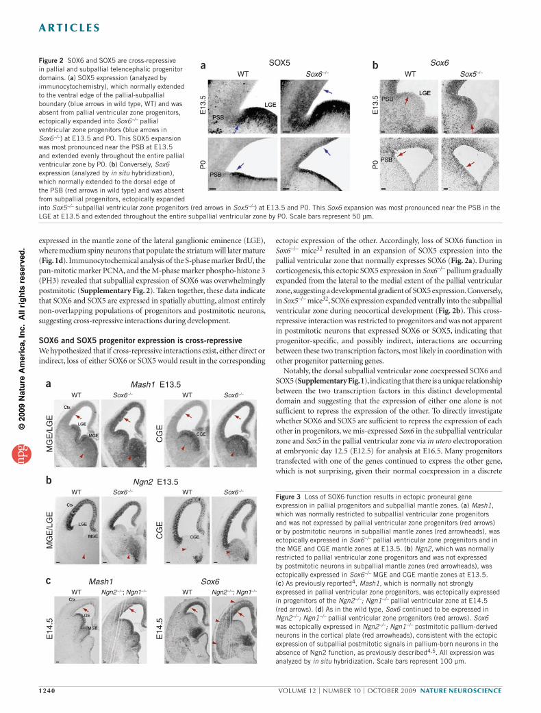
ectopic expression of the other. Accordingly, loss of SOX6 function in Sox6–/– mice32 resulted in an expansion of SOX5 expression into the pallial ventricular zone that normally expresses SOX6 (Fig. 2a). During corticogenesis, this ectopic SOX5 expression in Sox6–/– pallium gradually expanded from the lateral to the medial extent of the pallial ventricular zone, suggesting a developmental gradient of SOX5 expression. Conversely, in Sox5–/– mice32, SOX6 expression expanded ventrally into the subpallial ventricular zone during neocortical development (Fig. 2b). This cross-repressive interaction was restricted to progenitors and was not apparent in postmitotic neurons that expressed SOX6 or SOX5, indicating that progenitor-specific, and possibly indirect, interactions are occurring between these two transcription factors, most likely in coordination with other progenitor patterning genes.
Notably, the dorsal subpallial ventricular zone coexpressed SOX6 and SOX5 (Supplementary Fig. 1), indicating that there is a unique relationship between the two transcription factors in this distinct developmental domain and suggesting that the expression of either one alone is not sufficient to repress the expression of the other. To directly investigate whether SOX6 and SOX5 are sufficient to repress the expression of each other in progenitors, we mis-expressed Sox6 in the subpallial ventricular zone and Sox5 in the pallial ventricular zone via in utero electroporation at embryonic day 12.5 (E12.5) for analysis at E16.5. Many progenitors transfected with one of the genes continued to express the other gene, which is not surprising, given their normal coexpression in a discrete
expressed in the mantle zone of the lateral ganglionic eminence (LGE), where medium spiny neurons that populate the striatum will later mature (Fig. 1d). Immunocytochemical analysis of the S-phase marker BrdU, the pan-mitotic marker PCNA, and the M-phase marker phospho-histone 3 (PH3) revealed that subpallial expression of SOX6 was overwhelmingly postmitotic (Supplementary Fig. 2). Taken together, these data indicate that SOX6 and SOX5 are expressed in spatially abutting, almost entirely non-overlapping populations of progenitors and postmitotic neurons, suggesting cross-repressive interactions during development.
SOX6 and SOX5 progenitor expression is cross-repressiveWe hypothesized that if cross-repressive interactions exist, either direct or indirect, loss of either SOX6 or SOX5 would result in the corresponding
Figure 2 SOX6 and SOX5 are cross-repressive in pallial and subpallial telencephalic progenitor domains. (a) SOX5 expression (analyzed by immunocytochemistry), which normally extended to the ventral edge of the pallial-subpallial boundary (blue arrows in wild type, WT) and was absent from pallial ventricular zone progenitors, ectopically expanded into Sox6–/– pallial ventricular zone progenitors (blue arrows in Sox6–/–) at E13.5 and P0. This SOX5 expansion was most pronounced near the PSB at E13.5 and extended evenly throughout the entire pallial ventricular zone by P0. (b) Conversely, Sox6 expression (analyzed by in situ hybridization), which normally extended to the dorsal edge of the PSB (red arrows in wild type) and was absent from subpallial progenitors, ectopically expanded into Sox5–/– subpallial ventricular zone progenitors (red arrows in Sox5–/–) at E13.5 and P0. This Sox6 expansion was most pronounced near the PSB in the LGE at E13.5 and extended throughout the entire subpallial ventricular zone by P0. Scale bars represent 50 µm.
Figure 3 Loss of SOX6 function results in ectopic proneural gene expression in pallial progenitors and subpallial mantle zones. (a) Mash1, which was normally restricted to subpallial ventricular zone progenitors and was not expressed by pallial ventricular zone progenitors (red arrows) or by postmitotic neurons in subpallial mantle zones (red arrowheads), was ectopically expressed in Sox6–/– pallial ventricular zone progenitors and in the MGE and CGE mantle zones at E13.5. (b) Ngn2, which was normally restricted to pallial ventricular zone progenitors and was not expressed by postmitotic neurons in subpallial mantle zones (red arrowheads), was ectopically expressed in Sox6–/– MGE and CGE mantle zones at E13.5. (c) As previously reported4, Mash1, which is normally not strongly expressed in pallial ventricular zone progenitors, was ectopically expressed in progenitors of the Ngn2–/–; Ngn1–/– pallial ventricular zone at E14.5 (red arrows). (d) As in the wild type, Sox6 continued to be expressed in Ngn2–/–; Ngn1–/– pallial ventricular zone progenitors (red arrows). Sox6 was ectopically expressed in Ngn2–/–; Ngn1–/– postmitotic pallium-derived neurons in the cortical plate (red arrowheads), consistent with the ectopic expression of subpallial postmitotic signals in pallium-born neurons in the absence of Ngn2 function, as previously described4,5. All expression was analyzed by in situ hybridization. Scale bars represent 100 µm.
SOX5 Sox6WT Sox6 –/– Sox5–/–WT
E13
.5
E13
.5
P0
P0
a b
a
b
c
Mash1 E13.5
Ngn2 E13.5
Sox6Mash1
WT Sox6 –/– WT Sox6 –/–
WT Sox6 –/– WT Sox6 –/–
WT Ngn2 –/–; Ngn1–/– Ngn2 –/–; Ngn1–/–WT
MG
E/L
GE
CG
EC
GE
E14
.5
MG
E/L
GE
E14
.5
©20
09 N
atu
re A
mer
ica,
Inc.
All
rig
hts
res
erve
d.
nature neuroscience volume 12 | number 10 | oCTober 2009 1241
a r t i c l e s
the Sox6–/– pallium is indicative of the initiation of subpallium- specific programs of gene expression, we carried out comparative microarray analysis between wild-type and Sox6–/– pallium during mid- corticogenesis at E13.5 and identified several normally subpallium-expressed genes that were ectopically expressed in the Sox6–/– pallium, including Dlx1, Dlx2, Dlx4, Gsh2, Isl1, Meis1 and Sox5 (Supplementary Table 1). These data indicate that SOX6 critically maintains pallial progenitor identity by repressing subpallial programs of gene expression, but redundant and/or compensatory controls (for example, Ngn2 and Ngn1) persist that are sufficient to ensure largely appropriate pallial corticogenesis42. We conclude that SOX6 acts cooperatively with Ngn2 to control the segregation of telencephalic progenitor domains during development.
SOX6 controls cortical interneuron subtype differentiationSOX6 was also expressed in postmitotic interneurons as they reside in the subpallium and populate the neocortex (Fig. 1d and Supplementary Figs. 1 and 2). We therefore examined whether loss of SOX6 function would affect important sequential steps of interneuron differentiation: early postmitotic molecular identity, cortical laminar location and morphology, and interneuron molecular subtype differentiation. Our data indicate that SOX6 acts postmitotically at all three of these stages of cortical interneuron differentiation, controlling their appropriate development.
Because the early molecular programs of immature postmitotic cortical interneurons in the subpallial mantle zones largely determine and predict their appropriate differentiation43, we first examined early cortical interneuron molecular identity. We found that Sox6–/– MGE and CGE mantle zones abnormally expressed the proneural transcription factors Mash1 and Ngn2 (Fig. 3a,b). SOX6 repression of the ectopic and persistent expression of Mash1 (normally progenitor and subpallium specific) and Ngn2 (normally progenitor and pallium specific) in subpallial mantle zones strongly suggests that SOX6 controls the temporal segregation of transcriptional programs between progenitors and postmitotic neurons.
Because MGE and CGE Sox6–/– mantle zone cells ectopically expressed Ngn2, which normally represses subpallial and maintains pallial identity4, we hypothesized that these cells might abnormally initiate pallium-like gene expression. Consistent with this hypothesis, Sox6–/– subpallial mantle zone cells inappropriately expressed Vglut2, a vesicular glutamate transporter whose expression is normally restricted to pallium-born excitatory projection neurons (Fig. 4a). This indicates that at least a subpopulation of Sox6–/– subpallial immature neurons in the mantle zone are inappropriately acquiring pallial properties.
To determine whether this abnormal coexpression of pallial/ subpallial and progenitor/postmitotic molecular regulators (Mash1, Ngn2 and Vglut2) in Sox6–/– immature subpallial neurons affects their ability to broadly differentiate into GABAergic neurons, we examined whether they express GAD67 (also known as Gad1), an enzyme that is necessary for the synthesis of the inhibitory neurotransmitter GABA, a fundamental indicator of their identity. Using Gad67 in situ hybridization (data not shown) and Gad67-gfp (delta-neo) transgenic mice, in which green fluorescent protein (GFP) is expressed in most GABA-positive neurons28,44, we found that GAD67 was expressed in neurons leaving the subpallial mantle zones in Sox6–/– mice (Fig. 4b), suggesting appropriate GABAergic neuron specification. However, as cortical interneurons continued their migration tangentially in one of two streams toward the cortex (a superficial marginal zone stream or a deeper intermediate zone/ subventricular zone (SVZ) stream), they failed to migrate properly in Sox6–/– cortex, as indicated by the consistently less advanced leading edge of the marginal zone stream compared with the leading edge of the intermediate zone/SVZ stream (Fig. 4b,c and Supplementary Fig. 5). There was no change in the number of interneurons in either migratory stream at E13.5 (Fig. 4d and Supplementary Fig. 5). From these data,
region of dorsal subpallial progenitors (Supplementary Fig. 1), as well as in other developing systems32,33. Taken together, these data indicate that SOX6 and SOX5 are necessary, but not sufficient, to repress the expression of each other in forebrain progenitors, strongly suggesting that there are combinatorial interactions with other regional patterning signals during telencephalic development.
SOX6 and Ngn2 cooperatively control pallial identityThe complementary and mutually exclusive expression of SOX6 and SOX5 in forebrain progenitor domains is highly reminiscent of the generally non-overlapping expression of the patterning transcription factors Ngn2 (pallial) and Mash1 (subpallial)4,40. Loss of Ngn2 causes ectopic expansion of Mash1 expression into pallial progenitors, activating downstream subpallial differentiation programs4,5. Because the patterns of SOX6 and Ngn2 expression in telencephalic progenitors are similar, we examined whether the loss of SOX6 function would result in a similar ventralization of the pallium. Indeed, loss of SOX6 caused a marked expansion of Mash1 expression into the pallial ventricular zone throughout corticogenesis (Fig. 3a and Supplementary Fig. 3). Olig2, a transcription factor that is also expressed by subpallial progenitors during corticogenesis, was also ectopically expressed in Sox6–/– pallial ventricular zone (Supplementary Fig. 3). This domain-parcellating function is specific for SOX6, as loss of SOX5 function did not cause a reciprocal ventral expansion of pallium-specific Ngn2 expression, and simultaneous loss of both SOX6 and SOX5 function largely replicated the phenotype of Sox6–/– mice (Supplementary Fig. 3). These data indicate that SOX6 functions centrally in the molecular segregation of the pallial from the subpallial progenitor domain.
We next examined whether the partial ventralization of pallial progenitors in Sox6–/– mice is a result of the disruption of the expression of mostly pallium-restricted Pax6 or its direct downstream target Ngn2 (ref. 41). Pax6 (Supplementary Fig. 3) and Ngn2 (Fig. 3b) were still normally expressed in the Sox6–/– pallium, indicating that their expression was not centrally driven by SOX6. Because Ngn2 is known to normally repress Mash1 (ref. 4), and because this repression is lost in Sox6–/– pallial progenitors (where Ngn2 and Mash1 are abnormally coexpressed; Fig. 3a,b), we hypothesized that SOX6 maintains pallial identity either in and transcriptionally activated by the well-described Pax6-Ngn2-Mash1 pathway, mediating Ngn2 repression of Mash1, or in a previously undefined genetic cascade that does not require the Ngn2 pathway for transcriptional activation. To discriminate between these two possibilities, we examined the expression of SOX6 in the abnormally ventralized pallium of Ngn2–/–; Ngn1–/– mice4, in which the repression of Mash1 by Ngn2 (supplemented by additive repression by Ngn1) is lost4 (Fig. 3c). Loss of SOX6 expression in the Ngn2–/–; Ngn1–/– pallium would suggest that SOX6 is a downstream transcriptional target of Ngn signaling, acting in this canonical pathway. The data exclude this alternative, as SOX6 expression was maintained in Ngn2–/–; Ngn1–/– pallium (Fig. 3d). Just as Ngn2 was normally expressed in the pallial ventricular zone in the absence of SOX6, SOX6 was normally expressed in the absence of Ngn2. These data indicate that the cooperative convergence of both SOX6 and Ngn2 pathways is necessary to repress Mash1 and maintain the dorsal identity of pallial progenitors, although neither is sufficient on its own. In the absence of either of these critical regulators, pallial progenitors adopt a mixed dorsal-ventral identity, inappropriately coexpressing genes that are normally specific to one or the other developmental domain.
Despite the partial ventralization of Sox6–/– pallial progenitors, projection neuron laminar distribution and subtype- and layer-specific molecular expression appeared to be largely normal (Supplementary Fig. 4). Similarly, pallial progenitor proliferation was not affected by loss of SOX6 function, as assessed by BrdU uptake and PH3 expression (data not shown). To determine whether the expansion of Mash1 expression into
©20
09 N
atu
re A
mer
ica,
Inc.
All
rig
hts
res
erve
d.
1242 volume 12 | number 10 | oCTober 2009 nature neuroscience
a r t i c l e s
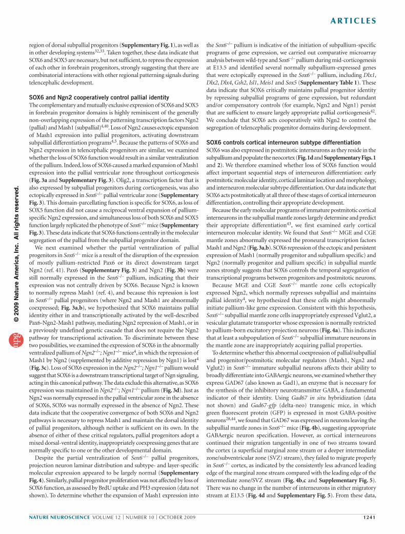
cortical invasion, we examined the laminar location and morphology of these interneurons as they populated the cortex. In Gad67-gfp mice at postnatal day 0 (P0; Fig. 5a), just after the interneurons have begun their radial migration into the maturing cortex, and at P14 (Fig. 5b), as they have more fully adopted their mature phenotypes, Sox6–/– interneurons preferentially populated deeper neocortical layers (Fig. 5c,d), without any change in their total numbers. Although the large majority of Sox6–/–
mice die perinatally, small numbers survive a few weeks postnatally, which allowed us to analyze them at P14 (ref. 32). Sox6+/– mice survived to adulthood, with a small number having occasional seizure behavior. We also examined the morphology of GAD67-GFP-positive neurons at P0 and found that Sox6–/–
we conclude that loss of SOX6 function perturbs the initial temporal segregation of progenitor-specific factors from postmitotic neurons, potentially causing their abnormal tangential migration, without affecting overall GABAergic neuron specification and abundance.
To further investigate whether the molecular and migratory irregularities at early stages of Sox6–/– subpallial neuron differentiation are associated with abnormalities in subsequent stages of interneuron
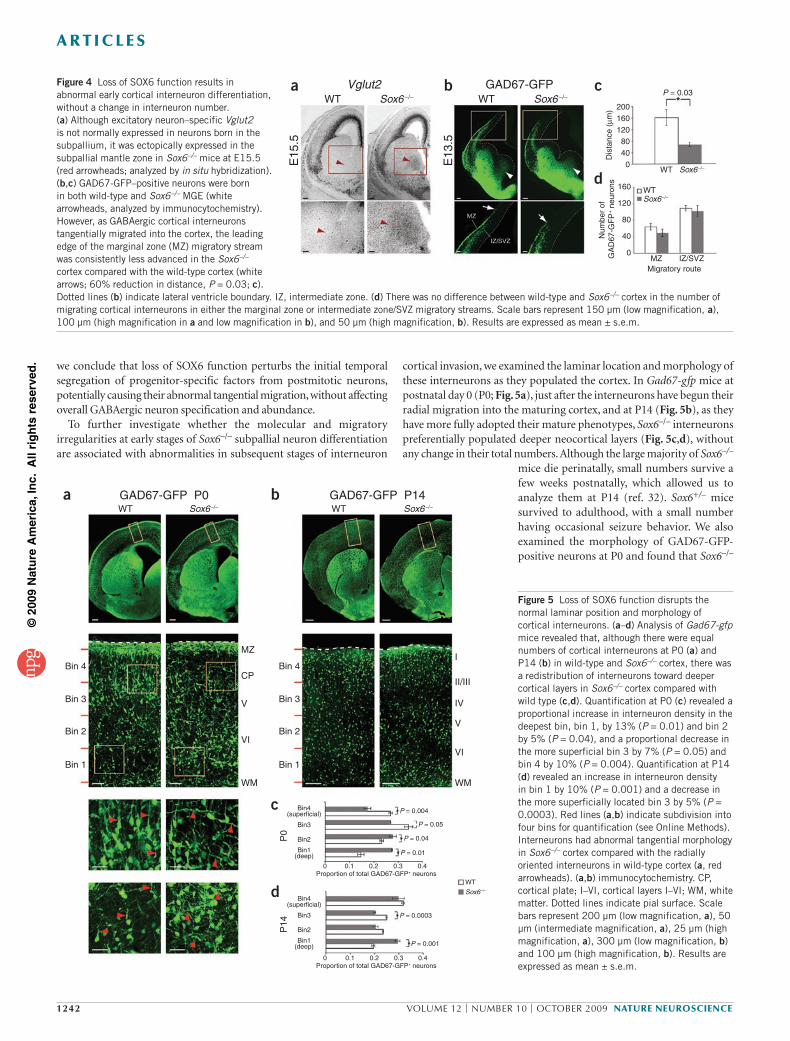
Figure 4 Loss of SOX6 function results in abnormal early cortical interneuron differentiation, without a change in interneuron number. (a) Although excitatory neuron–specific Vglut2 is not normally expressed in neurons born in the subpallium, it was ectopically expressed in the subpallial mantle zone in Sox6–/– mice at E15.5 (red arrowheads; analyzed by in situ hybridization). (b,c) GAD67-GFP–positive neurons were born in both wild-type and Sox6–/– MGE (white arrowheads, analyzed by immunocytochemistry). However, as GABAergic cortical interneurons tangentially migrated into the cortex, the leading edge of the marginal zone (MZ) migratory stream was consistently less advanced in the Sox6–/– cortex compared with the wild-type cortex (white arrows; 60% reduction in distance, P = 0.03; c). Dotted lines (b) indicate lateral ventricle boundary. IZ, intermediate zone. (d) There was no difference between wild-type and Sox6–/– cortex in the number of migrating cortical interneurons in either the marginal zone or intermediate zone/SVZ migratory streams. Scale bars represent 150 µm (low magnification, a), 100 µm (high magnification in a and low magnification in b), and 50 µm (high magnification, b). Results are expressed as mean ± s.e.m.
Figure 5 Loss of SOX6 function disrupts the normal laminar position and morphology of cortical interneurons. (a–d) Analysis of Gad67-gfp mice revealed that, although there were equal numbers of cortical interneurons at P0 (a) and P14 (b) in wild-type and Sox6–/– cortex, there was a redistribution of interneurons toward deeper cortical layers in Sox6–/– cortex compared with wild type (c,d). Quantification at P0 (c) revealed a proportional increase in interneuron density in the deepest bin, bin 1, by 13% (P = 0.01) and bin 2 by 5% (P = 0.04), and a proportional decrease in the more superficial bin 3 by 7% (P = 0.05) and bin 4 by 10% (P = 0.004). Quantification at P14 (d) revealed an increase in interneuron density in bin 1 by 10% (P = 0.001) and a decrease in the more superficially located bin 3 by 5% (P = 0.0003). Red lines (a,b) indicate subdivision into four bins for quantification (see Online Methods). Interneurons had abnormal tangential morphology in Sox6–/– cortex compared with the radially oriented interneurons in wild-type cortex (a, red arrowheads). (a,b) immunocytochemistry. CP, cortical plate; I–VI, cortical layers I–VI; WM, white matter. Dotted lines indicate pial surface. Scale bars represent 200 µm (low magnification, a), 50 µm (intermediate magnification, a), 25 µm (high magnification, a), 300 µm (low magnification, b) and 100 µm (high magnification, b). Results are expressed as mean ± s.e.m.
Vglut2 GAD67-GFPWT WTSox6 –/– Sox6 –/–
E15
.5
E13
.5
20016012080400
160
120
80
40
0
P = 0.03
WTSox6–/–
Sox6–/–
MZ
WT
IZ/SVZ
a b c
d
Dis
tanc
e (µ
m)
Num
ber
of
GA
D67
-GF
P+ n
euro
ns
Migratory route
a b
c
d
Bin 4
Bin 3
Bin 2
Bin 1
Bin 4
Bin 3
Bin 2
Bin 1
MZI
II/III
IV
V
VI
WM
CP
V
VI
WM
Bin4(superficial)
Bin3
Bin2
Bin1(deep)
Bin4(superficial)
Bin3
Bin2
Bin1(deep)
P = 0.004
P = 0.0003
P = 0.001
P = 0.05
P = 0.04
P = 0.01
0 0.1 0.2 0.3 0.4Proportion of total GAD67-GFP+ neurons
0 0.1 0.2 0.3 0.4Proportion of total GAD67-GFP+ neurons
WT
Sox6–/–
WT Sox6–/– Sox6–/–WTGAD67-GFP P0 GAD67-GFP P14
P0
P14
©20
09 N
atu
re A
mer
ica,
Inc.
All
rig
hts
res
erve
d.
nature neuroscience volume 12 | number 10 | oCTober 2009 1243
a r t i c l e s
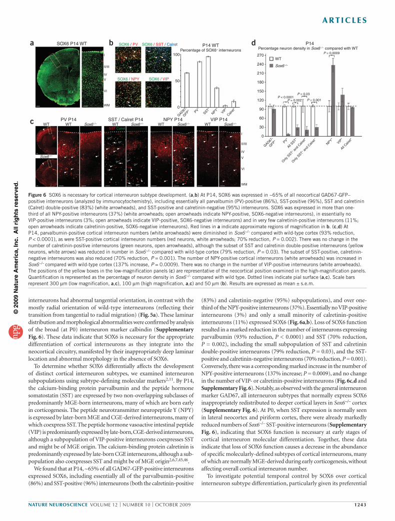
(83%) and calretinin-negative (95%) subpopulations), and over one-third of the NPY-positive interneurons (37%). Essentially no VIP-positive interneurons (3%) and only a small minority of calretinin-positive interneurons (11%) expressed SOX6 (Fig. 6a,b). Loss of SOX6 function resulted in a marked reduction in the number of interneurons expressing parvalbumin (93% reduction, P < 0.0001) and SST (70% reduction, P = 0.002), including the small subpopulation of SST and calretinin double-positive interneurons (79% reduction, P = 0.03), and the SST-positive and calretinin-negative interneurons (70% reduction, P = 0.001). Conversely, there was a corresponding marked increase in the number of NPY-positive interneurons (137% increase; P = 0.0009), and no change in the number of VIP- or calretinin-positive interneurons (Fig. 6c,d and Supplementary Fig. 6). Notably, as observed with the general interneuron marker GAD67, all interneuron subtypes that normally express SOX6 inappropriately redistributed to deeper cortical layers in Sox6–/– cortex (Supplementary Fig. 6). At P0, when SST expression is normally seen in lateral neocortex and piriform cortex, there were already markedly reduced numbers of Sox6–/– SST-positive interneurons (Supplementary Fig. 6), indicating that SOX6 function is necessary at early stages of cortical interneuron molecular differentiation. Together, these data indicate that loss of SOX6 function causes a decrease in the abundance of specific molecularly-defined subtypes of cortical interneurons, many of which are normally MGE-derived during early corticogenesis, without affecting overall cortical interneuron number.
To investigate potential temporal control by SOX6 over cortical interneuron subtype differentiation, particularly given its preferential
interneurons had abnormal tangential orientation, in contrast with the mostly radial orientation of wild-type interneurons (reflecting their transition from tangential to radial migration) (Fig. 5a). These laminar distribution and morphological abnormalities were confirmed by analysis of the broad (at P0) interneuron marker calbindin (Supplementary Fig. 6). These data indicate that SOX6 is necessary for the appropriate differentiation of cortical interneurons as they integrate into the neocortical circuitry, manifested by their inappropriately deep laminar location and abnormal morphology in the absence of SOX6.
To determine whether SOX6 differentially affects the development of distinct cortical interneuron subtypes, we examined interneuron subpopulations using subtype-defining molecular markers2,11. By P14, the calcium-binding protein parvalbumin and the peptide hormone somatostatin (SST) are expressed by two non-overlapping subclasses of predominantly MGE-born interneurons, many of which are born early in corticogenesis. The peptide neurotransmitter neuropeptide Y (NPY) is expressed by later-born MGE and CGE-derived interneurons, many of which coexpress SST. The peptide hormone vasoactive intestinal peptide (VIP) is predominantly expressed by late-born, CGE-derived interneurons, although a subpopulation of VIP-positive interneurons coexpresses SST and might be of MGE origin. The calcium- binding protein calretinin is predominantly expressed by late-born CGE interneurons, although a sub-population also coexpresses SST and might be of MGE origin2,6,7,45,46.
We found that at P14, ~65% of all GAD67-GFP-positive interneurons expressed SOX6, including essentially all of the parvalbumin-positive (86%) and SST-positive (96%) interneurons (both the calretinin-positive
Figure 6 SOX6 is necessary for cortical interneuron subtype development. (a,b) At P14, SOX6 was expressed in ~65% of all neocortical GAD67-GFP–positive interneurons (analyzed by immunocytochemistry), including essentially all parvalbumin (PV)-positive (86%), SST-positive (96%), SST and calretinin (Calret) double-positive (83%) (white arrowheads), and SST-positive and calretinin-negative (95%) interneurons. SOX6 was expressed in more than one-third of all NPY-positive interneurons (37%) (white arrowheads; open arrowheads indicate NPY-positive, SOX6-negative interneurons), in essentially no VIP-positive interneurons (3%; open arrowheads indicate VIP-positive, SOX6-negative interneurons) and in very few calretinin-positive interneurons (11%; open arrowheads indicate calretinin-positive, SOX6-negative interneurons). Red lines in a indicate approximate regions of magnification in b. (c,d) At P14, parvalbumin-positive cortical interneuron numbers (white arrowheads) were diminished in Sox6–/– compared with wild-type cortex (93% reduction, P < 0.0001), as were SST-positive cortical interneuron numbers (red neurons, white arrowheads; 70% reduction, P = 0.002). There was no change in the number of calretinin-positive interneurons (green neurons, open arrowheads), although the subset of SST and calretinin double-positive interneurons (yellow neurons, white arrows) was reduced in number in Sox6–/– compared with wild-type cortex (79% reduction, P = 0.03). The subset of SST-positive, calretinin-negative interneurons was also reduced (70% reduction, P = 0.001). The number of NPY-positive cortical interneurons (white arrowheads) was increased in Sox6–/– compared with wild-type cortex (137% increase, P = 0.0009). There was no change in the number of VIP-positive interneurons (white arrowheads). The positions of the yellow boxes in the low-magnification panels (c) are representative of the neocortical position examined in the high-magnification panels. Quantification is represented as the percentage of neuron density in Sox6–/– compared with wild type. Dotted lines indicate pial surface (a,c). Scale bars represent 300 µm (low magnification, a,c), 100 µm (high magnification, a,c) and 50 µm (b). Results are expressed as mean ± s.e.m.
a b d
c
SOX6 P14 WT P14 WT P14
PV P14 SST / Calret P14 NPY P14 VIP P14
SOX6 / PV
SOX6 / NPY
SOX6 / SST / Calret
SOX6 / VIP
Percentage of SOX6+ interneurons Percentage neuron density in Sox6 –/– compared with WT
100
50
0
GAD67-
GFP+
PV+
SST+
NPY+
VIP+
Calret+
GAD67-
GFP+
PV+
NPY+
VIP+
All SST
+
Only S
ST+ a
nd C
alret+
All Calr
et+
Only S
ST+ a
nd C
alret–
WT WT Sox6–/– WT Sox6–/– WT Sox6–/– WT Sox6–/–
Sox6–/–
I
I
II/III
P < 0.0001P = 0.03
P = 0.001
P = 0.0009
P = 0.002
IV
V
VI
WM
II/III
IV
V
VI
WM
SST Calret
270
240
210
180
150
120
90
60
30
0
WT
Sox6 –/–
©20
09 N
atu
re A
mer
ica,
Inc.
All
rig
hts
res
erve
d.
1244 volume 12 | number 10 | oCTober 2009 nature neuroscience
a r t i c l e s
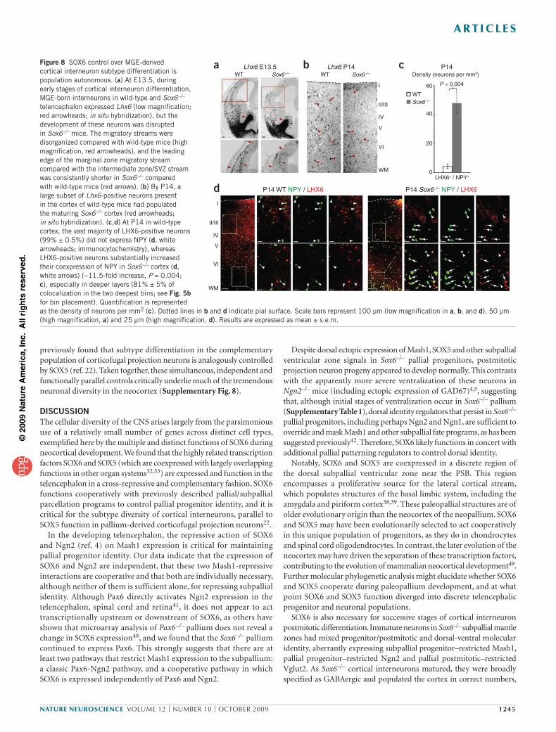
positive neurons in Sox6–/– cortex (35% reduction, P = 0.04), most LHX6-positive interneurons populated the cortex (Fig. 8b). LHX6 was not ectopically expressed in neurons born from abnormally ventralized Sox6–/– pallial progenitors (Fig. 8a) or in Sox6–/– CGE (data not shown), indicating that, as in the normal brain, LHX6-positive neurons in Sox6–/– cortex are MGE-derived.
To determine whether the abnormally abundant NPY-positive interneurons arise from the Sox6–/– MGE population itself (population autonomous, rather than a result of changes outside of this population), we investigated whether there was an increase in the number of MGE-derived LHX6-positive interneurons that express NPY in Sox6–/– cortex. In wild-type cortex, very few LHX6-positive neurons coexpressed NPY (1% ± 0.5% of LHX6-positive neurons), whereas the number that coexpressed NPY in Sox6–/– cortex markedly increased (23% ± 3% of LHX6-positive neurons; ~11.5-fold increase, P = 0.004; Fig. 8c,d). Similarly, there was a very large increase in the number of NPY-positive neurons that immunocytochemically colabeled with LHX6 in Sox6–/– cortex (22% ± 3% of NPY-positive neurons) as compared with wild type (4% ± 2% of NPY-positive neurons). In addition, the large majority of these Sox6–/– LHX6 and NPY double-positive neurons were found in deep cortical layers (81% ± 5%), further suggesting they were of early-born MGE origin (Fig. 8d). Taken together, these interneuron subtype analyses indicate that SOX6 functions in a population autonomous manner, controlling the appropriate molecular differentiation of MGE-derived cortical interneuron subtypes.
An additional (although not mutually exclusive) potential explanation for the increase in the number of Sox6–/– NPY-positive interneurons is that they might be born from abnormally ventralized Sox6–/– pallial progenitors. However, our data indicate that Sox6–/– NPY-positive neurons were not pallium derived; LHX6-positive interneurons in Sox6–/– cortex did not express TBR1 (Supplementary Fig. 7), a transcription factor that is broadly expressed by pallium-derived pyramidal neurons through P14, and all of the NPY-positive neurons in Sox6–/– cortex expressed GAD67-GFP (Supplementary Fig. 7), which was not expressed by neurons born from partially ventralized Sox6–/– pallial progenitors (Fig. 4b).
In sum, we found that SOX6 is largely mutually exclusively expressed and cross-repressively interacts with highly related SOX5 during telencephalic development, critically controlling pallial progenitor identity and cortical interneuron differentiation and diversity. We
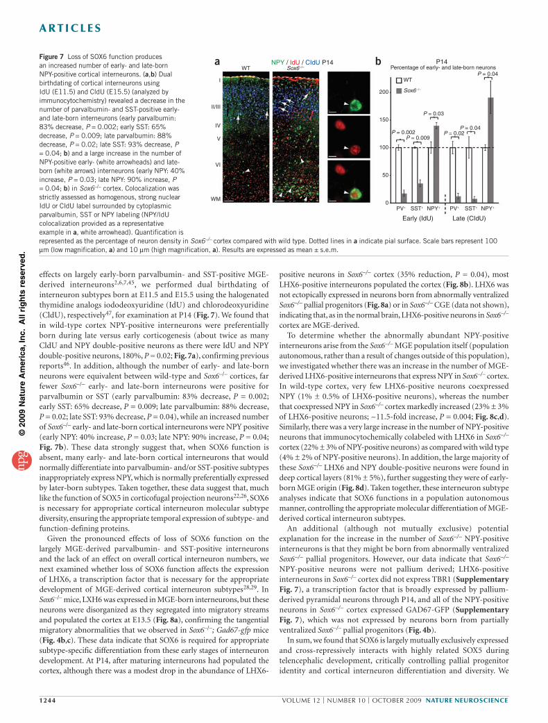
effects on largely early-born parvalbumin- and SST-positive MGE-derived interneurons2,6,7,45, we performed dual birthdating of interneuron subtypes born at E11.5 and E15.5 using the halogenated thymidine analogs iododeoxyuridine (IdU) and chlorodeoxyuridine (CldU), respectively47, for examination at P14 (Fig. 7). We found that in wild-type cortex NPY-positive interneurons were preferentially born during late versus early corticogenesis (about twice as many CldU and NPY double-positive neurons as there were IdU and NPY double- positive neurons, 180%, P = 0.02; Fig. 7a), confirming previous reports46. In addition, although the number of early- and late-born neurons were equivalent between wild-type and Sox6–/– cortices, far fewer Sox6–/– early- and late-born interneurons were positive for parvalbumin or SST (early parvalbumin: 83% decrease, P = 0.002; early SST: 65% decrease, P = 0.009; late parvalbumin: 88% decrease, P = 0.02; late SST: 93% decrease, P = 0.04), while an increased number of Sox6–/– early- and late-born cortical interneurons were NPY positive (early NPY: 40% increase, P = 0.03; late NPY: 90% increase, P = 0.04; Fig. 7b). These data strongly suggest that, when SOX6 function is absent, many early- and late-born cortical interneurons that would normally differentiate into parvalbumin- and/or SST-positive subtypes inappropriately express NPY, which is normally preferentially expressed by later-born subtypes. Taken together, these data suggest that, much like the function of SOX5 in corticofugal projection neurons22,26, SOX6 is necessary for appropriate cortical interneuron molecular subtype diversity, ensuring the appropriate temporal expression of subtype- and function-defining proteins.
Given the pronounced effects of loss of SOX6 function on the largely MGE-derived parvalbumin- and SST-positive interneurons and the lack of an effect on overall cortical interneuron numbers, we next examined whether loss of SOX6 function affects the expression of LHX6, a transcription factor that is necessary for the appropriate development of MGE-derived cortical interneuron subtypes28,29. In Sox6–/– mice, LXH6 was expressed in MGE-born interneurons, but these neurons were disorganized as they segregated into migratory streams and populated the cortex at E13.5 (Fig. 8a), confirming the tangential migratory abnormalities that we observed in Sox6–/–; Gad67-gfp mice (Fig. 4b,c). These data indicate that SOX6 is required for appropriate subtype-specific differentiation from these early stages of interneuron development. At P14, after maturing interneurons had populated the cortex, although there was a modest drop in the abundance of LHX6-
Figure 7 Loss of SOX6 function produces an increased number of early- and late-born NPY-positive cortical interneurons. (a,b) Dual birthdating of cortical interneurons using IdU (E11.5) and CldU (E15.5) (analyzed by immunocytochemistry) revealed a decrease in the number of parvalbumin- and SST-positive early- and late-born interneurons (early parvalbumin: 83% decrease, P = 0.002; early SST: 65% decrease, P = 0.009; late parvalbumin: 88% decrease, P = 0.02; late SST: 93% decrease, P = 0.04; b) and a large increase in the number of NPY-positive early- (white arrowheads) and late-born (white arrows) interneurons (early NPY: 40% increase, P = 0.03; late NPY: 90% increase, P = 0.04; b) in Sox6–/– cortex. Colocalization was strictly assessed as homogenous, strong nuclear IdU or CldU label surrounded by cytoplasmic parvalbumin, SST or NPY labeling (NPY/IdU colocalization provided as a representative example in a, white arrowhead). Quantification is represented as the percentage of neuron density in Sox6–/– cortex compared with wild type. Dotted lines in a indicate pial surface. Scale bars represent 100 µm (low magnification, a) and 10 µm (high magnification, a). Results are expressed as mean ± s.e.m.
a b
I
II/III
IV
V
VI
WM
WT Sox6 –/–NPY / IdU / CIdU P14 P14
Early (IdU) Late (CIdU)
WT
PV+ SST+ NPY+ PV+ SST+ NPY+
Sox6–/–
Percentage of early- and late-born neurons
200
150
100
50
0
P = 0.04
P = 0.04P = 0.02
P = 0.03
P = 0.009P = 0.002
©20
09 N
atu
re A
mer
ica,
Inc.
All
rig
hts
res
erve
d.
nature neuroscience volume 12 | number 10 | oCTober 2009 1245
a r t i c l e s
Despite dorsal ectopic expression of Mash1, SOX5 and other subpallial ventricular zone signals in Sox6–/– pallial progenitors, postmitotic projection neuron progeny appeared to develop normally. This contrasts with the apparently more severe ventralization of these neurons in Ngn2–/– mice (including ectopic expression of GAD67)4,5, suggesting that, although initial stages of ventralization occur in Sox6–/– pallium (Supplementary Table 1), dorsal identity regulators that persist in Sox6–/– pallial progenitors, including perhaps Ngn2 and Ngn1, are sufficient to override and mask Mash1 and other subpallial fate programs, as has been suggested previously42. Therefore, SOX6 likely functions in concert with additional pallial patterning regulators to control dorsal identity.
Notably, SOX6 and SOX5 are coexpressed in a discrete region of the dorsal subpallial ventricular zone near the PSB. This region encompasses a proliferative source for the lateral cortical stream, which populates structures of the basal limbic system, including the amygdala and piriform cortex38,39. These paleopallial structures are of older evolutionary origin than the neocortex of the neopallium. SOX6 and SOX5 may have been evolutionarily selected to act cooperatively in this unique population of progenitors, as they do in chondrocytes and spinal cord oligodendrocytes. In contrast, the later evolution of the neocortex may have driven the separation of these transcription factors, contributing to the evolution of mammalian neocortical development49. Further molecular phylogenetic analysis might elucidate whether SOX6 and SOX5 cooperate during paleopallium development, and at what point SOX6 and SOX5 function diverged into discrete telencephalic progenitor and neuronal populations.
SOX6 is also necessary for successive stages of cortical interneuron postmitotic differentiation. Immature neurons in Sox6–/– subpallial mantle zones had mixed progenitor/postmitotic and dorsal-ventral molecular identity, aberrantly expressing subpallial progenitor–restricted Mash1, pallial progenitor–restricted Ngn2 and pallial postmitotic–restricted Vglut2. As Sox6–/– cortical interneurons matured, they were broadly specified as GABAergic and populated the cortex in correct numbers,
previously found that subtype differentiation in the complementary population of corticofugal projection neurons is analogously controlled by SOX5 (ref. 22). Taken together, these simultaneous, independent and functionally parallel controls critically underlie much of the tremendous neuronal diversity in the neocortex (Supplementary Fig. 8).
DISCUSSIONThe cellular diversity of the CNS arises largely from the parsimonious use of a relatively small number of genes across distinct cell types, exemplified here by the multiple and distinct functions of SOX6 during neocortical development. We found that the highly related transcription factors SOX6 and SOX5 (which are coexpressed with largely overlapping functions in other organ systems32,33) are expressed and function in the telencephalon in a cross-repressive and complementary fashion. SOX6 functions cooperatively with previously described pallial/ subpallial parcellation programs to control pallial progenitor identity, and it is critical for the subtype diversity of cortical interneurons, parallel to SOX5 function in pallium-derived corticofugal projection neurons22.
In the developing telencephalon, the repressive action of SOX6 and Ngn2 (ref. 4) on Mash1 expression is critical for maintaining pallial progenitor identity. Our data indicate that the expression of SOX6 and Ngn2 are independent, that these two Mash1-repressive interactions are cooperative and that both are individually necessary, although neither of them is sufficient alone, for repressing subpallial identity. Although Pax6 directly activates Ngn2 expression in the telencephalon, spinal cord and retina41, it does not appear to act transcriptionally upstream or downstream of SOX6, as others have shown that microarray analysis of Pax6–/– pallium does not reveal a change in SOX6 expression48, and we found that the Sox6–/– pallium continued to express Pax6. This strongly suggests that there are at least two pathways that restrict Mash1 expression to the subpallium: a classic Pax6-Ngn2 pathway, and a cooperative pathway in which SOX6 is expressed independently of Pax6 and Ngn2.
Figure 8 SOX6 control over MGE-derived cortical interneuron subtype differentiation is population autonomous. (a) At E13.5, during early stages of cortical interneuron differentiation, MGE-born interneurons in wild-type and Sox6–/– telencephalon expressed Lhx6 (low magnification; red arrowheads; in situ hybridization), but the development of these neurons was disrupted in Sox6–/– mice. The migratory streams were disorganized compared with wild-type mice (high magnification, red arrowheads), and the leading edge of the marginal zone migratory stream compared with the intermediate zone/SVZ stream was consistently shorter in Sox6–/– compared with wild-type mice (red arrows). (b) By P14, a large subset of Lhx6-positive neurons present in the cortex of wild-type mice had populated the maturing Sox6–/– cortex (red arrowheads; in situ hybridization). (c,d) At P14 in wild-type cortex, the vast majority of LHX6-positive neurons (99% ± 0.5%) did not express NPY (d, white arrowheads; immunocytochemistry), whereas LHX6-positive neurons substantially increased their coexpression of NPY in Sox6–/– cortex (d, white arrows) (~11.5-fold increase, P = 0.004; c), especially in deeper layers (81% ± 5% of colocalization in the two deepest bins; see Fig. 5b for bin placement). Quantification is represented as the density of neurons per mm2 (c). Dotted lines in b and d indicate pial surface. Scale bars represent 100 µm (low magnification in a, b, and d), 50 µm (high magnification, a) and 25 µm (high magnification, d). Results are expressed as mean ± s.e.m.
Lhx6 E13.5
P14 WT NPY / LHX6 P14 Sox6 –/– NPY / LHX6
Lhx6 P14 P14WT Sox6 –/– WT Density (neurons per mm2)
LHX6+ / NPY+
WT
Sox6 –/–
Sox6 –/–
60
40
20
0
I
II/III
IV
V
VI
WM
I
II/III
IV
V
VI
WM
a
d
b c
P = 0.004
©20
09 N
atu
re A
mer
ica,
Inc.
All
rig
hts
res
erve
d.
1246 volume 12 | number 10 | oCTober 2009 nature neuroscience
a r t i c l e s
NPY, parvalbumin, SST or other major cortical interneuron subtype molecular markers. This suggests that some Sox6–/– MGE-derived interneurons might stall during later stages of subtype differentiation. In addition, these data do not exclude the possibility that SOX6 also functions in the population autonomous subtype differentiation of SOX6-positive CGE-derived cortical interneurons, or perhaps via additional non–population autonomous pathways.
NKX2-1 is a transcription factor that acts upstream of LHX6 (ref. 30), and was recently shown to be critical for multiple stages of cortical interneuron development, including the temporal fate specification of cortical interneuron subtypes31,50. Loss of NKX2-1 function results in a reduction in the number of parvalbumin- and SST-positive cortical interneurons and a corresponding increase in the number of VIP- and calretinin-positive interneurons. Given our results, SOX6 might be functioning, at least partially, in the postmitotic downstream execution of NKX2-1 signaling, potentially interacting with LHX6 (refs. 28,29), thereby regulating the temporal pacing of MGE-derived cortical interneuron fate specification and differentiation (Supplementary Fig. 8). Additional gain- and loss-of-function analyses might reveal potential functional interactions between these transcription factors during interneuron subtype specification and differentiation.
Much like pallium-born projection neuron subtypes, cortical interneuron subtype identity is largely determined by the time of birth. Fate-mapping experiments using H3-thymidine labeling and, more recently, genetic tools investigating subtype specification in MGE-born interneurons have shown that SST-positive interneurons, which are diminished in number in Sox6–/– cortex, are on average born at earlier stages of corticogenesis, whereas NPY-, VIP- and calretinin-positive interneurons, whose numbers are either increased or maintained in Sox6–/– cortex, are born later7,31,45,46. These data raise the hypothesis that, during cortical interneuron development, SOX6 participates in setting the pace for the proper timing of developmental transitions. In this model, loss of SOX6 might result in premature differentiation into neurons that are normally born at later developmental stages, at the expense of those born at earlier stages. Supporting this interpretation, our dual IdU/CldU birthdating analysis of the molecular differentiation of early- and late-born neurons in Sox6–/– cortex revealed that early-born neurons aberrantly differentiated and expressed the later-born subtype-defining protein NPY. As the lineage relationships of particular cortical interneuron subtypes are further clarified, it will be possible to discern whether loss of SOX6 function alters temporal development in a lineage (for example, those that would normally be SST-positive neurons aberrantly differentiate into NPY-positive neurons born later from potentially the same lineage) and/or whether loss of SOX6 function results in inappropriate differentiation across lineages (for example, those that would normally be parvalbumin-positive neurons aberrantly differentiate into NPY-positive neurons born later from a potentially distinct lineage).
We recently reported that the loss of SOX5 function in pallium-derived corticofugal projection neurons results in the premature adoption of subcerebral projection neuron features that are characteristic of later stages of cortical projection neuron development22. Therefore, it is possible that SOX6 and SOX5 both suppress coordinately regulated controls that promote premature transition into later stages of subtype differentiation. Consistent with this interpretation are the largely redundant roles of both SOX6 and SOX5 in chondroblasts during cartilage development and in oligodendroglial progenitors in the spinal cord in preventing the premature transition of these cell types to subsequent stages of development32,33. Given their analogous loss-of-function phenotypes, it is interesting to speculate that SOX6 and SOX5 separated in function during the evolution of the increasingly complex neuronal diversity of the telencephalon, and assumed complementary, but distinct, roles.
but finer molecular analysis revealed aberrant subtype differentiation, as exemplified by MGE-born cortical interneuron populations. In the absence of SOX6 function, there was a large increase in the abundance of NPY-positive interneurons at the expense of parvalbumin- and SST-positive interneurons, revealing abnormal subtype-defining neurotransmitter/molecular identity, one of multiple core contributing factors to the overall subtype identity and function of a neuron. Additional morphological and electrophysiological analyses might further examine whether these aberrant NPY-positive interneurons fully adopt functions that are normally associated with NPY expression.
Three potential (and not mutually exclusive) processes might account for the loss of Sox6–/– cortical interneuron molecular subtype diversity, without an overall reduction of interneuron number. One possibility is that population autonomous subtype specification is primarily affected, such that MGE-derived interneurons that would normally differentiate into subtypes that express parvalbumin and/or SST abnormally differentiate and express NPY. Another possibility is that MGE-born interneurons that normally would have been positive for either or both of parvalbumin and SST might selectively not populate the cortex, and CGE progenitors might simultaneously increase their NPY-positive interneuron output. A third, similar possibility is that abnormally partially ventralized Sox6–/– pallial progenitors are a source of these new NPY-positive neurons, which populate the cortex in place of MGE-born interneurons.
Our data very strongly favor the first interpretation of SOX6 control over population autonomous subtype differentiation. First, there was a very large increase in the number of MGE-derived LHX6-positive interneurons that expressed NPY concomitant with their loss of parvalbumin and SST expression. Second, our birthdating analysis revealed that, although the overall numbers of both early- and late-born neurons were unaffected by the loss of SOX6 function, a large number of early-born interneurons, which tend to arise from the MGE rather than the CGE, did not express parvalbumin or SST, but instead inappropriately expressed NPY. Third, abnormal molecular identity in the Sox6–/– MGE mantle zone (Ngn2, Mash1, Vglut2), observed as soon as the interneurons were born, strongly suggests a population autonomous effect of SOX6 function very early in neuronal differentiation. Fourth, there was no evidence of a substantial increase in the number of GAD67-GFP–positive migrating interneurons originating from Sox6–/– CGE, or any from the pallium, that would be required to compensate for the hypothetical loss of MGE-born interneurons (predicted for the second and third possibilities listed above). Fifth, regarding the second possibility, that the NPY-positive neurons were all CGE-derived, it is neither likely nor supported by any of the data that Sox6–/– CGE progenitor populations would increase their neurogenic rate in the absence of SOX6, as SOX6 is not normally expressed in CGE ventricular zone progenitors. Finally, regarding the third possibility, that the NPY-positive neurons were pallium derived, in Sox6–/– cortex, all of the NPY-positive neurons expressed GAD67, which was not ectopically expressed in Sox6–/– pallium-born neurons, and they did not express TBR1, which is broadly expressed by pallium-derived projection neurons, indicating that the NPY-positive neurons were not born from pallial progenitors. Taken together, these results reinforce previous findings on the population autonomous functions of SOX6 both in and outside of the nervous system32,33, indicating that SOX6 functions as a critical control over the appropriate molecular differentiation of MGE-derived cortical interneuron subtypes.
Additional interneuron developmental deficits might be occurring in the absence of SOX6 function. Although all of the LHX6-positive neurons in Sox6–/– cortex expressed GAD67-GFP, indicating their broad differentiation into GABAergic interneurons, some did not express
©20
09 N
atu
re A
mer
ica,
Inc.
All
rig
hts
res
erve
d.
nature neuroscience volume 12 | number 10 | oCTober 2009 1247
a r t i c l e s
17. Lewis, D.A. GABAergic local circuit neurons and prefrontal cortical dysfunction in schizophrenia. Brain Res. Brain Res. Rev. 31, 270–276 (2000).
18. Arlotta, P. et al. Neuronal subtype–specific genes that control corticospinal motor neuron development in vivo. Neuron 45, 207–221 (2005).
20. Chen, B., Schaevitz, L.R. & McConnell, S.K. Fezl regulates the differentiation and axon targeting of layer 5 subcortical projection neurons in cerebral cortex. Proc. Natl. Acad. Sci. USA 102, 17184–17189 (2005).
21. Chen, J.G., Rasin, M.R., Kwan, K.Y. & Sestan, N. Zfp312 is required for subcortical axonal projections and dendritic morphology of deep-layer pyramidal neurons of the cerebral cortex. Proc. Natl. Acad. Sci. USA 102, 17792–17797 (2005).
22. Lai, T. et al. SOX5 controls the sequential generation of distinct corticofugal neuron subtypes. Neuron 57, 232–247 (2008).
23. Alcamo, E.A. et al. Satb2 regulates callosal projection neuron identity in the develop-ing cerebral cortex. Neuron 57, 364–377 (2008).
24. Britanova, O. et al. Satb2 is a postmitotic determinant for upper-layer neuron speci-fication in the neocortex. Neuron 57, 378–392 (2008).
25. Joshi, P.S. et al. Bhlhb5 regulates the postmitotic acquisition of area identities in layers II–V of the developing neocortex. Neuron 60, 258–272 (2008).
26. Kwan, K.Y. et al. SOX5 postmitotically regulates migration, postmigratory differentia-tion and projections of subplate and deep-layer neocortical neurons. Proc. Natl. Acad. Sci. USA 105, 16021–16026 (2008).
27. Cobos, I. et al. Mice lacking Dlx1 show subtype-specific loss of interneurons, reduced inhibition and epilepsy. Nat. Neurosci. 8, 1059–1068 (2005).
28. Liodis, P. et al. Lhx6 activity is required for the normal migration and specification of cortical interneuron subtypes. J. Neurosci. 27, 3078–3089 (2007).
29. Zhao, Y. et al. Distinct molecular pathways for development of telencephalic interneu-ron subtypes revealed through analysis of Lhx6 mutants. J. Comp. Neurol. 510, 79–99 (2008).
30. Du, T., Xu, Q., Ocbina, P.J. & Anderson, S.A. NKX2.1 specifies cortical interneuron fate by activating Lhx6. Development 135, 1559–1567 (2008).
31. Butt, S.J. et al. The requirement of Nkx2-1 in the temporal specification of cortical interneuron subtypes. Neuron 59, 722–732 (2008).
32. Smits, P. et al. The transcription factors L-Sox5 and Sox6 are essential for cartilage formation. Dev. Cell 1, 277–290 (2001).
33. Stolt, C.C. et al. SoxD proteins influence multiple stages of oligodendrocyte develop-ment and modulate SoxE protein function. Dev. Cell 11, 697–709 (2006).
34. Wegner, M. From head to toes: the multiple facets of Sox proteins. Nucleic Acids Res. 27, 1409–1420 (1999).
35. Wegner, M. & Stolt, C.C. From stem cells to neurons and glia: a Soxist’s view of neural development. Trends Neurosci. 28, 583–588 (2005).
36. Connor, F., Wright, E., Denny, P., Koopman, P. & Ashworth, A. The Sry-related HMG box-containing gene Sox6 is expressed in the adult testis and developing nervous system of the mouse. Nucleic Acids Res. 23, 3365–3372 (1995).
37. Narahara, M., Yamada, A., Hamada-Kanazawa, M., Kawai, Y. & Miyake, M. cDNA cloning of the Sry-related gene Sox6 from rat with tissue-specific expression. Biol. Pharm. Bull. 25, 705–709 (2002).
38. Puelles, L. et al. Pallial and subpallial derivatives in the embryonic chick and mouse telencephalon, traced by the expression of the genes Dlx-2, Emx-1, Nkx-2.1, Pax-6 and Tbr-1. J. Comp. Neurol. 424, 409–438 (2000).
39. Carney, R.S. et al. Cell migration along the lateral cortical stream to the developing basal telencephalic limbic system. J. Neurosci. 26, 11562–11574 (2006).
40. Ma, Q., Sommer, L., Cserjesi, P. & Anderson, D.J. Mash1 and neurogenin1 expression patterns define complementary domains of neuroepithelium in the developing CNS and are correlated with regions expressing notch ligands. J. Neurosci. 17, 3644–3652 (1997).
41. Scardigli, R., Baumer, N., Gruss, P., Guillemot, F. & Le Roux, I. Direct and concentra-tion-dependent regulation of the proneural gene Neurogenin2 by Pax6. Development 130, 3269–3281 (2003).
42. Britz, O. et al. A role for proneural genes in the maturation of cortical progenitor cells. Cereb. Cortex 16 Suppl 1, i138–i151 (2006).
43. Batista-Brito, R., Machold, R., Klein, C. & Fishell, G. Gene expression in cortical interneuron precursors is prescient of their mature function. Cereb. Cortex 18, 2306–2317 (2008).
44. Tamamaki, N. et al. Green fluorescent protein expression and colocalization with cal-retinin, parvalbumin and somatostatin in the GAD67-GFP knock-in mouse. J. Comp. Neurol. 467, 60–79 (2003).
45. Cavanagh, M.E. & Parnavelas, J.G. Development of somatostatin immunoreactive neurons in the rat occipital cortex: a combined immunocytochemical-autoradiographic study. J. Comp. Neurol. 268, 1–12 (1988).
46. Cavanagh, M.E. & Parnavelas, J.G. Development of neuropeptide Y (NPY) immu-noreactive neurons in the rat occipital cortex: a combined immunohistochemical-autoradiographic study. J. Comp. Neurol. 297, 553–563 (1990).
47. Vega, C.J. & Peterson, D.A. Stem cell proliferative history in tissue revealed by temporal halogenated thymidine analog discrimination. Nat. Methods 2, 167–169 (2005).
48. Holm, P.C. et al. Loss- and gain-of-function analyses reveal targets of Pax6 in the developing mouse telencephalon. Mol. Cell. Neurosci. 34, 99–119 (2007).
49. Molnár, Z. & Butler, A.B. The corticostriatal junction: a crucial region for forebrain development and evolution. Bioessays 24, 530–541 (2002).
50. Nóbrega-Pereira, S. et al. Postmitotic Nkx2-1 controls the migration of telenceph-alic interneurons by direct repression of guidance receptors. Neuron 59, 733–745 (2008).
METHODSMethods and any associated references are available in the online version of the paper at http://www.nature.com/natureneuroscience/.
Note: Supplementary information is available on the Nature Neuroscience website.
ACKNOWLEDGMENTSWe thank K. Billmers, A. Palmer, L. Pasquina, K. Quinn, D. Schuback, E. Sievert, A. Wheeler and T. Yamamoto for superb technical assistance, G. Fishell, R. Batista-Brito, G. Miyoshi, P. Arlotta, B. Molyneaux, H. Padmanabhan, F. Guillemot, Q. Ma, C. Cepko and L. Goodrich for helpful discussions and input, U. Berger for technical assistance with in situ hybridization, C. Lois, R. Hevner, V. Lefebvre, F. Guillemot, V. Pachnis and Y. Yanagawa for generously sharing mice, antibodies and reagents, and current and past members of our laboratory for helpful suggestions. This work was partially supported by grants from the US National Institutes of Health (NS49553 and NS45523; additional infrastructure supported by NS41590), the Travis Roy Foundation, the Spastic Paraplegia Foundation, the Massachusetts Spinal Cord Injury research program, and the Harvard Stem Cell Institute to J.D.M., and by the Jane and Lee Seidman Fund for CNS Research, and the Emily and Robert Pearlstein Fund for Nervous System Repair. E.A. was partially supported by a US National Institutes of Health individual predoctoral National Research Service Award fellowship (F31 NS060421). D.J. was partially supported by fellowships from the Swiss National Science Foundation and the Holcim Foundation. R.M.F. was partially supported by a National Science Foundation Graduate Research Fellowship.
AUTHOR CONTRIBUTIONSE.A. and J.D.M. designed the overall experimental directions and specific analyses, and wrote and edited the manuscript. E.A. also performed all of the experiments and data analysis. D.J. co-performed the microarray experiments and assisted with interneuron quantification, microarray data evaluation, experimental design and data analysis, and manuscript writing and editing. R.M.F. performed whole-mount in situ hybridization/immunocytochemistry and assisted with BrdU/PH3 pallial progenitor analysis, microarray data evaluation, interneuron quantification, and manuscript editing. J.D.M. also contributed to data analysis and biological interpretation.
Published online at http://www.nature.com/natureneuroscience/.Reprints and permissions information is available online at http://www.nature.com/reprintsandpermissions/.
1. Schuurmans, C. & Guillemot, F. Molecular mechanisms underlying cell fate specifica-tion in the developing telencephalon. Curr. Opin. Neurobiol. 12, 26–34 (2002).
2. Wonders, C.P. & Anderson, S.A. The origin and specification of cortical interneurons. Nat. Rev. Neurosci. 7, 687–696 (2006).
3. Molyneaux, B.J., Arlotta, P., Menezes, J.R. & Macklis, J.D. Neuronal subtype specifica-tion in the cerebral cortex. Nat. Rev. Neurosci. 8, 427–437 (2007).
4. Fode, C. et al. A role for neural determination genes in specifying the dorsoventral identity of telencephalic neurons. Genes Dev. 14, 67–80 (2000).
5. Parras, C.M. et al. Divergent functions of the proneural genes Mash1 and Ngn2 in the specification of neuronal subtype identity. Genes Dev. 16, 324–338 (2002).
6. Butt, S.J. et al. The temporal and spatial origins of cortical interneurons predict their physiological subtype. Neuron 48, 591–604 (2005).
7. Miyoshi, G., Butt, S.J., Takebayashi, H. & Fishell, G. Physiologically distinct temporal cohorts of cortical interneurons arise from telencephalic Olig2-expressing precursors. J. Neurosci. 27, 7786–7798 (2007).
8. Flames, N. et al. Delineation of multiple subpallial progenitor domains by the com-binatorial expression of transcriptional codes. J. Neurosci. 27, 9682–9695 (2007).
9. Wonders, C.P. et al. A spatial bias for the origins of interneuron subgroups within the medial ganglionic eminence. Dev. Biol. 314, 127–136 (2008).
10. Fogarty, M. et al. Spatial genetic patterning of the embryonic neuroepithelium generates GABAergic interneuron diversity in the adult cortex. J. Neurosci. 27, 10935–10946 (2007).
11. Ascoli, G.A. et al. Petilla terminology: nomenclature of features of GABAergic interneu-rons of the cerebral cortex. Nat. Rev. Neurosci. 9, 557–568 (2008).
12. Flames, N. & Marin, O. Developmental mechanisms underlying the generation of cortical interneuron diversity. Neuron 46, 377–381 (2005).
13. Corbin, J.G., Nery, S. & Fishell, G. Telencephalic cells take a tangent: non-radial migra-tion in the mammalian forebrain. Nat. Neurosci. 4 Suppl, 1177–1182 (2001).
14. Levitt, P., Eagleson, K.L. & Powell, E.M. Regulation of neocortical interneuron develop-ment and the implications for neurodevelopmental disorders. Trends Neurosci. 27, 400–406 (2004).
15. Armijo, J.A., Valdizan, E.M., De Las Cuevas, I. & Cuadrado, A. Rev. Neurol. Advances in the physiopathology of epileptogenesis: molecular aspects. 34, 409–429 (2002).
16. Rubenstein, J.L. & Merzenich, M.M. Model of autism: increased ratio of excitation/inhibition in key neural systems. Genes Brain Behav. 2, 255–267 (2003).
©20
09 N
atu
re A
mer
ica,
Inc.
All
rig
hts
res
erve
d.
a r t i c l e s
nature neuroscience doi :10.1038/nn.2387
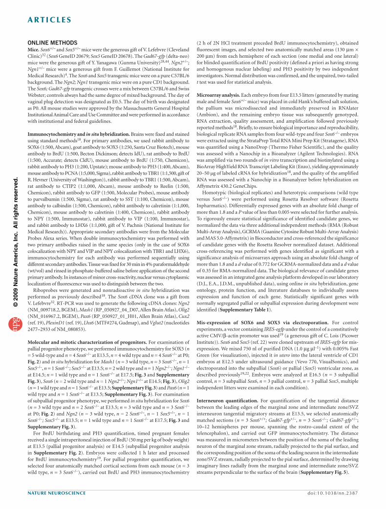
(2 h of 2N HCl treatment preceded BrdU immunocytochemistry), obtained fluorescent images, and selected two anatomically matched areas (130 µm × 200 µm) from each hemisphere of each section (one medial and one lateral) for blinded quantification of BrdU positivity (defined a priori as having strong and homogenous nuclear labeling) and PH3 positivity by two independent investigators. Normal distribution was confirmed, and the unpaired, two-tailed t test was used for statistical analysis.
Microarray analysis. Each embryo from four E13.5 litters (generated by mating male and female Sox6+/– mice) was placed in cold Hank’s buffered salt solution, the pallium was microdissected and immediately preserved in RNAlater (Ambion), and the remaining embryo tissue was subsequently genotyped. RNA extraction, quality assessment, and amplification followed previously reported methods18. Briefly, to ensure biological importance and reproducibility, biological replicate RNA samples from four wild-type and four Sox6–/– embryos were extracted using the StrataPrep Total RNA Mini Prep Kit (Stratagene), RNA was quantified using a NanoDrop (Thermo Fisher Scientific), and the quality was assessed with a Nanochip in a Bioanalyzer (Agilent Technologies). RNA was amplified via two rounds of in vitro transcription and biotinylated using a BioArray HighYield RNA Transcript Labeling Kit (Enzo), yielding approximately 20–50 µg of labeled cRNA for hybridization18, and the quality of the amplified RNA was assessed with a Nanochip in a Bioanalyzer before hybridization on Affymetrix 430.2 GeneChips.
Homotypic (biological replicates) and heterotypic comparisons (wild type versus Sox6–/–) were performed using Rosetta Resolver software (Rosetta Inpharmatics). Differentially expressed genes with an absolute fold change of more than 1.8 and a P value of less than 0.005 were selected for further analysis. To rigorously ensure statistical significance of identified candidate genes, we normalized the data via three additional independent methods (RMA (Robust Multi-Array Analysis), GCRMA (Guanine Cytosine Robust Multi-Array Analysis) and MAS 5.0-Affymetrix) in Bioconductor and cross-referenced the significance of candidate genes with the Rosetta Resolver normalized dataset. Additional cross-referencing was performed with genes identified as significant with a significance analysis of microarrays approach using an absolute fold change of more than 1.8 and a d value of 0.772 for GCRMA-normalized data and a d value of 0.35 for RMA-normalized data. The biological relevance of candidate genes was assessed in an integrated gene analysis platform developed in our laboratory (D.J., E.A., J.D.M., unpublished data), using online in situ hybridization, gene ontology, protein function, and literature databases to individually assess expression and function of each gene. Statistically significant genes with normally segregated pallial or subpallial expression during development were identified (Supplementary Table 1).
Mis-expression of SOX6 and SOX5 via electroporation. For control experiments, a vector containing IRES-egfp under the control of a constitutively active CMV/β-actin promoter was used19 (a generous gift of C. Lois (Picower Institute)). Sox6 and Sox5 (ref. 22) were cloned upstream of IRES-egfp for mis-expression. We mixed 750 nl of purified DNA (1.0 µg µl–1) with 0.005% Fast Green (for visualization), injected it in utero into the lateral ventricle of CD1 embryos at E12.5 under ultrasound guidance (Vevo 770, VisualSonics), and electroporated into the subpallial (Sox6) or pallial (Sox5) ventricular zone, as described previously19,22. Embryos were analyzed at E16.5 (n = 3 subpallial control, n = 3 subpallial Sox6, n = 3 pallial control, n = 3 pallial Sox5, multiple independent litters were examined in each condition).
Interneuron quantification. For quantification of the tangential distance between the leading edges of the marginal zone and intermediate zone/SVZ interneuron tangential migratory streams at E13.5, we selected anatomically matched sections (n = 3 Sox6+/+; Gad67-gfp+/–, n = 3 Sox6–/–; Gad67-gfp+/–; 10–12 hemispheres per mouse, spanning the rostro-caudal extent of the telencephalon), and carried out GFP immunocytochemistry. The distance was measured in micrometers between the position of the soma of the leading neuron of the marginal zone stream, radially projected to the pial surface, and the corresponding position of the soma of the leading neuron in the intermediate zone/SVZ stream, radially projected to the pial surface, determined by drawing imaginary lines radially from the marginal zone and intermediate zone/SVZ streams perpendicular to the surface of the brain (Supplementary Fig. 5).
ONLINE METHODSMice. Sox6+/– and Sox5+/– mice were the generous gift of V. Lefebvre (Cleveland Clinic)32 (Sox6 GeneID 20679; Sox5 GeneID 20678). The Gad67-gfp (delta-neo) mice were the generous gift of Y. Yanagawa (Gunma University)28,44. Ngn2+/–; Ngn1+/– mice were a generous gift from F. Guillemot (National Institute for Medical Research)4. The Sox6 and Sox5 transgenic mice were on a pure C57BL/6 background. The Ngn2; Ngn1 transgenic mice were on a pure CD1 background. The Sox6; Gad67-gfp transgenic crosses were a mix between C57BL/6 and Swiss Webster; controls always had the same degree of mixed background. The day of vaginal plug detection was designated as E0.5. The day of birth was designated as P0. All mouse studies were approved by the Massachusetts General Hospital Institutional Animal Care and Use Committee and were performed in accordance with institutional and federal guidelines.
Immunocytochemistry and in situ hybridization. Brains were fixed and stained using standard methods18. For primary antibodies, we used rabbit antibody to SOX6 (1:500, Abcam), goat antibody to SOX5 (1:250, Santa Cruz Biotech), mouse antibody to BrdU (1:500, Becton Dickinson; detects IdU), rat antibody to BrdU (1:500, Accurate; detects CldU), mouse antibody to BrdU (1:750, Chemicon), rabbit antibody to PH3 (1:200, Upstate); mouse antibody to PH3 (1:400, Abcam), mouse antibody to PCNA (1:5,000, Sigma), rabbit antibody to TBR1 (1:1,500, gift of R. Hevner (University of Washington)), rabbit antibody to TBR1 (1:500, Abcam), rat antibody to CTIP2 (1:1,000, Abcam), mouse antibody to Reelin (1:500, Chemicon), rabbit antibody to GFP (1:500, Molecular Probes), mouse antibody to parvalbumin (1:500, Sigma), rat antibody to SST (1:100, Chemicon), mouse antibody to calbindin (1:500, Chemicon), rabbit antibody to calretinin (1:1,000, Chemicon), mouse antibody to calretinin (1:400, Chemicon), rabbit antibody to NPY (1:500, Immunostar), rabbit antibody to VIP (1:100, Immunostar), and rabbit antibody to LHX6 (1:1,000, gift of V. Pachnis (National Institute for Medical Research)). Appropriate secondary antibodies were from the Molecular Probes Alexa series. When double immunocytochemistry was performed with two primary antibodies raised in the same species (only in the case of SOX6 colocalization with NPY and VIP and NPY colocalization with TBR1 and LHX6), immunocytochemistry for each antibody was performed sequentially using different secondary antibodies. Tissue was fixed for 30 min in 4% paraformaldehyde (wt/vol) and rinsed in phosphate-buffered saline before application of the second primary antibody. In instances of minor cross-reactivity, nuclear versus cytoplasmic localization of fluorescence was used to distinguish between the two.
Riboprobes were generated and nonradioactive in situ hybridization was performed as previously described18. The Sox6 cDNA clone was a gift from V. Lefebvre32. RT-PCR was used to generate the following cDNA clones: Ngn2 (NM_009718.2, BGEM), Mash1 (RP_050927_04_D07, Allen Brain Atlas), Olig2 (NM_016967.2, BGEM), Pax6 (RP_050927_01_H01, Allen Brain Atlas), Cux2 (ref. 19), PlexinD1 (ref. 19), Lhx6 (MTF#274, Gudmap), and Vglut2 (nucleotides 2477–2933 of NM_080853).
Molecular and mitotic characterization of progenitors. For examination of pallial progenitor phenotype, we performed immunocytochemistry for SOX5 (n = 5 wild-type and n = 4 Sox6–/– at E13.5, n = 4 wild type and n = 4 Sox6–/– at P0; Fig. 2) and in situ hybridization for Mash1 (n = 3 wild type, n = 3 Sox6–/–, n = 1 Sox5–/–, n = 1 Sox6–/–; Sox5–/– at E13.5; n = 2 wild type and n = 1 Ngn2–/–; Ngn1–/– at E14.5; n = 1 wild type and n = 1 Sox6–/– at E17.5; Fig. 3 and Supplementary Fig. 3), Sox6 (n = 2 wild type and n = 1 Ngn2–/–; Ngn1–/– at E14.5; Fig. 3), Olig2 (n = 1 wild type and n = 1 Sox6–/– at E13.5; Supplementary Fig. 3) and Pax6 (n = 1 wild type and n = 1 Sox6–/– at E13.5; Supplementary Fig. 3). For examination of subpallial progenitor phenotype, we performed in situ hybridization for Sox6 (n = 3 wild type and n = 2 Sox6–/– at E13.5; n = 3 wild type and n = 3 Sox6–/– at P0; Fig. 2) and Ngn2 (n = 3 wild type, n = 2 Sox6–/–, n = 1 Sox5–/–, n = 1 Sox6–/–; Sox5–/– at E13.5; n = 1 wild type and n = 1 Sox6–/– at E17.5; Fig. 3 and Supplementary Fig. 3).
For BrdU birthdating and PH3 quantification, timed pregnant females received a single intraperitoneal injection of BrdU (50 mg per kg of body weight) at E13.5 (pallial progenitor analysis) or E14.5 (subpallial progenitor analysis in Supplementary Fig. 2). Embryos were collected 1 h later and processed for BrdU immunocytochemistry19. For pallial progenitor quantification, we selected four anatomically matched cortical sections from each mouse (n = 3 wild type, n = 3 Sox6–/–), carried out BrdU and PH3 immunocytochemistry
©20
09 N
atu
re A
mer
ica,
Inc.
All
rig
hts
res
erve
d.
a r t i c l e s
doi:10.1038/nn.2387 nature neuroscience
killed the mice at P14 by perfusion, genotyped them and prepared brains for immunocytochemistry. Anatomically matched sections were selected, and IdU, CldU, parvalbumin, SST and NPY immunocytochemistry was performed (IdU and parvalbumin, IdU and NPY: n = 2 wild type, n = 3 Sox6–/–; IdU and SST, CldU and parvalbumin, CldU and SST, CldU and NPY: n = 2 wild type, n = 2 Sox6–/–; 7–8 hemispheres per mouse spanning the rostro-caudal extent of the telencephalon). Digital boxes of fixed width were superimposed on the cortices adjacent to the PSB, and height was adjusted to extend from the top of the white matter to the cortical surface. Interneurons coexpressing SST, parvalbumin or NPY, and either IdU or CldU (defined a priori as having strong and homogenous nuclear labeling) were quantified (colabeling was defined as clear nuclear label surrounded by cytoplasmic parvalbumin, SST or NPY label; Fig. 7a) and density values were calculated on the basis of the area of the box in each image.
All quantification was performed on 50-µm vibratome sections using well-established modified stereological methods, beginning at the rostral limit of the corpus callosum and continuing caudally, skipping four sections between samples, so that no cell could be counted twice in an adjacent section. We used strict a priori criteria, whereby the entire soma of a cell needed to be present to be counted, which was effectively accomplished by counting with high numerical aperture optics in the central approximately 30 µm of the thick 50-µm sections, avoiding cut neurons present in the top or bottom approximately 10 µm of each section. All quantifications were blinded, normal distribution was confirmed, and the unpaired, two-tailed t test was used for statistical analysis. Welch corrections were performed in the rare instances when the s.d. of the two groups varied significantly. All results are expressed as the mean ± s.e.m.
Microscopy and image analysis. Tissue sections were viewed on a Nikon E1000 microscope equipped with an X-Cite 120 illuminator (EXFO), and images were collected and analyzed with Volocity image analysis software (Improvision, v4.0.1). Images were optimized for size, color, and contrast using Photoshop 7.0 (Adobe). Single plane fluorescence images for E13.5 interneuron quantification were obtained using the Volocity Grid Confocal microscopy system (Improvision). Images were collected at the approximate midpoint between the top and bottom planes of focus.
For quantification of interneuron number at E13.5, anatomically matched sections were selected (n = 3 Sox6+/+; Gad67-gfp+/–, n = 3 Sox6–/–; Gad67-gfp+/–, 10–12 hemispheres per mouse spanning the rostro-caudal extent of the telencephalon), GFP immunocytochemistry was performed, and Grid Confocal (Improvision) images were obtained of a single plane of GFP-positive neurons. Digital boxes of fixed area (marginal zone stream, 85 µm × 175 µm; intermediate zone/SVZ stream, 115 µm × 175 µm) were superimposed at predetermined anatomical landmarks at the base, middle and leading edge of each migratory stream of each hemisphere, and GFP-positive neurons were quantified (Supplementary Fig. 5).
For P0 quantification, anatomically matched sections were selected (n = 3 wild type, n = 3 Sox6–/–; 10–12 hemispheres per mouse spanning the rostro-caudal extent of the telencephalon), GFP immunocytochemistry was performed, digital boxes of fixed width were equally divided into four bins (see Fig. 5a) and superimposed on the cortices adjacent to the PSB of each hemisphere, and height was adjusted to extend from the bottom of layer VI to directly under the marginal zone (at P0, the marginal zone was still very interneuron dense and was therefore not included in the quantification). GFP-positive neurons were quantified, and density values were calculated on the basis of the area of the box in each image; densities are expressed as neurons per mm2. Laminar distributions were determined by dividing the proportion of neurons in each bin by the total number of neurons in all bins.
For P14 quantification, anatomically matched sections were selected (n = 3 wild type, n = 3 Sox6–/–; 8–12 hemispheres per mouse spanning the rostro-caudal extent of the telencephalon), and SOX6, GFP, parvalbumin, SST, NPY (n = 6 wild type, n = 6 Sox6–/–), VIP (n = 4 wild type, n = 4 Sox6–/–), calretinin, calbindin and LHX6 immunocytochemistry was performed. Digital boxes of fixed width were equally divided into four bins (see Fig. 5b), superimposed on the cortices adjacent to the PSB of each hemisphere and height was adjusted to extend from the top of the white matter to the cortical surface. The percentages of wild-type interneurons that expressed SOX6 (Fig. 6b) were calculated from the total numbers in all four bins. Density values and laminar distributions in wild-type and Sox6–/– were calculated as above.
For IdU and CldU interneuron birthdating, equimolar delivery of IdU (57.5 mg per kg) at E11.5 and CldU (42.5 mg per kg) at E15.5 was performed47. We
©20
09 N
atu
re A
mer
ica,
Inc.
All
rig
hts
res
erve
d.