Sound-aided recovery from and persistence against visual filling-in
11
Sound-aided recovery from and persistence against visual filling-in Bhavin R. Sheth a, * , Shinsuke Shimojo a,b a Computation and Neural Systems, California Institute of Technology, 139-74, Caltech, Pasadena, CA 91125, USA b Human and Information Science Laboratory, NTT Communication Science Laboratories, Atsugi, Kanagawa 243-0198, Japan Received 6 May 2003; received in revised form 26 February 2004 Abstract Disappearance phenomena, in which salient visual stimuli do not register consciously, have been known to occur. Recovery from such phenomena typically occurs through change in some visual attribute, such as increase in luminance contrast or stimulus duration. Thus far, there have been no reports of cross-modal modulation of disappearance phenomena. In particular, what effect a cross-modal attentional cue has on sensory suppression is unknown. Here, we show that an adapted, flickered visual target that is synchronous with a brief sound appears more vivid than a similarly adapted, otherwise identical, visual target that is offset in time by more than 200 ms from the auditory cue. We argue that the brief auditory stimuli momentarily boost the concurrent signal of the adapted visual stimulus at a site downstream of the visual adaptation, thus causing the transient recovery from the visual adap- tation. Repetitive visual cues cause significantly less recovery from visual adaptation than repetitive auditory cues, implying that there are functions a cross-modal cue can perform that a cue of the same modality cannot. Moreover repetitive auditory cues selectively prevent synchronous visual targets from undergoing visual adaptation. Ours is the first report of cross-modal modulation of a disappearance phenomenon. Ó 2004 Elsevier Ltd. All rights reserved. Keywords: Multisensory; Adaptation; Disappearance phenomena; Attention; Cross-modal 1. Introduction Disappearance phenomena are phenomena in which information is ignored owing not from a failure to activate retinal photoreceptors, but from explicit ‘eras- ing’ or lack of conscious encoding later in the visual system. There are several examples of disappearance phenomena, including binocular (Blake, 1989) and monocular (Andrews & Purves, 1997; Campbell, Glin- sky, Howel, Riggs, & Atkinson, 1973) rivalry, motion- induced blindness (Bonneh, Cooperman, & Sagi, 2001) and after-images that disappear and reappear (Bennet- Clark & Evans, 1963). In these phenomena, the stimulus elicits a reliable response at the earliest sensory stages, but is then suppressed at some higher level in the brain prior to reaching visual awareness. A physically sub- threshold stimulus is not consciously registered either, but, unlike disappearance phenomena, it fails to elicit a reliable response even at the earliest sensory stages (including, perhaps, retinal photoreceptors). Modula- tion of disappearance phenomena occurs at multiple levels of the nervous system, and sometimes it takes the form of a high-level change of a visual attribute. For instance, the abrupt appearance of a stimulus in one eye causes the stimulus presented in the other eye to disap- pear from consciousness (Wolfe, 1986), or the visual grouping of target items changes the dynamics of mo- tion-induced blindness (Bonneh et al., 2001). An even clearer demonstration of a difference in the brain’s re- sponse to an above-threshold stimulus not consciously registered, and that to a sub-threshold stimulus is if a stimulus of a different modality modulates the percep- tion of a disappearance phenomenon but fails to im- prove the detection of a sub-threshold stimulus. Visual adaptation, in particular Troxler fading, is a known disappearance phenomenon. After one gazes on a flickering colored object for a minute or so, it fades from perception and is filled in perceptually by the surround, which is of a different color (Shimojo & Ka- mitani, 2001; Troxler, 1804). Neuronal fatigue resulting from the over-stimulation of neurons in the lateral geniculate nucleus (LGN; Clarke & Belcher, 1962) and * Corresponding author. Tel.: +1-626-395-2358; fax: +1-626-792- 8583. E-mail address: [email protected] (B.R. Sheth). 0042-6989/$ - see front matter Ó 2004 Elsevier Ltd. All rights reserved. doi:10.1016/j.visres.2004.03.009 Vision Research 44 (2004) 1907–1917 www.elsevier.com/locate/visres
Transcript of Sound-aided recovery from and persistence against visual filling-in

Vision Research 44 (2004) 1907–1917
www.elsevier.com/locate/visres
Sound-aided recovery from and persistence against visual filling-in
Bhavin R. Sheth a,*, Shinsuke Shimojo a,b
a Computation and Neural Systems, California Institute of Technology, 139-74, Caltech, Pasadena, CA 91125, USAb Human and Information Science Laboratory, NTT Communication Science Laboratories, Atsugi, Kanagawa 243-0198, Japan
Received 6 May 2003; received in revised form 26 February 2004
Abstract
Disappearance phenomena, in which salient visual stimuli do not register consciously, have been known to occur. Recovery from
such phenomena typically occurs through change in some visual attribute, such as increase in luminance contrast or stimulus
duration. Thus far, there have been no reports of cross-modal modulation of disappearance phenomena. In particular, what effect a
cross-modal attentional cue has on sensory suppression is unknown. Here, we show that an adapted, flickered visual target that is
synchronous with a brief sound appears more vivid than a similarly adapted, otherwise identical, visual target that is offset in time by
more than 200 ms from the auditory cue. We argue that the brief auditory stimuli momentarily boost the concurrent signal of the
adapted visual stimulus at a site downstream of the visual adaptation, thus causing the transient recovery from the visual adap-
tation. Repetitive visual cues cause significantly less recovery from visual adaptation than repetitive auditory cues, implying that
there are functions a cross-modal cue can perform that a cue of the same modality cannot. Moreover repetitive auditory cues
selectively prevent synchronous visual targets from undergoing visual adaptation. Ours is the first report of cross-modal modulation
of a disappearance phenomenon.
� 2004 Elsevier Ltd. All rights reserved.
Keywords: Multisensory; Adaptation; Disappearance phenomena; Attention; Cross-modal
1. Introduction
Disappearance phenomena are phenomena in which
information is ignored owing not from a failure toactivate retinal photoreceptors, but from explicit ‘eras-
ing’ or lack of conscious encoding later in the visual
system. There are several examples of disappearance
phenomena, including binocular (Blake, 1989) and
monocular (Andrews & Purves, 1997; Campbell, Glin-
sky, Howel, Riggs, & Atkinson, 1973) rivalry, motion-
induced blindness (Bonneh, Cooperman, & Sagi, 2001)
and after-images that disappear and reappear (Bennet-Clark & Evans, 1963). In these phenomena, the stimulus
elicits a reliable response at the earliest sensory stages,
but is then suppressed at some higher level in the brain
prior to reaching visual awareness. A physically sub-
threshold stimulus is not consciously registered either,
but, unlike disappearance phenomena, it fails to elicit a
reliable response even at the earliest sensory stages
* Corresponding author. Tel.: +1-626-395-2358; fax: +1-626-792-
8583.
E-mail address: [email protected] (B.R. Sheth).
0042-6989/$ - see front matter � 2004 Elsevier Ltd. All rights reserved.doi:10.1016/j.visres.2004.03.009
(including, perhaps, retinal photoreceptors). Modula-
tion of disappearance phenomena occurs at multiple
levels of the nervous system, and sometimes it takes the
form of a high-level change of a visual attribute. Forinstance, the abrupt appearance of a stimulus in one eye
causes the stimulus presented in the other eye to disap-
pear from consciousness (Wolfe, 1986), or the visual
grouping of target items changes the dynamics of mo-
tion-induced blindness (Bonneh et al., 2001). An even
clearer demonstration of a difference in the brain’s re-
sponse to an above-threshold stimulus not consciously
registered, and that to a sub-threshold stimulus is if astimulus of a different modality modulates the percep-
tion of a disappearance phenomenon but fails to im-
prove the detection of a sub-threshold stimulus.
Visual adaptation, in particular Troxler fading, is a
known disappearance phenomenon. After one gazes on
a flickering colored object for a minute or so, it fades
from perception and is filled in perceptually by the
surround, which is of a different color (Shimojo & Ka-mitani, 2001; Troxler, 1804). Neuronal fatigue resulting
from the over-stimulation of neurons in the lateral
geniculate nucleus (LGN; Clarke & Belcher, 1962) and
1908 B.R. Sheth, S. Shimojo / Vision Research 44 (2004) 1907–1917
the retinal ganglia (Kotulak & Schor, 1986; Millodot,
1967), two sites that lie upstream of striate cortex (V1),
is believed to underlie Troxler fading (cf. Dennett’s,
1991 criticism of the idea that there is a place in the
brain where consciousness happens as ‘‘vestigial Carte-
sianism’’). Of importance, there are some projections
from auditory centers to V1 (Falchier, Clavagnier, Ba-
rone, & Kennedy, 2002; Rockland & Ojima, 2002), butnone from auditory centers to the LGN or the retina.
Thus, if an auditory stimulus were to interact with
Troxler fading, it would have to be at a site downstream
of where the initial fade took place. An effect of sound
on Troxler fading would, therefore, be an example of
high-level modulation of a low-level disappearance
phenomenon. Although an auditory cue is known to
modulate visual salience (Stein, London, Wilkinson, &Price, 1996; Vroomen & de Gelder, 2000), disappearance
phenomena have never been studied in this regard.
Here, we report that repetitive auditory cues selecti-
vely enhance the visibility of synchronous visual targets,
preventing them from undergoing visual adaptation and
causing previously adapted targets to recover. Ours is
one of the first reports of cross-modal modulation of a
disappearance phenomenon.
2. Materials and methods
All stimuli were presented on a SONY Trinitron
monitor (75 Hz refresh; 37.5 cm · 30 cm viewing area)under control of a MAC G4 running MATLAB
(Mathworks Inc.) and Psychophysics Toolbox (Brai-
nard, 1997; Pelli, 1997). The observers sat comfortablyin a chair in front of the computer screen at a viewing
distance of 57 cm, with their heads in a chinrest (Han-
daya Co., Japan). Two loudspeakers (Altec Lansing
Satellite 31 speakers) were positioned 25 cm on either
side of the middle and at the same vertical level as the
visual display. Viewing was binocular. All experiments
were conducted in room illumination.
2.1. Adaptation recovery task
The fixation point (FP), located in the center of a
green screen (CIE coordinates, x ¼ 0:446, y ¼ 0:568,37.9 cd/m2), was a white circular dot (CIE coordinates,
x ¼ 0:313, y ¼ 0:329, 100.4 cd/m2, diameter¼ 12.4 arc-min). A red circular target (diameter¼ 5.0�, CIE coor-dinates, x ¼ 0:541, y ¼ 0:451, 34.7 cd/m2), centered 10.4�left or right of the FP, appeared at the beginning of each
trial. It lasted for 27 ms before being turned off. 133–293
ms (the actual time was randomly chosen from within
this range) following its offset, a second target, identicalin all respects to the first, appeared on the opposite side
of the FP (Fig. 1A). It too stayed on for 27 ms and then
turned off, followed, as before, by a random (133–293
ms) time interval after which the first target appeared
again in its location. The adaptation cycle of target
alternations lasted for 4 min. Each trial of the test phase
lasted 24 s (50 alternations) total, and began with a top-
up adaptation phase with the same cycle of alternations
between the two targets for 16.8 s (35 alternations).
After this, a click (2.2 ms duration, r.m.s. intensity¼ 71or 42 dBa SPL measured in fast mode by a Bruel andKjaer Microphone Amplifier and Meter, Model 2604;
sound intensity made no difference to the results, see
Sheth & Shimojo, 2002), occurred on both speakers, 14
ms after the onset of one of the targets, and the trial
continued for another 7.2 s (15 alternations) with the
sound synchronously presented with the chosen target.
On a given trial, the selection of which target for syn-
chronous presentation with the sound was random, butwas counter-balanced between left and right choices
across trials. Timing of sound and target were such that
the sound was presented while the target was present on
the screen. At trial’s end, the targets and sound were
turned off, and the observer had to report, in a self-
paced manner, whether the left or right target appeared
more salient after the sound came on. There were 30
trials total. The task typically took 20–25 min total.
2.2. Adaptation blocking task
On each trial, two targets, identical in stimulus
parameters, alternately flickered on until the participantresponded (same values as in the adaptation recovery
task). One of the targets was accompanied by a syn-
chronous sound (equal, non-zero amplitudes at DC and
11 kHZ frequencies, 2.2 ms duration, r.m.s. inten-
sity¼ 71 dBa SPL) from the beginning of the trial. Eachtarget stayed on for 27 ms during each cycle of adap-
tation, and there was a random 133–293 ms interval
between targets. The participant viewed the screen untilone of the targets (left or right) disappeared from per-
ception. The trial ended when he or she responded with
a keypress (left or right). There were 10 trials total. The
sound was synchronous with each (left/right) of the two
targets on 5/10 trials. A typical observer took 30 min to
complete the task.
3. Results
3.1. Transient auditory cues transiently focus exogenous
attention on visual stimuli nearby in time
Auditory–visual synchrony is usually the optimal
temporal setting for audio–visual interaction. Various
cross-modal effects peak at auditory–visual synchrony(Sekuler, Sekuler, & Lau, 1997; Shimojo, Miyauchi, &
Hikosaka, 1997). Absolute judgments of effect strength
are susceptible to non-sensory variables such as the
0.4
0.5
0.6
0.7
Sound → Flash
n = 4
***
-213 -106 -53 53 106 2130ms
FP
FP
FP
FP
Fra
ctio
n of
tria
ls in
whi
ch s
timul
us n
eare
r in
time
with
sou
nd a
ppea
red
mor
e sa
lient
n = 6
Chromatic contrast of visual stimulus
Low High
0.6
0.7
0.5
SOA (Flash - Sound)
Flash → Sound
**
Fra
ctio
n of
tria
ls in
whi
ch s
timul
us n
eare
r in
time
with
sou
nd a
ppea
red
mor
e sa
lient
A
B
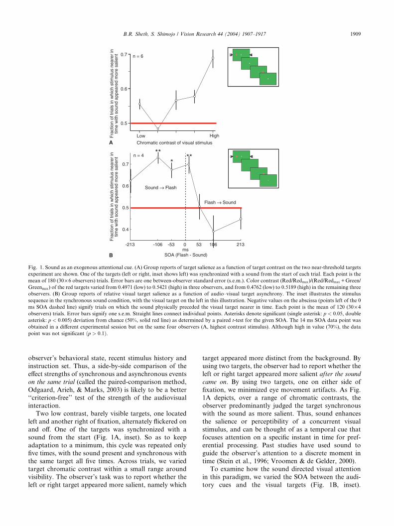
Fig. 1. Sound as an exogenous attentional cue. (A) Group reports of target salience as a function of target contrast on the two near-threshold targets
experiment are shown. One of the targets (left or right, inset shows left) was synchronized with a sound from the start of each trial. Each point is the
mean of 180 (30· 6 observers) trials. Error bars are one between-observer standard error (s.e.m.). Color contrast (Red/Redmax)/(Red/Redmax +Green/Greenmax) of the red targets varied from 0.4971 (low) to 0.5421 (high) in three observers, and from 0.4762 (low) to 0.5189 (high) in the remaining three
observers. (B) Group reports of relative visual target salience as a function of audio–visual target asynchrony. The inset illustrates the stimulus
sequence in the synchronous sound condition, with the visual target on the left in this illustration. Negative values on the abscissa (points left of the 0
ms SOA dashed line) signify trials on which the sound physically preceded the visual target nearer in time. Each point is the mean of 120 (30· 4observers) trials. Error bars signify one s.e.m. Straight lines connect individual points. Asterisks denote significant (single asterisk: p < 0:05, double
asterisk: p < 0:005) deviation from chance (50%, solid red line) as determined by a paired t-test for the given SOA. The 14 ms SOA data point wasobtained in a different experimental session but on the same four observers (A, highest contrast stimulus). Although high in value (70%), the data
point was not significant ðp > 0:1Þ.
B.R. Sheth, S. Shimojo / Vision Research 44 (2004) 1907–1917 1909
observer’s behavioral state, recent stimulus history and
instruction set. Thus, a side-by-side comparison of the
effect strengths of synchronous and asynchronous events
on the same trial (called the paired-comparison method,
Odgaard, Arieh, & Marks, 2003) is likely to be a better
‘‘criterion-free’’ test of the strength of the audiovisualinteraction.
Two low contrast, barely visible targets, one located
left and another right of fixation, alternately flickered on
and off. One of the targets was synchronized with a
sound from the start (Fig. 1A, inset). So as to keep
adaptation to a minimum, this cycle was repeated only
five times, with the sound present and synchronous with
the same target all five times. Across trials, we variedtarget chromatic contrast within a small range around
visibility. The observer’s task was to report whether the
left or right target appeared more salient, namely which
target appeared more distinct from the background. By
using two targets, the observer had to report whether the
left or right target appeared more salient after the sound
came on. By using two targets, one on either side of
fixation, we minimized eye movement artifacts. As Fig.
1A depicts, over a range of chromatic contrasts, theobserver predominantly judged the target synchronous
with the sound as more salient. Thus, sound enhances
the salience or perceptibility of a concurrent visual
stimulus, and can be thought of as a temporal cue that
focuses attention on a specific instant in time for pref-
erential processing. Past studies have used sound to
guide the observer’s attention to a discrete moment in
time (Stein et al., 1996; Vroomen & de Gelder, 2000).To examine how the sound directed visual attention
in this paradigm, we varied the SOA between the audi-
tory cues and the visual targets (Fig. 1B, inset).

1910 B.R. Sheth, S. Shimojo / Vision Research 44 (2004) 1907–1917
According to a stimulus-driven or exogenous attention
account based on studies of visual attention (Jonides,
1981), an exogenous cue has maximum impact on the
target if it precedes the target by no more than 50–100
ms. Assuming that exogenous cues of various modalities
all have maximum impact at similar timing, a auditory
cue preceding the visual target by 200 ms or less should
enhance visual salience the most (McDonald, Teder-S€alej€arvi, & Hillyard, 2000). On a second account,summation of auditory and visual signals enhances vi-
sual salience; so, the sound should have maximum im-
pact if it is synchronous with the visual target (0 SOA),
or, because auditory neural delays are known to be 50
ms shorter than visual (Goldstone & Lhamon, 1972;
Keele, 1986), if the sound follows the target by 50 ms.
On a third account, the sound is an endogenous, cog-nitive cue. Endogenous cues are typically slower because
top-down signals are slow, so the sound should have
maximum impact if it precedes the target by 200–400 ms
(Posner, 1978; Posner, Nissen, & Klein, 1976; Posner,
Nissen, & Ogden, 1978). Thus, SOA dependency pro-
vides a behavioral ‘‘fingerprint’’ for how auditory
attentional cues impact visual perception.
The data were consistent with the first account: theauditory cue maximally enhanced visual salience if the
sound preceded the visual target by 50–100 ms (Fig. 1B).
Contrary to the second account, if the sound followed
the target by 50–100 ms, it had little impact. Contrary to
the third account, if the sound preceded the target by
250 ms (or more), it decreased visual salience, if any-
thing. In sum, the sound behaved as an attentional cue
that exogenously cues attention to the visual targetappearing 50–100 ms later (McDonald et al., 2000;
Vroomen & de Gelder, 2000; Watanabe & Shimojo,
1998; see Driver & Spence, 1998 for a review).
3.2. Auditory cues cause previously adapted visual stimuli
synchronous with them to recover
Can an auditory stimulus that behaves as an exoge-
nous cue focusing visual attention on certain moments
in time modulate visual adaptation? Specifically, can
repetitive auditory cues cause a previously adapted
visual stimulus to recover from Troxler fading (i.e. de-
adapt the visual stimulus)? Endogenous visual atten-tional cues are known to enhance Troxler fading (Lou,
1999) but the effects of exogenous cues on Troxler fading
have not been explored.
From a multisensory perspective, it is known that an
auditory cue modulates visual salience. A visual target
interspersed among non-targets was more detectable if a
salient sound was played in approximate synchrony
(SOA<250 ms) with it (Vroomen & de Gelder, 2000).The target in Vroomen and de Gelder (2000) was an
object (four spots arranged at the corners of an imagi-
nary diamond); thus the auditory cue in their study
probably affected visual processing in high-level cortical
areas that process objects. In Stein et al. (1996), sound
enhanced the perceived brightness of a synchronous
foveal light; no comparison was made of the effects of
synchronous and non-synchronous sounds, however. V1
is a known neural correlate of the brightness percept
(Kinoshita & Komatsu, 2001; Rossi & Paradiso, 1999).
Thus, it seems that sound can modulate visual perceptsarising in cortex, including perhaps those that arise in
V1 (Odgaard et al., 2003 claimed that the enhancement
is eliminated if a paired-comparison method test is used
and if the sound accompanies the light on 25% and not
50% of trials; they proposed an alternative account
based on response bias). This claim is not surprising in
light of recent evidence for connections from auditory
centers to V1 (Bhattacharya, Shams, & Shimojo, 2002;Falchier et al., 2002; Giard & Peronnet, 1999; Rockland
& Ojima, 2002; Shams, Kamitani, Thompson, & Shim-
ojo, 2001). These findings beg the question of whether
sound can affect the processing in even lower levels of
the visual pathway. On the one hand, there are no
known projections from the auditory brain areas to the
LGN or the retina. On the other hand, the LGN receives
profuse feedback projections from V1 (Erisir, VanHorn, & Sherman, 1997; Montero, 1991; Murphy,
Duckett, & Sillito, 1999; Wilson, Friedlander, & Sher-
man, 1984), and V1 receives projections from auditory
areas. Thus, the auditory areasfiV1fiLGN polysyn-aptic pathway could be one route, albeit indirect, for
sound to affect visual processing in the LGN. Because
the LGN is a known neural correlate of Troxler fading
(Clarke & Belcher, 1962), studying the effects of soundon Troxler fading may help answer the question of
whether sound can effect processing in the LGN.
We employed near-threshold visual targets in an
experimental paradigm and tested for the effects of
sound on their perception. A target appeared momen-
tarily to the left or right of fixation (FP in Fig. 2A). A
random time (133–293 ms) following its offset, another
target appeared on the opposite side of fixation, fol-lowed, between 133 ms to 293 ms later, by the first
target. This adaptation cycle continued for 4 min, long
enough for the perception of the two targets to be
blurred and to be filled in by the background (Shimojo
& Kamitani, 2001). There was a seamless transition into
the test phase. Each trial of the test phase began with a
28 s long top-up adaptation phase. In the subsequent
‘‘sound on’’ phase of the trial, a clearly audible soundnearly synchronous with one of the two targets, was
played. This was repeated five times, with the sound
present and synchronous with the same target all five
times. The random nature of the time interval (266–486
ms) between two successive sounds prevented the ob-
server from ever acquiring prior knowledge of exactly
when the sound would be played. At trial’s end, the
observer had to report whether the left or right target

Fig. 2. Sound-induced recovery from visual adaptation (de-adaptation). (A) The sequence of stimuli and how the stimuli physically appear in the
adaptation experiment are shown. Two red targets––one on either side of fixation (FP, shown in white), flickered back and forth on a green
background in the adaptation phase (i). In the ‘‘sound on’’ phase that was at the end of each trial (ii), a sound was synchronously presented with one
of the targets (left target in the figure). (B) How the stimuli typically appear to the observer at the end of adaptation (i; the green background appears
to fill in), and in the ‘‘sound on’’ phase (ii; the left target that was accompanied in time by a sound appears more salient after the sound comes on).
Observers claimed that, on some trials, the targets disappeared entirely from their perception. (C) Individual reports of target salience for each of the
nine observers and the group mean in gray on the right is shown. The solid red line indicates chance performance (target synchronous with the sound
appears salient on 50% of trials), and the red dashed line indicates significance at the 0.05 level (sign test, 70% of trials).
B.R. Sheth, S. Shimojo / Vision Research 44 (2004) 1907–1917 1911
was more salient after the sound came on (paired-com-parison method). Salience was defined subjectively for
each observer––which target stood out more from the
background (i.e. distinctive in space and time from the
background) in terms of its flicker and red color. Having
two targets on opposite sides of fixation helped minimize
deviations of the eye from fixation.
Our observers typically found that, over the course of
adaptation, both targets gradually took on the color ofthe background i.e. the background filled in (Fig. 2B, i).
After the sound was turned on, perception changed: One
or both targets gradually became more salient from the
background (Fig. 2B, ii). Observers had to judge which
of the two targets appeared more salient from thebackground once the sound came on. They tended to
choose the target that was concurrent with the sound
(Fig. 2C, mean±one across-subject standard error:
84.3 ± 2.2% of all trials; also see Stein et al., 1996). This
preference was significantly above chance (Fig. 2C, 50%,
solid line) for each of the nine observers (p < 0:05, signtest for each. We nickname our effect the ‘‘beating heart
effect’’, as the sound is critical and the phenomenology isakin to that of a beating heart). Anecdotally, observers
reported that sometimes, only the synchronous flicker
was visible, and other times, both targets stood out from
the background but the one synchronous with the sound
1912 B.R. Sheth, S. Shimojo / Vision Research 44 (2004) 1907–1917
stood out first and more sharply. Still, the recovery from
filling-in was partial and transient. It never equaled the
visually guided recovery usually observed following an
overt shift in eye position and consequent refreshing of
the retinal image.
3.3. Response bias?
A critical issue is whether the de-adaptation was a
genuine perceptual effect or whether it was due to re-
sponse bias, namely that the sound did not affect per-
ception but rather biased observers to respond for the
light concurrent with the sound. That is to say, thesound merely affected post-perceptual decision pro-
cesses, and not attention or perception.
It is necessary––but not sufficient––that the targets be
clearly visible and temporally distinct so that the ob-
server’s post-perceptual mechanisms can match the
sound with the visible target synchronous with it. We
therefore conducted a new experiment to minimize the
effect of response bias by ensuring that the targets werenot visible following the adaptation. In order to achieve
this, one major change was made in experimental design.
Adaptation on a given trial did not terminate until the
observer reported, via a keypress, that both targets were
invisible. Following the keypress, the sound came on
synchronous with one of the targets, and stayed on until
the observer (n ¼ 4) responded ‘left’ or ‘right’. Observersðn ¼ 4Þ responded ‘left’ or ‘right’. The critical new ele-ment was that the targets were not visible when the
sound was played, thus response bias could not play a
major role. There was a significant preference
(60.0 ± 5.1% of all trials, p < 0:05, sign test) for thetarget synchronous with the sound, nonetheless. To
minimize further the influence of response bias, we did
an analogous experiment but now allowed the observer
ðn ¼ 5Þ three choices––‘left’, ‘right’ or ‘both’. Therefore,if the observer did not perceive either target to be more
visible after the sound began playing, he or she could
now report ‘both’. Observers still reported ‘left’ or
‘right’, and not ‘both’, on 88% of all trials. Of the trials
in which the observers did not report ‘both’, they re-
ported the target synchronous with the sound on 65.2%
of them. The preference was significantly above chance
(50%, p < 0:01, sign test). The overall preference for thetarget synchronous with the sound was smaller
(57.3%¼ 65.2% of 88% of all trials) than in the mainexperiment (84.3%, p < 0:001, Wilcoxon rank sum test).In sum, the experiments show that although response
bias was not a non-factor, it was not the key factor.
In visual cortex, attentional cues have an effect akin
to an increase in stimulus contrast (Martinez-Trujillo &
Treue, 2002). The auditory cue that focuses attention onthe concurrent visual target in our study may therefore
be able to compensate for an enhancement in chromatic
contrast in the non-concurrent target. To obtain a sen-
sory measure of the effect of sound synchrony, we pitted
the effect of sound synchrony with the sensory effect of
an increase in chromatic contrast following the adap-
tation. There were two kinds of trials––sound following
adaptation, and no sound following adaptation. Trials
of both conditions were randomly intermixed. As in the
previous experiment, observers ðn ¼ 5Þ waited until bothtargets were invisible before proceeding to the next stageof the trial. Immediately after the keypress (the FP
changed color from white to black upon keypress on all
trials) in the ‘‘no sound following adaptation’’ trials, the
chromatic contrast of one target was increased
(0.207fi 0.285 or 0.361, depending on the observer). Itwas perceptibly redder––in the absence of prior adap-
tation-than before. On the ‘‘sound following adapta-
tion’’ trials, simultaneous with the contrast increase, asound began playing in synchrony with the remaining
lower contrast target. The observer’s task was to judge
whether the target synchronous with the auditory cue or
the target asynchronous with the auditory cue but of
higher chromatic contrast appeared more salient. The
experiment thus provided a quantitative albeit crude,
sensory or perceptual measure of the impact of sound
synchrony on visual perception. Observers did not knowprior to the experiment that the contrast of one target
was increased, or that the other was paired with a sound,
nor did they acquire conscious knowledge of it later, as
post-task questioning revealed. All observers favored
the high chromatic contrast target on the ‘‘no sound
following adaptation’’ trials (65.0 ± 4.0% of all trials;
observers reported ‘both’ on 1% of trials). All favored it
substantially less on the ‘‘sound following adaptation’’trials (46 ± 6.2%; observers reported ‘both’ on 0% of
trials) when the target paired with a synchronous sound
was in competition with it (p < 0:05, sign test). Thissuggests that the auditory stimulus had a measurable
perceptual impact on vision.
3.4. Eye movements?
One account of sound-induced de-adaptation or
recovery from adaptation is based on ventriloquism.
Attraction of the perceived location of an unlocalizable
sound causes reflexive eye movements to be more fre-
quent toward the cued light. Studies of ventriloquizedsound (Spence & Driver, 2000; Vroomen, Bertelson, &
de Gelder, 2001) have shown that ventriloquism is
automatic. Thus, the ventriloquism account indirectly
supports our claim that the sound in our study was an
exogenous cue that automatically drew attention to the
concurrent light. Reflexive eye movements can thus be
thought of as an overt end-result of attentional redi-
rection toward the synchronous light by the unlocaliz-able sound, and not the cause of the de-adaptation.
Our observers were na€ıve to the purpose of the task,but were trained psychophysicists, so eye movements to

B.R. Sheth, S. Shimojo / Vision Research 44 (2004) 1907–1917 1913
the ‘‘cued’’ target synchronous with the sound were not
likely. We monitored the eye movements (Eyelink II, SR
Research) of two observers and observed almost no
saccadic eye movements in any direction even after the
sound came on. Moreover, the small drifts in eye posi-
tion that occurred during viewing did not appear to
correlate with their final choice (data not shown).
3.5. Attentional auditory cues prevent synchronous visual
stimuli from undergoing adaptation
We have shown that sound causes previously adaptedconcurrent visual targets to recover. Could the enhanced
salience imparted by the sound to the concurrent light
buffer it from visual adaptation and help it fade slower
than a second, identical light, but offset in time from the
sound? We devised a new experiment (adaptation
blocking task) to address the question. It differed from
previous experiments in two ways. From the trial’s start,
there was a sound concurrent with one of the targets,and the targets alternately flickered back and forth until
one or both disappeared from perception. The observer
had to report whether the left or the right target faded
first. If sound selectively prevents or slows down the
adaptation of the co-occurring visual target, it should
fade after the target not synchronous with the
sound fades. Indeed, the co-occurring target was re-
ported to fade first on 21.4 ± 5.5% of all trials. This wassignificantly less than chance (50%) for the group of
observers (n ¼ 7; v2c ¼ 21:73, corrected using Yatescorrection for continuity, p < 0:001 1). In this experi-ment, it is possible––in spite of the high visual presen-
tation frequency (2.1 Hz)––that observers knew which
target the sound was concurrent with, so cognitive
penetration may have contributed partly to the prefer-
ence. However, observers were explicitly warned tojudge target salience, not audio–visual synchrony, and
none stated to have explicitly used synchrony as a proxy
for salience. An account based on response bias would
be hard-pressed to explain why observers were biased to
respond for the synchronous target in the de-adaptation
task, but for the non-synchronous target in the blocking
task. Further, an endogenous attentional cue enhances
Troxler fading (Lou, 1999). This is in contrast to ourfinding that auditory cues delayed fading, which implies
that the sound in our study was an exogenous, and not
an endogenous, cue (see Jonides, 1981 for differences
between exogenous and endogenous cueing). Summing
up, repetitive auditory cues both prevent lights syn-
chronized with them from undergoing adaptation and
cause previously adapted lights synchronous with them
to recover.
1 Heterogeneity v2 ¼ 5:94, p > 0:25, which justifies the pooling of
data.
3.6. Can sound improve the detection of a synchronous
subthreshold visual stimulus
We have shown that auditory stimuli can modulate
the perception of a visual stimulus that is not con-
sciously registered but does elicit some response in the
earliest sensory stages. We now ask whether sound can
modulate the detection of a visual stimulus. Detection isrelated to adaptation, because adaptation to a visual
stimulus generally impairs its detection. There are,
however, important differences. The degree of adapta-
tion to a visual stimulus can be modulated in multiple
levels of the visual pathway, but the detection of a visual
stimulus may not.
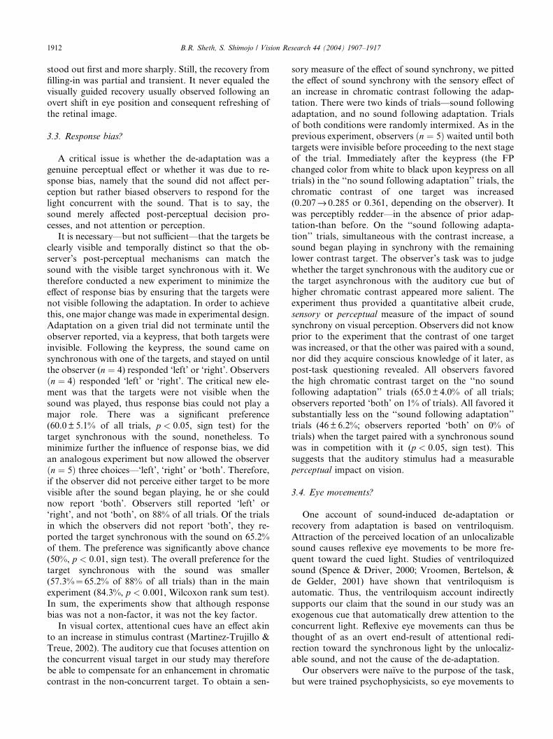
To explore this contrast further, we investigated
whether repetitive auditory cues improve the detectionof visual stimulus. A single target, located either left or
right of fixation, flickered five times over the course of
each trial (Fig. 3, inset). On a given trial, there was either
a brief sound synchronous with the target, a sound not
synchronous with the target, or no sound at all. The
observer (n ¼ 6, five na€ıve) had to report whether thetarget was left or right of fixation. The values of color
contrast were the same as those used in the first exper-iment (see Fig. 1). The results, graphed in Fig. 3, dem-
onstrate that sound, regardless of whether or not it was
synchronous with the visual target, failed to improve
detection performance (see Stein et al., 1996; Wuerger,
Hofbauer, & Meyer, 2003 for converging evidence).
3.7. Can visual cues effectively substitute for auditory
cues?
Is the effect of the auditory attentional cue on the
recovery from and resistance to visual adaptation be-
cause the cue was an auditory stimulus or because it
helped guide attention to the visual target (or both)?Replacing the auditory cues with visual cues will help
dissociate between the two possibilities. As a visual
analog to the auditory cue, a bright, horizontal line,
bilaterally symmetric about fixation, appeared for 14 ms
while either the left or the right target was on the screen
in the ‘‘cue on’’ phase (Fig. 4A, right). Thus, the line,
like the auditory cues of previous experiments, was a
temporal cue, and not a spatial one. Observers ðn ¼ 7Þweakly preferred the target simultaneous with the line
(Fig. 4B) on 57.6 ± 5.1% of all trials, 26% less than the
preference observed when auditory cues were used (cf.
Fig. 2).
The weaker effect of the line cue compared with the
sound could be because the line was not as salient as the
sound. However, the line was visually salient. We do not
think this was the case because the line was coloredwhite on a green background and was brighter than the
background. Nevertheless, in a second experiment, we
used an even more salient visual cue––a large, bright,
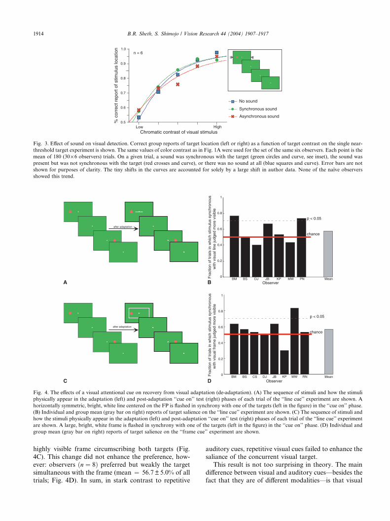
after adaptation
Observer
Fra
ctio
n of
tria
ls in
whi
ch s
timul
us s
ynch
rono
usw
ith v
isua
l fra
me
judg
ed m
ore
visi
ble
BM BS DJ JB KP MW RN MeanCS
p < 0.05
chance
after adaptation
BM BS DJ JB KP MW PN Mean0
0.2
0.4
0.6
0.8
1
0
0.2
0.4
0.6
0.8
1
p < 0.05
chance
Observer
Fra
ctio
n of
tria
ls in
whi
ch s
timul
us s
ynch
rono
usw
ith v
isua
l lin
e ju
dged
mor
e vi
sibl
e
B
D
A
C
Fig. 4. The effects of a visual attentional cue on recovery from visual adaptation (de-adaptation). (A) The sequence of stimuli and how the stimuli
physically appear in the adaptation (left) and post-adaptation ‘‘cue on’’ test (right) phases of each trial of the ‘‘line cue’’ experiment are shown. A
horizontally symmetric, bright, white line centered on the FP is flashed in synchrony with one of the targets (left in the figure) in the ‘‘cue on’’ phase.
(B) Individual and group mean (gray bar on right) reports of target salience on the ‘‘line cue’’ experiment are shown. (C) The sequence of stimuli and
how the stimuli physically appear in the adaptation (left) and post-adaptation ‘‘cue on’’ test (right) phases of each trial of the ‘‘line cue’’ experiment
are shown. A large, bright, white frame is flashed in synchrony with one of the targets (left in the figure) in the ‘‘cue on’’ phase. (D) Individual and
group mean (gray bar on right) reports of target salience on the ‘‘frame cue’’ experiment are shown.
Low High0.5
0.6
0.7
0.8
0.9
1.0n = 6
Chromatic contrast of visual stimulus
No sound
Synchronous sound
Asynchronous sound%
cor
rect
rep
ort o
f stim
ulus
loca
tion
Fig. 3. Effect of sound on visual detection. Correct group reports of target location (left or right) as a function of target contrast on the single near-
threshold target experiment is shown. The same values of color contrast as in Fig. 1A were used for the set of the same six observers. Each point is the
mean of 180 (30· 6 observers) trials. On a given trial, a sound was synchronous with the target (green circles and curve, see inset), the sound waspresent but was not synchronous with the target (red crosses and curve), or there was no sound at all (blue squares and curve). Error bars are not
shown for purposes of clarity. The tiny shifts in the curves are accounted for solely by a large shift in author data. None of the na€ıve observers
showed this trend.
1914 B.R. Sheth, S. Shimojo / Vision Research 44 (2004) 1907–1917
highly visible frame circumscribing both targets (Fig.
4C). This change did not enhance the preference, how-
ever: observers ðn ¼ 8Þ preferred but weakly the targetsimultaneous with the frame (mean ¼ 56.7 ± 5.0% of alltrials; Fig. 4D). In sum, in stark contrast to repetitive
auditory cues, repetitive visual cues failed to enhance the
saliance of the concurrent visual target.
This result is not too surprising in theory. The main
difference between visual and auditory cues––besides the
fact that they are of different modalities––is that visual

B.R. Sheth, S. Shimojo / Vision Research 44 (2004) 1907–1917 1915
cues, unlike auditory ones, must occupy discrete loca-
tions in space, which, in our experiments, are different
from the locations of the targets. Thus, repetitive visual
cues pull spatial attention away from targets located at
different positions from the cues, which reduces their
power to transiently enhance the signals of the targets
synchronous with them (Spence & Driver, 2000; Vroo-
men et al., 2001). Moreover, visual–visual suppression ismore potent than audio–visual suppression in masking
and inhibition of return (Spence, Lloyd, McGlone,
Nicholls, & Driver, 2000), which too would explain the
lower potency of the visual cue in enhancing attention to
the synchronous light. Regardless of cause, our experi-
ments are among the first, to our knowledge, to find that
an attentional cue of a different modality has a stronger
effect on perception than a cue of the same modality.This implies there are functions a cross-modal cue can
perform that a cue of the same modality cannot.
4. Discussion
We have shown that repetitive auditory cues selec-
tively enhance the visibility of synchronous visual tar-
gets, preventing them from undergoing visual
adaptation and causing previously adapted targets to
recover. Thus, sound enhances visual perception by
momentarily enhancing the visual signal that is near
simultaneous with it. We offer a tentative account of ourfindings. The sensory signal in response to the visual
stimulus competes with other signals from other visual
stimuli for awareness. Depending on a variety of factors,
including relative signal strength and the observer’s lo-
cus of attention, the higher-level cortical mechanisms
that gate or modulate conscious perception at the level
of objects will allow the stimulus to be consciously
perceived. If a cue helps focus one’s attention to a targetstimulus, the stimulus will be more likely, as compared
to its competitors, to be consciously perceived (Kastner,
De Weerd, Desimone, & Ungerleider, 1998). Audition
interacts with vision at the level of cortex. The brain
signal from the transient light that is synchronous with
the transient auditory cue is selectively enhanced. This
selective enhancement explains why a brief sound tem-
porarily causes previously adapted visual targets syn-chronous with it to recover and prevents visual targets
synchronous with it from undergoing visual adaptation.
There is evidence for high-level influence on filling-in,
and Troxler fading, in particular. For instance, inter-
mediate-level surface- and object-based fading has been
observed (Ramachandran & Gregory, 1991). Prolonged
observation of filled-in motion including the blind spot
of one eye causes a motion aftereffect to be perceived inthe other eye, again arguing for high-level influence over
filling-in (Murakami, 1995). Patients with parietal le-
sions report accelerated Troxler fading (Holliday, Ken-
nard, & Ruddock, 1985; Mennemeier et al., 1994) and
fading of moving peripheral stimuli contralateral to
their brain lesion (Mennemeier et al., 1994); in contrast,
patients with frontal lesions rarely report Troxler fading
(Mennemeier et al., 1994). These neurological results
implicate high-level parietal and frontal areas in Troxler
fading. Thus, Troxler fading can be modulated at higher
levels of processing. Our study may be understood inthis context: a cue of a different modality modulates
filling-in at some stage beyond visual detection. In the
context of multisensory integration, our study adds to
the mounting evidence for high-level modulation of low-
level percepts in the multimodal (Calvert et al., 1997;
Giard & Peronnet, 1999; Laurienti et al., 2002; Shams
et al., 2001; Taylor-Clarke, Kennett, & Haggard, 2002)
and unimodal domains (Chauduri, 1990; Rees, Frith, &Lavie, 1997).
On a different, and arguably, more important note,
this study is the first to show that an attentional cue of a
different modality (audition) can modulate the percep-
tion of a disappearance phenomenon (Troxler fading).
Our data further indicate what effects a cross-modal
attention cue has on perception––it can enhance per-
ception of an above-threshold stimulus, but cannot im-prove detection of a sub-threshold stimulus. Two
accounts have been provided of disappearance phe-
nomena––sensory suppression (Blake, 1989; Burbeck &
Kelly, 1984) and higher-level selection (Logothetis,
1998; MacKay, 1986). Undoubtedly, Troxler fading re-
sults from local adaptation––a form of sensory sup-
pression (Clarke & Belcher, 1962; Kotulak & Schor,
1986; Millodot, 1967). On the other hand, by all indi-cations, the sound in our study was an attentional or
selectional cue. Our study is among the first, therefore,
to demonstrate that one mechanism, namely selection,
can compensate, transiently and in part, for another,
namely sensory suppression, thereby implying that the
two mechanisms are inter-related.
Influential studies have claimed that filling-in is not
an active process, but one based on mid-level long-rangedisinhibition or post-inhibitory rebound. Hardage &
Tyler (1995) showed that the twinkle aftereffect observed
in a visual patch following stimulation by a dynamic
noise stimulus of a surrounding patch was dissociable
from the filling-in that occured during stimulation,
which supports the inhibition–disinhibition account of
filling-in. Tyler & Hardage (1998) further showed that
the disinhibition was restricted to the magnocellularsystem (no aftereffect in equiluminant noise or by noise
below a temporal frequency of 5 Hz). In the context of
this evidence, perhaps the sound in our study momen-
tarily disinhibited the synchronous visual target––a
transient disinhibition that lived and died with the
sound––and, thereby provided a brief recovery from the
visual adaptation. The fast temporal characteristics of
the magno system render it suitable as a basis for the
1916 B.R. Sheth, S. Shimojo / Vision Research 44 (2004) 1907–1917
fast disinhibition. We believe neurophysiological studies
will furnish the most rigorous test of our account.
Acknowledgements
We thank Dr. Chris Tyler and the two anonymous
reviewers for their insightful suggestions and criticisms.We are grateful to Daw-An Wu for discussions. Special
thanks to Prof. Mark Konishi for the generous loan of
his sound pressure meter, and to the volunteers who
kindly participated.
References
Andrews, T. J., & Purves, D. (1997). Similarities in normal and
binocularly rivalrous viewing. Proceedings of the National Academy
of Sciences USA, 94, 9905–9908.
Bennet-Clark, H., & Evans, C. (1963). Fragmentation of patterned
targets when viewed as prolonged after-images. Nature, 199, 1215–
1216.
Bhattacharya, J., Shams, L., & Shimojo, S. (2002). Sound-induced
illusory flash perception: role of gamma band responses. Neurore-
port, 13, 727–730.
Blake, R. (1989). A neural theory of binocular rivalry. Psychological
Review, 96, 145–167.
Bonneh, Y. S., Cooperman, A., & Sagi, D. (2001). Motion-induced
blindness in normal observers. Nature, 411, 798–801.
Brainard, D. H. (1997). The Psychophysics Toolbox. Spatial Vision,
10, 443–446.
Burbeck, C., & Kelly, D. (1984). Role of local adaptation in the fading
of stabilized images. Journal of Optical Society of America, 1, 216–
220.
Calvert, G. A., Bullmore, E. T., Brammer, M. J., Campbell, R.,
Williams, S. C., McGuire, P. K., Woodruff, P. W., Iversen, S. D., &
David, A. S. (1997). Activity of auditory cortex during silent
lipreading. Science, 276, 593–596.
Campbell, F. W., Glinsky, A. S., Howel, E. R., Riggs, L. A., &
Atkinson, J. (1973). The dependence of monocular rivalry on
orientation. Perception, 2, 123–125.
Chaudhuri, A. (1990). Modulation of the motion aftereffect by
selective attention. Nature, 344, 60–62.
Clarke, F. J. J., & Belcher, S. J. (1962). On the localization of Troxler’s
effect in the visual pathway. Vision Research, 2, 53–68.
Dennett, D. C. (1991). Consciousness explained. Boston, MA: BackBay
Books.
Driver, J., & Spence, C. (1998). Cross-modal attention. Current
Opinion in Neurobiology, 8, 245–253.
Erisir, A., Van Horn, S. C., & Sherman, S. M. (1997). Relative
numbers of cortical and brainstem inputs to the lateral geniculate
nucleus. Proceedings of the National Academy of Sciences USA, 94,
1517–1520.
Falchier, A., Clavagnier, S., Barone, P., & Kennedy, H. (2002).
Anatomical evidence of multimodal integration in primate striate
cortex. Journal of Neuroscience, 22, 5749–5759.
Giard, M. H., & Peronnet, M. (1999). Auditory–visual integration
during multimodal object recognition in humans: a behavioral and
electrophysiological study. Journal of Cognitive Neuroscience, 11,
473–490.
Goldstone, S., & Lhamon, W. T. (1972). Auditory–visual differences in
human temporal judgment. Perceptual & Motor Skills, 34, 623–633.
Hardage, L., & Tyler, C. W. (1995). Induced twinkle aftereffect as a
probe of dynamic visual processing mechanisms. Vision Research,
35, 757–766.
Holliday, I. E., Kennard, C., & Ruddock, K. H. (1985). Rapid fading
of visual sensations in a subject with a parietal–occipital tumour.
Ophthalmic and Physiological Optics, 5, 149–156.
Jonides, J. (1981). In J. Long & A. D. Baddeley (Eds.), Attention and
performance (Vol. 9, pp. 187–204). Hillsdale, NJ: Erlbaum.
Kastner, S., De Weerd, P., Desimone, R., & Ungerleider, L. G. (1998).
De Mechanisms of directed attention in the human extrastriate
cortex as revealed by functional MRI. Science, 282, 108–111.
Keele, S. W. (1986). In K. R. Boff, L. Kaufman, & J. P. Thomas (Eds.),
Handbook of perception and human performance: Vol. II. Cognitive
processes and performance (pp. 30.1–30.60). New York: Wiley and
Sons.
Kinoshita, M., & Komatsu, H. (2001). Neural representation of the
luminance and brightness of a uniform surface in the macaque
primary visual cortex. Journal of Neurophysiology, 86, 2559–2570.
Kotulak, J. C., & Schor, C. M. (1986). The accommodative response to
sub-threshold blur and to perceptual fading during the Troxler
phenomenon. Perception, 15, 7–15.
Laurienti, P. J., Burdette, J. H., Wallace, M. T., Yen, Y. F., Field, A.
S., & Stein, B. E. (2002). Deactivation of sensory-specific cortex by
cross-modal stimuli. Journal of Cognitive Neuroscience, 14, 420–
429.
Logothetis, N. K. (1998). Single units and conscious vision. Philo-
sophical Transactions of the Royal Society of London B, 353, 1801–
1818.
Lou, L. (1999). Selective peripheral fading: evidence for inhibitory
sensory effect of attention. Perception, 28, 519–526.
MacKay, D. M. (1986). In J. Pettigrew, K. Sanderson, & W. Levick
(Eds.), Visual neuroscience (pp. 365–373). Cambridge, UK: Cam-
bridge University Press.
Martinez-Trujillo, J., & Treue, S. (2002). Attentional modulation
strength in cortical area MT depends on stimulus contrast. Neuron,
35, 365–370.
McDonald, J. J., Teder-S€alej€arvi, W. A., & Hillyard, S. A. (2000).Involuntary orienting to sound improves visual perception. Nature,
407, 906–908.
Mennemeier, M. S., Chatterjee, A., Watson, R. T., Wertman, E.,
Carter, L. P., & Heilman, K. M. (1994). Contributions of the
parietal and frontal lobes to sustained attention and habituation.
Neuropsychologia, 32, 703–716.
Millodot, M. (1967). Extra-foveal variations of the phenomenon of
Troxler. Psychologie Francaise, 12, 190–196.
Montero, V. M. (1991). A quantitative study of synaptic contacts on
interneurons and relay cells of the cat lateral geniculate nucleus.
Experimental Brain Research, 86, 257–270.
Murakami, I. (1995). Motion aftereffect after monocular adaptation to
filled-in motion at the blind spot. Vision Research, 35, 1041–1045.
Murphy, P. C., Duckett, S. G., & Sillito, A. M. (1999). Feedback
connections to the lateral geniculate nucleus and cortical response
properties. Science, 286, 1552–1554.
Odgaard, C., Arieh, Y., & Marks, L. E. (2003). Cross-modal
enhancement of perceived brightness: sensory interaction versus
response bias. Perception & Psychophysics, 65, 123–132.
Pelli, D. G. (1997). The Video Toolbox software for visual psycho-
physics: transforming numbers into movies. Spatial Vision, 10,
437–442.
Posner, M. I. (1978). Chronometric explorations of the mind. Hillsdale,
NJ: Lawrence Erlbaum Associates.
Posner, M. I., Nissen, M. J., & Klein, R. M. (1976). Visual dominance:
an information-processing account of its origins and significance.
Psychological Review, 83, 157–171.
Posner, M. I., Nissen, M. J., & Ogden, W. C. (1978). In H. L. Pick & B.
J. Saltzman (Eds.), Modes of perceiving and processing information
(pp. 137–158). Hillsdale, NJ: Erlbaum.
Ramachandran, V. S., & Gregory, R. L. (1991). Perceptual filling in of
artificially induced scotomas in human vision. Nature, 350, 699–
702.
B.R. Sheth, S. Shimojo / Vision Research 44 (2004) 1907–1917 1917
Rees, G., Frith, C. D., & Lavie, N. (1997). Modulating irrelevant
motion perception by varying attentional load in an unrelated task.
Science, 278, 1616–1619.
Rockland, K. S., & Ojima, H. (2002). Multimodal convergence in
calcarine visual areas in macaque monkey, paper presented at the
3rd annual meeting of the International Multisensory Research
Forum, Geneva, Switzerland.
Rossi, A. F., & Paradiso, M. A. (1999). Neural correlates of perceived
brightness in the retina, lateral geniculate nucleus, and striate
cortex. Journal of Neuroscience, 19, 6145–6156.
Sekuler, R., Sekuler, A. B., & Lau, R. (1997). Sound alters visual
motion perception. Nature, 385, 308.
Shams, L., Kamitani, Y., Thompson, S., & Shimojo, S. (2001). Sound
alters visual evoked potentials in humans. Neuroreport, 12, 3849–
3852.
Sheth, B. R., & Shimojo, S. (2002). Recovery of visual perception from
adaptation by sound: The cross-modal ‘‘beating-heart’’ effect,
poster presented at the 2nd annual meeting of the Vision Sciences
Society, Sarasota, FL.
Shimojo, S., & Kamitani, Y. (2001). Filling-in induced by high-
contrast edge adaptation. Journal of Vision, 1, 53a.
Shimojo, S., Miyauchi, S., & Hikosaka, O. (1997). Visual motion
sensation yielded by non-visually driven attention. Vision Research,
37, 1575–1580.
Spence, C., & Driver, J. (2000). Attracting attention to the illusory
location of a sound: reflexive crossmodal orienting and ventrilo-
quism. Neuroreport, 11, 2057–2061.
Spence, C., Lloyd, D., McGlone, F., Nicholls, M. E., & Driver, J.
(2000). Inhibition of return is supramodal: a demonstration
between all possible pairings of vision, touch and audition.
Experimental Brain Research, 134, 42–48.
Stein, B. E., London, N., Wilkinson, L. K., & Price, D. D. (1996).
Enhancement of perceived visual intensity by auditory stimuli: a
psychophysical analysis. Journal of Cognitive Neuroscience, 8, 497–
506.
Taylor-Clarke, M., Kennett, S., & Haggard, P. (2002). Vision
modulates somatosensory cortical processing. Current Biology,
12, 233–236.
Troxler, D. (1804). In K. Himley & J. A. Schmidt (Eds.), Ophthalmo-
logisches bibliothek (pp. 51–53). Fromman, Germany: Jena.
Tyler, C. W., & Hardage, L. (1998). Long-range twinkle induction: an
achromatic rebound effect in the magnocellular processing system?
Perception, 27, 203–214.
Vroomen, J., Bertelson, P., & de Gelder, B. (2001). Directing spatial
attention towards the illusory location of a ventriloquized sound.
Acta Psychologica, 108, 21–33.
Vroomen, J., & de Gelder, B. (2000). Sound enhances visual
perception: cross-modal effects of auditory organization on vision.
Journal of Experimental Psychology: Human Perception & Perfor-
mance, 26, 1583–1590.
Watanabe, K., & Shimojo, S. (1998). Attentional modulation in
perception of visual motion events. Perception, 27, 1041–1054.
Wilson, J. R., Friedlander, M. J., & Sherman, S. M. (1984). Fine
structural morphology of identified X- and Y-cells in the cat’s
lateral geniculate nucleus. Proceedings of the Royal Society of
London Series B––Biological Science, 221, 411–436.
Wolfe, J. M. (1986). Briefly presented stimuli can disrupt constant
suppression and binocular rivalry suppression. Perception, 15, 413–
417.
Wuerger, S. M., Hofbauer, M., & Meyer, G. F. (2003). The integration
of auditory and visual motion signals at threshold. Perception &
Psychophysics, 65, 1188–1196.