Soluble Biodegradable Polymer-Based Cytokine Gene Delivery for Cancer Treatment
10
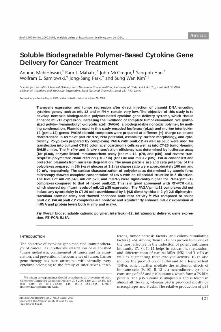
121 ARTICLE MOLECULAR THERAPY Vol. 2, No. 2, August 2000 Copyright The American Society of Gene Therapy 1525-0016/00 $35.00 doi:10.1006/mthe.2000.0105, available online at http://www.idealibrary.com on IDEAL INTRODUCTION The objective of cytokine gene-mediated immunothera- py of cancer lies in effective retardation of established tumor metastasis, confinement of tumor and its elimi- nation, and prevention of reoccurrence of tumor. Cancer gene therapy has been attempted with virtually every cytokine belonging to the family of interleukins, inter- ferons, tumor necrosis factors, and colony stimulating factors (1–6). Among them IL-12 has proven to be one of the most effective in the induction of potent antitumor immunity (7, 8). IL-12 helps in activation, maturation, and differentiation of natural killer (NK) and T cells as well as augmenting their cytolytic activity. IL-12 also induces the production of IFN-γ and to a lesser extent TNF-α, which further mediate the antitumor effects of immune cells (9, 10). IL-12 is a heterodimeric cytokine consisting of p35 and p40 subunits, which form a 75-kDa protein. The p35 subunit is ubiquitous and is found in almost all the cells, whereas p40 is produced mostly by macrophages and B cells. The relative production of p35 Soluble Biodegradable Polymer-Based Cytokine Gene Delivery for Cancer Treatment Anurag Maheshwari, * Ram I. Mahato, * John McGregor, † Sang-oh Han, * Wolfram E. Samlowski, † Jong-Sang Park, ‡ and Sung Wan Kim *,1 *Center for Controlled Chemical Delivery and †Huntsman Cancer Institute, University of Utah, Salt Lake City, Utah 84112-5820 ‡School of Chemistry and Molecular Engineering, Seoul National University, Seoul 151-742, Korea Received for publication May 4, 2000, and accepted in revised form June 27, 2000 Transgene expression and tumor regression after direct injection of plasmid DNA encoding cytokine genes, such as mIL-12 and mIFN-γ, remain very low. The objective of this study is to develop nontoxic biodegradable polymer-based cytokine gene delivery systems, which should enhance mIL-12 expression, increasing the likelihood of complete tumor elimination. We synthe- sized poly[α-(4-aminobutyl)-L-glycolic acid] (PAGA), a biodegradable nontoxic polymer, by melt- ing condensation. Plasmids used in this study encoded luciferase (pLuc) and murine interleukin- 12 (pmIL-12) genes. PAGA/plasmid complexes were prepared at different (±) charge ratios and characterized in terms of particle size, zeta potential, osmolality, surface morphology, and cyto- toxicity. Polyplexes prepared by complexing PAGA with pmIL-12 as well as pLuc were used for transfection into cultured CT-26 colon adenocarcinoma cells as well as into CT-26 tumor-bearing BALB/c mice. The in vitro and in vivo transfection efficiency was determined by luciferase assay (for pLuc), enzyme-linked immunosorbent assay (for mIL-12, p70, and p40), and reverse tran- scriptase–polymerase chain reaction (RT–PCR) (for Luc and mIL-12 p35). PAGA condensed and protected plasmids from nuclease degradation. The mean particle size and zeta potential of the polyplexes prepared in 5% (w/v) glucose at 3:1 (±) charge ratio were approximately 100 nm and 20 mV, respectively. The surface characterization of polyplexes as determined by atomic force microscopy showed complete condensation of DNA with an ellipsoidal structure in Z direction. The levels of mIL-12 p40, mIL-12 p70, and mIFN-γ were significantly higher for PAGA/pmIL-12 complexes compared to that of naked pmIL-12. This is in good agreement with RT–PCR data, which showed significant levels of mIL-12 p35 expression. The PAGA/pmIL-12 complexes did not induce any cytotoxicity in CT-26 cells as evidenced by 3-{4,5-dimethylthiazol-2-yl}-2,5-diphenylte- trazolium bromide assay and showed enhanced antitumor activity in vivo compared to naked pmIL-12. PAGA/pmIL-12 complexes are nontoxic and significantly enhance mIL-12 expression at mRNA and protein levels both in vitro and in vivo. Key Words: biodegradable cationic polymer; interleukin-12; intratumoral delivery; gene expres- sion; RT–PCR; ELISA. 1 To whom correspondence should be addressed at University of utah, Center for Controlled Chemical Delivery, 30 S 2000 E RM 201 {SK H}. Salt lake City, UT 84112-5820. Fax: (801) 581-7848. E-mail: [email protected].
-
Upload
independent -
Category
Documents
-
view
1 -
download
0
Transcript of Soluble Biodegradable Polymer-Based Cytokine Gene Delivery for Cancer Treatment
121
ARTICLE
MOLECULAR THERAPY Vol. 2, No. 2, August 2000Copyright � The American Society of Gene Therapy1525-0016/00 $35.00
doi:10.1006/mthe.2000.0105, available online at http://www.idealibrary.com on IDEAL
INTRODUCTION
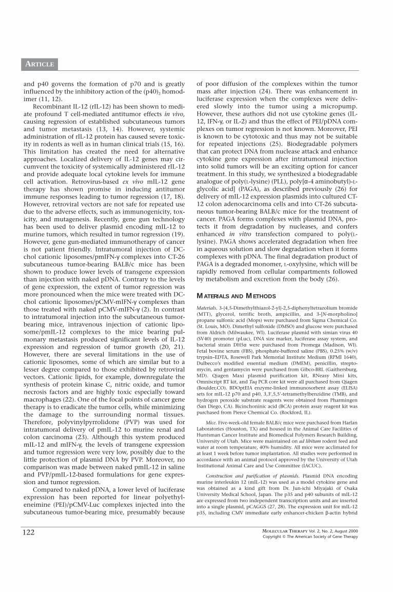
The objective of cytokine gene-mediated immunothera-py of cancer lies in effective retardation of establishedtumor metastasis, confinement of tumor and its elimi-nation, and prevention of reoccurrence of tumor. Cancergene therapy has been attempted with virtually everycytokine belonging to the family of interleukins, inter-
ferons, tumor necrosis factors, and colony stimulatingfactors (1–6). Among them IL-12 has proven to be one ofthe most effective in the induction of potent antitumorimmunity (7, 8). IL-12 helps in activation, maturation,and differentiation of natural killer (NK) and T cells aswell as augmenting their cytolytic activity. IL-12 alsoinduces the production of IFN-γ and to a lesser extentTNF-α, which further mediate the antitumor effects ofimmune cells (9, 10). IL-12 is a heterodimeric cytokineconsisting of p35 and p40 subunits, which form a 75-kDaprotein. The p35 subunit is ubiquitous and is found inalmost all the cells, whereas p40 is produced mostly bymacrophages and B cells. The relative production of p35
Soluble Biodegradable Polymer-Based Cytokine GeneDelivery for Cancer TreatmentAnurag Maheshwari,* Ram I. Mahato,* John McGregor,† Sang-oh Han,*Wolfram E. Samlowski,† Jong-Sang Park,‡ and Sung Wan Kim*,1
*Center for Controlled Chemical Delivery and †Huntsman Cancer Institute, University of Utah, Salt Lake City, Utah 84112-5820‡School of Chemistry and Molecular Engineering, Seoul National University, Seoul 151-742, Korea
Received for publication May 4, 2000, and accepted in revised form June 27, 2000
Transgene expression and tumor regression after direct injection of plasmid DNA encodingcytokine genes, such as mIL-12 and mIFN-γ, remain very low. The objective of this study is todevelop nontoxic biodegradable polymer-based cytokine gene delivery systems, which shouldenhance mIL-12 expression, increasing the likelihood of complete tumor elimination. We synthe-sized poly[α-(4-aminobutyl)-L-glycolic acid] (PAGA), a biodegradable nontoxic polymer, by melt-ing condensation. Plasmids used in this study encoded luciferase (pLuc) and murine interleukin-12 (pmIL-12) genes. PAGA/plasmid complexes were prepared at different (±) charge ratios andcharacterized in terms of particle size, zeta potential, osmolality, surface morphology, and cyto-toxicity. Polyplexes prepared by complexing PAGA with pmIL-12 as well as pLuc were used fortransfection into cultured CT-26 colon adenocarcinoma cells as well as into CT-26 tumor-bearingBALB/c mice. The in vitro and in vivo transfection efficiency was determined by luciferase assay(for pLuc), enzyme-linked immunosorbent assay (for mIL-12, p70, and p40), and reverse tran-scriptase–polymerase chain reaction (RT–PCR) (for Luc and mIL-12 p35). PAGA condensed andprotected plasmids from nuclease degradation. The mean particle size and zeta potential of thepolyplexes prepared in 5% (w/v) glucose at 3:1 (±) charge ratio were approximately 100 nm and20 mV, respectively. The surface characterization of polyplexes as determined by atomic forcemicroscopy showed complete condensation of DNA with an ellipsoidal structure in Z direction.The levels of mIL-12 p40, mIL-12 p70, and mIFN-γ were significantly higher for PAGA/pmIL-12complexes compared to that of naked pmIL-12. This is in good agreement with RT–PCR data,which showed significant levels of mIL-12 p35 expression. The PAGA/pmIL-12 complexes did notinduce any cytotoxicity in CT-26 cells as evidenced by 3-{4,5-dimethylthiazol-2-yl}-2,5-diphenylte-trazolium bromide assay and showed enhanced antitumor activity in vivo compared to nakedpmIL-12. PAGA/pmIL-12 complexes are nontoxic and significantly enhance mIL-12 expression atmRNA and protein levels both in vitro and in vivo.
Key Words: biodegradable cationic polymer; interleukin-12; intratumoral delivery; gene expres-sion; RT–PCR; ELISA.
1To whom correspondence should be addressed at University of utah,Center for Controlled Chemical Delivery, 30 S 2000 E RM 201 {SK H}. Saltlake City, UT 84112-5820. Fax: (801) 581-7848. E-mail:[email protected].
MOLECULAR THERAPY Vol. 2, No. 2, August 2000Copyright � The American Society of Gene Therapy
122
ARTICLE
and p40 governs the formation of p70 and is greatlyinfluenced by the inhibitory action of the (p40)2 homod-imer (11, 12).
Recombinant IL-12 (rIL-12) has been shown to medi-ate profound T cell-mediated antitumor effects in vivo,causing regression of established subcutaneous tumorsand tumor metastasis (13, 14). However, systemicadministration of rIL-12 protein has caused severe toxic-ity in rodents as well as in human clinical trials (15, 16).This limitation has created the need for alternativeapproaches. Localized delivery of IL-12 genes may cir-cumvent the toxicity of systemically administered rIL-12and provide adequate local cytokine levels for immunecell activation. Retrovirus-based ex vivo mIL-12 genetherapy has shown promise in inducing antitumorimmune responses leading to tumor regression (17, 18).However, retroviral vectors are not safe for repeated usedue to the adverse effects, such as immunogenicity, tox-icity, and mutagenesis. Recently, gene gun technologyhas been used to deliver plasmid encoding mIL-12 tomurine tumors, which resulted in tumor regression (19).However, gene gun-mediated immunotherapy of canceris not patient friendly. Intratumoral injection of DC-chol cationic liposomes/pmIFN-γ complexes into CT-26subcutaneous tumor-bearing BALB/c mice has beenshown to produce lower levels of transgene expressionthan injection with naked pDNA. Contrary to the levelsof gene expression, the extent of tumor regression wasmore pronounced when the mice were treated with DC-chol cationic liposomes/pCMV-mIFN-γ complexes thanthose treated with naked pCMV-mIFN-γ (2). In contrastto intratumoral injection into the subcutaneous tumor-bearing mice, intravenous injection of cationic lipo-some/pmIL-12 complexes to the mice bearing pul-monary metastasis produced significant levels of IL-12expression and regression of tumor growth (20, 21).However, there are several limitations in the use ofcationic liposomes, some of which are similar but to alesser degree compared to those exhibited by retroviralvectors. Cationic lipids, for example, downregulate thesynthesis of protein kinase C, nitric oxide, and tumornecrosis factors and are highly toxic especially towardmacrophages (22). One of the focal points of cancer genetherapy is to eradicate the tumor cells, while minimizingthe damage to the surrounding normal tissues.Therefore, polyvinylpyrrolidone (PVP) was used forintratumoral delivery of pmIL-12 to murine renal andcolon carcinoma (23). Although this system producedmIL-12 and mIFN-γ, the levels of transgene expressionand tumor regression were very low, possibly due to thelittle protection of plasmid DNA by PVP. Moreover, nocomparison was made between naked pmIL-12 in salineand PVP/pmIL-12-based formulations for gene expres-sion and tumor regression.
Compared to naked pDNA, a lower level of luciferaseexpression has been reported for linear polyethyl-eneimine (PEI)/pCMV-Luc complexes injected into thesubcutaneous tumor-bearing mice, presumably because
of poor diffusion of the complexes within the tumormass after injection (24). There was enhancement inluciferase expression when the complexes were deliv-ered slowly into the tumor using a micropump.However, these authors did not use cytokine genes (IL-12, IFN-γ, or IL-2) and thus the effect of PEI/pDNA com-plexes on tumor regression is not known. Moreover, PEIis known to be cytotoxic and thus may not be suitablefor repeated injections (25). Biodegradable polymersthat can protect DNA from nuclease attack and enhancecytokine gene expression after intratumoral injectioninto solid tumors will be an exciting option for cancertreatment. In this study, we synthesized a biodegradableanalogue of poly(L-lysine) (PLL), poly[α-4 aminobutyl)-L-glycolic acid] (PAGA), as described previously (26) fordelivery of mIL-12 expression plasmids into cultured CT-12 colon adenocarcinoma cells and into CT-26 subcuta-neous tumor-bearing BALB/c mice for the treatment ofcancer. PAGA forms complexes with plasmid DNA, pro-tects it from degradation by nucleases, and confersenhanced in vitro transfection compared to poly(L-lysine). PAGA shows accelerated degradation when freein aqueous solution and slow degradation when it formscomplexes with pDNA. The final degradation product ofPAGA is a degraded monomer, L-oxylysine, which will berapidly removed from cellular compartments followedby metabolism and excretion from the body (26).
MATERIALS AND METHODS
Materials. 3-{4,5-Dimethylthiazol-2-yl}-2,5-diphenyltetrazolium bromide(MTT), glycerol, terrific broth, ampicillin, and 3-[N-morpholino]propane sulfonic acid (Mops) were purchased from Sigma Chemical Co.(St. Louis, MO). Dimethyl sulfoxide (DMSO) and glucose were purchasedfrom Aldrich (Milwaukee, WI). Luciferase plasmid with simian virus 40(SV40) promoter (pLuc), DNA size marker, luciferase assay system, andbacterial strain DH5α were purchased from Promega (Madison, WI).Fetal bovine serum (FBS), phosphate-buffered saline (PBS), 0.25% (w/v)trypsin–EDTA, Rosewell Park Memorial Institute Medium (RPMI 1640),Dulbecco’s modified essential medium (DMEM), penicillin, strepto-mycin, and gentamycin were purchased from Gibco-BRL (Gaithersburg,MD). Qiagen Maxi plasmid purification kit, RNeasy Mini kits,Omniscript RT kit, and Taq PCR core kit were all purchased from Qiagen(Boulder,CO). BDOptEIA enzyme-linked immunosorbent assay (ELISA)sets for mIL-12 p70 and p40, 3,3′,5,5′-tetramethylbenzidine (TMB), andhydrogen peroxide substrate reagents were obtained from Pharmingen(San Diego, CA). Bicinchoninic acid (BCA) protein assay reagent kit waspurchased from Pierce Chemical Co. (Rockford, IL).
Mice. Five-week-old female BALB/c mice were purchased from HarlanLaboratories (Houston, TX) and housed in the Animal Care Facilities ofHuntsman Cancer Institute and Biomedical Polymers Research Building,University of Utah. Mice were maintained on ad libitum rodent feed andwater at room temperature, 40% humidity. All mice were acclimated forat least 1 week before tumor implantation. All studies were performed inaccordance with an animal protocol approved by the University of UtahInstitutional Animal Care and Use Committee (IACUC).
Construction and purification of plasmids. Plasmid DNA encodingmurine interleukin 12 (mIL-12) was used as a model cytokine gene andwas obtained as a kind gift from Dr. Jun-ichi Miyajaki of OsakaUniversity Medical School, Japan. The p35 and p40 subunits of mIL-12are expressed from two independent transcription units and are insertedinto a single plasmid, pCAGGS (27, 28). The expression unit for mIL-12p35, including CMV immediate early enhancer-chicken β-actin hybrid
MOLECULAR THERAPY Vol. 2, No. 2, August 2000Copyright � The American Society of Gene Therapy
123
ARTICLE
promoter and rabbit β-globin poly(A) signal, is excised from pCAGGS-p35and is inserted downstream of the mIL-12 p40 expression unit ofpCAGGS-p40 (27, 28). Another plasmid for mIL-12, pCMV-mIL-12, wascompared with pCAGGS-mIL-12 and was obtained as a kind gift from Dr.Steven Dow of the University of Colorado (20). Plasmid encodingluciferase (pLuc) was used as a reporter gene and was purchased fromPromega. These plasmids were amplified in Escherichia coli DH5α strainand then isolated and purified using Qiagen Plasmid Maxi purificationkit (Qiagen, Valencia, CA). The plasmid purity and integrity were con-firmed by 1% agarose gel electrophoresis followed by ethidium bromidestaining, and DNA concentration was measured by ultraviolet (UV)absorbance at 260 nm. The optical density ratios at 260 to 280 nm ofthese plasmid preparations were in the range of 1.7–1.8.
Restriction enzyme analysis. To confirm that pmIL-12 contains thewhole p40 and p35 genes and there is no rearrangement of the gene dur-ing cloning and propagation, restriction enzyme digestion assay wasdone as described by Yoshimoto et al. (29) with EcoRI and BamHI for p40and EcoRI and HindIII for p35, followed by 1% agarose gel electrophore-sis.
Synthesis of poly[α-(4-aminobutyl)-L-glycolic acid)]. Poly[α-(4-aminobutyl)-L-glycolic acid] (PAGA) was synthesized and characterized asdescribed previously by Lim et al. (26). Briefly, 10 g of sodium nitrite solu-tion was added dropwise to 20 g of CBZ-L-lysine in a mixture of 200 ml 2N H2SO4 and 200 ml acetonitrile with constant stirring on ice. Stirringwas continued for an additional 7 h on ice and 12 h at room temperature.The resulting solution was then extracted with ether and precipitatedwith petroleum ether. CBZ-L-oxylysine was polymerized by melting con-densation at 150�C under vacuum for 5 days. The polymer was cooledand dissolved in chloroform followed by precipitation with methanol.Two grams of the dried polymer was dissolved in 28 ml DMF containing2 g of activated palladium on carbon (10% Pd-C). Ninety milliliters of85% formic acid was added slowly to the polymer solution, and 15 h afterstirring at room temperature the solution was filtered to remove Pd-C.Forty milliliters of 2 N HCl was added and the volume was reduced undervacuum. PAGA was finally precipitated with a large excess of acetone,dried under vacuum, and stored at −70�C until further use. PAGA wascharacterized in terms of molecular weight and biodegradability usingMALDI-TOF–mass spectrometry and in terms of purity using NMR spec-troscopy.
Gel retardation assay. Various formulations of PAGA/pmIL-12 orPAGA/pLuc complexes, ranging from charge ratio 0.5/1 (±) to 7/1 (±),were prepared in the presence of 5% (w/v) glucose to adjust the osmolal-ity to 285–295 mOsm. The polyplexes were incubated for 15–20 min atroom temperature. The samples were electrophoresed on 1% agarose gelin 1� Tris–boric acid–EDTA (TBE) buffer at 80 V until the 1� orange–blueloading dye ran through 80% of the gel. A 200-bp marker (Promega) wasused as a DNA size marker. The gel was stained with 0.5 µg/ml ethidiumbromide for 45 min and analyzed on a UV illuminator to show the loca-tion of DNA.
DNase protection assay. PAGA/pmIL-12 (3/1, ±) complexes were incu-bated at 37�C in the presence of DNase Ι (45 µl). At 0, 5, 10, 15, 30, and60 min postincubation, 50 µl of the samples was taken into Eppendorftubes and mixed with 100 µl of stop solution (400 mM NaCl, 100 mMEDTA) under mild vortexing. The samples were then mixed with 12 µl of10% sodium dodecyl sulfate (SDS) and incubated at 65�C overnight. TheDNA was then extracted with a mixture of phenol:chloroform:isoamylalcohol (25:24:1, v/v). The extracted DNA was precipitated with 700 µl ofabsolute ethanol. The DNA precipitate was dried by air and dissolved in10 µl Tris–EDTA buffer. DNA integrity was assessed by 0.8% agarose gelelectrophoresis, as mentioned above.
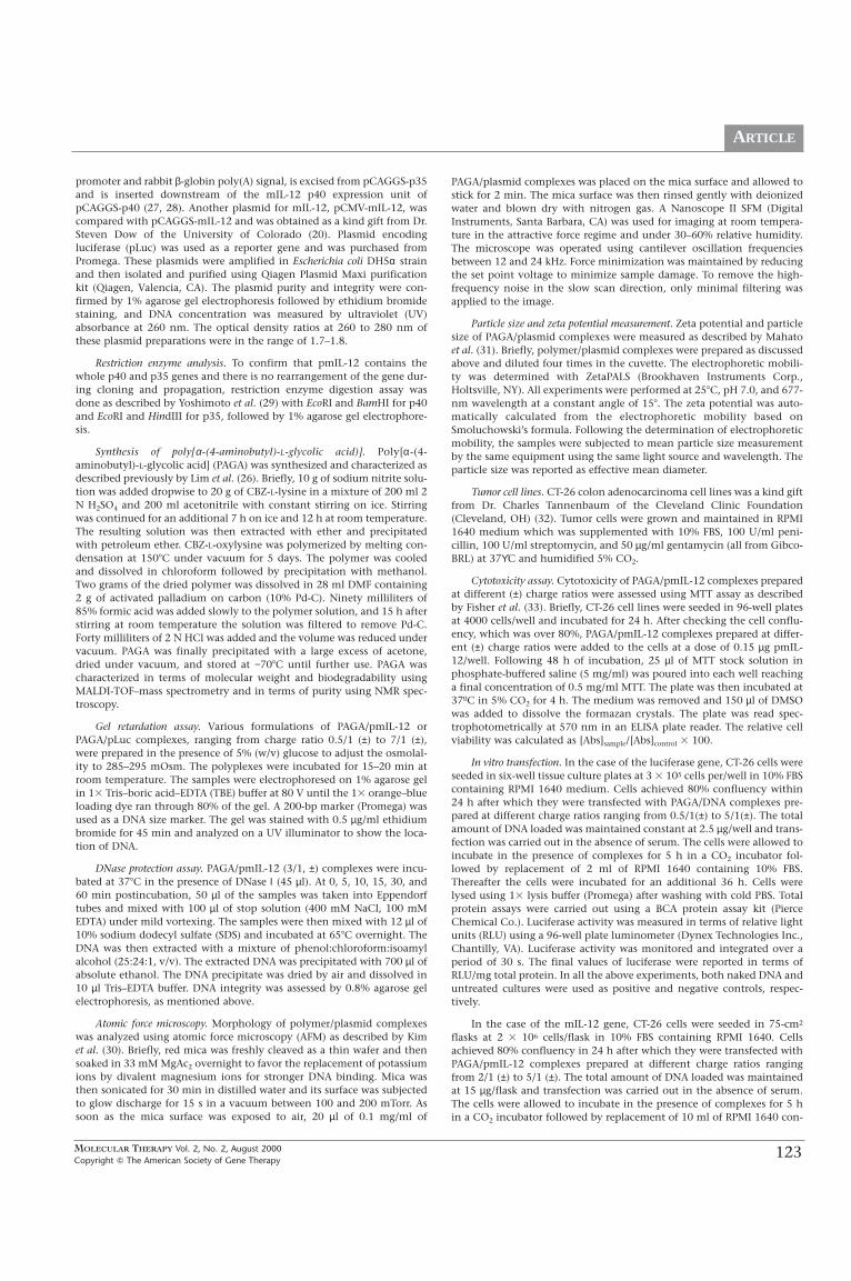
Atomic force microscopy. Morphology of polymer/plasmid complexeswas analyzed using atomic force microscopy (AFM) as described by Kimet al. (30). Briefly, red mica was freshly cleaved as a thin wafer and thensoaked in 33 mM MgAc2 overnight to favor the replacement of potassiumions by divalent magnesium ions for stronger DNA binding. Mica wasthen sonicated for 30 min in distilled water and its surface was subjectedto glow discharge for 15 s in a vacuum between 100 and 200 mTorr. Assoon as the mica surface was exposed to air, 20 µl of 0.1 mg/ml of
PAGA/plasmid complexes was placed on the mica surface and allowed tostick for 2 min. The mica surface was then rinsed gently with deionizedwater and blown dry with nitrogen gas. A Nanoscope II SFM (DigitalInstruments, Santa Barbara, CA) was used for imaging at room tempera-ture in the attractive force regime and under 30–60% relative humidity.The microscope was operated using cantilever oscillation frequenciesbetween 12 and 24 kHz. Force minimization was maintained by reducingthe set point voltage to minimize sample damage. To remove the high-frequency noise in the slow scan direction, only minimal filtering wasapplied to the image.
Particle size and zeta potential measurement. Zeta potential and particlesize of PAGA/plasmid complexes were measured as described by Mahatoet al. (31). Briefly, polymer/plasmid complexes were prepared as discussedabove and diluted four times in the cuvette. The electrophoretic mobili-ty was determined with ZetaPALS (Brookhaven Instruments Corp.,Holtsville, NY). All experiments were performed at 25�C, pH 7.0, and 677-nm wavelength at a constant angle of 15�. The zeta potential was auto-matically calculated from the electrophoretic mobility based onSmoluchowski’s formula. Following the determination of electrophoreticmobility, the samples were subjected to mean particle size measurementby the same equipment using the same light source and wavelength. Theparticle size was reported as effective mean diameter.
Tumor cell lines. CT-26 colon adenocarcinoma cell lines was a kind giftfrom Dr. Charles Tannenbaum of the Cleveland Clinic Foundation(Cleveland, OH) (32). Tumor cells were grown and maintained in RPMI1640 medium which was supplemented with 10% FBS, 100 U/ml peni-cillin, 100 U/ml streptomycin, and 50 µg/ml gentamycin (all from Gibco-BRL) at 37ϒC and humidified 5% CO2.
Cytotoxicity assay. Cytotoxicity of PAGA/pmIL-12 complexes preparedat different (±) charge ratios were assessed using MTT assay as describedby Fisher et al. (33). Briefly, CT-26 cell lines were seeded in 96-well platesat 4000 cells/well and incubated for 24 h. After checking the cell conflu-ency, which was over 80%, PAGA/pmIL-12 complexes prepared at differ-ent (±) charge ratios were added to the cells at a dose of 0.15 µg pmIL-12/well. Following 48 h of incubation, 25 µl of MTT stock solution inphosphate-buffered saline (5 mg/ml) was poured into each well reachinga final concentration of 0.5 mg/ml MTT. The plate was then incubated at37ºC in 5% CO2 for 4 h. The medium was removed and 150 µl of DMSOwas added to dissolve the formazan crystals. The plate was read spec-trophotometrically at 570 nm in an ELISA plate reader. The relative cellviability was calculated as [Abs]sample/[Abs]control � 100.
In vitro transfection. In the case of the luciferase gene, CT-26 cells wereseeded in six-well tissue culture plates at 3 � 105 cells per/well in 10% FBScontaining RPMI 1640 medium. Cells achieved 80% confluency within24 h after which they were transfected with PAGA/DNA complexes pre-pared at different charge ratios ranging from 0.5/1(±) to 5/1(±). The totalamount of DNA loaded was maintained constant at 2.5 µg/well and trans-fection was carried out in the absence of serum. The cells were allowed toincubate in the presence of complexes for 5 h in a CO2 incubator fol-lowed by replacement of 2 ml of RPMI 1640 containing 10% FBS.Thereafter the cells were incubated for an additional 36 h. Cells werelysed using 1� lysis buffer (Promega) after washing with cold PBS. Totalprotein assays were carried out using a BCA protein assay kit (PierceChemical Co.). Luciferase activity was measured in terms of relative lightunits (RLU) using a 96-well plate luminometer (Dynex Technologies Inc.,Chantilly, VA). Luciferase activity was monitored and integrated over aperiod of 30 s. The final values of luciferase were reported in terms ofRLU/mg total protein. In all the above experiments, both naked DNA anduntreated cultures were used as positive and negative controls, respec-tively.
In the case of the mIL-12 gene, CT-26 cells were seeded in 75-cm2
flasks at 2 � 106 cells/flask in 10% FBS containing RPMI 1640. Cellsachieved 80% confluency in 24 h after which they were transfected withPAGA/pmIL-12 complexes prepared at different charge ratios rangingfrom 2/1 (±) to 5/1 (±). The total amount of DNA loaded was maintainedat 15 µg/flask and transfection was carried out in the absence of serum.The cells were allowed to incubate in the presence of complexes for 5 hin a CO2 incubator followed by replacement of 10 ml of RPMI 1640 con-
MOLECULAR THERAPY Vol. 2, No. 2, August 2000Copyright � The American Society of Gene Therapy
124
ARTICLE
taining 10% FBS. Thereafter the cells were incubated for an additional 36h. Culture supernatants were assayed for mIL-12 p70 and p40 using ELISAkits as suggested by the manufacturer.
Tumor implantation and treatment. Subcutaneous tumor-bearingBALB/c mice were used as an animal model for evaluation of PAGA/pLucand PAGA/pmIL-12 complexes for transgene expression. To generatetumors, 5-week-old BALB/c mice were subcutaneously injected in themiddle of the left flank with 100 µl of a single-cell suspension containing4 � 105 CT-26 cells. Tumor volume was calculated by using its meandiameter measured with calipers across its two perpendicular diametersand its depth using the formula v = � abc. Treatment of the tumors wasstarted after about 2 weeks when they reached a size of about 100–120mm3. PAGA/pmIL-12 complexes were prepared at a 3/1 (±) charge ratioand 50 µl of the complexes was injected directly into the tumors ofBALB/c mice at a dose of 25 µg DNA/mouse. The treated mice were sacri-ficed on days 1, 3, 5, and 7 postinjection, whereas the control mice weresacrificed on day 7. Tumors were isolated and cut into two pieces, one forELISA and the other for RT–PCR. The tumor for RT–PCR was frozen imme-diately in liquid nitrogen and stored at −70�C until further use. For ELISA,the tumor was chopped into small pieces, recultured into six-well plates,and incubated at 37�C for 24 h. Supernatants were separated from thecells by centrifugation and measured by ELISA for mIL-12 p70, mIL-12p40, and mIFN-γ. In the case of the luciferase gene, isolated tumors werecut into two pieces, one for luciferase assay and the other for RT–PCR.Luciferase activity was assessed following homogenization and digestionof the tumor with 1� passive lysis buffer.
ELISA for mIL-12 and induced mIFN-γ. Measurement of mIL-12 p70and p40 as well as induced mIFN-γ was done using ELISA set BDOptEIAfor mIL-12 p70, mIL-12 p40, and mIFN-γ (Pharmingen, San Diego, CA)and used according to the manufacturer’s instructions. Briefly, ELISAplates (Nunc, Maxisorp, Denmark) were coated with capture antibody,sealed, and kept overnight for antibody binding. The plate was washedseveral times followed by incubation with assay diluent to block any non-specific binding for 1 h. After washing several times, the plate was thenincubated with samples and standards for 2 h. After incubating withdetection antibody solution containing avidin–HRP reagent for 1 h, thesubstrate solution was added to carry out the enzymatic reaction. Thereaction was stopped with 2 N H2SO4 and the plate was read at 450 nmusing a Bio-Rad (Hercules, CA) Model 3550 ELISA reader. The mIL-12 p70and p40 as well as mIFN-γ concentrations were reported in terms ofpg/ml.
Reverse transcriptase–polymerase chain reaction. RT–PCR was performedto detect mRNA transcripts for mIL-12 and luciferase in transfected tumor
43
cells as well as in tumor tissues of mice. In the case of the mIL-12 gene,tumors were isolated on days 1, 3, 5, and 7 after intratumoral injectionof PAGA/pmIL-12 complexes (3/1, ±) and total RNA was isolated using anRNeasy Qiagen kit (Qiagen Inc., Valencia, CA). Samples were lysed andhomogenized in the presence of guanidine isothiocyanate and thenreverse transcribed using an Omniscript reverse transcriptase kit (Qiagen,Valencia, CA). The reverse-transcribed samples were amplified by PCRtechnique using a Taq polymerase core kit (Qiagen, Valencia, CA).RT–PCR was used to detect the p35 subunit as well as β-actin promoterand pCAGGS. The primers synthesized from 5′ to 3′ were as follows: forpmIL-12 (p35), 5′-GTC TCC CAA GGT CAG CGT TCC-3′ upstream and5′-CTG GTT TGG TCC CGT GTG ATG-3′ downstream; for β-actin, 5′-ATGGTG GGA ATG GGT CAG AAG-3′ upstream and 5′-CAC GCA GCT CATTGT AGA AGG-3′ downstream; and for pCAGGS, 5′-GCC AAT AGG GACTTT CCA T-3′ upstream and 5′-GGT CAT GTA CTG GGC ATA ATG-3′downstream, respectively. The PCR cycling conditions were as follows:Denaturing at 95�C for 15 s, annealing at 56�C for 15 s, and extension at72�C for 30 s. A total of 35 cycles were run for product amplification. ThePCR product was separated by electrophoresis using a 1% agarose gel.The expected sizes of the PCR products from mIL-12 p35 mRNA and β-actin were 297 and 150 bp, respectively. For detection of luciferase tran-scripts, the following primers from 5′ to 3′ were used: 5′-ATG AAG AGATAC GCC CTG GTT-3′ upstream and 5′-CGG GAG GTA GAT GAG ATGTGA-3′ downstream. The primers for β-actin were the same as mentionedabove. The PCR cycling conditions for luciferase were as follows: dena-turing at 95�C for 1 min, annealing at 54.4�C for 1 min, and extensionat 72�C for 2 min. A total of 35 cycles were run for product amplification.
Antitumor effect after single injection. PAGA/pmIL-12 (3/1, ±) complex-es were injected directly into CT-26 subcutaneous tumor-bearing BALB/cmice at a dose of 25 µg pmIL-12/mouse. Naked pmIL-12 (25 µg/mouse)as well as 5% glucose was also injected into separate CT-26 tumor-bear-ing mice and these were used as controls. After a single intratumoralinjection, mice were closely monitored twice a week for tumor growth..Tumor progression was reported in terms of tumor volume (mm3) over aperiod of 56 days.
RESULTS
Physicochemical Properties of PAGA/Plasmid Complexes
MALDI-TOF–mass spectrometric analysis of PAGAshowed its molecular weight to be approximately 3200.
FIG. 1. Atomic force microscopy (AFM) image of aPAGA/pDNA complex (3/1, ±). Condensed plasmid DNAforms an ellipsoidal structure as seen in Z direction.
MOLECULAR THERAPY Vol. 2, No. 2, August 2000Copyright � The American Society of Gene Therapy
125
ARTICLE
PAGA was characterized in terms of purity using NMR[1H NMR(D2O); 1.4(2H), 1.6(2H), 1.8(2H), 2.9(2H),5.2(1H)] and the disappearance of the CBZ peak at 7.5ppm indicated that the deprotection was complete (26).Fast degradation of PAGA could be attributed mainly tothe autohydrolysis of the ester bond, which was replacedby the ε-amine bond.
DNA condensing ability of PAGA was determined bygel retardation assay. The positively charged PAGAmakes strong complexes with the negatively chargedphosphate ions on sugar backbone of DNA. When thecharge ratio (±) reached 1.5:1, no free DNA was seen(data not shown). PAGA could protect plasmids fromdegradation by nucleases up to 60 min postincubation inthe presence of DNase at charge ratios 2/1 (±) (data notshown). DNA condensation, its reduction in overall size,and the resulting surface morphology were captured byAFM (Fig. 1). pmIL-12 when complexed with PAGA atcharge ratio 3/1 (±) was completely condensed andformed an ellipsoidal structure in Z dimensions. Theellipsoidal particle morphology was supplemented withits particle size of 100 nm along the longest axis of thecomplexes. The AFM imaging revealed that the nakedDNA was condensed from its natural extended state inwater to about one-sixth of its size in the complex. Thisellipsoid morphology might be of special significance asit could facilitate enhanced mobility of the complexthrough the plasma membrane as well as cytosol.
The mean particle size of PAGA/pmIL-12 complexeswhen measured using laser light scattering was within arange of 57–130 nm for charge ratios 1.5/1 (±). The nar-row particle size distribution, for example at 3/1 (±)charge ratio, revealed that the equal volume mixing ofDNA and PAGA during formulation resulted in well-dis-persed, uniform-sized particles with little aggregation.The decreasing size of the particles with the increase incharge ratio reaffirmed the calculations based on molec-ular weight that beyond 1/1 (±) charge ratio and espe-cially at 3/1 (±) ratio the size of the polyplexes was anorder of magnitude lower than that of naked DNA due tocharge–charge interactions.
Zeta potential was in the range of 12–20 mV for thepolyplexes prepared at different charge ratios rangingfrom 1.5/1 (±) to 3/1 (±). The zeta potential proportion-ally increased with the increase in charge ratios and atthe ratio 3/1 (±) was close to 20 mV. A zeta potentialclose to zero would be the most ideal for gene delivery asit would not induce electric polarizability inside the celldue to low positive charge. The gel retardation data (notshown) confirm the above finding as there was no DNAmovement seen at the 1.5/1 (±) charge ratio or above.
Cytotoxicity
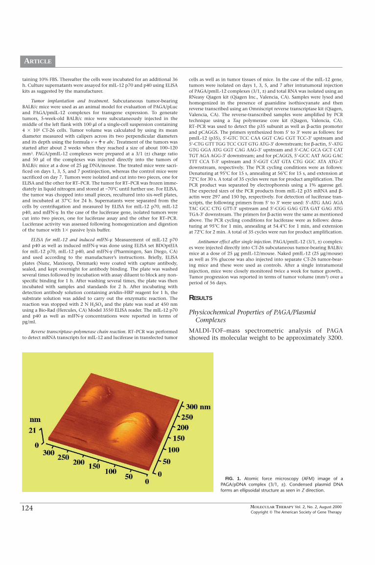
PAGA-based polyplexes were tested for cytotoxicityusing MTT assay in CT-26 cells over a wide range ofcharge ratios. Commercially available cationic liposomes(LipofectAMINE)/pmIL-12 (5/1, w/w) and dendrimer
(SuperFect)/pmIL-12 (6/1, w/w) complexes were used forcomparison. LipofectAMINE reagent is a 3:1 (w/w) lipo-some formulation of the polycationic lipid 2,3-dioley-loxy-N-[2(sperminecarboxamido)ethyl]-N,N-dimethyl-1-propanaminium trifluoroacetate (DOSPA) (MW 867) andthe neutral lipid dioleoyl phosphatidylethanolamine(DOPE) (MW 744) in membrane-filtered water. Based onits chemical structure, LipofectAMINE has two primaryamines, two secondary amines, and one quaternaryamine, totaling five positive charges per molecule (34).Subsequent calculations show that 5/1 (w/w) corre-sponds to 6.823/1 (±) for LipofectAMINE/pDNA com-plexes. Similarly, SuperFect, a G6 dendrimer, has a30,000 MW, with approximately 140 total protonatableamines, so that 6/1 (w/w) corresponds to 8.8/1 (±) forSuperFect/pDNA complexes. Following normalization by(±) charge ratios, we thus confirmed that PAGA/pmIL-12complexes were indeed nontoxic to the cells when for-mulated at a charge ratio of 7/1 (±) and below (Fig. 2). Incontrast, SuperFect/pmIL-12 and LipofectAMINE/pmIL-12 were toxic to the cells. Superfect- and Lipofectamine-treated cells granulated and the cell populationdecreased under the same or lower concentration asPAGA. PAGA alone was also put to test for its cytotoxici-ty at amounts equivalent to those used in charge ratios7/1 (±) and 9/1 (±). Even at such high charge ratios thecell viability attained a decent range of 72–79% (data notshown). Previously, we compared PAGA/pβ-Gal andPLL/pβ-Gal complexes for cytotoxicity in 293 cells andfound PAGA-based formulations to be nontoxic (26). Bytaking care of high cytotoxicity and a reported low levelof transgene expression in subcutaneous tumor models(2, 24), we did not test cationic liposome-
FIG. 2. Viability of CT-26 colon adenocarcinoma cells after transfection withPAGA/pmIL-12 (±) complexes prepared at different (±) charge ratios in 5%(w/v) glucose. Naked pmIL-12, SuperFect/pmIL-12 (6/1, w/w), andLipofectAMINE/pmIL-12 (5/1, w/w) were used for comparison. Relative cellviability was at least 80% for all PAGA/pmIL-12 complexes. In contrast,SuperFect/pmIL-12 and LipofectAMINE/pmIL-12 complex-based transfectionresulted in less than 60% cell viability.
MOLECULAR THERAPY Vol. 2, No. 2, August 2000Copyright � The American Society of Gene Therapy
126
ARTICLE
(LipofectAMINE) and cationic polymer (SuperFect)-basedformulations for direct intratumoral injections.
In Vitro Transfection
PAGA/pLuc and PAGA/pmIL-12 complexes formulat-ed at different charge ratios in 5% (w/v) glucose wereevaluated for their transfection efficiency in CT-26 coloncarcinoma cell lines. In the case of the luciferase gene,cells were lysed after transfection and RLU and total pro-tein concentration were determined. PAGA/pLuc com-plexes were used over a wide range of charge ratios 1/1(±) but <9/1 (±). The transfection efficiency increasedwith the increase in charge ratios but diminished slight-ly with ratios 7/1 (±) (data not shown). We found similarlevels of luciferase expression at charge ratios 3/1 (±) and5/1 (±). Thus, we chose polyplexes at charge ratio 3/1 (±)for further evaluation. As shown in Fig. 3, the RLU val-ues for PAGA/pLuc complexes gave several orders ofmagnitude higher luciferase levels compared to nakedpLuc. We have previously compared PAGA with PLL forin vitro transfection efficiency and cytotoxicity in 293cells. In vitro transfection efficiency of PAGA/pDNA com-plexes was higher than that of the PLL/pDNA complex(26).
In the case of cytokine genes, the culture medium wascollected after transfection and the levels of mIL-12 p70,mIL-12 p40, and mIFN-γ were determined using ELISA.Trends similar to luciferase transfection followed when agradient of various charge ratios 1/1 (±) was used.However, in the case of mIL-12, the transfection efficien-cy upward of 7/1 (±) regressed more sharply than thatexhibited for condensed luciferase at various ratios (datanot shown). As shown in Fig. 4, the mIL-12 levels forPAGA/pmIL-12 complexes (3/1, ±) were substantiallyhigher than those for naked pmIL-12. Plasmid DNA withCMV promoter produced only slightly higher levels of
mIL-12 compared to the plasmid with β-actin promoter(576 pg/ml vs 690 pg/ml).
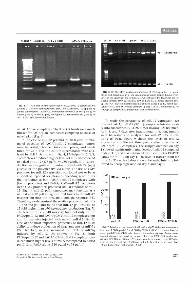
To complement with the ELISA results of in vitro trans-fected samples, RT–PCR was performed to assess thetransfection efficiency of PAGA/pmIL-12 at mRNA levelsand the results are shown in Fig. 5. RT–PCR results showthat the mIL-12 p35 production at the mRNA level is suf-ficient enough to induce the formation of mIL-12 p70 byforming disulfide linkages with mIL-12 p40. The β-actincontrol confirmed that mIL-12 gene expression wasindeed from the plasmid encoding mIL-12 and not fromendogenous production of mIL-12 by the cells. Thebands obtained from RT–PCR suggest that the mIL-12expression at protein levels should be considerably highand mIL-12 p70 secreted from the transfected CT-26 cellsshould also be very high if the relative production levelsof mIL-12 p35 and IL-12 p40 are close to each other. Thiswas not the case when the levels of mIL-12 p70 and p40were compared by ELISA (Fig. 7).
In Vivo Gene Expression
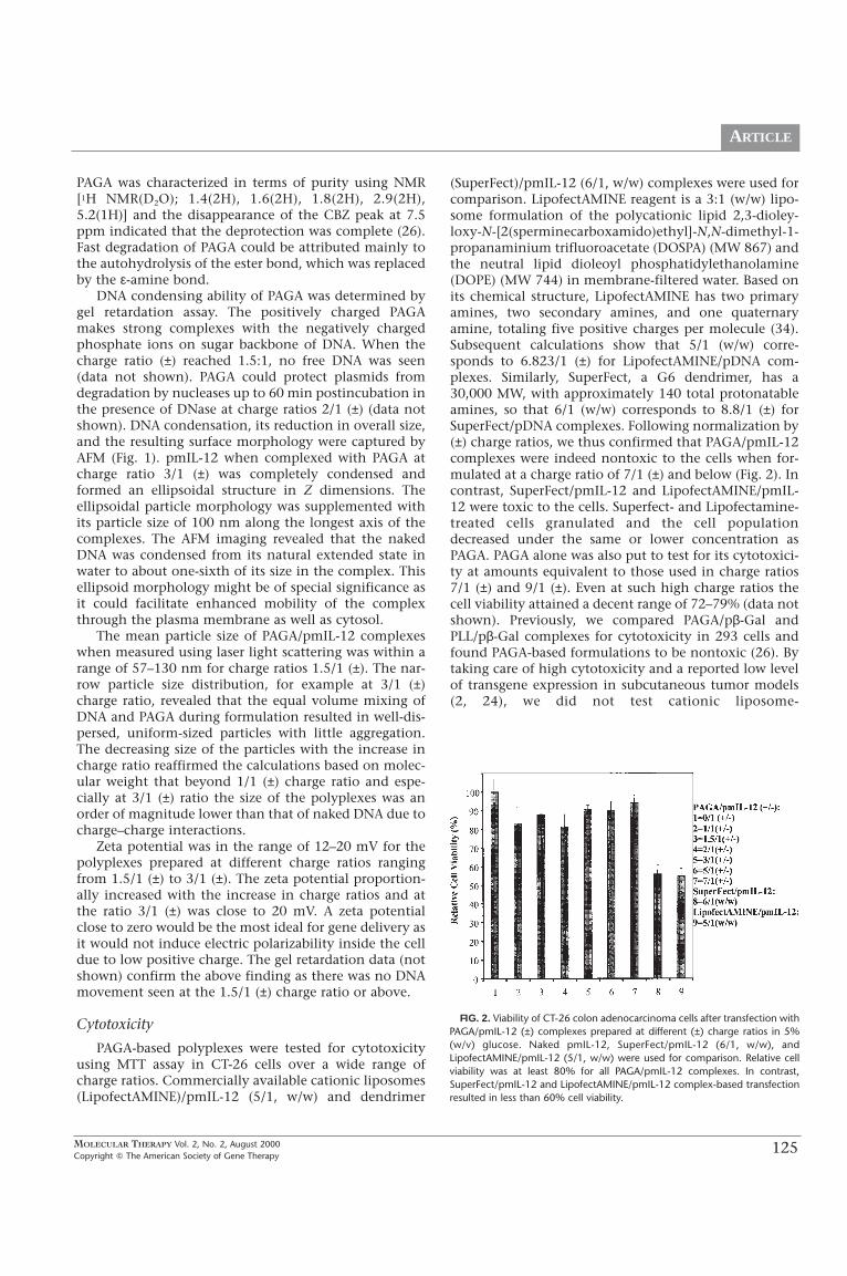
We implanted CT-26 colon adenocarcinoma cells intoBALB/c mice to assess the in vivo gene transfer efficiencyof PAGA, PAGA/pLuc (3/1, ±), PAGA/pmIL-12 (3/1, ±),and PAGA/pCMV-IL-12 (3/1, ±) complexes injected intra-tumorally. For the luciferase gene, transgene expressionwas assessed at mRNA and protein levels using RT–PCRand luciferase assay, respectively (Fig. 3 and Fig. 6).RLU/mg of protein was significantly higher forPAGA/pLuc complexes, compared to naked pLuc in cul-tured cells. RLU/mg protein values for intratumorallyinjected naked pLuc were only twofold lower than that
FIG. 3. Luciferase activity in cultured cells as well as CT-26 subcutaneoustumors 48 h after transfection. PAGA/pLuc (3/1, ±) complexes and nakedpLuc were used for in vitro transfection as well as intratumoral injections insubcutaneous tumor-bearing BALB/c mice. Nontransfected cells (in vitro) and5% (w/v) glucose-injected mice (in vivo) were used as negative controls.Luciferase activity is expressed as RLU/mg of total protein.
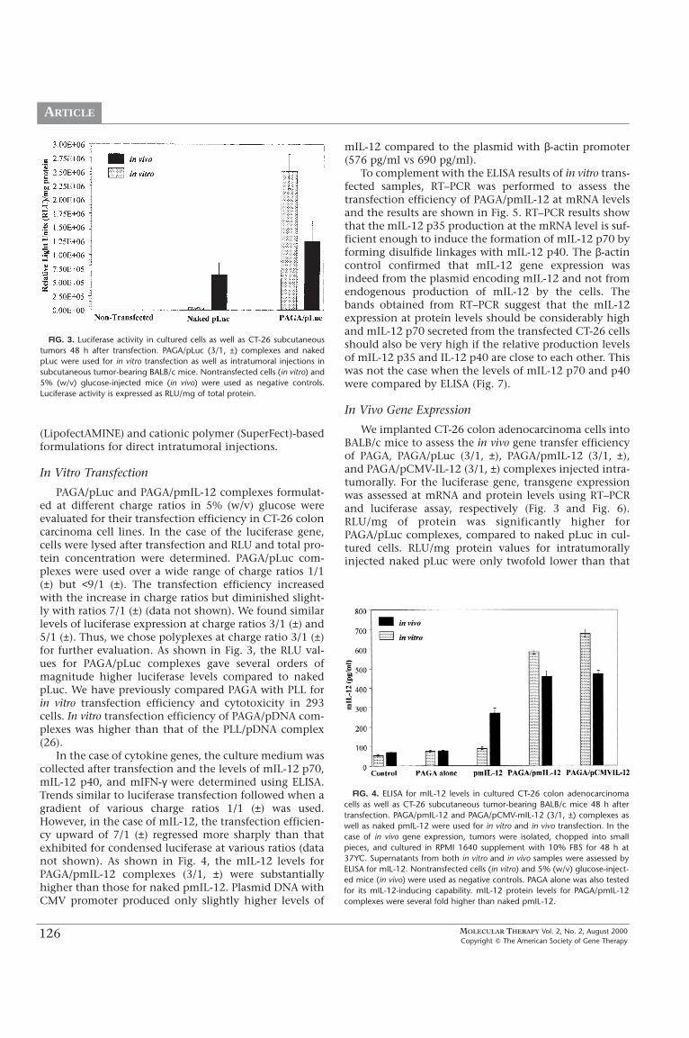
FIG. 4. ELISA for mIL-12 levels in cultured CT-26 colon adenocarcinomacells as well as CT-26 subcutaneous tumor-bearing BALB/c mice 48 h aftertransfection. PAGA/pmIL-12 and PAGA/pCMV-mIL-12 (3/1, ±) complexes aswell as naked pmIL-12 were used for in vitro and in vivo transfection. In thecase of in vivo gene expression, tumors were isolated, chopped into smallpieces, and cultured in RPMI 1640 supplement with 10% FBS for 48 h at37ϒC. Supernatants from both in vitro and in vivo samples were assessed byELISA for mIL-12. Nontransfected cells (in vitro) and 5% (w/v) glucose-inject-ed mice (in vivo) were used as negative controls. PAGA alone was also testedfor its mIL-12-inducing capability. mIL-12 protein levels for PAGA/pmIL-12complexes were several fold higher than naked pmIL-12.
MOLECULAR THERAPY Vol. 2, No. 2, August 2000Copyright � The American Society of Gene Therapy
127
ARTICLE
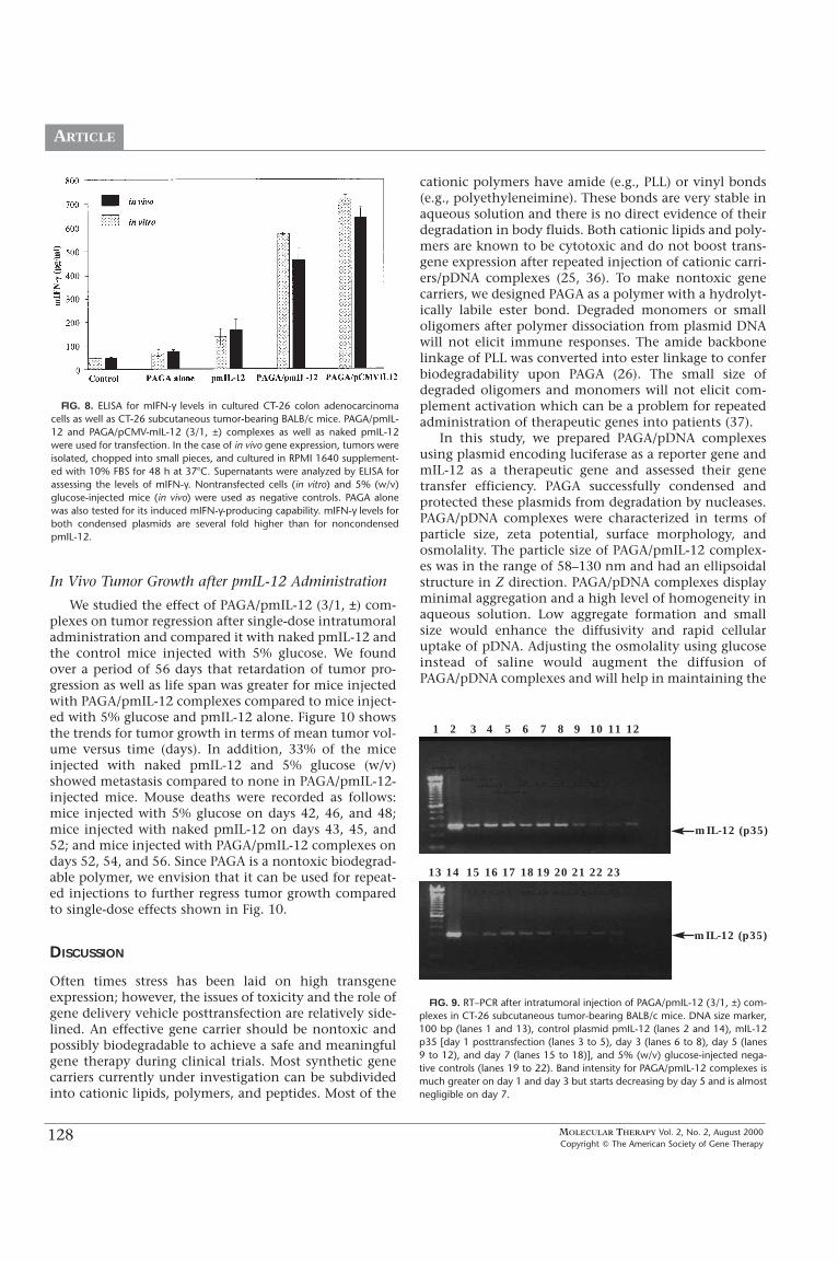
of PAGA/pLuc complexes. The RT–PCR bands were muchthicker for PAGA/pLuc complexes compared to those ofnaked pLuc (Fig. 6).
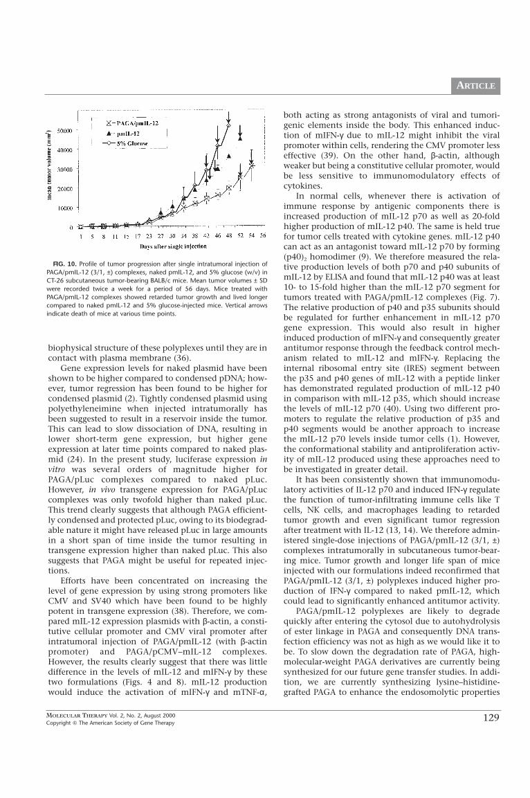
In the case of mIL-12 plasmid, at 48 h after intratu-moral injection of PAGA/pmIL-12 complexes, tumorswere harvested, chopped into small pieces, and recul-tured for 24 h and the culture supernatants were ana-lyzed by ELISA. As shown in Fig. 4, PAGA/pmIL-12 (3/1,±) complexes produced higher levels of mIL-12 comparedto naked pmIL-12 (473 pg/ml vs 250 pg/ml). mIL-12 pro-duction was insignificant in mice injected with 5% (w/v)glucose or the polymer (PAGA) alone. The use of CMVpromoter for mIL-12 expression was found not to be asefficient as reported for plasmids encoding genes otherthan cytokines, as both PAGA/pmIL-12 complexes (withβ-actin promoter) and PAGA/pCMV-mIL-12 complexes(with CMV promoter) produced similar amounts of mIL-12 (Fig. 4). mIL-12 p40 homodimer may function as anatural mIL-12 p70 antagonist that binds to the mIL-12receptor but does not mediate a biologic response (35).Therefore, we determined the relative production of mIL-12 p70 and p40 and found that mIL-12 p40 was 10- to15-fold higher than p70 heterodimer production (Fig. 7).The level of mIL-12 p40 was very high not only for thePAGA/pmIL-12 and PAGA/pCMV-mIL-12 complexes, butalso for the mice injected with naked pmIL-12 (Fig. 7).One of the most important properties of mIL-12 is itsability to induce production of large amounts of mIFN-γ(9). Therefore, we also measured the levels of mIFN-γinduced by mIL-12. As shown in Fig. 8, bothPAGA/pmIL-12 and PAGA/pCMV-mIL-12 complexes pro-duced much higher levels of mIFN-γ compared to nakedpmIL-12 or PAGA alone (530 pg/ml vs 78 pg/ml).
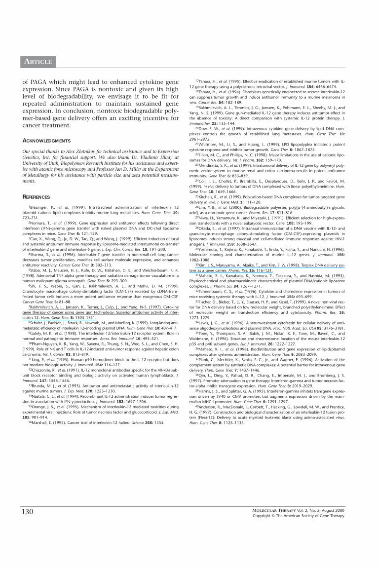
To study the persistence of mIL-12 expression, weinjected PAGA/pmIL-12 (3/1, ±) complexes intratumoral-ly into subcutaneous CT-26 tumor-bearing BALB/c mice.At 1, 3, and 7 days after intratumoral injection, tumorswere harvested and analyzed for mIL-12 p35 mRNAusing RT–PCR. Figure 9 shows the levels of mIL-12expression at different time points after injection ofPAGA/pmIL-12 complexes. The samples obtained on day1 showed significantly higher levels of mIL-12 comparedto days 3, 5, and 7 as evidenced by much thicker RT–PCRbands for mIL-12 on day 1. The level of transcription formIL-12 p35 on day 3 does show substantial intensity fol-lowed by sharp regression on day 5 and day 7.
FIG. 5. RT–PCR after in vitro transfection of PAGA/pmIL-12 complexes intocultured CT-26 colon adenocarcinoma cells. DNA size marker, 100 bp (lane 1),control plasmid pmIL-12 (lane 2), and nontransfected CT-26 cells (lane 3) forβ-actin, (lane 4) for mIL-12 p35, PAGA/pmIL-12 transfected cells, (lane 5) formIL-12 p35, and (lane 6) for β-actin.
FIG. 6. RT–PCR after intratumoral injection of PAGA/pLuc (3/1, ±) com-plexes and naked pLuc in CT-26 subcutaneous tumor-bearing BALB/c mice.Lanes in the upper half are for luciferase while those in the lower half are forβ-actin controls. DNA size marker, 100 bp (lane 1), luciferase plasmid (lane2), 5% (w/v) glucose-injected negative controls (lanes 3 to 5), naked pLuc(lanes 6 to 8), and PAGA/pLuc complexes (lanes 9 to 11). Band intensity forPAGA/pLuc complexes is greater than that of naked DNA.
Marker Plasmid CT-26 cells PAGA/pmIL-12 M P Control pLuc PAGA/pLuc
Luciferase
�-actin
FIG. 7. Relative production of mIL-12 p40 and p70 48 h after intratumoralinjection of PAGA/pmIL-12 and PAGA/pCMV-mIL-12 (3/1, ±) complexes ornaked pmIL-12 into CT-26 subcutaneous tumor-bearing mice. Tumors wereisolated, chopped into small pieces, and cultured in RPMI 1640 supplement-ed with 10% FBS for 48 h at 37�C. Supernatants were analyzed by ELISA forassessing the levels of mIL-12 p40 and p70. mIL-12 p40 levels are more than10-fold higher than that of pmIL-12 p70.
MOLECULAR THERAPY Vol. 2, No. 2, August 2000Copyright � The American Society of Gene Therapy
128
ARTICLE
1 2 3 4 5 6 7 8 9 10 11 12
13 14 15 16 17 18 19 20 21 22 23
In Vivo Tumor Growth after pmIL-12 Administration
We studied the effect of PAGA/pmIL-12 (3/1, ±) com-plexes on tumor regression after single-dose intratumoraladministration and compared it with naked pmIL-12 andthe control mice injected with 5% glucose. We foundover a period of 56 days that retardation of tumor pro-gression as well as life span was greater for mice injectedwith PAGA/pmIL-12 complexes compared to mice inject-ed with 5% glucose and pmIL-12 alone. Figure 10 showsthe trends for tumor growth in terms of mean tumor vol-ume versus time (days). In addition, 33% of the miceinjected with naked pmIL-12 and 5% glucose (w/v)showed metastasis compared to none in PAGA/pmIL-12-injected mice. Mouse deaths were recorded as follows:mice injected with 5% glucose on days 42, 46, and 48;mice injected with naked pmIL-12 on days 43, 45, and52; and mice injected with PAGA/pmIL-12 complexes ondays 52, 54, and 56. Since PAGA is a nontoxic biodegrad-able polymer, we envision that it can be used for repeat-ed injections to further regress tumor growth comparedto single-dose effects shown in Fig. 10.
DISCUSSION
Often times stress has been laid on high transgeneexpression; however, the issues of toxicity and the role ofgene delivery vehicle posttransfection are relatively side-lined. An effective gene carrier should be nontoxic andpossibly biodegradable to achieve a safe and meaningfulgene therapy during clinical trials. Most synthetic genecarriers currently under investigation can be subdividedinto cationic lipids, polymers, and peptides. Most of the
cationic polymers have amide (e.g., PLL) or vinyl bonds(e.g., polyethyleneimine). These bonds are very stable inaqueous solution and there is no direct evidence of theirdegradation in body fluids. Both cationic lipids and poly-mers are known to be cytotoxic and do not boost trans-gene expression after repeated injection of cationic carri-ers/pDNA complexes (25, 36). To make nontoxic genecarriers, we designed PAGA as a polymer with a hydrolyt-ically labile ester bond. Degraded monomers or smalloligomers after polymer dissociation from plasmid DNAwill not elicit immune responses. The amide backbonelinkage of PLL was converted into ester linkage to conferbiodegradability upon PAGA (26). The small size ofdegraded oligomers and monomers will not elicit com-plement activation which can be a problem for repeatedadministration of therapeutic genes into patients (37).
In this study, we prepared PAGA/pDNA complexesusing plasmid encoding luciferase as a reporter gene andmIL-12 as a therapeutic gene and assessed their genetransfer efficiency. PAGA successfully condensed andprotected these plasmids from degradation by nucleases.PAGA/pDNA complexes were characterized in terms ofparticle size, zeta potential, surface morphology, andosmolality. The particle size of PAGA/pmIL-12 complex-es was in the range of 58–130 nm and had an ellipsoidalstructure in Z direction. PAGA/pDNA complexes displayminimal aggregation and a high level of homogeneity inaqueous solution. Low aggregate formation and smallsize would enhance the diffusivity and rapid cellularuptake of pDNA. Adjusting the osmolality using glucoseinstead of saline would augment the diffusion ofPAGA/pDNA complexes and will help in maintaining the
FIG. 8. ELISA for mIFN-γ levels in cultured CT-26 colon adenocarcinomacells as well as CT-26 subcutaneous tumor-bearing BALB/c mice. PAGA/pmIL-12 and PAGA/pCMV-mIL-12 (3/1, ±) complexes as well as naked pmIL-12were used for transfection. In the case of in vivo gene expression, tumors wereisolated, chopped into small pieces, and cultured in RPMI 1640 supplement-ed with 10% FBS for 48 h at 37�C. Supernatants were analyzed by ELISA forassessing the levels of mIFN-γ. Nontransfected cells (in vitro) and 5% (w/v)glucose-injected mice (in vivo) were used as negative controls. PAGA alonewas also tested for its induced mIFN-γ-producing capability. mIFN-γ levels forboth condensed plasmids are several fold higher than for noncondensedpmIL-12.
FIG. 9. RT–PCR after intratumoral injection of PAGA/pmIL-12 (3/1, ±) com-plexes in CT-26 subcutaneous tumor-bearing BALB/c mice. DNA size marker,100 bp (lanes 1 and 13), control plasmid pmIL-12 (lanes 2 and 14), mIL-12p35 [day 1 posttransfection (lanes 3 to 5), day 3 (lanes 6 to 8), day 5 (lanes9 to 12), and day 7 (lanes 15 to 18)], and 5% (w/v) glucose-injected nega-tive controls (lanes 19 to 22). Band intensity for PAGA/pmIL-12 complexes ismuch greater on day 1 and day 3 but starts decreasing by day 5 and is almostnegligible on day 7.
mIL-12 (p35)
mIL-12 (p35)
MOLECULAR THERAPY Vol. 2, No. 2, August 2000Copyright � The American Society of Gene Therapy
129
ARTICLE
biophysical structure of these polyplexes until they are incontact with plasma membrane (36).
Gene expression levels for naked plasmid have beenshown to be higher compared to condensed pDNA; how-ever, tumor regression has been found to be higher forcondensed plasmid (2). Tightly condensed plasmid usingpolyethyleneimine when injected intratumorally hasbeen suggested to result in a reservoir inside the tumor.This can lead to slow dissociation of DNA, resulting inlower short-term gene expression, but higher geneexpression at later time points compared to naked plas-mid (24). In the present study, luciferase expression invitro was several orders of magnitude higher forPAGA/pLuc complexes compared to naked pLuc.However, in vivo transgene expression for PAGA/pLuccomplexes was only twofold higher than naked pLuc.This trend clearly suggests that although PAGA efficient-ly condensed and protected pLuc, owing to its biodegrad-able nature it might have released pLuc in large amountsin a short span of time inside the tumor resulting intransgene expression higher than naked pLuc. This alsosuggests that PAGA might be useful for repeated injec-tions.
Efforts have been concentrated on increasing thelevel of gene expression by using strong promoters likeCMV and SV40 which have been found to be highlypotent in transgene expression (38). Therefore, we com-pared mIL-12 expression plasmids with β-actin, a consti-tutive cellular promoter and CMV viral promoter afterintratumoral injection of PAGA/pmIL-12 (with β-actinpromoter) and PAGA/pCMV–mIL-12 complexes.However, the results clearly suggest that there was littledifference in the levels of mIL-12 and mIFN-γ by thesetwo formulations (Figs. 4 and 8). mIL-12 productionwould induce the activation of mIFN-γ and mTNF-α,
both acting as strong antagonists of viral and tumori-genic elements inside the body. This enhanced induc-tion of mIFN-γ due to mIL-12 might inhibit the viralpromoter within cells, rendering the CMV promoter lesseffective (39). On the other hand, β-actin, althoughweaker but being a constitutive cellular promoter, wouldbe less sensitive to immunomodulatory effects ofcytokines.
In normal cells, whenever there is activation ofimmune response by antigenic components there isincreased production of mIL-12 p70 as well as 20-foldhigher production of mIL-12 p40. The same is held truefor tumor cells treated with cytokine genes. mIL-12 p40can act as an antagonist toward mIL-12 p70 by forming(p40)2 homodimer (9). We therefore measured the rela-tive production levels of both p70 and p40 subunits ofmIL-12 by ELISA and found that mIL-12 p40 was at least10- to 15-fold higher than the mIL-12 p70 segment fortumors treated with PAGA/pmIL-12 complexes (Fig. 7).The relative production of p40 and p35 subunits shouldbe regulated for further enhancement in mIL-12 p70gene expression. This would also result in higherinduced production of mIFN-γ and consequently greaterantitumor response through the feedback control mech-anism related to mIL-12 and mIFN-γ. Replacing theinternal ribosomal entry site (IRES) segment betweenthe p35 and p40 genes of mIL-12 with a peptide linkerhas demonstrated regulated production of mIL-12 p40in comparison with mIL-12 p35, which should increasethe levels of mIL-12 p70 (40). Using two different pro-moters to regulate the relative production of p35 andp40 segments would be another approach to increasethe mIL-12 p70 levels inside tumor cells (1). However,the conformational stability and antiproliferation activ-ity of mIL-12 produced using these approaches need tobe investigated in greater detail.
It has been consistently shown that immunomodu-latory activities of IL-12 p70 and induced IFN-γ regulatethe function of tumor-infiltrating immune cells like Tcells, NK cells, and macrophages leading to retardedtumor growth and even significant tumor regressionafter treatment with IL-12 (13, 14). We therefore admin-istered single-dose injections of PAGA/pmIL-12 (3/1, ±)complexes intratumorally in subcutaneous tumor-bear-ing mice. Tumor growth and longer life span of miceinjected with our formulations indeed reconfirmed thatPAGA/pmIL-12 (3/1, ±) polyplexes induced higher pro-duction of IFN-γ compared to naked pmIL-12, whichcould lead to significantly enhanced antitumor activity.
PAGA/pmIL-12 polyplexes are likely to degradequickly after entering the cytosol due to autohydrolysisof ester linkage in PAGA and consequently DNA trans-fection efficiency was not as high as we would like it tobe. To slow down the degradation rate of PAGA, high-molecular-weight PAGA derivatives are currently beingsynthesized for our future gene transfer studies. In addi-tion, we are currently synthesizing lysine–histidine-grafted PAGA to enhance the endosomolytic properties
FIG. 10. Profile of tumor progression after single intratumoral injection ofPAGA/pmIL-12 (3/1, ±) complexes, naked pmIL-12, and 5% glucose (w/v) inCT-26 subcutaneous tumor-bearing BALB/c mice. Mean tumor volumes ± SDwere recorded twice a week for a period of 56 days. Mice treated withPAGA/pmIL-12 complexes showed retarded tumor growth and lived longercompared to naked pmIL-12 and 5% glucose-injected mice. Vertical arrowsindicate death of mice at various time points.
MOLECULAR THERAPY Vol. 2, No. 2, August 2000Copyright � The American Society of Gene Therapy
130
ARTICLE
of PAGA which might lead to enhanced cytokine geneexpression. Since PAGA is nontoxic and given its highlevel of biodegradability, we envisage it to be fit forrepeated administration to maintain sustained geneexpression. In conclusion, nontoxic biodegradable poly-mer-based gene delivery offers an exciting incentive forcancer treatment.
ACKNOWLEDGMENTS
Our special thanks to Alex Zlotnikov for technical assistance and to ExpressionGenetics, Inc. for financial support. We also thank Dr. Vladimir Hlady atUniversity of Utah, Biopolymers Research Institute for his assistance and expert-ise with atomic force microscopy and Professor Jan D. Miller at the Departmentof Metallurgy for his assistance with particle size and zeta potential measure-ments.
REFERENCES
1Blezinger, P., et al. (1999). Intratracheal administration of interleukin 12plasmid–cationic lipid complexes inhibits murine lung metastases. Hum. Gene. Ther. 10:723–731.
2Nomura, T., et al. (1999). Gene expression and antitumor effects following directinterferon (IFN)-gamma gene transfer with naked plasmid DNA and DC-chol liposomecomplexes in mice. Gene Ther. 6: 121–129.
3Cao, X., Wang, Q., Ju, D. W., Tao, Q., and Wang, J. (1999). Efficient induction of localand systemic antitumor immune response by liposome-mediated intratumoral co-transferof interleukin-2 gene and interleukin-6 gene. J. Exp. Clin. Cancer Res. 18: 191–200.
4Sharma, S., et al. (1996). Interleukin-7 gene transfer in non-small-cell lung cancerdecreases tumor proliferation, modifies cell surface molecule expression, and enhancesantitumor reactivity. Cancer Gene Ther. 3: 302–313.
5Staba, M. J., Mauceri, H. J., Kufe, D. W., Hallahan, D. E., and Weichselbaum, R. R.(1998). Adenoviral TNF-alpha gene therapy and radiation damage tumor vasculature in ahuman malignant glioma xenograft. Gene Ther. 5: 293–300.
6Shi, F. S., Weber, S., Gan, J., Rakhmilevich, A. L., and Mahvi, D. M. (1999).Granulocyte–macrophage colony-stimulating factor (GM-CSF) secreted by cDNA-trans-fected tumor cells induces a more potent antitumor response than exogenous GM-CSF.Cancer Gene Ther. 6: 81–88.
7Rakhmilevich, A. L., Janssen, K., Turner, J., Culp, J., and Yang, N-S. (1997). Cytokinegene therapy of cancer using gene gun technology: Superior antitumor activity of inter-leukin-12. Hum. Gene Ther. 8: 1303–1311.
8Schultz, J., Pavlovic, J., Strack, B., Nawrath, M., and Moelling, K. (1999). Long-lasting anti-metastatic efficiency of interleukin 12-encoding plasmid DNA. Hum. Gene Ther. 10: 407–417.
9Gately, M. K., et al. (1998). The interleukin-12/interleukin-12 receptor system: Role innormal and pathogenic immune responses. Annu. Rev. Immunol. 16: 495–521.
10Pham-Nguyen, K. B., Yang, W., Saxena, R., Thung, S. N., Woo, S. L., and Chen, S. H.(1999). Role of NK and T cells in IL-12-induced anti-tumor response against hepatic coloncarcinoma. Int. J. Cancer 81: 813–819.
11Ling, P., et al. (1995). Human p40 homodimer binds to the IL-12 receptor but doesnot mediate biologic activity. J. Immunol. 154: 116–127.
12Chizzonite, R., et al. (1991). IL-12 monoclonal antibodies specific for the 40-kDa sub-unit block receptor binding and biologic activity on activated human lymphoblasts. J.Immunol. 147: 1548–1556.
13Brunda, M. J., et al. (1993). Antitumor and antimetastatic activity of interleukin-12against murine tumors. J. Exp. Med. 178: 1223–1230.
14Nastala, C. L., et al. (1994). Recombinant IL-12 administration induces tumor regres-sion in association with IFN-γ production. J. Immunol. 153: 1697–1706.
15Orange, J. S., et al. (1995). Mechanism of interleukin-12 mediated toxicities duringexperimental viral injections: Role of tumor necrosis factor and glucocorticoid. J. Exp. Med.181: 901–914.
16Marshall, E. (1995). Cancer trial of interleukin-12 halted. Science 268: 1555.
17Tahara, H., et al. (1995). Effective eradication of established murine tumors with IL-12 gene therapy using a polycistronic retroviral vector. J. Immunol. 154: 6466–6474.
18Tahara, H., et al. (1994). Fibroblasts genetically engineered to secrete interleukin-12can suppress tumor growth and induce antitumor immunity to a murine melanoma invivo. Cancer Res. 54: 182–189.
19Rakhmilevich, A. L., Timmins, J. G., Janssen, K., Pohlmann, E. L., Sheehy, M. J., andYang, N. S. (1999). Gene gun-mediated IL-12 gene therapy induces antitumor effect inthe absence of toxicity: A direct comparison with systemic IL-12 protein therapy. J.Immunother. 22: 135–144.
20Dow, S. W., et al. (1999). Intravenous cytokine gene delivery by lipid–DNA com-plexes controls the growth of established lung metastases. Hum. Gene Ther. 10:2961–2972.
21Whitmore, M., Li, S., and Huang, L. (1999). LPD lipopolyplex initiates a potentcytokine response and inhibits tumor growth. Gene Ther. 6: 1867–1875.
22Filion, M. C., and Phillips, N. C. (1998). Major limitations in the use of cationic lipo-somes for DNA delivery. Int. J. Pharm. 162: 159–170.
23Mendiratta, S. K., et al. (1999). Intratumoral delivery of IL-12 gene by polyvinyl poly-meric vector system to murine renal and colon carcinoma results in potent antitumorimmunity. Gene Ther. 6: 833–839.
24Coll, J. L., Chollet, P., Brambilla, E., Desplanques, D., Behr, J. P., and Favrot, M.(1999). In vivo delivery to tumors of DNA complexed with linear polyethyleneimine. Hum.Gene Ther. 10: 1659–1666.
25Kircheis, R., et al. (1999). Polycation-based DNA complexes for tumor-targeted genedelivery in vivo. J. Gene Med. 1: 111–120.
26Lim, Y.-B., et al. (2000). Biodegradable polyester, poly[α-(4-aminobutyl)-L-glycolicacid], as a non-toxic gene carrier. Pharm. Res. 17: 811–816.
27Niwa, H., Yamamura, K., and Miyazaki, J. (1991). Efficient selection for high-expres-sion transfectants with a novel eukaryotic vector. Gene. 108: 193–199.
28Okada, E., et al. (1997). Intranasal immunization of a DNA vaccine with IL-12- andgranulocyte–macrophage colony-stimulating factor (GM-CSF)-expressing plasmids inliposomes induces strong mucosal and cell-mediated immune responses against HIV-1antigens. J. Immunol. 159: 3638–3647.
29Yoshimoto, T., Kojima, K., Funakoshi, T., Endo, Y., Fujita, T., and Nariuchi, H. (1996).Molecular cloning and characterization of murine IL-12 genes. J. Immunol. 156:1082–1088.
30Kim, J. S., Maruyama, A., Akaike, T., and Kim, S. W. (1998). Terplex DNA delivery sys-tem as a gene carrier. Pharm. Res. 15: 116–121.
31Mahato, R. I., Kawabata, K., Nomura, T., Takakura, Y., and Hashida, M. (1995).Physicochemical and pharmacokinetic characteristics of plasmid DNA/cationic liposomecomplexes. J. Pharm. Sci. 84: 1267–1271.
32Tannenbaum, C. S., et al. (1996). Cytokine and chemokine expression in tumors ofmice receiving systemic therapy with IL-12. J. Immunol. 156: 693–699.
33Fischer, D., Bieber, T., Li, Y., Elsasser, H. P., and Kissel, T. (1999). A novel non-viral vec-tor for DNA delivery based on low molecular weight, branched polyethyleneimine: Effectof molecular weight on transfection efficiency and cytotoxicity. Pharm. Res. 16:1273–1279.
34Lewis, J. G., et al. (1996). A serum-resistant cytofectin for cellular delivery of anti-sense oligodeoxynucleotides and plasmid DNA. Proc. Natl. Acad. Sci. USA 93: 3176–3181.
35Tone, Y., Thompson, S. A., Babik, J. M., Nolan, K. F., Tone, M., Raven, C., andWaldmann, H. (1996). Structure and chromosomal location of the mouse interleukin-12p35 and p40 subunit genes. Eur. J. Immunol. 26: 1222–1227.
36Mahato, R. I., et al. (1998). Biodistribution and gene expression of lipid/plasmidcomplexes after systemic administration. Hum. Gene Ther. 9: 2083–2099.
37Plank, C., Mechtler, K., Szoka, F. C., Jr., and Wagner, E. (1996). Activation of thecomplement system by synthetic DNA complexes: A potential barrier for intravenous genedelivery. Hum. Gene Ther. 7: 1437–1446.
38Qin, L., Ding, Y., Pahud, D. R., Chang, E., Imperiale, M. J., and Bromberg, J. S.(1997). Promoter attenuation in gene therapy: Interferon-gamma and tumor necrosis fac-tor-alpha inhibit transgene expression. Hum. Gene Ther. 8: 2019–2029.
39Harms, J. S., and Splitter, G. A. (1995). Interferon-gamma inhibits transgene expres-sion driven by SV40 or CMV promoters but augments expression driven by the mam-malian MHC I promoter. Hum. Gene Ther. 6: 1291–1297.
40Anderson, R., MacDonald, I., Corbett, T., Hacking, G., Lowdell, M. W., and Prentice,H. G. (1997). Construction and biological characterization of an interleukin-12 fusion pro-tein (Flexi-12): Delivery to acute myeloid leukemic blasts using adeno-associated virus.Hum. Gene Ther. 8: 1125–1135.