Bacterial cellulose as a biodegradable material for textile industries
56
TO ASSESS BACTERIAL CELLULOSE AS AN ALTERNATIVE TO PLANT CELLULOSE IN TEXTILE INDUSTRIES IN KENYA BY MARERI BRENDA MORAA 1
Transcript of Bacterial cellulose as a biodegradable material for textile industries
TO ASSESS BACTERIAL CELLULOSE AS AN ALTERNATIVE TO
PLANT CELLULOSE IN TEXTILE INDUSTRIES IN KENYA
BY
MARERI BRENDA MORAA
1

DECLARATION
I declare that this is my own research work presented to the
future ideas forum.
DEDICATION
I dedicate this project to my family and friends for theirsupport and encouragement.
i

ABSTRACT
Bacterial cellulose is an alternative to using plant cellulose
due to its high level of purity due to the absence of
hemicelluloses and lignin and does not require extensive
processing. Bacterial cellulose is an exopolysaccharide produced
by Acetobacter xylinum inoculated in suitable medium consisting
glucose, yeast, citric acid referred to as Hestrin-schramm media.
The concentration of glucose was varied to determine at what
concentration there is maximum production of cellulose. Acetobacter
xylinum was isolated from rotting apples and confirmed by
biochemical and morphological tests shown in Plate 6, 7 and 8. A
bacterial cellulose pellicle formed on the surface of the media
as shown in Plate 9. There was a greater yield of bacterial
cellulose at greater concentration of glucose as seen in Table 1.
Plant cellulose was obtained from crushed maize seeds, hydrolyzed
and compared with the bacterial cellulose. The method used for
comparison was high performance liquid chromatography method.
Plant cellulose contained hemicelluloses and lignin as shown by
Graph 3 compared to bacterial cellulose shown in Graph 2 which
ii

lacked the impurities. It is a cheap alternative since less
processing is required for removal of impurities as it is pure
cellulose.
iii
TABLE OF CONTENTS
DECLARATION..........................................................i
DEDICATION...........................................................i
ABSTRACT............................................................ii
CHAPTER ONE..........................................................1
INTRODUCTION.........................................................1
1.1 Background......................................................1
1.1 Statement of problem.............................................2
1.2 Justification....................................................2
1.3 Objectives.......................................................3
1.3.1 Main objective.................................................3
1.3.2 Specific objectives............................................3
1.4 Null hypothesis..................................................3
CHAPTER TWO..........................................................4
LITERATURE REVIEW....................................................4
2.1 Description and Ecology of Acetobacter xylinum.......................4
2.2 Bacterial cellulose..............................................5
2.3 Production of bacterial cellulose................................6
iv

CHAPTER THREE.......................................................11
MATERIALS AND METHODS...............................................11
3.1 Preparation of fermentation media and culture media conditions. .11
3.2 Isolation of Acetobacter xylinum....................................11
3.3 Initial culture and Subculture of diluted sample................11
3.4 Morphological tests.............................................12
3.5 Gram negative test of Acetobacter xylinum and Biochemical tests......12
3.6 Growth of Bacterial cellulose...................................12
3.7 Purification of the bacterial cellulose pellicle................12
3.8 Lignin determination............................................13
3.9 Extraction of plant cellulose and bacterial cellulose...........13
3.1.1 Dissolution in NaOH...........................................13
3.1.2 Percentage yield of bacterial cellulose.......................14
3.1.3 Data Analysis.................................................14
CHAPTER FOUR........................................................15
RESULTS.............................................................15
4.1 Initial culture of diluted sample...............................15
Plate 3: Colonies after initial culture at 10-4 and 10 -3 dilution factor
....................................................................15
v

4.2 Gram negative test, Endospore staining and Biochemical test.....16
4.3 Morphological tests.............................................17
4.4 Growth of Bacterial Cellulose...................................18
4.5 Dissolution in NaOH (aq)........................................19
4.6 Lignin Determination............................................19
4.7 Bacterial cellulose dry weights.................................19
4.8 Percentage yield of Bacterial Cellulose.........................19
4.9 Data Analysis...................................................20
4.1.0 Analysis of purity of the cellulose extracted.................22
CHAPTER FIVE........................................................24
DISCUSSION..........................................................24
5.1 Acetobacter xylinum characterization................................24
5.2 Bacterial Cellulose growth......................................24
5.3 Analysis of purity..............................................26
CONCLUSION..........................................................28
RECOMMENDATION......................................................29
Recommendations put forward after analysis of the project results to
obtain a greater yield of glucose and obtain more information about
the bacterial glucose include;......................................29
vi

References..........................................................30
vii

List of plates, tables and graphs
Plate 1: Scanning electron micrograph of bacterial cellulose
(Moosavi-Nasab et al., 2011)..................................17
Plate 2: FTIR spectra of bacterial cellulose (Gor et al., 2012)
..............................................................17
Plate 3: Colonies after initial culture at 10-4 and 10 -3 dilution
factor........................................................23
Plate 4: Pure colonies after subculturing.....................24
Plate 5: Green dots of the cream colony and lack of green dots of
translucent colony............................................24
Plate 6: Rod like bacteria x10................................25
Plate 7: Pink rods after gram staining........................26
Plate 8: Gas bubbles in hydrogen peroxide.....................26
Plate 9: Bacterial cellulose after 7 days of incubation.......27
Table 1: Table of dry weights of bacterial cellulose and
different glucose concentrations replicates...................27
Table 2: Table of percentage yield of bacterial cellulose and
different glucose concentrations..............................28
Table 3: Table of bacterial cellulose weights and square roots of
the weights...................................................28
Graph 1: Chromatogram showing peaks for pure cellulose........30
Graph 2: Chromatogram showing peaks for bacterial cellulose. . .31
Graph 3: Chromatogram showing peaks for plant cellulose.......31
viii

ix

CHAPTER ONE
INTRODUCTION
1.1 Background
Bacterial cellulose, a biopolymer produced by several strains of
acetic acid bacteria, has the same chemical structure compared to
plant-derived cellulose, which is a homogeneous polymer composed
of β-1, 4-glycosidic linkages between the glucose molecules.
Paper and textile industries require a significant amount of the
plant derived cellulose, which leads to a considerable demand on
wood biomass. The bacterial cellulose is distinguished from the
plant-derived cellulose by its high degree of polymerization,
high purity, and high water-holding capacity free from lignin and
hemicellulose. In addition, bacterial cellulose has high polymer
crystallinity and excellent physicochemical characteristics
superior to the plant derived cellulose (Wee et al., 2011).
Some species of Acetobacter, recently named as Gluconacetobacter, are
known to produce bacterial cellulose exhibiting superior features
over plant cellulose although being chemically identical. The
1

unique features of this material such as extreme purity, high
crystallinity and degree of polymerization have gained
considerable commercial and scientific interest (Aydin, 2009).
Bacterial cellulose is a suitable biomaterial instead of plant
cellulose because of the high tensile strength, insolubility in
most solvents, non-toxicity and good shape retention. The
bacterial cellulose can be dried upon synthesis and used as raw
material for textile industry as a cheaper alternative and
requires minimal processing. Main disadvantage is the high water
retention capacity which can be dealt with by adding water
repellent biomolecules in the media which is still being
researched
1.1 Statement of problem
Use of bacterial cellulose in Kenya as an alternative raw
material in textile industry has not been exploited. There has
hardly been any study done to establish purity of bacterial
cellulose and its tensile strength due to its ultrafine fibril
structure. Bacterial cellulose has been used in other countries
as food additive, in paper and textile industries and has proven
2
to be a better alternative. By determination of the purity of
bacterial cellulose it can be incorporated into certain
industries as a raw material for textiles. There is need to
sensitize the community on the use of bacterial cellulose as an
eco-friendly and cheaper alternative due to little need for
further processing and high rate of biodegradability. Bacterial
cellulose will reduce the need for wood and plant fibres in
industries hence preserving the environment.
1.2 Justification
Bacterial cellulose can be synthesized at lab scale using locally
available chemicals and can be more cost effective option due to
the little processing required. Bacterial cellulose has higher
level of purity than plant cellulose which contains
hemicelluloses and lignin. Bacterial cellulose deals with
environmental concerns of production, consumption and disposal of
textiles that is most related to using plant cellulose and
fibres. Bacterial cellulose is a suitable biomaterial instead of
plant cellulose because of the high tensile strength,
insolubility in most solvents, non-toxicity and good shape
3
retention. The bacterial cellulose can be dried upon synthesis
and used as raw material for textile industry as a cheaper
alternative and requires minimal processing.
1.3 Objectives
1.3.1 Main objective
To determine purity of bacterial cellulose compared to plantcellulose in textile industries.
1.3.2 Specific objectives
1. To isolate Acetobacter xylinum from rotting apples
2. To characterize Acetobacter xylinum from morphological and
biochemical tests
3. To extract plant and bacterial cellulose
4. To determine purity of bacterial cellulose and plant
cellulose
1.4 Null hypothesisBacterial cellulose derived from Acetobacter xylinum in suitable
media is not purer than plant cellulose.
4

CHAPTER TWO
LITERATURE REVIEW
2.1 Description and Ecology of Acetobacter xylinum
Acetobacter xylinum is a microorganism found in symbiotic
relationships with plants such as coffee and sugarcane. It is
Gram negative and anaerobic. It has been used as model organism
in the research on synthesis of bacterial cellulose. The
microorganism can be isolated from rotting fruits such as apples
and grapes. By performing suitable biochemical tests and
morphological tests the microorganism can be identified.
Although synthesis of an extracellular gelatinous mat by
Acetobacter xylinum was reported for the first time in 1886 by Brown
using Acetobacter xylinum as a model bacterium, practical work was
started by Hestrin and Schramm in 1954, who proved that resting
and lyophilized Acetobacter cells synthesized cellulose in the
presence of glucose and oxygen (Bielecki et al.,2010).
5

Acetobacter xylinum is an aerobic soil bacterium in the family of
bacteria that ferments carbohydrates to vinegar (Acetobacter aceti).
Acetobacter xylinum, a Gram negative bacterium found in the soil can
frequently be isolated from decaying fruit such as apples and
grapes. A. xylinum is an unusual member of this family because it
synthesizes and extrudes fibrils of cellulose as part of the
metabolism of glucose. The glucose subunits that form the
cellulose micro fibrils are extruded through pores in the cell
wall of the bacteria. In standing laboratory culture of the
bacteria, the cellulose fibrils bundle together to form a mat or
pellicle within which the bacteria are held. The pellicle floats
on the surface of the medium allowing the bacteria to obtain
plenty of oxygen, which they require for growth, multiplication,
and more cellulose synthesis (Cannon and Skinner, 2000).
2.2 Bacterial cellulose
Extensive research on bacterial cellulose revealed that it is
chemically identical to plant cellulose, but its macromolecular
structure and properties differ from plant cellulose (Bielecki et
al., 2010). Researchers have tried to increase the productivity of
6

cellulose from Acetobacter xylinum using various biochemicals.
Recently different carbon sources, such as monosaccharides,
oligosaccharides, alcohol and organic acids, were used in
bacterial cellulose production (Kesh and Sameshima, 2005).
Bacterial cellulose has gained attention in the research realm
for the favorable properties it possesses; such as its remarkable
mechanical properties in both dry and wet states, porosity, water
absorbency, moldability, biodegrability and excellent biological
affinity. Because of these properties, bacterial cellulose has a
wide range of potential applications including use as a
separation medium for water treatment, a specialty carrier for
battery fluids and fuel cells, a mixing agent, a viscosity
modifier, immobilization matrices of proteins or chromatography
substances (Marzieh and Yousefi, 2010).
The first scientific paper was written by Brown in 1886 on a
peculiar fermentative substance.
Under pure cultivation in carbohydrate media, it was observed
that the whole surface of the liquid is covered with a gelatinous
membrane, which may attain a thickness of 25 mm under favorable
circumstances. On removing the membrane from the liquid, it was
7

found to be very tough, especially if an attempt is made to tear
it across its plane of growth. From chemical analysis and various
reactions, the substance was concluded without doubt to be
cellulose, although microscopy at that time only gave a picture
of living bacteria embedded in a transparent structureless film
(Amano et al., 2005).
In recent research work bacterial cellulose has been modified to
be used as electronic paper. Due to the nanostructured nature and
paper-like optical properties when completely dried, microbial
cellulose has been used instead of commercial paper for creating
certain devices (Shah and Brown, 2005).
There have been some studies on breeding bacterial cellulose
producing bacteria. Previously, bacterial cellulose producers
with increased bacterial cellulose synthase activity have been
bred by genetic engineering, with branches of their metabolic
pathway blocked to decrease the amounts of by-products (Tsuchida
and Yoshinaga, 1997). Bacterial cellulose (BC) is chemically
pure, free of undesirable components such as lignin and
hemicellulose (there is no need for chlorine chemical bleaching)
and has high polymer crystallinity and high degree of
8

polymerization that distinguishes it from other forms of
cellulose (Marzieh and Yousefi, 2011)
2.3 Production of bacterial cellulose
Usually, glucose and sucrose are used as carbon sources for
cellulose production, although other carbohydrates such as
fructose, maltose, xylose, starch and glycerol have also been
tried. The effect of initial glucose concentration on cellulose
production is also important, since the formation of gluconic
acid as a byproduct in the medium decreases the pH of the culture
and ultimately decreases the production of cellulose. The
addition of acetic acid in the media has proven to decrease the
production of gluconic acid.
Shihara and colleagues in 1945 used xylose as a carbon source for
the production of cellulose by A. xylinum and obtained a yield of
3.0 g/L. Sucrose, mannitol and glucose were found to be the
optimal carbon sources for cellulose production by A. xylinum
(Chawla et al., 2009).
9

Components of sugarcane molasses were added such as sucrose,
fructose, glucose, nitrogenous compounds, non-nitrogenous acids,
nucleic acids, vitamins, other carbohydrates, minerals and black
colour substances individually or in combined forms into Hestrin-
Schramm medium. Their effect on bacterial cellulose production by
Acetobacter xylinum was investigated. They concluded that the
addition of vitamins, amino acids, other carbohydrates, minerals
and black colour substances to the molasses in the Hestrin-
Schramm medium with a mixture of sucrose and fructose as the
carbon source increased the bacterial cellulose yield.
The effect of the addition of sodium alginate on bacterial
cellulose production by Acetobacter xylinum was studied. It was
observed that sodium alginate hindered the formation of large
clumps of bacterial cellulose, and enhanced cellulose yield. The
effect of green tea on cellulose production by a G. xylinus strain
isolated from kombucha was also investigated. Green tea at a
level of 3 g/L gave the highest cellulose yield of 3.34 g/L after
7 days of incubation (Chawla, 2009).
10
The results of various carbon sources for maximum cellulose
production were performed by Panesar and colleagues. It was
observed that the maximum cellulose production was obtained in
mannitol followed by glucose. Similar trend of cellulose
production has also been reported by other researchers. Mannitol
gave best yield but due to the cost factor glucose was selected
as carbon source for further experimentation. Peptone, sodium
nitrate and methionine were found to be most effective among
these nitrogen sources. Panesar reported that vitamins and amino
acids played important role for cell growth and cellulose
production. Considering the cost factor sodium nitrate was
screened as nitrogen source for further experimental studies
(Panesar et al., 2009).
Cellulose was isolated for gas liquid analysis by extraction
procedure involving acetic acid and nitric acid modified to
recover product. Materials such as corn were proven to have high
levels of lignin by hydrolyzation with 72% sulphuric acid.
Complete dissolution will not be achieved fully due to presence
of lignin (Sloneker, 1971).
11
It was concluded that the synthesis of cellulose involved several
enzymatic processes that Acetobacter xylinum is involved in.
Microbial cellulose have wide application in various fields such
as food, healthcare, cosmetics and beauty, clothing and shoes,
outdoor sports, baby care products, audio products like speaker
diaphragms (Cannon and Skinner, 2000).
It is also being used in paper industry for making electronic
paper display packaging industry for building materials;
pharmaceuticals, cosmetics, gelling agents and medicines for
wound dressing (Panesar et al., 2009). In the Institute of
Chemical Fibres, Poland, an ecological method has been developed
for manufacturing of bacterial cellulose materials suitable for
medical applications. Being similar to human skin, bacterial
cellulose can be applied as skin substitute in treating extensive
burns (Ciechanska, 2004).
2.4 Analysis methods
12

To determine the physical structure of the bacterial cellulose
and standard cellulose fibers, scanning electron microscopy (SEM)
was carried out. The results revealed more delicacy in structure
of bacterial cellulose (Moosavi-Nasab et al., 2011). Scanning
electron micrograph of microbial cellulose, which is
characterized by an ultrafine network structure and the microbial
cellulose layer, constituted by a compact cellulose network
structure.
Plate 1: Scanning electron micrograph of bacterial cellulose(Moosavi-Nasab et al., 2011)
The analysis of crystallinity for the microbial cellulose was
carried out by Fourier transformed infrared spectroscopy (FTIR).
The position and intensity of absorption bands of FTIR
13

spectrometer of a substance are extremely specific to that
substance (Gor et al., 2012).
Plate 2: FTIR spectra of bacterial cellulose (Gor et al., 2012)
FTIR spectra showed that microbial cellulose is free from
contaminants such as lignin or hemicellulose, which is often
present in plant cellulose. According to Gor et al.,2012 a weak and
broad band centered at 891.59 cm-1, and a strong band centered at
1424.18 cm-1 were present in the spectra of the microbial
cellulose samples, defining them as true cellulose (Gor et al.,
2012).
The characteristic wave numbers of cellulose, hemicelluloses and
lignin were used to determine the peak height for individual
components in different mixtures. Lignin was identified to absorb
14

at maximum wavelength of 1514cm-1, cellulose at 1427cm-1 and
hemicellulose 1044cm-1
(Adapa et al., 2009).Analysis of hemicellulose fractions was done
by Karaaslan et al., 2010 from plant matter and results were
obtained from gas liquid chromatography(Karaaslan et al., 2010).
CHAPTER THREE
MATERIALS AND METHODS
3.1 Preparation of fermentation media and culture media conditions
Hestrin-schramm media was used as the basic media. It was
composed of 20g glucose, 5g yeast, 5g peptone, 2.7g Na2HPO4 and
1.15g citric acid. The materials were measured using an
analytical balance and placed in a beaker. Small amount of water
was added and the contents mixed until dissolution. This was
15

topped up to 1 litre and the pH will be adjusted to 6.0 using
citric acid and sodium hydroxide at room temperature.
Agar was be added to 500ml of the media which was heated for
12minutes until boiling point then autoclave. The media was
allowed to cool then dispensed to petri dishes. After
solidification the dishes were sealed with parafilm strips.
3.2 Isolation of Acetobacter xylinum
A sample of a rotting apple was crushed and serial diluted from
dilution factor of 10-0 to 10-4 using sterile water.
3.3 Initial culture and Subculture of diluted sample
The diluted sample was inoculated onto solid Hestrin Schramm
media and incubated for 24hours at 300C.The colonies that
appeared were subcultured further into solid Hestrin Schramm and
incubated at 300C for 24hours. The resulting pure colonies were
tested for presence of Acetobacter xylinum.
3.4 Morphological tests
16

Pure colonies were picked from the culture and spread on a glass
slide and a cover slip was placed. Simple staining was done using
safranin to determine shape and configuration of the colonies,
observations were recorded.
3.5 Gram negative test of Acetobacter xylinum and Biochemical tests
Pure colony was picked from the culture and spread on a glass
slide, a drop of crystal violet dye was placed on the sample on
the slide. Counter stain safranin dye was added after crystal
violet dye. A cover slip was placed on the glass slide. The
sample was observed under the microscope at magnification of x10
to x100, observations were recorded. Catalase test was performed
and endospore test, observations were recorded.
3.6 Growth of Bacterial cellulose
The colonies of Acetobacter xylinum were picked and inoculated into
liquid Hestrin Schramm media followed by incubation at 370C for 7
days. The observations were made after 7 days.
17

3.7 Purification of the bacterial cellulose pellicle
Cellulose pellicle obtained was be washed with distilled water to
remove medium components and treated with 4%NaOH at 800C for 1 hr
to eliminate bacterial cells. The pellicle was weighed and
results were recorded.
3.8 Lignin determination
Maize seeds were finely ground using a pestle and mortar. The
ground seeds were placed in a glass beaker and 72% sulphuric acid
added while stirring. The reaction was observed and recorded.
Small amount of bacterial cellulose was placed in a glass beaker.
72% sulphuric acid was added while stirring and the reaction was
observed.
3.9 Extraction of plant cellulose and bacterial cellulose
Maize seeds was ground finely and 30-150mg used. 3ml Acetic –
nitric acid reagent consisting 150ml of 80% acetic acid and 15ml
18

of concentrated nitric acid added slowly to the ground powder
while mixing.
The test tubes were capped and heated at 1000C for 30 minutes,
cooled then centrifuged. Fibrous precipitate was washed twice
with 3ml of the acetic-nitric acid reagent and twice with 2ml of
acetone. The residue was analyzed using High performance liquid
chromatography (HPLC). A portion of bacterial cellulose was
placed in a beaker and 72% sulphuric acid added until
dissolution. A solution of pure microcrystalline cellulose was
used as a standard for comparison. The resulting mixture was
analyzed by HPLC. Pure microcrystalline cellulose was also
analyzed for comparison.
3.1.1 Dissolution in NaOH
Maize seeds were crushed in a pestle and mortar and the powder
placed in a beaker. 17.5% NaOH (aq) at 20oC was added to the
ground maize seeds and reaction observed and recorded. A portion
of the bacterial cellulose was placed in a beaker and 17.5% NaOH
19
(aq) at 200C added to the bacterial cellulose and the reaction
observed and recorded.
3.1.2 Percentage yield of bacterial cellulose
The percentage yield of bacterial cellulose from the different
concentrations of glucose was determined by a formula after
drying the bacterial cellulose pellicles. The formula was used by
Wee et al., 2011
Percentage yield = Dry weight of bacterial cellulose pellicle
x 100
Weight of carbon source
used in media
3.1.3 Data Analysis
Different concentrations of glucose were used; 20g/l, 30g/l,
40g/l representing the treatments. There were 3 replicates for
each treatment. The experimental units used were petri dishes.
The parameter being evaluated was the percentage yield of
bacterial cellulose with the different concentrations of glucose.
20

The completely randomized block design was used because there was
no extraneous variable.
Non-destructive method was used to weigh the weight of the
resultant bacterial cellulose pellicles. The data obtained was
subjected to analysis using significance level of 0.05 using
ANOVA statistical method.
CHAPTER FOUR
RESULTS
4.1 Initial culture of diluted sample
After 24hours of incubating the diluted rotten apple sample at300C two distinct colonies were observed. Cream and translucentcolonies were observed. Both colonies appeared raised inelevation and smooth in appearance.
21
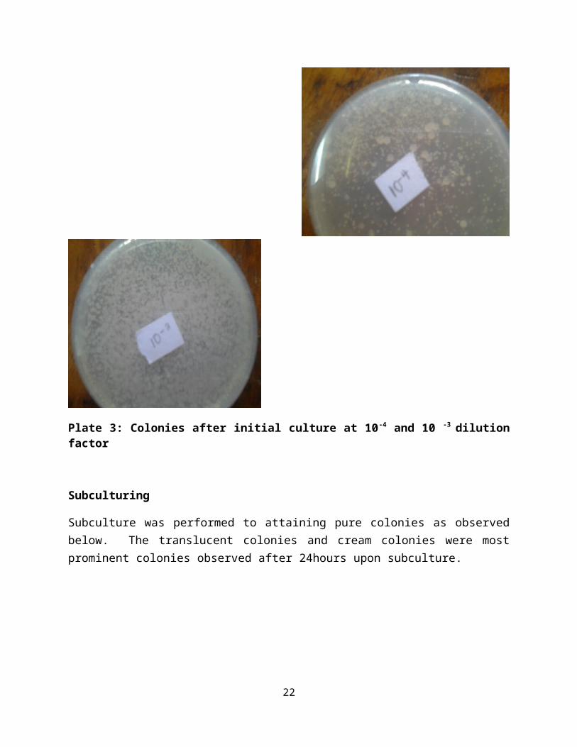
Plate 3: Colonies after initial culture at 10-4 and 10 -3 dilutionfactor
Subculturing
Subculture was performed to attaining pure colonies as observedbelow. The translucent colonies and cream colonies were mostprominent colonies observed after 24hours upon subculture.
22
.
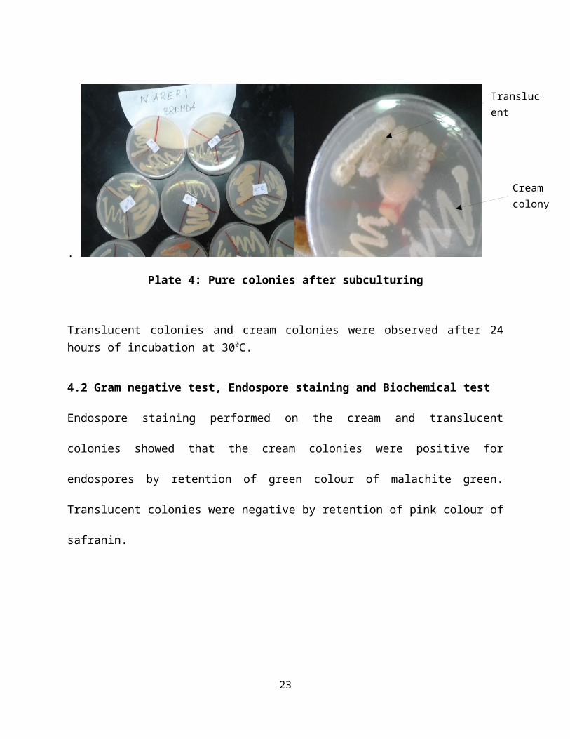
Plate 4: Pure colonies after subculturing
Translucent colonies and cream colonies were observed after 24hours of incubation at 300C.
4.2 Gram negative test, Endospore staining and Biochemical test
Endospore staining performed on the cream and translucent
colonies showed that the cream colonies were positive for
endospores by retention of green colour of malachite green.
Translucent colonies were negative by retention of pink colour of
safranin.
23
Translucent
Cream colony

Plate 5: Green dots of the cream colony pink dots of translucentcolony
4.3 Morphological tests
Morphological tests were carried out on the translucent coloniesand the bacteria viewed under the microscope appeared rod likeafter simple staining with safranin. They appeared singly, inpairs and chains.
Plate 6: Rod like bacteria x10
Gram Staining
Gram negative test was performed on the translucent colonies andpink rods were observed under the microscope confirming that the
24
Rod like bacteria

bacteria are Gram negative by retention of the colour ofsafranin.
Plate 7: Pink rods after gram staining
Catalase test
Catalase test performed in hydrogen peroxide was positive asbubbling was seen when Acetobacter xylinum colony was dipped insidethe hydrogen peroxide.
Plate 8: Gas bubbles in hydrogen peroxide
25
Gas bubbles observed

4.4 Growth of Bacterial Cellulose
After incubation for 7 days at 370C bacterial cellulose pelliclewas observed floating on the surface of the Hestrin Schrammmedia.
Plate 9: Bacterial cellulose after 7 days of incubation
4.5 Dissolution in NaOH (aq)
The maize sample showed dissolution and precipitation when NaOH(aq) was added to the ground maize. The bacterial cellulose didnot dissolve in NaOH (aq).
4.6 Lignin Determination
Upon addition of 72% Sulphuric acid the ground maize seeds didnot dissolve but the bacterial cellulose dissolved in the 72%sulphuric acid.
26
Bacterial cellulo
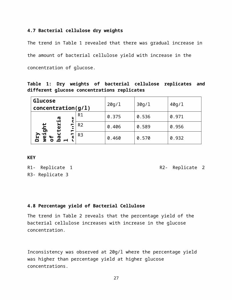
4.7 Bacterial cellulose dry weights
The trend in Table 1 revealed that there was gradual increase in
the amount of bacterial cellulose yield with increase in the
concentration of glucose.
Table 1: Dry weights of bacterial cellulose replicates anddifferent glucose concentrations replicates
Glucoseconcentration(g/l) 20g/l 30g/l 40g/l
Dry
weight
of bacteria
l cellulos
R1 0.375 0.536 0.971R2 0.406 0.589 0.956R3 0.460 0.570 0.932
KEY
R1- Replicate 1 R2- Replicate 2R3- Replicate 3
4.8 Percentage yield of Bacterial Cellulose
The trend in Table 2 reveals that the percentage yield of the bacterial cellulose increases with increase in the glucose concentration.
Inconsistency was observed at 20g/l where the percentage yield was higher than percentage yield at higher glucose concentrations.
27
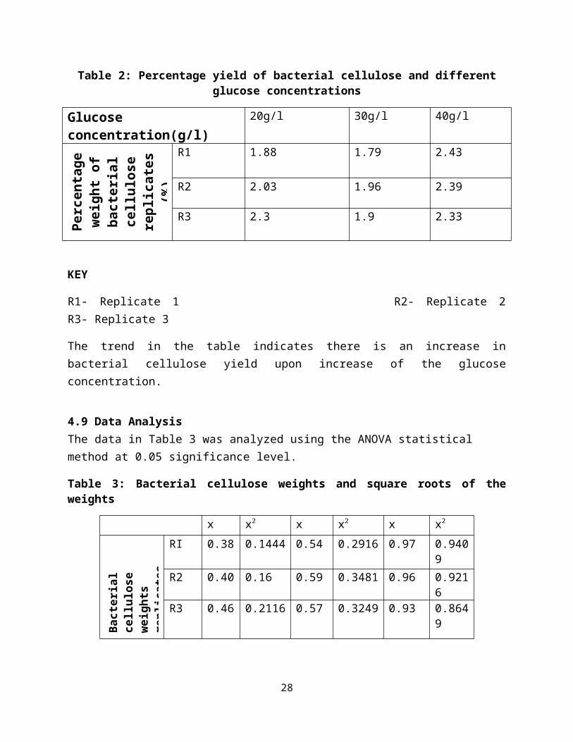
Table 2: Percentage yield of bacterial cellulose and differentglucose concentrations
Glucoseconcentration(g/l)
20g/l 30g/l 40g/l
Percentage
weight of
bacterial
cellulose
replicates
(%)
R1 1.88 1.79 2.43
R2 2.03 1.96 2.39
R3 2.3 1.9 2.33
KEY
R1- Replicate 1 R2- Replicate 2R3- Replicate 3
The trend in the table indicates there is an increase inbacterial cellulose yield upon increase of the glucoseconcentration.
4.9 Data AnalysisThe data in Table 3 was analyzed using the ANOVA statistical method at 0.05 significance level.
Table 3: Bacterial cellulose weights and square roots of theweights
x x2 x x2 x x2
Bact
erial
cell
ulose
weig
hts
repl
icates
RI 0.38 0.1444 0.54 0.2916 0.97 0.9409
R2 0.40 0.16 0.59 0.3481 0.96 0.9216
R3 0.46 0.2116 0.57 0.3249 0.93 0.8649
28

X: dry weight of bacterial cellulose X2: square of x R1-Replicate 1 R2- Replicate 2 R3-Replicate 3
Grand total of x (GT) = 5.8
Sum of x 2 ( x∑ 2) = 4.208
Samples (N) = 9
Stage 2: Correction factorStage 3: total sum of squares (SST)
C= GT2 = 3.74 C= 3.74( x2) – C = ∑ 0.47
N
Stage 4: sum of squares between groups (SSB) Stage 5: sumof squares within group
(SSW)
Tc - C = ∑ 0.46∑(x2) - Tc = ∑ 0.01
nc
nc
Stage 6: degrees of freedom
Degree of freedom for SST = 8
Degree of freedom for SSB = 2
Degree of freedom for SSW = 6
Stage 7: Mean squares
Mean square between groups (MSB) = 0.23
Mean square within groups (MSW) = 0.00229
Stage 8: calculated F valueTabulated F value
F = MSB = 11.5 F value = 11.5F = 5.14
MSW
Comparison of tabulated and calculated F value
Calculated statistic < tabulated statistic
Reject null hypothesis
Conclusion
There is significant difference between mean weights of bacterialcellulose pellicles in different glucose concentrations at 5%significance level.
4.1.0 Analysis of purity of the cellulose extracted
The chromatographs generated after HPLC analysis of plant,bacterial and pure microcrystalline cellulose. The peaksrepresented different retention times of the components in eachsample.
30
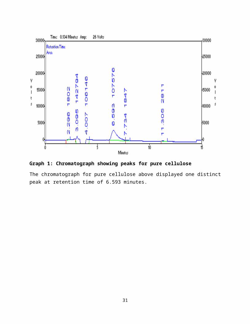
Graph 1: Chromatograph showing peaks for pure cellulose
The chromatograph for pure cellulose above displayed one distinctpeak at retention time of 6.593 minutes.
31
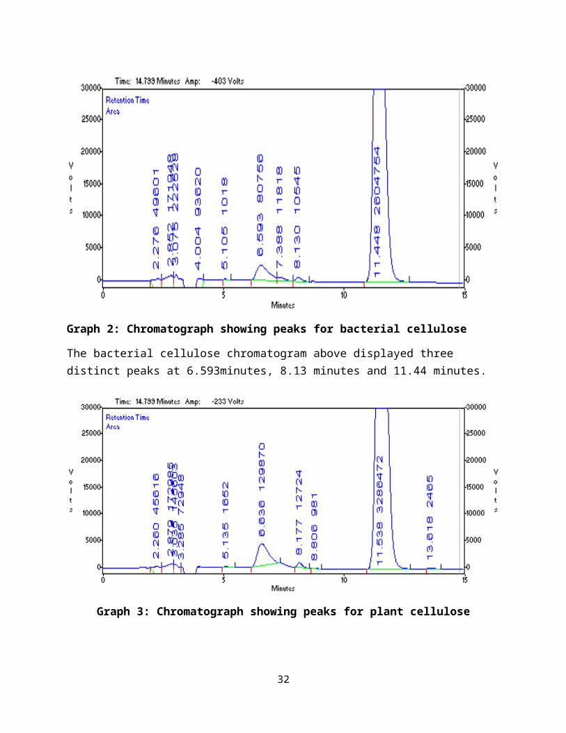
Graph 2: Chromatograph showing peaks for bacterial cellulose
The bacterial cellulose chromatogram above displayed three distinct peaks at 6.593minutes, 8.13 minutes and 11.44 minutes.
Graph 3: Chromatograph showing peaks for plant cellulose
32

The plant cellulose chromatograph above displayed 4 distinct peaks at 6.63 minutes, 8.17 minutes, 11.53 minutes and 13.618 minutes.
CHAPTER FIVE
DISCUSSION
5.1 Acetobacter xylinum characterization
Acetobacter xylinum was successfully isolated from rotten apples and
this was confirmed by morphological and biochemical tests. From
the results, the translucent colonies isolated were found to be
motile rod shaped, non spore forming catalase positive, and Gram
negative growing at pH 6.0. According to the eighth edition of
Bergeys manual of determinative Bacteriology, these strains
should be classified into the genera Acetobacter. Acetobacter xylinum
strain showed positive growth at 300C in Hestrin Schramm media
(Kadere et al., 2008).The endospore test was able to confirm the
colonies were non-sporing by observation of pink spots. The cream
colonies when flooded with malachite green appeared green hence
not considered being in the Acetobacter genera (Bielecki et al.,
2010).
33

5.2 Bacterial Cellulose growth
The yield of cellulose, relative to the amount of glucose
consumed, increased with increase in the initial glucose
concentration (Table 1). Bacterial cellulose production was
enhanced with increasing amount of glucose (Coban and Biyik,
2011). This was as a result of the presence of more glucose for
the bacteria to breakdown to form the bacterial cellulose.
The trend in Table 2. showed inconsistencies in percentage yield
of bacterial cellulose as there was lower yield at higher glucose
concentrations of glucose. This was attributed to conversion of
glucose to gluconic acid by glucose dehydrogenase which lowered
the Hestrin Schramm media pH hence lowering yield of the
bacterial cellulose (Wee et al., 2011).
Yeast extract added contained abundant nitrogen compounds as well
as many growth factors, its addition stimulated cellulose
production by Acetobacter (Son et al., 2001). A. xylinum converts glucose
into cellulose from direct cellulose precursor UDPGlc to glucose-
6-phosphate, catalyzed by glucokinase, followed by isomerization
of this intermediate to Glc-1-P, catalyzed by phosphoglucomutase,
34

and conversion of the latter metabolite to UDPGlc by UDPGlc
pyrophosphorylase (Bielecki et al., 2010).
The glucose subunits that form the cellulose micro fibrils
extruded through pores in the cell wall of the bacteria. The
cellulose fibrils bundled together to form a mat or pellicle
within which the bacteria are held. The pellicle floated on the
surface of the medium allowing the bacteria to obtain plenty of
oxygen, which they require for growth, multiplication, and more
cellulose synthesis (Cannon and Skinner, 2000).The synthesis of
cellulose involved several enzymatic processes that Acetobacter
xylinum is involved in.
Glucose (glucokinase)
Glucose-6-Phosphate
(Phosphoglucomutase)
Glucose-1-Phosphate
(UDP-glucose pyrophosphorylase)
UDP-Glucose
(Cellulose synthase)
35
Cellulose
5.3 Analysis of purity
Cellulose is insoluble in most of the organic solvents due to its
crystalline nature hence insoluble in sodium hydroxide proving it
is a α cellulose which is true cellulose. Plant cellulose from
the maize seeds dissolved and precipitated when sulphuric acid
was added proving that it contained lignin which is an impurity
while bacterial cellulose completely dissolved proving there was
no lignin (Abbot et al., 1988).
The results from the statistical analysis of bacterial cellulose
dry weights showed that there was significant difference between
the mean weights of bacterial cellulose at different glucose
concentrations at 5% significance level. This was determined by
the ANOVA analysis of the results obtained which showed that the
calculated statistic < tabulated statistic.
36
Chemical nature of the bacterial cellulose was defined by
performing high performance liquid chromatography (Graph 1).
Graph 2 shows the chromatograph for the bacterial cellulose while
Graph 3 shows the chromatograph for plant cellulose. The pure
cellulose chromatogram displayed a distinct peak at retention
time of 6.593 minutes which was the cellulose. The bacterial
cellulose displayed peaks at retention time of 6.593 minutes
representing cellulose, 8.13 minutes representing glucose and
11.44 minutes representing sucrose. The plant extract showed
several peaks on the chromatogram which represented 6.63 minutes
retention time for cellulose, 8.177minutes for glucose, 11.53
minutes for hemicellulose and 13.618 minutes for lignin. The
peaks were interpreted by referring to carbohydrate retention
time’s table for HPLC with mobile phase as Acetonitrile: Water
(75:25), using column c18, run time 15minutes and flow rate of
0.6ml/min (Adapa et al., 2009).
The only difference between the peaks of the pure cellulose and
the bacterial cellulose is the presence of glucose and sucrose in
the bacterial cellulose. Graph 2 showed that microbial cellulose
is free from contaminants such as lignin or hemicellulose (Gor et37
al., 2012). Plant cellulose was showed to contain hemicellulose
and lignin contaminants.
By comparing the plant cellulose and bacterial cellulose
chromatographs it can be concluded that plant cellulose contains
contaminants lignin and hemicellulose. The presence of glucose
and sucrose in bacterial cellulose (Graph 2) is as a result of
the enzymatic activities that resulted in formation of the
bacterial cellulose pellicle.
38

CONCLUSION
From the findings, it was concluded that Acetobacter xylinum can be
isolated from rotten apples successfully which was confirmed by
morphological and biochemical tests. Lignin test carried out and
dissolution in NaOH (aq) proved that bacterial cellulose is true
cellulose and did not contain contaminants. Greater amount of
bacterial cellulose was yield from greater concentration of
glucose in the Hestrin Schramm media. Bacterial cellulose was
proven to be purer than plant cellulose by comparison of the HPLC
chromatographs.
The results are of great significance since they indicate that
bacterial cellulose is purer than plant cellulose hence can be
chosen as an alternative to plant cellulose. Due to the high
degree of purity it is easily purified by washing in acetic acid.
Growth of bacterial cellulose was fast taking only two weeks.
Therefore this will reduce the destruction of the plant
vegetation to obtain fibres hence conserving the environment. To
conclude, high purity bacterial cellulose can be successfully
39

produced by Acetobacter xylinum in Hestrin Schramm media and can be
used in the textile industry as an alternative since it requires
minimal processing and no chemical bleaching since it is free of
contaminants.
40
RECOMMENDATIONRecommendations put forward after analysis of the project resultsto obtain a greater yield of glucose and obtain more information about the bacterial glucose include;
1. The effect of initial glucose concentration on cellulose
production is also important, since the formation of
gluconic acid as a byproduct in the medium decreases the pH
of the culture and ultimately decreases the production of
cellulose. The addition of acetic acid in the media is
recommended to decrease the production of gluconic acid.
2. For a higher yield commercial production of bacterial
cellulose it is recommended that agitated shaking culture
techniques be employed.
3. The tensile properties of bacterial cellulose could be
determined for further information on the bacterial
cellulose mechanical strength and use of the scanning
electron microscope to determine physical structure is
recommended.
4. To determine the optimum concentration of glucose for
maximum bacterial cellulose yield, higher glucose
41

concentrations are recommended to be used in further
experimentation.
42
References
1. Adapa.P.K, C. Karunakaran, L.G Tabil and G.J Schoenau
(2009), Qualitative and Quantitative Analysis of
Lignocellulosic Biomass using Infrared spectroscopy,
CSBE/SCGAB 2009 Annual conference, Paper No. CSBE09-307
2. Amano.Y, F.Ito and T.Kanda,( 2009), Novel cellulose
producing system by microorganisms such as Acetobacter sp.,
Journal of Biology and Macromology, Vol.5 (1), pp3-10
3. Aydin A.Y and N.D Aksoy, (2009), Isolation of cellulose
producing bacteria from wastes of vinegar fermentation,
World Congress on Engineering and Computer Science, Vol.1
4. Bielecki .S, A. Krystynowicz, M.Turkiewicz and
H.Kalinowska, (2010), Bacterial cellulose, Institute of
technical biochemistry of Lodz Stefanowskiego, pp90-924
5. Cannon R.E and P.O Skinner, (2000), Acetobacter xylinum: An
inquiry into cellulose biosynthesis, The American Biology
Teacher, Vol.62, No.6
6. Chawla P.R, I.B Bajaj, S.A Survase and R.S Singhal, (2009),
Microbial cellulose: Fermentative production and
43

Applications, Journal of Food Technology and Biotechnology, Vol.47
(2), pp 107-124.
7. Ciechanska .D, (2004), Multifunctional bacterial cellulose,
Institute of Chemical Fibres,
Vol.12, No.4, pp48
8. Goh W.N, A.Rasma, B.Kaur, A.Fazilah, A.A.Karim and
B.Rajeev,(2012), Microstructure and physical properties of
microbial cellulose produced during fermentation of black
tea broth (Kombucha).II, International Food research Journal
19(1):153-158
9. Karaaslan A.M, M.A.Tshabalala and G.Buschle-Diller ,
(2010), Wood hemicellulose/chitosan based semi-
interpenetrating network hydrogels: Mechanical swelling
and controlled drug released properties, Bioresources 5(2),
1036-1054
10. Kesk M.A and K.Sameshima, (2005), Evaluation of
different carbon sources for bacterial cellulose
production, African Journal of Biotechnology, Vol.4, pp 478-482
11. Moosavi-Nasab.M and A.R Yousefi, (2010), Investigation
of physicochemical properties of the bacterial cellulose
44

produced by Glucenobacter xylinus from Date syrup, World Academy
of Science Engineering and Technology, Vol.68, pp893-899.
12. Moosavi-Nasab .M and A.R Yousefi, (2011),
Biotechnology production of cellulose by
Glucenobacter xylinus from agricultural waste, Iranian Journal of
Biotechnology,
Vol.9, No.2
13. Panesar P.S, Y.V Chavan, M.B.Bera, O. Chand and H.
Kumar, (2009), Evaluation of Acetobacter strain for the
production of Microbial cellulose, Asian journal of chemistry,
Vol. 21, No.10, SO99-102.
14. Shah. J and M. Brown, (2005), towards electronic paper
displays made from microbial cellulose, Applied microbiology and
Biotechnology, Vol.66, pp 352-355.
15. Sloneker J.H, (1971), Determination of cellulose and
Apparent Hemicellulose in Plant tissue by Gas-liquid
chromatography, Analytical biochemistry, Vol.43, No.2, pp 539-
546
16. Thomas.P.Abbot, Doris.M.Palmer, Sherald.H.Gordon and
Marvin.O.Barghy, (1988), Solid state analysis of Plant
45
polymers, Journal of Wood Chemistry and Technology, Vol.8 (1), pp 1-
28.
17. Tsuchida. T and F. Yoshinaga, (1997), Production of
bacterial cellulose by agitation culture systems, Pure and
applied Chemistry, Vol.69, No.11, pp2453-2458.
18. T. T. Kadere, T. Miyamoto, R. K. Oniang`o, P. M. Kutima
and S. M. Njoroge, (2008), Isolation and identification of
the genera Acetobacter and Gluconobacter in coconut toddy
(mnazi), African Journal of Biotechnology Vol. 7 (16), pp.
2963-2971
19. Wee.Y, S. Kim, S. Yoon and H. Ryu, (2011), Isolation
and characterization of bacterial cellulose producing
bacterium derived from the persimmon vinegar, African journal of
biotechnology, Vol.10 (72), pp 16267-16276.
46