Solid Lipid Nanoparticle (SLN): A Modern Approach for Drug Delivery
9
Journal of Pharma Research 2013, 2(11) 13-21 Journal of P harma Research Review Article Available online through ISSN: 2319-5622 www.jprinfo.com Solid Lipid Nanoparticle (SLN): A Modern Approach for Drug Delivery Roohi Kesharwani, Suresh Kumar Nair, Dilip Patel * Chandra Shekhar Singh College of Pharmacy, Kausambi, Allahabad, U.P., INDIA. Received on: 17-10-2013; Revised and Accepted on: 29-10-2013 ABSTRACT Lipid nanoparticles were developed in the last decade of the last century as alternative carrier system to emulsions, liposomes and polymeric nanoparticles. Solid lipid nanoparticles (SLN) and nanostructured lipid carriers (NLC) are the two main types of li pid nanoparticles. The present review focuses on the utility of SLN in terms of their advantages, production methodology, characterization and applications. Due to their unique size-dependent properties, lipid nanoparticles offer the possibility to develop new therapeutics. The ability to incorporate drugs into nanocarriers offers a new prototype in drug delivery that could be used for secondary and tertiary levels of drug targeting. Hence, solid lipid nanoparticles hold great promise for reaching the goal of controlled and site specific drug delivery and hence have attracted wide attention of researchers. Keywords: Solid Lipid Nanoparticle, SLN, Lipid Nanoparticle, Nanoparticle, Colloidal Drug Delivery. INTRODUCTION Colloidal particles ranging in size between 10 and 1000 nm are known as nanoparticles. They are manufactured from synthetic/natural polymers and ideally suited to optimize drug delivery and reduce toxicity. Over the years, they have emerged as a variable substitute to liposomes as drug carriers. The successful implementation of nanoparticles for drug delivery depends on their ability to penetrate through several anatomical barriers, sustained release of their contents and their stability in the nanometer size [1, 2] . To overcome these limitations of polymeric nanoparticles, lipids have been put forward as an alternative carrier, particularly for lipophilic pharmaceuticals. These lipid nanoparticles are known as solid lipid nanoparticles (SLNs), which are attracting wide attention of formulators world-wide. SLNs are colloidal carriers developed in the last decade as an alternative system to the existing traditional carriers (emulsions, liposomes and polymeric nanoparticles). They are a new generation of submicron-sized lipid emulsions where the liquid lipid (oil) has been substituted by a solid lipid. SLN offer unique properties such as small size, large surface area, high drug loading and the interaction of phases at the interfaces, and are attractive for their potential to improve performance of pharmaceuticals, neutraceuticals and other materials. A solid lipid nanoparticle (SLN) is typically spherical with -an average diameter between 10 to 1000 nanometers. Solid lipid nanoparticles possess a solid lipid core matrix that can solubilize lipophilic molecules. The lipid core is stabilized by surfactants (emulsifiers). The term lipid is used here in a broader sense and includes triglycerides (e.g. tristearin), diglycerides (e.g. glycerol bahenate), monoglycerides (e.g. glycerol monostearate), fatty acids (e.g. stearic acid), steroids (e.g. cholesterol), and waxes (e.g. cetyl palmitate). All classes of emulsifiers (with respect to charge and molecular weight) have been used to stabilize the lipid dispersion. It has been found that the combination of emulsifiers might prevent particle agglomeration more efficiently [3, 4] . In order to overcome the disadvantages associated with the liquid state of the oil droplets, the liquid lipid was replaced by a solid lipid, which eventually transformed into *Corresponding author: Dilip Patel Chandra Shekhar Singh College of Pharmacy, Kausambi, Allahabad, U.P., INDIA. Phone No. +91-9936764324, Fax: 0532-2584090. *E-Mail: [email protected] solid lipid nanoparticles. The reasons for the increasing interest in lipid based system are many fold and include [4] . Better control over release kinetics of encapsulated compound Engineering via size and lipid composition. Melting can serve as trigger. Enhanced bioavailability of entrapped bioactive. Chemical protection of labile incorporated compounds. Much easier to manufacture than biopolymeric nanoparticles. No special solvents required. Wider range of base materials (lipids). Conventional emulsion manufacturing methods applicable. Raw materials essential the same as in emulsions. Very high long-term stability. Application versatility: Can be subjected to commercial sterilization procedures. Can be freeze-dried to produce powdered formulation [4, 5] . Definition of SLN: Solid lipid nanoparticles (SLN) introduced in 1991 represent an alternative carrier system to tradition colloidal carriers such as emulsions, liposomes and polymeric micro and nanoparticles1. Nanoparticles made from solid lipids are attracting major attention as novel colloidal drug carrier for intravenous applications as they have been proposed as an alternative particulate carrier system. SLN are sub-micron colloidal carriers ranging from 50 to 1000 nm, which are composed of physiological lipid, dispersed in water or in aqueous surfactant solution. SLN offer unique properties such as small size, large surface area, high drug loading and the interaction of phases at the interface and are attractive for their potential to improve performance of pharmaceuticals [5] . The SLN system can be easily explained. It is identical to an oil-in-water emulsion for parenteral nutrition (e.g., Intralipid, Lipofundin), but the liquid lipid (oil) of the emulsion has been replaced by a solid lipid, i.e., yielding solid lipid nano particles. SLN are particles made from solid lipid or lipid blends produced by high pressure homogenization. The mean photon correlation spectroscopy (PCS) diameter is typically between approximately 80nm to 1000nm. Particles below 80nm are more difficult to produce because very often they do not recrystallized8 The SLN are dispersed in an aqueous outer phase and stabilized by surfactants, e.g., Tween80, sodium dodecyl sulfate (SDS), lecithin. Alternatively, they can be produced surfactant free using steric stabilizers (e.g.poloxamer180) or an outer of increased viscosity (e.g. ethyl
Transcript of Solid Lipid Nanoparticle (SLN): A Modern Approach for Drug Delivery

Dilip Patel et al., J. Pharm. Res. 2013, 2(11), 13-21
Journal of Pharma Research 2013, 2(11) 13-21
Journal of Pharma Research Review Article Available online through ISSN: 2319-5622
www.jprinfo.com
Solid Lipid Nanoparticle (SLN): A Modern Approach for Drug Delivery
Roohi Kesharwani, Suresh Kumar Nair, Dilip Patel* Chandra Shekhar Singh College of Pharmacy, Kausambi, Allahabad, U.P., INDIA.
Received on: 17-10-2013; Revised and Accepted on: 29-10-2013
ABSTRACT
Lipid nanoparticles were developed in the last decade of the last century as alternative carrier system to emulsions, liposomes and
polymeric nanoparticles. Solid lipid nanoparticles (SLN) and nanostructured lipid carriers (NLC) are the two main types of li pid nanoparticles. The
present review focuses on the utility of SLN in terms of their advantages, production methodology, characterization and applications. Due to their
unique size-dependent properties, lipid nanoparticles offer the possibility to develop new therapeutics. The ability to incorporate drugs into
nanocarriers offers a new prototype in drug delivery that could be used for secondary and tertiary levels of drug targeting. Hence, solid lipid
nanoparticles hold great promise for reaching the goal of controlled and site specific drug delivery and hence have attracted wide attention of
researchers.
Keywords: Solid Lipid Nanoparticle, SLN, Lipid Nanoparticle, Nanoparticle, Colloidal Drug Delivery.
INTRODUCTION
Colloidal particles ranging in size between 10 and 1000
nm are known as nanoparticles. They are manufactured from synthetic/natural polymers and ideally suited to optimize drug delivery and reduce toxicity. Over the years, they have emerged as a variable substitute to liposomes as drug carriers. The successful implementation of nanoparticles for drug delivery depends on their ability to penetrate through several anatomical barriers, sustained release of their contents and their stability in the nanometer size [1,
2]. To overcome these limitations of polymeric nanoparticles, lipids have been put forward as an alternative carrier, particularly for lipophilic pharmaceuticals. These lipid nanoparticles are known as solid lipid nanoparticles (SLNs), which are attracting wide attention of formulators world-wide. SLNs are colloidal carriers developed in the last decade as an alternative system to the existing traditional carriers (emulsions, liposomes and polymeric nanoparticles). They are a new generation of submicron-sized lipid emulsions where the liquid lipid (oil) has been substituted by a solid lipid. SLN offer unique properties such as small size, large surface area, high drug loading and the interaction of phases at the interfaces, and are attractive for their potential to improve performance of pharmaceuticals, neutraceuticals and other materials. A solid lipid nanoparticle (SLN) is typically spherical with -an average diameter between 10 to 1000 nanometers. Solid lipid nanoparticles possess a solid lipid core matrix that can solubilize lipophilic molecules. The lipid core is stabilized by surfactants (emulsifiers). The term lipid is used here in a broader sense and includes triglycerides (e.g. tristearin), diglycerides (e.g. glycerol bahenate), monoglycerides (e.g. glycerol monostearate), fatty acids (e.g. stearic acid), steroids (e.g. cholesterol), and waxes (e.g. cetyl palmitate). All classes of emulsifiers (with respect to charge and molecular weight) have been used to stabilize the lipid dispersion. It has been found that the combination of emulsifiers might prevent particle agglomeration more efficiently [3, 4]. In order to overcome the disadvantages associated with the liquid state of the oil droplets, the liquid lipid was replaced by a solid lipid, which eventually transformed into
*Corresponding author: Dilip Patel Chandra Shekhar Singh College of Pharmacy, Kausambi, Allahabad, U.P., INDIA. Phone No. +91-9936764324, Fax: 0532-2584090. *E-Mail: [email protected]
solid lipid nanoparticles. The reasons for the increasing interest in lipid based system are many fold and include [4].
Better control over release kinetics of encapsulated compound Engineering via size and lipid composition. Melting can serve as trigger.
Enhanced bioavailability of entrapped bioactive. Chemical protection of labile incorporated compounds. Much easier to manufacture than biopolymeric nanoparticles. No special solvents required. Wider range of base materials (lipids). Conventional emulsion manufacturing methods applicable. Raw materials essential the same as in emulsions.
Very high long-term stability. Application versatility: Can be subjected to commercial sterilization procedures. Can be freeze-dried to produce powdered formulation [4, 5].
Definition of SLN: Solid lipid nanoparticles (SLN) introduced in 1991
represent an alternative carrier system to tradition colloidal carriers such as emulsions, liposomes and polymeric micro and nanoparticles1. Nanoparticles made from solid lipids are attracting major attention as novel colloidal drug carrier for intravenous applications as they have been proposed as an alternative particulate carrier system. SLN are sub-micron colloidal carriers ranging from 50 to 1000 nm, which are composed of physiological lipid, dispersed in water or in aqueous surfactant solution. SLN offer unique properties such as small size, large surface area, high drug loading and the interaction of phases at the interface and are attractive for their potential to improve performance of pharmaceuticals [5]. The SLN system can be easily explained. It is identical to an oil-in-water emulsion for parenteral nutrition (e.g., Intralipid, Lipofundin), but the liquid lipid (oil) of the emulsion has been replaced by a solid lipid, i.e., yielding solid lipid nano particles. SLN are particles made from solid lipid or lipid blends produced by high pressure homogenization. The mean photon correlation spectroscopy (PCS) diameter is typically between approximately 80nm to 1000nm. Particles below 80nm are more difficult to produce because very often they do not recrystallized8 The SLN are dispersed in an aqueous outer phase and stabilized by surfactants, e.g., Tween80, sodium dodecyl sulfate (SDS), lecithin. Alternatively, they can be produced surfactant free using steric stabilizers (e.g.poloxamer180) or an outer of increased viscosity (e.g. ethyl
Dilip Patel et al., J. Pharm. Res. 2013, 2(11), 13-21
Journal of Pharma Research 2013, 2(11) 13-21
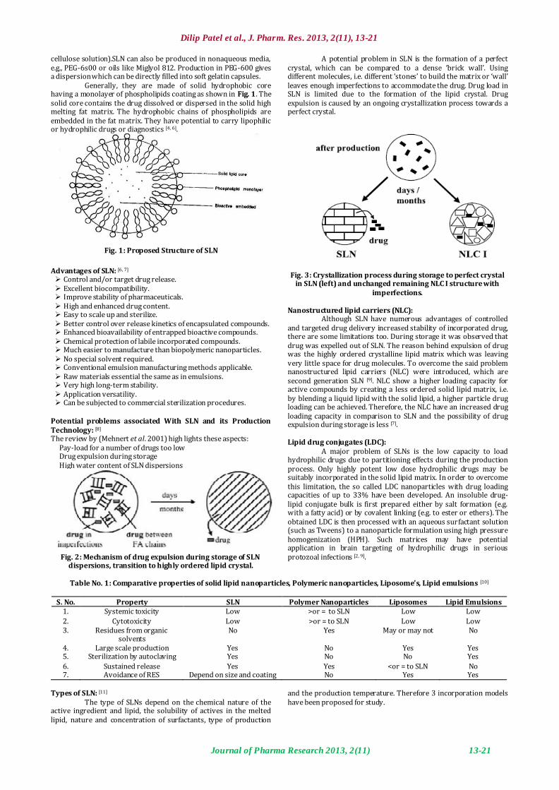
cellulose solution).SLN can also be produced in nonaqueous media, e.g., PEG-6s00 or oils like Miglyol 812. Production in PEG-600 gives a dispersion which can be directly filled into soft gelatin capsules.
Generally, they are made of solid hydrophobic core having a monolayer of phospholipids coating as shown in Fig. 1. The solid core contains the drug dissolved or dispersed in the solid high melting fat matrix. The hydrophobic chains of phospholipids are embedded in the fat matrix. They have potential to carry lipophilic or hydrophilic drugs or diagnostics [4, 6].
Fig. 1: Proposed Structure of SLN
Advantages of SLN: [6, 7] Control and/or target drug release. Excellent biocompatibility. Improve stability of pharmaceuticals. High and enhanced drug content. Easy to scale up and sterilize. Better control over release kinetics of encapsulated compounds. Enhanced bioavailability of entrapped bioactive compounds. Chemical protection of labile incorporated compounds. Much easier to manufacture than biopolymeric nanoparticles. No special solvent required. Conventional emulsion manufacturing methods applicable. Raw materials essential the same as in emulsions. Very high long-term stability. Application versatility. Can be subjected to commercial sterilization procedures.
Potential problems associated With SLN and its Production Technology: [8] The review by (Mehnert et al. 2001) high lights these aspects:
Pay-load for a number of drugs too low Drug expulsion during storage High water content of SLN dispersions
Fig. 2: Mechanism of drug expulsion during storage of SLN
dispersions, transition to highly ordered lipid crystal.
A potential problem in SLN is the formation of a perfect crystal, which can be compared to a dense ‘brick wall’. Using different molecules, i.e. different ‘stones’ to build the matrix or ‘wall’ leaves enough imperfections to accommodate the drug. Drug load in SLN is limited due to the formation of the lipid crystal. Drug expulsion is caused by an ongoing crystallization process towards a perfect crystal.
Fig. 3: Crystallization process during storage to perfect crystal
in SLN (left) and unchanged remaining NLC I structure with imperfections.
Nanostructured lipid carriers (NLC):
Although SLN have numerous advantages of controlled and targeted drug delivery increased stability of incorporated drug, there are some limitations too. During storage it was observed that drug was expelled out of SLN. The reason behind expulsion of drug was the highly ordered crystalline lipid matrix which was leaving very little space for drug molecules. To overcome the said problem nanostructured lipid carriers (NLC) were introduced, which are second generation SLN [9]. NLC show a higher loading capacity for active compounds by creating a less ordered solid lipid matrix, i.e. by blending a liquid lipid with the solid lipid, a higher particle drug loading can be achieved. Therefore, the NLC have an increased drug loading capacity in comparison to SLN and the possibility of drug expulsion during storage is less [7]. Lipid drug conjugates (LDC):
A major problem of SLNs is the low capacity to load hydrophilic drugs due to partitioning effects during the production process. Only highly potent low dose hydrophilic drugs may be suitably incorporated in the solid lipid matrix. In order to overcome this limitation, the so called LDC nanoparticles with drug loading capacities of up to 33% have been developed. An insoluble drug-lipid conjugate bulk is first prepared either by salt formation (e.g. with a fatty acid) or by covalent linking (e.g. to ester or ethers). The obtained LDC is then processed with an aqueous surfactant solution (such as Tweens) to a nanoparticle formulation using high pressure homogenization (HPH). Such matrices may have potential application in brain targeting of hydrophilic drugs in serious protozoal infections [2, 9].
Table No. 1: Comparative properties of solid lipid nanoparticles, Polymeric nanoparticles, Liposome’s, Lipid emulsions [10]
S. No. Property SLN Polymer Nanoparticles Liposomes Lipid Emulsions 1. Systemic toxicity Low >or = to SLN Low Low
2. Cytotoxicity Low >or = to SLN Low Low 3. Residues from organic
solvents No Yes May or may not No
4. Large scale production Yes No Yes Yes 5. Sterilization by autoclaving Yes No No Yes
6. Sustained release Yes Yes <or = to SLN No 7. Avoidance of RES Depend on size and coating No Yes Yes
Types of SLN: [11]
The type of SLNs depend on the chemical nature of the active ingredient and lipid, the solubility of actives in the melted lipid, nature and concentration of surfactants, type of production
and the production temperature. Therefore 3 incorporation models have been proposed for study.

Dilip Patel et al., J. Pharm. Res. 2013, 2(11), 13-21
Journal of Pharma Research 2013, 2(11) 13-21
1. SLN, Type I or homogenous matrix model: The SLN type I is derived from a solid solution of lipid and active ingredient. A solid solution can be obtained when SLN are produced by the cold homogenation method. A lipid blend can be produced containing the active in a molecularly dispersed form. After solidification of this blend it is ground in its solid state to avoid or minimize the enrichment of active molecules in different parts of the lipid nanoparticles.
2. SLN, Type II or drug enriched shell model: It is achieved when SLN are produced by the hot technique, and the active ingredient concentration in the melted lipid is low during the cooling process of the hot o/w nanoemulsion the lipid will precipitate first, leading to a steadily increasing concentration of active molecules in the remaining melt, an outer shell will
solidify containing both active and lipid. The enrichment of the outer area of the particles causes burst release. The percentage of active ingredient localized in the outer shell can be adjusted in a controlled shell model is the incorporation of coenzyme Q10.
3. SLN, Type III or drug enriched core model: Core model can take place when the active ingredient concentration in the lipid melt is high & relatively close to its saturation solubility. Cooling down of the hot oil droplets will in most cases reduce the solubility of the active in melt. When the saturation solubility exceeds, active molecules precipitate leading to the formation of a drug enriched core.
Fig. 4: Models of Incorporation of Active compounds into SLN [10]
Preparation techniques for solid lipid nanoparticles:
SLNs are prepared from lipid, emulsifier and water/ solvent by using different methods and are discussed below; 1. High pressure homogenization a. Hot homogenization b. Cold homogenization 2. Ultrasonication/high speed homogenization 3. Solvent Emulsification Evaporation Technique 4. Solvent emulsification-diffusion method 5. Supercritical fluid method 6. Microemulsion based method 7. Spray drying method 8. Double emulsion method 9. Precipitation technique 10. Solvent injection technique 11. Membrane contractor technique 12. Film-ultrasound dispersion
1. High Pressure Homogenization Technique: [8, 12, 13] In high pressure homogenization technique, melted lipid
solution is pushed with high pressure (100-200 bars) through a narrow gap of few micron ranges at high velocity with a rapid pressure drop causing cavitation. Subsequently, the mixture hits the solid surface causing further disruption and finally discharged as homogenized product. So, cavitation and shear stress are the forces which cause the disruption of particle to submicron range. Normally, the lipid contents are in the range of 5-10%, but even higher concentration (40%) of lipid can be homogenized to nanodispersion. Basically, there are two approaches for SLN production by high pressure homogenization namely hot- and cold homogenization techniques. In both the techniques as depicted in Fig. 5, the drug is dissolved or dispersed or solubilized in the lipid being melted at approximately 5-10oC above the melting point of lipid.
Hot Homogenization Technique: For the hot homogenization technique (HHT), the drug loaded in melted lipid is dispersed under high shear device (e.g. Ultra Turrax) in the aqueous surfactant solution of identical temperature. The hot pre-emulsion obtained is then processed in a temperature controlled high pressure homogenizer (e.g. piston gap homogenizer like Macron LAB 40 or Macron LAB 60 or APV-2000), generally a maximum of three cycles at 500 bars are sufficient. The resultant
hot o/w nanoemulsion recrystallizes upon cooling down to room temperature to form SLNs. It is necessary to cool the nanoemulsion to lower temperature than room temperature to start recrystallization where the melting point of lipid is very close to room temperature and in the case of glycerides composed of short chain fatty acids (e.g. Dynasan-112). In general, high temperature results in HHT, lowers the particle size due to the decreased viscosity of the inner phase and is also suitable for drugs showing temperature sensitivity to some extent because the exposure of drug to an increased temperature is relatively short. In addition, either medium scale or large scale production is possible for SLN by HHT. Furthermore, differently designed homogenizer from different manufacturers can be used for successful production of SLN by the above method.
The disadvantage associated with this technique is that at high temperature the rate of drug and carrier degradation is more and further due to small particle size and presence of emulsifier, lipid crystallization may be highly retarded and the sample remains as super cooled melt for several months. HHT is a poor technique for hydrophilic drug candidate because partitioning of drug into aqueous phase during homogenization and when cooled most of drug particle remained at the outer layer of the SLNs, which lead to burst release [8, 14].
Cold Homogenization Technique: Cold homogenization technique is carried out with the solid
lipid containing drug and therefore called as milling of a suspension. The first step of preparation is same as HHT, which includes dispersion or dissolving or solubilization of the drug in the melted lipid. Then, the drug lipid mixture is rapidly cooled either by means of liquid nitrogen or dry ice to convert it to solid state. The drug containing solid lipid is milled by means of mortar or ball mill to micron size (50-100 micron) and followed by the dispersion in chilled emulsifier solution to yield a pre-suspension. This pre-suspension is subjected to high pressure homogenization at room or below room temperature, where the cavitation force is strong enough to break the microparticles to SLNs. CHT avoids or minimizes the melting of lipid and thereby reducing the loss of hydrophilic drug to aqueous phase. Another way to minimize the loss of hydrophilic drug to aqueous phase is to replace water with other media (e.g. oil or PEG 600) with low solubility for the drug. In CHT, particle size and polydispersity index (less the polydispersity index more the stability of nanosuspension) are higher as compared

Dilip Patel et al., J. Pharm. Res. 2013, 2(11), 13-21
Journal of Pharma Research 2013, 2(11) 13-21
to HHT. The cold homogenization only minimizes the thermal exposure of drug, but it does not avoid it completely due to melting of the lipid/drug mixture in the first step of preparation. However,
an effective temperature control is needed to ensure unmolten state of lipid as temperature increases during homogenization [8].
Fig. 5: Schematic representation of SLN preparation by hot and cold homogenization.
2. Ultrasonication/high speed homogenization:
This ultrasonication technique is a dispersing technique, which was initially used for the production of solid lipid nanodispersion. Ultrasonication based on the mechanism of cavitation. In first step, the drug was added to previously melt solid lipid. In second step, the heated aqueous phase (heated to same temperature) was added to the melted lipid and emulsified by probe sonication or by using high speed stirrer or aqueous phase added to lipid phase drop bydrop followed by magnetic stirring. The obtained pre-emulsion was ultrasonicated using probe sonicator with water bath (at 0ºC). In order to prevent recrystalization during the process, the production temperature kept at least 5ºC above the lipid melting point. The obtained nanoemulsion (o/w) was filtered through a 0.45μm membrane in order to remove impurities carried in during ultrasonication. Then they obtained SLN is stored at 4ºC. To increase the stability of the formulation, was lyophilized by a lyophilizer to obtain freeze-dried powder and sometime mannitol (5%) was added into SLNs as cryoprotector [10]. 3. Solvent Emulsification Evaporation Technique:
SLNs can also prepared by solvent evaporation method. Sjo¨stro¨m and Bergenst°ahl described a production method to prepare nanoparticle dispersions by precipitation in o/w emulsions. The lipophilic material is dissolved in a water-immiscible organic solvent (e.g. cyclohexane) that is emulsified in an aqueous phase. Upon evaporation of the solvent, nanoparticles dispersion is formed by precipitation of the lipid in the aqueous medium by giving the nanoparticles of 25 nm mean size. Siekmann and Westesen also prepared solid lipid nanoparticles of 30 to 100 nm by dissolving tripalmitin in chloroform.This solution was emulsified in an aqueous phase by high pressure homogenization. The organic solvent was removed from the emulsion by evaporation under reduced pressure (40-60 mbar) [15]. 4. Solvent emulsification-diffusion method:
SLNs can also be produced by solvent emulsifica-tion-diffusion technique. The mean particle size depends upon lipid concentration in the organic phase and the emulsifier used. Particles with average diameters of 30-100 nm can be obtained by this technique. Avoidance of heat during the preparation is the most important advantage of this technique. Here, the lipid matrix is dissolved in water-immiscible organic solvent followed by emulsification in an aqueous phase. The solvent is evaporated under
reduced pressure resulting in nanoparticles dispersion formed by precipitation of the lipid in aqueous medium [16].
Fig. 6: Systematic representation for emulsification-diffusion
method [5] 5. Supercritical fluid technology:
This is a novel technique recently applied for the production of SLNs (Cavalli et al., 1996). A fluid is termed supercritical when its pressure and temperature exceed their respective critical value. The ability of the fluid to dissolve compounds increases. This technology comprises of several processes for nanoparticle production such as rapid expansion of supercritical solution (RESS), particles from gas saturated solution (PGSS), aerosol solvent extraction solvent (ASES), supercritical fluid extraction of emulsions (SFEE). The advantages of this technique includes avoidance of the use of solvents, particles obtained as a dry powder, instead of suspensions, requires mild pressure and tempera-ture conditions. Carbon dioxide solution is the good choice as a solvent for this method [16]. 6. Micro emulsion based SLN preparations:
Gasco and co-workers developed SLN preparation techniques which are based on the dilution of microemulsions . They

Dilip Patel et al., J. Pharm. Res. 2013, 2(11), 13-21
Journal of Pharma Research 2013, 2(11) 13-21
are made by stirring an optically transparent mixture at 65-700 which is typically composed of a low melting fatty acid (stearic acid), an emulsifier (polysorbate 20, polysorbate 60, soy phosphatidylcholine, and sodium taurodeoxycholate), co-emulsifiers (sodium monooctylphosphate) and water. The hot microemulsion is dispersed in cold water (2-30) under stirring. Typical volume ratios of the hot microemulsion to cold water are in the range of 1:25 to 1:50. The dilution process is critically determined by the composition of the microemulsion. According to the literature, the droplet structure is already contained in the microemulsion and therefore, no energy is required to achieve submicron particle sizes.
With respect to the similarities of the production procedure of polymer nanoparticles described by French scientists, different mechanisms might be considered. According to De Labouret et al., the particle size is critically determined by the velocity of the distribution processes. Nanoparticles were produced only with solvents which distribute very rapidly into the aqueous phase (acetone), while larger particle sizes were obtained with more lipophilic solvents. The hydrophilic co-solvents of the microemulsion might play a similar role in the formation of lipid nanoparticles as the acetone for the formation of polymer nanoparticles [3, 17].
Fig. 7: Microemulsion method
7. Spray drying method: It is an alternative technique to lyophilization in order to
transform an aqueous SLN dispersion into a drug product. This is a cost-effective method than lyophilization and recommends the use of lipid with melting point >70°C. This method causes particle aggregation due to high temperature shear forces and partial melting of the particle. According to Freitas and Muller (1998) best results were obtained with SLN concentration of 1% in a solution of trehalose in water or 20% trehalose in ethanol-water mixtures (10/90 v/v) [2]. 8. Double emulsion method:
In double emulsion technique the drug (mainly hydrophilic drugs) was dissolved in aqueous solution, and then was emulsified in melted lipid. This primary was stabilized by stabilizer. Then this stabilised primary emulsion was dispersed in aqueous phase containing hydrophilic emulsifier. Thereafter, the double emulsion was stirred and was isolated by filtration. Double emulsion technique avoids the necessity to melt the lipid for the preparation of peptide-loaded lipid nanoparticles and the surface of the nanoparticles could be modified in order to sterically stabilize them by means of a lipid-PEG derivative. A major drawback of this is the formation of high percentage of micro particles [18].
9. Precipitation technique: The glycerides are dissolved in an organic solvent (e.g.
chloroform) and the solution will be emulsified in an aqueous phase. After evaporation of the organic solvent the lipid will be precipitated forming nanoparticles [18].
10. Solvent Injection Technique :
Solvent injection technique is a novel approach to manufacture SLN which has following advantages over other production techniques like use of pharmacologically acceptable organic solvent, easy handling and fast production process without technically sophisticated equipment. The principle of preparation is based on lipid precipitation from the dissolved lipid in solution. The solid lipid was dissolved in water- miscible solvent (e.g. ethanol, acetone, isopropanol) or a water-miscible solvent mixture. Then, this lipid solvent mixture was injected through an injection needle in to stirred aqueous phase with or without surfactant as shown in the Fig. 8. The resulted dispersion is filtered through a filter paper in order to remove any excess lipid. The presence of emulsifier within the aqueous phase helps to produce lipid droplets at the site of injection and stabilize SLN until solvent diffusion was complete by reducing the surface tension between water and solvent [19, 20].
Fig. 8: Mechanism of formation of SLN by solvent injection technique
11. Membrane contactor technique:
It is a novel technique to prepare the SLN. In membrane contactor technique the liquid phase was pressed at a temperature
above the melting point of the lipid through the membrane pores allowing the formation of small droplets as indicated in Figure. The aqueous phase was stirred continuously and circulates tangentially

Dilip Patel et al., J. Pharm. Res. 2013, 2(11), 13-21
Journal of Pharma Research 2013, 2(11) 13-21
inside the membrane module, and sweeps away the droplets being formed at the pore outlets. SLNs were formed by the cooling of the preparation at the room temperature. Here both the phases were placed in the thermostated bath to maintain the required temperature and nitrogen was used to create the pressure for the liquid phase. The influence of various process parameters (aqueous phase cross flow velocity, the lipid phase pressure, aqueous and lipid phase temperature, lipid phase amount and membrane pore size) were studied. The membrane contactor method is also used for the preparation of polymeric nanoparticles, by methods involving a polymerization of dispersed monomers (interfacial polymerization method) or a dispersion of preformed polymers (nanoprecipitation method). The advantages of this process of SLN preparation using a membrane contactor are shown to be its facility of use, the control of the SLN size by an appropriate choice of process parameters and it’s scaling up ability [10].
12. Film-ultrasound dispersion:
The lipid and the drug were put into suitable organic solutions, after decompression, rotation and evaporation of the organic solutions, a lipid film is formed, then the aqueous solution which includes the emulsions was added. Using the ultrasound with the probe to diffuser at last, the SLN with the little and uniform particle size is formed [5]. Characterization of SLNs:
Characterization of the SLNs is necessary for its quality control. Characterization of SLN is a serious challenge due to the colloidal size of the particles and the complexity and dynamic nature of the delivery system [30]. The important parameters evaluated for the SLNs include particle size, size distribution kinetics (zeta potential), degree of crystallintity and lipid modification (polymorphism), coexistence of additional colloidal structures (miscelles, liposome, super cooled melts, drug nanoparticles), time scale of distribution processes, drug content, in-vitro drug release and surface morphology [16].
Particle Size and Zeta Potential:
There are so many techniques for the particle size determination like photon correlation spectroscopy (PCS), transmission electron microscopy (TEM), scanning electron microscopy (SEM), atomic force microscopy (AFM), scanning tunneling microscopy (STM) or freeze fracture electron microscopy (FFEM). For the routine measurement of particle size Photon correlation spectroscopy (PCS) and laser diffraction (LD) are important techniques used. Coulter counter are rarely used to measure particle size because of difficulties in the assessment of small nanoparticle and the need of electrolytes which may destabilize colloidal dispersions. PCS (also known dynamic light scattering) measures the fluctuation of the intensity of the scattered light which is caused by the particle movement. This method covers a size range from a few nanometers to about 3 microns. This means that PCS is a good tool to characterize nanoparticles, but it is not able to detect larger microparticles. They can be visualized by means of LD measurements. This method is based on the dependence of the diffraction angle on the particle radius (Fraunhofer spectra). Smaller particles cause more intense scattering at high angles compared to the larger ones. A clear advantage of LD is the coverage of a broad size range from the nanometer to the lower millimeter range. The development of polarization intensity differential scattering (PIDS) technology greatly enhanced the sensitivity of LD to smaller particles. However, despite this progress, it is highly recommended to use PCS and LD simultaneously. It should be kept in mind that both methods do not 'measure' particle size. Rather, they detect light scattering effects which are used to calculate particle size. For example, uncertainties may result from non-spherical particle shapes. Platelet structures commonly occur during lipid crystallization and have also been suggested in the SLN.
Further, difficulties may arise both in PCS and LD measurements for samples which contain several populations of different size. Therefore, additional techniques might be useful. For example, light microscopy is recommended, although it is not sensitive to the nanometer size range. It gives a fast indication of the presence and character of microparticles (microparticles of unit form or microparticles consisting of aggregates of smaller particles).
Electron microscopy provides, in contrast to PCS and LD, direct information on the particle shape. However, the investigator should pay special attention to possible artifacts which may be caused by the sample preparation. For example, solvent removal may cause modifications which will influence the particle shape.Almost all particles in contact with a liquid acquire an electric charge on their surface. The electric potential at the shear plane is called the zeta potential. The shear plane is an imaginary surface separating the thin layer of liquid (liquid layer constituted of counter-ions) bound to the solid surfacein motion [18, 20, 21].
Zeta potential measurement can be carried out using zeta potential analyzer or zetameter. Before measurement, SLN dispersions are diluted 50-fold with the original dispersion preparation medium for size determination and zeta potential measurement (Luo et al., 2006). Higher value of zeta potential may lead to deaggregation of particles in the absence of other complicating factors such as steric stabilizers or hydrophilic surface appendages. Zeta potential measurements allow predictions about the storage stability of colloidal dispersions.The greater the zeta potential more likely the suspension is to be stable because the charged particles repel one another and thus overcome the natural tendency to aggregate. The storage stability of a colloid system can be predicted by the measurement of the zeta potential. It is currently accepted that the zeta potentials under 30mV, optimum = 60 mV, are required for full electrostatic stabilization [8, 16, 20]. Static light scattering/Fraunhofer diffraction:
The method is fast and rugged, but requires more cleanliness than DLS, and advance knowledge of the particles' optical qualities Static light scattering (SLS) is an ensemble method in which the pattern of light scattered from a solution of particles is collected and fit to fundamental electromagnetic equations in which size is the primary variable [36]. Dynamic light scattering (DLS):
DLS, also known as PCS or quasi-elastic light scattering (QELS) records the variation in the intensity of scattered light on the microsecond time scale. This variation results from interference of light scattered by individual particles under the influence of Brownian motion and is quantified by compilation of an autocorrelation function. This function is fit to an exponential, or some combination or modification thereof, with the corresponding decay constant(s) being related to the diffusion coefficient(s). Using standard assumptions of spherical size, low concentration, and known viscosity of the suspending medium, particle size is calculated from this coefficient. The advantages of the method are the speed of analysis, lack of required calibration, and sensitivity to submicrometer particles [2]. Electron Microscopy:
Scanning electron microscopy (SEM) and transmission electron microscopy (TEM) provide a way to directly observe nanoparticles and physical characterization of nanoparticles with the former method being better for morphological examination. TEM has a smaller size limit of detection, is a good validation for other methods and one must be cognizant of the statistically small sample size and the effect that vacuum can have on the particles [18]. Acoustic methods:
Another ensemble approach, acoustic spectroscopy, measures the attenuation of sound waves as a means of determining size through the fitting of physically relevant equations. In addition, the oscillating electric field generated by the movement of charged particles under the influence of acoustic energy can be detected to provide information on surface charge [16].
Atomic force microscopy (AFM):
In this technique, a probe tip with atomic scale sharpness is rastered across a sample to produce a topological map based on the forces at play between the tip and the surface. The probe can be dragged across the sample (contact mode), or allowed to hover just above (noncontact mode), with the exact nature of the particular force employed serving to distinguish among the subtechniques. That ultrahigh resolution is obtainable with this approach, which along with the ability to map a sample according to properties in

Dilip Patel et al., J. Pharm. Res. 2013, 2(11), 13-21
Journal of Pharma Research 2013, 2(11) 13-21
addition to size, e.g., colloidal attraction or resistance to deformation, makes AFM a valuable tool [8, 16]. Nuclear magnetic resonance (NMR):
NMR can be used to determine both the size and the qualitative nature of nanoparticles. The selectivity afforded by chemical shift complements the sensitivity to molecular mobility to provide information on the physicochemical status of components within the nanoparticle [16]. X-ray diffraction or Differential scanning calorimetry (DSC):
DSC and powder X-ray diffractometry (PXRD) is performed for the determination of the degree of crystallinity of the particle dispersion. The rate of crystallinity using DSC is estimated by comparison of the melting enthalpy/g of the bulk material with the melting enthalpy/g of the dispersion [8, 16].
Principles of Drug Release from SLN:
The general principles of drug release from lipid nanoparticles are as follows-
There is an inverse relationship between drug release and the partition coefficient of the drug.
Higher surface area due to smaller particle size in nanometer range gives higher drug release.
Slow drug release can be achieved when the drug is homogenously dispersed in the lipid matrix. It depends on type and drug entrapment model of SLN.
Crystallinization behaviour of the lipid carrier and high mobility of the drug lead to fast drug release. There is an inverse relationship between crystallization degree and mobility of drug.
The drug incorporation model of SLN is crucial to the
drug release pattern. It is related to the composition and production method of SLN as explained above. For instance, the drug-loaded lipid phase remains mainly in the solid state in the case of production by cold homogenization technique. The solid solution drug incorporation model appears here. Drug release is prolonged over several weeks since mobility of the drug molecularly dispersed in colloidal particles is very limited. Fast initial drug release (burst effect) exists in the first 5 minutes in the drug-enriched shell model (ie, about 100% within <5 min) as a result of the outer layer of the particles due to the large surface area of drug depositon on the particle surface. The burst release is reduced with increasing particle size and prolonged release could be obtained when the particles were sufficiently large, ie, lipid microparticles. The type of surfactant and its concentration, which will interact with the outer shell and affect its structure, should be noted as the other important factor, because a low surfactant concentration leads to a minimal burst and prolonged drug release. In the drug-enriched core model, the drug release is membrane controlled and is governed by the Fick law diffusion since the lipid surrounds the drug as a membrane [8, 22]. Sterilization of SLNs:
For intravenous and ocular administration SLN must be sterile. The high temperature reach during sterilization by autoclaving presumably causes a hot o/w microemulsion to form in the autoclave, and probably modifies the size of the hot nanodroplets. On subsequent slow cooling, the SLN reformed, but some nanodroplets may coalesce, producing larger SLN than the initial ones. Since SLN are washed before sterilization, amounts of surfactant and cosurfactant present in the hot system are smaller, so that the nanodroplets may be not sufficiently stabilized. For parenteral administration, SLN dispersions must be sterile. The mean particle diameter of SLNs is often more than 200 nm, so sterile filtration is not possible in these cases. Autoclaving the finished dispersion is not practical as the lipids melt at temperatures used to terminally heat-sterilize pharmaceutical products, and the molten lipid droplets coalesce as there is no applied shear to prevent this. Options are therefore limited to aseptic manufacturing processes following sterilization of the starting materials (gamma or e-beam irradiation of the final dispersion) or exposure to ethylene oxide gas (EO). Bacterial endotoxins in raw materials need to be monitored, especially when raw materials are of natural origin [2, 8].
Applications of SLN: SLNs form the basis of colloidal drug delivery systems,
which are biodegradable and capable of being stored for atleast one year. They can deliver drugs to the liver in vivo and in vitro to cells which are are actively phagocytic [2]. There are several potential applications of SLNs some of which are given below: 1. Oral SLNs in antitubercular chemotherapy:
Antitubercular drugs such as rifampicin, isonizide, pyrazinamide-loaded SLN systems,were able to decrease the dosing frequency and improve patient compliance. By using the emulsion solvent diffusion technique this antitubercular drug loaded solid lipid nanoparticles are prepared [10].
2. SLN for Parenteral Application:
SLN are very suitable for systemic delivery because they consist of physiologically well-tolerated ingredients and they have good storage capabilities after lyophilization and/or sterilization. When injected intravenously, SLN are sufficiently small to circulate in the microvascular system and prevent macrophage uptake in case of hydrophilic coating. Therefore, SLN have been suggested for viral and non-viral gene delivery. Cationic SLN has been demonstrated to bind genes directly via electrostatic interactions, and have potential benefits in targeted gene therapy in treatment of cancer. The charge of particles can also be modulated via the composition, thus allowing binding of oppositely charged molecules. Treatment of central nervous system diseases such as brain tumors, AIDS, neurological and psychiatric disorders is often constrained by the inability of potent drugs to pass blood brain barrier (BBB). Hydrophilic coating of colloids improves the transport of these through BBB and tissue distribu-tion ,doxorubicin loaded stealth and non-stealth SLN and observed that the stealth nanopar-ticles were present in blood at higher concentrations than non-stealth SLN after 24 h following intravenous administration [8, 10, 16].
3. SLN for Nasal Application:
Nasal administration was a promising alternative noninvasive route of drug administration due to fast absorption and rapid onset of drug action, avoiding degradation of labile drugs (such as peptides and proteins) in the GI tract and insufficient transport across epithelial cell layers. In order to improve drug absorption through the nasal mucosa, approaches such as formulation development and prodrug derivatization have been employed. SLN has been proposed as alternative transmucosal delivery systems of macromolecular therapeutic agents and diagnostics by various research groups. In a recent report, coating polymeric nanoparticles with PEG gave promising results as vaccine carriers. The role of PEG coating of polylactic acid nanoparticles in improving the transmucosal transport of the encapsulated bioactive molecule reported. This concept can be useful for solid lipid nanoparticles [10, 16].
4. SLN for Respiratory Application:
The lungs offer a high surface area for drug absorp-tion by avoiding first-pass effects. Rapid drug absorption by aerosolization of drugs (in the 1-3 μm size range) occurs since the walls of alveoli in the deep lung are extremely thin. Lymphatic drainage plays an important role in the uptake of particulates in the respiratory system. SLN can be proposed as carriers of anti-cancer drugs in lung cancer treatment or peptide drugs to improve their bioavailability. Assessment of inhaled radio-labeled SLN bio distribution has been described and the data showed an important and significant uptake of the radio-labeled SLN into the lymphatic after inhalation. In a recent study, antitubercular drugs (rifampicin, isoniazid and pyrazinamide) were incorporated into various formulations of solid lipid particles ranged from 1.1-2.1 μm and formulations were nebulized to guinea pigs by mouth for direct pulmonary delivery. Nebulization of solid lipid particles carrying antitubercular drugs was observed to be successful in improving drug bioavailability and reducing the dosing frequency for better management of pulmonary tuberculosis [16].
5. SLNs as gene vector carrier:
SLN can be used in the gene vector formulation. There are several recent reports of SLN carrying genetic/peptide materials

Dilip Patel et al., J. Pharm. Res. 2013, 2(11), 13-21
Journal of Pharma Research 2013, 2(11) 13-21
such as DNA, plasmid DNA and other nucleic acids. The gene transfer was optimized by incorporation of a diametric HIV-1 HAT peptide (TAT 2) into SLN gene vector. The lipid nucleic acid nanoparticles were prepared from a liquid nanophase containing water and a water miscible organic solvent where both lipid and DNA are separately dissolved by removing the organic solvent, stable and homogeneously sized lipid-nuclic acid nanoparticle (70-100 nm) were formed. It's called genospheres. It is targeted specific by insertion of an antibody-lipo polymer conjugated in the particle [10].
6. Solid lipid nanoparticles for parasitic diseases:
Parasitic diseases (like malaria, leishmaniasis, tryanosomiasis) are one of the major problems around the globe. Antiparasitic chemotherapy is the only choice of treatment for these parasitic infections, the reason for this is that these infections do not elicit pronounced immune response hence effective vaccination may not be possible. Solid lipid nanoparticles (SLNs) and nanostructured lipid carriers (NLCs) represent a second generation of colloidal carriers and have emerged as an effective alternative to liposomes mainly due to their better stability profile, ease of scalability and commercialization and relative cost efficacy. Moreover, SLN and NLC due to their particulate nature and inherent structure exhibit good potential in the treatment of parasitic infections. Recent reports including our investigation have validated their utility at least to some extent. However, the need of hour is to undertake extensive investigations on SLN and NLC matrices in order to extend their versatility with respect to encapsulation ability and target ability and to arrive at a versatile, effective and economical approach for the delivery of anti-parasitic drugs [5].
7. SLNs for topical use:
SLNs used for topical application for various drug such as anticancers, vitamin-A, isotretinoin , flurbiprofen and aceclofenac Using glyceryl behenate, vitamine A-loaded nanoparticles can be prepared. The penetration of podophyllotoxin-SLN into stratum corneum along with skin surface lead to the epidermal targeting. The methods are useful for the improvement of penetration with sustained release. The isotretinoin-loaded lipid nanoparticles was formulated for topical delivery of drug. The soyabean lecithin and Tween 80 are used for the hot homogenization method for this. The methodology is useful because of the increase of accumulative uptake of isotretinoin in skin . Production of the flurbiprofen loaded SLN gel for topical application affer a potential advantages of delivering the drug directly to the site of action, which will produce higher tissue concentrations. Polyacrylamide, glycerol and water were used for the preparation of this type of SLN gel [8, 23].
8. SLNs for potential agriculture application:
Essential oil extracted from Artemisia arboreseens L when incorporated in SLN, were able to reduce the rapid evaporation compared with emulsions and the systems have been used in agriculture as a suitable carrier of ecologically safe pesticides. The SLN were prepared here by using compritol 888 ATO as lipid and poloxamer 188 or Miranol Ultra C32 as surfactant [10, 16].
9. SLNs in breast cancer and lymph node metastases:
Mitoxantrone-loaded SLN local injections were formulated to reduce the toxicity and improve the safety and bioavailability of drug. Efficacy of doxorubicin (Dox) has been reported to be enhanced by incorporation in SLNs. In the methodology the Dox was complexed with soybean-oil-based anionic polymer and dispersed together with a lipid in water to form Dox loaded solid lipid nanoparticles. The system is enhanced its efficacy and reduced breast cancer cells [10, 16].
10. SLN in cosmetic and dermatological preparations:
The SLNs have been applied in the preparation of sunscreens and as an active carrier agent for molecular sunscreens and UV blockers . The in vivo study showed that skin hydration will be increased by 31% after 4 weeks by addition of 4% SLN to a conventional cream. SLN and NLCs have proved to be controlled release innovative occlusive topical . Better localization has been achieved for vitamin A in upper layers of skin with
glyceryl behenate SLNs compared to conventional formulations.
An area of big potential for SLN and with a short time-to market are topical products based on the SLN technology, that means pharmaceutical but also cosmetic formulations. SLN are considered as being the next generation of delivery system after liposomes. Due to the lower risk of systemic side effects topical treatment of skin disease appears favourable, yet the stratum corneum counteracts the penetration of xenobiotics into viable skin. Particulate carrier systems may mean an option to improve dermal penetration. Since epidermal lipids are found in high amounts within the penetration barrier, lipid carriers attaching themselves to the skin surface and allowing lipid exchange between the outermost layers of the stratum corneum and the carrier appear promising. Besides liposomes, solid lipid nanoparticles (SLN) and nanostructured lipid carriers (NLC) have been studied intensively. Following the evaporation of water from the lipid nanodispersion applied to the skin surface, lipid particles form an adhesive layer occluding the skin surface. Then hydration of the stratum corneum may increase by which reducing corneocyte packing and widening of the inter-corneocytes gaps can facilitate drug penetration into deeper skin strata. Occlusive effects appear strongly related to particle size. Nanoparticles have turned out 15-fold more occlusive than microparticles, and particles smaller than 400 nm in a dispersion containing at least 35% lipid of high crystallinity has been most potent [2, 5, 24].
CONCLUSION
SLN as colloidal drug carrier combines the advantage of
polymeric nanoparticles, fat emulsions and liposomes. SLNs are prepared by hot and cold homogenization technique and by various advanced techniques. The site specific and sustained release effect of drug can better achieved by using SLNs. In contrast the solid lipid nanoparticles (SLNs) have the edge of having certain specific advantages like low cost of ingredients, ease of preparation and scale up. They possess high dispersibility in an aqueous medium and high entrapment of hydrophobic drugs with controlled particle size and extended release of entrapped drug.Physiological lipids are employed in manufacturing of solid lipid nanoparticles which offers high biocompatibility and biodegradability to the carrier system with good storage stabilities. They have also shown specific and wide potential application spectrum which includes dermal and intravenous administration. REFERENCE:
1. Westesen, K. “Novel lipid-based colloidal dispersions as potential drug administration systems-expectations and reality”. Coll. Polym. Sci., 278(7): 608.
2. Mukherjee, S., Ray, S., Thakur, R.S. “Solid lipid nanoparticles (SLN): A Modern Formulation Approach in Drug Delivery System”. Indian Journal of Pharmaceutical Sciences, 71(4): 349-358.
3. Khan Shagufta. “Solid Lipid Nanoparticles: A Review” World Journal of Pharmacy and Pharmaceutical Sciences, 1(1): 96-115.
4. Muller, R.H., Mehnert, W., Lucks, J.S., Schwarz, C., zur Muhlen, A., Weyhers, H., Freitas, C. an Ruhl, D. “Solid lipid nanoparticles (SLN)-an alternative colloidal carrier system for controlled drug delivery.” Eur. J. Pharm. Biopharm., 41: 62-69.
5. P. Ekambaram, A.Abdul Hasan Sathali, K. Priyanka. “Solid Lipid Nanoparticles: A Review” Sci. Revs. Chem. Commun., 2012; 2(1): 80-102.
6. Muller, R.H. and Runge, S.A. “Solid lipid nanoparticles (SLN) for controlled drug delivery. In: S. Benita (Ed.), Submicron Emulsions in Drug Targeting and Delivery Harwood Academic Publishers, Amsterdam, pp. 219-234.
7. Patel Dilip K., Tripathy Surendra, Nair Suresh K., Kesharwani Roohi. “Nanostructured Lipid Carrier (NLC) A Modern Approach For Topical Delivery: A Review” World Journal of Pharmacy and Pharmaceutical Sciences, 2(3): 1-15.
8. Mehnert, W., Mader, K. “Solid lipid nanoparticles: production, characterization and applications,” Adv. Drug Deliv. Rev., 2001; 47(2-3): 165-96.

Dilip Patel et al., J. Pharm. Res. 2013, 2(11), 13-21
Journal of Pharma Research 2013, 2(11) 13-21
9. Muller R H, Shegokar R, Keck C M. “20 Years of Lipid Nanoparticles (SLN & NLC): Present State of Development & Industrial Applications,” Current Drug Discovery Technologies, 2011; 8: 207-227.
10. Ramteke K.H, Joshi S.A, Dhole S.N. “Solid Lipid Nanoparticle: A Review”, IOSR Journal of Pharmacy, 2012; 2(6): pp. 34-44,
11. Mehnert W, Zur Muhlen A, Dingler A, Weyhers H, Muller RH. “Solid lipid nanoparticles (SLN)-ein neuartiger Wirkstoff-Carrier für Kosmetika und Pharmazeutika. II. Wirkstoff Inkorporation, Freisetzung und Sterilizierbarkeit.” Pharm. Ind., 1997; 59: 511-514
12. Rabinarayan Parhi, Padilama Suresh. “Preparation and Characterization of Solid Lipid Nanoparticles-A Review”, Current Drug Discovery Technologies, 2012; 9: 4-5.
13. Zur Muhlen, A., Schwarz, C. and Mehnert, W. “Solid lipid nanoparticles (SLN) for controlled drug delivery drug release and release mechanism.” Eur. J. Pharm. Biopharm., 45: 149-155.
14. Siekmann, B. and Westesen, K. “Melt-homogenized solid lipid nanoparticles stabilized by the nonionic surfactant tyloxapol. I. Preparation and particle size determination.” Pharm. Pharmacol. Lett., 3: 194-197.
15. Siekmann, B. and Westesen, K. “Investigations on solid lipid nanoparticles prepared by precipitation in o/w emulsions.” Eur. J. Pharm. Biopharm., 43: 104-109.
16. Garud Akanksha, Singh Deepti, Garud Navneet. “Solid Lipid Nanoparticles (SLN): Method, Characterization and Applications,” International Current Pharmaceutical Journal 2012: 1(11): 387.
17. Gasco, M. R. “Method for producing solid lipid microspheres having a narrow size distribution.” United States Patent 5: 250, 236.
18. Kamble Meghana S., Vaidya Krunal K., Bhosale Ashok V. “Solid Lipid Nanoparticles And Nanostructured Lipid Carriers - An Overview,” International Journal of Pharmaceutical, Chemical And Biological Sciences, IJPCBS, 2012; 2(4): 686.
19. Schubert, M. A., Muller R. H., Goymann, C. C. “Solvent injection as a new approach for manufacturing lipid nanoparticles-evaluation of the method and process parameters.” EJPB. 55(1): 125.
20. Rabinarayan Parhi, Padilama Suresh. “Preparation and Characterization of Solid Lipid Nanoparticles-A Review”,Current Drug Discovery Technologies, 2012; 9(1): 8.
21. Siekmann, B., Westesen K. “Submicron-sized parenteral car rier systems based on solid lipids,” Pharm. Pharmacol. Lett., 1992; 1: 123-126.
22. Melike Üner and Gülgün Yener. “Importance of solid lipid nanoparticles (SLN) in various administration routes and future perspectives”, Int. J. Nanomedicine., 2007; 2(3): 289-300.
23. Patel Dilip, Sandipan dasgupta et al., Nanostructured Lipid Carriers (NLC)-Based Gel for the Topical Delivery of Aceclofenac: Preparation, Characterization, and In Vivo Evaluation, Sci. Pharm., 2012; 80: 749-764.
24. Muller, R. H., Petersen, R. D., Hommoss, A., Pardeike, J. Nanostructured lipid carriers (NLC) in cosmetic dermal products. Advanced Drug Delivery Reviews., 2007; 59: 522-530.
Conflict of interest: The authors have declared that no conflict of interest exists.
Source of support: Nil