Single Domain Antibody Multimers Confer Protection against ...
10
Single Domain Antibody Multimers Confer Protection against Rabies Infection Bhargavi M. Boruah 1,2. , Dawei Liu 1. , Duan Ye 3 , Tie-jun Gu 3 , Chun-lai Jiang 3 , Mingsheng Qu 1,2 , Edward Wright 4 , Wei Wang 1 , Wen He 1,2,5 , Changzhen Liu 1 , Bin Gao 1,6 * 1 CAS Key Laboratory for Pathogenic Microbiology and Immunology (CASPMI), Institute of Microbiology, Chinese Academy of Sciences, Beijing, China, 2 University of Chinese Academy of Sciences, Beijing, China, 3 National Engineering Laboratory for AIDS Vaccine, College of Life Science, Jilin University, Changchun, China, 4 Viral Pseudotype Unit, School of Life Sciences, University of Westminster, London, United Kingdom, 5 Biochemistry Teaching and Research Office, Hebei Medical University, Shijiazhuang, China, 6 China-Japan Joint Laboratory of Molecular Immunology and Microbiology, Institute of Microbiology, Chinese Academy of Sciences, Beijing, China Abstract Post-exposure prophylactic (PEP) neutralizing antibodies against Rabies are the most effective way to prevent infection- related fatality. The outer envelope glycoprotein of the Rabies virus (RABV) is the most significant surface antigen for generating virus-neutralizing antibodies. The small size and uncompromised functional specificity of single domain antibodies (sdAbs) can be exploited in the fields of experimental therapeutic applications for infectious diseases through formatting flexibilities to increase their avidity towards target antigens. In this study, we used phage display technique to select and identify sdAbs that were specific for the RABV glycoprotein from a naı ¨ve llama-derived antibody library. To increase their neutralizing potencies, the sdAbs were fused with a coiled-coil peptide derived from the human cartilage oligomeric matrix protein (COMP48) to form homogenous pentavalent multimers, known as combodies. Compared to monovalent sdAbs, the combodies, namely 26424 and 26434, exhibited high avidity and were able to neutralize 85-fold higher input of RABV (CVS-11 strain) pseudotypes in vitro, as a result of multimerization, while retaining their specificities for target antigen. 26424 and 26434 were capable of neutralizing CVS-11 pseudotypes in vitro by 90–95% as compared to human rabies immunoglobulin (HRIG), currently used for PEP in Rabies. The multimeric sdAbs were also demonstrated to be partially protective for mice that were infected with lethal doses of rabies virus in vivo. The results demonstrate that the combodies could be valuable tools in understanding viral mechanisms, diagnosis and possible anti-viral candidate for RABV infection. Citation: Boruah BM, Liu D, Ye D, Gu T-j, Jiang C-l, et al. (2013) Single Domain Antibody Multimers Confer Protection against Rabies Infection. PLoS ONE 8(8): e71383. doi:10.1371/journal.pone.0071383 Editor: Gary P. Kobinger, Public Health Agency of Canada, Canada Received May 16, 2013; Accepted July 2, 2013; Published August 20, 2013 Copyright: ß 2013 Boruah et al. This is an open-access article distributed under the terms of the Creative Commons Attribution License, which permits unrestricted use, distribution, and reproduction in any medium, provided the original author and source are credited. Funding: This work was supported by grants from the Ministry of Science and Technology of China (S & T Major Program: No. 2012ZX10004701-001-002, 2009ZX10004103-002, 2008ZX10003012-002-008, 2009ZX10004101-003-002, 2009ZX09503-007, 2009ZX10004-305) and the National Natural Science Foundation of China (No. 30771953, 31070783, 81021003). The funding agencies had no role in study design, data collection and analysis, decision to publish, or preparation of the manuscript. Competing Interests: The authors have declared that no competing interests exist. * E-mail: [email protected] . These authors contributed equally to this work. Introduction Rabies virus (RABV), a member of the Rhabdoviridae family, is a bullet-shaped virus with a non-segmented, negative-sense, single- stranded RNA genome of approximately 11 kb that encodes the following five proteins: nucleocapsid protein (N), phosphoprotein (P), matrix protein (M), glycoprotein (G), and the large subunit (L) of the RNA-dependent RNA polymerase protein (RdRp) [1]. The glycoprotein (G protein) or the envelope protein is crucial for the adsorption of RABV to the cognate host cellular receptor, which induces endocytosis of the virion. In the endosome, the acidic pH induces conformational changes in the trimeric G protein, which triggers fusion between the virus and the cell membrane [2,3,4]. In vitro studies have shown that the muscular form of the nicotinic acetylcholine receptor (nAChR) [5,6], and the neuronal cell adhesion molecule (NCAM) [7] bind to the G protein, thereby facilitating RABV entry into cells. Although the p75 neurotrophin receptor (p75NTR) was previously reported to be a ligand for the soluble form of the RABV-G protein [8], the role of p75NTR as a RABV receptor remains obscure, as it was later reported that p75NTR is not required for RABV infection of primary neurons [9]. The mature G protein consists of the following three main moieties: the extracellular domain (20–459 aa), the transmem- brane region (460–480 aa) and the cytoplasmic domain (481–524 aa). The extracellular domain is the only region in the G protein that interacts with the host cell receptor, thereby facilitating viral entry. The G protein is also considered to be the primary surface antigen that is capable of inducing and reacting with virus- neutralizing antibodies [10]. Therefore, the design of most human and veterinary vaccines is based on the functional aspects of this protein. Current rabies post-exposure prophylaxis (PEP) includes the combined administration of the rabies vaccine and the rabies immunoglobulin (RIG), the latter of which is derived from the pooled sera of either horses (ERIG) or humans (HRIG) that have been immunized using the rabies vaccine. However, PEP is reportedly ineffective upon the manifestation of the first non- specific symptoms. Additionally, factors including health risks associated with blood-derived RIG, batch-to-batch variations, and PLOS ONE | www.plosone.org 1 August 2013 | Volume 8 | Issue 8 | e71383 brought to you by CORE View metadata, citation and similar papers at core.ac.uk provided by WestminsterResearch
-
Upload
khangminh22 -
Category
Documents
-
view
0 -
download
0
Transcript of Single Domain Antibody Multimers Confer Protection against ...

Single Domain Antibody Multimers Confer Protectionagainst Rabies InfectionBhargavi M. Boruah1,2., Dawei Liu1., Duan Ye3, Tie-jun Gu3, Chun-lai Jiang3, Mingsheng Qu1,2,
Edward Wright4, Wei Wang1, Wen He1,2,5, Changzhen Liu1, Bin Gao1,6*
1CAS Key Laboratory for Pathogenic Microbiology and Immunology (CASPMI), Institute of Microbiology, Chinese Academy of Sciences, Beijing, China, 2University of
Chinese Academy of Sciences, Beijing, China, 3National Engineering Laboratory for AIDS Vaccine, College of Life Science, Jilin University, Changchun, China, 4 Viral
Pseudotype Unit, School of Life Sciences, University of Westminster, London, United Kingdom, 5 Biochemistry Teaching and Research Office, Hebei Medical University,
Shijiazhuang, China, 6China-Japan Joint Laboratory of Molecular Immunology and Microbiology, Institute of Microbiology, Chinese Academy of Sciences, Beijing, China
Abstract
Post-exposure prophylactic (PEP) neutralizing antibodies against Rabies are the most effective way to prevent infection-related fatality. The outer envelope glycoprotein of the Rabies virus (RABV) is the most significant surface antigen forgenerating virus-neutralizing antibodies. The small size and uncompromised functional specificity of single domainantibodies (sdAbs) can be exploited in the fields of experimental therapeutic applications for infectious diseases throughformatting flexibilities to increase their avidity towards target antigens. In this study, we used phage display technique toselect and identify sdAbs that were specific for the RABV glycoprotein from a naı̈ve llama-derived antibody library. Toincrease their neutralizing potencies, the sdAbs were fused with a coiled-coil peptide derived from the human cartilageoligomeric matrix protein (COMP48) to form homogenous pentavalent multimers, known as combodies. Compared tomonovalent sdAbs, the combodies, namely 26424 and 26434, exhibited high avidity and were able to neutralize 85-foldhigher input of RABV (CVS-11 strain) pseudotypes in vitro, as a result of multimerization, while retaining their specificities fortarget antigen. 26424 and 26434 were capable of neutralizing CVS-11 pseudotypes in vitro by 90–95% as compared tohuman rabies immunoglobulin (HRIG), currently used for PEP in Rabies. The multimeric sdAbs were also demonstrated to bepartially protective for mice that were infected with lethal doses of rabies virus in vivo. The results demonstrate that thecombodies could be valuable tools in understanding viral mechanisms, diagnosis and possible anti-viral candidate for RABVinfection.
Citation: Boruah BM, Liu D, Ye D, Gu T-j, Jiang C-l, et al. (2013) Single Domain Antibody Multimers Confer Protection against Rabies Infection. PLoS ONE 8(8):e71383. doi:10.1371/journal.pone.0071383
Editor: Gary P. Kobinger, Public Health Agency of Canada, Canada
Received May 16, 2013; Accepted July 2, 2013; Published August 20, 2013
Copyright: � 2013 Boruah et al. This is an open-access article distributed under the terms of the Creative Commons Attribution License, which permitsunrestricted use, distribution, and reproduction in any medium, provided the original author and source are credited.
Funding: This work was supported by grants from the Ministry of Science and Technology of China (S & T Major Program: No. 2012ZX10004701-001-002,2009ZX10004103-002, 2008ZX10003012-002-008, 2009ZX10004101-003-002, 2009ZX09503-007, 2009ZX10004-305) and the National Natural Science Foundationof China (No. 30771953, 31070783, 81021003). The funding agencies had no role in study design, data collection and analysis, decision to publish, or preparationof the manuscript.
Competing Interests: The authors have declared that no competing interests exist.
* E-mail: [email protected]
. These authors contributed equally to this work.
Introduction
Rabies virus (RABV), a member of the Rhabdoviridae family, is a
bullet-shaped virus with a non-segmented, negative-sense, single-
stranded RNA genome of approximately 11 kb that encodes the
following five proteins: nucleocapsid protein (N), phosphoprotein
(P), matrix protein (M), glycoprotein (G), and the large subunit (L) of
the RNA-dependent RNA polymerase protein (RdRp) [1]. The
glycoprotein (G protein) or the envelope protein is crucial for the
adsorption of RABV to the cognate host cellular receptor, which
induces endocytosis of the virion. In the endosome, the acidic pH
induces conformational changes in the trimeric G protein, which
triggers fusion between the virus and the cell membrane [2,3,4]. In
vitro studies have shown that the muscular form of the nicotinic
acetylcholine receptor (nAChR) [5,6], and the neuronal cell
adhesion molecule (NCAM) [7] bind to the G protein, thereby
facilitating RABV entry into cells. Although the p75 neurotrophin
receptor (p75NTR) was previously reported to be a ligand for the
soluble form of the RABV-G protein [8], the role of p75NTR as a
RABV receptor remains obscure, as it was later reported that
p75NTR is not required for RABV infection of primary neurons [9].
The mature G protein consists of the following three main
moieties: the extracellular domain (20–459 aa), the transmem-
brane region (460–480 aa) and the cytoplasmic domain (481–524
aa). The extracellular domain is the only region in the G protein
that interacts with the host cell receptor, thereby facilitating viral
entry. The G protein is also considered to be the primary surface
antigen that is capable of inducing and reacting with virus-
neutralizing antibodies [10]. Therefore, the design of most human
and veterinary vaccines is based on the functional aspects of this
protein. Current rabies post-exposure prophylaxis (PEP) includes
the combined administration of the rabies vaccine and the rabies
immunoglobulin (RIG), the latter of which is derived from the
pooled sera of either horses (ERIG) or humans (HRIG) that have
been immunized using the rabies vaccine. However, PEP is
reportedly ineffective upon the manifestation of the first non-
specific symptoms. Additionally, factors including health risks
associated with blood-derived RIG, batch-to-batch variations, and
PLOS ONE | www.plosone.org 1 August 2013 | Volume 8 | Issue 8 | e71383
brought to you by COREView metadata, citation and similar papers at core.ac.uk
provided by WestminsterResearch

safety concerns related to blood-derived products, as well as the
issue of limited supply to endemic areas, highlight the need for
cheaper and more effective approaches for PEP against rabies
virus infection. Alternative approaches using human monoclonal
antibodies (mAbs) from transgenic mice [11] and the development
of human mAb cocktails [12] have been extensively studied. The
identification of RABV-specific antigen-binding fragments (Fabs)
from immunized humans using a phage-display library has also
been reported [13].
Single-domain antibodies (sdAbs) are derived from heavy chain
antibody fragments (VHHs) occurring naturally in the sera of
Camelidae and other dromedaries and have proven to be effective
viral neutralizers [14,15,16,17]. Moreover, sdAbs possess several
advantages, including efficiency of expression and purification in
E. coli, thermal stability [18], high refolding capacity [19] and
efficient tissue penetration in vivo [20]. sdAbs can be readily used in
various formats by fusion to other proteins or peptides [21]. These
properties make them excellent modalities for prophylactic and
therapeutic purposes. However, the small size of sdAbs (,15 kDa)
results in faster renal clearance, and the dissociation constant (Kd)
of such antibody fragments (particularly from naı̈ve or non-
immune libraries) typically ranges from 1028 to 1029 M, making
them inappropriate for several applications.
Enhancing the neutralizing potential is by far, the best way to
improve the therapeutic applications of sdAbs against viral
diseases, which can be achieved through several multimerization
strategies. Apart from improving avidity, the multivalent format
might also decrease the dissociation rates of sdAbs from target
antigens and optimize their biodistribution [22]. The potency of
anti-viral molecules can be further improved through exploitation
of unique formatting flexibilities of sdAbs or VHHs by either
fusing multiple copies of the same VHH gene or fusion of VHH
genes that recognize different epitopes [23].
In this study, we have isolated and characterized sdAbs against
RABV-G protein from a naı̈ve (non-immune) llama library
through phage display. To increase the binding avidity of the
sdAbs, we have attempted to fuse them with a coiled-coil assembly
peptide derived from the human cartilage oligomeric matrix
protein (COMP48), resulting in a homogenous pentavalent
structure known as combody. The alpha-helical coiled-coil peptide
has been largely exploited for protein design mainly due to the
efficient oligomerization and stability conferred by the comple-
mentary hydrophobic interactions between neighboring helices
[24,25,26,27,28,29]. COMP48 also aids in cell-cell adhesion by
mimicking the cluster formation of E-cadherin on the cell surface
[30]. The use of COMP48 has also been reported in the design for
soluble inhibitors of FasL and CD40L [31]. The applicability of
COMP48 to generate potent sdAb multimers has been extensively
demonstrated in our laboratory [32]. We have selected and
expressed a number of sdAbs specific for the RABV-G protein and
Figure 1. Schematic illustration of the isolation of sdAbs from naı̈ve llama library through phage display.Whole RABV (inactivated) wasused as antigen to screen the phages during bio-panning. Positive control (PC) and negative control (NC) were included in each plate during phageELISA. The strongest positive clones were selected for subsequent cloning and expression as monomer and multimer (combody) in E.coli.doi:10.1371/journal.pone.0071383.g001
One-Domain Antibody Multimer against Rabies
PLOS ONE | www.plosone.org 2 August 2013 | Volume 8 | Issue 8 | e71383
have chosen two pentamers or combodies, namely 26424 and
26434, as potent neutralizing multimeric sdAbs. BR 2.3, a sdAb
isolated from the same naive llama through phage display, has
been used as a control sdAb in the monomer format. Both the
monomer and combodies could be expressed homogeneously
using a prokaryotic cell expression system. These data can help to
demonstrate the applicability of sdAb multimers as potent anti-
viral molecules for the diagnosis and therapy of viral infections.
Methods
Ethics StatementAll animal experiments were performed in strict accordance
with the animal ethics guidelines recommended by the Chinese
Academy of Sciences (CAS). The protocol was approved by the
ethical committee of the Institute of Microbiology, CAS (Permit
Number: CASPMI 012). All measures were taken to minimize
sufferings of the animals and sacrifices were made at humane
endpoint. The mice were examined daily for definitive clinical
signs of rabies infection and were euthanized in extreme
conditions by CO2 intoxication. The experiment was carried out
for a total of 28 days post rabies virus inoculation, after which the
survivors were similarly euthanized.
Cells and virusesHuman embryonic kidney-293T cells (HEK-293T; ATCC
CRL-11268) and baby hamster kidney-21 clone 13 cells (BHK-
21; ATCC CCL-10) were grown at 37uC and 5% CO2 in
DMEM-10 (Dulbecco’s Minimal Essential Medium (Gibco)
supplemented with 10% fetal calf serum (FCS) and 100 mg ml21
streptomycin). The RABV, aG strain [33], was used as the antigen
in enzyme-linked immunosorbent assays (ELISAs) during the bio-
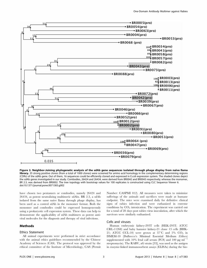
Figure 2. Neighbor-Joining phylogenetic analysis of the sdAb gene sequences isolated through phage display from naı̈ve llamalibrary. 35 strong positive clones (from a total of 1000 clones) were screened for amino acid homology in the complementary determining regions(CDRs) of the sdAb gene. Out of them, 16 sequences could be efficiently cloned and expressed in E.coli expression system. The shaded clones depictthe sdAb genes investigated in our study. Combodies, 26424 and 26434, were derived from BR0042 and BR0043 respectively; whereas the monomer,BR 2.3, was derived from BR0002. The tree topology with bootstrap values for 100 replicates is constructed using CLC Sequence Viewer 6.doi:10.1371/journal.pone.0071383.g002
One-Domain Antibody Multimer against Rabies
PLOS ONE | www.plosone.org 3 August 2013 | Volume 8 | Issue 8 | e71383

panning process for binder selection as well as for purified sdAb
ELISA. The virus was inactivated using 0.05% (v/v) b-propiolac-
tone to eliminate viral infectivity completely while maintaining
antigenicity [34].
Isolation of positive clones specific to RABV-G proteinThe repertoire of sdAbs was isolated from a naı̈ve llama library
(Wellcome Trust Sanger Institute, Cambridge, UK) by phage
display technique through infection into TG1 bacteria and KM13
helper phage as previously reported [35]. The frozen antibody
library was thawed on ice and diluted with 500 ml of 26TY
medium containing 100 mg ml21 of ampicillin. The TY medium
was supplemented with 4% (w/v) glucose for suppressing antibody
expression during bacterial culture. The cultures were grown in 2-
liter flasks at 37uC and 216 r.p.m. until the OD600 increased from
an initial absorbance of ,0.1 to approximately 0.5. KM13 helper
phages were added to a concentration of 261012 phages to the
bacterial culture and incubated in a water bath at 37uC for 30–
60 min. The cells were recovered by centrifugation at 3,200 g for
10 min at 4uC in 50-ml Falcon tubes, and the pellets were
resuspended in 500 ml of 26TY medium containing 0.1% (w/v)
glucose, 100 mg ml21 of ampicillin and 50 mg ml21 of kanamycin.
The culture was further grown at 25uC and 216 r.p.m. for 16–
20 h in a 2-liter flask. Cells were recovered by centrifugation at
3,200 g for 10 min at 4uC in Falcon tubes. The resulting
supernatants were filtered through 0.45 mm filters. The phages
were precipitated from the filtered supernatant by incubation in a
polyethylene glycol (PEG) solution (20% PEG 6000, 2.5 M NaCl)
on ice for 1 h, followed by centrifugation at 3,200 g for 30 min at
4uC in ten Falcon tubes. The pellets were resuspended in 5 ml of
PBS buffer and pooled together in a 15-ml Falcon tube to which
1 ml of PEG solution was added. The suspension was further
incubated on ice for 10 min and then centrifuged at 3,200 g for
30 min at 4uC. The resulting pellet was resuspended in PBS. The
Figure 3. Characterization of sdAb monomer and combodies. (A). Primary structure of monomer and combodies of the sdAbs used in ourstudy are shown. (B) Size-exclusion chromatography of BR 2.3, 26424 and 26434. The size of the monomeric and multimeric sdAbs was analyzedthrough Sephadex 200 chromatography and the elution positions have been depicted. (C) The size of BR 2.3, 26424 and 26434 has been furtherconfirmed through SDS-PAGE. BR 2.3 elutes as a 14 kDa monomer in both reducing (+DTT) and non-reducing (-DTT) conditions. Combodies 26424and 26434 elutes as 25 kDa protein in reducing conditions and appears to be more than 130 kDa in non-reducing SDS-PAGE, suggestingpentamerization of the coiled-coil peptide. (D) The monomeric and pentameric sdAbs were further analyzed in Western blot. The purified proteinswere run in a 12% SDS-PAGE in both reducing and non-reducing conditions. The antibodies were detected using Mouse anti-myc IgG and HRP-labeled goat anti-mouse IgG followed by chemiluminiscence detection. The figure depicts Western blot for 26434 and BR 2.3.doi:10.1371/journal.pone.0071383.g003
One-Domain Antibody Multimer against Rabies
PLOS ONE | www.plosone.org 4 August 2013 | Volume 8 | Issue 8 | e71383

phage solution was diluted in 1% Casein-PBS (CPBS) and
incubated for 30 min on Nunc Maxisorp plates coated with whole
RABV (inactivated using 0.05% (v/v) b-propiolactone) as the
antigen. The plates were washed with PBS-0.05% Tween-20 ten
times, and the bound phages were eluted with 200 ml of trypsin-
PBS. The eluted phages were used to infect fresh TG1 cultures
(OD600,0.5) or concentrated by PEG precipitation. After three
subsequent rounds of bio-panning, 56 colonies were selected, and
periplasmic extracts containing the sdAb gene were prepared
according to standard protocols. Selected clones were sent for
sequencing.
Construction of sdAb monomers and combodiesThe encoding sequences of the selected sdAbs were cloned into
the NcoI and NotI restriction sites of the C-terminal His6 tag-
containing pET20b vector (Novagen). The sdAb gene (monomer)
was amplified using the following primers: forward, 59-
CAGCCGGCCATGGCCCAGG-39; and reverse, 59-ATTAT-
TATGCGGCCGCTCAATGGTGATGGTGATGGTG-39. The
generation of the pentameric constructs (combodies) was per-
formed by cloning the sdAb sequences into the N-terminus along
with the coiled-coil domain of human COMP (Asp29-Gln76,
COMP48), myc-epitope and polyhistidine tag into the C-terminus
of the vector pET26b(+) (Novagen) [32]. The oligonucleotides 59-
TAATAAGAAGACCGCAGGCCCAGGTGCAGCTGGTG-
GAG-39 and 59-ATTATTTGGGC CCTGAAGAGACGGTGA-
CATTGT-39 were annealed and cloned into the NcoI and NotI
sites of the vector. The respective vectors were chosen based on
their suitability for obtaining periplasmic proteins from the E.coli
strains.
Expression and purification of the monomeric sdAbs andcombodies
For the production of the soluble sdAbs, we used the E.coli strain
BL21 Gold. The cells were grown in 5 ml LB Broth (100 mg ml21
kanamycin or ampicillin) and grown at 37uC with shaking at
220 r.p.m. overnight. The cultures were diluted to 1 L or 2 L LB
Broth (with 100 mg ml21 kanamycin or ampicillin) at a ratio of 2:1
and grown at 37uC until a 600 nm absorbance of between 0.5–1
was obtained. Protein expression was induced by treatment with
1 mM isopropyl-B-thio-galactoside (IPTG) at a lower temperature
of 22uC with shaking at 180 r.p.m. for 20 h. Cells were pelleted at
8,000 g at 4uC for 15 min and re-suspended in 1 M PBS (pH 7.4).
5 mg of lysozyme was added and incubated for 45 min at RT.
The lysed cells were sonicated using a sonic dismembrator to
reduce the viscosity of the lysate and centrifuged at 12,000 r.p.m.
to obtain clear supernatants containing the periplasmic protein.
The His6-tagged proteins (BR 2.3, 26424 and 26434) were purified
using Immobilized Metal ion Affinity Chromatography (IMAC)
and Nickel SepharoseTM Fast Flow (GE Biosciences) according to
the manufacturer’s protocols. The desired sdAbs were eluted using
500 mM imidazole after extensive washing with buffer containing
lower concentrations of imidazole.
BR 2.3, 26424 and 26434 were further purified by size
exclusion chromatography using a Sephadex 200 (GE Biosciences)
column on an AKTA purifier 2000 system (GE Biosciences). The
pentameric and monomeric proteins were collected at the
indicated elution volumes (see Results).
Binding specifity through ELISAFor the analysis of the specificity of monomer and the
combodies, 96-well Maxisorp plates (Nunc) were coated with
purified RABV (aG strain, inactivated using 0.05% (v/v) b-
propiolactone) at 100-fold dilution overnight at 4uC. For negative
control, influenza (H1N1) virus (PR8) with selective mutations in
the PB1 and PB2 genes [36], was used as the coating antigen
diluted to similar concentration. After thorough washing, the wells
were blocked in MT Buffer (PBS/2% skimmed milk/2% Tween-
20) for 2 h at 37uC. After three washes with PBS, optimized
concentration or dilutions of the sdAbs were added to the wells
and incubated for another 2 h at RT with shaking. After 5 washes
with PBST (PBS/0.05% Tween-20), the binding of the sdAbs was
detected using a mouse anti-myc mAb (Epigen) followed by
secondary probing with a goat anti-mouse horseradish peroxidase
(HRP) conjugate. HRP activity was determined using 3,39,5,59-
Tetramethylbenzidine (TMB) substrate. 2 M sulphuric acid
(H2SO4) was used to stop the reaction, and the readings at
450 nm wavelength were measured using an ELISA-plate reader.
Figure 4. Binding of 26424, 26434 and BR 2.3 in ELISA to RABV. (A) The combodies, 26424 and 26434, and BR 2.3 (control monovalent sdAb)were analyzed in a binding ELISA to confirm their specificity to RABV (inactivated). PR8 (H1N1) was used as the negative control to determine cross-neutralization of the sdAbs. The graph depicts binding specificity of 26424, 26434, and BR 2.3 at a concentration of 2.5 mg ml21. (B) Binding of 26424,26434 and BR 2.3 to RABV at different concentrations of the purified sdAbs. Mouse anti-myc IgG was used as primary antibody followed by HRP-labeled goat anti-mouse IgG. The binding reactivity was confirmed in four independent experiments and the figure represents the average value.Mean 6 standard deviations (SD) for each sample at different dilutions have been depicted.doi:10.1371/journal.pone.0071383.g004
One-Domain Antibody Multimer against Rabies
PLOS ONE | www.plosone.org 5 August 2013 | Volume 8 | Issue 8 | e71383
Preparation of CVS-11 pseudotypesWe used the CVS-11 strain (Challenge Virus Standard-11,
ATCC reference VR-959) for viral pseudotype preparation, as this
is the standard internationally recognized virulent strain for
laboratory use. The CVS-11 pseudotypes were prepared as
previously described [37]. For transfections, 56106 HEK-293T
cells were grown in DMEM-10 (Dulbecco’s Minimal Essential
Medium (DMEM) supplemented with 10% fetal calf serum (FCS)
and 100 mgml21 streptomycin) for 24 h prior to the addition of the
combination of constructs. The plasmids pLP1 (HIV gag-pol)and
pLP2 (RSV promoter) (BLOCK-iT TM Lentiviral RNAi Expres-
sion System, Invitrogen), which supply the helper functions as well
as structural and replication proteins to produce lentivirus, were
transfected together with the firefly luciferase reporter pCSFLW
plasmid and with the CVS-11 envelope construct, pI.18-CVS-11.
The supernatants containing the pseudotype viruses were
harvested at 72 h post-transfection and were stored in aliquots
for short-term at 4uC or for long-term at 280uC.
In vitro analysis using pseudotype neutralization assayIn 96-well flat-bottomed plates (Corning, USA), threefold serial
diluted or optimized concentration of the sdAbs (diluted in
DMEM without serum or antibiotics) ranging from 0.6 mg ml21
upto 50 mg ml21 were incubated with 50 ml of CVS-11
pseudotype viruses (final volume of 200 ml) for 1 h at 37uC in a
BOD incubator (5% CO2). For control experiment, Human
Rabies Immunoglubulin (HRIG) (Shandong Taibang Biological
Products Co. Ltd.) at similar concentrations was used as a positive
control. The antibody-pseudotype mixtures were then added to
96-well plates, pre-seeded overnight with monolayer cultures of
BHK-21 cells at a concentration of 5000 cells per well and
incubated for 5 h at 37uC in a BOD incubator (5% CO2).
Negative control comprised of wells containing CVS-11 pseudo-
types without any antibody treatment. The medium was then
replaced with DMEM containing 5% FCS (heat-inactivated at
56uC for 30 min) and incubated further for 48 h.
Luciferase reporter activity in transduced cells was quantified
using the Fire-Lucy Assay Kit (Vigorous Biotechnology Beijing
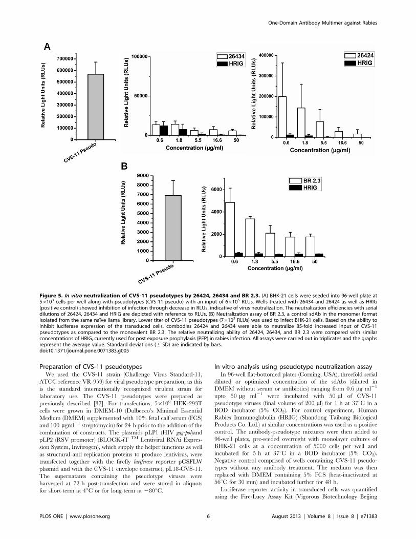
Figure 5. In vitro neutralization of CVS-11 pseudotypes by 26424, 26434 and BR 2.3. (A) BHK-21 cells were seeded into 96-well plate at56103 cells per well along with pseudotypes (CVS-11 pseudo) with an input of 66105 RLUs. Wells treated with 26434 and 26424 as well as HRIG(positive control) showed inhibition of infection through decrease in RLUs, indicative of virus neutralization. The neutralization efficiencies with serialdilutions of 26424, 26434 and HRIG are depicted with reference to RLUs. (B) Neutralization assay of BR 2.3, a control sdAb in the monomer formatisolated from the same naı̈ve llama library. Lower titer of CVS-11 pseudotypes (76103 RLUs) was used to infect BHK-21 cells. Based on the ability toinhibit luciferase expression of the transduced cells, combodies 26424 and 26434 were able to neutralize 85-fold increased input of CVS-11pseudotypes as compared to the monovalent BR 2.3. The relative neutralizing ability of 26424, 26434, and BR 2.3 were compared with similarconcentrations of HRIG, currently used for post exposure prophylaxis (PEP) in rabies infection. All assays were carried out in triplicates and the graphsrepresent the average value. Standard deviations (6 SD) are indicated by bars.doi:10.1371/journal.pone.0071383.g005
One-Domain Antibody Multimer against Rabies
PLOS ONE | www.plosone.org 6 August 2013 | Volume 8 | Issue 8 | e71383

Co. Ltd.). The cells were washed twice in PBS (pH 7.4) and lysed
with 1X Universal Lysis Buffer according to the manufacturer’s
protocols. The cells were detached by shaking for 5 min, and the
sdAb dilution-treated lysates were collected in fresh eppendorf
tubes and immediately placed in ice. For detecting luciferase
activity, 20 ml of each lysate was added to individual wells of a
white opaque 96-well plate, and 100 ml of luciferase substrate
solution was added prior to measurement. The plate was
measured using a Glomax 96 microplate luminometer (Promega),
and the relative light unit (RLU) values were compared to the
negative control containing CVS-11 pseudotypes only. A 50% or
more reduction in RLUs of the cells was considered indicative of
virus neutralization.
RLU output against each dilution of the sdAbs (26424, 26434
and BR 2.3) were plotted in a graph and the mean standard
deviations (6SD) were calculated. The RLU values of candidate
sdAbs were compared with that of HRIG (currently used in PEP
for rabies in China), which served as a positive control for all
subsequent neutralization assays.
Assessment of the efficacy of combodies using mouseneutralization test (MNT)
Based on their efficacy to neutralize the CVS-11 pseudotypes,
the combodies, 26424 and 26434 were chosen for testing their
neutralizing ability in a mouse RABV challenge model, according
to guidelines from World Health Organization (WHO). Rapid
Fluorescent Focus Inhibition test (RFFIT) was conducted for the
combodies (including ERIG) and mouse neutralization test (MNT)
was performed as previously described [38,39]. The titers of 26424
and 26434 were estimated to be 1.6 IU ml21 and 0.2 IU ml21.
Briefly, 26424 (0.3 mg ml21) and 26434 (0.6 mg ml21) were
individually mixed with 2500 LD50/5 ml RABV (CVS-24 strain)
[40] in separate tubes and incubated in ice for 1 hour. The
antibody-virus mixture was then injected in the hind leg of the
mouse (Kunming strain, 6 mice per group, each weighing 11–
13 g). The positive control consisted of ERIG (Wuhan Institute of
Biological Products Co. Ltd.) at 15.4 IU ml21 using a similar
administration procedure. The mice were vaccinated intraperito-
neally on days 0, 3, and 7 with 0.5 ml rabies vaccine (Jilin Maifeng
Pharmaceutical Co. Ltd.) that was diluted at a ratio of 1:25 (v/v)
with PBS. The negative control consisted of the two following
groups: one injected with PBS only and one injected with PBS and
vaccine. The mice were examined daily for definitive clinical signs
of rabies infection and were euthanized in extreme conditions by
CO2 intoxication. The experiment was carried out for a total of
28 days post rabies virus inoculation, after which the survivors
were similarly euthanized. Postmortem diagnosis of rabies
infection by direct fluorescent antibody testing was performed on
each mouse [41].
Kaplan–Meier survival curves were generated using GraphPad
Prism5. The statistical analysis of the survival curves was done
according to Mantel–Cox test.
Figure 6. Percentage neutralization of 26424, 26434 and BR2.3. Neutralization potencies have been calculated in percentage withreference to the decrease in RLUs of the antibody-treated samples ascompared to negative control containing CVS-11 pseudotypes alone.Percentage neutralization of samples treated with 26424 and 26434 hasbeen calculated against 66105 RLUs input of CVS-11 pseudotypes(Figure 5A), while that for BR 2.3 (control sdAb in monomer format) wascalculated against 76103 RLUs of pseudovirus input (Figure 5B). HRIGwas used as the positive control at similar concentrations of the testsamples in all in vitro neutralization assays.doi:10.1371/journal.pone.0071383.g006
Figure 7. In vivo lethal challenge of rabies infection. (A) Kaplan–Meier survival curve for mice in post exposure prophylaxis with thesdAb pentamer constructs. Mice were challenged with 2500 LD50 CVS-24 strain of RABV mixed with 26424 (1.6 IU ml21) and 26434 (0.2 IUml21) individually on Day 0. Negative control groups received PBSalong with vaccine (CVS+Vac) or without vaccine (CVS) whereas positivecontrol received 15.4 IU ml21 equine rabies immunoglobulin (ERIG).Vaccination was done on day 0, 3, and 7 in all groups including negativeand positive control. Animals were monitored daily for viability andweight change for a total of 28 days. Kaplan–Meier curves are shown byplotting percent survival against days (0 to 28). (B) Percentage deathrate of each group after 28 days of observation. Mantel-Cox statisticalanalysis has been performed for all the groups. 26424 showed statisticalsignificance in survival rate, compared to 26434 (* 0.01,P,0.05).doi:10.1371/journal.pone.0071383.g007
One-Domain Antibody Multimer against Rabies
PLOS ONE | www.plosone.org 7 August 2013 | Volume 8 | Issue 8 | e71383
Results
Selection of RABV-G specific sdAbsThe sdAb genes were isolated using phage display technique
from a naı̈ve llama library and screening was performed using
whole, inactivated RABV (aG strain). After extensive washing
steps, bound phages were rescued or eluted though reactions
with trypsin-PBS. Enrichment in specific binders was performed
over three rounds of in vitro selection or ‘‘bio-panning’’, The
positive clones were further assessed for antigen specificity and
binding by phage ELISA (Figure 1). A total of 35 clones were
identified (out of 1,000 clones) for further screening and
characterization (Figure 2).
Preparation of the sdAbs in monomeric and multimericformats
Based on binding assays and subsequent cloning into TG1
bacteria, 18 clones exhibiting the strongest binding specificities
were selected and re-cloned into a suitable bacterial expression
system for generating monomeric forms of sdAbs containing His-
and myc-tags at the C-terminus for affinity purification and
immuno-detection, respectively. For construction of pentamer or
combody, the monomeric sdAb gene was fused with the N-
terminus of COMP48 along with His- and myc-tags at the C-
terminus. An illustration of the construction of the monomer and
combody is shown in Figure 3A. Of the 18 clones generated, 16
were efficiently expressed in the bacterial periplasmic fraction
and were purified using His-tag affinity columns (IMAC, GE
Biosciences). The average yield of purified sdAbs ranged from
0.5 to 0.8 mg L21 for both monomer and combodies. The purity
of the protein was assessed by sodium-dodecyl sulfate polyacryl-
amide gel electrophoresis (SDS-PAGE) after purification.
The characterization of COMP48-conjugated pentameric
sdAbs has been extensively discussed previously [32]. COMP is
reported to exhibit inter-chain disulfide bonds at the C-terminus of
the assembly domain. The pentamerization of 26424 and 26434
was confirmed by size-exclusion chromatography. Under non-
reducing conditions, 26424 and 26434 migrated with a molecular
weight of more than 130 kDa, suggesting that multimerization was
attributed to inter-chain disulfide bonds (Figure 3B). Under
reducing conditions, the 26424 and 26434 appeared as 25 kDa
proteins (slightly higher molecular weight than the monomeric
form due to the presence of linker sequences), confirming the
presence of disulfide bonds (Figure 3C). The proteins were further
analyzed by Western Blot using an anti-myc antibody and
peroxidase-labeled anti-mouse IgG, followed by chemilumines-
cence detection (Figure 3D). By contrast, BR 2.3 appeared as
14 kDa monomer in SDS-PAGE under both reducing and non-
reducing conditions and was further confirmed through size-
exclusion chromatography (Sephadex 200, GE Healthcare),
whereby monomeric sdAbs eluted quickly as a 14 kDa protein
(Figure 3B and 3C).
Specific recognition of sdAb monomer and combodiesfor RABV
The antigen-binding specificities of 26424, 26434 and BR 2.3,
were determined via indirect enzyme-linked immunosorbent assay
(Indirect ELISA) against immobilized whole RABV (inactivated)
with influenza H1N1 virus (PR8) as a negative control. The
binding of the sdAbs to the corresponding immobilized antigens
were assessed using a mouse anti-myc mAb followed by anti-mouse
horseradish peroxidase (HRP)–conjugated immunoglobulin (IgG).
The purified combodies and monomer exhibited strong binding
specificity to immobilized RABV, in contrast to a negligible or
weak binding to H1N1 virus (PR8). Figure 4A depicts the binding
specificities of 26424, 26434 and BR 2.3 to RABV at a median
concentration of 2.5 mg ml21. Binding specificity for both
combodies and monomer was found to increase with higher
concentrations of 26424, 26434 and BR 2.3 (Figure 4B).
In vitro neutralizing efficacy of combodies against RABVpseudotypes
Neutralization abilities of 26424, 26434 and BR 2.3 were
assessed in a standardized neutralization assay using the rabies
CVS-11 pseudotype, a standard laboratory RABV strain.
Pseudotypes or pseudoviruses, replaces the need for use of live
viruses as they are antigenically similar to the native proteins on
wild-type live viruses, and give high specificity as well as
sensitivity for detection of virus neutralizing antibodies. Another
advantage is that pseudotypes are replication-incompetent and
can be used in biosafety level 2 (BL-2) laboratory conditions
without the need for BL-4, which is essential in case of handling
live pathogenic viruses.
Pseudotypes for the CVS-11 strain were prepared by incorpo-
rating the envelope construct (G protein) of CVS-11 into a
lentiviral vector (Invitrogen), which consists of the plasmids
carrying the HIV gag-pol (pLP1) and the RSV promoter (pLP2).
A firefly luciferase construct was used as a reporter for detection
and analysis of neutralizing efficiencies of the sdAbs. Decrease in
the relative light units (RLUs) of the luciferase activity in
transduced BHK-21 cells was indicative of binding and neutral-
ization of viral pseudotypes by the sdAb constructs. Combodies
26434 and 26424 could efficiently neutralize 85-fold higher input
of CVS-11 pseudotypes (66105 RLUs) at a lower concentration of
5.5 mg ml21 (Figure 5A). In contrast, however, at a relatively lower
input range of CVS-11 pseudotypes (76103 RLUs), monovalent
BR 2.3 was unable to inhibit infection of BHK-21 cells even at a
much lower concentration (5.5 mg ml21), but, could significantly
neutralize CVS-11 pseudotypes at higher concentrations
(Figure 5B). For 26424 and 26434, a dose of 16.6 mg ml21 could
neutralize 90% and 95% of the viral pseudotypes, respectively
(Figure 6). HRIG, a standard positive control included in all the
assays, could achieve 95–100% neutralization at similar range of
concentrations. Furthermore, increase in the level of viral
pseudotype titer was associated with concomitant decrease in the
neutralizing ability of the sdAb constructs. This trend was found to
be parallel with that of HRIG-treated samples. The data suggest
that multimerization has contributed to the efficacies of 26434 and
26424 to be able to neutralize higher input of viral pseudotypes,
resulting in increased neutralization potency in vitro.
Protection of mice challenged by a lethal dose of RABV invivo by combodies
On the basis of their binding affinities to RABV in ELISA and
effective neutralization of CVS-11 pseudotypes, the protection
abilities of 26424 and 26434 for animals challenged by live rabies
virus were tested in a mouse challenge model. The mouse
neutralization test (MNT) was conducted under specific labora-
tory conditions to assess the protection of mice challenged by a
lethal dose of RABV by the concurrent administration of the
rabies vaccine together with 26424 and 26434 individually.
ERIG, used as a positive control for MNT, is a cheaper and safe
alternative to human RIG and is used for post exposure
treatment of Rabies in developing countries. The survival rates
of the mice during the 28-day observation period were plotted as
Kaplan-Meier curves (Figure 7A) [42]. A death rate of 100% was
observed in the negative control group receiving either CVS-24
One-Domain Antibody Multimer against Rabies
PLOS ONE | www.plosone.org 8 August 2013 | Volume 8 | Issue 8 | e71383

virus only (CVS) or vaccine only (CVS+Vac), since the latter is
not capable for immediate generation of neutralizing antibodies
against RABV. The death rate in the group treated with 26424
(1.6 IU ml21) was 50%, while the group treated with 26434 (0.2
IU ml21) exhibited 60% death rate (Figure 7B). Positive control
group, consisting of mice treated with vaccine and ERIG (15.4
IU ml21), exhibited no death rate. The data implies that
combodies, 26424 and 26434 were capable of neutralizing live
RABV and could offer partial protection at a lower level of
dosage. Taken together, the studies demonstrate that combodies,
26424 and 26434, could prove to be promising anti-viral
molecules for Rabies infection in vivo.
Discussion
In this study, we report the isolation of two neutralizing sdAbs,
namely 26424 and 26434, from a naı̈ve llama library against the
trimeric glycoprotein (G) of RABV, and the influence of multi-
merization of the sdAbs to increase their neutralizing potential
through oligomerization was investigated. The multimerization
strategy for our study was adopted by fusing the coiled-coil peptide
of the human cartilage oligomeric matrix protein 48 (COMP48)
with sdAbs resulting in a pentavalent structure or combody. In
contrast to single-chain variable fragments (scFvs), sdAbs are ideal
candidates for oligomerization purposes, as they are half the size of
scFvs and therefore produce smaller oligomeric forms. Further-
more, sdAbs can exist as monomers, whereas scFvs tend to form
dimers, trimers, etc. [43]. COMP48 has been successfully used to
generate high-avidity combodies specifically against the melanoma
peptide-HLA A2 complex [32]. To our knowledge, this is the first
attempt to evaluate the effect of multimerization of sdAb
fragments using COMP48 for targeting antigens against infectious
diseases such as Rabies.
In our experiments, the neutralizing potencies of combodies
against RABV have been analyzed both in vitro and in vivo. Whole
inactivated virus (aG strain) was used for selection, to obtain sdAbs
specific for the RABV epitopes accessible in the intact viral
particle. Initially, the binding properties of the sdAbs were
evaluated using ELISA; the differences in the binding affinities
of the various clones were presumed through initial rounds of bio-
panning and phage ELISA. The clones exhibiting the strongest
binding specificity were screened for further assessment of in vitro
neutralizing ability using RABV (CVS-11) pseudotypes.
Several mechanisms might be responsible for the antiviral
activities of the sdAbs. One possible mechanism is the blockade of
the RABV-G protein interaction with its cognate cellular receptor,
which thereby inhibits the virus to enter the cell and replicate. We
have established a neutralization assay for testing the abilities of
sdAbs to neutralize pseudoviruses in vitro. BHK-21 cells, which are
routinely used in CVS-11 fluorescent antibody virus neutralization
(FAVN) tests, are highly permissive for CVS-11 pseudotypes
[44,45]. Initially, 16 clones (data not shown), consisting of both
monomers and combodies were tested in the neutralization assay,
of which 26424 and 26434 could neutralize the CVS-11
pseudotypes with relatively high efficacy. The neutralizing abilities
of the sdAbs have been compared with that of HRIG, currently
used for post-exposure prophylaxis of rabies. As a proof-of-
principle, further rabies pseudotype neutralization assays verified
that the combodies, 26424 and 26434, could neutralize 85-fold
increased input of CVS-11 pseudotypes in vitro at lower
concentrations as compared to monovalent sdAb (BR 2.3), which
highlights the improvement in avidity due to multimerization
(Figure 5A, B and Figure 6).
It is to be noted that the sdAb genes were screened using the aG
strain (also known as pG strain) of RABV as target antigen during
bio-panning process. The G protein has eight amino substitutions
(His69, Pro184, Pro250, Gly427, Ile431, Ile477, Lys481, and Asn160)
which are unique to aG strain [33]. However, the antigenic sites,
namely antigenic site I (231), antigenic site II (residues 34–42,198–
200), antigenic site III (residues 330–338), antigenic site IV
(residue 264) and antigenic site a (residue 342) were found to be
conserved as compared to other vaccine strains [33]. in vitro assays
of 26424 and 26434 (and also BR 2.3) against CVS-11
pseudotypes, suggest that the sdAb genes are specific for the G
protein whose antigenic sites are conserved across wide variety of
RABV strains. However, further investigations are needed for
identifying the epitopes on the RABV G protein recognized by the
combodies, in order to fully understand their future diagnostic or
therapeutic value.
To investigate the neutralizing potencies of both the combodies,
26424 and 26434 against a lethal challenge in vivo, we performed
the mouse neutralization test (MNT). The relative survival rate of
mice treated with 26424 was approximately 50% compared to the
40% survival rate of mice that received 26434 (Figure 7). In the
control groups, the mice receiving virus (CVS-24) alone or with
vaccine exhibited 100% mortality within 10 days post-infection,
whereas all of the ERIG-treated mice survived until day 28, post-
infection or completion of the test. Combodies with a molecular
weight of more than 130 kDa, sufficiently exceed the renal
clearance threshold, could result in longer serum retention and
produce effective viral neutralization. However, 26424 and 26434,
could achieve partial protection (40–50%) as compared to 100%
survival rate by ERIG. This may be partly due to the introduction
of a human protein fragment (COMP48) with the sdAb gene that
might elicit additional immunogenicity when injected in mice,
resulting in decreased neutralization efficiencies in vivo. Moreover,
as stated earlier, the relative concentration of 26434 (0.2 IU ml21)
and 26424 (1.6 IU ml21) were lower than the standard dosage
level required for effective virus neutralization in vivo. Our data
comprises of preliminary investigations into the efficacy of
multimeric sdAbs to be able to neutralize live RABV in mouse
challenge model. Future work relating to dose-response studies is
necessary to fully elucidate the prophylactic efficacies of 26434 and
26424 for achieving 100% protection in mice against rabies
infection. Nonetheless, this study indicates that the neutralizing
abilities of sdAbs have been significantly increased in vitro as well as
in vivo as a result of multimerization.
We further addressed the issue of possible immunogenicity
attributable to the repeated administration of non-human
therapeutic proteins. The sdAbs are derived from Camelidae and
exhibit significant homology to the human VH fragment.
[46,47,48] The reduced immunogenic potential of the llama-
derived heavy chain fragments (VHHs) have been further
substantiated in primate studies performed by Ablynx (http://
www.ablynx.com). On the other hand, the oligomeric matrix
protein, COMP48, is of human origin, thus reducing the risk of
immunogenicity upon administration in humans, despite possibly
having elicited an immune response in the mouse neutralization
test (MNT) as stated earlier. The shortcomings of such limitations
might be negated, as long as the therapeutic protein is efficient in
treating infectious diseases in humans. Furthermore, human-
derived COMP48 has the added advantage of improved stability
that results from complementary hydrophobic interactions and
disulfide bridges between its a-helices.
In conclusion, multivalent sdAbs obtained through fusion with
human COMP have been proven to exhibit increased avidity to
target antigens. Moreover, fusion-antibodies exhibit a correct
One-Domain Antibody Multimer against Rabies
PLOS ONE | www.plosone.org 9 August 2013 | Volume 8 | Issue 8 | e71383

domain folding without compromising target specificity. The
multivalent sdAbs isolated in our study could be useful anti-viral
molecules for the treatment of RABV infection, as well as for
investigations of mechanisms underlying viral infection, which
remain poorly understood.
Author Contributions
Conceived and designed the experiments: BG. Performed the experiments:
BMB DL DY. Analyzed the data: BG BMB DL. Contributed reagents/
materials/analysis tools: TG CJ MQ EW WW WH CL. Wrote the paper:
BMB BG.
References
1. Finke S, Conzelmann KK (2005) Replication strategies of rabies virus. Virus Res
111: 120–131.2. Gaudin Y, Tuffereau C, Segretain D, Knossow M, Flamand A (1991) Reversible
conformational-changes and fusion activity of rabies virus glycoprotein. Journal
of Virology 65: 4853–4859.3. Mifune K, Ohuchi M, Mannen K (1982) Hemolysis and cell-fusion by
Rhabdoviruses Febs Letters 137: 293–297.4. Whitt MA, Buonocore L, Prehaud C, Rose JK (1991) Membrane-fusion activity,
oligomerization, and assembly of the rabies virus glycoprotein. Virology 185:681–688.
5. Gastka M, Horvath J, Lentz TL (1996) Rabies virus binding to the nicotinic
acetylcholine receptor alpha subunit demonstrated by virus overlay proteinbinding assay. Journal of General Virology 77: 2437–2440.
6. Lentz TL, Burrage TG, Smith AL, Tignor GH (1983) The acetylcholine-receptor as a cellular receptor for rabies virus. Yale Journal of Biology and
Medicine 56: 315–322.
7. Thoulouze MI, Lafage M, Schachner M, Hartmann U, Cremer H, et al. (1998)The neural cell adhesion molecule is a receptor for rabies virus. J Virol 72:
7181–7190.8. Tuffereau C, Benejean J, Blondel D, Kieffer B, Flamand A (1998) Low-affinity
nerve-growth factor receptor (p75NTR) can serve as a receptor for rabies virus.
EMBO J 17: 7250–7259.9. Tuffereau C, Schmidt K, Langevin C, Lafay F, Dechant G, et al. (2007) The
rabies virus glycoprotein receptor p75NTR is not essential for rabies virusinfection. J Virol 81: 13622–13630.
10. Dietzschold B, Cox JH, Schneider G (1978) Structure and function of rabiesvirus glycoprotein. Dev Biol Stand 40: 45–55.
11. Sloan SE, Hanlon C, Weldon W, Niezgoda M, Blanton J, et al. (2007)
Identification and characterization of a human monoclonal antibody thatpotently neutralizes a broad panel of rabies virus isolates. Vaccine 25: 2800–
2810.12. de Kruif J, Bakker ABH, Marissen WE, Kramer RA, Throsby M, et al. (2007) A
human monoclonal antibody cocktail as a novel component of rabies
postexposure prophylaxis. Annual Review of Medicine. 359–368.13. Houimel M, Dellagi K (2009) Isolation and characterization of human
neutralizing antibodies to rabies virus derived from a recombinant immuneantibody library. J Virol Methods 161: 205–215.
14. Forsman A, Beirnaert E, Aasa-Chapman MMI, Hoorelbeke B, Hijazi K, et al.(2008) Llama Antibody Fragments with Cross-Subtype Human Immunodefi-
ciency Virus Type 1 (HIV-1)-Neutralizing Properties and High Affinity for HIV-
1 gp120. Journal of Virology 82: 12069–12081.15. Garaicoechea L, Olichon A, Marcoppido G, Wigdorovitz A, Mozgovoj M, et al.
(2008) Llama-derived single-chain antibody fragments directed to rotavirus VP6protein possess broad neutralizing activity in vitro and confer protection against
diarrhea in mice. Journal of Virology 82: 9753–9764.
16. Serruys B, Van Houtte F, Verbrugghe P, Leroux-Roels G, Vanlandschoot P(2009) Llama-Derived Single-Domain Intrabodies Inhibit Secretion of Hepatitis
B Virions in Mice. Hepatology 49: 39–49.17. van der Vaart JM, Pant N, Wolvers D, Bezemer S, Hermans PW, et al. (2006)
Reduction in morbidity of rotavirus induced diarrhoea in mice by yeastproduced monovalent llama-derived antibody fragments. Vaccine 24: 4130–
4137.
18. Muyldermans S (2001) Single domain camel antibodies: current status.J Biotechnol 74: 277–302.
19. Cortez-Retamozo V, Lauwereys M, Hassanzadeh Gh G, Gobert M, Conrath K,et al. (2002) Efficient tumor targeting by single-domain antibody fragments of
camels. Int J Cancer 98: 456–462.
20. Els Conrath K, Lauwereys M, Wyns L, Muyldermans S (2001) Camel single-domain antibodies as modular building units in bispecific and bivalent antibody
constructs. J Biol Chem 276: 7346–7350.21. Dumoulin M, Last AM, Desmyter A, Decanniere K, Canet D, et al. (2003) A
camelid antibody fragment inhibits the formation of amyloid fibrils by human
lysozyme. Nature 424: 783–788.22. Deyev SM, Lebedenko EN (2008) Multivalency: the hallmark of antibodies used
for optimization of tumor targeting by design. BioEssays 30: 904–918.23. Hultberg A, Temperton NJ, Rosseels V, Koenders M, Gonzalez-Pajuelo M, et
al. (2011) Llama-derived single domain antibodies to build multivalent,superpotent and broadened neutralizing anti-viral molecules. Plos One 6:
e17665.
24. Cohen C, Parry DA (1990) Alpha-helical coiled coils and bundles: how to design
an alpha-helical protein. Proteins 7: 1–15.
25. Kammerer RA (1997) Alpha-helical coiled-coil oligomerization domains in
extracellular proteins. Matrix Biol 15: 555–565; discussion 567–558.
26. Kohn WD, Mant CT, Hodges RS (1997) Alpha-helical protein assembly motifs.J Biol Chem 272: 2583–2586.
27. Lupas A (1996) Coiled coils: new structures and new functions. Trends BiochemSci 21: 375–382.
28. O’Neil K, DeGrado W (1990) A thermodynamic scale for the helix-formingtendencies of the commonly occurring amino acids. Science 250: 646–651.
29. Malashkevich VN, Kammerer RA, Efimov VP, Schulthess T, Engel J (1996)The crystal structure of a five-stranded coiled coil in COMP: A prototype ion
channel? Science 274: 761–765.
30. Tomschy A, Fauser C, Landwehr R, Engel J (1996) Homophilic adhesion of E-
cadherin occurs by a co-operative two-step interaction of N-terminal domains.EMBO J 15: 3507–3514.
31. Holler N, Kataoka T, Bodmer JL, Romero P, Romero J, et al. (2000)Development of improved soluble inhibitors of FasL and CD40L based on
oligomerized receptors. J Immunol Methods 237: 159–173.
32. Zhu X, Wang L, Liu R, Flutter B, Li S, et al. (2010) COMBODY: one-domain
antibody multimer with improved avidity. Immunol Cell Biol 88: 667–675.
33. Jiao W, Yin X, Li Z, Lan X, Li X, et al. (2011) Molecular characterization of
China rabies virus vaccine strain. Virology Journal 8: 521.
34. Goldstein MA, Tauraso NM (1970) Effect of Formalin, b-Propiolactone,Merthiolate, and Ultraviolet Light Upon Influenza Virus Infectivity, Chicken
Cell Agglutination, Hemagglutination, and Antigenicity. Applied Microbiology
19: 290–294.
35. Lee CMY, Iorno N, Sierro F, Christ D (2007) Selection of human antibody
fragments by phage display. Nat Protocols 2: 3001–3008.
36. He W, Wang W, Han H, Wang L, Zhang G, et al. (2013) Molecular Basis ofLive-Attenuated Influenza Virus. Plos One 8: e60413.
37. Wright E, Temperton NJ, Marston DA, McElhinney LM, Fooks AR, et al.(2008) Investigating antibody neutralization of lyssaviruses using lentiviral
pseudotypes: a cross-species comparison. Journal of General Virology 89: 2204–2213.
38. Duan Y, Gu TJ, Jiang CL, Yuan RS, Zhang HF, et al. (2012) A novel disulfide-stabilized single-chain variable antibody fragment against rabies virus G protein
with enhanced in vivo neutralizing potency. Mol Immunol 51: 188–196.
39. Haase M, Seinsche D, Schneider W (1985) The mouse neutralization test in
comparison with the rapid fluorescent focus inhibition test: Differences in theresults in rabies antibody determinations. Journal of Biological Standardization
13: 123–128.
40. Wenqiang J, Xiangping Y, Xuerui L, Jixing L (2013) Complete genome
sequence of rabies virus CVS-24 from China. Archives of Virology: 1–8.
41. Bakker AB, Marissen WE, Kramer RA, Rice AB, Weldon WC, et al. (2005)
Novel human monoclonal antibody combination effectively neutralizing naturalrabies virus variants and individual in vitro escape mutants. J Virol 79: 9062–
9068.
42. Kaplan EL, Meier P (1958) Nonparametric-estimation from incomplete
observations. Journal of the American Statistical Association 53: 457–481.
43. Zhang J, Tanha J, Hirama T, Khieu NH, To R, et al. (2004) Pentamerization of
single-domain antibodies from phage libraries: a novel strategy for the rapidgeneration of high-avidity antibody reagents. J Mol Biol 335: 49–56.
44. Wright E, Temperton NJ, Marston DA, McElhinney LM, Fooks AR, et al.(2008) Investigating antibody neutralization of lyssaviruses using lentiviral
pseudotypes: a cross-species comparison. J Gen Virol 89: 2204–2213.
45. Wright E, McNabb S, Goddard T, Horton DL, Lembo T, et al. (2009) A robust
lentiviral pseudotype neutralisation assay for in-field serosurveillance of rabiesand lyssaviruses in Africa. Vaccine 27: 7178–7186.
46. Roovers RC, Laeremans T, Huang L, De Taeye S, Verkleij AJ, et al. (2007)Efficient inhibition of EGFR signaling and of tumour growth by antagonistic
anti-EFGR Nanobodies. Cancer Immunol Immunother 56: 303–317.
47. Terskikh AV, Le Doussal JM, Crameri R, Fisch I, Mach JP, et al. (1997)
‘‘Peptabody’’: a new type of high avidity binding protein. Proc Natl AcadSci U S A 94: 1663–1668.
48. Cortez-Retamozo V, Backmann N, Senter PD, Wernery U, De Baetselier P, etal. (2004) Efficient cancer therapy with a nanobody-based conjugate. Cancer
Res 64: 2853–2857.
One-Domain Antibody Multimer against Rabies
PLOS ONE | www.plosone.org 10 August 2013 | Volume 8 | Issue 8 | e71383