Collisional effects on diffusion scaling laws in electrostatic turbulence
Simultaneous Characterization of Glyco- andPhosphoproteomes of Mouse Brain MembraneProteome with Electrostatic RepulsionHydrophilic Interaction Chromatography*□S
Huoming Zhang‡, Tiannan Guo‡, Xin Li‡, Arnab Datta‡, Jung Eun Park‡, Jie Yang‡,Sai Kiang Lim§, James P. Tam‡, and Siu Kwan Sze‡¶
Characterization of glyco- and phosphoproteins as wellas their modification sites poses many challenges, thegreatest being loss of their signals during mass spectro-metric detection due to substoichiometric amounts andthe ion suppression effect caused by peptides of highabundance. We report here an optimized protocol usingelectrostatic repulsion hydrophilic interaction chromatog-raphy for the simultaneous enrichment of glyco- andphosphopeptides from mouse brain membrane proteindigest. With this protocol, we successfully identified 544unique glycoproteins and 922 glycosylation sites, whichwere significantly higher than those from the commonlyused hydrazide chemistry method (192 glycoproteins and345 glycosylation sites). Moreover, a total of 383 phospho-proteins and 915 phosphorylation sites were recoveredfrom the sample, suggesting that this protocol has the po-tential to enrich both glycopeptides and phosphopeptidessimultaneously. Of the total 995 glycosylation sites identi-fied from both methods, 96% were considered new as theywere either annotated as putative or not documented in thenewly released Swiss-Prot database. Thus, this study couldbe of significant value in complementing the current glyco-protein database and provides a unique opportunity tostudy the complex interaction of two different post-transla-tional modifications in health and disease without beingaffected by interexperimental variations. Molecular &Cellular Proteomics 9:635–647, 2010.
Protein glycosylation and phosphorylation are two impor-tant post-translational modifications. In mammals, it has beenestimated that nearly 50% of all proteins are glycosylated (1),and at least one-third of all proteins are phosphorylated (2).The modification of a protein has an important role in deter-mining its stability, activity, localization, and interactions withother proteins. For example, N-linked glycosylation of a pro-
tein enhances its stability and targets it mainly to extracellularlocations such as the plasma membrane (3), whereas phos-phorylation of tyrosine kinases initiates signal cascades inboth proproliferative and antiapoptotic cellular responses (4).
Membrane proteins are essential for cells to maintain theintegrity of their structure and perform signal transduction inresponse to extracellular stimulation. For most membraneproteins, receptor activities of their extracellular domains areoften mediated via N-linked glycosylation, whereas the cyto-plasmic domains can be phosphorylated reversibly and func-tion as signal transducers. Alteration of these modificationscorrelates with cellular differentiation, implantation, and tissuedevelopment (5, 6). Such alteration is required for the induc-tion of many forms of synaptic plasticity such as long termpotentiation and depression in learning and memory forma-tion. For example, aberrant glycosylation and abnormal phos-phorylation have been found to be associated with many neu-rological disorders such as Alzheimer disease (7–9), Parkinsondisease (10, 11), Guillain-Barre and Miller-Fisher syndromes(12), and even muscle-eye-brain disease (13). Understandingthe brain glyco- and phosphoproteomes is therefore not onlyessential for studying the biology of these diseases but alsoaiding drug discovery and translational research as many of theglycoproteins have the potential to be viable drug targets due tothe ease of their accessibility on the cell surface.
Study of the glyco- and phosphoproteomes of cell mem-brane presents a number of challenges mainly due to their lowabundance, their large dynamic range, and the inherent hy-drophobicity of membrane proteins. It has been estimatedthat less than 5% of the peptides in a typical complex proteindigest are either N-linked glycosylated (14) or phosphorylated(15). Therefore, development of efficient protocols for theenrichment of glycopeptides and phosphopeptides is essen-tial for their subsequent detection and identification.
Methods used for the enrichment of glycopeptides andglycoproteins have varied with time and among investigators.Lectin-based affinity enrichment is one of the earliest andmost widely used approaches for the analysis of glycopro-teins and their associated carbohydrates (16–19). Generally,lectins are highly specific for a particular type of carbohydrate
From the ‡School of Biological Sciences, Nanyang TechnologicalUniversity, 60 Nanyang Drive, Singapore 637551 and §Institute ofMedical Biology, 8A Biomedical Grove, 05-505 Immunos,Singapore 138648, Singapore
Received, July 8, 2009, and in revised form, December 30, 2009Published, MCP Papers in Press, January 4, 2010, DOI 10.1074/
mcp.M900314-MCP200
Research
© 2010 by The American Society for Biochemistry and Molecular Biology, Inc. Molecular & Cellular Proteomics 9.4 635This paper is available on line at http://www.mcponline.org
moiety, such as wheat germ lectin, which mainly recognizesGlcNAc, and concanavalin A lectin specific for mannose (20).Combining the use of two or more types of lectins with po-tentially complementary binding properties is often requiredto recover a significant number of glycoproteins from a com-plex sample (18, 21, 22). Another commonly used method isutilizing hydrophilic affinity physicochemical chromatography.This method is based on hydrogen bonding between thehydroxyl group of carbohydrate moieties in glycopeptides andhydrophilic materials such as Sepharose or cellulose (23, 24).Thus, this enrichment has a much wider range in terms of thetypes of glycoproteins recovered as compared with the lectin-based method. In addition, Ding et al. (25) demonstrated thattryptic glycopeptides can be eluted as a set after the trypticnon-glycopeptides in the pure hydrophilic interaction liquidchromatography mode by increasing the hydrophobicity ofpeptides with trifluoroacetate as an ion-pairing agent. Re-cently, a method utilizing hydrazide chemistry has gainedincreasing popularity for the study of the N-linked glycopro-teome (26, 27). It has been applied to high throughput globalanalysis of N-glycoproteins from various samples such ashuman serum (28), saliva (29), platelet (30), liver (31), andcancer tissue (32). Although this approach is theoretically ableto capture all types of glycoproteins or glycopeptides, a totalof only 1522 unique glycosylation sites was collectively iden-tified in nearly 10 studies, representing about 3% of the totalpredicted glycosylation sites in the human proteome (14). Inrecent attempts to identify more glycoproteins in a sampleand expand coverage of the N-linked glycoproteins, differentgroups have used different methods in parallel to simulta-neously analyze a sample. Cao et al. (33) utilized the hydrazidemethod and hydrophilic affinity to identify glycosylation sitesin secreted proteins and reported a total of 300 glycosylationsites with 159 and 261 from each of the methods, respec-tively. Lee et al. (22) used the hydrazide method and threelectins for a study of rat liver glycoproteins. They identified atotal of 335 glycoproteins with 202 from the lectin method and210 from the hydrazide method. These studies demonstratedthat current methods are complementary; hence, combineduse of them could enhance glycoprotein recovery, althoughthe overall efficiency remains relatively low.
As with the need to develop protocols for glycopeptideenrichment, many methods for phosphopeptide enrichmenthave also been described. These include phosphoramidatechemistry (34), immunoprecipitation with phosphospecific an-tibodies (35), IMAC (36), strong cation exchange (SCX)1 chro-matography (37), and titanium dioxide (TiO2) chromatography
(38). Each method has its unique advantages and shortcom-ings and analyzing a sample either by using different methodsin parallel or combining different methods into one wouldoften enrich more phosphopeptides and therefore identifymore phosphoproteins. Indeed, Villen et al. (39) were able toidentify more than 5,600 non-redundant phosphorylation siteson 2,300 proteins from mouse liver when using SCX chroma-tography followed by IMAC affinity purification. Similarly,when coupling SCX with TiO2 chromatography, Olsenet al. (40) reported a total of 6,600 phosphorylation sites on2,200 HeLa cell proteins. In addition, the TiO2 and the IMACmethod were found to be complementary (41), and using bothmethods in parallel to analyze a sample generated a com-bined set of information that surpassed the outcome derivedusing one method. However, an effective method for simul-taneous enrichment of both glyco- and phosphopeptides ishighly desirable.
Recently, a novel mode of chromatography termed electro-static repulsion hydrophilic interaction chromatography (ER-LIC) has been introduced for enrichment of phosphopeptidesbased on both their electrostatic and hydrophilic properties(42). With the low pH and high organic content of the mobilephase, the majority of peptides with carboxyl groups at as-partic acid and glutamic acid residues and the C terminus arelargely un-ionized and thus poorly retained by the weak anionexchange (WAX) column, whereas phosphopeptides andhighly hydrophilic peptides will interact strongly with the col-umn and are retained. A salt and aqueous gradient can thenbe used to gradually elute phosphopeptides from the column.Typically, buffer A (10 mM sodium methyl phosphonate and70% acetonitrile, pH 2.0) and buffer B (200 mM triethylaminephosphate with 60% acetonitrile, pH 2.0) are used to create agradient for the enrichment and fractionation of the phos-phopeptides from a cell lysate digest (43). This enrichmentmethod has been found to be comparable with the hydrazidemethod in the identification of glycoproteins when a plateletdigest was used as a starting material (44). The ERLIC enrich-ment is mainly based on the negatively charged sialyl group inglycopeptides (44). However, it might be able to enrich otherhydrophilic glycopeptides when an oligosaccharide side chainis a large and hydrophilic domain that causes a significantincrease in retention time of the peptide in any hydrophilicinteraction liquid chromatography-based mode of ERLIC.
We report here an improved protocol using ERLIC for thesimultaneous enrichment of glyco- and phosphopeptidesfrom mouse brain membrane preparation. With this protocol,the yields of glycoproteins (544) and glycosylation sites (922)were significantly higher than those from the hydrazide chem-istry method (192 glycoproteins and 345 glycosylation sites).In total, 995 glycosylation sites were identified, 96% of whichwere considered new as they were either annotated as puta-tive or not documented in the newly released Swiss-Protdatabase. Moreover, a total of 383 phosphoproteins including915 phosphorylation sites was recovered from the ERLIC
1 The abbreviations used are: SCX, strong cation exchange; ERLIC,electrostatic repulsion hydrophilic interaction chromatography; LTQ,linear quadrupole ion trap; NCAM, neural cell adhesion molecule;TiO2, titanium dioxide; WAX, weak anion exchange; IPI, InternationalProtein Index; GO, gene ontology; MGI, Mouse Genome Informatics;KEGG, Kyoto Encyclopedia of Genes and Genomes; APP, amyloid �precursor protein.
Characterization of Mouse Brain Membrane Proteome with ERLIC
636 Molecular & Cellular Proteomics 9.4
approach, suggesting that this protocol has potential for thesimultaneous study of both glyco- and phosphoproteomes.
MATERIALS AND METHODS
Mice and Tissue Preparation—All animal procedures were per-formed according to the protocols approved by Nanyang Technolog-ical University for Biological Studies and Animal Care and Use Com-mittees. A single C57BL/6J inbred strain mouse purchased fromCentre for Animal Care, National University of Singapore was used inthis study. At the age of 8 weeks, the mouse was euthanized withexcess isoflurane anesthesia. The whole intact brain was collected,snap frozen in liquid nitrogen, and then stored at �80 °C.
Membrane Protein Extraction and Digestion—Three frozen brains(�1.5 g, wet weight) were ground in liquid nitrogen in a prechilledmortar with pestle. The fine powder was transferred to a 2-ml Eppen-dorf tube and HES buffer (20 mM HEPES, pH 7.4, 1 mM EDTA, 250 mM
sucrose) supplemented with protease inhibitor (10 �l/mg of tissue)(Roche Diagnostics). The sample was sonicated three times on iceusing a Vibra CellTM high intensity ultrasonic processor (Jencon,Leighton Buzzard, Bedfordshire, UK). The remaining debris and un-broken cells were removed by centrifugation at 1000 � g at 4 °C for10 min. The supernatant was transferred to a new tube and centri-fuged at 100,000 � g at 4 °C for 45 min. The pellet containingmembrane fractions was washed once with Na2CO3 (0.1 M, pH 11)and twice with Milli-Q water, respectively, followed by centrifugationat 100,000 � g at 4 °C. The membrane pellet was then dissolved in8 M urea solution, and protein content was determined with a 2-DQuant kit (GE Healthcare) according to the manufacturer’s instruc-tions. Approximately 6 mg of protein was aliquoted into six tubes andused for subsequent experiments. The proteins were reduced with 10mM tris(carboxyethylphosphine) hydrochloride for 30 min at 37 °C andalkylated with 40 mM iodoacetamide for 1 h at room temperature.Proteins were then diluted 7-fold with 50 mM NH4HCO3 prior todigestion with trypsin (Promega, Madison, WI) overnight at 37 °C in a1:50 trypsin-to-protein mass ratio. The protein digests were desaltedusing Sep-Pak C18 cartridges (Waters) and dried in a SpeedVac(Thermo Electron, Waltham, MA).
Enrichment of N-Linked Glycopeptides and Phosphopeptides Us-ing ERLIC—ERLIC buffer A (10 mM sodium methyl phosphonate with70% acetonitrile, pH 2.0) was prepared by addition of NaOH to asolution of methylphosphonic acid (Sigma-Aldrich) in water followedby addition of acetonitrile. Buffer B (200 mM triethylamine phosphatewith 25% acetonitrile, pH 2.0) was prepared by addition of triethyl-amine (Sigma-Aldrich) to a solution of phosphoric acid in water fol-lowed by addition of acetonitrile. A total of 3 mg of protein digest wasused for three replicate ERLIC runs. For each run, �1 mg of digestreconstituted in 200 �l of buffer A was loaded into a PolyWAX LPTM
column (4.6 � 200 mm, 5-�m particle size, 300-Å pore size; PolyLC,Columbia, MD) on a ProminenceTM HPLC unit (Shimadzu, Kyoto,Japan). The sample was fractionated using a gradient of 100% bufferA for 10 min, 0–30% buffer B for 25 min, 30–100% buffer B for 5 min,and finally 100% buffer B for 10 min at a constant flow rate of 1 ml/minfor a total of 50 min. The eluted fractions were monitored via a UVdetector at 214 nm wavelength. Fractions were collected at 1-minintervals and dried in a SpeedVac. The consecutive fractions werecombined, and finally a total of seven fractions was obtained prior todesalting using Sep-Pak C18 solid phase extraction cartridges (Wa-ters). The samples were then dried, reconstituted into 30 �l of 50 mM
NH4HCO3, and treated with 500 units/�l peptide-N-glycosidase F(New England Biolabs, Ipswich, MA) at 37 °C overnight. All sampleswere acidified with addition of 1.5 �l of 100% formic acid and dried ina SpeedVac.
Enrichment of N-Linked Glycopeptides Using Hydrazide Chemis-try—As with the ERLIC enrichment, a total of 3 mg of protein digest
was used for experiments in triplicate with each using 1 mg of digest.The glycopeptides were enriched using the published protocols (26,27) with some modifications. Briefly, the dried protein digests weredissolved in 200 �l of coupling buffer (100 mM sodium acetate, 150mM sodium chloride, pH 5.5) and oxidized by incubation with sodiumperiodate (final concentration of 10 mM) in the dark for 30 min at roomtemperature. The reaction was stopped by incubating with a finalconcentration of 20 mM sodium sulfite solution for 10 min. The oxi-dized peptides were then coupled with 50 �l of prewashed Affi-Gelhydrazide gel (Bio-Rad) overnight at room temperature with end-over-end rotation. After the coupling, the gel was washed twice sequen-tially with 1 ml of H2O, 1.5 M sodium chloride, methanol, acetonitrile,and 50 mM NH4HCO3 to remove nonspecific binding. The glycopep-tides were then enzymatically released from the gel by incubation with200 �l of peptide-N-glycosidase F in 50 mM NH4HCO3 (500 units/�l)at 37 °C overnight. After centrifugation at 500 � g, the supernatantwas collected, and the gel was washed with 200 �l of 50 mM
NH4HCO3, 50% acetonitrile, and 80% acetonitrile sequentially. Thesupernatant and all washings were combined, acidified with formicacid, and dried in a SpeedVac.
Mass Spectrometric Analysis—Each dried fraction was reconsti-tuted in 100 �l of 0.1% formic acid and analyzed twice using anLTQ-FT Ultra mass spectrometer (Thermo Electron) coupled with aProminenceTM HPLC unit (Shimadzu). For each analysis, 50 �l of thesamples was injected from an autosampler (Shimadzu) and concen-trated in a Zorbax peptide trap (Agilent, Palo Alto, CA). The peptideseparation was performed in a capillary column (200-�m inner diam-eter � 10 cm) packed with C18 AQ (5-�m particles, 300-Å pore size;Michrom Bioresources, Auburn, CA). Mobile phase A (0.1% formicacid in H2O) and mobile phase B (0.1% formic acid in acetonitrile)were used to establish the 90-min gradient comprising 3 min of 0–5%B and then 52 min of 5–25% B followed by 19 min of 25–80% B,maintenance at 80% B for 8 min, and finally re-equilibration at 5% Bfor 8 min. The HPLC system was operated at a constant flow rate of30 �l/min, and a splitter was used to create a flow rate of �500 nl/minat the electrospray emitter (Michrom Bioresources). The sample wasinjected into an LTQ-FT through an ADVANCETM CaptiveSprayTM
source (Michrom Bioresources) with an electrospray potential of 1.5kV. The gas flow was set at 2, ion transfer tube temperature was180 °C, and collision gas pressure was 0.85 millitorr. The LTQ-FT wasset to perform data acquisition in the positive ion mode as describedpreviously (43). Briefly, a full MS scan (350–2000 m/z range) wasacquired in the FT-ICR cell at a resolution of 100,000 and a maximumion accumulation time of 1000 ms. The automatic gain control targetfor FT was set at 1e�06, and precursor ion charge state screeningwas activated. The linear ion trap was used to collect peptides and tomeasure peptide fragments generated by CID. The default automaticgain control setting was used (full MS target at 3.0e�04, MSn at1e�04) in the linear ion trap. The 10 most intense ions above a500-count threshold were selected for fragmentation in CID (MS2),which was performed concurrently with a maximum ion accumulationtime of 200 ms. An MS3 scan was followed after each MS2 scan whenneutral losses of 98 Da for 1�, 49 Da for 2�, or 32.7 Da for 3� ionswere detected. Dynamic exclusion was activated for this process witha repeat count of 1, exclusion duration of 20 s, and �5-ppm masstolerance. For CID, the activation Q was set at 0.25, isolation width(m/z) was 2.0, activation time was 30 ms, and normalized collisionenergy was 35%.
Database Searching—All MS and MS/MS data were searched us-ing both Sequest in Bioworks Browser (version 3.3, Thermo FisherScientific Inc.) and Mascot (version 2.2.04, Matrix Science, Boston,MA) search engines. The IPI mouse protein database (45) (version3.55; 55,956 sequences) and its reversed complement were com-bined and used for the searches. For the Sequest search, the peak
Characterization of Mouse Brain Membrane Proteome with ERLIC
Molecular & Cellular Proteomics 9.4 637
lists (dta files) were first generated from the raw data by BioworksBrowser (version 3.3, Thermo Fisher Scientific Inc.) for the search. Forthe Mascot search, the raw data were first converted into the dtaformat using the extract_msn (version 4.0) in the Bioworks Browser.These dta files were then converted into Mascot generic file formatusing an in-house program prior to the Mascot search as described(46). In both searches, enzyme limits were set at full tryptic cleavageat both ends; a maximum of two missed cleavages was allowed;mass tolerances of 10 ppm for peptide precursors was used; carbox-amidomethylation (�57.02) at cysteine residues was set as a fixedmodification; and oxidation (�15.99) at methionine, phosphorylationat serine, threonine, or tyrosine (�79.96), and deamidation (�0.98) atasparagine or glutamine were set as variable modifications. A masstolerance of 0.5 Da was set for fragment ions in both Mascot andSequest searches. To achieve high confidence identification, peptidematches were filtered with an expectation value of less than 0.05 inthe Mascot search and peptide probability cutoff of 0.05, �Cn � 0.08,and XCorr cutoffs of 1.5, 2.0, and 2.5 for 1�, 2�, and 3� chargedpeptides, respectively, in the Sequest search. After filtering, the falsepositive rates (FPR � 2 � Nrev/Ntotal where Nrev is the number ofpeptides identified from the reversed sequences and Ntotal is thenumber of total peptides identified) for the ERLICs and the hydrazideswere estimated to be 0.82 and 1.3% via Mascot search and 0.75 and1.7%, respectively, via Sequest by the “target-decoy” databasesearch strategy (47). Peptides identified with a consensus NX(S/T)(with X not proline) and a modification of deamidation at the Asn wereregarded as N-linked glycopeptides. The deamidation (Asn to Asp)can be wrongly assigned from database searches if an isotopic peakof a precursor is incorrectly assigned as a “monoisotopic peak.” Thisoccurs occasionally when a precursor ion signal is weak. To eliminatesuch false positive assignments, we integrated the area of the peaklocated immediately before the assigned monoisotopic peak andfiltered out the assignments when the peak area over the monoiso-topic peak was above 1%. Furthermore, a set of non-redundantglycoproteins was reported by inclusion of only the protein isoformwith a higher protein score assigned by the database search engineswhen the protein isoforms share the same glycopeptides. The pep-tide/protein lists obtained were either exported to Microsoft Excel orprocessed using in-house scripts for further analysis.
Protein Classification and Functional Annotation—Subcellular andfunctional categories were based on the annotations of gene ontology(GO) using the MGI GO_Slim Chart Tool. Signal peptides were pre-dicted using SignalP 3.0 (48). The number of transmembrane helicesof all identified glycoproteins was predicted using TMHMM 2.0 (49).Cell surface, secreted, and transmembrane proteins were classifiedbased on SignalP and TMHMM information and were grouped asextracellular proteins as described (14). Pathway analysis was per-formed based on the Kyoto Encyclopedia of Genes and Genomes(KEGG) pathway collection (50).
RESULTS AND DISCUSSION
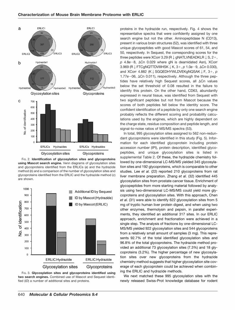
Experimental Design—Recovery of glyco- and phosphopro-teins as well as their modification sites poses many chal-lenges, the greatest being the loss of the low abundance
glyco- and phosphopeptides during isolation and detection.To maximize the recovery of glyco- and phosphoproteinsfrom the mouse brain membrane preparation, we adopted aunique combination of individually successful approachessuch as subfractionation to obtain a purified membrane frac-tion (51) followed by parallel use of two enrichment ap-proaches with potentially high efficiency (hydrazide chemistryand ERLIC). This was finally coupled to duplicate MS analysisand two types of database searches (Table I) that ensuredstatistical consistency and better coverage for identification.
The glycopeptide capture based on hydrazide chemistryhas been standardized and widely used (26, 27), whereasERLIC is a relatively new method. Initial adaptations of theERLIC protocol for phosphopeptide binding and elution to thecapture of glycopeptides yielded a number of glycoproteinscomparable to that of the hydrazide method (44). As phos-phopeptides and glycopeptides differ in their charge and hy-drophilicity, we hypothesized that the retention time and op-timal elution conditions for glycopeptides would be differentfrom those for phosphopeptides. Indeed, we found that manyglycopeptides did not elute until a low organic content(�30%) was reached. The broader gradient window for theelution of glycopeptides (70–30% organic content) as com-pared with phosphopeptides (70–60%) is probably due to thehigher complexity and heterogeneity of carbohydrate moi-eties. Thus, a new buffer system consisting of buffer A (10 mM
sodium methyl phosphonate and 70% acetonitrile, pH 2.0)and buffer B (200 mM triethylamine phosphate with 25%acetonitrile, pH 2.0) was used for subsequent glyco- andphosphopeptide enrichment and fractionation.
Fig. 1 shows the ERLIC chromatograms for the fraction-ation of the mouse brain membrane digest. As this studyfocused on the identification of post-translational modifica-tions, the bulk of peptides eluted in the first 7 min was notcollected. In each ERLIC run, a total of 41 fractions from 7 to48 min was collected, but only seven fractions were finallysubmitted for LC-MS/MS analysis by combining five consec-utive fractions with the exception of the last fraction (Fig. 1, aand b) as we anticipated the peptide complexity to be sub-stantially reduced such that it was below the detection sen-sitivity of one-dimensional LC-MS/MS.
Identification of N-Linked Glycoproteins and GlycosylationSites—Protein identification was achieved by using twosearch engines, Mascot and Sequest, for the interpretation ofthe MS/MS spectra. The false positive rate of overall peptide
TABLE ISummary of experiment statistics
Peptides/experiment
ReplicatesNo. of
fractionsNo. of MSinjections
Samplepreparation time
MS timeDatabasesearches
mg days h
ERLIC 1 3 21 42 �1 63 Mascot, SequestHydrazide method 1 3 3 6 �2 9 Mascot, Sequest
Characterization of Mouse Brain Membrane Proteome with ERLIC
638 Molecular & Cellular Proteomics 9.4

assignment in both searches was controlled within 1%. Thenumbers of total peptides, glycopeptides, and proteins iden-tified by the two engines were comparable. Identifying thecorrect isoform of the protein is a common problem of shot-gun proteomics studies. This poses a greater challenge inglycoproteomics as a glycopeptide can possibly be assignedto multiple proteins with each having supporting unique pep-tides. For example, Nrcam isoform 2 and isoform 3 share fourglycopeptides but also have respective unique peptides(K2EDAHADPEIQPMKEDDGTFGEYSDAEDHKPLK2Kin iso-form 2 and K2EKEDAHADPEIQPMKEDDGTFGEYR2S inisoform 3). In this case, we reported only the glycoproteinisoform with a higher protein score assigned by the data-base search engine to achieve a set of non-redundant gly-coproteins. The statistics of identified proteins, peptides,and post-translational modification sites are summarized insupplemental Table 1.
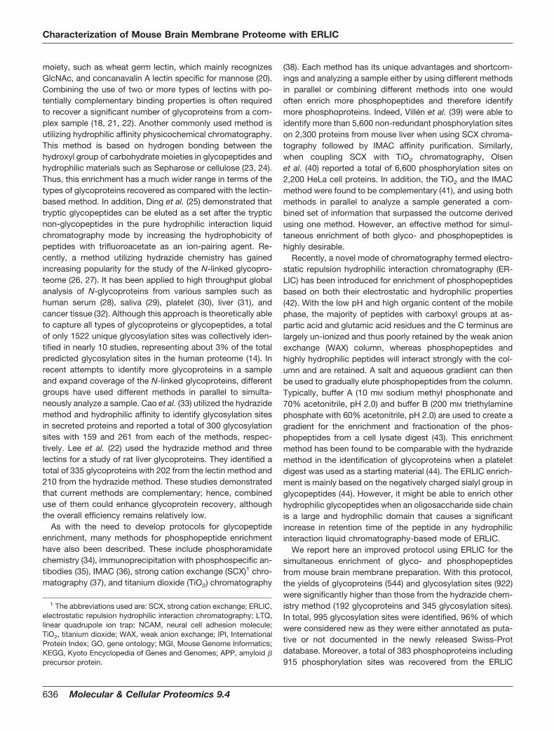
Using the Mascot search engine, a total of 738 uniqueglycosylation sites assigned to 446 glycoproteins was identi-fied from the three ERLIC replicates (Fig. 2). In contrast, only259 unique glycosylation sites and 153 glycoproteins wererecovered from the three hydrazide replicates. The non-re-dundant MS/MS spectra of the glycopeptides enriched by theERLIC and the hydrazide chemistry in Mascot peptide viewformat are shown in supplemental Data 1 and 4. The numbers ofidentified glycosylation sites and glycoproteins in the ERLICapproach (mean � S.D., 560 � 70 sites; 352 � 30 glycopro-
teins) were significantly higher than those from the hydrazidemethod (216 � 9 sites; 131 � 4 glycoproteins) (Fig. 2c). In theERLIC approach, 52.4% of glycosylation sites were common inall three replicates, and an average of 65.5% was found to beoverlapping between any two replicates. About 58.3% of theglycoproteins were identified from all three replicates with anaverage of 70.1% overlapping between any two replicates(Fig. 2a). A slightly higher proportion of glycosylation sites(68.3%) and glycoproteins (72.7%) was observed in all threereplicates from the hydrazide method (Fig. 2b). Clearly, re-peated analyses of a single sample enhanced the number ofglycosylation sites and glycoproteins identified from the sam-ple as is true of shotgun proteomics in general. Similar resultswere obtained via a Sequest search: a total of 827 glycosy-lation sites (528, 704, and 604 from each ERLIC) and 495glycoproteins (348, 430, and 373, respectively) were identifiedfrom the three ERLIC replicates, whereas 283 glycosylationsites (232, 219, and 220 from each hydrazide) and 174 gly-coproteins (148, 136, and 140, respectively) were recoveredfrom the three hydrazide replicates. We found that the ERLICand hydrazide results contained 12.1 and 9.5% non-NX(S/T)motif sites, respectively; these sites are likely generated bynonspecific deamidation during the sample preparation.
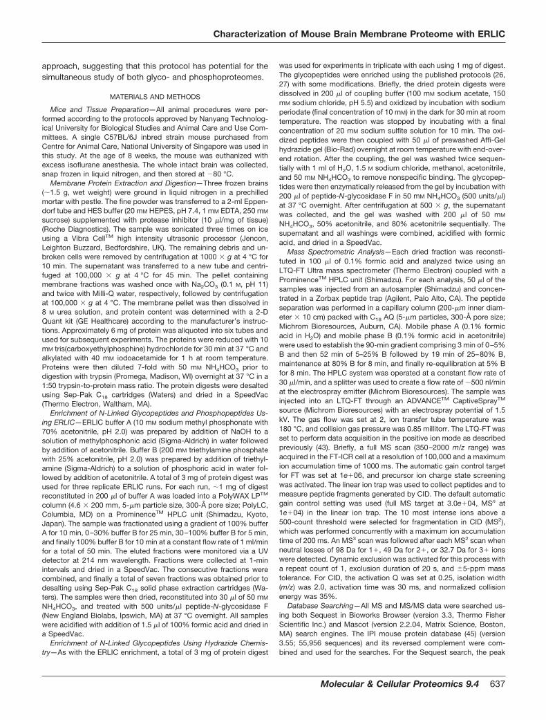
The advantage of using a combination of search engines isshown in Fig. 3. The use of a second search engine (Sequest)yielded 184 additional glycosylation sites and 98 additional gly-coproteins in the ERLICs and an additional 92 sites and 38
FIG. 1. ERLIC chromatograms. a, a blank run shows the LC gradient and background UV absorption chromatogram at 214 nm. b, 1 mg ofmouse brain tryptic digest. Forty fractions from 7 to 50 min were collected and then combined to seven final fractions as shown for LC-MS/MSanalysis.
Characterization of Mouse Brain Membrane Proteome with ERLIC
Molecular & Cellular Proteomics 9.4 639
proteins in the hydrazide run, respectively. Fig. 4 shows therepresentative spectra that were confidently assigned by onesearch engine but not the other. Aminopeptidase N (CD13),present in various brain structures (52), was identified with threeunique glycopeptides with good Mascot scores of 61, 54, and50, respectively. In Sequest, the corresponding scores for thethree peptides were XCorr 3.29 (R2gNATLVNEADKLR2S, 2�,p 4.8e�8, �Cn 0.029 where gN is deamidated Asn), XCorr3.969 (R2FTCgNQTTDVIIIHSK2K, 3�, p 1.0e�9, �Cn 0.030),and XCorr 4.882 (K2SGQEDHYWLDVEKgNQSAK2F, 3�, p1.77e�06, �Cn 0.011), respectively. Although the three pep-tides have relatively high Sequest scores, all �Cn valuesbelow the set threshold of 0.08 resulted in the failure toidentify this protein. On the other hand, CD63, abundantlyexpressed in neural tissue, was identified from Sequest withtwo significant peptides but not from Mascot because thescores of both peptides fell below the identity score. Theconfident identification of a peptide by only one search engineprobably reflects the different scoring and probability calcu-lations used by the engines, which are highly dependent onthe charge state, residue composition and peptide length, andsignal-to-noise ratios of MS/MS spectra (53).
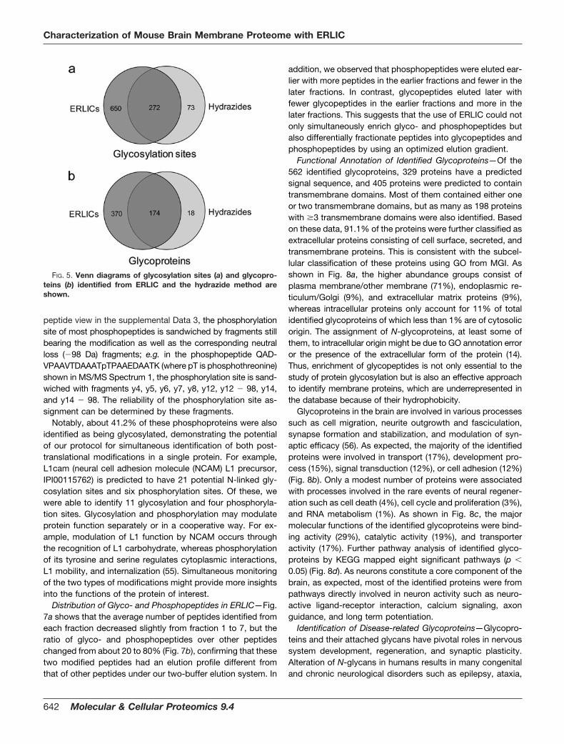
In total, 995 glycosylation sites assigned to 562 non-redun-dant glycoproteins were identified in this study (Fig. 5). Infor-mation for each identified glycoprotein including proteinaccession number (IPI), protein description, identified glyco-peptides, and unique glycosylation sites is listed insupplemental Table 2. Of these, the hydrazide chemistry fol-lowed by one-dimensional LC-MS/MS yielded 345 glycosyla-tion sites and 192 glycoproteins, which is comparable to otherstudies. Lee et al. (22) reported 210 glycoproteins from ratliver membrane preparation. Zhang et al. (32) identified 445glycosylation sites from prostate cancer tissue. Enrichment ofglycopeptides from more starting material followed by analy-sis using two-dimensional LC-MS/MS could yield more gly-coproteins and glycosylation sites. With this approach, Chenet al. (31) were able to identify 622 glycosylation sites from 5mg of tryptic human liver protein digest, and when using twoother enzymes, thermolysin and pepsin, in parallel experi-ments, they identified an additional 317 sites. In our ERLICapproach, enrichment and fractionation were achieved in asingle step. The analysis of fractions by one-dimensional LC-MS/MS yielded 922 glycosylation sites and 544 glycoproteinsfrom a relatively small amount of samples (3 mg). This repre-sents 92.7% of the total identified glycosylation sites and96.8% of the total glycoproteins. The hydrazide method pro-vided an additional 73 glycosylation sites (7.3%) and 18 gly-coproteins (3.2%). The higher percentage of new glycosyla-tion sites over new glycoproteins from the hydrazidechemistry method suggests that higher glycosylation site cov-erage of each glycoprotein could be achieved when combin-ing the ERLIC and hydrazide methods.
We next matched these 995 glycosylation sites with thenewly released Swiss-Prot knowledge database for rodent
FIG. 2. Identification of glycosylation sites and glycoproteinsusing Mascot search engine. Venn diagrams of glycosylation sitesand glycoproteins identified from the ERLICs (a) and the hydrazidemethod (b) and a comparison of the number of glycosylation sites andglycoproteins identified from the ERLIC and the hydrazide method (c)are shown.
FIG. 3. Glycosylation sites and glycoproteins identified usingtwo search engines. Combined use of Mascot and Sequest identi-fied (ID) a number of additional sites and proteins.
Characterization of Mouse Brain Membrane Proteome with ERLIC
640 Molecular & Cellular Proteomics 9.4

species (released on May 26, 2009). It is worth mentioningthat the positions of the glycosylation sites identified from theIPI database were different from those in the Swiss-Prot da-tabase in some of the glycoproteins. For example, CD98heavy chain (IPI00114641) was identified with four glycosyla-tion sites at Asn-172, Asn-265, Asn-391, and Asn-405,whereas in the Swiss-Prot database, these sites becomeAsn-166, Asn-259, Asn-385, and Asn-399 because of se-quence cleavage or protein truncation/isoform. Such changesin site position were manually verified based on sequencematch and included in supplemental Table 2. Of the 995glycosylation sites, only 39 are documented as valid N-linkedglycosylation sites with experimental proof of which two arehigh mannose type; 62.0% (617) of them are annotated aspotential (601 sites), probable (five sites), or by similarity (11sites); and 34.1% (339) of them were not documented asN-linked glycosylation sites (Fig. 6a). This shows that glyco-proteins and glycosylation sites are largely underrepresentedin the current databases, and the use of a glycoproteomicsapproach allows not only a large scale validation of potential
glycosylation sites but also identification of novel glycosyla-tion sites (Fig. 6b).
Identification of Membrane Phosphoproteins—Phosphory-lation of membrane proteins often initiates signal transductionpathways or attenuates plasma membrane transport pro-cesses (54). However, study of these proteins is challengingdue to the inherent hydrophobicity of membrane proteins andlow abundance of phosphopeptides. Here, we prepared pro-tein digests from purified membrane proteins and enrichedhydrophilic and negatively charged peptides using ERLIC. Weshowed that in addition to the identification of a high numberof glycoproteins a total of 383 phosphoproteins was alsoidentified with 915 unique phosphorylation sites identified(supplemental Table 3). Of these, 232 proteins (60.1%) aremembrane phosphoproteins, and 140 are known plasmamembrane proteins, indicative of the efficacy of this approachfor studying the membrane phosphoproteome. The MS/MSspectra of the ERLIC enriched phosphopeptides in Mascotpeptide view format are shown in the supplemental Data2 and 3. As shown in the MS/MS spectrum in the Mascot
FIG. 4. Representative mass spectrum that was confidently assigned to peptide R2gNATLVNEADKLR2S from aminopeptidase N(CD13) by Mascot but not Sequest.
Characterization of Mouse Brain Membrane Proteome with ERLIC
Molecular & Cellular Proteomics 9.4 641
peptide view in the supplemental Data 3, the phosphorylationsite of most phosphopeptides is sandwiched by fragments stillbearing the modification as well as the corresponding neutralloss (�98 Da) fragments; e.g. in the phosphopeptide QAD-VPAAVTDAAATpTPAAEDAATK (where pT is phosphothreonine)shown in MS/MS Spectrum 1, the phosphorylation site is sand-wiched with fragments y4, y5, y6, y7, y8, y12, y12 � 98, y14,and y14 � 98. The reliability of the phosphorylation site as-signment can be determined by these fragments.
Notably, about 41.2% of these phosphoproteins were alsoidentified as being glycosylated, demonstrating the potentialof our protocol for simultaneous identification of both post-translational modifications in a single protein. For example,L1cam (neural cell adhesion molecule (NCAM) L1 precursor,IPI00115762) is predicted to have 21 potential N-linked gly-cosylation sites and six phosphorylation sites. Of these, wewere able to identify 11 glycosylation and four phosphoryla-tion sites. Glycosylation and phosphorylation may modulateprotein function separately or in a cooperative way. For ex-ample, modulation of L1 function by NCAM occurs throughthe recognition of L1 carbohydrate, whereas phosphorylationof its tyrosine and serine regulates cytoplasmic interactions,L1 mobility, and internalization (55). Simultaneous monitoringof the two types of modifications might provide more insightsinto the functions of the protein of interest.
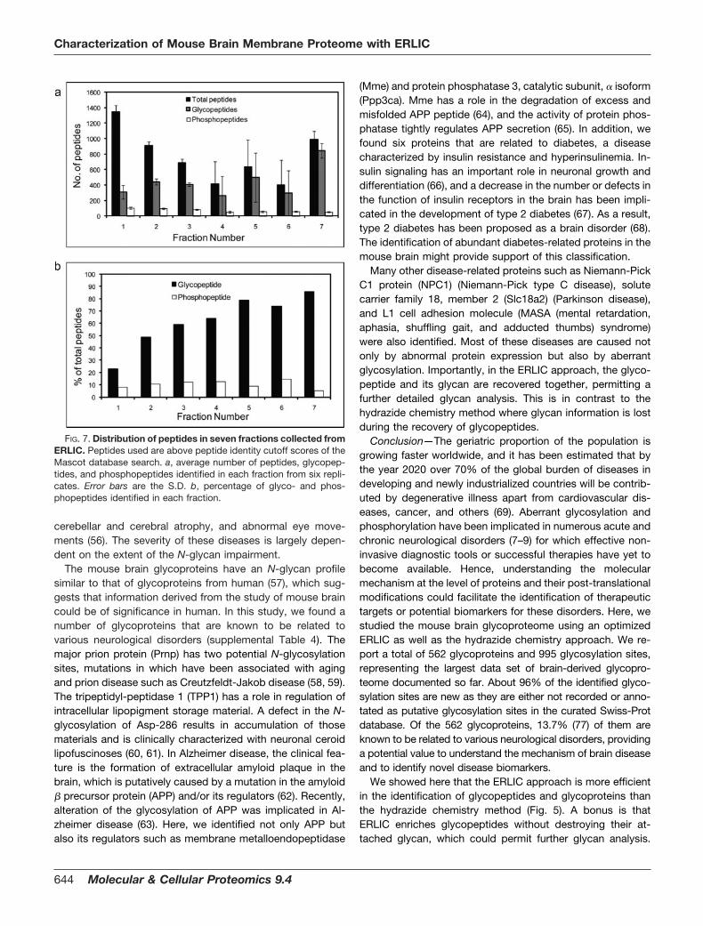
Distribution of Glyco- and Phosphopeptides in ERLIC—Fig.7a shows that the average number of peptides identified fromeach fraction decreased slightly from fraction 1 to 7, but theratio of glyco- and phosphopeptides over other peptideschanged from about 20 to 80% (Fig. 7b), confirming that thesetwo modified peptides had an elution profile different fromthat of other peptides under our two-buffer elution system. In
addition, we observed that phosphopeptides were eluted ear-lier with more peptides in the earlier fractions and fewer in thelater fractions. In contrast, glycopeptides eluted later withfewer glycopeptides in the earlier fractions and more in thelater fractions. This suggests that the use of ERLIC could notonly simultaneously enrich glyco- and phosphopeptides butalso differentially fractionate peptides into glycopeptides andphosphopeptides by using an optimized elution gradient.
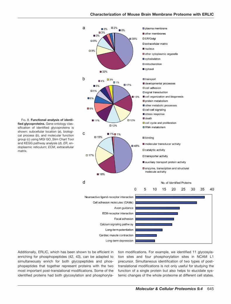
Functional Annotation of Identified Glycoproteins—Of the562 identified glycoproteins, 329 proteins have a predictedsignal sequence, and 405 proteins were predicted to containtransmembrane domains. Most of them contained either oneor two transmembrane domains, but as many as 198 proteinswith �3 transmembrane domains were also identified. Basedon these data, 91.1% of the proteins were further classified asextracellular proteins consisting of cell surface, secreted, andtransmembrane proteins. This is consistent with the subcel-lular classification of these proteins using GO from MGI. Asshown in Fig. 8a, the higher abundance groups consist ofplasma membrane/other membrane (71%), endoplasmic re-ticulum/Golgi (9%), and extracellular matrix proteins (9%),whereas intracellular proteins only account for 11% of totalidentified glycoproteins of which less than 1% are of cytosolicorigin. The assignment of N-glycoproteins, at least some ofthem, to intracellular origin might be due to GO annotation erroror the presence of the extracellular form of the protein (14).Thus, enrichment of glycopeptides is not only essential to thestudy of protein glycosylation but is also an effective approachto identify membrane proteins, which are underrepresented inthe database because of their hydrophobicity.
Glycoproteins in the brain are involved in various processessuch as cell migration, neurite outgrowth and fasciculation,synapse formation and stabilization, and modulation of syn-aptic efficacy (56). As expected, the majority of the identifiedproteins were involved in transport (17%), development pro-cess (15%), signal transduction (12%), or cell adhesion (12%)(Fig. 8b). Only a modest number of proteins were associatedwith processes involved in the rare events of neural regener-ation such as cell death (4%), cell cycle and proliferation (3%),and RNA metabolism (1%). As shown in Fig. 8c, the majormolecular functions of the identified glycoproteins were bind-ing activity (29%), catalytic activity (19%), and transporteractivity (17%). Further pathway analysis of identified glyco-proteins by KEGG mapped eight significant pathways (p
0.05) (Fig. 8d). As neurons constitute a core component of thebrain, as expected, most of the identified proteins were frompathways directly involved in neuron activity such as neuro-active ligand-receptor interaction, calcium signaling, axonguidance, and long term potentiation.
Identification of Disease-related Glycoproteins—Glycopro-teins and their attached glycans have pivotal roles in nervoussystem development, regeneration, and synaptic plasticity.Alteration of N-glycans in humans results in many congenitaland chronic neurological disorders such as epilepsy, ataxia,
FIG. 5. Venn diagrams of glycosylation sites (a) and glycopro-teins (b) identified from ERLIC and the hydrazide method areshown.
Characterization of Mouse Brain Membrane Proteome with ERLIC
642 Molecular & Cellular Proteomics 9.4
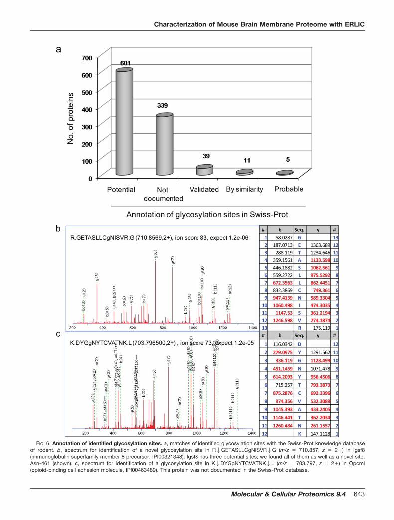
FIG. 6. Annotation of identified glycosylation sites. a, matches of identified glycosylation sites with the Swiss-Prot knowledge databaseof rodent. b, spectrum for identification of a novel glycosylation site in R2GETASLLCgNISVR2G (m/z � 710.857, z � 2�) in Igsf8(immunoglobulin superfamily member 8 precursor, IPI00321348). Igsf8 has three potential sites; we found all of them as well as a novel site,Asn-461 (shown). c, spectrum for identification of a glycosylation site in K2DYGgNYTCVATNK2L (m/z � 703.797, z � 2�) in Opcml(opioid-binding cell adhesion molecule, IPI00463489). This protein was not documented in the Swiss-Prot database.
Characterization of Mouse Brain Membrane Proteome with ERLIC
Molecular & Cellular Proteomics 9.4 643
cerebellar and cerebral atrophy, and abnormal eye move-ments (56). The severity of these diseases is largely depen-dent on the extent of the N-glycan impairment.
The mouse brain glycoproteins have an N-glycan profilesimilar to that of glycoproteins from human (57), which sug-gests that information derived from the study of mouse braincould be of significance in human. In this study, we found anumber of glycoproteins that are known to be related tovarious neurological disorders (supplemental Table 4). Themajor prion protein (Prnp) has two potential N-glycosylationsites, mutations in which have been associated with agingand prion disease such as Creutzfeldt-Jakob disease (58, 59).The tripeptidyl-peptidase 1 (TPP1) has a role in regulation ofintracellular lipopigment storage material. A defect in the N-glycosylation of Asp-286 results in accumulation of thosematerials and is clinically characterized with neuronal ceroidlipofuscinoses (60, 61). In Alzheimer disease, the clinical fea-ture is the formation of extracellular amyloid plaque in thebrain, which is putatively caused by a mutation in the amyloid� precursor protein (APP) and/or its regulators (62). Recently,alteration of the glycosylation of APP was implicated in Al-zheimer disease (63). Here, we identified not only APP butalso its regulators such as membrane metalloendopeptidase
(Mme) and protein phosphatase 3, catalytic subunit, � isoform(Ppp3ca). Mme has a role in the degradation of excess andmisfolded APP peptide (64), and the activity of protein phos-phatase tightly regulates APP secretion (65). In addition, wefound six proteins that are related to diabetes, a diseasecharacterized by insulin resistance and hyperinsulinemia. In-sulin signaling has an important role in neuronal growth anddifferentiation (66), and a decrease in the number or defects inthe function of insulin receptors in the brain has been impli-cated in the development of type 2 diabetes (67). As a result,type 2 diabetes has been proposed as a brain disorder (68).The identification of abundant diabetes-related proteins in themouse brain might provide support of this classification.
Many other disease-related proteins such as Niemann-PickC1 protein (NPC1) (Niemann-Pick type C disease), solutecarrier family 18, member 2 (Slc18a2) (Parkinson disease),and L1 cell adhesion molecule (MASA (mental retardation,aphasia, shuffling gait, and adducted thumbs) syndrome)were also identified. Most of these diseases are caused notonly by abnormal protein expression but also by aberrantglycosylation. Importantly, in the ERLIC approach, the glyco-peptide and its glycan are recovered together, permitting afurther detailed glycan analysis. This is in contrast to thehydrazide chemistry method where glycan information is lostduring the recovery of glycopeptides.
Conclusion—The geriatric proportion of the population isgrowing faster worldwide, and it has been estimated that bythe year 2020 over 70% of the global burden of diseases indeveloping and newly industrialized countries will be contrib-uted by degenerative illness apart from cardiovascular dis-eases, cancer, and others (69). Aberrant glycosylation andphosphorylation have been implicated in numerous acute andchronic neurological disorders (7–9) for which effective non-invasive diagnostic tools or successful therapies have yet tobecome available. Hence, understanding the molecularmechanism at the level of proteins and their post-translationalmodifications could facilitate the identification of therapeutictargets or potential biomarkers for these disorders. Here, westudied the mouse brain glycoproteome using an optimizedERLIC as well as the hydrazide chemistry approach. We re-port a total of 562 glycoproteins and 995 glycosylation sites,representing the largest data set of brain-derived glycopro-teome documented so far. About 96% of the identified glyco-sylation sites are new as they are either not recorded or anno-tated as putative glycosylation sites in the curated Swiss-Protdatabase. Of the 562 glycoproteins, 13.7% (77) of them areknown to be related to various neurological disorders, providinga potential value to understand the mechanism of brain diseaseand to identify novel disease biomarkers.
We showed here that the ERLIC approach is more efficientin the identification of glycopeptides and glycoproteins thanthe hydrazide chemistry method (Fig. 5). A bonus is thatERLIC enriches glycopeptides without destroying their at-tached glycan, which could permit further glycan analysis.
FIG. 7. Distribution of peptides in seven fractions collected fromERLIC. Peptides used are above peptide identity cutoff scores of theMascot database search. a, average number of peptides, glycopep-tides, and phosphopeptides identified in each fraction from six repli-cates. Error bars are the S.D. b, percentage of glyco- and phos-phopeptides identified in each fraction.
Characterization of Mouse Brain Membrane Proteome with ERLIC
644 Molecular & Cellular Proteomics 9.4
Additionally, ERLIC, which has been shown to be efficient inenriching for phosphopeptides (42, 43), can be adapted tosimultaneously enrich for both glycopeptides and phos-phopeptides that together represent proteins with the twomost important post-translational modifications. Some of theidentified proteins had both glycosylation and phosphoryla-
tion modifications. For example, we identified 11 glycosyla-tion sites and four phosphorylation sites in NCAM L1precursor. Simultaneous identification of two types of post-translational modifications is not only useful for studying thefunction of a single protein but also helps to elucidate sys-temic changes of the whole proteome at different cell states.
FIG. 8. Functional analysis of identi-fied glycoproteins. Gene ontology clas-sification of identified glycoproteins isshown: subcellular location (a), biologi-cal process (b), and molecular functiongroup (c) using MGI GO_Slim Chart Tooland KEGG pathway analysis (d). ER, en-doplasmic reticulum; ECM, extracellularmatrix.
Characterization of Mouse Brain Membrane Proteome with ERLIC
Molecular & Cellular Proteomics 9.4 645
In summary, we demonstrate that ERLIC is a simple androbust approach for not only the recovery of glycoproteins butalso the simultaneous recovery of both glycoproteins andphosphoproteins. This approach enabled us to identify thelargest set of glycoproteins from mouse brain so far, whichcould be of significant value in complementing the currentglycoprotein database. We expect that ERLIC could help touncover more glycoproteins and novel glycosylation sitesfrom other samples such as body fluid, cells, or tissues.
Supplemental Data—Detailed information on all identifiedproteins is listed in supplemental Table 5 (from the Mascotsearch) and supplemental Table 6 (from the Sequest search).All MS2 spectra of glyco- and phosphopeptide assignmentsare clearly shown in supplemental spectra hosted onhttps://proteomecommons.org/tranche/ (titled “SimultaneousCharacterization of Glyco- and Phospho-proteomes of MouseBrain Membrane Proteome Using ERLIC Chromatography”)and downloadable using the following hash: LCcjHCB0H/g-7PpRF7Q5uAo0vh2MIEWGi56q8jy/dqQ3OBOk6JNsXXU-PoyUEgctUS�BmB1PpyRSOYRc54Mu46im1RxeoAAAAAA-AACwA��. Alternatively, the non-redundant MS2 spectra ofglyco- and phosphopeptide assignments are provided inSupplemental Data 1–4.
Acknowledgment—We express our gratitude to Andrew Alpert ofPolyLC, Inc. for invaluable advice. We thank Wei Meng, Yi Zhu, PaulHon Sen Tan, and Manavalan Arulmani for invaluable discussions.
* This work was supported by Biomedical Research Council Grant07/1/22/19/531 and Ministry of Education ARC Grant T206B3211 ofSingapore.
□S This article contains supplemental Tables 1–6 and spectralData 1–4.
¶ To whom correspondence should be addressed. Tel.: 65-6514-1006; Fax: 65-6791-3856; E-mail: [email protected].
REFERENCES
1. Apweiler, R., Hermjakob, H., and Sharon, N. (1999) On the frequency ofprotein glycosylation, as deduced from analysis of the SWISS-PROTdatabase. Biochim. Biophys. Acta 1473, 4–8
2. Cohen, P. (2000) The regulation of protein function by multisitephosphorylation–a 25 year update. Trends Biochem. Sci. 25, 596–601
3. Roth, J. (2002) Protein N-glycosylation along the secretory pathway: rela-tionship to organelle topography and function, protein quality control,and cell interactions. Chem. Rev. 102, 285–303
4. Schmidt-Ullrich, R. K., Contessa, J. N., Lammering, G., Amorino, G., andLin, P. S. (2003) ERBB receptor tyrosine kinases and cellular radiationresponses. Oncogene 22, 5855–5865
5. Dennis, J. W., Granovsky, M., and Warren, C. E. (1999) Protein glycosyla-tion in development and disease. BioEssays 21, 412–421
6. Ballif, B. A., Villen, J., Beausoleil, S. A., Schwartz, D., and Gygi, S. P. (2004)Phosphoproteomic analysis of the developing mouse brain. Mol. Cell.Proteomics 3, 1093–1101
7. Guevara, J., Espinosa, B., Zenteno, E., Vazguez, L., Luna, J., Perry, G., andMena, R. (1998) Altered glycosylation pattern of proteins in Alzheimerdisease. J. Neuropathol. Exp. Neurol. 57, 905–914
8. Takahashi, M., Tsujioka, Y., Yamada, T., Tsuboi, Y., Okada, H., Yamamoto,T., and Liposits, Z. (1999) Glycosylation of microtubule-associated pro-tein tau in Alzheimer’s disease brain. Acta Neuropathol. 97, 635–641
9. Mi, K., and Johnson, G. V. (2006) The role of tau phosphorylation in thepathogenesis of Alzheimer’s disease. Curr. Alzheimer Res. 3, 449–463
10. Castellani, R., Smith, M. A., Richey, P. L., and Perry, G. (1996) Glycoxida-
tion and oxidative stress in Parkinson disease and diffuse Lewy bodydisease. Brain Res. 737, 195–200
11. Muntane, G., Dalfo, E., Martinez, A., and Ferrer, I. (2008) Phosphorylation oftau and alpha-synuclein in synaptic-enriched fractions of the frontalcortex in Alzheimer’s disease, and in Parkinson’s disease and relatedalpha-synucleinopathies. Neuroscience 152, 913–923
12. Guerry, P. (1997) Nonlipopolysaccharide surface antigens of Campy-lobacter species. J. Infect. Dis. 176, Suppl. 2, S122–S124
13. Liu, J., Ball, S. L., Yang, Y., Mei, P., Zhang, L., Shi, H., Kaminski, H. J.,Lemmon, V. P., and Hu, H. (2006) A genetic model for muscle-eye-braindisease in mice lacking protein O-mannose 1,2-N-acetylglucosaminyl-transferase (POMGnT1). Mech. Dev. 123, 228–240
14. Zhang, H., Loriaux, P., Eng, J., Campbell, D., Keller, A., Moss, P., Bonneau,R., Zhang, N., Zhou, Y., Wollscheid, B., Cooke, K., Yi, E. C., Lee, H.,Peskind, E. R., Zhang, J., Smith, R. D., and Aebersold, R. (2006) Uni-Pep—a database for human N-linked glycosites: a resource for biomar-ker discovery. Genome Biol. 7, R73
15. Chalmers, M. J., Kolch, W., Emmett, M. R., Marshall, A. G., and Mischak, H.(2004) Identification and analysis of phosphopeptides. J. Chromatogr. BAnalyt. Technol. Biomed. Life Sci. 803, 111–120
16. Mintz, G., and Glaser, L. (1979) Glycoprotein purification on a high-capacitywheat germ lectin affinity column. Anal. Biochem. 97, 423–427
17. Mechref, Y., Madera, M., and Novotny, M. V. (2008) Glycoprotein enrich-ment through lectin affinity techniques. Methods Mol. Biol. 424, 373–396
18. Drake, R. R., Schwegler, E. E., Malik, G., Diaz, J., Block, T., Mehta, A., andSemmes, O. J. (2006) Lectin capture strategies combined with massspectrometry for the discovery of serum glycoprotein biomarkers. Mol.Cell. Proteomics 5, 1957–1967
19. Hourani, B. T., Chace, N. M., and Pincus, J. H. (1973) Plasma membraneglycoproteins from nucleated cells. I. Preparative techniques for isolationand partial characterization of a membrane glycoprotein extract fromL1210 cells with lectin receptor activity. Biochim. Biophys. Acta 328,520–532
20. Debray, H., Decout, D., Strecker, G., Spik, G., and Montreuil, J. (1981)Specificity of twelve lectins towards oligosaccharides and glycopeptidesrelated to N-glycosylproteins. Eur. J. Biochem. 117, 41–55
21. Ghosh, D., Krokhin, O., Antonovici, M., Ens, W., Standing, K. G., Beavis,R. C., and Wilkins, J. A. (2004) Lectin affinity as an approach to theproteomic analysis of membrane glycoproteins. J. Proteome Res. 3,841–850
22. Lee, A., Kolarich, D., Haynes, P. A., Jensen, P. H., Baker, M. S., and Packer,N. H. (2009) Rat liver membrane glycoproteome: enrichment by phasepartitioning and glycoprotein capture. J. Proteome Res. 8, 770–781
23. Wada, Y., Tajiri, M., and Yoshida, S. (2004) Hydrophilic affinity isolation andMALDI multiple-stage tandem mass spectrometry of glycopeptides forglycoproteomics. Anal. Chem. 76, 6560–6565
24. Tajiri, M., Yoshida, S., and Wada, Y. (2005) Differential analysis of site-specific glycans on plasma and cellular fibronectins: application of ahydrophilic affinity method for glycopeptide enrichment. Glycobiology15, 1332–1340
25. Ding, W., Nothaft, H., Szymanski, C. M., and Kelly, J. (2009) Identificationand quantification of glycoproteins using ion-pairing normal-phase liquidchromatography and mass spectrometry. Mol. Cell. Proteomics 8,2170–2185
26. Zhang, H., Li, X. J., Martin, D. B., and Aebersold, R. (2003) Identification andquantification of N-linked glycoproteins using hydrazide chemistry, sta-ble isotope labeling and mass spectrometry. Nat. Biotechnol. 21,660–666
27. Sun, B., Ranish, J. A., Utleg, A. G., White, J. T., Yan, X., Lin, B., and Hood,L. (2007) Shotgun glycopeptide capture approach coupled with massspectrometry for comprehensive glycoproteomics. Mol. Cell. Proteomics6, 141–149
28. Zhang, H., Yi, E. C., Li, X. J., Mallick, P., Kelly-Spratt, K. S., Masselon, C. D.,Camp, D. G., 2nd, Smith, R. D., Kemp, C. J., and Aebersold, R. (2005)High throughput quantitative analysis of serum proteins using glycopep-tide capture and liquid chromatography mass spectrometry. Mol. Cell.Proteomics 4, 144–155
29. Ramachandran, P., Boontheung, P., Xie, Y., Sondej, M., Wong, D. T., andLoo, J. A. (2006) Identification of N-linked glycoproteins in human salivaby glycoprotein capture and mass spectrometry. J. Proteome Res. 5,1493–1503
Characterization of Mouse Brain Membrane Proteome with ERLIC
646 Molecular & Cellular Proteomics 9.4

30. Lewandrowski, U., Moebius, J., Walter, U., and Sickmann, A. (2006) Eluci-dation of N-glycosylation sites on human platelet proteins: a glycopro-teomic approach. Mol. Cell. Proteomics 5, 226–233
31. Chen, R., Jiang, X., Sun, D., Han, G., Wang, F., Ye, M., Wang, L., and Zou,H. (2009) Glycoproteomics analysis of human liver tissue by combinationof multiple enzyme digestion and hydrazide chemistry. J. Proteome Res.8, 651–661
32. Zhang, H., Liu, A. Y., Loriaux, P., Wollscheid, B., Zhou, Y., Watts, J. D., andAebersold, R. (2007) Mass spectrometric detection of tissue proteins inplasma. Mol. Cell. Proteomics 6, 64–71
33. Cao, J., Shen, C., Wang, H., Shen, H., Chen, Y., Nie, A., Yan, G., Lu, H., Liu,Y., and Yang, P. (2009) Identification of N-glycosylation sites on secretedproteins of human hepatocellular carcinoma cells with a complementaryproteomics approach. J. Proteome Res. 8, 662–672
34. Tao, W. A., Wollscheid, B., O’Brien, R., Eng, J. K., Li, X. J., Bodenmiller, B.,Watts, J. D., Hood, L., and Aebersold, R. (2005) Quantitative phospho-proteome analysis using a dendrimer conjugation chemistry and tandemmass spectrometry. Nat. Methods 2, 591–598
35. Rush, J., Moritz, A., Lee, K. A., Guo, A., Goss, V. L., Spek, E. J., Zhang, H.,Zha, X. M., Polakiewicz, R. D., and Comb, M. J. (2005) Immunoaffinityprofiling of tyrosine phosphorylation in cancer cells. Nat. Biotechnol. 23,94–101
36. Ficarro, S. B., McCleland, M. L., Stukenberg, P. T., Burke, D. J., Ross,M. M., Shabanowitz, J., Hunt, D. F., and White, F. M. (2002) Phospho-proteome analysis by mass spectrometry and its application to Saccha-romyces cerevisiae. Nat. Biotechnol. 20, 301–305
37. Beausoleil, S. A., Jedrychowski, M., Schwartz, D., Elias, J. E., Villen, J., Li,J., Cohn, M. A., Cantley, L. C., and Gygi, S. P. (2004) Large-scalecharacterization of HeLa cell nuclear phosphoproteins. Proc. Natl. Acad.Sci. U.S.A. 101, 12130–12135
38. Larsen, M. R., Thingholm, T. E., Jensen, O. N., Roepstorff, P., and Jør-gensen, T. J. (2005) Highly selective enrichment of phosphorylated pep-tides from peptide mixtures using titanium dioxide microcolumns. Mol.Cell. Proteomics 4, 873–886
39. Villen, J., Beausoleil, S. A., Gerber, S. A., and Gygi, S. P. (2007) Large-scalephosphorylation analysis of mouse liver. Proc. Natl. Acad. Sci. U.S.A.104, 1488–1493
40. Olsen, J. V., Blagoev, B., Gnad, F., Macek, B., Kumar, C., Mortensen, P.,and Mann, M. (2006) Global, in vivo, and site-specific phosphorylationdynamics in signaling networks. Cell 127, 635–648
41. Bodenmiller, B., Mueller, L. N., Mueller, M., Domon, B., and Aebersold, R.(2007) Reproducible isolation of distinct, overlapping segments of thephosphoproteome. Nat. Methods 4, 231–237
42. Alpert, A. J. (2008) Electrostatic repulsion hydrophilic interaction chroma-tography for isocratic separation of charged solutes and selective isola-tion of phosphopeptides. Anal. Chem. 80, 62–76
43. Gan, C. S., Guo, T., Zhang, H., Lim, S. K., and Sze, S. K. (2008) Acomparative study of electrostatic repulsion-hydrophilic interaction chro-matography (ERLIC) versus SCX-IMAC-based methods for phos-phopeptide isolation/enrichment. J. Proteome Res. 7, 4869–4877
44. Lewandrowski, U., Lohrig, K., Zahedi, R., Walter, D., and Sickmann, A.(2008) Glycosylation site analysis of human platelets by electrostaticrepulsion hydrophilic interaction chromatography. Clin. Proteomics 4,25–36
45. Kersey, P. J., Duarte, J., Williams, A., Karavidopoulou, Y., Birney, E., andApweiler, R. (2004) The International Protein Index: an integrated data-base for proteomics experiments. Proteomics 4, 1985–1988
46. Guo, T., Gan, C. S., Zhang, H., Zhu, Y., Kon, O. L., and Sze, S. K. (2008)Hybridization of pulsed-Q dissociation and collision-activated dissocia-tion in linear ion trap mass spectrometer for iTRAQ quantitation. J.Proteome Res. 7, 4831–4840
47. Elias, J. E., and Gygi, S. P. (2007) Target-decoy search strategy for in-creased confidence in large-scale protein identifications by mass spec-trometry. Nat. Methods 4, 207–214
48. Bendtsen, J. D., Nielsen, H., von Heijne, G., and Brunak, S. (2004) Improvedprediction of signal peptides: SignalP 3.0. J. Mol. Biol. 340, 783–795
49. Krogh, A., Larsson, B., von Heijne, G., and Sonnhammer, E. L. (2001)Predicting transmembrane protein topology with a hidden Markov mod-el: application to complete genomes. J. Mol. Biol. 305, 567–580
50. Mi, H., Lazareva-Ulitsky, B., Loo, R., Kejariwal, A., Vandergriff, J., Rabkin,S., Guo, N., Muruganujan, A., Doremieux, O., Campbell, M. J., Kitano, H.,and Thomas, P. D. (2005) The PANTHER database of protein families,subfamilies, functions and pathways. Nucleic Acids Res. 33, D284–D288
51. Zhang, H., Lin, Q., Ponnusamy, S., Kothandaraman, N., Lim, T. K., Zhao, C.,Kit, H. S., Arijit, B., Rauff, M., Hew, C. L., Chung, M. C., Joshi, S. B., andChoolani, M. (2007) Differential recovery of membrane proteins afterextraction by aqueous methanol and trifluoroethanol. Proteomics 7,1654–1663
52. Danziger, R. S. (2008) Aminopeptidase N in arterial hypertension. Heart Fail.Rev. 13, 293–298
53. Elias, J. E., Haas, W., Faherty, B. K., and Gygi, S. P. (2005) Comparativeevaluation of mass spectrometry platforms used in large-scale proteom-ics investigations. Nat. Methods 2, 667–675
54. Thingholm, T. E., Larsen, M. R., Ingrell, C. R., Kassem, M., and Jensen,O. N. (2008) TiO(2)-based phosphoproteomic analysis of the plasmamembrane and the effects of phosphatase inhibitor treatment. J. Pro-teome Res. 7, 3304–3313
55. Kenwrick, S., Watkins, A., and De Angelis, E. (2000) Neural cell recognitionmolecule L1: relating biological complexity to human disease mutations.Hum. Mol. Genet. 9, 879–886
56. Kleene, R., and Schachner, M. (2004) Glycans and neural cell interactions.Nat. Rev. Neurosci. 5, 195–208
57. Albach, C., Klein, R. A., and Schmitz, B. (2001) Do rodent and human brainshave different N-glycosylation patterns? Biol. Chem. 382, 187–194
58. Owen, F., Poulter, M., Lofthouse, R., Collinge, J., Crow, T. J., Risby, D.,Baker, H. F., Ridley, R. M., Hsiao, K., and Prusiner, S. B. (1989) Insertionin prion protein gene in familial Creutzfeldt-Jakob disease. Lancet 1,51–52
59. Goh, A. X., Li, C., Sy, M. S., and Wong, B. S. (2007) Altered prion proteinglycosylation in the aging mouse brain. J. Neurochem. 100, 841–854
60. Mole, S. E. (2006) Neuronal ceroid lipofuscinoses (NCL). Eur. J. Paediatr.Neurol. 10, 255–257
61. Tsiakas, K., Steinfeld, R., Storch, S., Ezaki, J., Lukacs, Z., Kominami, E.,Kohlschutter, A., Ullrich, K., and Braulke, T. (2004) Mutation of theglycosylated asparagine residue 286 in human CLN2 protein results inloss of enzymatic activity. Glycobiology 14, 1C–5C
62. Sennvik, K., Fastbom, J., Blomberg, M., Wahlund, L. O., Winblad, B., andBenedikz, E. (2000) Levels of alpha- and beta-secretase cleaved amyloidprecursor protein in the cerebrospinal fluid of Alzheimer’s disease pa-tients. Neurosci. Lett. 278, 169–172
63. Akasaka-Manya, K., Manya, H., Sakurai, Y., Wojczyk, B. S., Spitalnik, S. L.,and Endo, T. (2008) Increased bisecting and core-fucosylated N-glycanson mutant human amyloid precursor proteins. Glycoconj. J. 25, 775–786
64. Helisalmi, S., Hiltunen, M., Vepsalainen, S., Iivonen, S., Mannermaa, A.,Lehtovirta, M., Koivisto, A. M., Alafuzoff, I., and Soininen, H. (2004)Polymorphisms in neprilysin gene affect the risk of Alzheimer’s disease inFinnish patients. J. Neurol. Neurosurg. Psychiatry 75, 1746–1748
65. Tian, Q., and Wang, J. (2002) Role of serine/threonine protein phosphatasein Alzheimer’s disease. Neurosignals 11, 262–269
66. Wan, Q., Xiong, Z. G., Man, H. Y., Ackerley, C. A., Braunton, J., Lu, W. Y.,Becker, L. E., MacDonald, J. F., and Wang, Y. T. (1997) Recruitment offunctional GABA(A) receptors to postsynaptic domains by insulin. Nature388, 686–690
67. Bruning, J. C., Gautam, D., Burks, D. J., Gillette, J., Schubert, M., Orban,P. C., Klein, R., Krone, W., Muller-Wieland, D., and Kahn, C. R. (2000)Role of brain insulin receptor in control of body weight and reproduction.Science 289, 2122–2125
68. Das, U. N. (2002) Is type 2 diabetes mellitus a disorder of the brain?Nutrition 18, 667–672
69. World Health Organization (2002) Active Ageing: a Policy Framework, WorldHealth Organization, Geneva
Characterization of Mouse Brain Membrane Proteome with ERLIC
Molecular & Cellular Proteomics 9.4 647
Copyright © 2022 FDOKUMEN


![Water-soluble aminocalix[4]arene receptors with hydrophobic and hydrophilic mouths](https://static.fdokumen.com/doc/165x107/63133b5cc32ab5e46f0c535e/water-soluble-aminocalix4arene-receptors-with-hydrophobic-and-hydrophilic-mouths.jpg)