Simulations of DNA topoisomerase 1B bound to supercoiled DNA reveal changes in the flexibility...
9
Nucleic Acids Research, 2014 1 doi: 10.1093/nar/gku654 Simulations of DNA topoisomerase 1B bound to supercoiled DNA reveal changes in the flexibility pattern of the enzyme and a secondary protein–DNA binding site Ilda D’Annessa 1 , Andrea Coletta 1 , Thana Sutthibutpong 2 , Jonathan Mitchell 3 , Giovanni Chillemi 4 , Sarah Harris 2 and Alessandro Desideri 1,* 1 Department of Biology and Interuniversity Consortium, National Institute Biostructure and Biosystem (INBB), University of Rome Tor Vergata, ViaDella Ricerca Scientifica, Rome 00133, Italy, 2 School of Physics and Astronomy, University of Leeds, Leeds, LS2 9JT, UK, 3 Division of Genetics and Epidemiology, Institute of Cancer Research, Sutton, SM2 5NG, UK and 4 Cineca, via dei Tizii 3, 00166 Roma, Italy Received March 19, 2014; Revised June 13, 2014; Accepted July 8, 2014 ABSTRACT Human topoisomerase 1B has been simulated cova- lently bound to a negatively supercoiled DNA minicir- cle, and its behavior compared to the enzyme bound to a simple linear DNA duplex. The presence of the more realistic supercoiled substrate facilitates the formation of larger number of protein–DNA interac- tions when compared to a simple linear duplex frag- ment. The number of protein–DNA hydrogen bonds doubles in proximity to the active site, affecting all of the residues in the catalytic pentad. The clamp over the DNA, characterized by the salt bridge be- tween Lys369 and Glu497, undergoes reduced fluctu- ations when bound to the supercoiled minicircle. The linker domain of the enzyme, which is implicated in the controlled relaxation of superhelical stress, also displays an increased number of contacts with the minicircle compared to linear DNA. Finally, the more complex topology of the supercoiled DNA minicircle gives rise to a secondary DNA binding site involving four residues located on subdomain III. The simula- tion trajectories reveal significant changes in the in- teractions between the enzyme and the DNA for the more complex DNA topology, which are consistent with the experimental observation that the protein has a preference for binding to supercoiled DNA. INTRODUCTION Topoisomerases are ubiquitous enzymes that control the topological state of DNA during fundamental cellular pro- cesses such as replication, transcription and chromosomal segregation (1–3). Human topoisomerase 1B (hTop1B) re- laxes positive and negative supercoils by creating a nick on one strand of the DNA duplex and forming a tran- sient phosphor-tyrosine bond (4). The enzyme mechanism is composed of five steps: (i) DNA binding and formation of a non-covalent complex; (ii) nucleophilic attack by a ty- rosine residue on one DNA strand; (iii) rotation of the in- tact strand around the nick to resolve the supercoil; (iv) re- ligation of the DNA strand and (v) enzyme release (4). The enzyme is highly active in cells undergoing a high degree of replication, such as cancer cells, and for this reason it is an important drug target. hTop1B is the unique molecular target of a class of anticancer compounds, camptothecins (CPTs) (5), which interact with the protein–DNA complex after cleavage and block the religation step (6,7). The structure of the protein has been solved as a cova- lent and non-covalent complex with a 22 bp DNA substrate (8,9), and in the presence of different classes of inhibitors (6,10). The enzyme is composed of (i) an N-terminal do- main, that due to its high degree of flexibility has never been crystallized, but which is dispensable for the catalytic activ- ity and thought to be involved in nuclear localization and in- teraction with other proteins; (ii) a core domain further di- vided into subdomain I, II and III; (iii) a C-terminal domain owning the catalytic residue Tyr723 and (iv) a linker domain connecting subdomain III with the C-terminal (9,11). The protein first clamps the DNA forming a non-covalent com- plex that is stabilized by a salt bridge formed by residues Lys369 and Glu497 which are located on two loops defined as Lip1 and Lip2 (containing Lys369 and Glu497 respec- tively) and which stabilize the complex during the cataly- sis (1). The linker domain is formed by two long helices that are directly involved in the relaxation mechanism. The shape and positive charge of the linker enable it to interact * To whom correspondence should be addressed. Tel: +39 06 72594376; Fax: +39 06 2022798; E-mail: [email protected] C ⃝ The Author(s) 2014. Published by Oxford University Press on behalf of Nucleic Acids Research. This is an Open Access article distributed under the terms of the Creative Commons Attribution License (http://creativecommons.org/licenses/by-nc/4.0/), which permits non-commercial re-use, distribution, and reproduction in any medium, provided the original work is properly cited. For commercial re-use, please contact [email protected] Nucleic Acids Research Advance Access published July 23, 2014 at University of Leeds on November 6, 2014 http://nar.oxfordjournals.org/ Downloaded from
-
Upload
independent -
Category
Documents
-
view
0 -
download
0
Transcript of Simulations of DNA topoisomerase 1B bound to supercoiled DNA reveal changes in the flexibility...

Nucleic Acids Research, 2014 1doi: 10.1093/nar/gku654
Simulations of DNA topoisomerase 1B bound tosupercoiled DNA reveal changes in the flexibilitypattern of the enzyme and a secondary protein–DNAbinding siteIlda D’Annessa1, Andrea Coletta1, Thana Sutthibutpong2, Jonathan Mitchell3,Giovanni Chillemi4, Sarah Harris2 and Alessandro Desideri1,*
1Department of Biology and Interuniversity Consortium, National Institute Biostructure and Biosystem (INBB),University of Rome Tor Vergata, Via Della Ricerca Scientifica, Rome 00133, Italy, 2School of Physics and Astronomy,University of Leeds, Leeds, LS2 9JT, UK, 3Division of Genetics and Epidemiology, Institute of Cancer Research,Sutton, SM2 5NG, UK and 4Cineca, via dei Tizii 3, 00166 Roma, Italy
Received March 19, 2014; Revised June 13, 2014; Accepted July 8, 2014
ABSTRACT
Human topoisomerase 1B has been simulated cova-lently bound to a negatively supercoiled DNA minicir-cle, and its behavior compared to the enzyme boundto a simple linear DNA duplex. The presence of themore realistic supercoiled substrate facilitates theformation of larger number of protein–DNA interac-tions when compared to a simple linear duplex frag-ment. The number of protein–DNA hydrogen bondsdoubles in proximity to the active site, affecting allof the residues in the catalytic pentad. The clampover the DNA, characterized by the salt bridge be-tween Lys369 and Glu497, undergoes reduced fluctu-ations when bound to the supercoiled minicircle. Thelinker domain of the enzyme, which is implicated inthe controlled relaxation of superhelical stress, alsodisplays an increased number of contacts with theminicircle compared to linear DNA. Finally, the morecomplex topology of the supercoiled DNA minicirclegives rise to a secondary DNA binding site involvingfour residues located on subdomain III. The simula-tion trajectories reveal significant changes in the in-teractions between the enzyme and the DNA for themore complex DNA topology, which are consistentwith the experimental observation that the proteinhas a preference for binding to supercoiled DNA.
INTRODUCTION
Topoisomerases are ubiquitous enzymes that control thetopological state of DNA during fundamental cellular pro-cesses such as replication, transcription and chromosomal
segregation (1–3). Human topoisomerase 1B (hTop1B) re-laxes positive and negative supercoils by creating a nickon one strand of the DNA duplex and forming a tran-sient phosphor-tyrosine bond (4). The enzyme mechanismis composed of five steps: (i) DNA binding and formationof a non-covalent complex; (ii) nucleophilic attack by a ty-rosine residue on one DNA strand; (iii) rotation of the in-tact strand around the nick to resolve the supercoil; (iv) re-ligation of the DNA strand and (v) enzyme release (4). Theenzyme is highly active in cells undergoing a high degreeof replication, such as cancer cells, and for this reason it isan important drug target. hTop1B is the unique moleculartarget of a class of anticancer compounds, camptothecins(CPTs) (5), which interact with the protein–DNA complexafter cleavage and block the religation step (6,7).
The structure of the protein has been solved as a cova-lent and non-covalent complex with a 22 bp DNA substrate(8,9), and in the presence of different classes of inhibitors(6,10). The enzyme is composed of (i) an N-terminal do-main, that due to its high degree of flexibility has never beencrystallized, but which is dispensable for the catalytic activ-ity and thought to be involved in nuclear localization and in-teraction with other proteins; (ii) a core domain further di-vided into subdomain I, II and III; (iii) a C-terminal domainowning the catalytic residue Tyr723 and (iv) a linker domainconnecting subdomain III with the C-terminal (9,11). Theprotein first clamps the DNA forming a non-covalent com-plex that is stabilized by a salt bridge formed by residuesLys369 and Glu497 which are located on two loops definedas Lip1 and Lip2 (containing Lys369 and Glu497 respec-tively) and which stabilize the complex during the cataly-sis (1). The linker domain is formed by two long helicesthat are directly involved in the relaxation mechanism. Theshape and positive charge of the linker enable it to interact
*To whom correspondence should be addressed. Tel: +39 06 72594376; Fax: +39 06 2022798; E-mail: [email protected]
C⃝ The Author(s) 2014. Published by Oxford University Press on behalf of Nucleic Acids Research.This is an Open Access article distributed under the terms of the Creative Commons Attribution License (http://creativecommons.org/licenses/by-nc/4.0/), whichpermits non-commercial re-use, distribution, and reproduction in any medium, provided the original work is properly cited. For commercial re-use, please [email protected]
Nucleic Acids Research Advance Access published July 23, 2014 at U
niversity of Leeds on Novem
ber 6, 2014http://nar.oxfordjournals.org/
Dow
nloaded from
2 Nucleic Acids Research, 2014
with the DNA substrate downstream of the cleavage site;it is then thought to guide the relaxation of the supercoiledDNA, giving rise to the ‘controlled rotation’ mechanism (4).The direct correlation between the mobility of the linker andDNA relaxation has been demonstrated by showing thatpoint mutations affecting the dynamics of the linker also al-ter the catalytic religation rate and the susceptibility of theenzyme to drugs (12–17). Reduced drug sensitivity and var-ied flexibility has also been shown in a chimeric enzyme inwhich the plasmodial linker has been substituted into thehuman protein (18).
One major limitation of both the X-ray structure and thesimulations carried out to date is that the DNA substratesstudied have all been linear. hTop1B in fact shows preferen-tial binding to supercoiled DNA (19); however, mutationalanalysis of basic residues on the protein surface reduces thispreference (20). Through improvements in computing tech-nology, which now permit calculations on far larger sys-tems, we have performed a series of atomistic molecular dy-namics simulations of hTop1B in a covalent complex witha 240 bp negatively supercoiled plasmid with explicit watermolecules, and we have compared the data obtained with aprevious simulation of the protein in complex with a 22 bplinear substrate (21). Analysis of the dynamical propertiesof the protein bound to the more biologically relevant su-percoiled substrate reveals an increased number of protein–DNA interactions and, most strikingly, the presence of asecondary protein–DNA binding site.
MATERIALS AND METHODS
In order to provide a negatively supercoiled substrate, a 5-ns GB/SA implicit solvent simulation of the supercoiled 240bp DNA minicircle with linking number Lk = 20 and !Lk= −1.5 was performed, using the simulation protocols andparameters described in our previous work (22). The twistof DNA is slightly underestimated by the AMBER forcefield (32.2 bp/turn), consequently, the relaxed linking (Lk0)number for a minicircle of this size is Lk0 = 21.5, whichgives a superhelical density of σ = !Lk/Lk0 = −1.5/21.5= −0.07, which is marginally higher in magnitude than theσ = −0.06 found in bacterial DNA in physiological con-ditions. This simulation produced a DNA substrate with awrithe of −0.9, as calculated using the methodology pre-viously described (22) and a global under-twist of !Tw =−0.6 helical turns, as calculated using the CURVES+ pro-gram (23).
To position the protein on the supercoiled plasmid, astructural alignment between the 22 bp nicked DNA co-crystallized with the enzyme and the supercoiled DNA wasperformed with the SwissPdbViewer program (24). By def-inition, the base that is cleaved is referred to as −1; thebases upstream are then numbered starting from −2 andthe bases downstream start from +1. Particular attentionwas paid to the alignment of the bases −5/+2, which displaythe largest number of interaction with the protein and com-prise the cleavage site at the −1 position. The coordinatesfor the 22 bp covalent DNA were taken from the crystalstructure 1K4S (10). Once the alignment identified the mostsuitable position for the protein, it was positioned on the su-percoiled DNA (Figure 1) and the system was energy mini-
mized using the Sybyl program version 6.0 (TRIPOS, http://www.tripos.com/) with the Powell method (25). The topol-ogy of the system was built using the Amber10 all atomforce field, with the parmbsc0 forcefield corrections (26)using tleap, and at this stage the 3′-phosphotyrosyl bondbetween the Tyr723 and the thymine in position −1 wasconstructed. The octahedral simulation box consisted of243288 water molecules. Four hundred and fifty-nine Na+
counterions were added to neutralize the system, giving afinal number of 754972 atoms. Simulations were performedusing the program NAMD 2.8b361 (27). The system wassimulated for 50 ns in periodic boundary conditions, using acutoff of 10 A for the evaluation of short-range non bondedinteractions and the Particle Mesh Ewald method for thelong-range electrostatic interactions (28). The temperaturewas fixed at 300 K, using Langevin dynamics (29) whereaspressure was kept constant at 1 Atmosphere through theLangevin piston method (30). The SHAKE (31) and SET-TLE (32) algorithms were used to restrain bond lengths,for the solute (DNA and protein) and water molecules, re-spectively. The atomic positions were saved every 250 steps(i.e. 0.5 ps) for the analysis with the Gromacs 4.5 package(33), or with code written in-house. The results were com-pared with a previous simulation in which the protein wascovalently bound to a linear 22 bps DNA substrate (21).Clustering analysis was performed by fitting the structureof the protein during the simulation trajectories relative tothe C! atoms of the core and C-terminal domains (residues201–624 and 713–765), and then clustering the structures ofthe linker C! atoms (residues 625–712) using a root meansquared deviation (RMSD) cut-off of 1.6 A.
In order to check the reproducibility of the results, fourreplicas of the simulation were performed. These replicaswere generated by reassigning the atomic velocities for twoconfigurations taken at 20 ns (replicas R1 and R2) and at 30ns (replicas R3 and R4), respectively, in accordance with therequired Maxwell-Boltzmann distribution. All quantitativecomparisons of the behaviour of the primary and replicasimulations considered only the last 20 ns of the trajectoriesto ensure that the trajectories are all of equivalent lengths.
To characterize the structure of the DNA during the MDtrajectory, the each DNA base pair was firstly classified aseither disrupted (e.g. containing a kink, bubble or wrinkle)(34) or non-disrupted. All DNA helical parameters werecalculated using CURVES+ (23). Disrupted base pairs wereidentified using the following criteria: (i) if the time aver-age displacement between the two complementary bases is>1 A, the base pair is considered to contain a bubble; (ii)if the fluctuation in roll is >8◦, the base pair step is classi-fied as kinked and (iii) if the chi-dihedral angle is outside ofthe anti-region (between −75◦ and −150◦), then the DNA isclassified as wrinkled. Since DNA twist is highly sequence-dependent (for instance, MD simulations using parmbsc0force field have shown that GC steps have an equilibriumtwist of 35.6◦, while AT steps have an equilibrium twist ofonly 30.5◦ (35)), to assess the change in DNA structure dueto interactions with Top1B at its binding site, we have plot-ted the twist relative to the values reported for MD simula-tions of linear DNA (!tw) rather than the absolute values.
at University of Leeds on N
ovember 6, 2014
http://nar.oxfordjournals.org/D
ownloaded from

Nucleic Acids Research, 2014 3
Figure 1. Molecular structure of the hTop1B in covalent complex with a 240 bp negatively supercoiled DNA after 20 ns of atomistic molecular dynamics,showing the formation of the secondary protein–DNA binding site.
RESULTS AND DISCUSSION
Analysis of protein dynamics
The simulations of the hTop1B in covalent complex witha 240 bp plasmid DNA negatively supercoiled (hTop1Bsc)enable us to compare the dynamical behavior of the pro-tein bound to a supercoiled DNA substrate with a simplelinear 22 bp DNA fragment (hTop1Blin) (21). In both thelinear and supercoiled DNA, the RMSD of the protein C!atoms does not converge (Supplementary Figure S1A, fulllines). However, when the contribution of the linker domainis eliminated, the RMSD of protein reaches a plateau withan average RMSD value lower than 0.15 nm (Supplemen-tary Figure S1A, dashed lines), indicating that in both sys-tems the linker is the most dynamic protein domain. Thiswas also the case for each of the four replica simulations(Supplementary Figure S1B).
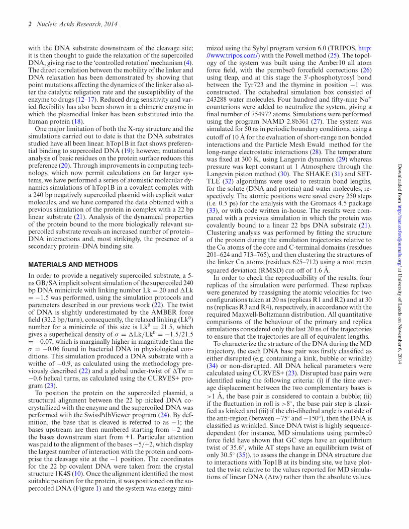
The highly mobile nature of the linker is confirmed bycalculating the per-residue root mean square fluctuation(RMSF), as shown in Figure 2. The profile of the fluctu-ations is similar in the two systems, with the linker reachingvalues of 0.8 and 0.7 nm in the hTop1Bsc and hTop1Blin,respectively (Figure 2). A second peak is found for residues635–644, which are located in part of the loop that con-nects subdomain III to the linker, and which have alreadybeen reported to have a role in modulating the linker mo-bility (36). The per residue profiles of the protein fluctu-ations are similar for all replica simulations (Supplemen-tary Figure S2). Clustering of the C! atoms of residuesAla625-Lys712, involving the linker and the loop precedingit, shows that the linker explores a different region of con-formational space in the two systems. We identified 15 and 4clusters for hTop1Blin and hTop1Bsc, respectively; the mostoccupied clusters are compared in red in Figure 3, where theless occupied clusters are shown in gray. For the linear DNAsubstrate, the first four clusters account for 96% of the to-tal structures (cl1 = 62%, cl2 = 18%, cl3 = 9%, cl4 = 7%),whereas for the supercoiled substrate more than 98% of thetotal conformations sampled belong to the first cluster, asshown in Supplementary Figure S3. In the supercoiled sys-
tem, the linker undergoes a conformational change fromthe starting structure after around 100 ps (SupplementaryFigure S3) which allows it to participate in additional in-teractions with the longer DNA substrate. SupplementaryFigure S4 shows molecular configurations extracted fromthe trajectory every 10 ns. These structures show that thecomplex topological state of the supercoiled DNA substratepermits additional interactions of the N- and C-terminalregions of the linker domain with the double helix, whichis accompanied by a rotation of the coiled–coiled helicesaround their central axis (Supplementary Figure S4).
Similarly, analysis of the persistent hydrogen bonds (e.g.those present for more than 60% of total simulation time)between the DNA and residues Ala625-Lys712 (which formthe linker and the loop preceding it) shows that thereare seven hydrogen bonds in the hTop1Bsc system, whileonly a single bond is found in for hTop1Blin (Table 1).Our simulations therefore show that the three-dimensionalarrangement of the supercoiled double helix facilitates alarger number of hydrogen bonding interactions with thelinker. Moreover, comparing the per-residue RMSF in thehTop1Blin system with that for the supercoiled DNA in-dicates that Glu497 (which is located on Lip2) undergoeslarger fluctuations when the complex is formed with lin-ear DNA. This residue is significant because in the X-raycrystal structures of both the protein–DNA covalent andnon-covalent complexes it forms an electrostatic interac-tion with Lys369, (located on Lip1) and is implicated inthe stabilization of the protein clamp around the DNA (1).In the hTop1Bsc simulation, as well as in the four replicas,this fluctuation is not observed, suggesting the presence ofa tighter interaction between the Lip1 and Lip2, and a re-duced conformational diversity of the protein–DNA com-plex than is observed for the linear substrate.
Protein–DNA interactions and evidence of a secondary DNAbinding site
The static X-ray structures of both the covalent and non-covalent complex of hTopoI with a 22 bp linear DNA frag-
at University of Leeds on N
ovember 6, 2014
http://nar.oxfordjournals.org/D
ownloaded from
4 Nucleic Acids Research, 2014
Figure 2. Per-residue RMSF of the protein in hTop1Blin (black line) and hTop1Bsc (red line) complexes. The different protein domains are defined byvertical lines.
Figure 3. Clustering of the structures of the linker domain, comprising residues Ala625-Lys712, in the simulation of hTop1Blin (A) and hTop1Bsc com-plexes (B). The centroids of the four families representing 96% of total structures in hTop1Blinear and of the only one representing 98% of total structuresin hTop1Bsc are reported in red.
at University of Leeds on N
ovember 6, 2014
http://nar.oxfordjournals.org/D
ownloaded from

Nucleic Acids Research, 2014 5
Table 1. Hydrogen bonds between residues Ala625-Lys712 and DNAwhich are present for more than 60% of total simulation time
hTop1Blin hTop1Bsc
Arg634-Gua12 71% His632-Gua436 64%Lys654-Ade447 69%Arg693-Ade446 76%Gln697-Ade446 76%Lys700-Ade446 68%Gln704-Gua39O 81%Arg708-Cyt38O 73%
ment show that the protein establishes a large number ofhydrogen bonds with DNA, in particular with the segmentof the DNA corresponding to the −5/+2 bases of bothstrands. In the simulation of the enzyme bound to the lin-ear substrate, 25 hydrogen bonds between the protein andDNA bases −10 to +12 DNA are present for more than 60%of the total simulation time. When we consider the sameregion in hTop1Bsc, then the number of hydrogen bondsincreases to 54. Among these bonds, 19 are conserved be-tween the linear and supercoiled DNA substrates, demon-strating that the network of interactions is similar in thesimulations of the two systems, but that additional bondsare occupied in presence of the supercoiled DNA relative tothe linear fragment (21). Among the new interactions ap-pearing in hTop1Bsc, six involve the five residues of the cat-alytic pentad with both bases of the intact (i) or cleaved (c)strands, Arg488-Gua+1(c), Lys532-Ade-2(i), Lys532-Thy-1(c), Hys632-Gua+1(c), His632-Gua+2(c) and Gua+1(c)-Tyr723, and seven interactions involve the linker domainresidues, as reported in Table 1. These six specific hydrogenbonding interactions, and indeed additional contacts notobserved in the primary MD simulation, are also present inall of the replica simulations (see Supplementary Table S1).
In addition to the increased hydrogen bonding interac-tions within the enzyme active site and the tighter com-plex with the linker domain, the simulations of hTop1Bscshow the presence of a secondary DNA binding site. Withinthe core subdomain III, the four residues Lys466, Lys468,Lys545 and Lys549 form direct contacts with the DNA inspite of the fact that they are far from the canonical DNAbinding site. The list of the interactions and their percent-age occurrences are provided in Table 2 and in Figure 4,and the formation and persistence of the secondary bind-ing site is illustrated as a function of simulation time inSupplementary Figure S5. The replica simulations indicatethat the same amino acid residues are involved in forma-tion of this additional protein–DNA interaction (see Ta-ble 2). This secondary DNA binding site involves positivelycharged and polar surface exposed residues located on sub-domain III. A secondary binding site for hTopoIB was pos-tulated by Champoux and co-workers, who demonstratedthat reversing the charge of residues Lys466/Lys468 orLys545/Lys549 affects the preferential binding of hTop1Bto supercoiled DNA (36). In our simulations, the mostpersistent interactions involve these residues; in particu-lar Lys545 and Lys549 are engaged in Lys545-Gua218 andLys549-Cyt217 hydrogen bonding interactions for 64 and41% of the total simulation time, respectively (Table 2, col-umn 1). The simulations suggest that Lys545 is the residue
that plays the primary role in the formation of this sec-ond binding site; the interaction observed between this pos-itively charge residue and the DNA in the replica sim-ulations reaches values of up to 95% (Table 2, columns2–6). A secondary DNA binding site has been also de-scribed in the minimal functional enzyme from vacciniavirus (37,38), and was observed in the crystal structure ofthe Deinococcus radiorurans topoisomerase in complex withDNA, where it involved equivalent residues to those thatare implicated in our simulations (e.g. residues 114, 116,181 and 185 (39) which are equivalent to the human 466,468, 545 and 549) (19). Moreover, the ability of the hu-man enzyme to promote the formation of intra- and inter-molecular synapses between distal DNA segments has beendemonstrated by atomic force microscopy (40). It is possi-ble that this secondary binding site plays an important bio-logical role; most interestingly, a mutation close to Lys545,namely Arg546Gln, has been recently reported in an indi-vidual with a rare form of autism (41).
The presence of this secondary binding site, the tighterinteraction we observe with the linker region of the proteinand the increased number of hydrogen bonding interactionswithin the active site are all consistent with the experimen-tal observation that hTop1B prefers supercoiled over linearDNA substrates.
Structure and dynamics of the DNA in the hTop1B–DNAcomplex
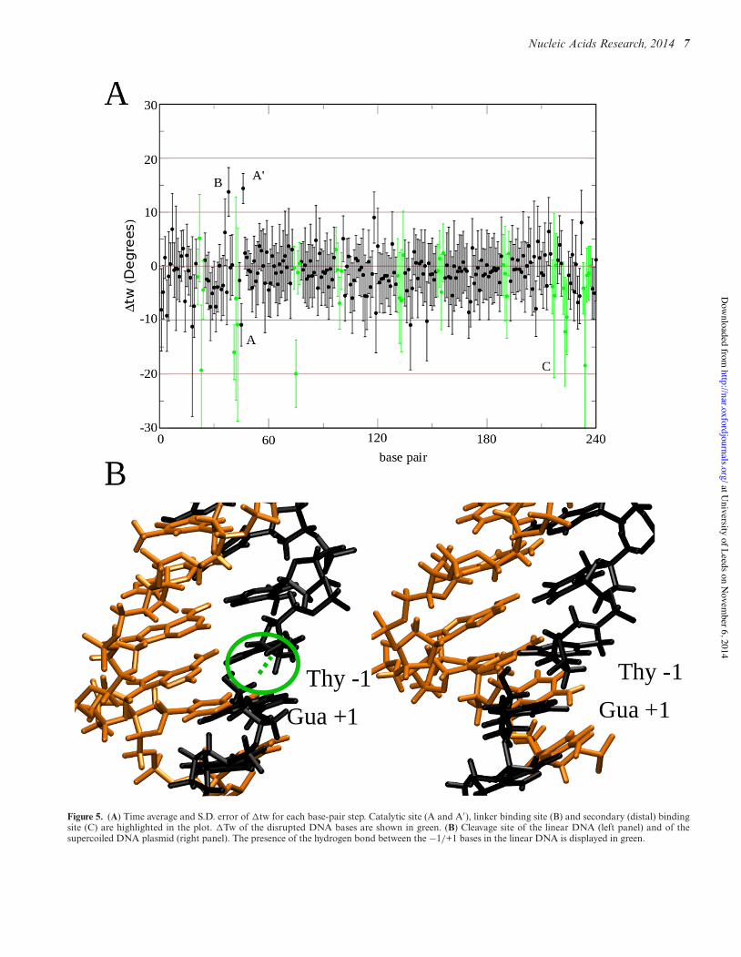
The effect of the protein binding on the structural featuresof a negatively supercoiled DNA plasmid in complex withhTop1B have been monitored by following the twist (tw) be-tween each base pair and plotting the !tw as a function oftime in Figure 5A. !tw within undisrupted regions of theDNA was found to be slightly negative (−1.1◦) due to thenegative supercoiling, as would be expected. The plot alsoshows the presence of disrupted regions located throughoutthe supercoil (depicted in green). Interestingly, the regionsshowing the most evident disruption, with values lower than−20 to −30◦, are those in proximity of the regions contact-ing the protein active site, the linker and the secondary bind-ing site on subdomain III (Figure 5A), which we hypothe-sise is most likely due to the strong interaction between theDNA and the protein.
The catalytic reaction of topoisomerase results in thecleavage of base pair step T−1s/G+1s, which allows thestressed negatively supercoiled DNA to relax. In the linearDNA, the hydrogen bond between the two ends of the cleav-age site persists, in spite of the absence of the phosphategroup (Figure 5B, left panel), while in supercoiled DNA,this hydrogen bond is lost (Figure 5B, right panel), mostlikely because the torsional stress has been partly relievedduring the 50 ns timescale. The restoring torque within thesupercoiled structure is reflected in the over-twisting of thetwist at the cleavage site and the under-twisting of the nextbase pair step downstream, as shown in Figure 5B.
CONCLUSIONS
Molecular dynamics simulations of hTop1B in covalentcomplex with a negatively supercoiled plasmid containing
at University of Leeds on N
ovember 6, 2014
http://nar.oxfordjournals.org/D
ownloaded from

6 Nucleic Acids Research, 2014
Figure 4. Protein–DNA hydrogen bonding network within the secondary binding site.
Table 2. Hydrogen bonds between residues in the secondary binding site and supercoiled DNA along the total 50 ns and last 20 ns of the main (M)simulation (columns 1 and 2) and along the last 20 ns of replica (R1–4) simulations (columns 3–6)
M (50 ns) M (last 20 ns) R1 (last 20 ns) R2 (last 20 ns) R3 (last 20 ns) R4 (last 20 ns)
Lys466-Ade267 28% 44%
Lys466-Thy268 30% 47% 60% 84% 51% 33%Lys468-Gua269 18% 36% 25%
Lys545-Cyt217 30% 29% 20% 26%
Lys545-Gua218 64% 59% 95% 72% 40% 92%Lys545-Ade219 27% 65% 25%
Lys549-Cyt217 41% 46% 42% 20% 14% 56%Lys549-Gua218 26% 40% 52% 95% 14% 44%
240 bp (Figure 1) have provided the first atomistic insightinto the importance of supercoiling in mediating the inter-action between the enzyme and its DNA substrate. Whilethe 240 bp minicircle investigated in these calculations iscertainly far smaller than either the plasmids generally em-ployed in biochemical assays or the size of topological do-mains within the human genome (42), these tight loops maynevertheless be representative of the ends of plectonemically
supercoiled loops and moreover are the largest DNA mini-circles simulated atomistically in the presence of a boundprotein in aqueous solution to date.
The simulations provide new insight into the preferencefor hTop1B for supercoiled over linear DNA substrates.Firstly, the calculations show that DNA supercoiling can in-fluence the structure and the dynamical behavior of hTop1Blinker, which is implicated in the controlled rotation mech-
at University of Leeds on N
ovember 6, 2014
http://nar.oxfordjournals.org/D
ownloaded from
Nucleic Acids Research, 2014 7
Figure 5. (A) Time average and S.D. error of !tw for each base-pair step. Catalytic site (A and A′), linker binding site (B) and secondary (distal) bindingsite (C) are highlighted in the plot. !Tw of the disrupted DNA bases are shown in green. (B) Cleavage site of the linear DNA (left panel) and of thesupercoiled DNA plasmid (right panel). The presence of the hydrogen bond between the −1/+1 bases in the linear DNA is displayed in green.
at University of Leeds on N
ovember 6, 2014
http://nar.oxfordjournals.org/D
ownloaded from

8 Nucleic Acids Research, 2014
anism associated with supercoil relaxation. In the presenceof supercoiled DNA, the linker undergoes a rotation to anew conformation, which then has a tighter interaction withthe DNA substrate. In addition, the protein active site hasadditional hydrogen bonding interactions with the super-coiled minicircle compared to the linear DNA fragment.Most striking, however, is the formation of a secondaryDNA binding site which can only occur within complexDNA topologies, and which would not be detectable fora simple linear substrate. These simulations therefore em-phasize the importance of DNA supercoiling in mediatingin DNA recognition events, and consequently highlight therole of topology in genome regulation more generally.
SUPPLEMENTARY DATA
Supplementary Data are available at NAR Online.
ACKNOWLEDGMENT
We acknowledge PRACE for awarding us access to resourceFERMI based in Italy at Cineca.
FUNDING
Associazione Italiana Ricerca Cancro (AIRC) with theGrant No. 10121 to AD. Funding for open access charge:Associazione Italiana Ricerca Cancro (AIRC) with theGrant No. 10121 to AD and the Grant No. BB/I019472/1to SH.Conflict of interest statement. None declared.
REFERENCES1. Champoux,J.J. (2001) DNA topoisomerase I-mediated nicking of
circular duplex DNA. Methods Mol. Biol., 95, 81–87.2. Wang,J.C. (2002) Cellular roles of DNA topoisomerases: a molecular
perspective. Nat. Rev. Mol. Cell Biol., 3, 430–440.3. Corbett,K.D. and Berger,J.M. (2004) Structure, molecular
mechanisms, and evolutionary relationships in DNA topoisomerases.Annu. Rev. Biophys. Biomol. Struct., 33, 95–118.
4. Stewart,L., Redinbo,M.R., Qiu,X., Hol,W.G. and Champoux,J.J.(1998) A model for the mechanism of human topoisomerase I.Science, 279, 1534–1541.
5. Castelli,S., Coletta,A., D’Annessa,I., Fiorani,P., Tesauro,C. andDesideri,A. (2012) Interaction between natural compounds andhuman topoisomerase I. Biol. Chem., 393, 1327–1340.
6. Staker,B.L., Feese,M.D., Cushman,M., Pommier,Y., Zembower,D.,Stewart,L. and Burgin,A.B. (2005) Structures of three classes ofanticancer agents bound to the human topoisomerase I-DNAcovalent complex. J. Med. Chem., 48, 2336–2345.
7. Pommier,Y. (2006) Topoisomerase I inhibitors: camptothecins andbeyond. Nat. Rev. Cancer, 6, 789–802.
8. Redinbo,M.R., Stewart,L., Champoux,J.J. and Hol,W.G. (1999)Structural flexibility in human topoisomerase I revealed in multiplenon-isomorphous crystal structures. J. Mol. Biol., 292, 685–696.
9. Redinbo,M.R., Stewart,L., Kuhn,P., Champoux,J.J. and Hol,W.G.(1998) Crystal structures of human topoisomerase I in covalent andnoncovalent complexes with DNA. Science, 279, 1504–1513.
10. Staker,B.L., Hjerrild,K., Feese,M.D., Behnke,C.A., Burgin,A.B. Jr.and Stewart,L. (2002) The mechanism of topoisomerase I poisoningby a camptothecin analog. Proc. Natl. Acad. Sci. U.S.A.,99, 15387–15392.
11. Stewart,L., Ireton,G.C., Parker,L.H., Madden,K.R. andChampoux,J.J. (1996) Biochemical and biophysical analyses ofrecombinant forms of human topoisomerase I. J. Biol. Chem., 271,7593–7601.
12. Fiorani,P., Bruselles,A., Falconi,M., Chillemi,G., Desideri,A. andBenedetti,P. (2003) Single mutation in the linker domain confersprotein flexibility and camptothecin resistance to humantopoisomerase I. J. Biol. Chem., 278, 43268–43275.
13. Fiorani,P., Tesauro,C., Mancini,G., Chillemi,G., D’Annessa,I.,Graziani,G., Tentori,L., Muzi,A. and Desideri,A. (2009) Evidence ofthe crucial role of the linker domain on the catalytic activity of humantopoisomerase I by experimental and simulative characterization ofthe Lys681Ala mutant. Nucleic Acids Res., 37, 6849–6858.
14. Chillemi,G., D’Annessa,I., Fiorani,P., Losasso,C., Benedetti,P. andDesideri,A. (2008) Thr729 in human topoisomerase I modulatesanti-cancer drug resistance by altering protein domaincommunications as suggested by molecular dynamics simulations.Nucleic Acids Res., 36, 5645–5651.
15. D’Annessa,I., Tesauro,C., Fiorani,P., Chillemi,G., Castelli,S.,Vassallo,O., Capranico,G. and Desideri,A. (2012) Role of flexibilityin protein-DNA-drug recognition: the case of Asp677Gly-Val703Iletopoisomerase mutant hypersensitive to camptothecin. J. AminoAcids, 206083, 1–8.
16. Tesauro,C., Morozzo Della Rocca,B., Ottaviani,A., Coletta,A.,Zuccaro,L., Arno,B., D’Annessa,I., Fiorani,P. andDesideri,A. (2013) Molecular mechanism of the camptothecinresistance of Glu710Gly topoisomerase IB mutant analyzed in vitroand in silico. Mol. Cancer, 12, 100–113.
17. D’Annessa,I., Tesauro,C., Wang,Z., Arno,B., Zuccaro,L., Fiorani,P.and Desideri,A. (2013) The human topoisomerase 1B Arg634Alamutation results in camptothecin resistance and loss of inter-domainmotion correlation. Biochim. Biophys. Acta, 1834, 2712–2721.
18. Arno,B., D’Annessa,I., Tesauro,C., Zuccaro,L., Ottaviani,A.,Knudsen,B., Fiorani,P. and Desideri,A. (2013) Replacement of thehuman topoisomerase linker domain with the plasmodial counterpartrenders the enzyme camptothecin resistant. PLoS One, 8, e68404.
19. Madden,K.R., Stewart,L. and Champoux,J.J. (1995) Preferentialbinding of human topoisomerase I to superhelical DNA. EMBO J.,14, 5399–5409.
20. Yang,Z., Carey,J.F. and Champoux,J.J. (2009) Mutational analysis ofthe preferential binding of human topoisomerase I to supercoiledDNA. FEBS J., 276, 5906–5919.
21. Mancini,G., D’Annessa,I., Coletta,A., Chillemi,G., Pommier,Y.,Cushman,M. and Desideri,A. (2012) Binding of anIndenoisoquinoline to the topoisomerase-DNA complex inducesreduction of linker mobility and strengthening of protein-DNAinteraction. PLoS One, 7, e51354.
22. Mitchell,J. and Harris,S.A. (2013) The thermodynamics of DNAwrithe from atomistic molecular dynamics simulations. Phys. Rev.Lett., 110, 148105–148110.
23. Lavery,R., Moakher,M., Maddocks,J.H., Petkeviciute,D. andZakrzewska,K. (2009) Conformational analysis of nucleic acidsrevisited: curves+. Nucleic Acids Res., 37, 5917–5929.
24. Guex,N. and Peitsch,M.C. (1997) SWISS-MODEL and theSwiss-PdbViewer: an environment for comparative protein modeling.Electrophoresis, 18, 2714–2723.
25. Powell,M.J.D. (1964) An efficient method for finding the minimum ofa function of several variables without calculating derivatives.Comput. J., 17, 155–162.
26. Duan,Y., Wu,C., Chowdhury,S., Lee,M.C., Xiong,G., Zhang,W.,Yang,R., Cieplak,P., Luo,R., Lee,T. et al. (2003) A point-charge forcefield for molecular mechanics simulations of proteins based oncondensed-phase quantum mechanical calculations. J. Comput.Chem., 24, 1999–2012.
27. Phillips,J.C., Braun,R., Wang,W., Gumbart,J., Tajkhorshid,E.,Villa,E., Chipot,C., Skeel,R.D., Kale,L. and Schulten,K. (2005)Scalable molecular dynamics with NAMD. J. Comput. Chem., 26,1781–1802.
28. Cheatham,T.E., Miller,J.L., Fox,T., Darden,T.A. and Kollman,P.A.(1995) Molecular-dynamics simulations on solvated biomolecularsystems––the particle mesh Ewald method leads to stable trajectoriesof DNA, RNA, and proteins. J. Am. Chem. Soc., 117, 4193–4194.
29. Ceriotti,M., Bussi,G. and Parrinello,M. (2009) Langevin equationwith colored noise for constant-temperature molecular dynamicssimulations. Phys. Rev. Lett., 102, 020601–020604.
30. Feller,S.E., Zhang,Y., Pastor,R.W. and Brooks,B.R. (1995) Constantpressure molecular dynamics simulation: the Langevin pistonmethod. J. Chem. Phys., 103, 4613–4621.
at University of Leeds on N
ovember 6, 2014
http://nar.oxfordjournals.org/D
ownloaded from

Nucleic Acids Research, 2014 9
31. Ryckaert,J.P., Ciccotti,G. and Berendsen,H.J.C. (1977) Numericalintegration of the cartesian equations of motion of a system withconstraints: molecular dynamics of n-alkanes. J. Comput.Phys., 23, 327–341.
32. Miyamoto,S. and Kollman,P.A. (1992) Settle: an analytical version ofthe SHAKE and RATTLE algorithm for rigid water models. J.Comput. Chem., 13, 952–962.
33. Hess,B., Kutzner,C., van der Spoel,D. and Lindahl,E. (2008)GROMACS 4: algorithm for highly Efficient, load-balanced, andscalable molecular simulation. J. Chem. Theory Comput., 4, 435–447.
34. Mitchell,J.S., Laughton,C.A. and Harris,S.A. (2011) Atomisticsimulations reveal bubbles, kinks and wrinkles in supercoiled DNA.Nucleic Acids Res., 39, 3928–3938.
35. Lavery,R., Zakrzewska,K., Beveridge,D., Bishop,T.C., Case,D.A.,Cheatham,T. 3rd, Dixit,S., Jayaram,B., Lankas,F., Laughton,C. et al.(2010) A systematic molecular dynamics study of nearest-neighboreffects on base pair and base pair step conformations and fluctuationsin B-DNA. Nucleic Acids Res., 38, 299–313.
36. Mancini,G., D’Annessa,I., Coletta,A., Sanna,N., Chillemi,G. andDesideri,A. (2010) Structural and dynamical effects induced by theanticancer drug topotecan on the human topoisomerase I - DNAcomplex. PLoS One, 5, e10934.
37. Shuman,S., Bear,D.G. and Sekiguchi,J. (1997) Intramolecularsynapsis of duplex DNA by vaccinia topoisomerase. EMBO J., 16,6584–6589.
38. Moreno-Herrero,F., Holtzer,L., Koster,D.A., Shuman,S., Dekker,C.and Dekker,N.H. (2005) Atomic force microscopy shows thatvaccinia topoisomerase IB generates filaments on DNA in acooperative fashion. Nucleic acids Res., 33, 5945–5953.
39. Patel,A., Yakovleva,L., Shuman,S. and Mondragon,A. (2010) Crystalstructure of a bacterial topoisomerase IB in complex with DNAreveals a secondary DNA binding site. Structure, 18, 725–733.
40. Subramani,R., Juul,S., Rotaru,A., Andersen,F.F., Gothelf,K.V.,Mamdouh,W., Besenbacher,F., Dong,M. and Knudsen,B.R. (2010) Anovel secondary DNA binding site in human topoisomerase Iunravelled by using a 2D DNA origami platform. ACS Nano, 4,5969–5977.
41. Neale,B.M., Kou,Y., Liu,L., Ma’ayan,A., Samocha,K.E., Sabo,A.,Lin,C.F., Stevens,C., Wang,L.S., Makarov,V. et al. (2012) Patternsand rates of exonic de novo mutations in autism spectrum disorders.Nature, 485, 242–245.
42. Dekker,J., Marti-Renom,M.A. and Mirny,L.A. (2013) Exploring thethree-dimensional organization of genomes: interpreting chromatininteraction data. Nat. Rev. Genet., 14, 390–403.
at University of Leeds on N
ovember 6, 2014
http://nar.oxfordjournals.org/D
ownloaded from

![Bisindenoisoquinoline bis-1,3-{(5,6-dihydro-5,11-diketo-11H-indeno[1,2-c]isoquinoline)-6-propylamino}propane bis(trifluoroacetate) (NSC 727357), a DNA intercalator and topoisomerase](https://static.fdokumen.com/doc/165x107/63195970e9c87e0c0910145e/bisindenoisoquinoline-bis-13-56-dihydro-511-diketo-11h-indeno12-cisoquinoline-6-propylaminopropane.jpg)


![Substituted dibenzo[ c,h]cinnolines: topoisomerase I-targeting anticancer agents](https://static.fdokumen.com/doc/165x107/631871c065e4a6af370f5e52/substituted-dibenzo-chcinnolines-topoisomerase-i-targeting-anticancer-agents.jpg)