Spinigerin induces apoptotic like cell death in a caspase independent manner in Leishmania donovani
Characterization of the DNA-binding domain andidentification of the active site residue in the ‘Gyr A’half of Leishmania donovani topoisomerase IITanushri Sengupta, Mandira Mukherjee, Rakhee Das, Aditi Das1 and Hemanta K. Majumder*
Molecular Parasitology Laboratory, Indian Institute of Chemical Biology 4, Raja S.C. Mullick Road, Kolkata-700032,India and 1Sealy Center for Molecular Sciences, University of Texas, Medical Branch, Galveston, USA
Received January 19, 2005; Revised and Accepted April 4, 2005
ABSTRACT
DNA topoisomerase II is a multidomain homodimericenzyme that changes DNA topology by coupling ATPhydrolysis to the transport of one DNA helix through atransient double-stranded break in another. To invest-igate the biochemical properties of the individualdomains of Leishmania donovani topoisomerase II,four truncation mutants were generated. Deletion of178 aminoacids from the C-terminus (core and Ld-DC1058) had no apparent effect on the DNA-bindingor cleavage activities of the enzymes. However, when429 aminoacids from the N-terminus and 451 amino-acids from the C-terminus were removed (LdDNDC),the enzyme was no longer active. Moreover, theremoval of 429 aminoacids from the N-terminus(LdDNDC, core and LdDN429) render the mutant pro-teinsincapableofperformingATPhydrolysis.Themut-ant proteins show cleavage activities at wide rangeof KCl concentrations (25–350 mM). In addition, themutant proteins, excepting LdDNDC, can also act onkDNA and linearize the minicircles. Surprisingly, themutant proteins fail to show the formation of theenhanced cleavable complex in the presence ofetoposide. Our findings suggest that the conformationrequired for interaction with the drug is absent in themutant proteins. Here, we have also identified Tyr775
through direct sequencing of the DNA linked peptideas the catalytic residue implicated in DNA-breakageand rejoining. Taken together, our results demonstratethat topoisomerase II are functionally and mechanist-ically conserved enzymes and the variations in activityseem to reflect functional optimization for its physio-logical role during parasite genome replication.
INTRODUCTION
The participation of the DNA topoisomerases in a number ofvital processes, including replication, transcription, geneticrecombination, chromosomal decondensation and segrega-tion is well documented (1). These enzymes catalyze thetransient breakage of DNA strands to form enzyme-mediated gates through which an intact DNA strand ora pair of DNA strands of a double helix can pass. DNAtopoisomerases can be divided into three sub-families,namely type IA, type IB and type II on the basis of sequencehomology and mechanism of action (2). Type II DNA topo-isomerases catalyze ATP-dependent passage of a DNAsegment through a transient double-strand break in anothersegment. In such a reaction, the enzyme-mediated reversiblecleavage is generated by the trans-esterification reactionsbetween a pair of active site tyrosines and two DNAphosphodiester bonds staggered 4 bp apart. Eukaryotic topo-isomerase II are dimeric, whereas its prokaryotic counterpartsincluding gyrase and topo IV are A2B2 tetramers. Moreover,the N-terminal and the central parts of eukaryotic topo-isomerase II are homologous to bacterial GyrB and GyrAsubunits, respectively.
Even though cells cannot survive without the double-stranded DNA passage activity of topoisomerase II, the forma-tion of a cleavage complex is potentially toxic. DNApolymerases or helicases can convert such transient topoII-DNA intermediates into permanent double-stranded DNAbreaks that can eventually trigger cell death pathways (3).Besides its physiological activities, topoisomerase II is a targetfor a number of antineoplastic and antiparasitic drugs (4,5).Topoisomerase II inhibitors can be divided into topo IIcatalytic inhibitors and topo II poisons (6). Topoisomerasecatalytic inhibitors like bis-dioxopiperizines do not stabilizeDNA cleavage complexes whereas topo II poisons inhibit theenzyme by increasing the steady-state levels of DNA cleavagecomplexes (7,8).
*To whom correspondence should be addressed. Tel: +91 33 2412 3207; Fax: +91 33 2473 5197; Email: [email protected]
The authors wish it to be known that, in their opinion, the first two authors should be regarded as joint First Authors
ª The Author 2005. Published by Oxford University Press. All rights reserved.
The online version of this article has been published under an open access model. Users are entitled to use, reproduce, disseminate, or display the open accessversion of this article for non-commercial purposes provided that: the original authorship is properly and fully attributed; the Journal and Oxford University Pressare attributed as the original place of publication with the correct citation details given; if an article is subsequently reproduced or disseminated not in its entirety butonly in part or as a derivative work this must be clearly indicated. For commercial re-use, please contact [email protected]
2364–2373 Nucleic Acids Research, 2005, Vol. 33, No. 8doi:10.1093/nar/gki527
by guest on March 14, 2014
http://nar.oxfordjournals.org/D
ownloaded from
Despite the importance of DNA cleavage/religation reactionto the function of topoisomerase, relatively little is knownabout the specific enzyme–DNA interaction that drivesthis reaction. Therefore, the molecular interaction ofLdTOP2 and DNA remains an enigma. In conjunctionwith this fact, even though topoisomerase II from protozoanparasites have been cloned and sequenced (9), the mechanisticfeatures of the enzyme have never been explored. In kineto-plastid parasites topoisomerase II undoubtedly functionat several steps in the minicircle replication processes,decatenation of the parental circles from the network andrecatenation of the daughter circles to the network (10).In Leishmania donovani, we had previously reported theexistence of a type II topoisomerase containing 1236 amino-acids localized both in nucleus and kinetoplast (11). Giventhe importance of cleavage/religation reactions mediated bytopoisomerase in the life cycle of the parasite, in the presentstudy we report the properties of the DNA-binding domainof LdTOP2.
In human topoisomerase IIa a fragment from aminoacid1–439 was shown to have an intrinsic ATPase activity (12),which could be stimulated by DNA. In another study, the coredomain of drosophila topoisomerase II covering aminoacid406–1207 was demonstrated to have wild-type levels of cleav-age and religation activities (13). These results indicate thatthe individual domains in topoisomerase II preserve theirintrinsic activities even when separated from the rest of theenzyme, demonstrating that they fold up as independentcatalytic domains.
In the present study, to further investigate the biochemicalproperties of the DNA-binding or core domain of LdTOP2,we have constructed four truncation mutants namely LdDN429(aminoacid residues from 430–1236), core (aminoacid resi-dues 430–1058), LdDC1058 (aminoacid residues 1–1058) andLdDNDC (aminoacid residues 430–785). The mutant proteinshave been characterized with respect to ATPase, DNA-binding and cleavage activities. Our results indicate that theresidues from 1–1058 which mimics the wild-type enzymein complementing the temperature-sensitive topoisomeraseII mutant yeast strain (14), retains its DNA-dependent ATPaseactivity and can also perform cleavage of supercoiled DNA.Moreover, the catalytic amino acid implicated in DNA break-age and reunion was mapped by sequencing the DNA boundprotein. Taken together, this is the first report that demon-strates the domain demarcations of LdTOP2 and providesan insight into the mechanistic aspects of the core/DNA-binding domain of the protein and the catalytic residueimplicated in DNA breakage and rejoining.
MATERIALS AND METHODS
Construction of truncation mutants
For construction of all truncation mutants, the regions cor-responding to amino acids 430–785, 430–1058, 430–1236and 1–1058 were amplified by PCR. For aminoacids 430–785 (1 kb DNA fragment), aminoacids 430–1058 (1.9 kbDNA fragment), aminoacids 430–1236 (2.4 kb DNA frag-ment), the sense primer used was 50 GGGAATTCCATATGA-CAGAGGGTGACTCGGCG 30 and the antisense primers
were 50 CGGGATCCCTTGTACAAGTCAAGGCG 30,50CGGGATCCGATGTAGTCGAAGCTCTC30 and 50CGG-GATCCCCCGCTTCACATGCTCCCCTTACA 30, respect-ively. For the construction of 1–1058 (3.2 kb) mutant, thesense and the antisense primers used were 50 GGGAATTC-CATATGACAGACGCTTCCAAG- 30 and 50 CGGGATCCT-CTAGAGAAGCTCTCGTCCACGCG- 30 respectively. Theamplified products were cloned in the NdeI and BamHI sites ofbacterial expression vector pET16b (Novagen) and expressedin Escherichia coli BL21 (DE3) pLysS. The full lengthLdTOP2 was cloned in the BamHI and HindIII sites of expres-sion vector pET28C, using sense primer 50-CG GGATCCT-CATGACAGACGCTTCCAAG-30 and anti sense primer 50-CCCAAGCTTCATCAAAACATGTGGCAG-30. PCR wasperformed for 30 cycles of denaturation at 94�C for 45 s,annealing at 52.5�C and extension at 68�C for 1 kb/minusing Pwo polymerase (Roche Applied Science) and200 mM of each dNTP.
Expression and protein purification
Expression from constructs pET16b/1, pET16b/1.9, pET16b/2.4, pET16b/3.2 and pET28c/3.7 yielded 40, 70, 89, 116 and137 kDa proteins, respectively. Purification of all the His-tagged proteins was done as follows. Briefly, the cells con-taining the recombinant plasmids were induced at 0.6 OD600
with 0.5 mM IPTG at 22�C for 10 h. The cells were harvestedand resuspended in phosphate buffer (pH 7.8) containing300 mM NaCl, 200 mg/ml lysozyme, 0.1% Triton X-100,0.25% Sarkosyl and 1 mM PMSF. Final lysis was achievedby sonication on ice and the lysate was cleared by centrifu-gation at 12 000 rpm in a SS34 rotor for 20 min. The clearedlysate was passed through Ni- nitrilo triacetic acid (Ni-NTA)agarose column (Qiagen). After washing with phosphate buf-fer (pH 7.8) containing 300 mM NaCl and 30 mM imidazole,elution was done using 250 mM imidazole. For further puri-fication, the fractions were pooled from Ni2+ column, dialyzedand loaded onto a 2 ml packed phosphocellulose column (P11cellulose, Whatman). The column was washed with 10 ml ofwash buffer (50 mM Tris–HCl, pH 7.5, 0.5 mM EDTA and20% glycerol) containing 600 mM KCl and finally eluted withthe same buffer containing 800 mM KCl. After elution thepeak fractions were dialyzed overnight against storage buffercontaining 10 mM Tris–HCl (pH 7.8), 100mM NaCl, 5 mMMgCl2, 1 mM PMSF and 15% glycerol and stored at �70�Cuntil further use.
ATPase assay
ATPase measurements were carried out by the pyruvatekinase/lactate dehydrogenase assay described previously(12) with the following modifications. Reaction mixture(1 ml) contained: 0.1 mM NADH, 2 mM phosphoenol-pyruvate, 3 units of pyruvate kinase and 4 units of lactatedehydrogenase. Reactions were initiated by mixing thereaction mixture and ATP, both pre-equilibrated to 30�C.Decrease in NADH concentration was monitored by measur-ing the absorbance at 340 nm in a UV-visible spectrophoto-meter (Shimadzu Corp.) All assays were performed intriplicate.
Nucleic Acids Research, 2005, Vol. 33, No. 8 2365
by guest on March 14, 2014
http://nar.oxfordjournals.org/D
ownloaded from
Electrophoretic mobility shift assay
The labeling of 36 mer oligonucleotide 1 (50ATGAAATCT-AACAATGCGCTCATCGTCATCCTCGGC30) containinghigh affinity topoisomerase II binding site (15) was doneusing polynucleotide kinase (Invitrogen). The labeledoligonucleotide 1 was annealed to oligonucleotide 2 (50
TACTTTAGATTGTTACGCGAGTAGCAGTAGGACCG 30)in a buffer containing 40 mM Tris–HCl (pH 7.5), 20 mMMgCl2 and 50 mM NaCl at 70�C for 1 h and allowed tocool down slowly to room temperature. Briefly, the reactionwas done in a 20 ml binding buffer containing 50 mM Tris–HCl (pH 7.5), 1 mM DTT, 4 mM MgCl2, 50 mM KCl and15 mg/ml BSA and 5 pmol of 36 bp duplex oligonucleotide andvarying concentrations of wild type and truncated LdTOP2enzymes. Reaction mixtures were incubated at 4�C for 30 minand electrophoresed through 7% non-denaturing polyacry-lamide gel (acrylamide:bis:: 29:1) in 0.25· TBE and auto-radiographed. Quantitation of the unbound oligonucleotidewas done by densitometry and Kd was estimated from proteinconcentration at which half of the duplex oligonucleotide wasbound to the protein (16).
Cleavage reaction
Cleavage reaction was performed as described (17). A 20 mlreaction mixture contained 200 ng negatively supercoiledpRYG DNA, 10 mM Tris–HCl (pH 7.5), 100 mM KCl,0.1 mM EDTA, 5 mM MgCl2, 0.5 mM DTT, 30 mg BSAand 10 pmol of enzymes. Reaction was started by incubatingat 37�C for 30 min and terminated by the addition of 0.5% SDSand 10 mM EDTA. The mixture was further incubated with100 mg/ml proteinase K at 37�C for 30 min and analyzed byelectrophoresis on a 1% agarose gel. Ethidium bromide at afinal concentration of 0.5 mg/ml was included in the gel toresolve the linear product (Form III) from the supercoiledmolecule (Form I). Control assays always contained anamount of drug diluent (Dimethyl sulfoxide) equivalent tothat present in drug containing reaction. Salt dependence ofthe mutant enzymes was determined by performing cleavagereaction as described above, using varying concentrations ofsalt. When KCl dependence was estimated, Mg2+ concentra-tion was kept at 5 mM and for Ca2+ and Mg2+ requirement,KCl concentration was kept at 100 mM.
DNA relaxation
DNA relaxation reactions containing 0.5 mg of negativelysupercoiled pRYG DNA were carried out in the absence orpresence of etoposide in 25 mM Tris–HCl(pH 7.9), 10 mMMgCl2, 0.1 mM EDTA, 1 mM DTT, 2 mM ATP, 100 mMNaCl, 10% glycerol and 500 ng of recombinant L.donovanitopoisomerase II. Following incubation at 37�C for 30 min,the reaction was terminated by the addition of loading buffer.The products were analyzed by gel electrophoresis in 1%agarose gel, stained with ethidium bromide and photographedunder UV Transillumination.
Cleavage of kDNA
Plasmid pGEM4Z was used to clone the full-lengthL.donovani minicircles sequence resulting in the formationof pLURKE3 (18). The insert was excised with EcoRI andradiolabeled by random priming using [a32P]dCTP
(Amersham pharmacia) and used as a probe. As describedpreviously, (19) kinetoplast DNA (kDNA) was isolated fromLeishmania strain UR6. About 200 ng kDNA was incubatedwith 50 pmol enzyme for 30 min at 37�C in the cleavage buffermentioned above. DNA was transferred from agarose gel tonitro cellulose membrane. The membrane was hybridized withradiolabeled E3 probe.
Covalent complex between truncated LdTOP2 enzymesand radiolabeled DNA
Covalent complex between protein and DNA was formed asdescribed (20). Reactions were initiated with 50 pmol trun-cated LdTOP2 enzymes (70, 89 and 116 kDa) in the cleavagebuffer mentioned above, containing 4ng 32P-labeled negat-ively supercoiled pRYG plasmid DNA. Reactions werequenched with NaOH and neutralized with HCl. DNase(FPLC pure, Amersham pharmacia) treatment was done for1 h at 37�C. Reactions were terminated by the addition of0.5% SDS and 10 mM EDTA before the complexes were pro-cessed through two cycles of a Sephadex G-25 spin columnand proteins were precipitated and separated by SDS–PAGE. The gel was dried and autoradiographed to exposethe protein bands.
For thin layer chromatography, the protein-DNA complexespurified through G-25 column were treated with proteinase Kat 37�C for 30 min the digested products were vaccum driedand re-suspended in 30 ml of constant boiling with 6N HCl.After removal of HCl, the hydrolysates were dried undervaccum and redissolved in H2O containing unlabeled phos-phoserine, phosphotyrosine and phosphothreonine. The hydro-lysates were spotted on silica gel plate and chromatographed ina solvent containing ethanol/n-butyl alcohol/ammonia/water(volume ratios 4:1/:2:3). After drying, the plates were sprayedwith 0.25% ninhydrin in acetone and heated to 65�C tovisualize the phospho aminoacid standards. The radioactivesignals of the phospho aminoacid were detected by autoradio-graphy (21).
Preparation of LdTOP2-derived tryptic peptidecovalently linked to DNA
A 100 ml mixture containing the protein and 32P labeled pRYGDNA was incubated in the above-mentioned cleavage bufferat 37�C for 10 min. SDS was added to a final concentrationof 0.5% to form a covalent complex between DNA and protein.The protein DNA complex was subjected to SDS–PAGE elec-trophoresis and stained with Coomassie brilliantblue. The proteinband was excised and in gel trypsin digestion was performed.The trypsin-digested peptides were fractionated by reversephase HPLC. Aliquots of the fractions were assayed for 32P.The positive fraction was subjected to Edman sequencing.
RESULTS
Expression of N-terminal deletion mutants andcore domain of LdTOP2
In order to study the function of core domain of kinetoplastidtoposiomerase II, we constructed four truncation mutants ofL.donovani topoisomerase II (1236 amino acids). The trunca-tion start points and end points were determined on the basis of
2366 Nucleic Acids Research, 2005, Vol. 33, No. 8
by guest on March 14, 2014
http://nar.oxfordjournals.org/D
ownloaded from

our previous analysis, where the amino terminal 1–385 resi-dues have an intrinsic ATPase activity (T. Sengupta,M. Mukherjee, A. Das, C. Mandal, R. Das, T. Mukherjeeand H. K. Majumder, unpublished data) and 1–1058 aminoacids can decatenate kDNA and complement temperature-sensitive mutant yeast strain (14). So, to further investigatethe interaction of DNA with the core domain (homologous tothe core/ DNA-binding domain of other eukaryotic topoi-somerase II), we constructed four truncation mutants ofLdTOP2 containing amino acids 430–785 (LdDNDC), 430–1058(core), 430–1236 (LdDN429) and 1–1058 (LdDC1058)(Figure 1A). All the mutants were generated by PCR ampli-fication of the full-length LdTOP2 gene using site-specificprimers. The PCR products were cloned in pET16b and trans-formed in E.coli BL21 (DE3) plysS. The bacterial cells har-boring the truncation constructs were induced with 0.5 mMIPTG and the resultant overexpressed proteins were purifiedby Ni-NTA agarose, as the mutant enzymes had six histidine
residues at their N-terminus, and the wild-type LdTOP2 hadhistidine tagged both at the N-terminus and the C-terminus.Further purification was achieved by passing the Ni-NTAeluate through the phosphocellulose column as described inMaterials and methods (Figure 1B).
ATPase activity
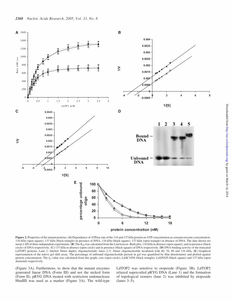
It is known that upon binding to ATP, topoisomerase II under-goes a conformational change that triggers double-strandedDNA passage (20). We checked if any of the mutant proteinsretain their ATPase activities. Apart from the wild-typeenzyme, only the 116 kDa enzyme (1–1058 aminoacidsresidues) had an ATPase activity. Moreover, the ATPaseactivity of this protein was found to be stimulated by DNA(Figure 2A). The KM of the 116 kDa protein was found to be0.33 mM and 0.55 mM in the presence and absence of DNA,respectively (Figure 2B). DNA was found to stimulate itsactivity 2-fold. These values are comparable with those ofthe wild-type enzyme having KM of 0.32 mM and 0.50 mMin the presence and absence of DNA, respectively (Figure 2C).
The truncation mutants bind to DNA ingel retardation assay
A polyacrylamide gel retardation assay was carried out to studythe DNA binding characteristics of LdTOP2 truncationmutants. The linear DNA substrate was a 36 bp duplex con-taining high affinity topoisomerase II cleavage site (15). Theability of LdTOP2 truncation mutants to bind the 36 bp duplexwas tested under standard gel retardation assay condition, asdescribed in Materials and Methods. The constructs containingamino acid 1–1058 (116 kDa), 430–1058 (70 kDa) and 430–1236 (89 kDa) were able to bind to the oligonucleotide butthe construct containing amino acids 430–785 (40 kDa)failed to bind to the duplex DNA. The electrophoreticpatterns were typical of the binding studies (Figure 2D). Thebound DNA migrated as a high molecular weight species ascompared to the unbound DNA. Since the electrophoreticmobility of the complexed DNA was greatly retarded as com-pared to that of the free substrate, the amount of unbound DNAcould be quantitated directly by densitometric scanning. Thisquantitation, in turn, allowed the straightforward calculation ofbound plasmid molecules by subtraction from the total DNAemployed (Figure 2E). The Kd value of LdDC1058, LdDN429and core were found to be 3.3, 4.09 and 4.2 nM, respectively,while that of the wild-type enzyme was 3.0 nM.
Analysis of the cleavage activity of the mutant proteins
An essential step in the relaxation of supercoiled DNA isthe cleavage of DNA in both strands by topoisomerase II.To test the possibility that a mutant enzyme can neverthe-less cleave DNA via trans-esterification and may possess anormal level of cleavage activity, the purified mutant enzymeswere assayed for their cleavage of plasmid DNA. The 40 kDaprotein (430–785 amino acids residues) which does not bind toDNA also fails to show any cleavage of supercoiled DNA(Figure 3A), whereas all other purified proteins (70, 89 and116 kDa and and the wild-type LdTOP2) cleave supercoiledDNA. However, etoposide enhanced the formation of cleav-able complex only by the wild-type LdTOP2 protein and didnot have any effect on the activities of the mutant proteins
A
B
Figure 1. Schematic representation of topoisomerase II of L.donovani andpurification of the truncated proteins. (A) A coordinate of Leishmania TOP2peptide. (A) LdDC1058, (B) LdDN429, (C) Core, (D) LdDNDC protein. (B)10% SDS-polyacrylamide gel showing purification of the truncation proteins.Lanes 1–4, 2 mg of 40, 70, 89 and 116 kDa, respectively, purified through Ni-NTA and phosphocellulose columns. The gel was stained by silver stain. Sizesof molecular mass markers (MBI Fermentas) in kDa are shown.
Nucleic Acids Research, 2005, Vol. 33, No. 8 2367
by guest on March 14, 2014
http://nar.oxfordjournals.org/D
ownloaded from
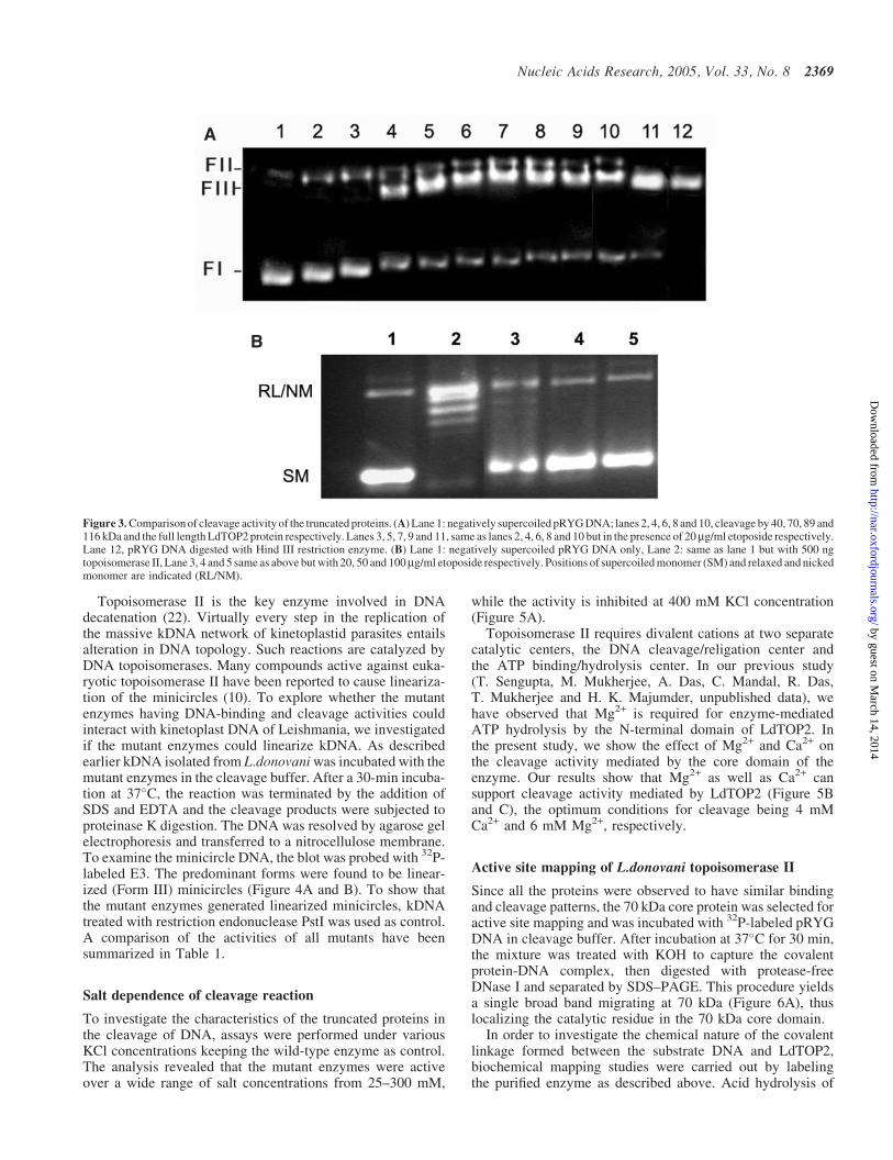
(Figure 3A). Furthermore, to show that the mutant enzymesgenerated linear DNA (Form III) and not the nicked form(Form II), pRYG DNA treated with restriction endonucleaseHindIII was used as a marker (Figure 3A). The wild-type
LdTOP2 was sensitive to etoposide (Figure 3B). LdTOP2relaxed supercoiled pRYG DNA (Lane 1) and the formationof topological isomers (lane 2) was inhibited by etoposide(lanes 3–5).
A B
C D
E
Figure 2. Properties of the mutant proteins. (A) Dependence of ATPase rate of the 116 and 137 kDa protein on ATP concentration at constant enzyme concentration.116 kDa (open square), 137 kDa (black triangle) in presence of DNA, 116 kDa (black square), 137 kDa (open triangle) in absence of DNA. The data shown aremean – SD of three independent experiments. (B) The KM was calculated from the Lineweaver–Burk plot, 116 kDa in absence (open square), and in presence (blackcircle) of DNA respectively. (C) 137 kDa in absence (open circle) and in presence (black square) of DNA respectively. (D) DNA-binding activity of the truncatedLdTOP2 proteins. Lane 1, labeled 36mer duplex oligonucleotide; lanes 2–5, 36mer oligonucleotide incubated with 40, 70, 89 and 116 kDa. (E) Graphicalrepresentation of the native gel shift assay. The percentage of unbound oligonucleotide present in gel was quantified by film densitometry and plotted againstprotein concentration. The kd value was calculated from this graph, core (open circle), LdDC1058 (black triangle), LdDN429 (black square) and 137 kDa (opendiamond) respectively.
2368 Nucleic Acids Research, 2005, Vol. 33, No. 8
by guest on March 14, 2014
http://nar.oxfordjournals.org/D
ownloaded from
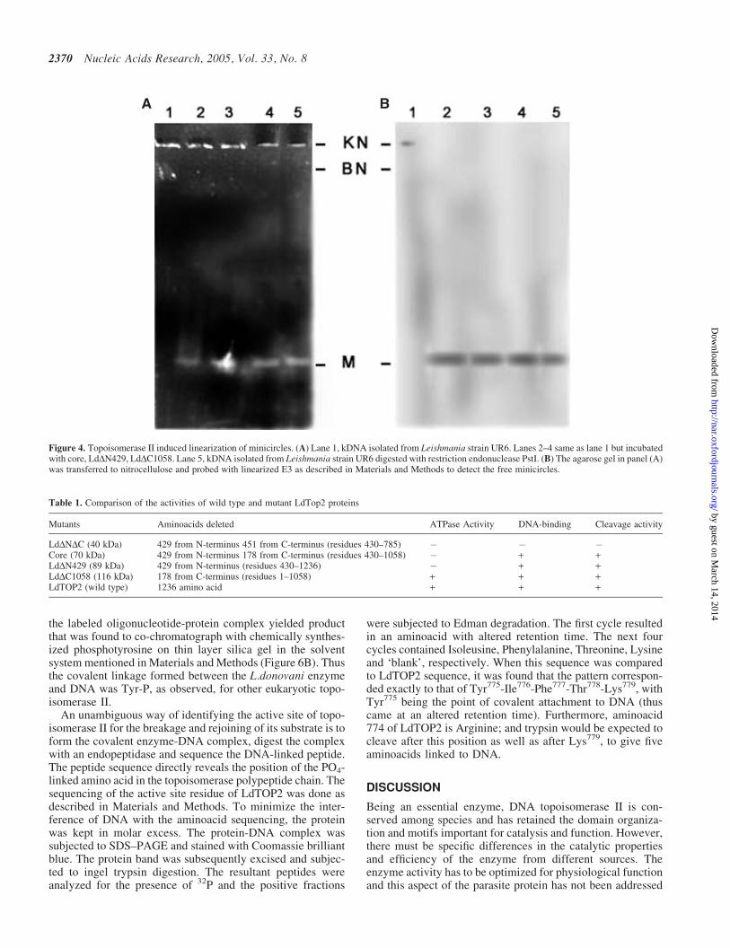
Topoisomerase II is the key enzyme involved in DNAdecatenation (22). Virtually every step in the replication ofthe massive kDNA network of kinetoplastid parasites entailsalteration in DNA topology. Such reactions are catalyzed byDNA topoisomerases. Many compounds active against euka-ryotic topoisomerase II have been reported to cause lineariza-tion of the minicircles (10). To explore whether the mutantenzymes having DNA-binding and cleavage activities couldinteract with kinetoplast DNA of Leishmania, we investigatedif the mutant enzymes could linearize kDNA. As describedearlier kDNA isolated from L.donovani was incubated with themutant enzymes in the cleavage buffer. After a 30-min incuba-tion at 37�C, the reaction was terminated by the addition ofSDS and EDTA and the cleavage products were subjected toproteinase K digestion. The DNA was resolved by agarose gelelectrophoresis and transferred to a nitrocellulose membrane.To examine the minicircle DNA, the blot was probed with 32P-labeled E3. The predominant forms were found to be linear-ized (Form III) minicircles (Figure 4A and B). To show thatthe mutant enzymes generated linearized minicircles, kDNAtreated with restriction endonuclease PstI was used as control.A comparison of the activities of all mutants have beensummarized in Table 1.
Salt dependence of cleavage reaction
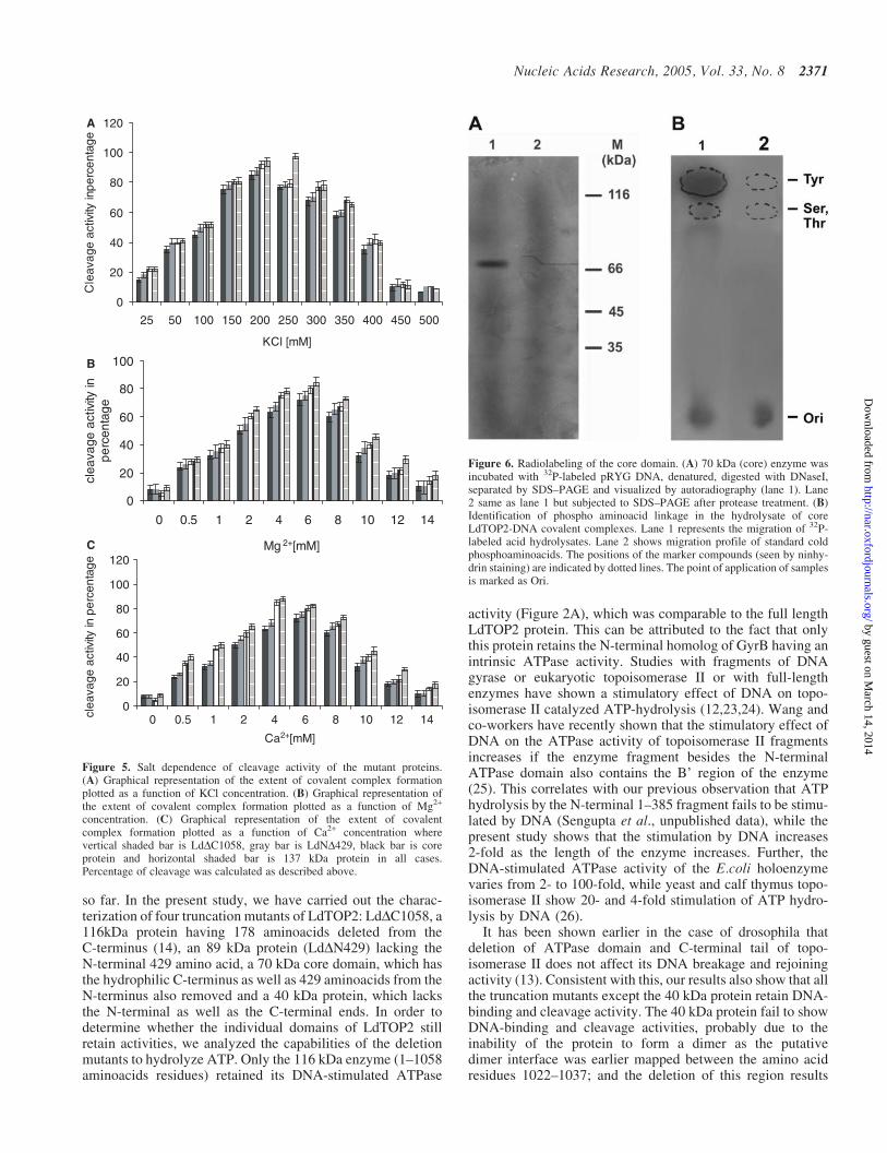
To investigate the characteristics of the truncated proteins inthe cleavage of DNA, assays were performed under variousKCl concentrations keeping the wild-type enzyme as control.The analysis revealed that the mutant enzymes were activeover a wide range of salt concentrations from 25–300 mM,
while the activity is inhibited at 400 mM KCl concentration(Figure 5A).
Topoisomerase II requires divalent cations at two separatecatalytic centers, the DNA cleavage/religation center andthe ATP binding/hydrolysis center. In our previous study(T. Sengupta, M. Mukherjee, A. Das, C. Mandal, R. Das,T. Mukherjee and H. K. Majumder, unpublished data), wehave observed that Mg2+ is required for enzyme-mediatedATP hydrolysis by the N-terminal domain of LdTOP2. Inthe present study, we show the effect of Mg2+ and Ca2+ onthe cleavage activity mediated by the core domain of theenzyme. Our results show that Mg2+ as well as Ca2+ cansupport cleavage activity mediated by LdTOP2 (Figure 5Band C), the optimum conditions for cleavage being 4 mMCa2+ and 6 mM Mg2+, respectively.
Active site mapping of L.donovani topoisomerase II
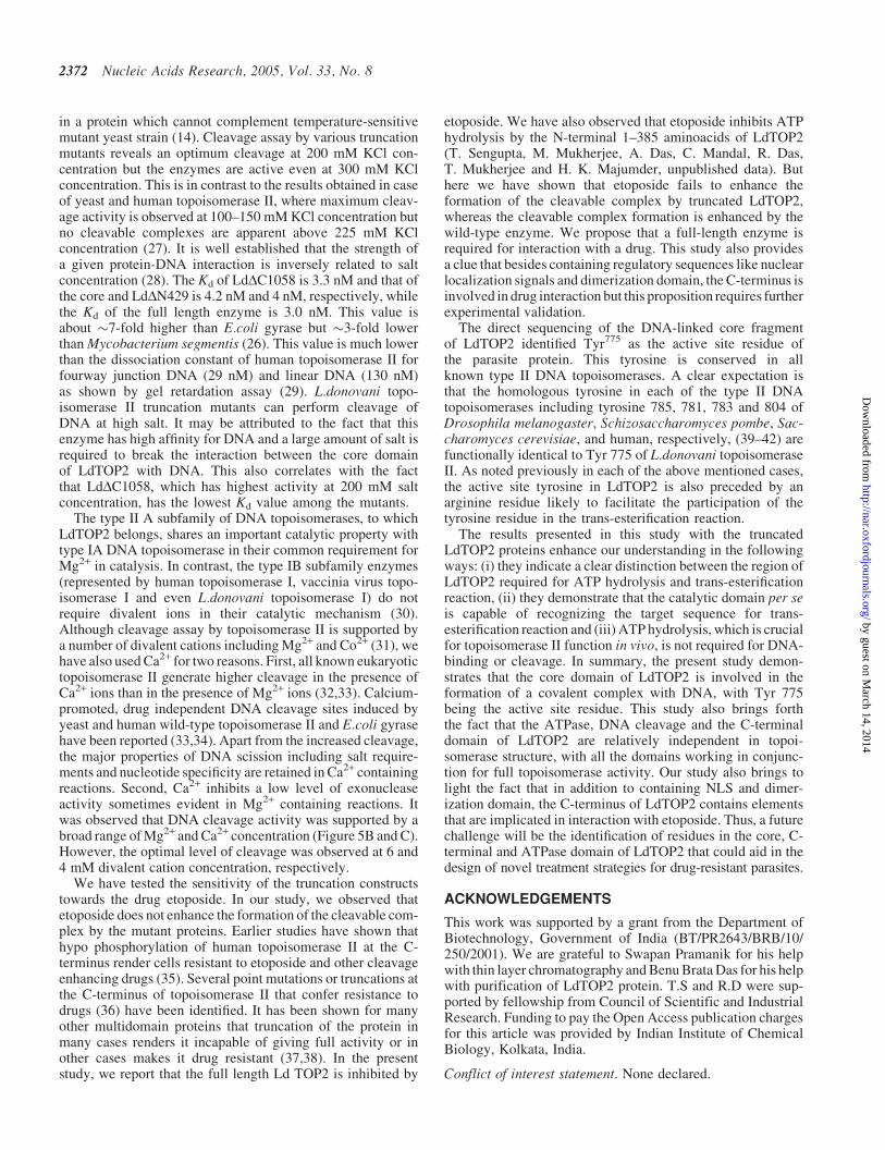
Since all the proteins were observed to have similar bindingand cleavage patterns, the 70 kDa core protein was selected foractive site mapping and was incubated with 32P-labeled pRYGDNA in cleavage buffer. After incubation at 37�C for 30 min,the mixture was treated with KOH to capture the covalentprotein-DNA complex, then digested with protease-freeDNase I and separated by SDS–PAGE. This procedure yieldsa single broad band migrating at 70 kDa (Figure 6A), thuslocalizing the catalytic residue in the 70 kDa core domain.
In order to investigate the chemical nature of the covalentlinkage formed between the substrate DNA and LdTOP2,biochemical mapping studies were carried out by labelingthe purified enzyme as described above. Acid hydrolysis of
A
B
Figure 3. Comparison of cleavage activity of the truncated proteins. (A) Lane 1: negatively supercoiled pRYG DNA; lanes 2, 4, 6, 8 and 10, cleavage by 40, 70, 89 and116 kDa and the full length LdTOP2 protein respectively. Lanes 3, 5, 7, 9 and 11, same as lanes 2, 4, 6, 8 and 10 but in the presence of 20mg/ml etoposide respectively.Lane 12, pRYG DNA digested with Hind III restriction enzyme. (B) Lane 1: negatively supercoiled pRYG DNA only, Lane 2: same as lane 1 but with 500 ngtopoisomerase II, Lane 3, 4 and 5 same as above but with 20, 50 and 100mg/ml etoposide respectively. Positions of supercoiled monomer (SM) and relaxed and nickedmonomer are indicated (RL/NM).
Nucleic Acids Research, 2005, Vol. 33, No. 8 2369
by guest on March 14, 2014
http://nar.oxfordjournals.org/D
ownloaded from
the labeled oligonucleotide-protein complex yielded productthat was found to co-chromatograph with chemically synthes-ized phosphotyrosine on thin layer silica gel in the solventsystem mentioned in Materials and Methods (Figure 6B). Thusthe covalent linkage formed between the L.donovani enzymeand DNA was Tyr-P, as observed, for other eukaryotic topo-isomerase II.
An unambiguous way of identifying the active site of topo-isomerase II for the breakage and rejoining of its substrate is toform the covalent enzyme-DNA complex, digest the complexwith an endopeptidase and sequence the DNA-linked peptide.The peptide sequence directly reveals the position of the PO4-linked amino acid in the topoisomerase polypeptide chain. Thesequencing of the active site residue of LdTOP2 was done asdescribed in Materials and Methods. To minimize the inter-ference of DNA with the aminoacid sequencing, the proteinwas kept in molar excess. The protein-DNA complex wassubjected to SDS–PAGE and stained with Coomassie brilliantblue. The protein band was subsequently excised and subjec-ted to ingel trypsin digestion. The resultant peptides wereanalyzed for the presence of 32P and the positive fractions
were subjected to Edman degradation. The first cycle resultedin an aminoacid with altered retention time. The next fourcycles contained Isoleusine, Phenylalanine, Threonine, Lysineand ‘blank’, respectively. When this sequence was comparedto LdTOP2 sequence, it was found that the pattern correspon-ded exactly to that of Tyr775-Ile776-Phe777-Thr778-Lys779, withTyr775 being the point of covalent attachment to DNA (thuscame at an altered retention time). Furthermore, aminoacid774 of LdTOP2 is Arginine; and trypsin would be expected tocleave after this position as well as after Lys779, to give fiveaminoacids linked to DNA.
DISCUSSION
Being an essential enzyme, DNA topoisomerase II is con-served among species and has retained the domain organiza-tion and motifs important for catalysis and function. However,there must be specific differences in the catalytic propertiesand efficiency of the enzyme from different sources. Theenzyme activity has to be optimized for physiological functionand this aspect of the parasite protein has not been addressed
A B
Figure 4. Topoisomerase II induced linearization of minicircles. (A) Lane 1, kDNA isolated from Leishmania strain UR6. Lanes 2–4 same as lane 1 but incubatedwith core, LdDN429, LdDC1058. Lane 5, kDNA isolated from Leishmania strain UR6 digested with restriction endonuclease PstI. (B) The agarose gel in panel (A)was transferred to nitrocellulose and probed with linearized E3 as described in Materials and Methods to detect the free minicircles.
Table 1. Comparison of the activities of wild type and mutant LdTop2 proteins
Mutants Aminoacids deleted ATPase Activity DNA-binding Cleavage activity
LdDNDC (40 kDa) 429 from N-terminus 451 from C-terminus (residues 430–785) � � �Core (70 kDa) 429 from N-terminus 178 from C-terminus (residues 430–1058) � + +LdDN429 (89 kDa) 429 from N-terminus (residues 430–1236) � + +LdDC1058 (116 kDa) 178 from C-terminus (residues 1–1058) + + +LdTOP2 (wild type) 1236 amino acid + + +
2370 Nucleic Acids Research, 2005, Vol. 33, No. 8
by guest on March 14, 2014
http://nar.oxfordjournals.org/D
ownloaded from
so far. In the present study, we have carried out the charac-terization of four truncation mutants of LdTOP2: LdDC1058, a116kDa protein having 178 aminoacids deleted from theC-terminus (14), an 89 kDa protein (LdDN429) lacking theN-terminal 429 amino acid, a 70 kDa core domain, which hasthe hydrophilic C-terminus as well as 429 aminoacids from theN-terminus also removed and a 40 kDa protein, which lacksthe N-terminal as well as the C-terminal ends. In order todetermine whether the individual domains of LdTOP2 stillretain activities, we analyzed the capabilities of the deletionmutants to hydrolyze ATP. Only the 116 kDa enzyme (1–1058aminoacids residues) retained its DNA-stimulated ATPase
activity (Figure 2A), which was comparable to the full lengthLdTOP2 protein. This can be attributed to the fact that onlythis protein retains the N-terminal homolog of GyrB having anintrinsic ATPase activity. Studies with fragments of DNAgyrase or eukaryotic topoisomerase II or with full-lengthenzymes have shown a stimulatory effect of DNA on topo-isomerase II catalyzed ATP-hydrolysis (12,23,24). Wang andco-workers have recently shown that the stimulatory effect ofDNA on the ATPase activity of topoisomerase II fragmentsincreases if the enzyme fragment besides the N-terminalATPase domain also contains the B’ region of the enzyme(25). This correlates with our previous observation that ATPhydrolysis by the N-terminal 1–385 fragment fails to be stimu-lated by DNA (Sengupta et al., unpublished data), while thepresent study shows that the stimulation by DNA increases2-fold as the length of the enzyme increases. Further, theDNA-stimulated ATPase activity of the E.coli holoenzymevaries from 2- to 100-fold, while yeast and calf thymus topo-isomerase II show 20- and 4-fold stimulation of ATP hydro-lysis by DNA (26).
It has been shown earlier in the case of drosophila thatdeletion of ATPase domain and C-terminal tail of topo-isomerase II does not affect its DNA breakage and rejoiningactivity (13). Consistent with this, our results also show that allthe truncation mutants except the 40 kDa protein retain DNA-binding and cleavage activity. The 40 kDa protein fail to showDNA-binding and cleavage activities, probably due to theinability of the protein to form a dimer as the putativedimer interface was earlier mapped between the amino acidresidues 1022–1037; and the deletion of this region results
Figure 6. Radiolabeling of the core domain. (A) 70 kDa (core) enzyme wasincubated with 32P-labeled pRYG DNA, denatured, digested with DNaseI,separated by SDS–PAGE and visualized by autoradiography (lane 1). Lane2 same as lane 1 but subjected to SDS–PAGE after protease treatment. (B)Identification of phospho aminoacid linkage in the hydrolysate of coreLdTOP2-DNA covalent complexes. Lane 1 represents the migration of 32P-labeled acid hydrolysates. Lane 2 shows migration profile of standard coldphosphoaminoacids. The positions of the marker compounds (seen by ninhy-drin staining) are indicated by dotted lines. The point of application of samplesis marked as Ori.
0
20
40
60
80
100
120A
B
C
25 50 100 150 200 250 300 350 400 450 500
KCl [mM]
Cle
avag
e ac
tivity
inpe
rcen
tage
0
20
40
60
80
100
0 0.5 1 2 4 6 8 10 12 14
Mg 2+[mM]
clea
vage
act
ivity
inpe
rcen
tage
0
20
40
60
80
100
120
0 0.5 1 2 4 6 8 10 12 14
Ca2+[mM]
clea
vage
act
ivity
in p
erce
ntag
e
Figure 5. Salt dependence of cleavage activity of the mutant proteins.(A) Graphical representation of the extent of covalent complex formationplotted as a function of KCl concentration. (B) Graphical representation ofthe extent of covalent complex formation plotted as a function of Mg2+
concentration. (C) Graphical representation of the extent of covalentcomplex formation plotted as a function of Ca2+ concentration wherevertical shaded bar is LdDC1058, gray bar is LdND429, black bar is coreprotein and horizontal shaded bar is 137 kDa protein in all cases.Percentage of cleavage was calculated as described above.
Nucleic Acids Research, 2005, Vol. 33, No. 8 2371
by guest on March 14, 2014
http://nar.oxfordjournals.org/D
ownloaded from
in a protein which cannot complement temperature-sensitivemutant yeast strain (14). Cleavage assay by various truncationmutants reveals an optimum cleavage at 200 mM KCl con-centration but the enzymes are active even at 300 mM KClconcentration. This is in contrast to the results obtained in caseof yeast and human topoisomerase II, where maximum cleav-age activity is observed at 100–150 mM KCl concentration butno cleavable complexes are apparent above 225 mM KClconcentration (27). It is well established that the strength ofa given protein-DNA interaction is inversely related to saltconcentration (28). The Kd of LdDC1058 is 3.3 nM and that ofthe core and LdDN429 is 4.2 nM and 4 nM, respectively, whilethe Kd of the full length enzyme is 3.0 nM. This value isabout �7-fold higher than E.coli gyrase but �3-fold lowerthan Mycobacterium segmentis (26). This value is much lowerthan the dissociation constant of human topoisomerase II forfourway junction DNA (29 nM) and linear DNA (130 nM)as shown by gel retardation assay (29). L.donovani topo-isomerase II truncation mutants can perform cleavage ofDNA at high salt. It may be attributed to the fact that thisenzyme has high affinity for DNA and a large amount of salt isrequired to break the interaction between the core domainof LdTOP2 with DNA. This also correlates with the factthat LdDC1058, which has highest activity at 200 mM saltconcentration, has the lowest Kd value among the mutants.
The type II A subfamily of DNA topoisomerases, to whichLdTOP2 belongs, shares an important catalytic property withtype IA DNA topoisomerase in their common requirement forMg2+ in catalysis. In contrast, the type IB subfamily enzymes(represented by human topoisomerase I, vaccinia virus topo-isomerase I and even L.donovani topoisomerase I) do notrequire divalent ions in their catalytic mechanism (30).Although cleavage assay by topoisomerase II is supported bya number of divalent cations including Mg2+ and Co2+ (31), wehave also used Ca2+ for two reasons. First, all known eukaryotictopoisomerase II generate higher cleavage in the presence ofCa2+ ions than in the presence of Mg2+ ions (32,33). Calcium-promoted, drug independent DNA cleavage sites induced byyeast and human wild-type topoisomerase II and E.coli gyrasehave been reported (33,34). Apart from the increased cleavage,the major properties of DNA scission including salt require-ments and nucleotide specificity are retained in Ca2+ containingreactions. Second, Ca2+ inhibits a low level of exonucleaseactivity sometimes evident in Mg2+ containing reactions. Itwas observed that DNA cleavage activity was supported by abroad range of Mg2+ and Ca2+ concentration (Figure 5B and C).However, the optimal level of cleavage was observed at 6 and4 mM divalent cation concentration, respectively.
We have tested the sensitivity of the truncation constructstowards the drug etoposide. In our study, we observed thatetoposide does not enhance the formation of the cleavable com-plex by the mutant proteins. Earlier studies have shown thathypo phosphorylation of human topoisomerase II at the C-terminus render cells resistant to etoposide and other cleavageenhancing drugs (35). Several point mutations or truncations atthe C-terminus of topoisomerase II that confer resistance todrugs (36) have been identified. It has been shown for manyother multidomain proteins that truncation of the protein inmany cases renders it incapable of giving full activity or inother cases makes it drug resistant (37,38). In the presentstudy, we report that the full length Ld TOP2 is inhibited by
etoposide. We have also observed that etoposide inhibits ATPhydrolysis by the N-terminal 1–385 aminoacids of LdTOP2(T. Sengupta, M. Mukherjee, A. Das, C. Mandal, R. Das,T. Mukherjee and H. K. Majumder, unpublished data). Buthere we have shown that etoposide fails to enhance theformation of the cleavable complex by truncated LdTOP2,whereas the cleavable complex formation is enhanced by thewild-type enzyme. We propose that a full-length enzyme isrequired for interaction with a drug. This study also providesa clue that besides containing regulatory sequences like nuclearlocalization signals and dimerization domain, the C-terminus isinvolved in drug interaction but this proposition requires furtherexperimental validation.
The direct sequencing of the DNA-linked core fragmentof LdTOP2 identified Tyr775 as the active site residue ofthe parasite protein. This tyrosine is conserved in allknown type II DNA topoisomerases. A clear expectation isthat the homologous tyrosine in each of the type II DNAtopoisomerases including tyrosine 785, 781, 783 and 804 ofDrosophila melanogaster, Schizosaccharomyces pombe, Sac-charomyces cerevisiae, and human, respectively, (39–42) arefunctionally identical to Tyr 775 of L.donovani topoisomeraseII. As noted previously in each of the above mentioned cases,the active site tyrosine in LdTOP2 is also preceded by anarginine residue likely to facilitate the participation of thetyrosine residue in the trans-esterification reaction.
The results presented in this study with the truncatedLdTOP2 proteins enhance our understanding in the followingways: (i) they indicate a clear distinction between the region ofLdTOP2 required for ATP hydrolysis and trans-esterificationreaction, (ii) they demonstrate that the catalytic domain per seis capable of recognizing the target sequence for trans-esterification reaction and (iii) ATP hydrolysis, which is crucialfor topoisomerase II function in vivo, is not required for DNA-binding or cleavage. In summary, the present study demon-strates that the core domain of LdTOP2 is involved in theformation of a covalent complex with DNA, with Tyr 775being the active site residue. This study also brings forththe fact that the ATPase, DNA cleavage and the C-terminaldomain of LdTOP2 are relatively independent in topoi-somerase structure, with all the domains working in conjunc-tion for full topoisomerase activity. Our study also brings tolight the fact that in addition to containing NLS and dimer-ization domain, the C-terminus of LdTOP2 contains elementsthat are implicated in interaction with etoposide. Thus, a futurechallenge will be the identification of residues in the core, C-terminal and ATPase domain of LdTOP2 that could aid in thedesign of novel treatment strategies for drug-resistant parasites.
ACKNOWLEDGEMENTS
This work was supported by a grant from the Department ofBiotechnology, Government of India (BT/PR2643/BRB/10/250/2001). We are grateful to Swapan Pramanik for his helpwith thin layer chromatography and Benu Brata Das for his helpwith purification of LdTOP2 protein. T.S and R.D were sup-ported by fellowship from Council of Scientific and IndustrialResearch. Funding to pay the Open Access publication chargesfor this article was provided by Indian Institute of ChemicalBiology, Kolkata, India.
Conflict of interest statement. None declared.
2372 Nucleic Acids Research, 2005, Vol. 33, No. 8
by guest on March 14, 2014
http://nar.oxfordjournals.org/D
ownloaded from
REFERENCES
1. Wang,J.C. (2002) Cellular roles of DNA topoisomerases: a molecularperspective. Nature Rev. Mol. Cell. Biol., 3, 429–440.
2. Corbett,K.D. and Berger,J.M. (2004) Structure, molecular mechanisms,and evolutionary relationships in DNA topoisomerases. Annu. Rev.Biophys. Biomol. Struct., 33, 95–118.
3. Burden,D.A. and Osheroff,N. (1998) Mechanism of action of eukaryotictopoisomerase II and drugs targeted to the enzyme. Biochim. Biophys.Acta, 1400, 139–154.
4. Li,T.K. and Liu,L.F. (2001) Tumor cell death induced by topoisomerase-targeting drugs. Annu. Rev. Pharmacol. Toxicol., 41, 53–77.
5. Fortune,J.M. and Osheroff,N. (2000) Topoisomerase II as a target foranticancer drugs: when enzymes stop being nice. Prog. Nucleic Acid Res.Mol. Biol., 64, 221–253.
6. Corbett,A.H. and Osheroff,N. (1993) When good enzymes go bad:conversion of topoisomeraseII to a cellular toxin by antineoplastic drugs.Chem. Res. Toxicol., 5, 585–597.
7. Froelich-Ammon,S.J. and Osheroff,N. (1995) Topoisomerase poisons:harnessing the dark side of enzyme mechanism. J. Biol. Chem., 270,21429–21432.
8. Liu,L.F. (1994) DNA topoisomerases: Topoisomerase targeting drugs.Academic press, New York.
9. Das,A., Dasgupta,A., Sengupta,T. and Majumder,H.K. (2004)Topoisomerases of kinetoplastid parasites as potential chemotherapeutictargets. Trends Parasitol., 20, 381–387.
10. Shapiro,T.A. and Showalter,A.F. (1994) In vivo inhibition oftrypanosome mitochondrial topoisomerase II: effects on kinetoplastDNA maxicircles. Mol. Cell Biol., 14, 5891–58977.
11. Das,A., Dasgupta,A., Sharma,S., Ghosh,M., Sengupta,T.,Bandopadhyay,S. and Majumder,H.K. (2001) Characterisation of thegene encoding type II DNA topoisomerase from Leishmania donovani:a key molecular target in antileishmanial therapy. Nucleic Acids Res.,29, 1844–1851.
12. Gardiner,L.P., Roper,D.I., Hammonds,T.R. and Maxwell,A. (1998) TheN- terminal domain of human topoisomerase IIalpha is a DNA-dependentATPase. Biochemistry, 37, 6997–7004.
13. Chang,S., Hu,T. and Hsieh,T.S. (1998) Analysis of a core domain inDrosophila DNA topoisomerase II. Targeting of an antitumor agentICRF-159. J. Biol. Chem., 273, 19822–19828.
14. Sengupta,T., Mukherjee,M., Mandal,C.N., Das,A. and Majumder,H.K.(2003) Functional dissection of the C-terminal domain of type II DNAtopoisomerase from the kinetoplastid hemoflagellate Leishmaniadonovani. Nucleic Acids Res., 30, 5305–5316.
15. Andersen,A.H., Christiansen,K., Zechiedrich,E.L., Jensen,P.S.,Osheroff,N. and Westergaard,O. (1989) Strand specificity of thetopoisomerase II mediated double- stranded DNA cleavage reaction.Biochemistry, 28, 6237–6244.
16. Carey,J. (1991) Gel retardation. Methods Enzymol., 208, 103–117.17. Ostergaard,V.H., Giangiacomo,L., Bjergbaek,L., Knudson,B.R. and
Anderson,A.H. (2004) Hindering the strand passage reaction of humantopoisomerase Iialpha without disturbing DNA cleavage, ATPhydrolysis, or the operation of the N-terminal clamp. J. Biol. Chem.,279, 28093–28099.
18. Singh,R., Dutta,C. and Majumder,H.K. (1994) Analysis of sequences oftwo different classes of kinetoplast DNA minicircles of Leishmaniaspecies. J. Biosci., 19, 117–182.
19. Dasgupta,S., Adhya,S. and Majumder,H.K. (1986) A simple procedurefor the preparation of pure kinetoplast DNA network free of nuclear DNAfrom the kinetoplast hemoflagellate Leishmania donovani. Anal.Biochem., 158, 189–194.
20. Bodley,A.L., Chakraborty,A.K., Xie,S., Burri,A. and Shapiro,T.A.(2003) An unusual typeIB topoisomerase from African trypanosomes.Proc. Natl Acad. Sci. USA, 100, 7539–7544.
21. Tse,Y.C., Kirkegaard,K. and Wang,J.C. (1980) Covalent bondsbetween protein and DNA. Formation of phosphotyrosinelinkage between certain DNA topoisomerases and DNA. J. Biol. Chem.,255, 5560–5565.
22. Skoufias,D.A., Lacroix,F.B., Andreassen,P.R., Wilson,L. andMargolis,R.L. (2004) Inhibition of DNA decatenation, but not DNAdamage, arrests cells at metaphase. Mol. Cell., 15, 977–990.
23. Lindsley,J.E. and Wang,J.C. (1993) On the coupling between ATP usageand DNA transport by yeast DNA topoisomerase II. J. Biol. Chem.,268, 8096–8104.
24. Campbell,S. and Maxwell,A. (2002) The ATP-operated clamp ofhuman DNA topoisomerase IIalpha: hyperstimulation of ATPase by‘piggy-back’ binding. J. Mol. Biol., 320, 171–188.
25. Olland,S. and Wang,J.C. (1999) Catalysis of ATP hydrolysis by twoNH(2)- terminal fragments of yeast DNA topoisomerase II. J. Biol.Chem., 274, 21688–21694.
26. Manjunatha,U.H., Dalal,M., Chaterjee,M., Radha,D.R.,Visweswariah,S.S. and Nagaraja,V. (2002) Functional characterisationof mycobacterial DNA gyrase: an efficient decatenase. Nucleic AcidsRes., 30, 2144–2153.
27. Scala,D., Escargueil,A.E., Couprie,J. and Larsen,A.K. (1999) Thecatalytic activities of DNA topoisomerase II are most closely associatedwith the DNA cleavage/religation steps. Biochimie, 81, 771–779.
28. Record,M.T.,Jr, Ha,J.H. and Fisher,M.A. (1991) Analysis of equilibriumand kinetic measurements to determine thermodynamic origins ofstability and specificity and mechanism of formation of site-specificcomplexes between proteins and helical DNA. Methods Enzymol.,208, 291–343.
29. West,K.L. and Austin,C.A. (1999) Human DNA topoisomerase II betabinds and cleaves four-way junction DNA in vitro. Nucleic Acids Res.,27, 984–992.
30. Das,B.B., Sen,N., Ganguly,A. and Majumder,H.K. (2004) Reconstitutionand functional characterization of the unusual bi-subunit type I DNAtopoisomerase from Leishmania donovani. FEBS Lett., 565, 81–88.
31. Baldwin,E.L., Byl,J.A. and Osheroff,N. (2004) Cobalt enhances DNAcleavage mediated by human topoisomerase II alpha in vitro andin cultured cells. Biochemistry, 43, 728–735.
32. Osheroff,N. and Zechiedrich,E.L. (1987) Calcium-promoted DNAcleavage by eukaryotic topoisomerase II: trapping the covalentenzyme-DNA complex in an active form. Biochemistry, 26, 4293–4313.
33. Strumberg,D., Nitiss,J.L., Dong,J., Kohn,K.W. and Pommier,Y. (1999)Molecular analysis of yeast and human type II topoisomerases.Enzyme-DNA and drug interactions. J. Biol. Chem., 274, 28246–28255.
34. Capranico,G., Guano,F., Moro,S., Zagotto,G., Sissi,C., Gotto,B.,Zunino,F., Menta,E. and Palumbo,M. (1998) Mapping drug interactionsat the covalent topoisomerase II-DNA complex by bisantrene/amsacrinecongeners. J. Biol. Chem., 273, 12732–12739.
35. Chikamori,K., Grabowski,D.R., Kinter,M., Willard,B.B., Yadav,S.,Aebersold,R.H., Bukowski,R.M., Hickson,I.D., Anderson,A.H.,Ganapathi,M.K. et al. (2003) Phosphorylation of serine 1106 in thecatalytic domain of topoisomerase II alpha regulates enzymatic activityand drug sensitivity. J. Biol. Chem., 278, 12696–12702.
36. Vassetzsky,Y.S., Alghisi,G.C. and Gasser,S.M. (1995) DNAtopoisomerase II mutations and resistance to anti-tumor drugs.Bioessays, 17, 767–774.
37. Brandt,P.A. and Jacobs,B.L. (2001) Both carboxy- and amino-terminaldomains of the vaccinia virus interferon resistance gene, E3L, arerequired for pathogenesis in a mouse model. J. Virol., 75, 850–856.
38. Khoo,K.H., Douglas,E., Azadi,P., Inamine,J.M., Besra,G.S.,Mikusova,K., Brennan,P.J. and Chaterjee,D. (1996) Truncated structuralvariants of lipoarabinomannan in ethambutol drug-resistant strainsof Mycobacterium smegmatis. Inhibition of arabinan biosynthesis byethambutol. J. Biol. Chem., 271, 28682–28690.
39. Wortland,S.T. and Wang,J.C. (1989) Inducible overexpression,purification, and active site mapping of DNA topoisomerase II from theyeast Saccharomyces cerevisiae. J. Biol. Chem., 264, 4412–4416.
40. Uemura,T., Morikawa,K. and Yanagida,M. (1986) The nucleotidesequence of the fission yeast DNA topoisomerase II gene: structural andfunctional relationships to other DNA topoisomerases. EMBO J., 5,2355–2361.
41. Wycoff,E., Natalie,D., Nolan,J., Lee,M. and Hsieh,T.S. (1989) Structureof the Drosophila DNA topoisomerase II gene. Nucleotide sequence andhomology among topoisomerases II. J. Mol. Biol., 505, 1–13.
42. Tsai-Pflugfelder,M., Liu,L.F., Liu,A.A., Tewey,K.M., Whangpeng,J.,Knutsen,T., Croce,C.M. and Wang,J.C. (1988) Cloning and sequencingof cDNA encoding human DNA topoisomerase II and localization of thegene to chromosome region 17q21-22. Proc. Natl Acad. Sci. USA,85, 7177–7181.
Nucleic Acids Research, 2005, Vol. 33, No. 8 2373
by guest on March 14, 2014
http://nar.oxfordjournals.org/D
ownloaded from
Copyright © 2022 FDOKUMEN


![Substituted dibenzo[ c,h]cinnolines: topoisomerase I-targeting anticancer agents](https://static.fdokumen.com/doc/165x107/631871c065e4a6af370f5e52/substituted-dibenzo-chcinnolines-topoisomerase-i-targeting-anticancer-agents.jpg)