Genetic analysis of spermidine synthase from Leishmania donovani
Upload
independentCategory
view
1download
0
Critical Roles for LIGHT and Its Receptors in Generating TCell-Mediated Immunity during Leishmania donovaniInfectionAmanda C. Stanley1,2, Fabian de Labastida Rivera1, Ashraful Haque1, Meru Sheel1, Yonghong Zhou1,
Fiona H. Amante1, Patrick T. Bunn1, Louise M. Randall1,3, Klaus Pfeffer4, Stefanie Scheu4, Michael J.
Hickey5, Bernadette M. Saunders6, Carl Ware7, Geoff R. Hill1, Koji Tamada8, Paul M. Kaye9, Christian R.
Engwerda1*
1 Queensland Institute of Medical Research and the Australian Centre for Vaccine Development, Herston, Queensland, Australia, 2 Institute for Molecular Biology,
University of Queensland, St Lucia, Queensland, Australia, 3 Department of Pathobiology, School of Veterinary Sciences, University of Pennsylvania, Philadelphia,
Pennsylvania, United States of America, 4 Institute of Medical Microbiology and Hospital Hygiene, University of Duesseldorf, Duesseldorf, Germany, 5 Centre for
Inflammatory Diseases, Monash University, Department of Medicine, Monash Medical Centre, Clayton, Victoria, Australia, 6 Centenary Institute, Newtown, New South
Wales, Australia, 7 Infectious and Inflammatory Diseases Centre, Sanford|Burnham Medical Research Institute, La Jolla, California, United States of America, 8 Marlene and
Stewart Greenebaum Cancer Center, University of Maryland, Baltimore, Maryland, Unites States of America, 9 Hull York Medical School, Department of Biology, York
University, York, United Kingdom
Abstract
LIGHT (TNFSF14) is a member of the TNF superfamily involved in inflammation and defence against infection. LIGHT signalsvia two cell-bound receptors; herpes virus entry mediator (HVEM) and lymphotoxin-beta receptor (LTbR). We found thatLIGHT is critical for control of hepatic parasite growth in mice with visceral leishmaniasis (VL) caused by infection with theprotozoan parasite Leishmania donovani. LIGHT-HVEM signalling is essential for early dendritic cell IL-12/IL-23p40production, and the generation of IFNc- and TNF-producing T cells that control hepatic infection. However, we alsodiscovered that LIGHT-LTbR interactions suppress anti-parasitic immunity in the liver in the first 7 days of infection bymechanisms that restrict both CD4+ T cell function and TNF-dependent microbicidal mechanisms. Thus, we have identifieddistinct roles for LIGHT in infection, and show that manipulation of interactions between LIGHT and its receptors may beused for therapeutic advantage.
Citation: Stanley AC, de Labastida Rivera F, Haque A, Sheel M, Zhou Y, et al. (2011) Critical Roles for LIGHT and Its Receptors in Generating T Cell-MediatedImmunity during Leishmania donovani Infection. PLoS Pathog 7(10): e1002279. doi:10.1371/journal.ppat.1002279
Editor: David L. Sacks, National Institute of Health, United States of America
Received April 12, 2011; Accepted August 8, 2011; Published October 6, 2011
Copyright: � 2011 Stanley et al. This is an open-access article distributed under the terms of the Creative Commons Attribution License, which permitsunrestricted use, distribution, and reproduction in any medium, provided the original author and source are credited.
Funding: This work was supported by the Australian NHMRC Project grant number 613649, NHMRC Program number 496600 and a NHMRC Fellowship (552425)to Engwerda (http://www.nhmrc.gov.au). The funders had no role in study design, data collection and analysis, decision to publish, or preparation of themanuscript.
Competing Interests: The authors have declared that no competing interests exist.
* E-mail: [email protected]
Introduction
Tumour necrosis factor (TNF) superfamily members are
involved in many biological functions, including cell growth and
differentiation, apoptosis and organogenesis [1]. This broad range
of activities is achieved by TNF family members interacting with
functional receptors associated with distinct cell signalling
pathways [2]. TNF, lymphotoxin (LT)a, LTb and LIGHT
(TNFSF14) comprise a closely related set of ligands in the TNF
family [3,4]. TNF exists as a cell-bound or soluble homotrimer
that binds TNF receptor (TNFR)1 and TNFR2 [5,6]. LTa can
form a soluble homotrimer (LTa3) that binds TNFR1, TNFR2
and HVEM [5,7], but can also form a cell-bound heterotrimer
with LTb (LTa1b2) that binds and signals through LTbR [8].
LIGHT exists in cell-bound and soluble forms that interact with
both LTbR and herpes virus entry mediator (HVEM) [7,9,10].
HVEM also engages members of the immunoglobulin superfam-
ily; B and T lymphocyte attenuator (BTLA) [11] and CD160 [12],
as well as the envelope glycoprotein D of Herpes Simplex virus
[13]. HVEM activates BTLA inhibitory signalling via SHP
phosphatases suppressing T cell activation [14]. LIGHT, LTaand the Ig superfamily ligands can also activate HVEM-dependent
cell survival signalling via NF-kB [15].
LIGHT has emerged as a key mediator of inflammation and
immune homeostasis [4,14]. There is broad expression of LIGHT and
HVEM in the hematopoietic compartment [7,9,16,17,18], while
LTbR expression is largely restricted to stromal and myeloid cells
[7,19,20]. LTbR and HVEM are implicated as key host defence
mechanisms against persistent viral [21] and bacterial pathogens [22].
However, little is known about the role of these receptors in infection
with parasites that establish persistent infections in their hosts.
The protozoan parasite Leishmania donovani causes persistent
infections in humans and experimental animals [23,24]. We and
others have defined important roles for TNF and LTa in host
resistance in a mouse model of visceral leishmaniasis (VL) caused by
L. donovani [25,26,27]. This disease model is characterised by an
acute, resolving infection in the liver involving the formation of pro-
inflammatory granulomas around infected Kupffer cells, and the
PLoS Pathogens | www.plospathogens.org 1 October 2011 | Volume 7 | Issue 10 | e1002279
establishment of a chronic infection in the spleen (reviewed in
[24,28,29,30]). Mice deficient in TNF are highly susceptible to L.
donovani infection, and die in the second month of infection with
unchecked parasite growth [25,26,31]. However, TNF also induces
disease pathology in the spleen, including the loss of marginal zone
macrophages and down-regulation of chemokine receptor expres-
sion by dendritic cells (DCs) [31,32]. Mice lacking LTa display a less
severe phenotype characterised by disrupted cellular trafficking into
the liver and reduced control of hepatic parasite growth, although
ultimately, infection is resolved in this organ [26].
Here we investigated the impact of L. donovani infection in
LIGHT-deficient mice, as well as the roles of LIGHT binding
each of its functional, cognate receptors during infection. We
report a critical role for LIGHT in the resolution of hepatic
infection, and more specifically, identify an important role for
LIGHT-HVEM interactions in stimulating IL-12 production by
DCs, and hence in the control of parasitic infections. Conversely,
we also discovered that blockade of LIGHT-LTbR interactions
dramatically enhanced early anti-parasitic immunity. Thus, we
have identified distinct and opposing roles for LIGHT engage-
ment of each of its receptors during infection.
Results
Organ-specific expression of LIGHT in response toL. donovani infection
Homeostatic levels of LIGHT mRNA in liver (Figure 1A) and
spleen (Figure 1B) differed by an order of magnitude in naı̈ve mice.
Following L. donovani infection, LIGHT mRNA accumulation
increased in the liver over the first 28 days, and remained elevated
despite infection largely resolving (Figure 1C). In contrast, the
initially high splenic LIGHT mRNA levels decreased over the first
28 days of infection (Figure 1B), and remained diminished as a
persistent L. donovani infection became established (Figure S1A).
Thus, an organ-specific pattern of LIGHT mRNA expression
emerged in response to L. donovani infection.
To establish whether LIGHT was required to control infection,
we infected LIGHT-deficient and control C57BL/6 mice with L.
donovani and followed the course of infection in the spleen and liver
for 90 days. Despite no difference in hepatic parasite burdens in
the first 7 days of infection, parasite growth was significantly
greater in the livers of LIGHT-deficient mice from day 14 p.i.
onwards. Furthermore, these mice failed to fully resolve hepatic
infection in the time period studied (Figure 1C). TNF, IFNc and
nitric oxide (measured as the surrogate marker inducible nitric
oxide synthase; NOS2) are all critical for control of L. donovani in
the liver [26,27,31,33,34]. Serum TNF and IFNc levels were
reduced, and the accumulation of hepatic NOS2, IFNc and TNF
mRNA were all lower in LIGHT-deficient mice at 14 days,
compared with control animals (Figure S1B–E). In the spleen,
there were no significant differences in parasite burdens between
C57BL/6 and B6.LIGHT2/2 mice at any time point studied
(data not shown). The accumulation of NOS2 mRNA was much
lower in the spleen of C57BL/6 and B6.LIGHT2/2 mice at day
14 p.i., compared with the liver, and no difference in IFNc, TNF
and NOS2 mRNA accumulation in the spleen between mouse
strains was observed at this time point (Figure S1B–E). We
therefore focused our attention on the liver.
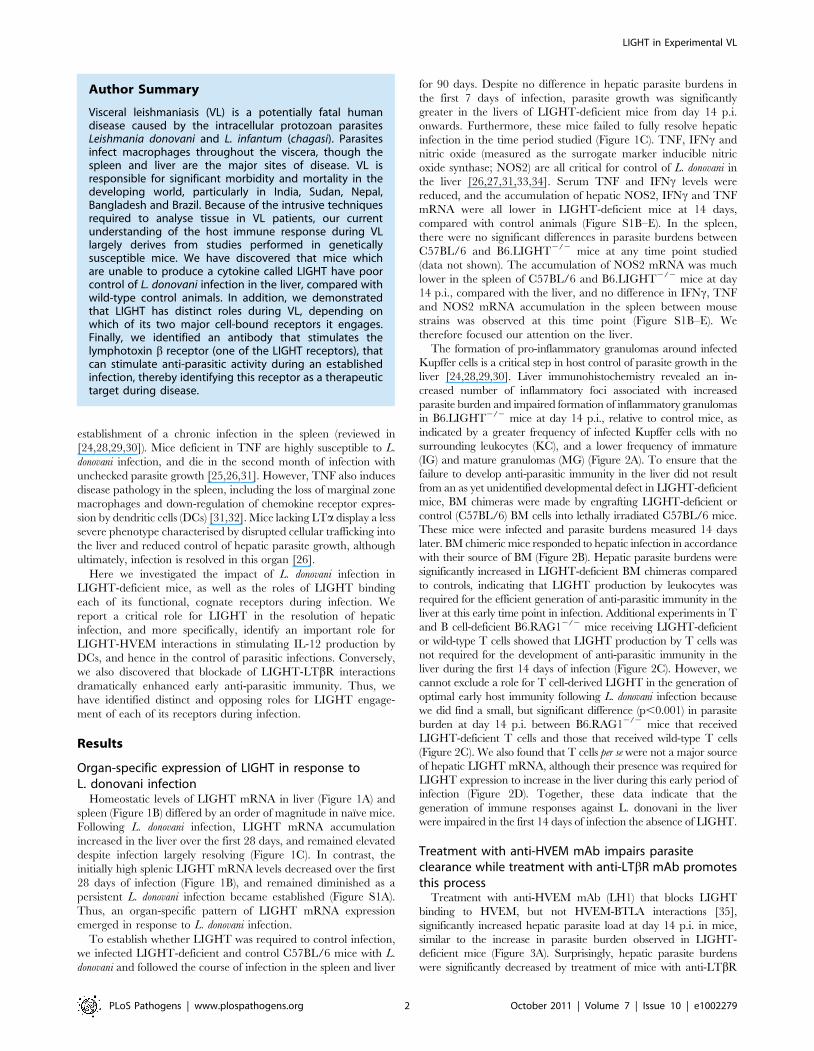
The formation of pro-inflammatory granulomas around infected
Kupffer cells is a critical step in host control of parasite growth in the
liver [24,28,29,30]. Liver immunohistochemistry revealed an in-
creased number of inflammatory foci associated with increased
parasite burden and impaired formation of inflammatory granulomas
in B6.LIGHT2/2 mice at day 14 p.i., relative to control mice, as
indicated by a greater frequency of infected Kupffer cells with no
surrounding leukocytes (KC), and a lower frequency of immature
(IG) and mature granulomas (MG) (Figure 2A). To ensure that the
failure to develop anti-parasitic immunity in the liver did not result
from an as yet unidentified developmental defect in LIGHT-deficient
mice, BM chimeras were made by engrafting LIGHT-deficient or
control (C57BL/6) BM cells into lethally irradiated C57BL/6 mice.
These mice were infected and parasite burdens measured 14 days
later. BM chimeric mice responded to hepatic infection in accordance
with their source of BM (Figure 2B). Hepatic parasite burdens were
significantly increased in LIGHT-deficient BM chimeras compared
to controls, indicating that LIGHT production by leukocytes was
required for the efficient generation of anti-parasitic immunity in the
liver at this early time point in infection. Additional experiments in T
and B cell-deficient B6.RAG12/2 mice receiving LIGHT-deficient
or wild-type T cells showed that LIGHT production by T cells was
not required for the development of anti-parasitic immunity in the
liver during the first 14 days of infection (Figure 2C). However, we
cannot exclude a role for T cell-derived LIGHT in the generation of
optimal early host immunity following L. donovani infection because
we did find a small, but significant difference (p,0.001) in parasite
burden at day 14 p.i. between B6.RAG12/2 mice that received
LIGHT-deficient T cells and those that received wild-type T cells
(Figure 2C). We also found that T cells per se were not a major source
of hepatic LIGHT mRNA, although their presence was required for
LIGHT expression to increase in the liver during this early period of
infection (Figure 2D). Together, these data indicate that the
generation of immune responses against L. donovani in the liver
were impaired in the first 14 days of infection the absence of LIGHT.
Treatment with anti-HVEM mAb impairs parasiteclearance while treatment with anti-LTbR mAb promotesthis process
Treatment with anti-HVEM mAb (LH1) that blocks LIGHT
binding to HVEM, but not HVEM-BTLA interactions [35],
significantly increased hepatic parasite load at day 14 p.i. in mice,
similar to the increase in parasite burden observed in LIGHT-
deficient mice (Figure 3A). Surprisingly, hepatic parasite burdens
were significantly decreased by treatment of mice with anti-LTbR
Author Summary
Visceral leishmaniasis (VL) is a potentially fatal humandisease caused by the intracellular protozoan parasitesLeishmania donovani and L. infantum (chagasi). Parasitesinfect macrophages throughout the viscera, though thespleen and liver are the major sites of disease. VL isresponsible for significant morbidity and mortality in thedeveloping world, particularly in India, Sudan, Nepal,Bangladesh and Brazil. Because of the intrusive techniquesrequired to analyse tissue in VL patients, our currentunderstanding of the host immune response during VLlargely derives from studies performed in geneticallysusceptible mice. We have discovered that mice whichare unable to produce a cytokine called LIGHT have poorcontrol of L. donovani infection in the liver, compared withwild-type control animals. In addition, we demonstratedthat LIGHT has distinct roles during VL, depending onwhich of its two major cell-bound receptors it engages.Finally, we identified an antibody that stimulates thelymphotoxin b receptor (one of the LIGHT receptors), thatcan stimulate anti-parasitic activity during an establishedinfection, thereby identifying this receptor as a therapeutictarget during disease.
LIGHT in Experimental VL
PLoS Pathogens | www.plospathogens.org 2 October 2011 | Volume 7 | Issue 10 | e1002279

mAb (LLBT2) that blocks LIGHT binding to LTbR, but not
LTa1b2-LTbR interactions [35] (Figure 3A). Antibody treatments
had no significant effect on the low splenic parasite burden at this
time point (Figure S2). The formation of granulomas was significantly
impaired by anti-HVEM mAb, as indicated by a greater frequency of
KC and a lower frequency of IG and MG (p,0.05, k2 analysis;
Figure 3B). In contrast, granuloma formation was significantly
enhanced by anti-LTbR mAb, as indicated by a lower frequency of
KC and a higher frequency of IG and MG (p,0.05, k2 analysis;
Figure 3B). Thus, these results indicate that HVEM and LTbR have
distinct and opposing roles during the first 14 days of infection.
Stimulation of LTbR improves parasite clearance duringan established infection
To further investigate the role of LTbR in VL, we treated mice
with the agonist anti-LTbR antibody (3C8) which blocks binding
of both LTa1b2 and LIGHT to LTbR, yet functions as an agonist
directly activating LTbR signalling pathways [36,37]. The anti-
LTbR 3C8 enhanced parasite clearance in the liver during an
established infection (Figure 4A), but had no anti-parasitic effect in
the first 14 days of infection (data not shown), unlike the anti-
LTbR mAb LLBT2 (Figure 3A). Importantly, 3C8 also prevented
parasite growth in the spleen between days 14 to 28 p.i.
(Figure 4B). In contrast, treatment with LLTB2 during established
infection (days 14–28 p.i.) had no effect on parasite clearance in
the liver or spleen (data not shown). Thus, treatment of L. donovani-
infected mice with two different anti-LTbR mAbs had distinct
effects on the course of infection, reflecting different functional
properties of these mAbs.
Treatment with anti-HVEM mAb impairs Th1-mediatedimmunity
We next sought to identify anti-parasitic mechanisms dependent
upon LIGHT-HVEM signalling. We previously showed that early
Figure 1. LIGHT is required for efficient parasite clearance in the liver. C57BL/6 mice were infected with L. donovani, and LIGHT mRNAaccumulation was measured in liver (A) and spleen (B) at indicated times (expressed as the number of LIGHT mRNA molecules per 1000 HPRT mRNAmolecules). (C) C57BL/6 (closed squares) and B6.LIGHT2/2 (open squares) mice were infected with L. donovani, and hepatic parasite burdens weremeasured from day 7 p.i. to day 90 p.i. and data represent the mean +/2SEM. One representative experiment of at least 2 performed is shown (n = 4–5 mice per treatment group in each experiment). Statistical differences of p,0.05 (*), p,0.01 (**) or p,0.001 (***) are indicated.doi:10.1371/journal.ppat.1002279.g001
LIGHT in Experimental VL
PLoS Pathogens | www.plospathogens.org 3 October 2011 | Volume 7 | Issue 10 | e1002279
splenic IL-12/IL-23p40 production by DC is critical for the efficient
generation of immunity in the liver [38,39]. Although no change in
IL-12p35 mRNA accumulation was observed in any treatment
group, the anti-HVEM mAb (LH1) inhibited splenic DC IL-12/IL-
23p40 mRNA accumulation in response to L. donovani infection
(Figure 5A). We next evaluated the importance of LIGHT-HVEM
co-stimulatory signals for the development of L. donovani-specific
CD4+ T cell priming and Th1 differentiation, the latter being a
known IL-12-dependent process [39,40,41]. Mice were injected
with CFSE-labelled OVA-specific CD4+ (OT-II) T cells, then
infected with transgenic OVA-expressing L. donovani [42], and
antigen-specific CD4+ T cell proliferation was assessed 4 days later
by CFSE dilution. No antigen-specific CD4+ T cell proliferation was
observed when mice were infected with wild-type parasites
(Figure 5B), so no bystander activation had occurred, and OT-II
cell proliferation occurred equally in control and anti-HVEM-
treated mice (Figure 5B), indicating that LIGHT-HVEM interac-
tions were not required for early priming of CD4+ T cell
proliferation. In support of this result, proliferation of polyclonal
antigen-specific, CD4+ T cells was similar between cells isolated
from the spleens of control-treated and anti-HVEM treated mice
(Figure 5C). However, production of IFNc and TNF by these
antigen-specific CD4+ T cells was inhibited by anti-HVEM mAb
(Figure 5C). Furthermore, direct ex vivo production of IFNc by
hepatic CD4+ T cells (both total number and frequency) was
significantly reduced by anti-HVEM mAb (Figure 5D), indicating
that LIGHT-HVEM interactions play an important role in
generating Th1 cell responses following L. donovani infection.
Figure 2. LIGHT from hematopoietic cells is required for efficient granuloma formation and control of L. donovani in the liver. Thenumber and maturity of hepatic granulomas were evaluated on liver sections taken at day 14 p.i. from C57BL/6 and B6.LIGHT2/2 mice, as indicated(A), and data represent the mean frequency of infected Kupffer cells (KC), immature granulomas (IG) and mature granulomas (MG) per liver +/2SEM.(B) C57BL/6 mice engrafted with BM from C57BL/6 mice (B6.B6) (closed bars) and B6.LIGHT2/2 mice (B6.LIGHT2/2.B6) (open bars) were infectedwith L. donovani and hepatic parasite burdens were measured at day 14 p.i. and are represented as the mean +/2SEM. Parasite burden (C) and LIGHTmRNA accumulation (D) in the livers of C57BL/6 and B6.RAG12/2 mice (plus or minus C57BL/6 or LIGHT-deficient CD4+ T cells and CD8+ T cells (equalnumbers), as indicated) at day 14 p.i.. One representative experiment of at least 2 performed is shown (n = 4–5 mice per treatment group in eachexperiment). Statistical differences of p,0.05 (*), p,0.01 (**) or p,0.001 (***) are indicated.doi:10.1371/journal.ppat.1002279.g002
LIGHT in Experimental VL
PLoS Pathogens | www.plospathogens.org 4 October 2011 | Volume 7 | Issue 10 | e1002279

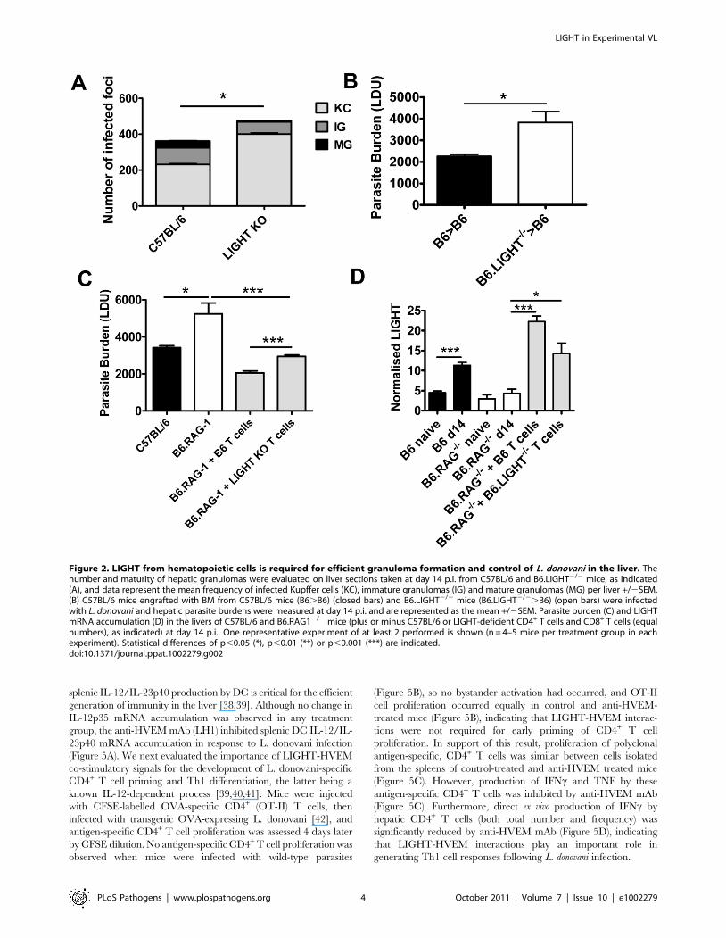
Treatment with anti- LTbR mAb promotes Th1-mediatedimmunity
The anti-LTbR mAb (LLTB2) inhibited parasite growth in
acute experimental VL (Figure 3A), but had no effect on splenic
DC IL-12/IL-23p40 or IL-12p35 mRNA accumulation at 5 hours
p.i. (Figure 6A), and no effect on the expansion of OVA-specific
CD4+ T cells (OT-II cells) in mice infected with OVA-transgenic
L. donovani (Figure 6B). We also found no differences in antigen-
specific recall responses in splenic CD4+ T cells isolated from
infected mice on day 14 p.i., yet the amount of TNF and IFNcproduced upon antigen-specific CD4+ T cell stimulation was
greatly enhanced in these cells from mice treated with anti-LTbR
mAb (Figure 6C). In addition, the number and frequency of IFNc-
producing hepatic CD4+ T cells measured directly ex vivo on day
14 p.i. was significantly increased in these mice (Figure 6D),
suggesting LIGHT-LTbR binding suppresses the development of
Th1 cell responses in VL.
Treatment with anti- LTbR mAb promotes parasiteclearance in the liver early during infection
We next investigated timing requirements for treatment with
the anti-LTbR mAb (LLTB2) during acute infection with L.
donovani. A single dose (100 mg) of anti-LTbR mAb at the time of
infection was sufficient to reduce hepatic parasite burden as early
as day 7 p.i. (Figure 7A). To test whether treatment with anti-
LTbR mAb was simply shunting available LIGHT onto HVEM,
we also co-treated mice with anti-LTbR (LLTB2) and anti-HVEM
(LH1) mAbs (Figure 7B), and found no additional effect of co-
administration over anti-LTbR alone by day 7 p.i., indicating that
increased, early anti-parasitic immunity observed after anti-LTbR
mAb (LLTB2) treatment was not caused by enhanced HVEM-
mediated co-stimulation. Of note, there was no effect of anti-
HVEM mAb treatment alone at day 7 p.i., indicating that the
effect of this treatment on parasite burden only becomes apparent
between days 7–14 p.i., similar to what was observed in LIGHT-
deficient mice (Figure 1C). To test whether anti-LTbR mAb
(LLTB2) agonist activity might account for the above effect, we
treated LIGHT-deficient mice with this antibody and measured
liver parasite burdens at day 7 p.i. (Figure 7C). Although a
significant reduction in parasite burden was found in C57BL/6
mice treated with anti-LTbR mAb (LLTB2), no such effect was
observed in LIGHT-deficient mice, indicating that the likely
mechanism of action was via the blockade of LIGHT binding
LTbR.
Enhanced parasite clearance observed followingtreatment with anti-LTbR mAb requires CD4+ T cells andTNF
We investigated the cellular requirements for the early anti-
parasitic effects of anti-LTbR mAb (LLTB2). Treatment with anti-
LTbR mAb had no impact on hepatic parasite burdens in
B6.RAG12/2 mice at day 7 p.i. (Figure S3), suggesting that B
and/or T lymphocytes are required for the enhanced parasite
clearance resulting from this treatment. We focused our attention
on T cells because we have previously shown that B cells play a
negative regulatory role in the liver during infection [43].
Depletion of CD4+ or CD8+ T cells alone during the first 7 days
of infection had no effect on hepatic parasite burden (Figure 8A),
despite T cells being required for the control of parasite growth at
later stages of infection [26,44,45]. However, depletion of CD4+ T
cells, but not CD8+ T cells, prevented the anti-parasitic effect
mediated by anti-LTbR at day 7 p.i. (Figure 8A). Given that NKT
cells comprise a significant proportion of hepatic CD4+ T cells, we
also investigated whether this cell subset was required for the
increased anti-parasitic activity. Treatment of NKT cell-deficient
(B6.Ja182/2) mice with the anti-LTbR mAb (Figure 8A) had no
impact on the decreased liver parasite burden, indicating that
conventional CD4+ T cells, but not NKT cells, are required for the
enhanced parasite clearance following anti-LTbR mAb treatment.
We observed increased CD4+ T cell TNF and IFNc production
was associated with improved control of parasite growth resulting
from anti-LTbR mAb treatment (Figure 6). We next assessed
whether these cytokines were required for the enhanced parasite
clearance in mice receiving anti-LTbR (LLTB2) mAb. Hepatic
parasite burdens were decreased similarly in anti-LTbR mAb
treated control and IFNc-deficient mice (Figure 8B). However,
anti-LTbR mAb treatment in TNF-deficient mice had no impact
Figure 3. Distinct effects of blocking LIGHT-HVEM and LIGHT-LTbR interactions on parasite growth in the liver. Parasiteburdens were determined in the livers of L. donovani infected micetreated with anti-HVEM (LH1) mAb or control hamster IgG or anti-LTbR(LLBT2) mAb or control rat IgG (A). Data are represented as the mean +/2 SEM at day 14 p.i.. The number and maturity of hepatic granulomas(B) were measured at day 14 p.i., as described in the legend of Figure 2.One representative experiment of 3 performed is shown (n = 5 mice pertreatment group in each experiment). Statistical differences of p,0.05(*) or p,0.001 (***) for control versus mAb-treated mice are shown.doi:10.1371/journal.ppat.1002279.g003
LIGHT in Experimental VL
PLoS Pathogens | www.plospathogens.org 5 October 2011 | Volume 7 | Issue 10 | e1002279

on hepatic parasite burden (Figure 8B), indicating that TNF is
critical for this enhanced parasite clearance. The failure of anti-
LTbR mAb treatment in TNF-deficient animals was not caused
by reduced expression of LTbR on the cells of these mice, as
LTbR expression levels were no different to those on immune cells
from C57BL/6 control mice (data not shown). Furthermore,
adoptive transfer of wild type and TNF-deficient CD4+ T cells into
B6.RAG12/2 mice and treatment with anti-LTbR mAb demon-
strated that CD4+ T cells did not have to produce TNF
(Figure 8C). Thus, anti-LTbR mAb treatment increased early
hepatic anti-parasitic immunity by mechanisms requiring conven-
tional CD4+ T cells and TNF, the latter potentially coming from a
non-T cell source.
Discussion
We have identified distinct and opposing roles for LIGHT and
its receptors during infection. LIGHT has important roles in T cell
costimulation [3,14]. Blockade of LIGHT impairs allogeneic T cell
responses and graft versus host disease [46,47], while over-
expression of LIGHT by T cells causes inflammatory disease of the
gut and reproductive tissues [48,49]. Our results indicate that
these effects could be mediated via the LIGHT-HVEM axis
between T cells and DC. Early DC IL-12 production depends on
the presence of T cells, and this IL-12 production is critical for
generating anti-parasitic immune mechanisms that control L.
donovani growth [24,39,40,50]. Our finding that anti- HVEM mAb
blocks IL-12/IL-23p40 mRNA accumulation during infection is
consistent with a previous study that reported BM-derived DCs
from LIGHT-deficient animals were impaired in their ability to
produce IL-12 following activation in vitro [51]. This study also
showed that blockade of LIGHT with soluble receptors in mice
infected with L. major, a cause of cutaneous leishmaniasis, resulted
in reduced IL-12 generation, associated with diminished CD4+ T
cell IFNc production and increased parasite growth and disease.
Our finding that T cells did not have to produce LIGHT in order
to promote anti-parasitic immunity, together with data from L.
major infection in mice [51], support a model whereby DC-derived
LIGHT interacts with T cell HVEM to promote DC IL-12
production.
The defect in anti-parasitic immunity observed in the absence of
LIGHT was restricted to the liver, and not the spleen. The reason
for this is unclear, but could relate to the requirement for cellular
recruitment and granuloma development for control of parasite
growth in the liver. Although increased tissue weight and cellular
expansion are features of L. donovani infection in the spleen,
organised inflammatory granulomas are rarely observed in this
tissue [24,28,29,30]. Importantly, parasite growth is contained in
the spleen after 1–2 months of infection rather than efficiently
controlled, as occurs in the liver. Therefore, it is possible that
different anti-parasitic immune mechanisms operate in these two
tissue sites during experimental VL with different requirements for
LIGHT.
The LIGHT-specific blocking mAbs we have employed (LH1
and LLTB2) have previously been shown to selectively block
interactions between LIGHT and its receptors (HVEM and
LTbR, respectively) [35]. However, we cannot exclude the
possibility that they may trigger some receptor activation following
engagement, and that this may contribute to biological effects we
have observed. In addition, because these mAbs cause the selective
blockade of LIGHT binding to their respective receptors, we
cannot rule out that they promote alternative receptor-ligand
interactions (Figure 9A). For example, blocking LIGHT interact-
ing with HVEM may allow HVEM to more readily engage BTLA
on cells to increase inhibitory signals (Figure 9B), as well as
increased CD160 signalling. Similarly, blockade of LIGHT
binding LTbR may allow greater amounts of LIGHT to bind
HVEM, thereby reducing negative signalling between HVEM and
BTLA and potentially promoting LIGHT-HVEM-mediated T
cell co-stimulation (Figure 9C). However, this latter possibility
Figure 4. Agonistic stimulation of LTbR only enhances parasite clearance in an established infection. C57BL/6 mice with an establishedL. donovani infection were treated with control rat IgG or agonistic anti-LTbR mAb (3C8) from day 14 p.i. onwards and parasite burdens weredetermined in the liver (A) and spleen (B) at day 28 p.i.. One representative experiment of two performed is shown (n = 5 mice per treatment group ineach experiment). Statistical differences of p,0.01 (**) for control versus treated mice are shown.doi:10.1371/journal.ppat.1002279.g004
LIGHT in Experimental VL
PLoS Pathogens | www.plospathogens.org 6 October 2011 | Volume 7 | Issue 10 | e1002279

Figure 5. LIGHT-HVEM interactions are required for early DC IL-12/IL-23p40 mRNA accumulation, and antigen-specific CD4+ T cellIFNc and TNF production. C57BL/6 mice were treated with anti-HVEM mAb or control hamster IgG or left untreated prior to infection with 16108
L. donovani amastigotes. IL-12/IL-23p40 mRNA accumulation was measured in splenic CD11c+ DC that had been enriched by positive selection by
LIGHT in Experimental VL
PLoS Pathogens | www.plospathogens.org 7 October 2011 | Volume 7 | Issue 10 | e1002279

seems unlikely in the current study given that co-administration of
LH1 and LLTB2 resulted in improved control of parasite growth
(Figure 7B). Instead, LIGHT may send inhibitory signals via
LTbR early during infection, although no such LTbR-mediated
negative signalling pathway has yet been defined.
The anti-parasitic effects of anti-LTbR mAb (LLTB2) were
observed when it was administered at the time of infection, but not
in mice with an established L. donovani infection, suggesting that a
mAb with similar functional characteristics would have limited
therapeutic potential for treatment of VL. However, our finding
that the defined agonist anti-LTbR mAb (3C8) improved the rate
of parasite clearance in the liver and reduced parasite load in the
spleen, not only demonstrated fundamentally different biological
activities for LLTB2 and 3C8 mAbs, but also shows that LTbR
activation can promote beneficial immune mechanisms during
established infection. This agonist antibody has previously been
shown to promote DC development and maturation in vivo
[20,37], and this may explain the anti-parasitic effects observed
after administration to L. donovani-infected mice because we have
previously shown that DC adoptive transfer can improve control
of parasite growth in infected mice [32]. Hence, the activation of
anti-parasitic immune mechanisms by stimulation of LTbR
represents a potential therapeutic strategy against chronic
infectious diseases like VL. However, a better understanding of
the functional characteristics of the different anti-LTbR mAbs will
be required in order to better harness their therapeutic potential,
including identification of the specific epitopes they recognise and
signalling pathways they activate.
We previously reported increased monocyte recruitment into
the spleen in an experimental model of cerebral malaria following
treatment of mice with the anti-LTbR mAb (LLTB2), and that this
treatment protected mice from disease [52]. Interestingly, no
protection from experimental cerebral malaria was afforded by
treatment with the anti-LTbR (3C8) mAb (Randall and
Engwerda, unpublished), again emphasising the functional differ-
ences between LLTB2 and 3C8 anti-LTbR mAbs. An intriguing
finding from our current studies was an increase in hepatic and
splenic monocyte recruitment following anti-LTbR mAb LLTB2
treatment (CD11b+ Ly6Chi cells; Figure S4A and B). Flow
cytometry analysis revealed that monocytes, along with DCs (both
cDC and pDC), and neutrophils expressed the highest levels of
LTbR in the liver, as previously reported [19,20,37], and
furthermore, that expression of LTbR did not appear to change
significantly on any of these cells during the first 5 days of infection
with L. donovani (Figure S5). However, the increased monocyte
recruitment was not necessary for improved early control of
parasite growth in treated animals in the current study because
mice lacking CCL2 that have an impairment in monocyte
mobilisation [53], also had improved control of parasite growth
following anti-LTbR (LLTB2) treatment at day 7 p.i. (Figure
S4C). Although the early anti-parasitic effect of anti-LTbR
(LLTB2) mAb appeared to involve blocking of LIGHT- LTbR
interactions, as indicated by the failure of this antibody to improve
parasite control in LIGHT-deficient mice (Figure 7C), we cannot
exclude the possibility that some effects of this antibody, such as
increased monocyte mobilisation, might involve agonist activities.
Regardless, given the important role for monocyte infiltration into
sites of infection and tumour growth [54], our results indicate that
manipulation of the LIGHT-LTbR signalling axis offers a
potential way to improve monocyte mobilisation for therapeutic
applications. Furthermore, given the recent report that monocytes
can migrate into secondary lymphoid tissues in response to
interactions with gram negative bacteria and/or their products,
and then develop into CD209a+, CD206+, CD14+, CD11chi DC
capable of activating CD4+ T cells and cross-priming CD8+ T cells
[55], our results suggest that manipulation of LIGHT signalling
pathways may be one way to promote this process that may have
applications in vaccination.
In summary, our findings further delineate the complex
interactions between LIGHT and its receptors and demonstrate
the therapeutic potential of modulating these immune regulatory
pathways to improve disease outcomes. Our results provide
mechanistic insight into the roles of LIGHT-HVEM interactions
on DC function and CD4+ T cell priming, as well as anti-parasitic
immune responses activated by blockade of LIGHT-LTbR
interactions. Finally, we have identified two different mAbs that
target LTbR with distinct functional outcomes on anti-parasitic
immunity at different stages of infection.
Material and Methods
MiceInbred female C57BL/6 and B6.SJL.Ptprca (B6.CD45.1) mice
were purchased from the Australian Resource Centre (Canning
Vale, Western Australia), and maintained under conventional
conditions. B6.RAG12/2 [56], B6.LIGHT2/2 [57], B6.TNF2/2
[58], B6.SJL.Ptprca6OT-II [59], B6.SJL.Ptprca6OT-I [60],
B6.IFNc2/2 [61] and B6.Ja182/2 [62] were bred and main-
tained at the Queensland Institute of Medical Research.
B6.CCL22/2 mice [53] were bred at Monash University and
maintained at the Queensland Institute of Medical Research. All
mice used were age- and sex-matched (6–10 weeks), and were
housed under specific-pathogen free conditions. Chimeric mice
were prepared by irradiating B6.SJL.Ptprca mice with 11Gy and
then engrafting with 36106 fresh bone marrow (BM) cells i.v. via
the lateral tail vein. Mice were maintained on antibiotics for 2
weeks after engraftment and infected with L. donovani 8 weeks after
receiving BM, as previously described [26]. Adoptive transfer of
equal numbers (106) of purified CD4+ and CD8+ T cells (98%
purity as determined by flow cytometry) into B6.RAG12/2 mice
was performed as previously described [26].
MACS from naı̈ve mice (hatched bars), untreated mice (grey bars), hamster IgG treated mice (open bars) or anti-HVEM treated mice (closed bars) at5 hours p.i. (A). C57BL/6 mice treated with control hamster IgG or anti-HVEM mAb received CD45.1+ CFSE-labelled OVA-specific CD4+ T cells (OT-II)prior to infection with 26107 OVA-expressing L. donovani. OT-II CFSE dilution in the spleen was assessed by flow cytometry on day 4 p.i., after gatingon CD45.1+CD4+TCRb+ cells (B) and % CFSE dilution +/2 SEM is shown on each plot (CFSE dilution for 4 individual mice in each treatment group isshown, as well as CSFE dilution in a control animal that was infected with wild type parasites). Splenic CD4+ T cells were purified on day 14 p.i. fromnaı̈ve mice or L. donovani-infected mice treated with control hamster IgG (open bars) or anti-HVEM mAb (closed bars) by positive MACS selection.CD4+ T cells were co-cultured with paraformaldehyde-fixed L. donovani amastigotes and irradiated spleen cells. Antigen-specific CD4+ T cellproliferation, as well as IFNc and TNF levels in cell culture supernatants were measured by cytometric bead array at 72 h post-stimulation (C). HepaticCD4+ T cell (CD4+TCRb+) IFNc production was measured in L. donovani-infected mice treated with either control hamster IgG (solid line) or anti-HVEM(dashed line) mAb on day 14 p.i.. Representative histograms showing IFNc production by gated CD4+ TCRb+ cells are shown and compared to isotypecontrol antibody (solid gray) (D). Both the total number and frequency of IFNc-producing CD4+ T cells are shown graphically. One representativeexperiment of 3 performed with similar outcome is shown (n = 4 mice per treatment group in each experiment). Statistical differences of p,0.05 (*),p,0.01 (**) or p,0.001 (***) for control versus mAb-treated mice are shown.doi:10.1371/journal.ppat.1002279.g005
LIGHT in Experimental VL
PLoS Pathogens | www.plospathogens.org 8 October 2011 | Volume 7 | Issue 10 | e1002279
Figure 6. Treatment with anti-LTbR mAb LLTB2 leads to increased antigen-specific CD4+ T cell IFNc and TNF production. DC IL-12/IL-23p40 mRNA accumulation (A), splenic OT-II CFSE dilution (B), antigen-specific splenic CD4+ T cell proliferation, IFNc and TNF production (C) andhepatic CD4+ T cell IFNc production (D) were measured as described in the legend of Figure 5. One representative experiment of 3 performed withsimilar outcome is shown (n = 4 mice per treatment group in each experiment). Statistical differences of p,0.05 (*), p,0.01 (**) or p,0.001 (***) forcontrol versus mAb-treated mice are shown.doi:10.1371/journal.ppat.1002279.g006
LIGHT in Experimental VL
PLoS Pathogens | www.plospathogens.org 9 October 2011 | Volume 7 | Issue 10 | e1002279
Ethics statementAll animal procedures were approved and monitored by the
Queensland Institute of Medical Research Animal Ethics
Committee. This work was conducted under QIMR animal ethics
approval number A02-634M, in accordance with the ‘‘Australian
code of practice for the care and use of animals for scientific
purposes’’ (Australian National Health & Medical Research
Council).
Parasites and infectionsL. donovani (LV9) and OVA-transgenic LV9 (PINK LV9) [42]
were maintained by passage in B6.RAG12/2 mice and amasti-
gotes were isolated from the spleens of chronically infected mice.
Mice were infected by injecting 26107 amastigotes i.v. via the
lateral tail vein, killed at the times indicated in the text by CO2
asphyxiation and bled via cardiac puncture. In experiments
examining DC IL-12/IL-23p40 production, mice were infected
with 16108 amastigotes intravenously, as previously described
[63]. Spleens and perfused livers were removed at times indicated
and parasite burdens were determined from Diff-Quik-stained
impression smears (Lab Aids, Narrabeen, Australia) and expressed
as Leishman-Donovan units (LDU) (the number of amastigotes per
1,000 host nuclei multiplied by the organ weight in grams) [64].
Liver and spleen tissue were also preserved in either RNAlater
(Sigma-Aldrich, Castle Hill, Australia) or Tissue-Tek O.C.T.
compound (Sakura, Torrence, USA). Hepatic mononuclear cells
and splenocytes were isolated as previously described [65].
AntibodiesAll antibody-producing hybridomas were grown in 5% (v/v)
foetal calf serum, RPMI containing 10 mM L-glutamine, 100 U/
ml penicillin and 100 mg/ml streptomycin. Purified antibody was
prepared as previously described [63]. Mice were administered
100 mg of anti-LTbR mAb (LLTB2) or anti-HVEM mAb (LH1)
[35] i.v. on the day of infection and every 5 days thereafter for 14
day experiments, or as a single dose on the day of infection for 7
day experiments.. The anti-LTbR mAb 3C8 was administered at
200 mg i.v. [36,37] starting at the times indicated in the text and
every 5 days thereafter. The anti-LTbR mAb (LLTB2) or anti-
HVEM mAb (LH1) specifically block the binding of LIGHT to
either LTbR or HVEM, respectively, but do not disrupt
interactions between these receptors and other functional ligands
(i.e., LTa1b2 for LTbR and BTLA for HVEM) [35]. The anti-
LTbR mAb (3C8) blocks binding of both LTa1b2 and LIGHT,
but is an agonist directly activating LTbR [66]. Mice were
depleted of CD4+ or CD8+ T cells with anti-CD4 (YTS191.1) or
anti-CD8b (53-5.8) mAbs, respectively, as previously described
[64]. Depletion of T cell subsets was confirmed at completion of
experiments by assessing T cell numbers in the spleen by flow
cytometry. Greater than 95% of CD4+ and CD8+ T cells were
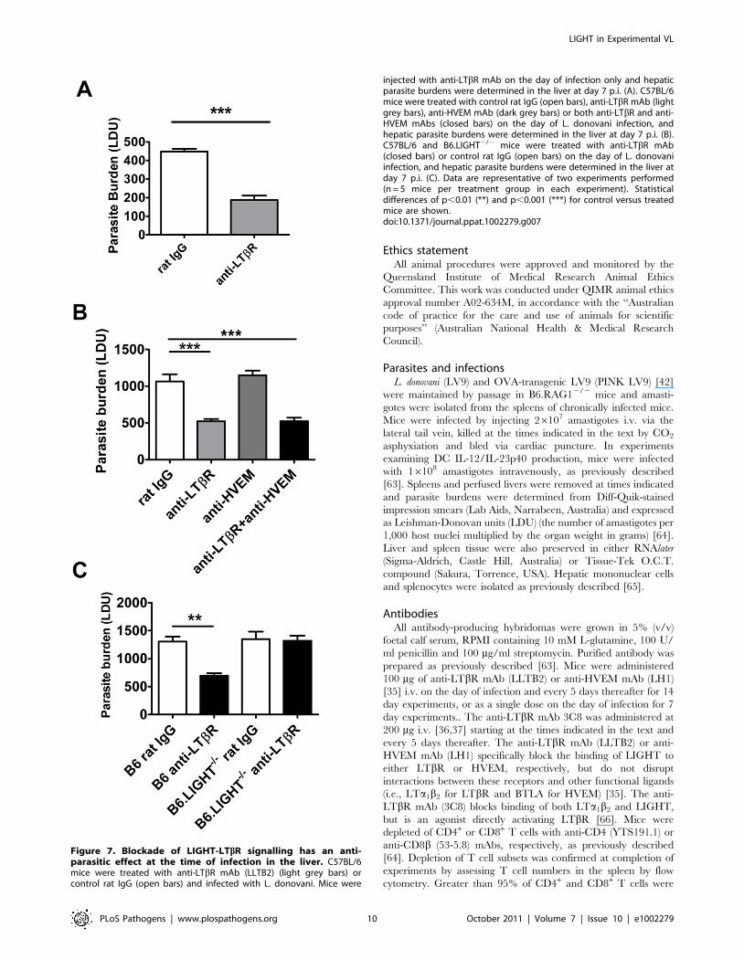
Figure 7. Blockade of LIGHT-LTbR signalling has an anti-parasitic effect at the time of infection in the liver. C57BL/6mice were treated with anti-LTbR mAb (LLTB2) (light grey bars) orcontrol rat IgG (open bars) and infected with L. donovani. Mice were
injected with anti-LTbR mAb on the day of infection only and hepaticparasite burdens were determined in the liver at day 7 p.i. (A). C57BL/6mice were treated with control rat IgG (open bars), anti-LTbR mAb (lightgrey bars), anti-HVEM mAb (dark grey bars) or both anti-LTbR and anti-HVEM mAbs (closed bars) on the day of L. donovani infection, andhepatic parasite burdens were determined in the liver at day 7 p.i. (B).C57BL/6 and B6.LIGHT2/2 mice were treated with anti-LTbR mAb(closed bars) or control rat IgG (open bars) on the day of L. donovaniinfection, and hepatic parasite burdens were determined in the liver atday 7 p.i. (C). Data are representative of two experiments performed(n = 5 mice per treatment group in each experiment). Statisticaldifferences of p,0.01 (**) and p,0.001 (***) for control versus treatedmice are shown.doi:10.1371/journal.ppat.1002279.g007
LIGHT in Experimental VL
PLoS Pathogens | www.plospathogens.org 10 October 2011 | Volume 7 | Issue 10 | e1002279
depleted by antibody treatment. In all experiments, control mice
received the same quantities of the appropriate control hamster
IgG (UC8-1B9; ATCC, Manassas, VA) or control rat IgG (Sigma-
Aldrich).
CD4+ T cell proliferationTo assess antigen-specific T cell proliferation in vivo, mice were
infected with OVA-transgenic PINK LV9 [42]. Splenic OVA-
specific OT-II T cells were isolated and labelled with CFSE, as
previously described [64]. CFSE-labelled OT-II cells (16106) were
adoptively transferred into mice 2 h prior to infection with LV9 or
PINK LV9. Expansion of CFSE+ cells in the spleen was monitored
by FACS 4 days later. In all of these experiments, control animals
were included that received the same number of CFSE-labelled
OT-II cells, but were infected with wild-type parasites. No OT-II
proliferation was ever observed in these animals. Re-stimulation
assays for endogenous splenic CD4+ T cells were performed as
previously described [63].
Assessment of granuloma formationThe maturation of granulomas was scored around infected
Kupffer cells in acetone-fixed liver sections as previously described
[64,65].
Flow cytometryAllophycocyanin (APC)-conjugated anti-TCRb chain (H57-
597), B220 (clone RA3-6B2), CD11c (clone N418), phycoerythrin
(PE)-Cy5-conjugated anti-CD4 (GK1.5), PE-conjugated IFNc(XMG1.2), CD8b (53-5.8), I-Ab (clone AF6-120.1), Ly6G (clone
1A8), CD45.1 (clone A20), CD45.2 (clone 104), rat IgG1
(RTK2071), fluorescein isothiocyanate (FITC)-conjugated CD19
(clone 6D5), BST2 (clone 120G8), Ly6C (clone AL-21) and
biotinylated anti-NK1.1 (PK136), CD11b (clone M1/70), LTbR
(3C8) were purchased from Biolegend (San Diego, CA) or BD
Biosciences (Franklin Lakes, NJ). Biotinylated antibodies were
detected with streptavidin conjugated alexa 488, PE or PE/Cy5
(Biolegend). Leukocyte populations were defined as follows; CD4+
T cells (CD4+, TCR+), CD8+ T cells (CD8+, TCR+), NKT cells
(NK1.1+, TCR+), NK cells (NK1.1+, TCR2), B cells (B220+,
CD19+), cDC (CD11chi, MHCIIhi, TCR2, B2202), pDC
(CD11cint, MHCIIint, 120G8+), monocytes (CD11b+, Ly6C+)
and neutrophils (CD11b+, Ly6G+). The staining of cell surface
antigens and intracellular cytokine staining was carried out as
described previously [63]. FACS was performed on a FACSCa-
libur or a FACS Canto II (BD Biosciences), and data were
analysed using FlowJo software (TreeStar, Oregon, USA). Serum
and/or tissue culture supernatants were assessed for the presence
of soluble cytokines using flexset bead array kits and a FACSArray
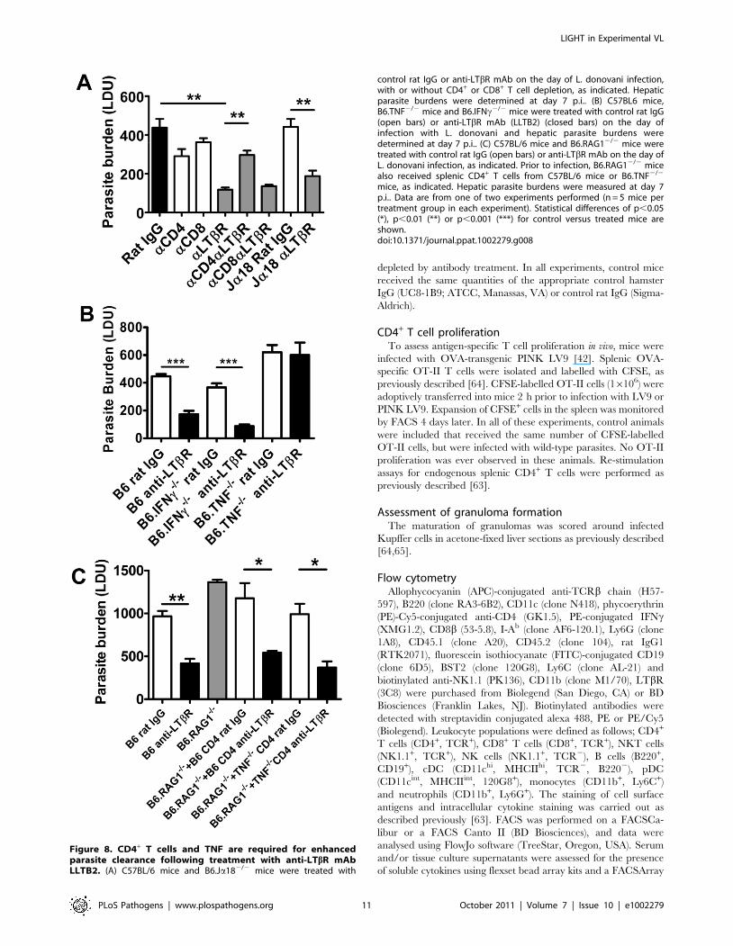
Figure 8. CD4+ T cells and TNF are required for enhancedparasite clearance following treatment with anti-LTbR mAbLLTB2. (A) C57BL/6 mice and B6.Ja182/2 mice were treated with
control rat IgG or anti-LTbR mAb on the day of L. donovani infection,with or without CD4+ or CD8+ T cell depletion, as indicated. Hepaticparasite burdens were determined at day 7 p.i.. (B) C57BL6 mice,B6.TNF2/2 mice and B6.IFNc2/2 mice were treated with control rat IgG(open bars) or anti-LTbR mAb (LLTB2) (closed bars) on the day ofinfection with L. donovani and hepatic parasite burdens weredetermined at day 7 p.i.. (C) C57BL/6 mice and B6.RAG12/2 mice weretreated with control rat IgG (open bars) or anti-LTbR mAb on the day ofL. donovani infection, as indicated. Prior to infection, B6.RAG12/2 micealso received splenic CD4+ T cells from C57BL/6 mice or B6.TNF2/2
mice, as indicated. Hepatic parasite burdens were measured at day 7p.i.. Data are from one of two experiments performed (n = 5 mice pertreatment group in each experiment). Statistical differences of p,0.05(*), p,0.01 (**) or p,0.001 (***) for control versus treated mice areshown.doi:10.1371/journal.ppat.1002279.g008
LIGHT in Experimental VL
PLoS Pathogens | www.plospathogens.org 11 October 2011 | Volume 7 | Issue 10 | e1002279

LIGHT in Experimental VL
PLoS Pathogens | www.plospathogens.org 12 October 2011 | Volume 7 | Issue 10 | e1002279

plate reader (BD Biosciences) according to the manufacturers’
instructions.
Real Time PCRRNA extraction and real-time RT-PCR was performed as
previously described [63]. The number of IFNc, TNF, NOS-2,
LIGHT and hypoxanthine phosphoribosyltransferase (HPRT)
cDNA molecules in each liver tissue sample were calculated using
Platinum Sybr Green Master Mix (Invitrogen Life Technologies)
[63]. Standard curves were generated with known amounts of
cDNA for each gene, and the number of cytokine molecules per
1000 HPRT molecules in each sample was calculated. The
number of IL-12/IL-23p40 and IL-12p35 cDNA molecules in
each DC sample were calculated using Taqman Gene Expression
Assays (Applied Biosystems). Relative quantitation of gene
expression was performed using the relative standard curve
method as described by Applied Biosystems. Briefly, standard
curves were prepared for all target and endogenous control genes
using an uninfected control sample. HPRT was used as the
endogenous control. The amount of target gene or endogenous
control in each sample was calculated from the appropriate
standard curves. The target amount was then divided by the
endogenous control amount to give the normalized target value.
The average normalized values for the four naı̈ve samples were
used as the calibrator.
Statistical analysisStatistical differences between groups was determined using the
Mann-Whitney U test using GraphPad Prism version 4.03 for
Windows (GraphPad Software, San Diego, CA) and p,0.05 was
considered statistically significant. The distribution of hepatic
histological responses was compared using X2 analysis with
Microsoft Excel software. All data are presented as the mean
values plus or minus standard error unless otherwise stated.
Supporting Information
Figure S1 A persistent L. donovani infection becomes established
in the spleen (A). Reduced cytokine production in L. donovani-
infected LIGHT-deficient mice. (B) Day 14 p.i. serum TNF and
IFNc levels in C57BL/6 (closed bars) and B6.LIGHT2/2 mice
(open bars).The accumulation of NOS2 (C), TNF (D) and IFNc(E) mRNA levels in naı̈ve or day 14 p.i. C57BL/6 and
B6.LIGHT2/2 mice was detected by real time RT-PCR and is
expressed as mRNA molecules per 1000 HPRT molecules (left
panels are from liver and right panels are from spleen). Data are
from one of two experiments performed (n = 4–5 mice per
treatment group in each experiment). Statistical differences of
p,0.05 (*) for C57BL/6 versus B6.LIGHT2/2 mice are shown.
(TIF)
Figure S2 No significant effect of blocking LIGHT-HVEM and
LIGHT-LTbR interactions on parasite growth in the spleen in the
first 14 days of infection. Parasite burdens were determined in the
spleens of L. donovani infected mice treated with anti-HVEM
(LH1) mAb or control hamster IgG or anti-LTbR (LLBT2) mAb
or control rat IgG. Data are represented as the mean +/2 SEM at
day 14 p.i.. No statistical differences for control versus mAb-
treated mice were found.
(TIF)
Figure S3 The anti-parasitic effect of blocking LIGHT-LTbR
interactions fails in B6.RAG12/2 mice. C57BL/6 or B6.RAG12/2
mice were treated with control rat IgG (open bars) or anti-LTbR
mAb (closed bars) the day prior to L. donovani infection, and
hepatic parasite burdens were measured at day 7 p.i. and are
represented as the mean +/2SEM. Statistical differences of p,0.05
(*) for control versus treated mice are shown.
(TIF)
Figure S4 Enhanced recruitment of inflammatory monocytes to
the liver in mice in which LIGHT-LTbR interactions are blocked.
(A) FACS profiles of hepatic monocytes (CD11b+Ly6Chi) from L.
donovani-infected C57BL/6 mice at day 7 p.i., following treatment
with either control rat IgG or anti-LTbR mAb prior to infection are
shown. (B) Total numbers of CD11b+Ly6Chi cells were measured
from the livers of infected C57BL/6 mice and CCL2-deficient mice
treated with either control rat IgG (open bars) or anti-LTbR mAb
(closed bars) at day 7 p.i.. (C) C57BL/6 mice and B6.CCL22/2
mice were treated with either control rat IgG (open bars) or anti-
LTbR (closed bars), prior to L. donovani infection and hepatic
parasite burdens were measured at day 7 p.i.. One representative
experiment of two performed is shown (n = 5 mice per treatment
group in each experiment). Statistical differences of p,0.05 (*) or
p,0.01 (**) for control versus treated mice are shown.
(TIF)
Figure S5 Mononuclear cells isolated from the livers of naı̈ve
and L. donovani-infected mice at day 5 p.i. were phenotyped,
labelled with a mAb against LTbR and enumerated by flow
cytometry. Representative histograms gated on appropriate
populations are shown for isotype control (solid grey shading),
naı̈ve mice (black line) and at day 5 p.i. with L. donovani (grey
line). Cells were identified as follows: B cells (B220+CD19+),
cDC(CD11chiMHCIIhi), monocytes (CD11b+Ly6chi), neutrophils
(CD11b+Ly6cint), NK cells (NK1.1+TCRb2), NKT cells
(NK1.1+TCRb+), pDC (CD11cint120G8+), CD4+ T cells
(CD4+TCRb+), CD8+ T cells (CD8+TCRb+). Data presented is
representative of 2 independent experiments.
(TIF)
Acknowledgments
The authors thank the staff of the QIMR Animal Facility for animal
husbandry, and Paula Hall and Grace Chojnowski for assistance with flow
cytometry.
Author Contributions
Conceived and designed the experiments: CRE ACS AH GRH CW.
Performed the experiments: CRE ACS FdLR AH MS YZ FHA PTB
LMR. Analyzed the data: CRE ACS FdLR GRH PMK. Contributed
reagents/materials/analysis tools: KP SS MJH BMS CW KT PMK.
Wrote the paper: CRE ACS CW.
Figure 9. Model of how manipulation of interactions between LIGHT and its receptors may impact on disease outcome. A. Undernormal circumstances LIGHT will interact with HVEM and/or LTbR to promote anti-parasitic immunity, as well as with DCR3 (not shown). HVEM canalso interact with BTLA to suppress immune responses, and can also interact with CD160 and HSV1 gD (not shown). LTa1b2 can also bind to LTbR andprovide signals essential for tissue organising and homeostasis. B. Blockade of LIGHT-HVEM with antibody will prevent HVEM-mediated costimulationand increase HVEM-BTLA-mediated inhibitory signals. LTbR-mediated inhibitory signals may also be increased via greater interactions with LIGHT.Together, this results in suppression of anti-parasitic immunity. C. Blockade of LIGHT-LTbR with antibody will potentially prevent LTbR-mediatedinhibitory signals, but will also promote LIGHT-HVEM co-stimulatory signalling and reduce HVEM-BLTA inhibitory signals. Together, this stimulatesincreased anti-parasitic immunity.doi:10.1371/journal.ppat.1002279.g009
LIGHT in Experimental VL
PLoS Pathogens | www.plospathogens.org 13 October 2011 | Volume 7 | Issue 10 | e1002279

References
1. Locksley RM, Killeen N, Lenardo MJ (2001) The TNF and TNF receptorsuperfamilies: integrating mammalian biology. Cell 104: 487–501.
2. Aggarwal BB (2003) Signalling pathways of the TNF superfamily: a double-edged sword. Nat Rev Immunol 3: 745–756.
3. Ware CF (2005) Network communications: lymphotoxins, LIGHT, and TNF.Annu Rev Immunol 23: 787–819.
4. Ware CF (2008) Targeting lymphocyte activation through the lymphotoxin andLIGHT pathways. Immunol Rev 223: 186–201.
5. Aggarwal BB, Eessalu TE, Hass PE (1985) Characterization of receptors forhuman tumour necrosis factor and their regulation by gamma-interferon.
Nature 318: 665–667.
6. Ruuls SR, Hoek RM, Ngo VN, McNeil T, Lucian LA, et al. (2001) Membrane-bound TNF supports secondary lymphoid organ structure but is subservient to
secreted TNF in driving autoimmune inflammation. Immunity 15: 533–543.
7. Mauri DN, Ebner R, Montgomery RI, Kochel KD, Cheung TC, et al. (1998)
LIGHT, a new member of the TNF superfamily, and lymphotoxin alpha areligands for herpesvirus entry mediator. Immunity 8: 21–30.
8. Crowe PD, VanArsdale TL, Walter BN, Ware CF, Hession C, et al. (1994) Alymphotoxin-beta-specific receptor. Science 264: 707–710.
9. Harrop JA, McDonnell PC, Brigham-Burke M, Lyn SD, Minton J, et al. (1998)Herpesvirus entry mediator ligand (HVEM-L), a novel ligand for HVEM/TR2,
stimulates proliferation of T cells and inhibits HT29 cell growth. J Biol Chem
273: 27548–27556.
10. Zhai Y, Guo R, Hsu TL, Yu GL, Ni J, et al. (1998) LIGHT, a novel ligand for
lymphotoxin beta receptor and TR2/HVEM induces apoptosis and suppressesin vivo tumor formation via gene transfer. J Clin Invest 102: 1142–1151.
11. Sedy JR, Gavrieli M, Potter KG, Hurchla MA, Lindsley RC, et al. (2005) B andT lymphocyte attenuator regulates T cell activation through interaction with
herpesvirus entry mediator. Nat Immunol 6: 90–98.
12. Cai G, Anumanthan A, Brown JA, Greenfield EA, Zhu B, et al. (2008) CD160
inhibits activation of human CD4+ T cells through interaction with herpesvirus
entry mediator. Nat Immunol 9: 176–185.
13. Whitbeck JC, Peng C, Lou H, Xu R, Willis SH, et al. (1997) Glycoprotein D of
herpes simplex virus (HSV) binds directly to HVEM, a member of the tumornecrosis factor receptor superfamily and a mediator of HSV entry. J Virol 71:
6083–6093.
14. Murphy TL, Murphy KM (2010) Slow down and survive: Enigmatic
immunoregulation by BTLA and HVEM. Annu Rev Immunol 28: 389–411.
15. Cheung TC, Steinberg MW, Oborne LM, Macauley MG, Fukuyama S, et al.
(2009) Unconventional ligand activation of herpesvirus entry mediator signals
cell survival. Proc Natl Acad Sci U S A 106: 6244–6249.
16. Otterdal K, Smith C, Oie E, Pedersen TM, Yndestad A, et al. (2006) Platelet-
derived LIGHT induces inflammatory responses in endothelial cells andmonocytes. Blood 108: 928–935.
17. Kwon BS, Wang S, Udagawa N, Haridas V, Lee ZH, et al. (1998) TR1, a newmember of the tumor necrosis factor receptor superfamily, induces fibroblast
proliferation and inhibits osteoclastogenesis and bone resorption. Faseb J 12:
845–854.
18. Marsters SA, Ayres TM, Skubatch M, Gray CL, Rothe M, et al. (1997)
Herpesvirus entry mediator, a member of the tumor necrosis factor receptor(TNFR) family, interacts with members of the TNFR-associated factor family
and activates the transcription factors NF-kappaB and AP-1. J Biol Chem 272:14029–14032.
19. Browning JL, Sizing ID, Lawton P, Bourdon PR, Rennert PD, et al. (1997)Characterization of lymphotoxin-alpha beta complexes on the surface of mouse
lymphocytes. J Immunol 159: 3288–3298.
20. Kabashima K, Banks TA, Ansel KM, Lu TT, Ware CF, et al. (2005) Intrinsiclymphotoxin-beta receptor requirement for homeostasis of lymphoid tissue
dendritic cells. Immunity 22: 439–450.
21. Schneider K, Loewendorf A, De Trez C, Fulton J, Rhode A, et al. (2008)
Lymphotoxin-mediated crosstalk between B cells and splenic stroma promotesthe initial type I interferon response to cytomegalovirus. Cell Host Microbe 3:
67–76.
22. Ehlers S, Holscher C, Scheu S, Tertilt C, Hehlgans T, et al. (2003) The
lymphotoxin beta receptor is critically involved in controlling infections with the
intracellular pathogens Mycobacterium tuberculosis and Listeria monocyto-genes. J Immunol 170: 5210–5218.
23. Murray HW, Berman JD, Davies CR, Saravia NG (2005) Advances inleishmaniasis. Lancet 366: 1561–1577.
24. Stanley AC, Engwerda CR (2007) Balancing immunity and pathology in visceralleishmaniasis. Immunol Cell Biol 85: 138–147.
25. Murray HW, Jungbluth A, Ritter E, Montelibano C, Marino MW (2000)Visceral leishmaniasis in mice devoid of tumor necrosis factor and response to
treatment. Infect Immun 68: 6289–6293.
26. Engwerda CR, Ato M, Stager S, Alexander CE, Stanley AC, et al. (2004)Distinct roles for lymphotoxin-alpha and tumor necrosis factor in the control of
Leishmania donovani infection. Am J Pathol 165: 2123–2133.
27. Tumang MC, Keogh C, Moldawer LL, Helfgott DC, Teitelbaum R, et al.
(1994) Role and effect of TNF-alpha in experimental visceral leishmaniasis.J Immunol 153: 768–775.
28. Engwerda CR, Kaye PM (2000) Organ-specific immune responses associatedwith infectious disease. Immunol Today 21: 73–78.
29. Engwerda CR, Ato M, Kaye PM (2004) Macrophages, pathology and parasite
persistence in experimental visceral leishmaniasis. Trends Parasitol 20: 524–530.
30. Kaye PM, Svensson M, Ato M, Maroof A, Polley R, et al. (2004) The
immunopathology of experimental visceral leishmaniasis. Immunol Rev 201:239–253.
31. Engwerda CR, Ato M, Cotterell SE, Mynott TL, Tschannerl A, et al. (2002) A
role for tumor necrosis factor-alpha in remodeling the splenic marginal zoneduring Leishmania donovani infection. Am J Pathol 161: 429–437.
32. Ato M, Stager S, Engwerda CR, Kaye PM (2002) Defective CCR7 expressionon dendritic cells contributes to the development of visceral leishmaniasis. Nat
Immunol 3: 1185–1191.
33. Squires KE, Schreiber RD, McElrath MJ, Rubin BY, Anderson SL, et al. (1989)Experimental visceral leishmaniasis: role of endogenous IFN-gamma in host
defense and tissue granulomatous response. J Immunol 143: 4244–4249.
34. Murray HW, Nathan CF (1999) Macrophage microbicidal mechanisms in vivo:
reactive nitrogen versus oxygen intermediates in the killing of intracellular
visceral Leishmania donovani. J Exp Med 189: 741–746.
35. Anand S, Wang P, Yoshimura K, Choi IH, Hilliard A, et al. (2006) Essential role
of TNF family molecule LIGHT as a cytokine in the pathogenesis of hepatitis.J Clin Invest 116: 1045–1051.
36. Dejardin E, Droin NM, Delhase M, Haas E, Cao Y, et al. (2002) Thelymphotoxin-beta receptor induces different patterns of gene expression via two
NF-kappaB pathways. Immunity 17: 525–535.
37. De Trez C, Schneider K, Potter K, Droin N, Fulton J, et al. (2008) Theinhibitory HVEM-BTLA pathway counter regulates lymphotoxin receptor
signaling to achieve homeostasis of dendritic cells. J Immunol 180: 238–248.
38. Gorak PM, Engwerda CR, Kaye PM (1998) Dendritic cells, but not
macrophages, produce IL-12 immediately following Leishmania donovani
infection. Eur J Immunol 28: 687–695.
39. Engwerda CR, Murphy ML, Cotterell SE, Smelt SC, Kaye PM (1998)
Neutralization of IL-12 demonstrates the existence of discrete organ- specificphases in the control of Leishmania donovani. Eur J Immunol 28: 669–680.
40. Murray HW (1997) Endogenous interleukin-12 regulates acquired resistance in
experimental visceral leishmaniasis. J Infect Dis 175: 1477–1479.
41. Trinchieri G (1995) Interleukin-12: a proinflammatory cytokine with immuno-
regulatory functions that bridge innate resistance and antigen-specific adaptiveimmunity. Annu Rev Immunol 13: 251–276.
42. Polley R, Stager S, Prickett S, Maroof A, Zubairi S, et al. (2006) Adoptive
immunotherapy against experimental visceral leishmaniasis with CD8+ T cellsrequires the presence of cognate antigen. Infect Immun 74: 773–776.
43. Smelt SC, Cotterell SE, Engwerda CR, Kaye PM (2000) B cell-deficient miceare highly resistant to Leishmania donovani infection, but develop neutrophil-
mediated tissue pathology. J Immunol 164: 3681–3688.
44. McElrath MJ, Murray HW, Cohn ZA (1988) The dynamics of granulomaformation in experimental visceral leishmaniasis. J Exp Med 167: 1927–1937.
45. Alexander CE, Kaye PM, Engwerda CR (2001) CD95 is required for the earlycontrol of parasite burden in the liver of Leishmania donovani-infected mice.
Eur J Immunol 31: 1199–1210.
46. Tamada K, Shimozaki K, Chapoval AI, Zhai Y, Su J, et al. (2000) LIGHT, a
TNF-like molecule, costimulates T cell proliferation and is required for dendritic
cell-mediated allogeneic T cell response. J Immunol 164: 4105–4110.
47. Tamada K, Shimozaki K, Chapoval AI, Zhu G, Sica G, et al. (2000) Modulation
of T-cell-mediated immunity in tumor and graft-versus-host disease modelsthrough the LIGHT co-stimulatory pathway. Nat Med 6: 283–289.
48. Wang J, Lo JC, Foster A, Yu P, Chen HM, et al. (2001) The regulation of T cell
homeostasis and autoimmunity by T cell-derived LIGHT. J Clin Invest 108:1771–1780.
49. Shaikh RB, Santee S, Granger SW, Butrovich K, Cheung T, et al. (2001)Constitutive expression of LIGHT on T cells leads to lymphocyte activation,
inflammation, and tissue destruction. J Immunol 167: 6330–6337.
50. Engwerda CR, Smelt SC, Kaye PM (1996) An in vivo analysis of cytokineproduction during Leishmania donovani infection in scid mice. Exp Parasitol 84:
195–202.
51. Xu G, Liu D, Okwor I, Wang Y, Korner H, et al. (2007) LIGHT Is critical for
IL-12 production by dendritic cells, optimal CD4+ Th1 cell response, and
resistance to Leishmania major. J Immunol 179: 6901–6909.
52. Randall LM, Amante FH, Zhou Y, Stanley AC, Haque A, et al. (2008) Cutting
edge: selective blockade of LIGHT-lymphotoxin beta receptor signaling protectsmice from experimental cerebral malaria caused by Plasmodium berghei
ANKA. J Immunol 181: 7458–7462.
53. Lu B, Rutledge BJ, Gu L, Fiorillo J, Lukacs NW, et al. (1998) Abnormalities inmonocyte recruitment and cytokine expression in monocyte chemoattractant
protein 1-deficient mice. J Exp Med 187: 601–608.
54. Geissmann F, Auffray C, Palframan R, Wirrig C, Ciocca A, et al. (2008) Blood
monocytes: distinct subsets, how they relate to dendritic cells, and their possibleroles in the regulation of T-cell responses. Immunol Cell Biol 86: 398–408.
55. Cheong C, Matos I, Choi JH, Dandamudi DB, Shrestha E, et al. (2010)
Microbial stimulation fully differentiates monocytes to DC-SIGN/CD209(+)dendritic cells for immune T cell areas. Cell 143: 416–429.
56. Mombaerts P, Iacomini J, Johnson RS, Herrup K, Tonegawa S, et al. (1992)RAG-1-deficient mice have no mature B and T lymphocytes. Cell 68: 869–877.
LIGHT in Experimental VL
PLoS Pathogens | www.plospathogens.org 14 October 2011 | Volume 7 | Issue 10 | e1002279
57. Scheu S, Alferink J, Potzel T, Barchet W, Kalinke U, et al. (2002) Targeted
disruption of LIGHT causes defects in costimulatory T cell activation andreveals cooperation with lymphotoxin beta in mesenteric lymph node genesis.
J Exp Med 195: 1613–1624.
58. Korner H, Cook M, Riminton DS, Lemckert FA, Hoek RM, et al. (1997)Distinct roles for lymphotoxin-alpha and tumor necrosis factor in organogenesis
and spatial organization of lymphoid tissue. Eur J Immunol 27: 2600–2609.59. Barnden MJ, Allison J, Heath WR, Carbone FR (1998) Defective TCR
expression in transgenic mice constructed using cDNA-based alpha- and beta-
chain genes under the control of heterologous regulatory elements. ImmunolCell Biol 76: 34–40.
60. Hogquist KA, Jameson SC, Heath WR, Howard JL, Bevan MJ, et al. (1994) Tcell receptor antagonist peptides induce positive selection. Cell 76: 17–27.
61. Dalton DK, Pitts-Meek S, Keshav S, Figari IS, Bradley A, et al. (1993) Multipledefects of immune cell function in mice with disrupted interferon-gamma genes.
Science 259: 1739–1742.
62. Cui J, Shin T, Kawano T, Sato H, Kondo E, et al. (1997) Requirement for
Valpha14 NKT cells in IL-12-mediated rejection of tumors. Science 278:1623–1626.
63. Stanley AC, Dalton JE, Rossotti SH, MacDonald KP, Zhou Y, et al. (2008)
VCAM-1 and VLA-4 modulate dendritic cell IL-12p40 production inexperimental visceral leishmaniasis. PLoS Pathog 4: e1000158.
64. Haque A, Stanley AC, Amante FH, Rivera Fde L, Zhou Y, et al. (2010)Therapeutic glucocorticoid-induced TNF receptor-mediated amplification of
CD4+ T cell responses enhances antiparasitic immunity. J Immunol 184:
2583–2592.65. Stanley AC, Zhou Y, Amante FH, Randall LM, Haque A, et al. (2008)
Activation of invariant NKT cells exacerbates experimental visceral leishman-iasis. PLoS Pathog 4: e1000028.
66. Sanjo H, Zajonc DM, Braden R, Norris PS, Ware CF (2010) Allostericregulation of the ubiquitin:NIK and ubiquitin:TRAF3 E3 ligases by the
lymphotoxin-beta receptor. J Biol Chem 285: 17148–17155.
LIGHT in Experimental VL
PLoS Pathogens | www.plospathogens.org 15 October 2011 | Volume 7 | Issue 10 | e1002279
Copyright © 2022 FDOKUMEN