Sequence-specific interactions of drugs interfering with the topoisomerase–DNA cleavage complex
Biochimica et Biophysica Acta 1834 (2013) 2712–2721
Contents lists available at ScienceDirect
Biochimica et Biophysica Acta
j ourna l homepage: www.e lsev ie r .com/ locate /bbapap
The human topoisomerase 1B Arg634Ala mutation results incamptothecin resistance and loss of inter-domain motion correlation
Ilda D'Annessa a,1, Cinzia Tesauro a,1, Zhenxing Wang a,1, Barbara Arnò a, Laura Zuccaro a,Paola Fiorani b, Alessandro Desideri a,⁎a Department of Biology and Interuniversity Consortium, National Institute Biostructure and Biosystem (INBB), University of Rome Tor Vergata, Via Della Ricerca Scientifica, Rome 00133, Italyb Institute of Translational Pharmacology, National Research Council, CNR, Via Del Fosso del Cavaliere 100, Rome 00133, Italy
Abbreviations: hTop1B, human topoisomerase 1B; CPTDMSO, dimethyl sulfoxide; EDTA, ethylenediaminetetraactetraacetic acid; DTT, dithiothreitol; BSA, bovine serine aPMSF, phenylmethanesulfonylfluoride⁎ Corresponding author at: Department of Biology, Univ
Della Ricerca Scientifica, Rome 00133, Italy.E-mail address: [email protected] (A. Desideri).
1 These authors contributed equally to the work.
1570-9639/$ – see front matter © 2013 Elsevier B.V. All rhttp://dx.doi.org/10.1016/j.bbapap.2013.09.017
a b s t r a c t
a r t i c l e i n f oArticle history:Received 11 July 2013Received in revised form 23 September 2013Accepted 25 September 2013Available online 2 October 2013
Keywords:Linker domainCamptothecinTopoisomeraseMolecular dynamicsSingle mutation
Human topoisomerase 1B, the unique target of the natural anticancer compound camptothecin, catalyzes theunwinding of supercoiled DNA by introducing transient single strand nicks and providing covalent protein–DNAadducts. The functional properties and the drug reactivity of the single Arg634Ala mutant have been investigatedin comparison to the wild type enzyme. The mutant is characterized by an identical relaxation and cleavage ratebut it displays resistance to camptothecin as indicated by a viability assay of the yeast cells transformed withthe mutated protein. The mutant also displays a very fast religation rate that is only partially reduced by the pres-ence of the drug, suggesting that this is themain reason for its resistance. A comparative analysis of the structural–dynamical properties of the native andmutant proteins bymolecular dynamics simulation indicates thatmutationof Arg634 brings to a loss ofmotion correlation between the different domains and in particular between the linkerand the C-terminal domain, containing the catalytic tyrosine residue. These results indicate that the loss of motioncorrelation and the drug resistance are two strongly correlated events.
© 2013 Elsevier B.V. All rights reserved.
1. Introduction
Human topoisomerase 1B (hTop1B) is a monomeric 765 residueenzyme whose role is to maintain the topological state of DNA duringthe progression of cellular processes such as transcription and duplica-tion [1–3]. The enzyme relaxes positive and negative supercoils by cre-ating a nick on one strand of the DNA duplex and forming a transientphospho-tyrosine bond. The catalytic cycle is composed by five steps:1) DNA binding and formation of a non-covalent complex; 2) nucleo-philic attack operated by a tyrosine residue (Tyr723) on oneDNA strandwith the formation of 3′-phosphotyrosine bond; 3) strand rotation ofthe intact strand around the nicked strand to resolve the supercoil;4) religation of the DNA strand; and 5) enzyme release [4]. hTop1Bis the unique molecular target of a class of anticancer compounds be-longing to the camptothecin family (CPTs) [5],which are able to interactwith the protein–DNA covalent complex only once the cleavage hasoccurred, slowing down the religation step [6,7]. The parental com-pound, extracted from the plant Camptotheca acuminata, is poorly
, camptothecin; TPT, topotecan;etic acid; EGTA, ethylene glycollbumin; TBE, tris-borate-EDTA;
ersity of Rome Tor Vergata, Via
ights reserved.
soluble and toxic for the organisms. A series of derivatives have been de-veloped and two of them, topotecan (TPT) and irinotecan, are in clinicaluse for the cure of ovarian, breast, lung and colorectal cancers [7,8].
The 3D structure of the protein has been solved both in covalent andnon-covalent complex with a 22 bp DNA substrate [9]. The enzyme iscomposed of 1) a N-terminal domain (residues 1–214) that, due to itshigh degree of flexibility, has never been crystallized, dispensable forthe catalytic activity and deputed to the nuclear localization and inter-actionwith other proteins; 2) a core domain (residues 215–635) furtherdivided in subdomains I, II and III (Fig. S1A, yellow, blue and redrespectively); 3) a C-terminal domain (residues 713–765) containingthe catalytic residue Tyr723 (Fig. S1A, cyan) and 4) a linker domain (res-idues 636–712) connecting subdomain III with the C-terminal (Fig. S1A,green) [9,10]. The linker domain, formed by two long helices, protrudesout of the globular shape of the protein and it is directly involved in therelaxationmechanism since, due to its shape and its positive charge, it in-teracts with the DNA substrate downstream the cleavage site driving therelaxation through a “controlled rotation” mechanism [4]. The impor-tance of the domain in modulating the activity and the reactivity to thedrug has been demonstrated by its deletion that gives rise to a still activeenzyme, characterized by an increased religation rate and a partial CPTresistance [11]. The linker deleted enzyme loses the motion correlationbetween the various protein domains, that is likely needed for the cor-rect functioning of the enzyme, as demonstrated by MD simulations[12,13]. Biochemical and MD studies, carried out on single and doublemutants, have confirmed that modulation of the linker flexibility also
2713I. D'Annessa et al. / Biochimica et Biophysica Acta 1834 (2013) 2712–2721
perturbs the inter-domain correlation and the reactivity of the enzymetoward anticancer drugs [14–19]. The correlation between the linkerand the drug reactivity is confirmed by the fact that the linker and partof the 629–640 loop connecting the linker to subdomain III are notalways observed in the X-ray structure of the protein–DNA binary com-plex, while are always detectable in the structures of the protein–DNA–drug ternary complexes [6,9,20,21]. In the case of the 629–640 residues,two amino acids, His632 and Arg634, form stable hydrogen bonds withDNA, as observed in the X-ray structures and in all the investigated MDsimulations [6,9,15,16,18,20–23]. His632, that belongs to the catalyticpentad (Arg488, Lys532, Arg590, His632, and Tyr723), interacts withboth the −1 and with the +2 bases of the scissile strand, whileArg634, that is a residue well conserved among the different organisms(Supplementary Fig. S1B), interacts with the +2 base of the scissilestrand. The occurrence of such hydrogen bonds likely influences thelinker flexibility and so the enzyme function and CPT reactivity. Thishypothesis can be tested upon their mutation that however cannot bedone on His632 that, belonging to the catalytic pentad, has a directrole on the enzyme activity.
In this work the role of Arg634 in affecting the linkermobility, via itsinteraction with DNA, has been investigated through an arginine to ala-ninemutation, analyzing the activity and drug sensitivity of themutantusing in vitro biochemical assays. A comparative molecular dynamicssimulation of the wild type and mutant proteins has been carried outin order to detect the structural and dynamical effects of the mutation.The results confirm a strategic role of Arg634 in modulating the linkerflexibility and the protein function and the concomitant experimentaland simulative approaches provide an explanation for this behavior.
2. Materials and methods
2.1. Chemicals, yeast strains and plasmids
DMSO and CPT were purchased from Sigma-Aldrich. CPT wasdissolved in 99.9% DMSO to a final concentration of 4 mg/ml (11.5 mM)and stored at−20 °C.
ANTI-FLAGM2monoclonal affinity gel, FLAGpeptide andANTI-FLAGM2 monoclonal antibody were purchased from Sigma-Aldrich.
Saccharomyces cerevisiae top1 null strain EKY3 (ura3-52, his3Δ200,leu2Δ1, trp1Δ63, top1::TRP1, MATα) was used to express the hTop1Bgene. YCpGAL1-e-hTop1B single copy plasmidwas described previously[24]. Arg634Ala was generated by oligonucleotide-directedmutagenesisof the YCpGAL1-hTop1B inwhich the hTop1B is expressed under the ga-lactose inducible promoter in a single-copy plasmid. The epitope-taggedconstruct YCpGAL1-e-hTop1B contains the N-terminal sequence FLAG:DYKDDDDY (indicated with ‘e’), recognized by the M2monoclonal anti-body. The epitope-tag was subcloned into YCpGAL1-hTop1BArg634Alato produce the YCpGAL1-e-hTop1BArg634Ala. The cloning reactionswere transformed into XL10-Gold E. coli cells (Agilent Technologies)and positive clones were identified by sequencing the extracted plasmidDNA.
2.2. Drug sensitivity assay
Yeast EKY3 strains were transformed with YCp50, YCpGAL1-e-hTop1B and YCpGAL1-e-hTop1BArg634Ala vectors by LiOAc treatment[25] and selected on synthetic complete (SC)-uracilmediumsupplement-ed with 2% dextrose. Transformants were grown to an A595 = 0.3 and5 μl aliquots of serial 10-fold dilutions were spotted onto SC-uracil platesplus 2%dextrose or 2%galactose,with orwithout the indicated concentra-tions of CPT.
2.3. hTop1B and hTop1BArg634Ala purification
EKY3 yeast cells, transformed with the YCpGAL1-e-hTop1B andYCpGAL1-e-hTop1BArg634Ala, were grown overnight on SC-uracil
plus 2% dextrose, at an optical density of A595 = 1.0 and they werediluted 1:100 in SC-uracil plus 2% raffinose. Cells were induced with2% galactose for 6 h at an optical density of A595 = 1.0. Cells werethen centrifuged, washed with cold water and resuspended in 2 mlbuffer/g cells [50 mM Tris–HCl, pH 7.4, 1 mM EDTA, 1 mM EGTA, 10%glycerol and protease inhibitors cocktail from Roche, supplementedwith 0.1 mg/ml sodium bisulfate, 0.8 mg/ml sodium fluoride, 1 mMPhenylmethanesulfonylfluoride (PMSF) and 1 mM DTT]. After additionof 0.5 volumes of 425–600 μm diameter glass beads, the cells weredisrupted by vortexing for 30 s alternating with 30 s on ice and thencentrifuged at 15,000 g for 30 min. For homogenous protein prepara-tions, the whole extracts were applied to an ANTIFLAG M2 affinity gel(Sigma-Aldrich) already equilibrated according to the manufacturerprotocol. Columns were then washed with 20 volumes of TBS(50 mM Tris–HCl pH 7.4 and 150 mM KCl) supplemented with theprotease inhibitors, prior to load the lysate. Elution of e-hTop1B ore-hTop1BArg634Ala, was performed by competition with five columnvolumes of a solution containing 1 mg of FLAG peptide (DYKDDDDK) inTBS. Fractions of 500 μl were collected and 80% glycerol was added inall preparations, which were stored at −20 °C [15]. Protein levels andintegrity were assessed by immunoblot with the monoclonal anti M2antibody (Sigma-Aldrich). The hTop1B and hTop1BArg634Ala similarconcentrated fractions were also compared to the purified hTop1Bwith a known concentration (provided from Topogen) by immunoblotusing the ab58313 Anti-hTop1B antibody (Abcam) and ab97240 goatpolyclonal secondary antibody (Abcam). The relative concentrations ofthe two chosen fractionswere estimated by adensitometry quantificationusing ImageJ software. The in vitro experiments have been performedusing equal amount of purified hTop1B and hTop1BArg634Ala.
2.4. DNA relaxation assays
The activity of 1 μl of hTop1B (24 ng/μl) or hTop1BArg634Ala(24 ng/μl) was assayed in 30 μl of reaction volume containing 0.5 μgof negatively supercoiled pBlue-Script KSII(+) DNA, that is present inboth dimeric and monomeric forms, and reaction buffer (20 mM Tris–HCl pH 7.5, 0.1 mMNa2EDTA, 10 mMMgCl2, 5 μg/ml acetylated bovineserum albumin and 150 mM KCl). The effect of CPT on enzyme activitywas measured by adding DMSO or 100 μM of the drug to the reactionsthat were stopped with 0.5% SDS after each time-course pointat 37 °C. The samples were resolved in a 1% (w/v) agarose gel in48 mM Tris, 45.5 mM boric acid, 1 mM EDTA at 10 V/cm. The gelswere stained with ethidium bromide (0.5 μg/ml), destained withwater and photographed using a UV transilluminator. To quantifythe disappearance of the bands due to the supercoiled DNA, the stainedgels were first exposed to UV light for 30 min, to induce photo-nickingof the DNA, then restained with ethidium bromide (0.5 μg/ml) for20 min [17]. Bands corresponding to supercoiled DNA were quantifiedusing the ImageJ software (http://rsbweb.nih.gov/ij/), normalized tothe total amount of DNA present in each lane and plotted as functionof time.
2.5. Cleavage kinetics
Oligonucleotide CL14-U (5′-GAAAAAAGACTUAG-3′) containing anhTop1B high affinity cleavage site, was 5′ end labeled with [γ 32P]ATP. The CP25 complementary strand (5′-TAAAAATTTTTCTAAGTCTTTTTTC-3′) was 5′-end phosphorylated with unlabeled ATP. The twostrands were annealed with a 2-fold molar excess of CP25 over CL14U.20 nM substrate has been incubated with an excess of hTop1B orhTop1BArg634Ala enzymes in 20 mMTris–HCl pH 7.5, 0.1 mMNa2EDTA,10 mM MgCl2, 5 μg/ml acetylated BSA, 150 mM KCl, at 25 °C in a finalvolume of 50 μl. At various time points 5 μl aliquots were removed andthe reaction stopped with 0.5% (w/v) SDS and directly loaded withoutethanol precipitation and trypsin digestion. Samples have been analyzedby denaturing 7 M urea/20% polyacrylamide gel electrophoresis in TBE

2714 I. D'Annessa et al. / Biochimica et Biophysica Acta 1834 (2013) 2712–2721
(48 mM Tris, 45.5 mM Boric Acid, 1 mM EDTA). The percentageof cleaved substrate (Cl1) was determined by PhosphorImagerand ImageQuant software and normalized on the total amount ofradioactivity in each lane.
2.6. Religation kinetics
OligonucleotideCL14 (5′-GAAAAAAGACTTAG-3′) containing ahTop1Bhigh affinity cleavage site was 5′-end labeled with [γ 32P] ATP. TheCP25 complementary strand (5′-TAAAAATTTTTCTAAGTCTTTTTTC-3′)was 5′-end phosphorylated with unlabeled ATP. The two strands wereannealed with a 2-fold molar excess of CP25 over CL14 [26].
20 nM of CL14/CP25 (suicide substrate) was incubated with anexcess of hTop1B or hTop1BArg634Ala for 60 min at 25 °C followedby 30 min at 37 °C in 20 mM Tris–HCl pH 7.5, 0.1 mM Na2EDTA,10 mM MgCl2, 50 μg/ml acetylated BSA, and 150 mM KCl [11]. Afterthe formation of the cleavage complex (Cl1p) a 5 μl aliquotwas removedand used as time 0 point, then DMSO or 100 μM CPT were added andreligation reaction was started by adding a 200-fold molar excessof R11 oligonucleotide (5′-AGAAAAATTTT-3′) over the CL14/CP25 [11].5 μl aliquots were removed at various time points, and the reactionstopped with 0.5% SDS. After ethanol precipitation, samples were resus-pended in 5 μl of 1 mg/ml trypsin and incubated at 37 °C for 60 min.Trypsin doesn't digest hTop1B completely so a trypsin resistant peptideremains attached to the substrate causing the 12 nt (Cl1p) oligo to runslower than the uncleaved in the gel. Sampleswere analyzed by denatur-ing 7 M urea/20% polyacrylamide gel electrophoresis in 48 mM Tris,45.5 mM Boric Acid, 1 mM EDTA. The percentage of remaining cleavagecomplexwas quantifiedby ImageQuant software, normalized to the totalradioactivity for each lane and to the value at t = 0 and finally plotted asa function of time.
2.7. Molecular dynamics
Molecular dynamics simulation of the hTop1B-DNA covalentcomplex has been carried out. The starting coordinates of the proteinresidues 201–765 and of the 22 bp DNA molecules have been obtainedas described inMancini et al. 2012 [22]. Themutation of residue Arg634to alanine has been directly introduced with the tleap module ofAmber 12 [27] during the building of the topology. The wild type andmutant complexes have been immersed in a triclinic box filled withTIP3P water molecules [28] and rendered neutral by the addictionof 22 and 21 Na+ counterions. The topologies have been createdusing tleap with the ff99bsc0 Amber force field [29]. The Amber topolo-gies have been then converted in the Gromacs v. 4.5 format usingAcpype [30,31]. The simulations have been then carried out usingGromacs. Electrostatic interactions have been taken into account bymeans of the Particle Mesh Ewald method [32] and the SHAKE algo-rithm [33] has been used to apply a constraint on all hydrogen bondlength. Optimization and relaxation of solvent and ions wereinitially performed keeping the protein–DNA atoms constrainedto their initial position with decreasing force constants of 1000and 500 kJ/(mol·nm), for 500 ps. The system has then been simu-lated for 75 ns at a constant temperature of 300 K using theBerendsen's method [34] and at a constant pressure of 1 bar; thepressure was kept constant (1 bar) using the Rahman–Parrinellobarostat [35] with a 2.0 fs time step. All the analyses have beenperformed with the GROMACS MD package v. 4.5 [30], imageswere obtained with the VMD program [36] and graphs with the Graceprogram.
The analyses have been performed considering the last 72 ns ofsimulation, once eliminated the equilibration time, and have beencompared to a previously performed simulation of the human wildtype enzyme [22].
3. Results
3.1. The Arg634Ala mutant is active and resistant to CPT when expressed inyeast cells
In Saccharomyces cerevisiae, where the Top1 gene isn't essential,camptothecin sensitivity of ΔTop1 yeast cells can be introduced ex-pressing human Top1 gene [37]. The effect of the Arg634Ala mutationhas been tested in a viability assay on a Top1Δ yeast strain (EKY3),transformedwith GAL1-e-hTop1B constructs (Fig. 1). At least five inde-pendent cloneswere selected from each transformation. Serial dilutionsof yeast cells, transformed with the indicated plasmids, have beenspotted on plates containing dextrose, to assess the general growth,or galactose, to monitor the effect of the inducible expression of theenzymes. Yeast cells are fully viable in dextrose while galactose slightlyaffects the viability (top of Fig. 1).
The same yeast cells have been spotted on galactose plates supple-mented with CPT concentration from 5 to 500 ng/mL (bottom of Fig. 1),to test the CPT sensitivity when the expression of hTop1B is induced.Yeast cells, expressing wild type hTop1B, show growth deficiency in thepresence of 10 ng/mL of CPT, while yeasts containing the Arg634Ala mu-tant produce viable colonies up to a concentration of 100 ng/mL, indicat-ing that the mutation confers resistance to the drug. Yeasts transformedwith the “empty” vector, used as control, show no response to CPT asexpected.
3.2. The mutant Arg634Ala is resistant to CPT in vitro
The ability of wild type and Arg634Ala enzymes to relax a super-coiled plasmid has been investigated in the absence and presence of100 μM CPT at physiological condition of 150 mM KCl. Equal amountsof purified proteins have been incubated with 0.5 μg of a negativesupercoiled plasmid in a time course experiment from 0.16 to 30 min(Fig. 2A). The assay in the absence of CPT has been carried out in thepresence of DMSO to control that the CPT solvent doesn't affect therelaxation activity of the two enzymes. The products have been resolvedby agarose gel electrophoresis (Fig. 2A). The amount of supercoiledDNAhas been quantified, normalized to the total amount of DNA for eachlane and plotted as a function of time (Fig. 2B).
The supercoiled DNA is completely relaxed by the wild type after4 min (Fig. 2A lane 7) while the mutant is slightly faster, completelyrelaxing the supercoiled DNA after 2 min (Fig. 2A lane 6). In thepresence of CPT the relaxation rate decreases. In detail, CPT inhibitsthe relaxation rate of wild type up to 30 min (Fig. 2A lane 19), whilein the case of the Arg634Ala mutant the inhibition is present only upto 8 min (Fig. 2A lane 17). Quantification of the disappearance of thebands, corresponding to supercoiled DNA, evidences that the twoproteins have almost the same relaxation rate in the absence of thedrug (Fig. 2B, compare black and gray full lines in the graph) whilethe mutant displays a faster relaxation rate in the presence of CPT(Fig. 2B, compare black and gray dashed lines in the graph). This resultindicates that the mutant is also resistant to CPT when it is purified.
3.3. Kinetics of cleavage of the wild type and Arg634Ala mutant
The time course of the cleavage of the wild type and Arg634Ala mu-tant has been followed using a suicide cleavage substrate made by a 5′-end radiolabeled oligonucleotide CL14-U (5′-GAAAAAAGACTUAG-3′)having a ribo-Uracil (rU) in position 12 (top of Fig. 3A), annealed to aCP25 (5′-TAAAAATTTTTCTAAGTCTTTTTTC-3′) complementary strand,to produce a duplexwith an 11-base 5′-single-strand extension. The en-zyme cuts the substrate at the preferred site close to the uracil, indicatedby an arrow in Fig. 3A. After the cutting, the 2′-OH of the ribose canattack the 3′-phosphotyrosyl linkage between the enzyme and ribonu-cleotide, permitting the release of hTop1B leaving a 2′,3′-cyclic phos-phate end [38].

Fig. 1. Yeast cell viability assay. Exponentially growingyeast cells transformedwith a single copyplasmid expressing vector, wild type andArg634Ala have been serially 10-fold diluted andspotted onto selective media in the presence of dextrose, galactose, or galactose plus the indicated CPT concentrations.
Fig. 2. Relaxation of supercoiled DNA. (A) Relaxation of negative supercoiled plasmid in a time course experiment for the wild type and the Arg634Ala mutant in presence of DMSO(lanes 2–9) and 100 μM CPT (lanes 10–19); lane 1 represents the control (C) where no protein is added. The reaction products are resolved in agarose gel and visualized with ethidiumbromide. The two forms of theplasmidDNA are indicated as “monomer” and “relaxed”. (B) Percentage of supercoiledDNA, relative to the total amount ofDNA for each lane, plotted againsttime in absence or in presence of CPT for the wild type (squares, full and dashed black lines, respectively) and the Arg634Ala mutant (diamonds, full and dashed gray lines, respectively).
2715I. D'Annessa et al. / Biochimica et Biophysica Acta 1834 (2013) 2712–2721

Fig. 3.Cleavage kinetics using ribo-modified substrate. (A) Time course (0.25–60 min) of the cleavage reaction of purifiedwild type (lanes 2–9), and Arg634Alamutant (lanes 10–19)withthe CL14-U/CP25 substrate (shown at the top of the figure). In lane 1 the protein has not been added. CL1 represents the DNA strand cleaved by the enzymes at the preferred cleavage site,indicated by an arrow at the top of the figure. (B) Percentage of cleaved substrate, normalized to themaximum value of the wild type, plotted against time for the reaction with wild type(black squares) and with Arg634Ala mutant (gray diamonds). Data shown are means ± SD from at least 3 independent experiments.
2716 I. D'Annessa et al. / Biochimica et Biophysica Acta 1834 (2013) 2712–2721
An excess of wild type and mutant enzymes have been incubatedwith the substrate to obtain the cleaved DNA fragments that havebeen resolved in a denaturing polyacrylamide gel, shown in Fig. 3A.The amount of cleaved fragment, normalized to the plateau valueof the wild type protein and plotted as a function of time, shows thatthe wild type protein and the Arg634Ala mutant have the same rate ofcleavage and reach the same plateau value (Fig. 3B).
3.4. Kinetics of religation of the wild type and Arg634Ala mutant
A directmeasure of the religation rate has been carried out using theoligonucleotide substrate CL14 (5′-GAAAAAAGACTTAG-3′), containinga preferred cleavage site for hTop1B, radiolabeled at its 5′ end andannealed to the CP25 complementary strand (5′-TAAAAATTTTTCTAAGTCTTTTTTC-3′) to generate a suicide cleavage substrate [11]. HTop1Bcut this substrate at the preferred site, indicate by an arrow in Fig. 4A,but the religation step is excluded because the generated AG-3′ dinucle-otide is too short to be religated and the enzyme remains covalentlyattached to the 12 oligonucleotide 3′ end.
The suicide cleavage substrate has been incubated with an excessof wild type and mutant enzymes to allow the cleavage to proceedto completion. A 200-fold molar excess of complementary R11 oligonu-cleotide (5′-AGAAAAATTTT-3′) has been then added in absence andpresence of CPT and aliquots have been removed and analyzed at differ-ent times on urea-polyacrylamide gel, as described in materials and
methods. The percentage of the remaining covalent complex (Cl1p)has been determined, normalized to the time 0 and plotted in Fig. 4B,as routinely quantified in religation experiments [39]. The data showthat the Arg634Ala mutant has a religation rate higher than the wildtype enzyme (Fig. 4A, compare lanes 2–6 with lanes 11–15; Fig. 4Bcompare full black with full gray line). The presence of CPT strongly de-creases the religation rate of the wild type protein (Fig. 4A lanes 7–10;Fig. 4B dashed black line). A decrease is also observed for the Arg634Alamutant (Fig. 4A lanes 16–19; Fig. 4B dashed gray line), but in this casethe presence of CPT brings the religation rate of the mutant to a valuecomparable to the one observed in the wild type in the absence of thedrug, indicating that the mutant undergoes an efficient religation alsoin the presence of the drug.
3.5. Local effect of the Arg634Ala mutation and influence onprotein flexibility
The structural and dynamical effect of the mutation, leading to anincrease in the religation rate, have been investigated by moleculardynamics simulation of the wild type and mutant enzymes in covalentcomplex with a 22 bp DNA substrate.
Analysis of the trajectories indicates that the local effect of themuta-tion is detectable at the level of the hydrogen bonds network betweenresidues 632–712 and the DNA bases as shown in Table 1, where theH-bonds present for at least 50% of total length in one of the two
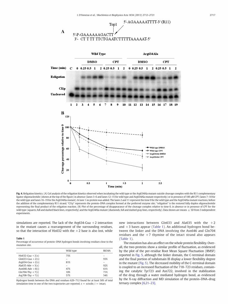
Fig. 4. Religation kinetics. (A) Gel analysis of the religation kinetics observedwhen incubating thewild type or the Arg634Alamutant-suicide cleavage complexwith the R11 complementaryligator oligonucleotide (shown at the top of the figure) in absence (lanes 3–6 and lanes 12–15 forwild type and Arg634Alamutant respectively) or in presence of 100 μMCPT (lanes 7–10 forthewild type and lanes 16–19 for theArg634Alamutant). In lane 1noproteinwas added. The lanes 2 and11 represent the time 0 for thewild type and theArg634Alamutant reactions, beforethe addition of the complementary R11 strand. “Cl1p” represents the protein–DNA complex formed at the preferred enzyme site; “religation” is the restored fully duplex oligonucleotiderepresenting the final product of the religation reaction. (B) Plot of the percentage of disappearance of the cleavage complex relative to time 0, in absence or in presence of CPT for thewild type (squares, full anddashed black lines, respectively) and theArg634Alamutant (diamonds, full anddashed gray lines, respectively). Data shown aremeans ± SD from3 independentexperiments.
2717I. D'Annessa et al. / Biochimica et Biophysica Acta 1834 (2013) 2712–2721
simulations are reported. The lack of the Arg634-Gua +2 interactionin the mutant causes a rearrangement of the surrounding residues,so that the interaction of His632 with the +2 base is also lost, while
Table 1Percentage of occurrence of protein–DNA hydrogen bonds involving residues close to themutation site.
Wild type R634A
His632-Gua +2(s) 75% –
Gln633-Gua +2(s) – 93%Arg634-Gua +2(s) 81% –
Ala635-Ade +3(s) – 92%Asn646-Ade +8(i) 47% 61%Gln704-Thy +7(i) 10% 71%Arg708-Thy +7(i) 57% 54%
Hydrogen bonds between the DNA and residues 629–712 found for at least 50% of totalsimulation time in one of the two trajectories are reported. s = scissile, i = intact.
new interactions between Gln633 and Ala635 with the +2and +3 bases appear (Table 1). An additional hydrogen bond be-tween the linker and the DNA involving the Asn646 and Gln704residues and the +7 thymine of the intact strand also appears(Table 1).
Themutation has also aneffect on thewhole proteinflexibility. Over-all, the two proteins show a similar profile of fluctuation, as evidencedby the plot of the per-residue Root Mean Square Fluctuation (RMSF)reported in Fig. 5, although the linker domain, the C-terminal domainand the final portion of subdomain III display a lower flexibility degreein the mutant (Fig. 5). The decreased mobility of the C-terminal domainis coupled to an increased fluctuation of the 718–723 residues, contain-ing the catalytic Tyr723 and Asn722, involved in the stabilizationof the drug through a water mediated hydrogen bond, as evidencedby the X-ray diffraction and MD simulation of the protein–DNA–drugternary complex [6,21–23].
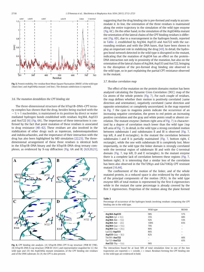
Fig. 5.Proteinmobility. Per-residueRootMean Square Fluctuation (RMSF) of thewild type(black line) and Arg634Ala mutant (red line). The domain subdivision is reported.
2718 I. D'Annessa et al. / Biochimica et Biophysica Acta 1834 (2013) 2712–2721
3.6. The mutation destabilizes the CPT binding site
The three-dimensional structure of the hTop1B–DNA–CPT terna-ry complex has shown that the drug, besides being stacked with the−1/+1 nucleotides, is maintained in its position by direct or watermediated hydrogen bonds established with residues Arg364, Asp533and Asn722 [6] (Fig. 6A). The importance of these interactions is con-firmed by the fact that point mutation of these residues is associatedto drug resistance [40–42]. These residues are also involved in thestabilization of other drugs such as topotecan, indenoisoquinolinesand indolocarbazoles, and the importance of their interaction with thedrug has also been highlighted by MD simulation [22,23]. The three-dimensional arrangement of these three residues is identical bothin the hTop1B–DNA binary and the hTop1B–DNA–drug ternary com-plexes, as evidenced by X-ray diffraction (Fig. 6A and B) [6,9,20,21],
Fig. 6. CPT binding site analysis. (A) hTop1B–DNA–CPT X-ray structure (PDB ID 1T8I),(B) hTop1B–DNA X-ray structure (PDB ID 1A31) and representative snapshot for (C) thewild type and (D) the Arg634Ala mutant simulation, of the CPT binding site residuesand of the DNA substrate. In (A) the CPT is also present.
suggesting that the drug binding site is pre-formed and ready to accom-modate it. In line, the orientation of the three residues is maintainedalong the entire trajectory in the simulation of the wild type enzyme(Fig. 6C). On the other hand, in the simulation of the Arg634Ala mutantthe orientation of the lateral chains of the CPT binding residues is differ-ent (Fig. 6D), due to a rearrangement in the hydrogen bonds, reportedin Table. 2, established by Arg364, Asp533 and Asn722 with the sur-rounding residues and with the DNA bases, that have been shown toplay an important role in stabilizing the drug [43]. In detail, the hydro-gen bond network detected in the wild type is disrupted in the mutant,indicating that the mutation of Arg634 has an effect on the protein–DNA interaction not only in proximity of the mutation, but also on theorientation of the lateral chains of Arg364, Asp533 andAsn722, bringingto the disruption of the pre-formed drug binding site observed inthe wild type, so in part explaining the partial CPT resistance observedin the mutant.
3.7. Residue correlation map
The effect of the mutation on the protein domains motion has beenanalyzed calculating the Dynamic Cross Correlation (DCC) map of theCα atoms of the whole protein (Fig. 7). For each couple of residues,the map defines whether their motion is positively correlated (samedirection and orientation), negatively correlated (same direction andopposite orientation) or completely uncorrelated. In the map reportedin Fig. 7 the cyan to magenta points indicate the occurrence of anincreasing negative correlation, the green to red points an increasingpositive correlation and the gray and white points small or absent cor-relation. The mutant enzyme (bottom right area of Fig. 7) is character-ized by a degree of correlation much lower than the wild type (topleft area of Fig. 7). In detail, in the wild type a strong correlated motionbetween subdomain I and subdomains II and III is observed (Fig. 7,top left, A and B rectangles). In the mutant the correlation betweensubdomains I and II is partially maintained (Fig. 7, bottom right, Crectangle), while the one with subdomain III is completely lost. Moreimportantly, in the wild type the linker domain is strongly correlatedwith the terminal region of subdomain III and with the C-terminaldomain (Fig. 7, top left, D and E rectangles). In the mutant enzymethere is a complete lack of correlation between these regions (Fig. 7,bottom right). It is interesting that a similar loss of the correlationhas been also observed in the Thr729Lys and Glu710Gly CPT resistantmutant [16,44].
The confinement of the motion of the linker, and of the wholemutated protein, in a reduced space is also evidenced by the analysisof the principal components of the motion (PCA). In the wild typeenzyme 60% of total motion is represented by the first 6 eigenvectorswhile in the mutant the same percentage is already covered by thefirst 3 eigenvectors. Projection of the motion along the plane formed
Table 2Percentage of occurrence of the hydrogen bonds involving residues composing the CPTbinding site in the wild type.
Wild type R634A
Arg364-Asp533 98% 57%Arg364-Cyt +1(i) 35% 68%Arg364-Ade−1(i) 92% 53%Arg364-Ade−2(i) 100% 83%Arg364-Gua +1(s) 70% –
Arg364-Gua +2(s) 80% –
Lys532-Asp533 86% –
Asp533-Gua−3(i) 82% –
Thr591-Asn722 – 60%Tyr723-Asn722 – 65%Asn722-Thy−1(s) 96% –
The interactions found for at least 50% of total simulation time in one of the twotrajectories are reported. s = scissile, i = intact. Residues forming the CPT binding sitein the wild type are evidenced in bold.
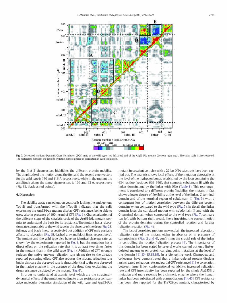
Fig. 7. Correlated motions. Dynamic Cross Correlation (DCC) map of the wild type (top left area) and of the Arg634Ala mutant (bottom right area). The color scale is also reported.The rectangles highlight the regions with the highest degree of correlation in each simulation.
2719I. D'Annessa et al. / Biochimica et Biophysica Acta 1834 (2013) 2712–2721
by the first 2 eigenvectors highlights the different protein mobility.The amplitude of themotion along the first and the second eigenvectorsfor thewild type is 170 and 116 Å, respectively, while in themutant theamplitude along the same eigenvectors is 109 and 93 Å, respectively(Fig. S2, black vs red points).
4. Discussion
The viability assay carried out on yeast cells lacking the endogenousTop1B and transformed with the hTop1B indicates that the cellsexpressing the Arg634Ala mutant display CPT resistance, being able togrow also in presence of 100 ng/ml of CPT (Fig. 1). Characterization ofthe different steps of the catalytic cycle of the Arg634Ala mutant per-mits to understand the basis for its resistance. The mutant has a relaxa-tion rate comparable to thewild type in the absence of the drug (Fig. 2B,full gray and black lines, respectively) but addition of CPT only partiallyaffects its relaxation (Fig. 2B, dashed gray and black lines, respectively).The mutant and the wild type also have an identical cleavage rate, asshown by the experiments reported in Fig. 3, but the mutation has adirect effect on the religation rate that it is at least two times fasterin the mutant than in the wild type (Fig. 4). Addition of CPT stronglyreduces the native enzyme religation rate giving rise to the alreadyreported poisoning effect. CPT also reduces the mutant religation ratebut in this case the observed rate is almost identical to the one observedin the native enzyme in the absence of the drug, thus explaining thedrug resistance displayed by the mutant (Fig. 4).
In order to understand at atomic level which are the structural–dynamical effects of the mutation leading to drug resistance a compar-ative molecular dynamics simulation of the wild type and Arg634Ala
mutant in covalent complex with a 22 bp DNA substrate have been car-ried out. The analysis shows local effects of the mutation detectable atthe level of the hydrogen bonds established by the loop containing the634 residue (residues 629–640), that connects subdomain III with thelinker domain, and by the linker with DNA (Table 1). This rearrange-ment is correlated to a different protein flexibility, the mutant in factshows a lower degree of flexibility at the level of the linker, C-terminaldomain and of the terminal region of subdomain III (Fig. 5) with aconsequent loss of motion correlation between the different proteindomains when compared to the wild type (Fig. 7). In detail, the linkerdomain loses the correlated motion with subdomain III and with theC-terminal domain when compared to the wild type (Fig. 7, comparetop left with bottom right areas), likely impairing the correct motionof the protein domains during the controlled rotation and furtherreligation reaction (Fig. 4).
The loss of correlatedmotionsmay explain the increased relaxation/religation rate of the mutant either in absence or in presence ofcamptothecin (Figs. 2 and 4), confirming the crucial role of the linkerin controlling the rotation/religation process [4]. The importance ofthis domain has been stated by several works carried out on a linker-deleted enzyme or on protein carrying point mutations at the level ofthe domain [11,13–15,18,19]. In a pioneering work Champoux andcolleagues have demonstrated that a linker-deleted protein displaysan increased religation rate and partial CPT resistance [11]. A correlationbetween large linker conformational variability, increased religationrate and CPT insensitivity has been reported for the single Ala653Promutation and more recently for a chimeric enzyme where the humanlinker has been substituted with plasmodial one [14,45]. CPT resistancehas been also reported for the Thr729Lys mutant, characterized by

2720 I. D'Annessa et al. / Biochimica et Biophysica Acta 1834 (2013) 2712–2721
comparable linker flexibility with the wild type, but where the linkerdomain loses motion correlation with the other protein domainsand in particular with the C-terminal domain containing the catalyticTyr723 residue [16,17]. In the here studied Arg634Ala mutant the sub-stitution brings to a reduction of the linker flexibility and to a completeloss of motion correlation between the linker and the C-terminal do-main (Figs. 5 and 7) and from the functional point of view to an in-creased relaxation and religation rate and to CPT resistance. This studythen confirms that perturbation of the linker has a direct effect onthe catalytic function and on the drug reactivity and we proposethat the occurrence of the motion correlation of the linker domainwith the C-terminal domain is a necessary requisite to have the “con-trolled rotation” and a CPT sensitive enzyme. The increased religationrate of the mutant (Fig. 4) coupled to an identical cleavage rate (Fig. 3)explains the in vivo CPT resistance since the number of hTop1–DNAcleavage complexes is lower in the yeast hosting the mutated than thewild type enzyme.
Finally, the presence of an alanine in position 634 also affects thethree-dimensional orientation of the Arg364, Asp533 and Asn722residues forming the CPT binding site in the wild type protein (Fig. 6).The different orientation of the lateral chains of these three residuescan be considered as an additional cause for a lowered stabilization ofthe CPT between the −1 and +1 bases, leading to partial resistance.
5. Conclusions
In conclusion, using a combined experimental and computationalapproach we have shown that mutation of arginine 634 to alanine ofhTop1B impairs the flexibility of the linker domain, affecting themotioncorrelation between the linker and the other protein domains presentin the wild type enzyme. The loss of interdomain communications iscorrelated to a faster religation rate and to partial drug resistance.
The combined biochemical and simulative approaches permits todemonstrate that mutation far from the active site affects the functionof the protein and its reactivity to the drug and disrupts protein domainmotion correlation, highlighting the importance of concerted domainsmotion for the correct function of the enzyme.
Supplementary data to this article can be found online at http://dx.doi.org/10.1016/j.bbapap.2013.09.017.
Acknowledgements
This work was supported by Associazione Italiana Ricerca Cancro(AIRC) with the project N. 10121. The authors thank Fabio Retico forthe help in obtaining the simulation of themutant. Calculations facilitiesto run the simulations where made available by Cineca Consortiumthrough the Iscra C project with code IscraC_Stra-Top.
References
[1] J.J. Champoux, DNA topoisomerase I-mediated nicking of circular duplex DNA,Methods Mol. Biol. 95 (2001) 81–87.
[2] J.C. Wang, Cellular roles of DNA topoisomerases: a molecular perspective, Nat. Rev.Mol. Cell. Biol. 3 (2002) 430–440.
[3] K.D. Corbett, J.M. Berger, Structure, molecular mechanisms, and evolutionaryrelationships in DNA topoisomerases, Annu. Rev. Biophys. Biomol. Struct. 33(2004) 95–118.
[4] L. Stewart, M.R. Redinbo, X. Qiu, W.G. Hol, J.J. Champoux, A model for the mecha-nism of human topoisomerase I, Science 279 (1998) 1534–1541.
[5] S. Castelli, A. Coletta, I. D'Annessa, P. Fiorani, C. Tesauro, A. Desideri, Interactionbetween natural compounds and human topoisomerase I, Biol. Chem. 393 (2012)1327–1340.
[6] B.L. Staker, M.D. Feese, M. Cushman, Y. Pommier, D. Zembower, L. Stewart, A.B.Burgin, Structures of three classes of anticancer agents bound to the humantopoisomerase I–DNA covalent complex, J. Med. Chem. 48 (2005) 2336–2345.
[7] Y. Pommier, Topoisomerase I inhibitors: camptothecins and beyond, Nat. Rev. Cancer6 (2006) 789–802.
[8] Y. Pommier, DNA topoisomerase I inhibitors: chemistry, biology, and interfacialinhibition, Chem. Rev. 109 (2009) 2894–2902.
[9] M.R. Redinbo, L. Stewart, P. Kuhn, J.J. Champoux, W.G. Hol, Crystal structuresof human topoisomerase I in covalent and noncovalent complexes with DNA,Science 279 (1998) 1504–1513.
[10] L. Stewart, G.C. Ireton, L.H. Parker, K.R. Madden, J.J. Champoux, Biochemical andbiophysical analyses of recombinant forms of human topoisomerase I, J. Biol.Chem. 271 (1996) 7593–7601.
[11] L. Stewart, G.C. Ireton, J.J. Champoux, A functional linker in human topoisomerase Iis required for maximum sensitivity to camptothecin in a DNA relaxation assay,J. Biol. Chem. 274 (1999) 32950–32960.
[12] G. Chillemi, P. Fiorani, P. Benedetti, A. Desideri, Protein concerted motionsin the DNA–human topoisomerase I complex, Nucleic Acids Res. 31 (2003)1525–1535.
[13] G. Chillemi, M. Redinbo, A. Bruselles, A. Desideri, Role of the linker domain and the203–214 N-terminal residues in the human topoisomerase I DNA complex dynamics,Biophys. J. 87 (2004) 4087–4097.
[14] P. Fiorani, A. Bruselles, M. Falconi, G. Chillemi, A. Desideri, P. Benedetti, Single muta-tion in the linker domain confers protein flexibility and camptothecin resistance tohuman topoisomerase I, J. Biol. Chem. 278 (2003) 43268–43275.
[15] P. Fiorani, C. Tesauro, G. Mancini, G. Chillemi, I. D'Annessa, G. Graziani, L.Tentori, A. Muzi, A. Desideri, Evidence of the crucial role of the linker domainon the catalytic activity of human topoisomerase I by experimental and simu-lative characterization of the Lys681Ala mutant, Nucleic Acids Res. 37 (2009)6849–6858.
[16] G. Chillemi, I. D'Annessa, P. Fiorani, C. Losasso, P. Benedetti, A. Desideri, Thr729 inhuman topoisomerase I modulates anti-cancer drug resistance by altering proteindomain communications as suggested by molecular dynamics simulations, NucleicAcids Res. 36 (2008) 5645–5651.
[17] C. Losasso, E. Cretaio, P. Fiorani, I. D'Annessa, G. Chillemi, P. Benedetti, A singlemutation in the 729 residue modulates human DNA topoisomerase IB DNA bindingand drug resistance, Nucleic Acids Res. 36 (2008) 5635–5644.
[18] I. D'Annessa, C. Tesauro, P. Fiorani, G. Chillemi, S. Castelli, O. Vassallo, G. Capranico, A.Desideri, Role of flexibility in protein–DNA–drug recognition: the case ofAsp677Gly-Val703Ile topoisomerase mutant hypersensitive to camptothecin, J.Amino Acids 2012 (2012) 206083.
[19] C. Gongora, N. Vezzio-Vie, S. Tuduri, V. Denis, A. Causse, C. Auzanneau, G.Collod-Beroud, A. Coquelle, P. Pasero, P. Pourquier, P. Martineau, M. Del Rio, Newtopoisomerase I mutations are associated with resistance to camptothecin, Mol.Cancer 10 (2011) 64.
[20] M.R. Redinbo, L. Stewart, J.J. Champoux, W.G. Hol, Structural flexibility in humantopoisomerase I revealed in multiple non-isomorphous crystal structures, J. Mol.Biol. 292 (1999) 685–696.
[21] B.L. Staker, K. Hjerrild, M.D. Feese, C.A. Behnke, A.B. Burgin Jr., L. Stewart, The mech-anism of topoisomerase I poisoning by a camptothecin analog, Proc. Natl. Acad. Sci.U. S. A. 99 (2002) 15387–15392.
[22] G. Mancini, I. D'Annessa, A. Coletta, G. Chillemi, Y. Pommier, M. Cushman, A.Desideri, Binding of an indenoisoquinoline to the topoisomerase–DNA complex in-duces reduction of linker mobility and strengthening of protein–DNA interaction,PLoS One (2013)(in press).
[23] G. Mancini, I. D'Annessa, A. Coletta, N. Sanna, G. Chillemi, A. Desideri, Structuraland dynamical effects induced by the anticancer drug topotecan on the humantopoisomerase I–DNA complex, PLoS One 5 (2010) e10934.
[24] M.A. Bjornsti, P. Benedetti, G.A. Viglianti, J.C. Wang, Expression of human DNAtopoisomerase I in yeast cells lacking yeast DNA topoisomerase I: restoration ofsensitivity of the cells to the antitumor drug camptothecin, Cancer Res. 49 (1989)6318–6323.
[25] C. Kaiser, S. Michaelis, A. Mitchell, Lithium Acetate Yeast Transformation, ColdSpring Harbor Laboratory Press, New York, 1994. 133–134.
[26] A.H. Andersen, E. Gocke, B.J. Bonven, O.F. Nielsen, O. Westergaard, Topoisomerase Ihas a strong binding preference for a conserved hexadecameric sequence in thepromoter region of the rRNA gene from Tetrahymena pyriformis, Nucleic Acids Res.13 (1985) 1543–1557.
[27] D.A. Case, T.E. Cheatham 3rd, T. Darden, H. Gohlke, R. Luo, K.M. Merz Jr., A. Onufriev,C. Simmerling, B. Wang, R.J. Woods, The Amber biomolecular simulation programs,J. Comput. Chem. 26 (2005) 1668–1688.
[28] W.L. Jorgensen, J. Chandrasekhar, J.D. Madura, R.W. Impery, M.L. Klein, Comparisonof simple potential functions for simulating liquid water, J. Chem. Phys. 79 (1983)926–935.
[29] Y. Duan, C. Wu, S. Chowdhury, M.C. Lee, G. Xiong, W. Zhang, R. Yang, P. Cieplak, R.Luo, T. Lee, J. Caldwell, J. Wang, P. Kollman, A point-charge force field for molecularmechanics simulations of proteins based on condensed-phase quantummechanicalcalculations, J. Comput. Chem. 24 (2003) 1999–2012.
[30] B. Hess, C. Kutzner, van der Spoel D. , E. Lindahl, GROMACS 4: algorithm for highlyefficient, load-balanced, and scalable molecular simulation, J. Chem. Theory Comput.4 (2008) 435–447.
[31] A.W. Sousa da Silva, W.F. Vranken, ACPYPE— AnteChamber PYthon Parser interfacE,BMC res. notes 5 (2012) 367.
[32] T.E. Cheatham, J.L. Miller, T. Fox, T.A. Darden, P.A. Kollman, Molecular-dynamicssimulations on solvated biomolecular systems — the particle mesh Ewald methodleads to stable trajectories of DNA, RNA, and proteins, J. Am. Chem. Soc. 117(1995) 4193–4194.
[33] J.P. Ryckaert, G. Ciccotti, H.J.C. Berendsen, Numerical integration of the Cartesianequations of motion of a system with constraints: molecular dynamics of n-alkanes,J. Comput. Phys. 23 (1977) 327–341.
[34] H.J.C. Berendsen, J.P.M. Pstma, W.F. van Gusteren, A. Di Nola, J.R. Haak,Molecular dynamics with coupling to an external bath, J. Comput. Phys. 81(1984) 3684–3690.

2721I. D'Annessa et al. / Biochimica et Biophysica Acta 1834 (2013) 2712–2721
[35] M. Parrinello, A. Rahman, Polymorphic transitions in single-crystals — a newmolecular-dynamics method, J. Appl. Phys. (1981) 7182–7190.
[36] W. Humphrey, A. Dalke, K. Schulten, VMD: visual molecular dynamics, J. Mol. Graph.14 (1996) 33–38(27–38).
[37] M.A. Bjornsti, A.M. Knab, P. Benedetti, Yeast Saccharomyces cerevisiae as a modelsystem to study the cytotoxic activity of the antitumor drug camptothecin, CancerChemother. Pharmacol. 34 Suppl. (1994) S1–S5.
[38] N. Kim, S.N. Huang, J.S. Williams, Y.C. Li, A.B. Clark, J.E. Cho, T.A. Kunkel, Y. Pommier,S. Jinks-Robertson, Mutagenic processing of ribonucleotides in DNA by yeasttopoisomerase I, Science 332 (2011) 1561–1564.
[39] H. Interthal, P.M. Quigley, W.G. Hol, J.J. Champoux, The role of lysine 532 in thecatalytic mechanism of human topoisomerase I, J. Biol. Chem. 279 (2004)2984–2992.
[40] A. Tanizawa, R. Beitrand, G. Kohlhagen, A. Tabuchi, J. Jenkins, Y. Pommier, Clon-ing of Chinese hamster DNA topoisomerase I cDNA and identification of a singlepoint mutation responsible for camptothecin resistance, J. Biol. Chem. 268(1993) 25463–25468.
[41] X.G. Li, P. Haluska Jr., Y.H. Hsiang, A.K. Bharti, D.W. Kufe, L.F. Liu, E.H. Rubin, Involve-ment of amino acids 361 to 364 of human topoisomerase I in camptothecin resis-tance and enzyme catalysis, Biochem. Pharmacol. 53 (1997) 1019–1027.
[42] J.E. Chrencik, B.L. Staker, A.B. Burgin, P. Pourquier, Y. Pommier, L. Stewart, M.R.Redinbo, Mechanisms of camptothecin resistance by human topoisomerase I muta-tions, J. Mol. Biol. 339 (2004) 773–784.
[43] A. Coletta, A. Desideri, Role of the protein in the DNA sequence specificityof the cleavage site stabilized by the camptothecin topoisomerase IB inhibitor: ametadynamics study, Nucleic Acids Res. (2013).
[44] C. Tesauro, B. Morozzo Della Rocca, A. Ottaviani, A. Coletta, L. Zuccaro, B. Arno, I.D'Annessa, P. Fiorani, A. Desideri, Molecular mechanism of the camptothecinresistance of Glu710Gly topoisomerase IB mutant analyzed in vitro and in silico,Mol. Cancer 12 (2013) 100.
[45] B. Arnò, I. D'Annessa, C. Tesauro, L. Zuccaro, A. Ottaviani, B. Knudsen, P. Fiorani, A.Desideri1, Replacement of the human topoisomerase linker domain with theplasmodial counterpart renders the enzyme camptothecin resistant, PLoS One 8(2013) e68404.
Copyright © 2022 FDOKUMEN