Application of a novel microtitre plate-based assay for the discovery of new inhibitors of DNA...
14
Application of a Novel Microtitre Plate-Based Assay for the Discovery of New Inhibitors of DNA Gyrase and DNA Topoisomerase VI James A. Taylor ¤ , Lesley A. Mitchenall, Martin Rejzek, Robert A. Field, Anthony Maxwell* Department of Biological Chemistry, John Innes Centre, Colney Lane, Norwich, United Kingdom Abstract DNA topoisomerases are highly exploited targets for antimicrobial drugs. The spread of antibiotic resistance represents a significant threat to public health and necessitates the discovery of inhibitors that target topoisomerases in novel ways. However, the traditional assays for topoisomerase activity are not suitable for the high-throughput approaches necessary for drug discovery. In this study we validate a novel assay for screening topoisomerase inhibitors. A library of 960 compounds was screened against Escherichia coli DNA gyrase and archaeal Methanosarcina mazei DNA topoisomerase VI. Several novel inhibitors were identified for both enzymes, and subsequently characterised in vitro and in vivo. Inhibitors from the M. mazei topoisomerase VI screen were tested for their ability to inhibit Arabidopsis topoisomerase VI in planta. The data from this work present new options for antibiotic drug discovery and provide insight into the mechanism of topoisomerase VI. Citation: Taylor JA, Mitchenall LA, Rejzek M, Field RA, Maxwell A (2013) Application of a Novel Microtitre Plate-Based Assay for the Discovery of New Inhibitors of DNA Gyrase and DNA Topoisomerase VI. PLoS ONE 8(2): e58010. doi:10.1371/journal.pone.0058010 Editor: Arkady B. Khodursky, University of Minnesota, United States of America Received December 28, 2012; Accepted January 29, 2013; Published February 26, 2013 Copyright: ß 2013 Taylor et al. This is an open-access article distributed under the terms of the Creative Commons Attribution License, which permits unrestricted use, distribution, and reproduction in any medium, provided the original author and source are credited. Funding: JAT was supported by a BBSRC-CASE studentship funded by the Biotechnological and Biological Science Research Council and Plant Bioscience Ltd; the work was supported by grant BB/J004561/1 from BBSRC and the John Innes Foundation. The funders had no role in study design, data collection and analysis, decision to publish, or preparation of the manuscript. Competing Interests: In relation funding from a commercial source (PBL), this has had no influence on the publication of this work and that this does not alter the authors’ adherence to all the PLOS ONE policies on sharing data and materials. * E-mail: [email protected] ¤ Current address: Dept. Biochemistry, University of Bristol, Bristol, United Kingdom. Introduction The emergence of pathogenic bacterial strains resistant to currently available antimicrobial agents is a universal problem of mounting importance [1–3]. Resistance mechanisms have been reported for all known classes of antibiotics with some strains exhibiting multiple resistance phenotypes, which is a consequence of natural selection and human mismanagement [4]. The danger that these strains pose is demonstrated by the increased mortality and morbidity rates for infected patients when compared to those infected with susceptible strains [5,6]. Unfortunately this increase in resistance has not been met with an increase in the development of new antibiotics, with the total number of new drugs being brought to market actually decreasing [7]. Clearly there is an urgent need for the development of new antibiotics and management strategies. Extensive attempts to validate new target enzymes for antimicrobials have met with little success [8], with the majority of successful drugs inhibiting a handful of cellular processes. One of the most successfully exploited drug targets is the DNA topoisomerase (topo) class of enzymes [9–12]. DNA topoisomer- ases are essential and ubiquitous enzymes responsible for controlling the topological state of DNA [13]. This is accom- plished by the reaction of an active-site tyrosine with the phosphate backbone of the DNA to generate a covalent intermediate (the so-called ‘cleavage complex’), followed by either strand passage of another segment of DNA or free rotation of the broken strand [14–17]. DNA topoisomerases are classified as either type I or type II based on whether they cleave one or both strands of the DNA [18], and further subdivided into IA, IB, IC, IIA or IIB based on structural and mechanistic differences [19]. The essential nature of these enzymes and the vulnerability of the cleavage complex, which, if stabilised, rapidly results in cell death, make them ideal drug targets. The type IIA topoisomerases have been the most exploited class, acting as targets for many anticancer and antibacterial drugs. DNA gyrase is a type IIA topoisomerase of particular importance due to it being a target for numerous antibacterial drugs and its distinct mechanism. All type IIA topoisomerases are capable of removing supercoils from DNA (relaxation) in an ATP-dependent manner [20]; gyrase introduces negative supercoils into DNA in the presence of ATP, but relaxes DNA when ATP is absent [21]. Whereas eukaryotic type IIA topoisomerases are dimeric in nature, gyrase forms a heterotetramer of two GyrB subunits, which contain the ATPase domains, and two GyrA subunits, which contain the active-site tyrosines [22]. During the reaction cycle, the segment of DNA to be cleaved (the ‘gate’ or ‘G’ segment) binds to the DNA-binding saddle in GyrA. ATP binding causes the GyrB subunits to dimerise and capture a second segment of DNA (the ‘transported’ or ‘T’ segment) [23]. The G segment is then cleaved and the break pried open by conformational changes, allowing the T segment to pass through. The G segment can then be religated. The differences in mechanism and structure between PLOS ONE | www.plosone.org 1 February 2013 | Volume 8 | Issue 2 | e58010
-
Upload
independent -
Category
Documents
-
view
0 -
download
0
Transcript of Application of a novel microtitre plate-based assay for the discovery of new inhibitors of DNA...
Application of a Novel Microtitre Plate-Based Assay forthe Discovery of New Inhibitors of DNA Gyrase and DNATopoisomerase VIJames A. Taylor¤, Lesley A. Mitchenall, Martin Rejzek, Robert A. Field, Anthony Maxwell*
Department of Biological Chemistry, John Innes Centre, Colney Lane, Norwich, United Kingdom
Abstract
DNA topoisomerases are highly exploited targets for antimicrobial drugs. The spread of antibiotic resistance represents asignificant threat to public health and necessitates the discovery of inhibitors that target topoisomerases in novel ways.However, the traditional assays for topoisomerase activity are not suitable for the high-throughput approaches necessaryfor drug discovery. In this study we validate a novel assay for screening topoisomerase inhibitors. A library of 960compounds was screened against Escherichia coli DNA gyrase and archaeal Methanosarcina mazei DNA topoisomerase VI.Several novel inhibitors were identified for both enzymes, and subsequently characterised in vitro and in vivo. Inhibitorsfrom the M. mazei topoisomerase VI screen were tested for their ability to inhibit Arabidopsis topoisomerase VI in planta.The data from this work present new options for antibiotic drug discovery and provide insight into the mechanism oftopoisomerase VI.
Citation: Taylor JA, Mitchenall LA, Rejzek M, Field RA, Maxwell A (2013) Application of a Novel Microtitre Plate-Based Assay for the Discovery of New Inhibitors ofDNA Gyrase and DNA Topoisomerase VI. PLoS ONE 8(2): e58010. doi:10.1371/journal.pone.0058010
Editor: Arkady B. Khodursky, University of Minnesota, United States of America
Received December 28, 2012; Accepted January 29, 2013; Published February 26, 2013
Copyright: � 2013 Taylor et al. This is an open-access article distributed under the terms of the Creative Commons Attribution License, which permitsunrestricted use, distribution, and reproduction in any medium, provided the original author and source are credited.
Funding: JAT was supported by a BBSRC-CASE studentship funded by the Biotechnological and Biological Science Research Council and Plant Bioscience Ltd; thework was supported by grant BB/J004561/1 from BBSRC and the John Innes Foundation. The funders had no role in study design, data collection and analysis,decision to publish, or preparation of the manuscript.
Competing Interests: In relation funding from a commercial source (PBL), this has had no influence on the publication of this work and that this does not alterthe authors’ adherence to all the PLOS ONE policies on sharing data and materials.
* E-mail: [email protected]
¤ Current address: Dept. Biochemistry, University of Bristol, Bristol, United Kingdom.
Introduction
The emergence of pathogenic bacterial strains resistant to
currently available antimicrobial agents is a universal problem of
mounting importance [1–3]. Resistance mechanisms have been
reported for all known classes of antibiotics with some strains
exhibiting multiple resistance phenotypes, which is a consequence
of natural selection and human mismanagement [4]. The danger
that these strains pose is demonstrated by the increased mortality
and morbidity rates for infected patients when compared to those
infected with susceptible strains [5,6]. Unfortunately this increase
in resistance has not been met with an increase in the development
of new antibiotics, with the total number of new drugs being
brought to market actually decreasing [7]. Clearly there is an
urgent need for the development of new antibiotics and
management strategies.
Extensive attempts to validate new target enzymes for
antimicrobials have met with little success [8], with the majority
of successful drugs inhibiting a handful of cellular processes. One
of the most successfully exploited drug targets is the DNA
topoisomerase (topo) class of enzymes [9–12]. DNA topoisomer-
ases are essential and ubiquitous enzymes responsible for
controlling the topological state of DNA [13]. This is accom-
plished by the reaction of an active-site tyrosine with the
phosphate backbone of the DNA to generate a covalent
intermediate (the so-called ‘cleavage complex’), followed by either
strand passage of another segment of DNA or free rotation of the
broken strand [14–17]. DNA topoisomerases are classified as
either type I or type II based on whether they cleave one or both
strands of the DNA [18], and further subdivided into IA, IB, IC,
IIA or IIB based on structural and mechanistic differences [19].
The essential nature of these enzymes and the vulnerability of the
cleavage complex, which, if stabilised, rapidly results in cell death,
make them ideal drug targets.
The type IIA topoisomerases have been the most exploited
class, acting as targets for many anticancer and antibacterial drugs.
DNA gyrase is a type IIA topoisomerase of particular importance
due to it being a target for numerous antibacterial drugs and its
distinct mechanism. All type IIA topoisomerases are capable of
removing supercoils from DNA (relaxation) in an ATP-dependent
manner [20]; gyrase introduces negative supercoils into DNA in
the presence of ATP, but relaxes DNA when ATP is absent [21].
Whereas eukaryotic type IIA topoisomerases are dimeric in
nature, gyrase forms a heterotetramer of two GyrB subunits,
which contain the ATPase domains, and two GyrA subunits,
which contain the active-site tyrosines [22]. During the reaction
cycle, the segment of DNA to be cleaved (the ‘gate’ or ‘G’ segment)
binds to the DNA-binding saddle in GyrA. ATP binding causes
the GyrB subunits to dimerise and capture a second segment of
DNA (the ‘transported’ or ‘T’ segment) [23]. The G segment is
then cleaved and the break pried open by conformational changes,
allowing the T segment to pass through. The G segment can then
be religated. The differences in mechanism and structure between
PLOS ONE | www.plosone.org 1 February 2013 | Volume 8 | Issue 2 | e58010
gyrase and eukaryotic topoisomerases, as well as its low homology
to human type IIA topoisomerases, have allowed the development
of bactericidal drugs that target bacterial topoisomerases with a
high degree of specificity.
The mechanism of action for DNA gyrase inhibitors is highly
varied, with different chemical families inhibiting different steps in
the reaction cycle [12]. The most successful class of gyrase
inhibitors is the ‘‘billion-dollar’’ quinolone family of drugs.
Quinolones have the potent ability to stabilise the cleavage
complex of DNA gyrase, resulting in double-strand breaks and cell
death [24,25]. The exact mechanism by which this occurs remains
unclear, but several crystal structures of quinolones bound to
gyrase or its sister enzyme topo IV have been published [26–29].
These structures suggest that quinolones bind in pockets near the
active-site tyrosines while simultaneously intercalating with the
cleaved DNA, presumably distorting it in such a way as to prevent
religation. In contrast, the aminocoumarin class of inhibitors target
the ATPase activity of the enzyme in a competitive manner,
binding in a pocket that overlaps with the ATP-binding site and
sterically hindering nucleotide binding [30]. Unfortunately these
compounds possess unfavourable pharmacokinetics and produce
too many side effects to be effective clinical antimicrobials [31,32].
The simocyclinone class of inhibitors prevents the binding of the
enzyme to DNA by a novel ‘‘double-warhead’’ mechanism
[33,34]. These compounds consist of an aminocoumarin group
linked to a polyketide group by a long linker, both groups bind to
two separate sites in the DNA-binding saddle of GyrA. A crystal
structure of the drug bound to GyrA shows two GyrA dimers
being cross-linked by four drug molecules, occluding the DNA-
binding saddle [34]. However an alternative model, supported by
mass spectrometry data, has been proposed in which one molecule
of SD8 bridges the two sites on the same GyrA subunit, hindering
DNA binding [35].
The type IIB family of topoisomerases has similarities to the
type IIA family, but has significant structural and mechanistic
differences. The family currently consists of a single enzyme: topo
VI. Similar to the bacterial type IIA enzymes topo VI is an A2B2
heterotetramer, with the ATPase sites located on the B subunits
and the active-site tyrosines located on the A subunit [36,37]. The
A subunits of topo VI lack some of the extensive protein-protein
interactions of the type IIA enzymes. In addition, whereas the type
IIAs are capable of cleaving DNA in the absence of ATP, ATP
hydrolysis and DNA cleavage have been shown to be tightly
coupled during the reaction cycle of topo VI from the
thermophilic archaea Sulfolobus shibatae [38]. It has been suggested
that these two features evolved in parallel, with the reduction in
protein-protein interactions necessitating tighter coupling between
strand cleavage and ATP binding in order to prevent accidental
double-strand lesion formation [23].
In contrast to the type IIA topoisomerases, the type IIB family
has received relatively little attention, being regarded for many
years an ‘‘archaeal curiosity’’. However the discovery of topo VI
homologues in plants [39–41] and the malaria parasite Plasmodium
falciparum [42] opens the possibility for this enzyme being used as a
target for herbicides or anti-malarial agents. In plants, topo VI has
been shown to play a key role in the process of endoreduplication
[39,40], in which the plant cell replicates its genome several times
without dividing, accompanied by cell expansion. Knock-out
mutants of topo VI in Arabidopsis thaliana have an extreme dwarf
phenotype, yellowish leaves, reduced trichome (‘‘leaf hair’’) size,
reduced chromosome counts (ploidy) and reduced root hair size
and frequency [39,40]. These plants die after 4–5 weeks of
growth, which indicates the essential nature of topo VI in plants.
Neither plant nor malarial topo VI have been successfully
purified, but extensive work has been carried out on the Sulfolobus
shibatae orthologue of the enzyme. Unlike the type IIA topoisom-
erases, high-resolution crystal structures of the entire A2B2
complex have been solved for S. shibatae topo VI and the topo
VI from the methanogenic archeon Methanosarcina mazei [36,37]. A
few inhibitors have been identified for the S. shibatae enzyme,
including the Hsp90 inhibitor radicicol, which has been shown to
inhibit the ATPase activity of the enzyme [43]. In addition, several
inhibitors of eukaryotic topo II have been shown to inhibit S.
shibatae topo VI, although their mechanism of action has not been
determined [44]. Novel inhibitors targeting topo VI will be
invaluable as probes of the enzyme’s mechanism and may well
provide the basis for novel chemotherapeutics.
Despite their great potential as drug targets, high-throughput
screening for inhibitors of topoisomerases has been limited by the
traditional assays for topoisomerase activity, which are poorly
suited to rapidly processing large numbers of reactions in a
quantitative fashion. We have previously described a novel
microtitre plate-based assay that allows the processing of a large
number of reactions simultaneously and has the potential to be
automated [45–47]. This assay is based on the observation that
supercoiled plasmids form intermolecular DNA triplexes more
readily than relaxed plasmids. A triplex-forming oligonucleotide is
immobilised on a microtitre plate surface and used to capture
supercoiled plasmids from solution, which can be subsequently
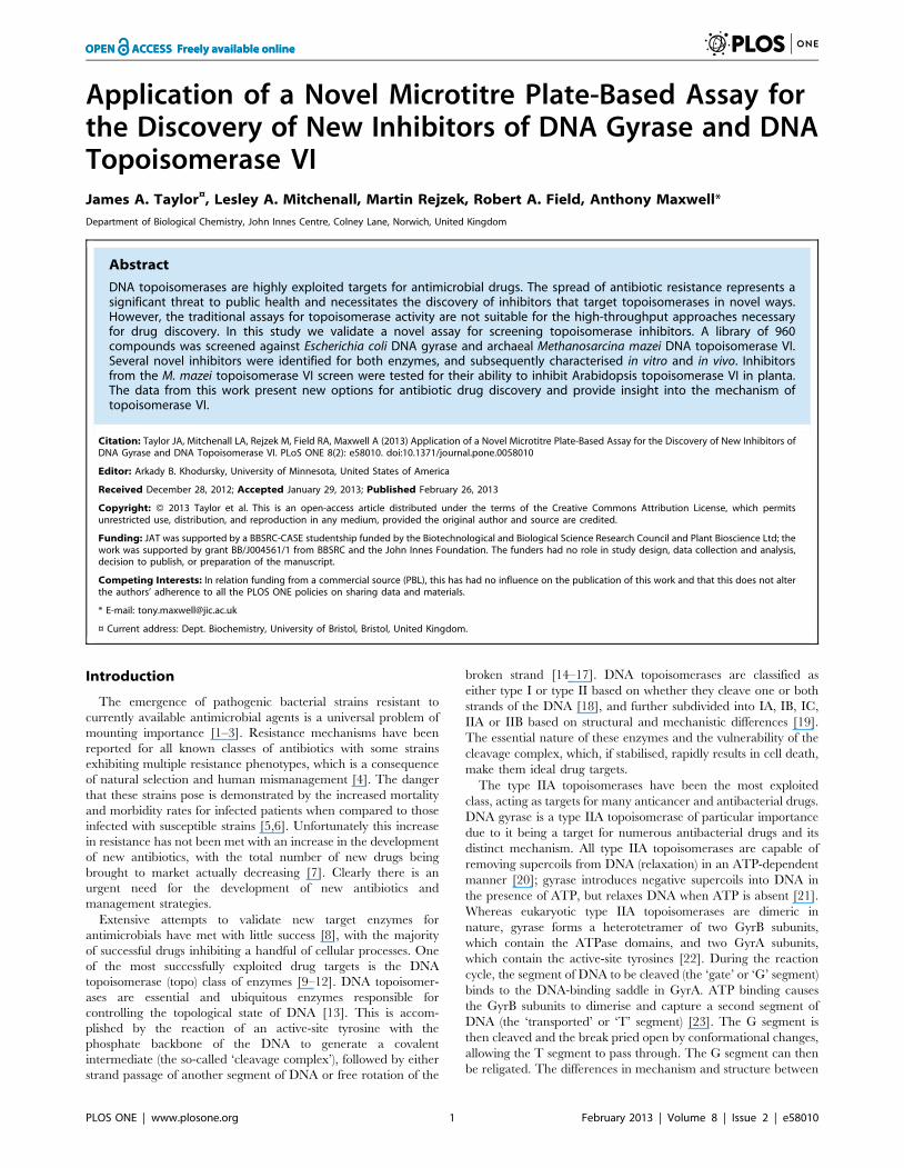
Figure 1. Screening the Microsource GenPlus chemical libraryagainst E. coli DNA gyrase and M. mazei DNA topoisomerase VI.A. Results from the DNA gyrase screen. B. Results from thetopoisomerase VI screen. Each bar represents the average percentageinhibition for a compound screened in duplicate. Compound bars havebeen coloured to indicate 96-well plate groupings. The arbitrary hitthreshold of 25% inhibition is demonstrated by the dotted lines.doi:10.1371/journal.pone.0058010.g001
New Inhibitors of Gyrase and Topoisomerase VI
PLOS ONE | www.plosone.org 2 February 2013 | Volume 8 | Issue 2 | e58010
stained with a fluorescent dye. This in turn allows for the
supercoiling or relaxation activity of topoisomerases to be
monitored.
Although this assay has been validated in a low-throughput
context, its efficacy in a high-throughput context has yet to be
reported. We have conducted a proof-of-principle screen of a
library of 960 compounds, consisting of 80% FDA-approved drugs
and 20% natural products, against DNA gyrase from E. coli and
M. mazei topo VI. It was expected that by screening against
compounds already shown to interact with biological molecules
the screen would achieve a higher hit rate than with a random
library [48–50]. M. mazei topo VI was selected over the S. shibatae
orthologue since the thermophilic nature of the S. shibatae enzyme
makes it a potentially less relevant model for eukaryotic topo VI
enzymes. Furthermore, few inhibitors have been so far discovered
for this enzyme. The mechanisms of action of the hits from both
screens were explored in vitro, and their antibiotic properties
assessed in cell-based assays against Gram-positive and Gram-
negative bacteria, for the DNA gyrase hits, or against Arabidopsis
thaliana, for the topo VI hits.
Results
Screening a chemical library against E. coli gyrase and M.mazei topoisomerase VI
Both screens were conducted in duplicate over three days at a
compound concentration of 25 mM (Figure 1). A hit threshold of
25% inhibition was set (based upon our previous unpublished
data), and any compounds exceeding this limit were validated
using agarose gel assays (DNA supercoiling or relaxation). The
quality of the screening data was determined by calculating the
mean fluorescent signals and standard deviations for the 192
negative (DNA alone) and 192 positive (DNA plus enzyme with no
drug) controls in each screen. The signal-to-background ratio was
calculated to be 5 for the gyrase screen and 4 for the topo VI
screen, while the signal-to-noise ratio was 10 for the gyrase screen
and 15 for the topo VI screen. The overall quality of the data for
both screens was good, with both having an average Z’ factor of
above 0.5 and no obvious patterns in the data [51]. The average of
the Z’ factors for the twelve plates was calculated to be 0.64 for the
gyrase screen and 0.69 for the topo VI screen, indicating that there
was a good degree of separation between the positive and negative
controls and implying a good overall quality of the data. The
distribution of the Z’ factor for each plate around the mean was
close, with no single plate giving a value below 0.4.
In the gyrase inhibitor screen, 22 compounds scored over the hit
threshold (Table 1 and Figure 2). The majority of these were
already characterised as DNA gyrase inhibitors including a
number of fluoroquinolones, novobiocin and acriflavinium
[12,52], which were not studied further. Out of the remaining
hits, mitoxantrone and suramin displayed inhibition while the
other 9 compounds tested did not significantly affect DNA gyrase
activity in the gel-based supercoiling assay. This gave 13 validated
hits, resulting in a hit rate of 1.35% and a novel hit rate of 0.21%.
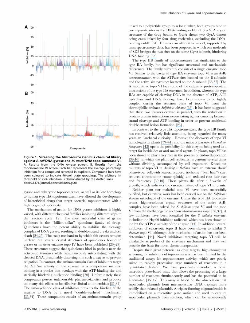
Figure 2. Structures of screen hits. The M. mazei topo VI inhibitors are circled in blue on the left and the DNA gyrase inhibitors are circled inpurple on the right. The 11 already well characterised inhibitors of DNA gyrase identified by the screen are not shown.doi:10.1371/journal.pone.0058010.g002
New Inhibitors of Gyrase and Topoisomerase VI
PLOS ONE | www.plosone.org 3 February 2013 | Volume 8 | Issue 2 | e58010
Four known gyrase inhibitors were missed by the screen: nalidixic
acid, cinoxacin, oxolinic acid and enoxacin. These false negatives
were likely to be due to low inhibitor potency.
For the M. mazei topo VI screen, 9 compounds that exceeded
the hit threshold were selected for further study (Figure 1B). Out of
these only m-amsacrine had been previously reported as an
inhibitor of topo VI [53], and only against the S. shibatae enzyme.
Six of these 9 were validated as hits in the gel-based relaxation
assay: m-amsacrine, suramin, hexylresorcinol, 9-aminoacridine,
purpurin and quinacrine (Figure 2). This gave a hit rate of 0.63%.
Of these compounds m-amsacrine, suramin and quinacrine have
been previously shown to inhibit type IIA topoisomerases [54–56],
whilst purpurin and 9-aminoacridine are structurally related to
known topo II inhibitors (mitoxantrone and m-amsacrine respec-
tively). Mitoxantrone was subsequently found to inhibit M. mazei
topo VI, this was missed during the initial screen due to its
disruptive effects on DNA triplex formation; this illustrates a
potential limitation of this assay. No false negatives were identified
but since there are few known inhibitors described for topo VI, this
analysis was less informative than with the DNA gyrase screen.
Apart from m-amsacrine, none of the other previously described
inhibitors [44,57] were present in the library.
Mitoxantrone and suramin are novel inhibitors of E. coligyrase
Out of the 13 hits identified in the gyrase screen, two were novel
gyrase inhibitors: mitoxantrone and suramin. Both of these
compounds have previously been shown to have activity against
eukaryotic topo II [55,58], but they had not previously been
shown to be active against DNA gyrase. The IC50 values for these
compounds against E. coli DNA gyrase were determined to be
12 mM for mitoxantrone and 80 mM for suramin in the gel assay
(Figures 3A and 3B).
Mitoxantrone is from the anthraquinone class of drugs and is
currently used as an antineoplastic agent [59]. It is thought to
inhibit topo II by stabilisation of the DNA-cleavage intermediate,
leading to generation of double-stranded breaks in DNA [58]. To
determine if this mode of action is the same for its inhibition of
gyrase, a gel-based DNA cleavage assay was conducted under
conditions which reveal formation of the cleavage intermediate
(Figure 3C). It was observed that mitoxantrone strongly induced
DNA cleavage by DNA gyrase at 10 mM, comparable to the
known cleavage-intermediate stabiliser ciprofloxacin, showing that
mitoxantrone stabilises the cleavage complex of gyrase as well as
topo II. This is likely to be due to the drug intercalating at or near
the DNA break sites generated in the cleavage complex in both
enzymes. It also appears that at 100 mM the drug’s ability to
stabilise the cleavage complex is reduced; this is probably due to its
binding to DNA and inhibiting enzyme binding. Suramin, on the
other hand did not display any ability to induce cleavage.
Suramin is an anti-protozoal drug that has been subjected to
clinical trials for the treatment of several forms of cancer [60].
Although it has been shown to protect against cleavage of DNA by
topo II induced by cleavage-intermediate stabilising agents [55], its
exact mode of inhibition has yet to be determined [61]. The ability
of suramin to protect DNA from gyrase-induced cleavage was
tested (Figure 3D). To eliminate the possibility of drug-drug
interactions, Ca2+ was used to induce cleavage by DNA gyrase
[62]. Suramin at 80 mM was able to completely protect DNA from
cleavage by gyrase in the presence of 4 mM calcium chloride,
indicating that its mode of action is similar to that found with topo
II and is independent of drug-drug interactions.
To determine if the drug was protecting from cleavage by
preventing binding of the protein to DNA, a native gel-shift assay
to measure the binding of DNA gyrase to a 147 bp DNA fragment
in the presence or absence of 100 mM suramin was carried out
(Figure 3E). In the absence of suramin, the conversion of free DNA
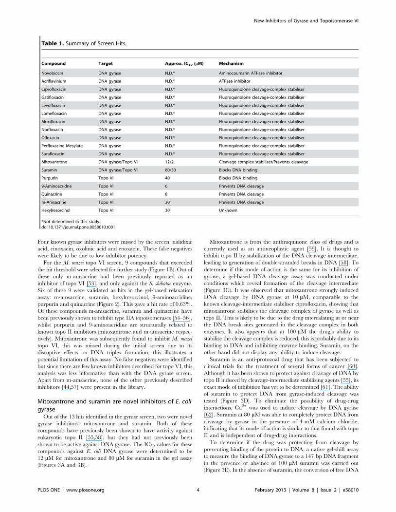
Table 1. Summary of Screen Hits.
Compound Target Approx. IC50 (mM) Mechanism
Novobiocin DNA gyrase N.D.* Aminocoumarin ATPase inhibitor
Acriflavinium DNA gyrase N.D.* ATPase inhibitor
Ciprofloxacin DNA gyrase N.D.* Fluoroquinolone cleavage-complex stabiliser
Gatifloxacin DNA gyrase N.D.* Fluoroquinolone cleavage-complex stabiliser
Levofloxacin DNA gyrase N.D.* Fluoroquinolone cleavage-complex stabiliser
Lomefloxacin DNA gyrase N.D.* Fluoroquinolone cleavage-complex stabiliser
Moxifloxacin DNA gyrase N.D.* Fluoroquinolone cleavage-complex stabiliser
Norfloxacin DNA gyrase N.D.* Fluoroquinolone cleavage-complex stabiliser
Ofloxacin DNA gyrase N.D.* Fluoroquinolone cleavage-complex stabiliser
Perfloxacine Mesylate DNA gyrase N.D.* Fluoroquinolone cleavage-complex stabiliser
Surafloxacin DNA gyrase N.D.* Fluoroquinolone cleavage-complex stabiliser
Mitoxantrone DNA gyrase/Topo VI 12/2 Cleavage-complex stabiliser/Prevents cleavage
Suramin DNA gyrase/Topo VI 80/30 Blocks DNA binding
Purpurin Topo VI 40 Blocks DNA binding
9-Aminoacridne Topo VI 6 Prevents DNA cleavage
Quinacrine Topo VI 8 Prevents DNA cleavage
m-Amsacrine Topo VI 30 Prevents DNA cleavage
Hexylresorcinol Topo VI 30 Unknown
*Not determined in this study.doi:10.1371/journal.pone.0058010.t001
New Inhibitors of Gyrase and Topoisomerase VI
PLOS ONE | www.plosone.org 4 February 2013 | Volume 8 | Issue 2 | e58010
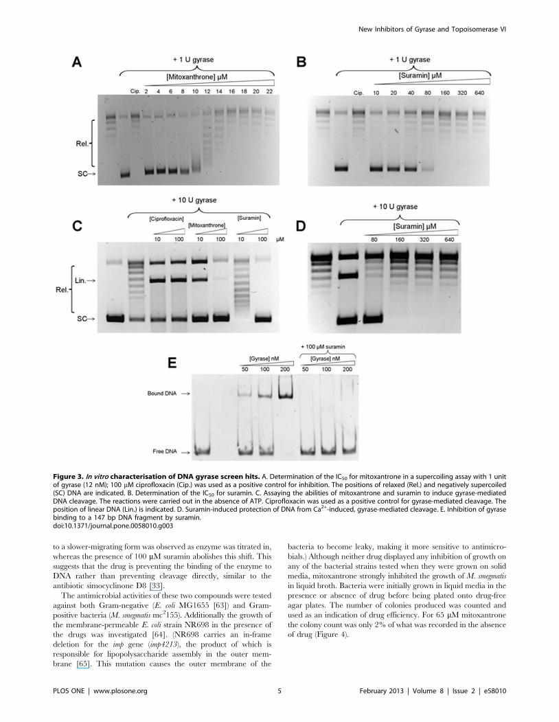
to a slower-migrating form was observed as enzyme was titrated in,
whereas the presence of 100 mM suramin abolishes this shift. This
suggests that the drug is preventing the binding of the enzyme to
DNA rather than preventing cleavage directly, similar to the
antibiotic simocyclinone D8 [33].
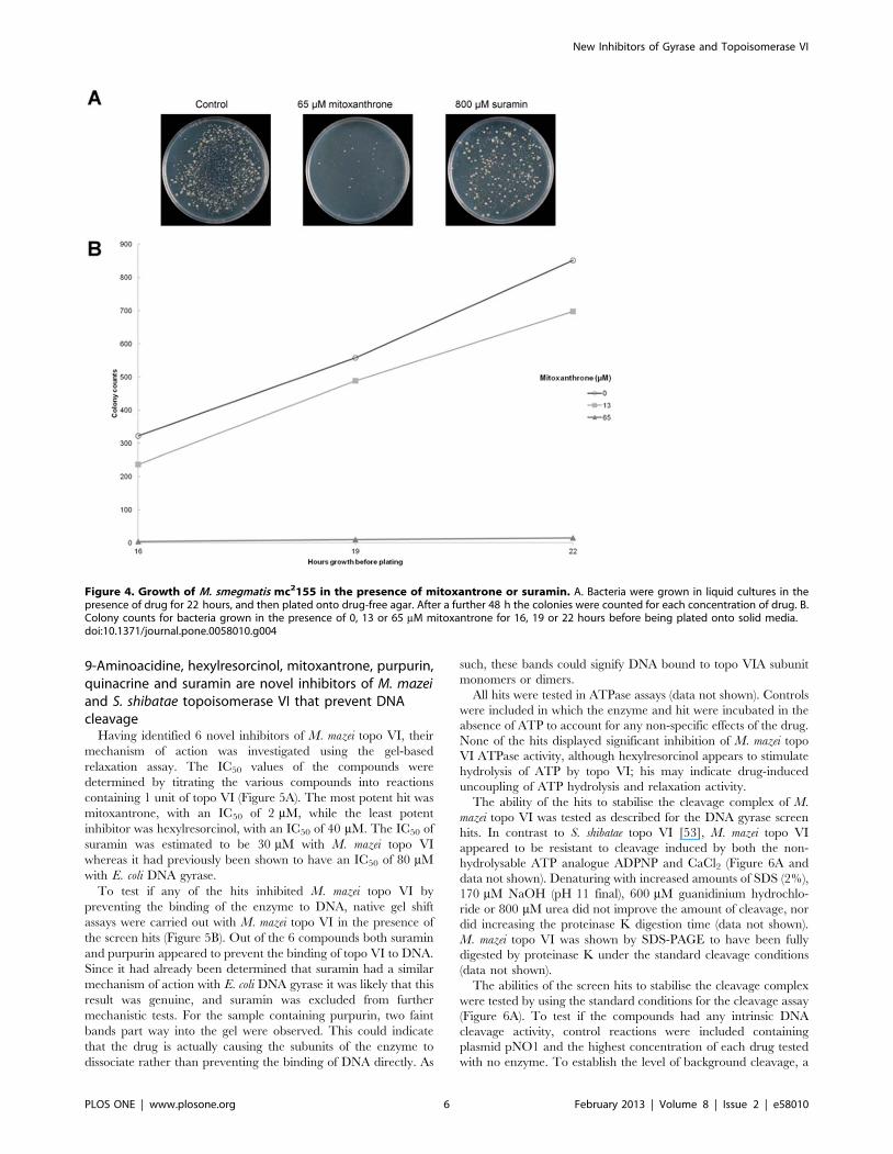
The antimicrobial activities of these two compounds were tested
against both Gram-negative (E. coli MG1655 [63]) and Gram-
positive bacteria (M. smegmatis mc2155). Additionally the growth of
the membrane-permeable E. coli strain NR698 in the presence of
the drugs was investigated [64]. (NR698 carries an in-frame
deletion for the imp gene (imp4213), the product of which is
responsible for lipopolysaccharide assembly in the outer mem-
brane [65]. This mutation causes the outer membrane of the
bacteria to become leaky, making it more sensitive to antimicro-
bials.) Although neither drug displayed any inhibition of growth on
any of the bacterial strains tested when they were grown on solid
media, mitoxantrone strongly inhibited the growth of M. smegmatis
in liquid broth. Bacteria were initially grown in liquid media in the
presence or absence of drug before being plated onto drug-free
agar plates. The number of colonies produced was counted and
used as an indication of drug efficiency. For 65 mM mitoxantrone
the colony count was only 2% of what was recorded in the absence
of drug (Figure 4).
Figure 3. In vitro characterisation of DNA gyrase screen hits. A. Determination of the IC50 for mitoxantrone in a supercoiling assay with 1 unitof gyrase (12 nM); 100 mM ciprofloxacin (Cip.) was used as a positive control for inhibition. The positions of relaxed (Rel.) and negatively supercoiled(SC) DNA are indicated. B. Determination of the IC50 for suramin. C. Assaying the abilities of mitoxantrone and suramin to induce gyrase-mediatedDNA cleavage. The reactions were carried out in the absence of ATP. Ciprofloxacin was used as a positive control for gyrase-mediated cleavage. Theposition of linear DNA (Lin.) is indicated. D. Suramin-induced protection of DNA from Ca2+-induced, gyrase-mediated cleavage. E. Inhibition of gyrasebinding to a 147 bp DNA fragment by suramin.doi:10.1371/journal.pone.0058010.g003
New Inhibitors of Gyrase and Topoisomerase VI
PLOS ONE | www.plosone.org 5 February 2013 | Volume 8 | Issue 2 | e58010
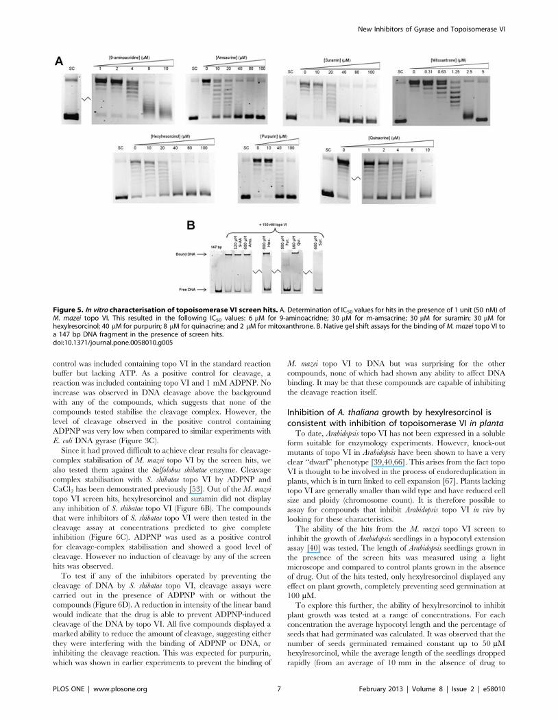
9-Aminoacidine, hexylresorcinol, mitoxantrone, purpurin,quinacrine and suramin are novel inhibitors of M. mazeiand S. shibatae topoisomerase VI that prevent DNAcleavage
Having identified 6 novel inhibitors of M. mazei topo VI, their
mechanism of action was investigated using the gel-based
relaxation assay. The IC50 values of the compounds were
determined by titrating the various compounds into reactions
containing 1 unit of topo VI (Figure 5A). The most potent hit was
mitoxantrone, with an IC50 of 2 mM, while the least potent
inhibitor was hexylresorcinol, with an IC50 of 40 mM. The IC50 of
suramin was estimated to be 30 mM with M. mazei topo VI
whereas it had previously been shown to have an IC50 of 80 mM
with E. coli DNA gyrase.
To test if any of the hits inhibited M. mazei topo VI by
preventing the binding of the enzyme to DNA, native gel shift
assays were carried out with M. mazei topo VI in the presence of
the screen hits (Figure 5B). Out of the 6 compounds both suramin
and purpurin appeared to prevent the binding of topo VI to DNA.
Since it had already been determined that suramin had a similar
mechanism of action with E. coli DNA gyrase it was likely that this
result was genuine, and suramin was excluded from further
mechanistic tests. For the sample containing purpurin, two faint
bands part way into the gel were observed. This could indicate
that the drug is actually causing the subunits of the enzyme to
dissociate rather than preventing the binding of DNA directly. As
such, these bands could signify DNA bound to topo VIA subunit
monomers or dimers.
All hits were tested in ATPase assays (data not shown). Controls
were included in which the enzyme and hit were incubated in the
absence of ATP to account for any non-specific effects of the drug.
None of the hits displayed significant inhibition of M. mazei topo
VI ATPase activity, although hexylresorcinol appears to stimulate
hydrolysis of ATP by topo VI; his may indicate drug-induced
uncoupling of ATP hydrolysis and relaxation activity.
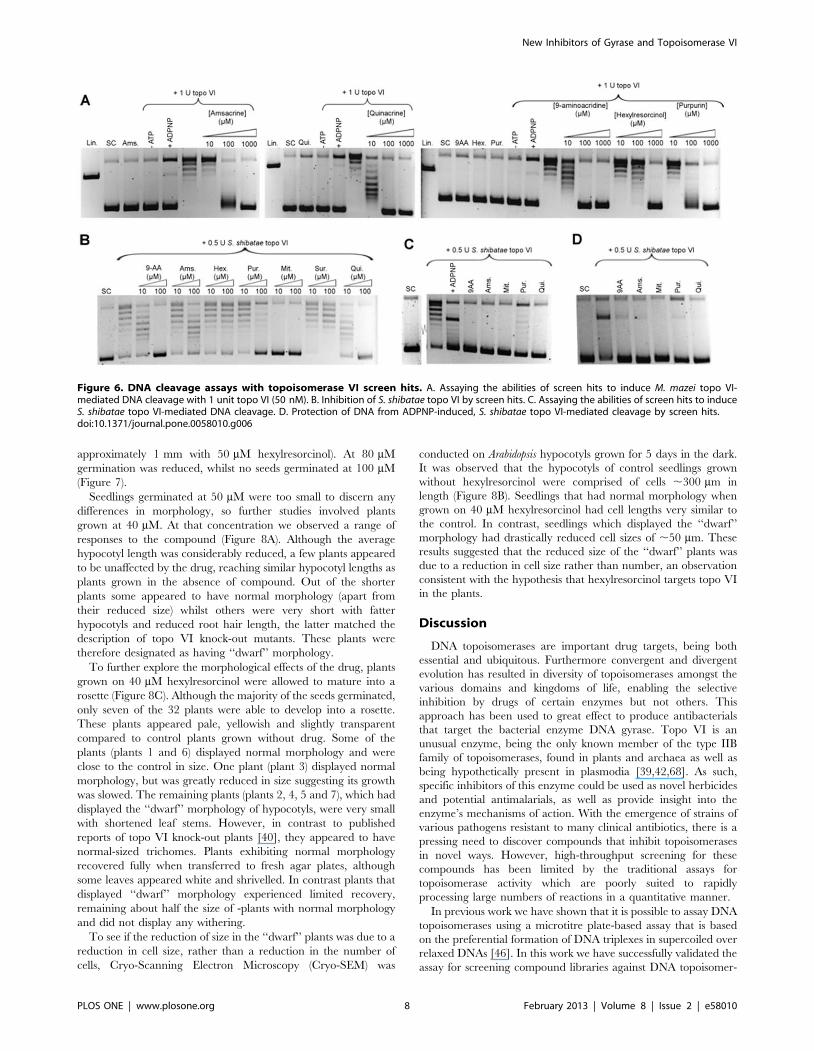
The ability of the hits to stabilise the cleavage complex of M.
mazei topo VI was tested as described for the DNA gyrase screen
hits. In contrast to S. shibatae topo VI [53], M. mazei topo VI
appeared to be resistant to cleavage induced by both the non-
hydrolysable ATP analogue ADPNP and CaCl2 (Figure 6A and
data not shown). Denaturing with increased amounts of SDS (2%),
170 mM NaOH (pH 11 final), 600 mM guanidinium hydrochlo-
ride or 800 mM urea did not improve the amount of cleavage, nor
did increasing the proteinase K digestion time (data not shown).
M. mazei topo VI was shown by SDS-PAGE to have been fully
digested by proteinase K under the standard cleavage conditions
(data not shown).
The abilities of the screen hits to stabilise the cleavage complex
were tested by using the standard conditions for the cleavage assay
(Figure 6A). To test if the compounds had any intrinsic DNA
cleavage activity, control reactions were included containing
plasmid pNO1 and the highest concentration of each drug tested
with no enzyme. To establish the level of background cleavage, a
Figure 4. Growth of M. smegmatis mc2155 in the presence of mitoxantrone or suramin. A. Bacteria were grown in liquid cultures in thepresence of drug for 22 hours, and then plated onto drug-free agar. After a further 48 h the colonies were counted for each concentration of drug. B.Colony counts for bacteria grown in the presence of 0, 13 or 65 mM mitoxantrone for 16, 19 or 22 hours before being plated onto solid media.doi:10.1371/journal.pone.0058010.g004
New Inhibitors of Gyrase and Topoisomerase VI
PLOS ONE | www.plosone.org 6 February 2013 | Volume 8 | Issue 2 | e58010
control was included containing topo VI in the standard reaction
buffer but lacking ATP. As a positive control for cleavage, a
reaction was included containing topo VI and 1 mM ADPNP. No
increase was observed in DNA cleavage above the background
with any of the compounds, which suggests that none of the
compounds tested stabilise the cleavage complex. However, the
level of cleavage observed in the positive control containing
ADPNP was very low when compared to similar experiments with
E. coli DNA gyrase (Figure 3C).
Since it had proved difficult to achieve clear results for cleavage-
complex stabilisation of M. mazei topo VI by the screen hits, we
also tested them against the Sulfolobus shibatae enzyme. Cleavage
complex stabilisation with S. shibatae topo VI by ADPNP and
CaCl2 has been demonstrated previously [53]. Out of the M. mazei
topo VI screen hits, hexylresorcinol and suramin did not display
any inhibition of S. shibatae topo VI (Figure 6B). The compounds
that were inhibitors of S. shibatae topo VI were then tested in the
cleavage assay at concentrations predicted to give complete
inhibition (Figure 6C). ADPNP was used as a positive control
for cleavage-complex stabilisation and showed a good level of
cleavage. However no induction of cleavage by any of the screen
hits was observed.
To test if any of the inhibitors operated by preventing the
cleavage of DNA by S. shibatae topo VI, cleavage assays were
carried out in the presence of ADPNP with or without the
compounds (Figure 6D). A reduction in intensity of the linear band
would indicate that the drug is able to prevent ADPNP-induced
cleavage of the DNA by topo VI. All five compounds displayed a
marked ability to reduce the amount of cleavage, suggesting either
they were interfering with the binding of ADPNP or DNA, or
inhibiting the cleavage reaction. This was expected for purpurin,
which was shown in earlier experiments to prevent the binding of
M. mazei topo VI to DNA but was surprising for the other
compounds, none of which had shown any ability to affect DNA
binding. It may be that these compounds are capable of inhibiting
the cleavage reaction itself.
Inhibition of A. thaliana growth by hexylresorcinol isconsistent with inhibition of topoisomerase VI in planta
To date, Arabidopsis topo VI has not been expressed in a soluble
form suitable for enzymology experiments. However, knock-out
mutants of topo VI in Arabidopsis have been shown to have a very
clear ‘‘dwarf’’ phenotype [39,40,66]. This arises from the fact topo
VI is thought to be involved in the process of endoreduplication in
plants, which is in turn linked to cell expansion [67]. Plants lacking
topo VI are generally smaller than wild type and have reduced cell
size and ploidy (chromosome count). It is therefore possible to
assay for compounds that inhibit Arabidopsis topo VI in vivo by
looking for these characteristics.
The ability of the hits from the M. mazei topo VI screen to
inhibit the growth of Arabidopsis seedlings in a hypocotyl extension
assay [40] was tested. The length of Arabidopsis seedlings grown in
the presence of the screen hits was measured using a light
microscope and compared to control plants grown in the absence
of drug. Out of the hits tested, only hexylresorcinol displayed any
effect on plant growth, completely preventing seed germination at
100 mM.
To explore this further, the ability of hexylresorcinol to inhibit
plant growth was tested at a range of concentrations. For each
concentration the average hypocotyl length and the percentage of
seeds that had germinated was calculated. It was observed that the
number of seeds germinated remained constant up to 50 mM
hexylresorcinol, while the average length of the seedlings dropped
rapidly (from an average of 10 mm in the absence of drug to
Figure 5. In vitro characterisation of topoisomerase VI screen hits. A. Determination of IC50 values for hits in the presence of 1 unit (50 nM) ofM. mazei topo VI. This resulted in the following IC50 values: 6 mM for 9-aminoacridne; 30 mM for m-amsacrine; 30 mM for suramin; 30 mM forhexylresorcinol; 40 mM for purpurin; 8 mM for quinacrine; and 2 mM for mitoxanthrone. B. Native gel shift assays for the binding of M. mazei topo VI toa 147 bp DNA fragment in the presence of screen hits.doi:10.1371/journal.pone.0058010.g005
New Inhibitors of Gyrase and Topoisomerase VI
PLOS ONE | www.plosone.org 7 February 2013 | Volume 8 | Issue 2 | e58010
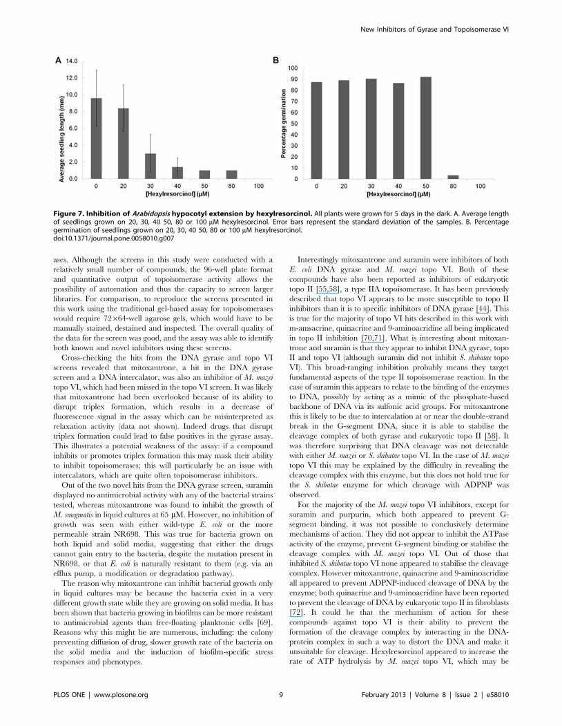
approximately 1 mm with 50 mM hexylresorcinol). At 80 mM
germination was reduced, whilst no seeds germinated at 100 mM
(Figure 7).
Seedlings germinated at 50 mM were too small to discern any
differences in morphology, so further studies involved plants
grown at 40 mM. At that concentration we observed a range of
responses to the compound (Figure 8A). Although the average
hypocotyl length was considerably reduced, a few plants appeared
to be unaffected by the drug, reaching similar hypocotyl lengths as
plants grown in the absence of compound. Out of the shorter
plants some appeared to have normal morphology (apart from
their reduced size) whilst others were very short with fatter
hypocotyls and reduced root hair length, the latter matched the
description of topo VI knock-out mutants. These plants were
therefore designated as having ‘‘dwarf’’ morphology.
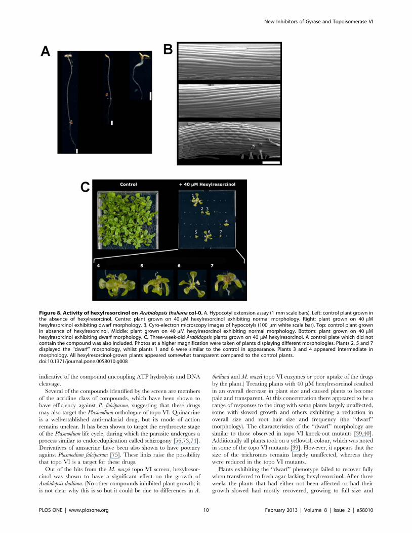
To further explore the morphological effects of the drug, plants
grown on 40 mM hexylresorcinol were allowed to mature into a
rosette (Figure 8C). Although the majority of the seeds germinated,
only seven of the 32 plants were able to develop into a rosette.
These plants appeared pale, yellowish and slightly transparent
compared to control plants grown without drug. Some of the
plants (plants 1 and 6) displayed normal morphology and were
close to the control in size. One plant (plant 3) displayed normal
morphology, but was greatly reduced in size suggesting its growth
was slowed. The remaining plants (plants 2, 4, 5 and 7), which had
displayed the ‘‘dwarf’’ morphology of hypocotyls, were very small
with shortened leaf stems. However, in contrast to published
reports of topo VI knock-out plants [40], they appeared to have
normal-sized trichomes. Plants exhibiting normal morphology
recovered fully when transferred to fresh agar plates, although
some leaves appeared white and shrivelled. In contrast plants that
displayed ‘‘dwarf’’ morphology experienced limited recovery,
remaining about half the size of -plants with normal morphology
and did not display any withering.
To see if the reduction of size in the ‘‘dwarf’’ plants was due to a
reduction in cell size, rather than a reduction in the number of
cells, Cryo-Scanning Electron Microscopy (Cryo-SEM) was
conducted on Arabidopsis hypocotyls grown for 5 days in the dark.
It was observed that the hypocotyls of control seedlings grown
without hexylresorcinol were comprised of cells ,300 mm in
length (Figure 8B). Seedlings that had normal morphology when
grown on 40 mM hexylresorcinol had cell lengths very similar to
the control. In contrast, seedlings which displayed the ‘‘dwarf’’
morphology had drastically reduced cell sizes of ,50 mm. These
results suggested that the reduced size of the ‘‘dwarf’’ plants was
due to a reduction in cell size rather than number, an observation
consistent with the hypothesis that hexylresorcinol targets topo VI
in the plants.
Discussion
DNA topoisomerases are important drug targets, being both
essential and ubiquitous. Furthermore convergent and divergent
evolution has resulted in diversity of topoisomerases amongst the
various domains and kingdoms of life, enabling the selective
inhibition by drugs of certain enzymes but not others. This
approach has been used to great effect to produce antibacterials
that target the bacterial enzyme DNA gyrase. Topo VI is an
unusual enzyme, being the only known member of the type IIB
family of topoisomerases, found in plants and archaea as well as
being hypothetically present in plasmodia [39,42,68]. As such,
specific inhibitors of this enzyme could be used as novel herbicides
and potential antimalarials, as well as provide insight into the
enzyme’s mechanisms of action. With the emergence of strains of
various pathogens resistant to many clinical antibiotics, there is a
pressing need to discover compounds that inhibit topoisomerases
in novel ways. However, high-throughput screening for these
compounds has been limited by the traditional assays for
topoisomerase activity which are poorly suited to rapidly
processing large numbers of reactions in a quantitative manner.
In previous work we have shown that it is possible to assay DNA
topoisomerases using a microtitre plate-based assay that is based
on the preferential formation of DNA triplexes in supercoiled over
relaxed DNAs [46]. In this work we have successfully validated the
assay for screening compound libraries against DNA topoisomer-
Figure 6. DNA cleavage assays with topoisomerase VI screen hits. A. Assaying the abilities of screen hits to induce M. mazei topo VI-mediated DNA cleavage with 1 unit topo VI (50 nM). B. Inhibition of S. shibatae topo VI by screen hits. C. Assaying the abilities of screen hits to induceS. shibatae topo VI-mediated DNA cleavage. D. Protection of DNA from ADPNP-induced, S. shibatae topo VI-mediated cleavage by screen hits.doi:10.1371/journal.pone.0058010.g006
New Inhibitors of Gyrase and Topoisomerase VI
PLOS ONE | www.plosone.org 8 February 2013 | Volume 8 | Issue 2 | e58010
ases. Although the screens in this study were conducted with a
relatively small number of compounds, the 96-well plate format
and quantitative output of topoisomerase activity allows the
possibility of automation and thus the capacity to screen larger
libraries. For comparison, to reproduce the screens presented in
this work using the traditional gel-based assay for topoisomerases
would require 72664-well agarose gels, which would have to be
manually stained, destained and inspected. The overall quality of
the data for the screen was good, and the assay was able to identify
both known and novel inhibitors using these screens.
Cross-checking the hits from the DNA gyrase and topo VI
screens revealed that mitoxantrone, a hit in the DNA gyrase
screen and a DNA intercalator, was also an inhibitor of M. mazei
topo VI, which had been missed in the topo VI screen. It was likely
that mitoxantrone had been overlooked because of its ability to
disrupt triplex formation, which results in a decrease of
fluorescence signal in the assay which can be misinterpreted as
relaxation activity (data not shown). Indeed drugs that disrupt
triplex formation could lead to false positives in the gyrase assay.
This illustrates a potential weakness of the assay: if a compound
inhibits or promotes triplex formation this may mask their ability
to inhibit topoisomerases; this will particularly be an issue with
intercalators, which are quite often topoisomerase inhibitors.
Out of the two novel hits from the DNA gyrase screen, suramin
displayed no antimicrobial activity with any of the bacterial strains
tested, whereas mitoxantrone was found to inhibit the growth of
M. smegmatis in liquid cultures at 65 mM. However, no inhibition of
growth was seen with either wild-type E. coli or the more
permeable strain NR698. This was true for bacteria grown on
both liquid and solid media, suggesting that either the drugs
cannot gain entry to the bacteria, despite the mutation present in
NR698, or that E. coli is naturally resistant to them (e.g. via an
efflux pump, a modification or degradation pathway).
The reason why mitoxantrone can inhibit bacterial growth only
in liquid cultures may be because the bacteria exist in a very
different growth state while they are growing on solid media. It has
been shown that bacteria growing in biofilms can be more resistant
to antimicrobial agents than free-floating planktonic cells [69].
Reasons why this might be are numerous, including: the colony
preventing diffusion of drug, slower growth rate of the bacteria on
the solid media and the induction of biofilm-specific stress
responses and phenotypes.
Interestingly mitoxantrone and suramin were inhibitors of both
E. coli DNA gyrase and M. mazei topo VI. Both of these
compounds have also been reported as inhibitors of eukaryotic
topo II [55,58], a type IIA topoisomerase. It has been previously
described that topo VI appears to be more susceptible to topo II
inhibitors than it is to specific inhibitors of DNA gyrase [44]. This
is true for the majority of topo VI hits described in this work with
m-amsacrine, quinacrine and 9-aminoacridine all being implicated
in topo II inhibition [70,71]. What is interesting about mitoxan-
trone and suramin is that they appear to inhibit DNA gyrase, topo
II and topo VI (although suramin did not inhibit S. shibatae topo
VI). This broad-ranging inhibition probably means they target
fundamental aspects of the type II topoisomerase reaction. In the
case of suramin this appears to relate to the binding of the enzymes
to DNA, possibly by acting as a mimic of the phosphate-based
backbone of DNA via its sulfonic acid groups. For mitoxantrone
this is likely to be due to intercalation at or near the double-strand
break in the G-segment DNA, since it is able to stabilise the
cleavage complex of both gyrase and eukaryotic topo II [58]. It
was therefore surprising that DNA cleavage was not detectable
with either M. mazei or S. shibatae topo VI. In the case of M. mazei
topo VI this may be explained by the difficulty in revealing the
cleavage complex with this enzyme, but this does not hold true for
the S. shibatae enzyme for which cleavage with ADPNP was
observed.
For the majority of the M. mazei topo VI inhibitors, except for
suramin and purpurin, which both appeared to prevent G-
segment binding, it was not possible to conclusively determine
mechanisms of action. They did not appear to inhibit the ATPase
activity of the enzyme, prevent G-segment binding or stabilise the
cleavage complex with M. mazei topo VI. Out of those that
inhibited S. shibatae topo VI none appeared to stabilise the cleavage
complex. However mitoxantrone, quinacrine and 9-aminoacridine
all appeared to prevent ADPNP-induced cleavage of DNA by the
enzyme; both quinacrine and 9-aminoacridine have been reported
to prevent the cleavage of DNA by eukaryotic topo II in fibroblasts
[72]. It could be that the mechanism of action for these
compounds against topo VI is their ability to prevent the
formation of the cleavage complex by interacting in the DNA-
protein complex in such a way to distort the DNA and make it
unsuitable for cleavage. Hexylresorcinol appeared to increase the
rate of ATP hydrolysis by M. mazei topo VI, which may be
Figure 7. Inhibition of Arabidopsis hypocotyl extension by hexylresorcinol. All plants were grown for 5 days in the dark. A. Average lengthof seedlings grown on 20, 30, 40 50, 80 or 100 mM hexylresorcinol. Error bars represent the standard deviation of the samples. B. Percentagegermination of seedlings grown on 20, 30, 40 50, 80 or 100 mM hexylresorcinol.doi:10.1371/journal.pone.0058010.g007
New Inhibitors of Gyrase and Topoisomerase VI
PLOS ONE | www.plosone.org 9 February 2013 | Volume 8 | Issue 2 | e58010
indicative of the compound uncoupling ATP hydrolysis and DNA
cleavage.
Several of the compounds identified by the screen are members
of the acridine class of compounds, which have been shown to
have efficiency against P. falciparum, suggesting that these drugs
may also target the Plasmodium orthologue of topo VI. Quinacrine
is a well-established anti-malarial drug, but its mode of action
remains unclear. It has been shown to target the erythrocyte stage
of the Plasmodium life cycle, during which the parasite undergoes a
process similar to endoreduplication called schizogony [56,73,74].
Derivatives of amsacrine have been also shown to have potency
against Plasmodium falciparum [75]. These links raise the possibility
that topo VI is a target for these drugs.
Out of the hits from the M. mazei topo VI screen, hexylresor-
cinol was shown to have a significant effect on the growth of
Arabidopsis thaliana. (No other compounds inhibited plant growth; it
is not clear why this is so but it could be due to differences in A.
thaliana and M. mazei topo VI enzymes or poor uptake of the drugs
by the plant.) Treating plants with 40 mM hexylresorcinol resulted
in an overall decrease in plant size and caused plants to become
pale and transparent. At this concentration there appeared to be a
range of responses to the drug with some plants largely unaffected,
some with slowed growth and others exhibiting a reduction in
overall size and root hair size and frequency (the ‘‘dwarf’’
morphology). The characteristics of the ‘‘dwarf’’ morphology are
similar to those observed in topo VI knock-out mutants [39,40].
Additionally all plants took on a yellowish colour, which was noted
in some of the topo VI mutants [39]. However, it appears that the
size of the trichromes remains largely unaffected, whereas they
were reduced in the topo VI mutants.
Plants exhibiting the ‘‘dwarf’’ phenotype failed to recover fully
when transferred to fresh agar lacking hexylresorcinol. After three
weeks the plants that had either not been affected or had their
growth slowed had mostly recovered, growing to full size and
Figure 8. Activity of hexylresorcinol on Arabidopsis thaliana col-0. A. Hypocotyl extension assay (1 mm scale bars). Left: control plant grown inthe absence of hexylresorcinol. Centre: plant grown on 40 mM hexylresorcinol exhibiting normal morphology. Right: plant grown on 40 mMhexylresorcinol exhibiting dwarf morphology. B. Cyro-electron microscopy images of hypocotyls (100 mm white scale bar). Top: control plant grownin absence of hexylresorcinol. Middle: plant grown on 40 mM hexylresorcinol exhibiting normal morphology. Bottom: plant grown on 40 mMhexylresorcinol exhibiting dwarf morphology. C. Three-week-old Arabidopsis plants grown on 40 mM hexylresorcinol. A control plate which did notcontain the compound was also included. Photos at a higher magnification were taken of plants displaying different morphologies. Plants 2, 5 and 7displayed the ‘‘dwarf’’ morphology, whilst plants 1 and 6 were similar to the control in appearance. Plants 3 and 4 appeared intermediate inmorphology. All hexylresorcinol-grown plants appeared somewhat transparent compared to the control plants.doi:10.1371/journal.pone.0058010.g008
New Inhibitors of Gyrase and Topoisomerase VI
PLOS ONE | www.plosone.org 10 February 2013 | Volume 8 | Issue 2 | e58010
regaining their pigmentation. Interestingly, a few leaves on these
plants turned white and withered. In contrast, plants that had
exhibited the dwarf morphology remained smaller and did not
have any withered leaves, although they did grow considerably
and retain their pigmentation. These observations suggest that the
withering of leaves is an immune response to the drug, most likely
sequestering the herbicide into certain leaves. A similar response
has been reported for weeds resistant to the common herbicide
glyphosate [76]. It is possible that plants which are able to
sequester hexylresorcinol successfully are able to grow normally or
have their growth slowed as a stress response when grown on sub-
lethal concentrations of drug, whereas those that cannot do so
have inhibited topo VI activity and therefore exhibit the ‘‘dwarf’’
morphology.
To determine whether the ‘‘dwarf’’ morphology was due to in
vivo inhibition of endoreduplication, cryo-SEM was carried out on
seedlings grown in the presence of hexylresorcinol. Since
endoreduplication results in increased cell expansion the expected
result for its inhibition is a reduction in cell size. Plants that had
grown normally or slowly both exhibited a cell size of around
300 mm whereas plants exhibiting the ‘‘dwarf’’ phenotype were
reduced to around 50 mm. As to whether this decrease in
endoreduplication is due to the inhibition of Arabidopsis topo VI
in vivo, it is difficult to say for certain since there is always the
possibility that hexylresorcinol affects other enzymes involved in
the process. The drug does provoke other morphologies (such as
reduction in pigmentation) that are not solely explainable as an
effect of reduced endoreduplication. It is therefore possible that it
has multiple targets within the plant. Additionally, although the
‘‘dwarf’’ morphology shares some of the features with the topo VI
knock-out mutants (overall size reduction, yellowish colour, and
reduced root hair size and frequency), there are some key
differences. Trichome size appears to be unaffected, whereas it was
reduced in the mutants, which suggests that hexylresorcinol is
targeted to specific tissues within the plant. Taken together the
results show that hexylresorcinol leads to a dwarf morphology that
is consistent with an effect on endoreduplication via topo VI
inhibition, but further work would be required to establish this.
One way to prove that topo VI is the target within the plant
would be to express and purify the plant enzyme and demonstrate
that hexylresorcinol inhibits it in vitro; our efforts to purify active
plant topo VI have so far been unsuccessful (data not shown).
Alternatively plants resistant to hexylresorcinol could be selected
and the resistance genes mapped and sequenced. Our observations
that the drug inhibits M. mazei topo VI in vitro and Arabidopsis
endoreduplication in planta, provides at least circumstantial
evidence for topo VI being the in vivo target for this compound.
Hexylresorcinol is an active ingredient of acrisorcin (Akrinol), a
topical antifungal [77] and has been used as an oral antiseptic
[78], and is an active ingredient in throat lozenges, but there has
been no previous reports of its herbicidal activity. Hence, the
significance of its effect on Arabidopsis and on topo VI remains to be
established.
The emergence of pathogenic bacterial strains resistant to
currently used antibiotics is a problem of growing importance.
Topoisomerases have proved to be extremely successful targets for
anticancer and antibacterial drugs, but the search for novel
inhibitors of these enzymes has been hampered by the low-
throughput nature of traditional assays. In this work we have
validated a novel assay for topoisomerase activity by successfully
screening a small library of compounds against E. coli DNA gyrase
and M. mazei topo VI. Several novel inhibitors for these enzymes
have been identified and their mechanisms of action explored in
vitro and in vivo. This work provides a blueprint for larger-scale
screens for inhibitors of topoisomerases which have the potential to
lead to new therapeutics.
Materials and Methods
Enzymes, DNA and compoundsE. coli DNA gyrase and his-tagged M. mazei topo VI were
prepared as described previously [36,79]. S. shibatae topo VI was a
gift from Danielle Gadelle (University of Paris XI, Orsay). One
unit of enzyme is defined as the amount required to fully supercoil
(in the case of E. coli DNA gyrase) or relax (in the case of M. mazei
topo VI) 0.5 mg of pNO1 in 30 min. at 37uC. In the case of S.
shibatae topo VI, one unit was defined as the amount required to
fully relax 0.5 mg of pNO1 in 5 min at 75uC. Wheat germ topo I
was purchased from Inspiralis Ltd (Norwich, UK). Chicken
erythrocyte topo I was a gift from Alison Howells (Inspiralis Ltd.).
Plasmid pNO1 is a modified version of plasmid pBR322*
containing a 16 bp triplex forming sequence (59-
CTCTCTCTCTCTCTCT) [46]. Supercoiled plasmid was puri-
fied by caesium chloride gradient [80]. Relaxed substrate was
prepared by incubating 10 mg of supercoiled plasmid with chicken
erythrocyte topo I for 1 h at 37uC in 50 mL of: 20 mM Tris?HCl
(pH 8.0), 200 mM NaCl, 0.25 mM EDTA, 5% glycerol. The
relaxed plasmid was subsequently purified by phenol-chloroform
extraction and ethanol precipitation. The biotinylated oligonucle-
otide TFO1 (59 biotin-TCTCTCTCTCTCTCTC) was synthe-
sised by Sigma-Aldrich Co. LLC.
Screening was carried out on the GenPlus library from
Microsource Ltd. Daughter plates were prepared with a
compound concentration of 250 mM in 100% DMSO. Fresh
stocks of potential hits were ordered either from Sigma-Aldrich
Co. LLC. or Microsource Ltd.
DNA triplex-based assay for topoisomerase activityThe triplex-based assay was conducted as previously described
[45–47]. In brief, oligonucleotide TFO1 was immobilised onto a
streptavidin-coated microtitre plate. Excess oligonucleotide was
removed by washing with buffer. Topoisomerase reactions were
carried out in a 30 mL reaction volume containing 1–2 units
topoisomerase and 1 mg relaxed or negatively supercoiled pNO1
under the published reaction conditions [36,47]; gyrase supercoil-
ing: 35 mM TrisNHCl (pH 7.5), 24 mM KCl, 4 mM MgCl2,
2 mM DTT, 1.8 mM spermidine, 1 mM ATP, 6.5% (w/v)
glycerol, 0.1 mg/mL bovine serum albumin; topo VI relaxation:
20 mM bis-tris propane (pH 6.5), 100 mM potassium glutamate,
10 mM MgCl2, 1 mM DTT, 1 mM ATP. The reaction was
stopped by the addition of a low pH, high salt buffer and triplex
formation was allowed to occur for 30 min at room temperature.
The DNA retained on the plate was stained with SYBR Gold
(Sigma) dye and the fluorescence readings for each well read in a
SpectraMax Gemini fluorimeter (Molecular Devices).
Topoisomerase screening conditionsTwelve plates were screened in duplicate over three days
manually using a multichannel pipette as previously described
[45]. Each reaction contained a final concentration of approxi-
mately 25 mM compound and 5% DMSO. Each plate contained
16 control reactions: eight positive for enzyme activity, which
contained enzyme but no library compounds, and eight negative
for enzyme activity, which lacked both enzyme and library
compounds. All controls were carried out with 5% DMSO. Once
the data had been collected from the screen, the fluorescence
signals for the duplicates were averaged and converted into
percentage inhibition using the positive and negative controls. An
New Inhibitors of Gyrase and Topoisomerase VI
PLOS ONE | www.plosone.org 11 February 2013 | Volume 8 | Issue 2 | e58010
arbitrary hit threshold of 25% inhibition was used to select
compounds for validation by the agarose gel-based assay. To
analyse the degree of separation between a negative and positive
result the Z’ factor [51] for the assay was calculated from the data
for the control reactions (Equation 1).
Z0~(3spositivecontrolsz3snegativecontrols)
Dmpositivecontrols{mnegativecontrolsDð1Þ
Agarose gel-based assay for DNA topoisomerase activityThe agarose gel-based assay was conducted as previously
described [36,44,62]. In brief, topoisomerases were incubated with
0.5 mg of plasmid and the desired concentration of compound for
30 min at 37uC (for DNA gyrase and M. mazei topo VI) or 5 min
at 75uC (for S. shibatae topo VI), in a total volume of 30 mL. The
final concentration of DMSO in these reactions did not exceed
5%. Reactions were stopped by the addition of a loading buffer
containing EDTA and the drug extracted by vortexing with either
chloroform or aqueous butanol as required. In the case of M. mazei
topo VI, it was necessary to include SDS in the loading buffer, at a
final concentration of 1%. Samples were loaded onto a 1%
agarose gel, run for the appropriate length of time and stained
with ethidium bromide before visualisation on a UV transillumi-
nator.
Agarose gel-based assay for DNA cleavage by DNAtopoisomerases
Assays to detect the stabilisation of cleavage complexes were
identical to the protocol for topoisomerase activity detailed above,
except as follows. Cleavage experiments with DNA gyrase were
carried out in the absence of ATP and spermidine and 30 mL
reactions were stopped with 3 mL 10% SDS and incubated with
3 mL 1 mg/mL proteinase K solution at 37uC for 1 h. For
cleavage complex protection assays with suramin, the reaction
buffer was modified to contain 4 mM CaCl2 rather than MgCl2.
Pyruvate kinase-linked assay for topoisomerase ATPaseactivity
ATPase assays were performed essentially as described previ-
ously [81] except they were adapted to a microplate format using
clear, colourless 96-well Microtitre plates (Pro-bindTM, Becton
Dickinson). Reactions were carried out in 100 mL of the
established reaction buffer for either DNA gyrase of M. mazei
topo VI [36,62] supplemented with: 800 mM phosphoenolpyr-
uvate (PEP), 400 mM NADH, 1% (vol/vol) PK/LDH (pyruvate
kinase-lactate dehydrogenase mixture in 50% (w/v) glycer-
ol,100 mM KCl, 10 mM HEPES (pH 7.0)) and the desired
concentration of hit compound. ATP was initially withheld from
the reaction. After 5 min incubation at room temperature the
reactions were initiated by the addition of ATP and the
absorbance at 340 nm was measured over the course of an hour
using a Spectra Max Plus absorbance reader (Molecular Devices).
Data were processed by omitting the first 10–15 min of collection
and normalising the first retained time point. This was done to
exclude artifacts observed within this time frame and for ease of
comparison of rates. Controls lacking topoisomerase were used to
assess the intrinsic ATPase activity of the test compounds.
Electrophoretic mobility shift assay for topoisomerase-DNA binding
In order to test the effects of hit compounds upon DNA binding
by topoisomerases, samples of enzyme and drug were prepared
under the following conditions in a final volume of 10 mL: 1 nM
147 bp linear fragment of DNA derived from pBR322, [82]
50 mM TrisNHCl (pH 7.5), 100 mM KCl, 5 mM MgCl2, 2 mM
DTT, 10% w/v glycerol. Samples were incubated for 30 min at
room temperature before being run on a 5% (29:1) Protogel
acrylamide (National Diagnostics) gel in 90 mM Tris?Borate,
5 mM MgCl2 at 150 V for 45 min. The DNA was stained by
soaking in 2 mg/mL ethidium bromide for 10 min and visualised
under UV light.
Assaying E. coli DNA gyrase hits for bactericidal activityFor E. coli work, 10 mL cultures of E. coli MG1655 or NR698
were grown overnight at 37uC in LB medium. A 100 mL sample
was then added to 10 mL of LB medium containing the desired
concentration of compound. The culture was then allowed to grow
at 37uC for 5 h and its OD600 measured every hour. For M.
smegmatis, a 50 mL culture was grown for 48 h in Middlebrooks
7H11 medium at 37uC. A 100 mL sample was then added to
10 mL of Middlebrooks 7H11 containing the desired concentra-
tion of compound. The culture was then allowed to grow at 37uCfor 9 h and its OD600 measured every three hours. For colony
counting experiments M. smegmatis was grown for 48 h in 50 mL of
Middlebrooks 7H9 medium at 37uC. A 100 mL sample was then
added to 10 mL of Middlebrooks 7H9 medium containing the
desired concentration test compound and allowed to grow at 37uCovernight. After 16 h, 20 mL samples were taken from these
cultures and diluted 1 in 10,000 with Middlebrooks 7H9 medium.
Petri dishes containing Middlebrooks 7H11 agar were prepared
and 100 mL of each diluted culture was spread out. The plates
were subsequently stored at 4uC. This was repeated every three
hours for six hours, after which the plates were transferred to 37uCfor 48 h. The numbers of colonies for each time point were
counted manually.
Arabidopsis hypocotyl extension assay and plant growthconditions
The hypocotyl extension assay was conducted as previously
described [40,66]. MS Salts medium [83] (containing micro and
macro elements including vitamins, pH 5.8) was supplemented
with 1 g/L sucrose and 0.7 g/L phytagel (Sigma) before
autoclaving. After the medium cooled to ,50uC it was divided
into 50 mL aliquots and compounds to be tested (or an equivalent
amount of appropriate solvent for control plates) then added at the
desired concentration under aseptic conditions. The media were
then poured into separate 100 mm square Petri dishes and allowed
to cool to room temperature in a laminar flow hood. Arabidopsis
thaliana columbia (Col-0) seeds were surface sterilised with 5%
bleach for 10 min immediately prior to use. The seeds were then
washed three times with sterile water under a laminar flow hood.
After surface sterilisation the seeds were planted in a grid pattern
(32 seeds per dish) and the dishes sealed with surgical tape. The
plates were transferred to 4uC and left for 64 h in the dark to
vernalise, after which they were transferred to a 22uC growth
cabinet. After 2 h of light exposure the plates were stacked
vertically in the dark at 22uC to allow for hypocotyl extension
along the agar surface. After 4–5 days the hypocotyls were
observed using an optical microscope and their length measured.
For experiments where the plants were allowed to form rosettes,
New Inhibitors of Gyrase and Topoisomerase VI
PLOS ONE | www.plosone.org 12 February 2013 | Volume 8 | Issue 2 | e58010
the Arabidopsis seedlings were grown horizontally under a 16 h day
in a 22uC growth cabinet for the indicated number of weeks.
Cryo-scanning electron microscopy (SEM) on Arabidopsishypocotyls
Five day old hypocotyls grown on media containing various
concentrations of compound were frozen in nitrogen slush and
loaded into a Philips XL30 SEM with a cryo stage installed.
Samples were sputter coated with platinum and transferred into
the microscope chamber for image collection.
Acknowledgments
We thank Kevin Corbett and James Berger for supplying the clones for the
M. mazei topo VI enzyme (University of California Berkley), Danielle
Gadelle and Patrick Forterre for providing S. shibatae topoisomerase VI
(University of Paris XI, Orsay), Alison Howells and Nicolas Burton for
advice and providing chicken erythrocyte topo I (Inspiralis Ltd.), Martin
Stocks for advice and comments (Plant Bioscience Ltd.), Nicola Stacey for
advice on the Arabidopsis thaliana work, Kim Findlay for assistance with the
cryo-SEM experiments, and Fred Collin for comments on the manuscript.
Author Contributions
Conceived and designed the experiments: JAT RAF AM. Performed the
experiments: JAT. Analyzed the data: JAT AM. Contributed reagents/
materials/analysis tools: LAM MR. Wrote the paper: JAT AM.
References
1. Woodford N, Livermore DM (2009) Infections caused by Gram-positivebacteria: a review of the global challenge. J Infect 59 Suppl 1: S4–16.
2. Dondorp AM, Yeung S, White L, Nguon C, Day NP, et al. (2010) Artemisinin
resistance: current status and scenarios for containment. Nat Rev Microbiol 8:272–280.
3. Lew W, Pai M, Oxlade O, Martin D, Menzies D (2008) Initial drug resistanceand tuberculosis treatment outcomes: Systematic review and meta-analysis.
Annals of Internal Medicine 149: 123–134.
4. Rice LB (2003) Do we really need new anti-infective drugs? Curr Opin
Pharmacol 3: 459–463.
5. Kock R, Becker K, Cookson B, van Gemert-Pijnen JE, Harbarth S, et al. (2010)
Methicillin-resistant Staphylococcus aureus (MRSA): burden of disease and
control challenges in Europe. Euro Surveill 15: 19688.
6. Flament-Saillour M, Robert J, Jarlier V, Grosset J (1999) Outcome of multi-
drug-resistant tuberculosis in France: a nationwide case-control study.Am J Respir Crit Care Med 160: 587–593.
7. Cooper MA, Shlaes D (2011) Fix the antibiotics pipeline. Nature 472: 32.
8. Payne DJ, Gwynn MN, Holmes DJ, Pompliano DL (2007) Drugs for bad bugs:
confronting the challenges of antibacterial discovery. Nat Rev Drug Discov 6:
29–40.
9. Pommier Y, Leo E, Zhang H, Marchand C (2010) DNA topoisomerases and
their poisoning by anticancer and antibacterial drugs. Chem Biol 17: 421–433.
10. Tse-Dinh YC (2007) Exploring DNA topoisomerases as targets of novel
therapeutic agents in the treatment of infectious diseases. Infect Disord DrugTargets 7: 3–9.
11. Bradbury BJ, Pucci MJ (2008) Recent advances in bacterial topoisomeraseinhibitors. Curr Opin Pharmacol 8: 574–581.
12. Collin F, Karkare S, Maxwell A (2011) Exploiting bacterial DNA gyrase as a
drug target: current state and perspectives. Appl Microbiol Biotechnol 92: 479–497.
13. Bates AD, Maxwell A (2005) DNA Topology. Oxford: Oxford University Press.
14. Berger JM, Gamblin SJ, Harrison SC, Wang JC (1996) Structure and
mechanism of DNA topoisomerase II. Nature 379: 225–232.
15. Stewart L, Redinbo MR, Qiu X, Hol WG, et al. (1998) A Model for the
Mechanism of Human Topoisomerase I. Science 279: 1534–1541.
16. Lima CD, Wang JC, Mondragon A (1994) Three-dimensional structure of the
67K N-terminal fragment of E. coli DNA topoisomerase I. Nature 367: 138–
146.
17. Sander M, Hsieh T (1983) Double strand DNA cleavage by type II DNA
topoisomerase from Drosophila melanogaster. J Biol Chem 258: 8421–8428.
18. Liu LF, Liu CC, Alberts BM (1980) Type II DNA topoisomerases: enzymes that
can unknot a topologically knotted DNA molecule via a reversible double-strandbreak. Cell 19: 697–707.
19. Wang JC (1998) Moving one DNA double helix through another by a type II
DNA topoisomerase: the story of a simple molecular machine. QuarterlyReviews of Biophysics 31: 107–144.
20. Bates AD, Maxwell A (2007) Energy coupling in type II topoisomerases: why dothey hydrolyze ATP? Biochemistry 46: 7929–7941.
21. Nollmann M, Crisona NJ, Arimondo PB (2007) Thirty years of Escherichia coliDNA gyrase: from in vivo function to single-molecule mechanism. Biochimie 89:
490–499.
22. Champoux JJ (2001) DNA topoisomerases: Structure, function, and mechanism.
Annual Review of Biochemistry 70: 369–413.
23. Bates AD, Berger JM, Maxwell A (2011) The ancestral role of ATP hydrolysis intype II topoisomerases: prevention of DNA double-strand breaks. Nucleic Acids
Res 39: 6327–6339.
24. Gellert M, Mizuuchi K, O’Dea MH, Itoh T, Tomizawa JI (1977) Nalidixic acid
resistance: a second genetic character involved in DNA gyrase activity. Proc NatlAcad Sci U S A 74: 4772–4776.
25. Sugino A, Peebles CL, Kreuzer KN, Cozzarelli NR (1977) Mechanism of action
of nalidixic acid: purification of Escherichia coli nalA gene product and its
relationship to DNA gyrase and a novel nicking-closing enzyme. Proceedings of
the National Academy of Sciences of the United States of America 74: 4767–
4771.
26. Bax BD, Chan PF, Eggleston DS, Fosberry A, Gentry DR, et al. (2010) Type IIA
topoisomerase inhibition by a new class of antibacterial agents. Nature 466: 935–
940.
27. Laponogov I, Pan XS, Veselkov DA, McAuley KE, Fisher LM, et al. (2010)
Structural basis of gate-DNA breakage and resealing by type II topoisomerases.
PLoS ONE 5: e11338.
28. Laponogov I, Sohi MK, Veselkov DA, Pan XS, Sawhney R, et al. (2009)
Structural insight into the quinolone-DNA cleavage complex of type IIA
topoisomerases. Nat Struct Mol Biol 16: 667–669.
29. Wohlkonig A, Chan PF, Fosberry AP, Homes P, Huang J, et al. (2010)
Structural basis of quinolone inhibition of type IIA topoisomerases and target-
mediated resistance. Nat Struct Mol Biol 17: 1152–1153.
30. Maxwell A, Lawson DM (2003) The ATP-binding site of type II topoisomerases
as a target for antibacterial drugs. Curr Top Med Chem 3: 283–303.
31. Maxwell A (1993) The interaction between coumarin drugs and DNA gyrase.
Mol Microbiol 9: 681–686.
32. Heide L, Li S-M (2002) Aminocoumarin antibiotics. In: Rubio F, editor.
Microbial Secondary Metabolites: Biosynthesis, Genetics and Regulation:
Research Signpost.
33. Flatman RH, Howells AJ, Heide L, Fiedler HP, Maxwell A (2005)
Simocyclinone D8, an inhibitor of DNA gyrase with a novel mode of action.
Antimicrob Agents Chemother 49: 1093–1100.
34. Edwards MJ, Flatman RH, Mitchenall LA, Stevenson CE, Le TB, et al. (2009) A
crystal structure of the bifunctional antibiotic simocyclinone D8, bound to DNA
gyrase. Science 326: 1415–1418.
35. Edwards MJ, Williams MA, Maxwell A, McKay AR (2011) Mass spectrometry
reveals that the antibiotic simocyclinone D8 binds to DNA gyrase in a ‘‘bent-
over’’ conformation: evidence of positive cooperativity in binding. Biochemistry
50: 3432–3440.
36. Corbett KD, Benedetti P, Berger JM (2007) Holoenzyme assembly and ATP-
mediated conformational dynamics of topoisomerase VI. Nature Structural &
Molecular Biology 14: 611–619.
37. Graille M, Cladiere L, Durand D, Lecointe F, Gadelle D, et al. (2008) Crystal
structure of an intact type II DNA topoisomerase: insights into DNA transfer
mechanisms. Structure 16: 360–370.
38. Buhler C, Lebbink JH, Bocs C, Ladenstein R, Forterre P (2001) DNA
topoisomerase VI generates ATP-dependent double-strand breaks with two-
nucleotide overhangs. J Biol Chem 276: 37215–37222.
39. Hartung F, Angelis KJ, Meister A, Schubert I, Melzer M, et al. (2002) An
archaebacterial topoisomerase homolog not present in other eukaryotes is
indispensable for cell proliferation of plants. Curr Biol 12: 1787–1791.
40. Sugimoto-Shirasu K, Stacey NJ, Corsar J, Roberts K, McCann MC (2002) DNA
topoisomerase VI is essential for endoreduplication in Arabidopsis. Curr Biol 12:
1782–1786.
41. Hartung F, Puchta H (2000) Molecular characterisation of two paralogous
SPO11 homologues in Arabidopsis thaliana. Nucleic Acids Res 28: 1548–1554.
42. Aravind L, Iyer LM, Wellems TE, Miller LH (2003) Plasmodium biology:
genomic gleanings. Cell 115: 771–785.
43. Gadelle D, Graille M, Forterre P (2006) The HSP90 and DNA topoisomerase
VI inhibitor radicicol also inhibits human type II DNA topoisomerase.
Biochemical Pharmacology 72: 1207–1216.
44. Bergerat A, Gadelle D, Forterre P (1994) Purification of a DNA topoisomerase II
from the hyperthermophilic archaeon Sulfolobus shibatae. A thermostable
enzyme with both bacterial and eucaryal features. Journal of Biological
Chemistry 269: 27663–27669.
45. Taylor JA, Burton NP, Maxwell A (2012) High-throughput microtitre plate-
based assay for DNA topoisomerases. Methods Mol Biol 815: 229–239.
New Inhibitors of Gyrase and Topoisomerase VI
PLOS ONE | www.plosone.org 13 February 2013 | Volume 8 | Issue 2 | e58010
46. Maxwell A, Burton NP, O’Hagan N (2006) High-throughput assays for DNA
gyrase and other topoisomerases. Nucleic Acids Res 34: e104.47. Burrell MR, Burton NP, Maxwell A (2010) A high-throughput assay for DNA
topoisomerases and other enzymes, based on DNA triplex formation. Methods
Mol Biol 613: 257–266.48. Chong CR, Sullivan DJ Jr (2007) New uses for old drugs. Nature 448: 645–646.
49. DiMasi JA, Hansen RW, Grabowski HG (2003) The price of innovation: newestimates of drug development costs. J Health Econ 22: 151–185.
50. Harvey AL (2007) Natural products as a screening resource. Curr Opin Chem
Biol 11: 480–484.51. Zhang JH, Chung TD, Oldenburg KR (1999) A simple statistical parameter for
use in evaluation and validation of high throughput screening assays. J BiomolScreen 4: 67–73.
52. Funatsuki K, Tanaka R, Inagaki S, Konno H, Katoh K, et al. (1998) Quinolone-resistant mutations of DNA gyrase increase sensitivity to acriflavine. Biol Pharm
Bull 21: 667–672.
53. Buhler C, Gadelle D, Forterre P, Wang JC, Bergerat A (1998) Reconstitution ofDNA topoisomerase VI of the thermophilic archaeon Sulfolobus shibatae from
subunits separately overexpressed in Escherichia coli. Nucleic Acids Res 26:5157–5162.
54. Finlay GJ, Atwell GJ, Baguley BC (1999) Inhibition of the action of the
topoisomerase II poison amsacrine by simple aniline derivatives: evidence fordrug-protein interactions. Oncol Res 11: 249–254.
55. Bojanowski K, Lelievre S, Markovits J, Couprie J, Jacquemin-Sablon A, et al.(1992) Suramin is an inhibitor of DNA topoisomerase II in vitro and in Chinese
hamster fibrosarcoma cells. Proceedings of the National Academy of Sciences ofthe United States of America 89: 3025–3029.
56. Chauhan PM, Srivastava SK (2001) Present trends and future strategy in
chemotherapy of malaria. Curr Med Chem 8: 1535–1542.57. Gadelle D, Bocs C, Graille M, Forterre P (2005) Inhibition of archaeal growth
and DNA topoisomerase VI activities by the Hsp90 inhibitor radicicol. NucleicAcids Research 33: 2310–2317.
58. Smith PJ, Morgan SA, Fox ME, Watson JV (1990) Mitoxantrone-DNA binding
and the induction of topoisomerase II associated DNA damage in multi-drugresistant small cell lung cancer cells. Biochemical Pharmacology 40: 2069–2078.
59. Schrappe M (2010) Mitoxantrone in first-relapse paediatric ALL: the ALL R3trial. Lancet 376: 1968–1970.
60. Stein CA (1993) Suramin: a novel antineoplastic agent with multiple potentialmechanisms of action. Cancer Research 53: 2239–2248.
61. Swift LP, Cutts SM, Nudelman A, Levovich I, Rephaeli A, et al. (2008) The
cardio-protecting agent and topoisomerase II catalytic inhibitor sobuzoxaneenhances doxorubicin-DNA adduct mediated cytotoxicity. Cancer Chemother-
apy and Pharmacology 61: 739–749.62. Reece RJ, Maxwell A (1989) Tryptic fragments of the Escherichia coli DNA
gyrase A protein. J Biol Chem 264: 19648–19653.
63. Blattner FR, Plunkett G 3rd, Bloch CA, Perna NT, Burland V, et al. (1997) Thecomplete genome sequence of Escherichia coli K-12. Science 277: 1453–1474.
64. Ruiz N, Falcone B, Kahne D, Silhavy TJ (2005) Chemical conditionality: agenetic strategy to probe organelle assembly. Cell 121: 307–317.
65. Ruiz N, Kahne D, Silhavy TJ (2006) Advances in understanding bacterial outer-membrane biogenesis. Nature Reviews Microbiology 4: 57–66.
66. Sugimoto-Shirasu K, Roberts GR, Stacey NJ, McCann MC, Maxwell A, et al.
(2005) RHL1 is an essential component of the plant DNA topoisomerase VI
complex and is required for ploidy-dependent cell growth. Proc Natl Acad
Sci U S A 102: 18736–18741.
67. Sugimoto-Shirasu K, Roberts K (2003) ‘‘Big it up’’: endoreduplication and cell-
size control in plants. Curr Opin Plant Biol 6: 544–553.
68. Bergerat A, de Massy B, Gadelle D, Varoutas P-C, Nicolas A, et al. (1997) An
atypical topoisomerase II from archaea with implications for meiotic
recombination. Nature 386: 414–417.
69. Mah TF, O’Toole GA (2001) Mechanisms of biofilm resistance to antimicrobial
agents. Trends in Microbiology 9: 34–39.
70. Finlay GJ, Wilson WR, Baguley BC (1989) Chemoprotection by 9-aminoacri-
dine derivatives against the cytotoxicity of topoisomerase II-directed drugs.
Eur J Cancer Clin Oncol 25: 1695–1701.
71. Nelson EM, Tewey KM, Liu LF (1984) Mechanism of antitumor drug action:
poisoning of mammalian DNA topoisomerase II on DNA by 49-(9-acridinyla-
mino)-methanesulfon-m-anisidide. Proc Natl Acad Sci U S A 81: 1361–1365.
72. Thielmann HW, Popanda O, Edler L (1991) The effects of inhibitors of
topoisomerase II and quinacrine on ultraviolet-light-induced DNA incision in
normal and xeroderma pigmentosum fibroblasts. J Cancer Res Clin Oncol 117:
19–26.
73. Gantt SM, Myung JM, Briones MR, Li WD, Corey EJ, et al. (1998) Proteasome
inhibitors block development of Plasmodium spp. Antimicrob Agents Che-
mother 42: 2731–2738.
74. Sturm A, Heussler V (2007) Live and let die: manipulation of host hepatocytes
by exoerythrocytic Plasmodium parasites. Med Microbiol Immunol 196: 127–
133.
75. Figgitt D, Denny W, Chavalitshewinkoon P, Wilairat P, Ralph R (1992) In vitro
study of anticancer acridines as potential antitrypanosomal and antimalarial
agents. Antimicrob Agents Chemother 36: 1644–1647.
76. Shaner DL, Lindenmeyer RB, Ostlie MH (2011) What have the mechanisms of
resistance to glyphosate taught us? Pest Manag Sci.
77. Nierman MM (1961) Akrinol, a new topical anti-infective agent. J Indiana State
Med Assoc 54: 614–617.
78. Kraal JH, Hussain AA, Gregorio SB, Akaho E (1979) Exposure time and the
effect of hexylresorcinol on bacterial aggregates. J Dent Res 58: 2125–2131.
79. Maxwell A, Howells AJ (1999) Overexpression and purification of bacterial
DNA gyrase. In: Bjornsti M-A, Osheroff N, editors. DNA Topoisomerase
Protocols I DNA Topology and Enzymes. Totowa, New Jersey: Humana Press.
pp. 135–144.
80. Jordan KL, Evans DL, Hall DJ (1999) Purification of supercoiled plasmid DNA.
Methods in Molecular Biology 94: 41–49.
81. Ali JA, Jackson AP, Howells AJ, Maxwell A (1993) The 43-kilodalton N-terminal
fragment of the DNA gyrase B protein hydrolyzes ATP and binds coumarin
drugs. Biochemistry 32: 2717–2724.
82. Pierrat OA, Maxwell A (2005) Evidence for the role of DNA strand passage in
the mechanism of action of microcin B17 on DNA gyrase. Biochemistry 44:
4204–4215.
83. Murashige T, Skoog F (1962) A Revised Medium for Rapid Growth and Bio
Assays with Tobacco Tissue Cultures. Physiologia Plantarum 15: 473–&.
New Inhibitors of Gyrase and Topoisomerase VI
PLOS ONE | www.plosone.org 14 February 2013 | Volume 8 | Issue 2 | e58010