Cleavage of single-stranded DNA by plasmid pT 181-encoded RepC protein
Upload
independentCategory
view
0download
0
Residues of E. coli topoisomerase I conserved forinteraction with a specific cytosine base to facilitateDNA cleavageGagandeep Narula and Yuk-Ching Tse-Dinh*
Department of Biochemistry and Molecular Biology, New York Medical College, Valhalla, NY 10595, USA
Received May 24, 2012; Revised June 20, 2012; Accepted June 21, 2012
ABSTRACT
Bacterial and archaeal topoisomerase I displayselectivity for a cytosine base 4 nt upstream fromthe DNA cleavage site. Recently, the solved crystalstructure of Escherichia coli topoisomerase I cova-lently linked to a single-stranded oligonucleotiderevealed that R169 and R173 interact with thecytosine base at the �4 position via hydrogenbonds while the phenol ring of Y177 wedgesbetween the bases at the �4 and the �5 position.Substituting R169 to alanine changed the selectivityof the enzyme for the base at the �4 position froma cytosine to an adenine. The R173A mutant dis-played similar sequence selectivity as the wild-type enzyme, but weaker cleavage and relaxationactivity. Mutation of Y177 to serine or alaninerendered the enzyme inactive. Although mutationof each of these residues led to different outcomes,R169, R173 and Y177 work together to interact witha cytosine base at the �4 position to facilitate DNAcleavage. These strictly conserved residues mightact after initial substrate binding as a MolecularRuler to form a protein–DNA complex with thescissile phosphate positioned at the active site foroptimal DNA cleavage by the tyrosine hydroxyl nu-cleophile to facilitate DNA cleavage in the reactionpathway.
INTRODUCTION
Maintenance of optimal DNA topology in cells relies onthe action of ubiquitous enzymes called topoisomerases(1). Topoisomerases relieve superhelical stress by transi-ently cleaving, shuffling and rejoining DNA. This isachieved via a tyrosine residue (Y319 in Escherichia colitopoisomerase I) in the active site of topoisomerases.Topoisomerases are classified into two types based on
the number of strands they cleave: Type I topoisomerasescleave a single strand of DNA while Type II topoisomer-ases cleave both strands of DNA. Type I and Type IItopoisomerases are further subdivided into subclasses,each consisting of enzymes that are structurally and mech-anistically similar (2). Although the distribution of topo-isomerases varies from organism to organism dependingon the functional roles to be fulfilled, the one unifyingobservation is the presence of at least one Type IA andone Type II topoisomerase in each organism (3). Type IBand Type IIA topoisomerases are well-established targetsfor anticancer and antibacterial drugs (4–8). The emer-gence of multi-drug resistance bacterial pathogens hasled to urgency for the development of novel drugtargets. Currently, there are no drugs available thattarget Type IA topoisomerases specifically. The presenceof at least one Type IA topoisomerase in every bacteriummakes this subclass of topoisomerases an attractive targetfor discovery of topoisomerase poison inhibitors that canlead to bacterial cell death through accumulation of thecovalent DNA cleavage complex (9). In order to developthem as drug targets, a detailed understanding of the exactcatalytic mechanism of action of Type IA topoisomerasesis imperative.DNA cleavage by topoisomerases does not occur at
random sites. Previous studies have shown that E. colitopoisomerase I (EcTOP1), a Type IA topoisomerase, se-lectively cleaves DNA at sites with a cytosine base fourbases 50 to the DNA scissile phosphate (10,11). The select-ively for this cytosine base has been noted for all otherbacterial and archeal topoisomerase I as well as reversegyrase enzymes characterized (12,13). There has been noindication of the biochemical basis for this conserved se-lectivity of the �4 cytosine base until the recent availabil-ity of the crystal structure of the EcTOP1 D111N mutantenzyme catalytic domain in covalent complex with acleaved single-stranded 13 base oligonucleotide substrate(14). There is a cytosine base at the �4 position from thecleavage site in this oligonucleotide substrate. It was alsoobserved in the crystal structure that there is a distinctive
*To whom correspondence should be addressed. Tel: +1 914 594 4061; Fax: +1 914 594 4058; Email: [email protected]
Published online 24 July 2012 Nucleic Acids Research, 2012, Vol. 40, No. 18 9233–9243doi:10.1093/nar/gks688
� The Author(s) 2012. Published by Oxford University Press.This is an Open Access article distributed under the terms of the Creative Commons Attribution Non-Commercial License (http://creativecommons.org/licenses/by-nc/3.0), which permits unrestricted non-commercial use, distribution, and reproduction in any medium, provided the original work is properly cited.

kink in the phosphate-deoxyribose chain of the oligo-nucleotide between �4 and �5 position bases. This kinkdisrupts the stacking of the bases of the oligonucleotide.Analysis of the residues around the �4C revealed twoarginine residues R169 and R173 interacting with thecytosine base via hydrogen bonds (SupplementaryFigure S1). Additionally, the phenol ring of a tyrosineresidue Y177 wedges between the �4 and the �5 bases(14).R169 and R173 are strictly conserved in bacterial and
archael topoisomerase I and reverse gyrase enzymes, whilea phenylalanine is seen in all reverse gyrase sequences atthe position corresponding to Y177 (14). In contrast,topoisomerase III enzymes have different amino acidresidues present at positions corresponding to R169,R173 and Y177 and are known to cleave DNA with dif-ferent sequence selectivity (14). The specific selectivity ofthe cytosine base at the �4 position and the inferencesfrom the crystal structure led to the hypothesis thatR169, R173 and Y177 might play an important role inthe catalytic mechanism of E. coli topoisomerase I. Inorder to analyze their role in DNA sequence selectivityand overall catalysis, residues R169, R173 and Y177 ofEcTOP1 were substituted by site-directed mutagenesis.Biochemical analysis of the mutant enzymes showed thateach of the mutations resulted in effects on DNA cleavageand relaxation activity distinct from each other. Based onthe results reported here, it can be proposed that R169,R173 and Y177 function as a group to interact with thecytosine base at the �4 position and act as a MolecularRuler to position the scissile phosphate at the active sitefor the DNA cleavage step of the catalytic cycle to proceedin the concerted reaction pathway following the initialbinding of DNA substrate.
MATERIALS AND METHODS
Expression and purification of EcTOP1 wild-type andR169A, R173A and Y177S mutant enzymes
The expression and purification of EcTOP1 wild-type andmutant enzymes was performed as described earlier(15,16). Briefly, the EcTOP1 mutant clones were generatedby site directed mutagenesis using the QuickChangeprocedures with Pfu Ultra II Fusion HS DNA polymerase(Stratagene). Plasmid pLIC-ETOP was used as a template.The ETOP coding sequence on pLIC-ETOP fused to aTEV protease cleavage 6� histidine tag is under thecontrol of the T7 promoter (17). Plasmid pLIC-ETOP ex-pressing either wild-type or mutant topoisomerase I wastransformed into E. coli strain BL21AI (Invitrogen) withthe T7 RNA polymerase coding sequence under thecontrol of lacI and the BAD promoter. Recombinantprotein expression was induced when A600=0.4 with theaddition of 0.02% arabinose and 1mM isopropyl1-thio-ß-D-galactopyranoside. After induction for 3 h at37�C, the cells were lysed by lysozyme treatment in lysisbuffer (50mM sodium phosphate, 0.3M NaCl and 10mMImidazole, pH 8.0) and freeze thaw cycles (18). Thesoluble lysate containing the recombinant proteins wasobtained by ultracentrifugation at 31 000 rpm for 2 h at
4�C. Initial purification was carried out by mixing thesoluble lysate with Ni-NTA Agarose (Qiagen) in washbuffer (50mM sodium phosphate, 0.3M NaCl and20mM Imidazole, pH 8.0) before packing into acolumn. After extensive washing, the recombinantprotein was eluted with elution buffer (50mM sodiumphosphate, 0.3 M NaCl, pH 8.0) containing 250mM imid-azole. Post elution, the N-terminal hexa-histidine tag wascleaved with tobacco etch virus protease and removedusing Ni-NTA agarose again. Additional purificationwas achieved using a single-stranded DNA cellulosecolumn (Sigma). The recombinant proteins were elutedusing an increasing concentration gradient of KCl (18).The EcTOP1 R169A and Y177S mutant proteins werefurther purified using an S200 gel filtration column toremove nucleases co-eluted during the previous steps.The recombinant proteins were checked for purity bySDS–PAGE and found to be >99% homogeneous atthe end of purification. Protein concentrations weredetermined by the Bio-Rad Protein Assay (Bio-Rad)using BSA as standard.
Assay of relaxation activity
Wild-type and mutant EcTOP1 topoisomerases wereserially diluted and assayed for relaxation activity in astandard reaction volume of 20 ml with 10mM Tris–HCl,pH 8.0, 50mM NaCl, 0.1mg/ml gelatin, 6mM MgCl2 and0.2 mg of supercoiled pBAD/thio plasmid DNA (purifiedby CsCl density gradient centrifugation). After incubationat 37�C for 30min, the reactions were stopped by additionof 4 ml of stop buffer [50mM EDTA, 50% glycerol and0.5% (v/v) bromophenol blue]. For relaxation assays per-formed to compare the rate of catalysis, 10 ng of wild-typeEcTOP1 and 200 ng of mutant enzymes were incubatedfor 30, 60, 90, 120 and 180min at 37�C before stoppingthe reactions. The DNA was electrophoresed in a 0.8%agarose gel with TAE buffer (40mM Tris–acetate, pH 8.1,2mM EDTA). The gel was stained with ethidium bromideand photographed over UV light.
Cleavage of 556 base single-stranded DNA substrate
A 50-32P labeled single-stranded DNA substrate of 556bases in length was generated by PCR using primers andplasmid pBAD/thio as template (12), followed by stranddenaturation. The reverse primer was labeled with [g-32P]ATP in the presence of T4 polynucleotide kinase (NewEngland BioLabs) prior to the PCR. The PCR productswere purified using the DNA Clean and Concentrator Kit(Zymos) and eluted in 10mM Tris–HCl, pH 8.0. Prior tothe addition of EcTOP1 and mutant enzymes in thecleavage assay, the labeled DNA substrate was denaturedto single strands by heating at 95�C for 10min and rapidlycooled on ice. After incubation with 200 ng of enzyme at37�C for 30min, trapping of the covalent enzyme–DNAcomplex and cleaved DNA was achieved by the additionof 0.1 M NaOH. The reactions were incubated at 37�C foran additional minute. The reactions were stopped byadding an equal volume of gel loading solution (85%formamide, 25mM EDTA, 0.1% bromophenol blue and0.1% xylene cyanol). The samples were heated at 95�C for
9234 Nucleic Acids Research, 2012, Vol. 40, No. 18

5min before electrophoresis on a 6% polyacrylamidesequencing gel. The 50-end-labeled DNA cleavageproducts on the dried gel were visualized using a Storm860 Phosphorimager. DNA sequencing reaction productswere generated with the same 50-end-labeled primer usedto generate the substrate for the cleavage assay and byfollowing the cycle sequencing procedures according tothe manufacturer’s instructions (SequiTherm DNAsequencing Kit, Epicentre). The sequencing reactionproducts were electrophoresed next to lanes containingcleavage products to identify the cleavage sites.
Cleavage and religation of 59 base hairpinoligonucleotide substrates
Oligonucleotide substrates Oligo A, Oligo C, Oligo G andOligo T (Figure 3), forming a hairpin structure with 16 bpin the stem and 27 bases in the loop were labeled with [g32P] ATP by T4 polynucleotide kinase (New EnglandBioLabs). Wild-type EcTOP1 or the mutant enzymeswere incubated with the labeled oligonucleotides at 37�Cfor 10min in 10mM Tris, pH 8.0 in the presence orabsence of 0.5mM MgCl2. The reactions were stoppedby adding an equal volume of stop solution (79%formamide, 0.2 M NaOH, 0.04% bromophenol blue).The samples were heated at 95�C for 5min before electro-phoresis in a 15% sequencing gel. The fraction of oligo-nucleotide cleaved by the enzymes was determined byanalysis with the Phosphoimager Storm 860. The graphsfor percent cleaved product represent the average and SDfrom at least three experiments performed separately.
For religation assays, post cleavage incubation, thecleavage reactions were cooled on ice for 10min beforethe simultaneous addition of 0.5mM MgCl2 to initiatereligation and 1 M NaCl to dissociate the enzyme fromthe religated DNA. The reactions were stopped byaddition of equal volume of stop solution. The sampleswere heated at 95�C for 5min before electrophoresis in a15% sequencing gel. The fraction of oligonucleotiderejoined by the enzymes was determined by the decreasein the intensity of the cleaved product band using theStorm 860 Phosphorimager.
Anisotropy experiments to measure enzyme–DNAbinding affinity
Enzyme–DNA binding affinity was measured using fluor-escence anisotropy. Increasing concentrations (2–210 nM)of either wild-type EcTOP1 or the R169A, R173A andY177S mutant enzymes were titrated into 5, 10, 15, 20,25 and 30 nM solutions of the 59 base hairpin oligonucleo-tide substrates (Oligo C, Oligo A, Oligo G) (Figure 3)modified with 6-carboxyfluorescein at the 30 end(synthesized by Biosearch Technologies). All measure-ments were performed at room temperature in 0.5ml ofbinding buffer (50mM Tris–HCl, pH 7.5, 100mM NaCl,0.1mM EDTA) (16). Excitation and emission wavelengthswere 495 and 520 nm, respectively. The excitation andemission slits were set at 5 and 10 nm, respectively. Datawas collected using the Advanced Reads program on aVarian Cary Eclipse fluorescence spectrophotometer.Control experiments were performed by titrating the
fluorescently labeled oligonucleotide with volume ofstorage buffer corresponding to the enzyme additions.The increase in volume from addition of buffer orenzyme did not exceed 3.5% of the initial volume.Binding data was fit to the following equation forBinding Ligand Depletion using Anisotropy to determineKd (dissociation constant) in GraphPad Prism software:
Y ¼ Amax=zð Þ � b� sqrt b� b� 4� a� cð Þð Þ= 2� að Þ
In this equation a=1, b=Kd+X/n+z, c=X/n� zand Kd is the dissociation constant, n is the number oftitrant molecules/fluorescent molecule, z is the fluorescentsubstance concentration and Amax is the maximum relativeanisotropy value.
Modeling of residues in wild-type or mutant EcTOP1using PyMOL
Figures depicting the structures of residues R169, R173and Y177 in wild-type EcTOP1 or their mutants weremodeled in PyMOL (The PyMOL Molecular GraphicsSystem, Version 1.5.0.1 Schrodinger, LLC) using thesolved crystal structure of the 67-kD N terminalfragment of EcTOP1 D111N mutant enzyme in covalentcomplex with a 13 base single-stranded oligonucleotide(PDB ID 3PX7). Amino acid mutations were generatedusing the Mutagenesis wizard in PyMOL, whereasnucleic acid substitutions were generated using thewinCOOT program (19).
RESULTS
Mutation of R169, R173 and Y177 residues led to loss inrelaxation activity
The relaxation activity of wild-type EcTOP1 and theR169A, R173A and Y177S mutant enzymes wasdetermined by incubating the purified enzymes with nega-tively supercoiled plasmid DNA. All three mutantenzymes were affected in their ability to relax DNA,when assayed for 30min at 37�C (Figure 1A). TheR169A mutant displayed approximately a 150-folddecrease in relaxation activity compared to wild-typeEcTOP1. The R173A mutant displayed approximately a100-fold decrease in relaxation activity compared to wild-type EcTOP1. The Y177S mutant showed a complete lossin relaxation activity (Figure 1A). The Y177A mutantenzyme was also purified and was found to also havenull relaxation activity (data not shown). These resultsindicated that mutation of residues seen in the crystalstructure to be interacting with the cytosine base in the�4 position could affect the functional activity of theenzyme by disrupting the catalytic cycle.In order to determine if the effect observed in the relax-
ation assay was due to a decreased rate of catalysis, thewild-type EcTOP1 and the R169A, R173A and Y177Smutant enzymes were incubated with the supercoiledplasmid DNA substrate for longer time periods. TheR169A and R173A mutant enzymes formed slightlymore relaxed DNA products after incubation for over120min (Figure 1B). However, the Y177S mutant was
Nucleic Acids Research, 2012, Vol. 40, No. 18 9235

incapable of relaxing DNA even with extended incubationtimes (Figure 1B). The decreased rate of DNA relaxationby the R169A and R173A mutants suggested thatmutation of these residues affected the rate of catalysisof the enzyme.
Effect of EcTOP1 R169A, R173A and Y177S mutationson DNA cleavage activity
The relaxation assay results suggested that mutation ofresidues R169, R173 and Y177 led to a disruption in thecatalytic cycle of the enzyme. Hence, cleavage, religationand binding assays were performed to determine whichstep of the catalytic cycle was disrupted.Since the three residues being studied were observed in
the crystal structure to interact with the cytosine base atthe �4 position, the disruption in the catalytic cycleobserved may be due to an inability of the mutantenzymes to recognize the cytosine base. To check ifmutation of the residues led to a change in selectivity ofcleavage sites, wild-type EcTOP1 and the R169A, R173Aand Y177S mutant enzymes were incubated with a 556base single-stranded DNA substrate labeled with 32P atthe 50 end. Sequencing reactions with the labeled substrateas template were run along with the cleavage reactions tomap the base at the �4 position of cleaved site. The resultsshowed that wild-type EcTOP1 cleaved the labeled sub-strate at multiple sites, all of which have a cytosine base atthe �4 position (Figure 2). The R173A mutant enzymedisplayed a cleavage pattern similar to wild-type,however the level of cleaved DNA formed was signifi-cantly lower. The Y177S mutant enzyme was incapableof DNA cleavage. However, a noticibly different resultwas observed when the labeled substrate was cleaved by
the R169A mutant enzyme. This enzyme cleaved DNA atsites distinct from the wild-type EcTOP1 DNA cleavagesites (Figure 2). When the cleavage sites were mapped onthe sequence of the substrate, it was observed that all thesites cleaved by the R169A mutant had an adenine base atthe �4 position. The result observed with the mutation ofthe R169 residue demonstrated a change in the sequenceselectivity of the mutant enzyme from a cytosine to anadenine base at the �4 position.
Designing oligonucleotide substrates for specific cleavage,religation and binding experiments
Type IA topoisomerases require a single-stranded sub-strate in order to relieve topological stress. Cleavageassays using long DNA substrates reveal multiplecleaved products. Shorter oligonucleotides can bedesigned which are cleaved specifically at one site asshown for Oligo C (Figure 3), a 59 base oligonucleotidewhich forms a hairpin structure. This structure providesthe enzyme with both single- and double-stranded regionsmimicking the negatively supercoiled duplex DNA sub-strates for topoisomerase I in vivo. Oligo C has a preferredcleavage site in the single-stranded region recognized byEcTOP1 (Figure 3, arrow). DNA cleavage experimentsusing Oligo C labeled with 32P at the 50 end confirmedthat wild-type EcTOP1 recognizes the cytosine base atthe 22nd position and cleaves the substrate at one site 4bases from this cytosine (Figure 4A). Using Oligo C as atemplate, three additional oligonucleotides were designed,each of them differing from Oligo C in the base at position22 from the 50 end. These oligonucleotides were namedOligo A, Oligo G and Oligo T, corresponding to thebase at the 22nd position from the 50 end (Figure 3).
Figure 1. Relaxation assay comparing wild-type EcTOP1 activity against R169A, R173A and Y177S mutant enzymes. (A) Wild-type EcTOP1 andR169A, R173A and Y177S mutant enzymes were serially diluted and added to a reaction mixture containing 0.2 mg of negatively supercoiled plasmidDNA. Incubation was at 37�C for 30min. (B) 10 ng of wild-type EcTOP1 and 200 ng R169A, R173A and Y177S mutant enzymes were incubatedwith 0.2 mg of negatively supercoiled plasmid DNA at 37�C for the indicated time periods. No enz, no enzyme.
9236 Nucleic Acids Research, 2012, Vol. 40, No. 18
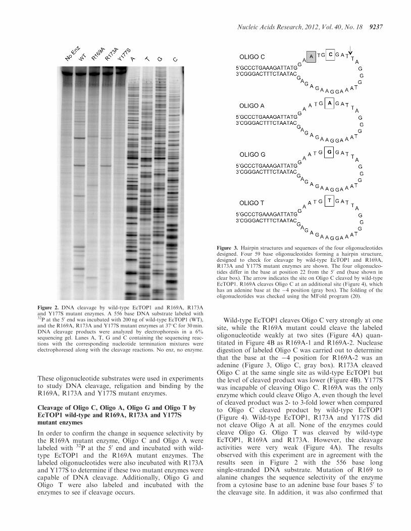
These oligonucleotide substrates were used in experimentsto study DNA cleavage, religation and binding by theR169A, R173A and Y177S mutant enzymes.
Cleavage of Oligo C, Oligo A, Oligo G and Oligo T byEcTOP1 wild-type and R169A, R173A and Y177Smutant enzymes
In order to confirm the change in sequence selectivity bythe R169A mutant enzyme, Oligo C and Oligo A werelabeled with 32P at the 50 end and incubated with wild-type EcTOP1 and the R169A mutant enzymes. Thelabeled oligonucleotides were also incubated with R173Aand Y177S to determine if these two mutant enzymes werecapable of DNA cleavage. Additionally, Oligo G andOligo T were also labeled and incubated with theenzymes to see if cleavage occurs.
Wild-type EcTOP1 cleaves Oligo C very strongly at onesite, while the R169A mutant could cleave the labeledoligonucleotide weakly at two sites (Figure 4A) quan-titated in Figure 4B as R169A-1 and R169A-2. Nucleasedigestion of labeled Oligo C was carried out to determinethat the base at the �4 position for R169A-2 was anadenine (Figure 3, Oligo C, gray box). R173A cleavedOligo C at the same single site as wild-type EcTOP1 butthe level of cleaved product was lower (Figure 4B). Y177Swas incapable of cleaving Oligo C. R169A was the onlyenzyme which could cleave Oligo A, even though the levelof cleaved product was 2- to 3-fold lower when comparedto Oligo C cleaved product by wild-type EcTOP1(Figure 4). Wild-type EcTOP1, R173A and Y177S didnot cleave Oligo A at all. None of the enzymes couldcleave Oligo G. Oligo T was cleaved by wild-typeEcTOP1, R169A and R173A. However, the cleavageactivities were very weak (Figure 4A). The resultsobserved with this experiment are in agreement with theresults seen in Figure 2 with the 556 base longsingle-stranded DNA substrate. Mutation of R169 toalanine changes the sequence selectivity of the enzymefrom a cytosine base to an adenine base four bases 50 tothe cleavage site. In addition, it was also confirmed that
Figure 2. DNA cleavage by wild-type EcTOP1 and R169A, R173Aand Y177S mutant enzymes. A 556 base DNA substrate labeled with32P at the 50 end was incubated with 200 ng of wild-type EcTOP1 (WT),and the R169A, R173A and Y177S mutant enzymes at 37�C for 30min.DNA cleavage products were analyzed by electrophoresis in a 6%sequencing gel. Lanes A, T, G and C containing the sequencing reac-tions with the corresponding nucleotide termination mixtures wereelectrophoresed along with the cleavage reactions. No enz, no enzyme.
Figure 3. Hairpin structures and sequences of the four oligonucleotidesdesigned. Four 59 base oligonucleotides forming a hairpin structure,designed to check for cleavage by wild-type EcTOP1 and R169A,R173A and Y177S mutant enzymes are shown. The four oligonucleo-tides differ in the base at position 22 from the 50 end (base shown inclear box). The arrow indicates the site on Oligo C cleaved by wild-typeEcTOP1. R169A cleaves Oligo C at an additional site (Figure 4), whichhas an adenine base at the �4 position (gray box). The folding of theoligonucleotides was checked using the MFold program (20).
Nucleic Acids Research, 2012, Vol. 40, No. 18 9237

although less active, R173A retained the wild-typeEcTOP1 cleavage sequence selectivity.
DNA religation by wild-type EcTOP1, R169A and R173A
The ability of these mutant enzymes to religate DNA wasalso compared to wild-type EcTOP1. The R169A enzymehas a preference for an adenine at the �4 position forDNA cleavage and since it cleaved Oligo A better thanOligo C, Oligo A was used to assay the DNA religationactivity of R169A enzyme. Oligo C was used to assay thereligation activity of wild-type EcTOP1 and R173Amutant enzyme.As seen in Figure 5, wild-type EcTOP1 efficiently
religated Oligo C post cleavage. Although cleavage of
Oligo C by R173A was weaker than wild-type, themutant was extremely efficient at religating Oligo C postcleavage. Similarly, R169A demonstrated efficientreligation of Oligo A post cleavage. These results suggestthat mutation of the R169 and R173 residues to alaninedoes not affect the ability of the enzyme to religate DNAdespite weaker/differential cleavage activity. Since theY177S mutant enzyme demonstrated a complete loss incleavage activity, the religation activity of this mutantcould not be assessed.
Figure 5. Oligo C religation by wild-type EcTOP1 and R173A; OligoA religation by R169A. Topoisomerase cleavage reactions wereincubated with 0.5mM MgCl2 and 1 M NaCl to dissociate theenzyme from DNA after religation of the covalent cleaved complex.Aliquots of reactions were stopped at the indicated time points andelectrophoresed in a 15% sequencing gel. The levels of DNA substrateand cleaved products were analyzed by Phosphorimager. No enz, noenzyme.
Figure 4. Cleavage of Oligo C, Oligo A, Oligo G and Oligo T byEcTOP1 wild-type, and R169A, R173A and Y177S mutant enzymes.(A) Oligo C, Oligo A, Oligo G and Oligo T labeled with 32P at the 50
end were incubated with wild-type EcTOP1 and R169A, R173A andY177S mutant enzymes at 37�C for 10min. DNA cleavage productswere analyzed by electrophoresis in a 15% sequencing gel. No, noenzyme; WT, wild-type; 169, R169A; 173, R173A; 77, Y177S. (B)The percent of Oligo C cleaved product by wild-type EcTOP1,R169A and R173A enzymes and percent of Oligo A cleaved productby R169A mutant enzyme was quantitated with spot densitometryanalysis. R169A-1 and R169A-2 represent the two cleaved productsof Oligo C from top to bottom observed in part A. The average andSD from at least three separate experiments is shown.
9238 Nucleic Acids Research, 2012, Vol. 40, No. 18

Binding of wild-type EcTOP1 and the R169A, R173A andY177S mutant enzymes to the hairpin oligonucleotides
The R169A and R173A mutant enzymes demonstratedefficient religation activity, while their ability to cleaveDNA was compromised when compared to wild-type.The Y177S mutant was unable to cleave DNA. Theseresults pose an important question: is the decreased/differ-ential DNA cleavage activity observed by the mutantenzymes due to decreased affinity to the DNA substrateor due to inefficient positioning of the DNA–proteininteractions for the cleavage step to take place? Theimportance of R169, R173 and Y177 residues in selectivityof the cytosine base at the �4 position encompasses thepossibility of accurately positioning the DNA substraterelative to the catalytic residues in the active site suchthat the tyrosine side chain responsible for DNAcleavage has access to the scissile DNA phosphate.Hence, to determine the effect of the selectivity of the�4 cytosine base on initial DNA binding, fluorescenceanisotropy binding assays were performed. Oligo C,Oligo A and Oligo G were labeled with6-carboxyfluorescein at the 30 end and six different con-centrations of the labeled oligonucleotides were titratedwith increasing concentrations of wild-type EcTOP1,R169A, R173A and Y177S mutant enzymes. LabeledOligo C, which has a cytosine base at the �4 position,was titrated with wild-type EcTOP1, R169A, R173Aand Y177S mutant enzymes. Oligo A, which has anadenine base at the �4 position, was cleaved only by theR169A mutant enzyme (Figure 4A). Hence, titrations forlabeled Oligo A were performed with the R169A mutantenzyme and wild-type EcTOP1 for comparison. Since nocleavage was observed for Oligo G by either wild-type orthe mutant enzymes, it would be interesting to see if theenzymes are still capable of binding to this oligonucleo-tide. Hence labeled Oligo G was titrated with wild-typeEcTOP1 and the R169A, R173A and Y177S mutantenzymes. The change in anisotropy upon increasing con-centration of enzyme was measured using a fluorescencespectrophotometer and the data were fit in GraphPadPrism using the equation for Binding Ligand Depletionusing Anisotropy. The Kd values obtained by fitting thedata are shown in Table 1.
Previous experiments performed with an EcTOP1mutant enzyme with a substitution of an arginineresidue R195 to alanine demonstrated the importance ofthis residue in DNA phosphate binding (21). Hence,EcTOP1 R195A mutant enzyme was used as a positivecontrol to check for the validity of using fluorescence an-isotropy as a technique to measure DNA binding affinity.The Kd value obtained with the EcTOP1 R195A mutantenzyme was �3.5-fold higher than wild-type (data notshown), similar to the estimation previously made fromthe results of gel shift assay (22).
As expected, wild-type EcTOP1 binds to Oligo Cstrongly (Kd=0.31 nM) (Table 1). R173A and Y177Senzymes bind to Oligo C with weaker affinity than wild-type EcTOP1; but the Kd values still indicate strongbinding (<2-fold reduction when compared to wild-type)(Table 1).
Wild-type EcTOP1 is incapable of cleaving Oligo A(Figure 4); however the binding affinity of the wild-typeenzyme to Oligo A is comparable to R169A (Table 1),which is the only enzyme which cleaves Oligo A. Thisresult suggests that recognition of the base at the �4position for DNA cleavage may not be indicated by thesubstrate binding affinity. This is confirmed by the com-parable Kd value observed for wild-type EcTOP1 bindingto Oligo G (Table 1). As observed in the oligonucleotidecleavage assay, neither the wild-type EcTOP1 nor themutant enzymes cleave Oligo G; but the binding affinityof wild-type EcTOP1 to Oligo G is comparable to thebinding affinity of wild-type EcTOP1 to Oligo C.It was interesting to see that the binding affinity of
R169A to Oligo A was stronger than its binding affinityto Oligo C or Oligo G (Table 1). The Kd value for R169Abinding to Oligo C was 0.86 nM, which is �2.7-foldweaker than wild-type-Oligo C binding. The R169Abinding data for all three oligonucleotides tested indicatethat along with the change in cleavage selectivity,mutation of this residue does affect the substrate bindingaffinity of the enzyme to some extent.The Kd values obtained for the R173A mutant enzyme
are more similar to the affinities for wild-type EcTOP1,indicating that this mutation does not affect the substratebinding affinity of the enzyme as significantly as R169A. Asimilar result was observed for the Y177S mutant enzyme;the substrate binding affinities of Y177S are comparableto wild-type. Hence, the total lack of DNA cleavage orrelaxation activity by the Y177S mutant enzyme is not dueto loss of its binding affinity for the DNA substrate. Themutation is also unlikely to have resulted in a large changein protein folding.
DISCUSSION
Bacterial and archaeal topoisomerase I as well as reversegyrase display a sequence selectivity of a cytosine base 4bases 50 from the DNA cleavage site. The biochemicalbasis for this selectivity was previously unknown.
Table 1. Dissociation constants (Kd) obtained for binding of
wild-type EcTOP1, R169A, R173A and Y177S mutant enzymes to
30-labeled Oligo C, Oligo A and Oligo G
30-labeled oligonucleotide Enzyme Kd (nM)
Oligo C Wild-type 0.31±0.05R169A 0.86±0.15R173A 0.57±0.14Y177S 0.48±0.02
Oligo A Wild-type 0.40±0.12R169A 0.55±0.12
Oligo G Wild-type 0.50±0.02R169A 0.97±0.19R173A 0.61±0.09Y177S 0.46±0.08
Kd values of the wild-type and mutant enzymes binding to the oligo-nucleotide substrates were obtained by fitting the fluorescence anisot-ropy binding data to an equation measuring binding ligand depletionusing anisotropy in GraphPad Prism. Values represent average and SDfrom three separate experiments.
Nucleic Acids Research, 2012, Vol. 40, No. 18 9239

Recently, the solved crystal structure of EcTOP1 D111Nmutant enzyme in covalent complex with a cleavedsingle-stranded DNA shed light upon the residues whichinteract with the cytosine at the �4 position relative to thecleavage site (14). These residues R169, R173 and Y177were mutated in order to analyze their biochemical role inthe recognition and selectivity of the cytosine at the �4position. Each of the mutant enzymes demonstrated bio-chemical properties distinct from each other.Mutation of only one of the residues, R169 to alanine,
resulted in a change in the sequence selectivity of EcTOP1.R169A mutant enzyme displayed higher cleavage activitywhen the base at the �4 position is an adenine comparedto a cytosine (Figure 4). Once cleaved, the R169A mutantwas efficient at religating cleaved DNA (Figure 5).Although R169A cleaved DNA with higher efficiencywhen the base at the �4 position is an adenine (Oligo A,Figure 4) as opposed to a cytosine (Oligo C, Figure 4), thecleavage activity of R169A was lower than wild-typeEcTOP1 acting on Oligo C (Figure 4B). The mutant wasfound to have a 150-fold decrease in DNA relaxationactivity compared to wild-type (Figure 1A). The decreasedrelaxation activity could be explained in part also by thelower binding affinity of R169A to DNA substrate whencompared to wild-type EcTOP1 (Table 1). The solvedcrystal structure of EcTOP1 D111N mutant enzyme incovalent complex with DNA determined that the pocketcreated at the �4 position is capable of only accomodatinga cytosine. R169 plays an important role in stericallyrestricting the choice of the base at the �4 position(Figure 6A). Mutation of R169 to alanine creates alarger pocket (Figure 6B, arrow) which could accom-modate and interact with a larger base like adenine(Figure 6C). Measurement of the distance between thealanine residue and the adenine base in the modelindicated the possibility of van der Waals interaction(data not shown). The extra space created due to thelarger pocket in this R169A mutant would not allowtight interactions between the residues and the �4 baseif the base is a pyrimidine. Although sterically thepocket could potentially fit a guanine base, the datasuggests that the R169A mutant might be incapable ofinteracting non-covalently with a guanine as the �4base, demonstrated by the inability of the R169Amutant to cleave Oligo G (Figure 4). Modelling studiesindicated a steric interference in interaction with R173when the base at the �4 position is a guanine (data notshown).Mutation of R173 to an alanine retained the selectivity
of a cytosine at the �4 position. The R173A mutantcleaved DNA with sequence selectivity similar to wild-type EcTOP1; however, the level of cleaved productobserved was lower (Figures 2 and 4B). Mutation ofresidue R173 did not affect the binding affinity of theenzyme significantly (Table 1) and also did not changethe ability of the enzyme to religate cleaved DNA(Figure 5). Since binding and religation were unaffecteddue to the mutation, the decrease in the cleavage activitycould account for the lower rate of relaxation by R173Awhen compared to wild-type EcTOP1 (Figure 1A).Modeling of the R173 residue around the cytosine at the
�4 position showed that this residue does not contributesterically to the selection of the cytosine base, but isimportant for non-covalently interacting with the base atthe �4 position (Figure 7A). Mutation of R173 to alaninewould interfere with the ability of the residue to hydrogenbond with the cytosine base at the �4 position (Figure 7A)which then might affect the cleavage activity of the mutantenzyme compared to wild-type EcTOP1.
Mutation of the Y177 residue to a serine renders theenzyme completely inactive. The Y177S mutant is notaffected in non-covalent binding affinity to DNA sub-strate (Table 1), but it is incapable of DNA relaxationand cleavage (Figures 1A, 2 and 4A). These resultssuggest a vital catalytic role for residue Y177 after theinitial binding of DNA substrate, even though it is distalto the active site Y319 and the scissile phosphate. Asobserved in the crystal structure, the side chain ofresidue Y177 forms a wedge between the bases at the �4and the �5 position. The phenol ring of Y177 stacksparallel to the guanine base at the �5 position andperpendicular to the cytosine base at the �4 position(Figure 7B). Y177 interacts with the bases at the �4 andthe �5 position via p–p interactions. Y177 is strictlyconserved in bacterial topoisomerase I and the corres-ponding residue in archaeal reverse gyrase is also strictlyconserved as the aromatic amino acid phenylalanine.Mutation of Y177 to a serine residue would abolish theinteraction of this residue with the bases at the �4 and the�5 position (Figure 7B). The major impact of mutatingY177 to a serine, however, would be on the wedgingaction of this residue. The selectivity of the cytosine atthe �4 position might be an important step in the con-formational change of the enzyme–DNA complex afterinitial binding so that the active site tyrosine could beprecisely positioned for cleaving the single strand. Thewedge created by Y177 could be a mechanism to latchon to the DNA strand upon appropriate recognition ofthe cytosine base, so that the active site tyrosine hydroxylnucleophile has access to the scissile phosphate. Thismechanism of recognizing the substrate at a site distantfrom the cleavage site is known as Molecular RulerMechanism. An example of the Molecular Ruler mechan-ism has been demonstrated in Type I restriction modifica-tion enzymes. These enzymes consist of three subunits;restriction, methylation and specificity subunits. The spe-cificity subunit binds to two DNA sequences spaced5–8 bp apart and determines whether the enzyme wouldact as an endonuclease or methylase. These enzymes differfrom Type II and Type III restriction modificationenzymes, because cleavage or methylation by Type Ienzymes occur at the site distinct from the recognitionsite (23). The crystal structure of the specificity subunitof a Type I R-M enzyme from Methanococcus jannaschiirevealed the presence of two highly conserved regionswhich form a coiled coil that separates the two DNAbinding domains, thereby acting as a Molecular Rulerfor the spacing between the two sequences bound by theDNA binding domain. Their results also suggestedbending of the target DNA for exposure of the adeninebases to be methylated by the methylation subunit of theenzyme (24). Molecular rulers are very common among
9240 Nucleic Acids Research, 2012, Vol. 40, No. 18

protein–protein interactions as well. An example is thecleavage of the thyrotropin receptor into two subunitsby what is presumed to be a matrix metalloprotease.Cleavage of the receptor by the enzyme occurs at thefixed distance from the protease attachment site (25).Deletion of residues on the receptor at the protease attach-ment site decreased the cleavage activity and shifted thecleavage site to an upstream region.Hence, recognition and selectivity of the cytosine base
at the �4 position could be due to R169, R173 and Y177residues functioning as a Molecular Ruler. These residuesare therefore strictly conserved only in Type IA topoisom-erases that maintain this cleavage sequence selectivity. Thecombined action of these three residues might be import-ant in inducing conformational change in the enzyme–DNA complex after initial DNA binding to appropriatelyposition or activate the active site tyrosine for cleavage ofDNA. Initial DNA binding is likely to occur via residuesrecognizing DNA ribose-phosphate as seen in previouscrystal structures of non-covalent complex betweenDNA and Type IA topoisomerases (26). These includeresidues E115, R168, D172, S192, R195 and Q197 ofEcTOP1 that have been shown in previous mutagenesisstudies to be important for DNA relaxation activity(14,21). Unlike R169, R173 and Y177 responsible forinteraction with the cytosine base, these residues thatinteract with the DNA backbone are conserved amongall Type IA topoisomerases regardless of their cleavagesequence selectivity (27).Based on the results obtained in this study along with
previous studies of EcTOP1, we propose the followingsequence of events: (a) EcTOP1 binds to DNA viaresidues that recognize the DNA ribose-phosphatebackbone leading to initial complex formation. (b) Afterinitial complex formation, residue R169 recognizes acytosine base at the �4 position relative to the site thatneeds to be cleaved. (c) Once recognized, non-covalentinteractions of residues R169, R173 and Y177 with thecytosine base at the �4 position and the wedging actionof the Y177 residue lead to a change in conformation ofthe enzyme–DNA complex so that the active site tyrosineis positioned or activated for efficient DNA cleavage,thereby acting as a Molecular Ruler.The crystal structure of the D111N mutant in covalent
complex in single-stranded DNA oligonucleotide sub-strate revealed a kink in the DNA phosphate-deoxyribosechain of the oligonucleotide between the �4 and �5 baseswhich disrupts the stacking of the bases (14). A recentstudy reported a novel, previously uncharacterizedsequence of events during DNA cleavage by human topo-isomerase II wherein post non-specific DNA binding, the
Figure 6. Modeling of the interaction of R169 and its alanine substi-tution mutant with the base at the �4 position. (A) Figure shows thesurface representation of the wild-type enzyme bound to a single-stranded oligonucleotide with a cytosine base at the �4 position.R169 sterically restricts the choice of the base at the �4 position.
Figure 6. Continued(B) Mutating R169 to alanine creates a larger pocket in the regionsurrounding the base at the �4 position (arrow). (C) This largerpocket created due to the R169A mutant could accommodate alarger base like adenine. The figures were made by mutating theR169 residue in PyMOL using the solved crystal structure of D111NEcTOP1 mutant enzyme in covalent complex with single-strandedDNA (PDB ID 3PX7). Substitution of the cytosine base to adeninewas performed using winCOOT (19).
Nucleic Acids Research, 2012, Vol. 40, No. 18 9241

enzyme induces a sharp bend in DNA in a sequencespecific manner prior to DNA cleavage (28). Their datasuggested a tight coordination between DNA bending andDNA cleavage at a selected site by topoisomerase II. It ispossible that the kink observed in the sugar-phosphatebackbone in the crystal structure of EcTOP1 D111Nbound to single-stranded DNA could be employed in co-ordination with the recognition of the cytosine base at the�4 position to facilitate DNA cleavage. Further studiesshould be carried out to elucidate further details of thismechanism in bacterial topoisomerase I action.
SUPPLEMENTARY DATA
Supplementary Data are available at NAR Online:Supplementary Figure 1.
FUNDING
National Institutes of Health (NIH) [R01 GM054226 toY.T.]. Funding for open access charge: NIH [R01GM054226].
Conflict of interest statement. None declared.
REFERENCES
1. Schoeffler,A.J. and Berger,J.M. (2008) DNA topoisomerases:Harnessing and constraining energy to govern chromosometopology. Q. Rev. Biophys., 41, 41–101.
2. Corbett,K.D. and Berger,J.M. (2004) Structure, molecularmechanisms, and evolutionary relationships in DNAtopoisomerases. Annu. Rev. Biophys. Biomol. Struct., 33, 95–118.
3. Forterre,P., Gribaldo,S., Gadelle,D. and Serre,M.C. (2007) Originand evolution of DNA topoisomerases. Biochimie, 89, 427–446.
4. Pommier,Y., Leo,E., Zhang,H. and Marchand,C. (2010) DNAtopoisomerases and their poisoning by anticancer andantibacterial drugs. Chem. Biol., 17, 421–433.
5. Nitiss,J.L. (2009) Targeting DNA topoisomerase II in cancerchemotherapy. Nat. Rev. Cancer, 9, 338–350.
6. Tse-Dinh,Y.C. (2007) Exploring DNA topoisomerases as targetsof novel therapeutic agents in the treatment of infectious diseases.Infect. Disord. Drug Targets, 7, 3–9.
7. Collin,F., Karkare,S. and Maxwell,A. (2011) Exploiting bacterialDNA gyrase as a drug target: current state and perspectives.Appl. Microbiol. Biotechnol., 92, 479–497.
8. Bradbury,B.J. and Pucci,M.J. (2008) Recent advances in bacterialtopoisomerase inhibitors. Curr. Opin. Pharmacol., 8, 574–581.
9. Tse-Dinh,Y.C. (2009) Bacterial topoisomerase I as a target fordiscovery of antibacterial compounds. Nucleic Acids Res., 37,731–737.
10. Tse,Y.C., Kirkegaard,K. and Wang,J.C. (1980) Covalent bondsbetween protein and DNA. formation of phosphotyrosine linkagebetween certain DNA topoisomerases and DNA. J. Biol. Chem.,255, 5560–5565.
11. Dean,F., Krasnow,M.A., Otter,R., Matzuk,M.M., Spengler,S.J.and Cozzarelli,N.R. (1983) Escherichia coli type-1 topoisomerases:identification, mechanism, and role in recombination. Cold SpringHarb. Symp. Quant. Biol., 47(Pt 2), 769–777.
12. Annamalai,T., Dani,N., Cheng,B. and Tse-Dinh,Y.C. (2009)Analysis of DNA relaxation and cleavage activities ofrecombinant Mycobacterium tuberculosis DNA topoisomerase Ifrom a new expression and purification protocol. BMC Biochem.,10, 18.
13. Viard,T. and de la Tour,C.B. (2007) Type IA topoisomerases: Asimple puzzle? Biochimie, 89, 456–467.
14. Zhang,Z., Cheng,B. and Tse-Dinh,Y.C. (2011) Crystal structureof a covalent intermediate in DNA cleavage and rejoining byEscherichia coli DNA topoisomerase I. Proc. Natl Acad. Sci.USA, 108, 6939–6944.
15. Narula,G., Annamalai,T., Aedo,S., Cheng,B., Sorokin,E.,Wong,A. and Tse-Dinh,Y.C. (2011) The strictly conserved arg-321residue in the active site of Escherichia coli topoisomerase I playsa critical role in DNA rejoining. J. Biol. Chem., 286,18673–18680.
16. Sorokin,E.P., Cheng,B., Rathi,S., Aedo,S.J., Abrenica,M.V. andTse-Dinh,Y.C. (2008) Inhibition of Mg2+ binding and DNAreligation by bacterial topoisomerase I via introduction of anadditional positive charge into the active site region.Nucleic Acids Res., 36, 4788–4796.
17. Doyle,S.A. (2005) High-throughput cloning for proteomicsresearch. Methods Mol. Biol., 310, 107–113.
18. Zhu,C.X. and Tse-Dinh,Y.C. (1999) Overexpression andpurification of bacterial DNA topoisomerase I. MethodsMol. Biol., 94, 145–151.
19. Emsley,P., Lohkamp,B., Scott,W.G. and Cowtan,K. (2010)Features and development of coot. Acta Crystallogr.D. Biol. Crystallogr., 66, 486–501.
20. Zuker,M. (2003) Mfold web server for nucleic acid folding andhybridization prediction. Nucleic Acids Res., 31, 3406–3415.
21. Cheng,B., Feng,J., Mulay,V., Gadgil,S. and Tse-Dinh,Y.C. (2004)Site-directed mutagenesis of residues involved in G strand DNAbinding by Escherichia coli DNA topoisomerase I. J. Biol. Chem.,279, 39207–39213.
22. Cheng,B., Feng,J., Gadgil,S. and Tse-Dinh,Y.C. (2004) Flexibilityat Gly-194 is required for DNA cleavage and relaxation activity
Figure 7. Modeling of the interaction of R173 and Y177 and theirsubstitution mutants with the cytosine base at the �4 position.(A) Figure depicts the effect of mutating R173 to alanine (A173, red)on its interaction with the cytosine base. The hydrogen bond formed isdepicted by the yellow dashed line. (B) Figure depicts the effect ofmutating Y177 to serine (S177, red) on its interaction with thecytosine base. S177 would be incapable of forming the wedge. Thefigures were made by mutating the R173 and Y177 residues inPyMOL starting from the solved crystal structure of D111N EcTOP1mutant enzyme in covalent complex with single-stranded DNA (PDBID 3PX7).
9242 Nucleic Acids Research, 2012, Vol. 40, No. 18

of Escherichia coli DNA topoisomerase I. J. Biol. Chem., 279,8648–8654.
23. Murray,N.E. (2000) Type I restriction systems: Sophisticatedmolecular machines (a legacy of bertani and weigle). Microbiol.Mol. Biol. Rev., 64, 412–434.
24. Kim,J.S., DeGiovanni,A., Jancarik,J., Adams,P.D., Yokota,H.,Kim,R. and Kim,S.H. (2005) Crystal structure of DNA sequencespecificity subunit of a type I restriction-modification enzyme andits functional implications. Proc. Natl Acad. Sci. USA, 102,3248–3253.
25. Tanaka,K., Chazenbalk,G.D., McLachlan,S.M. and Rapoport,B.(2000) Evidence that cleavage of the thyrotropin receptor involves
a ‘‘Molecular Ruler’’ mechanism: Deletion of amino acid residues305-320 causes a spatial shift in cleavage site 1 independent ofamino acid motif. Endocrinology, 141, 3573–3577.
26. Baker,N.M., Rajan,R. and Mondragon,A. (2009) Structuralstudies of type I topoisomerases. Nucleic Acids Res., 37, 693–701.
27. Caron,P.R. (1999) Compendium of DNA topoisomerasesequences. Methods Mol. Biol., 94, 279–316.
28. Lee,S., Jung,S.R., Heo,K., Byl,J.A., Deweese,J.E., Osheroff,N.and Hohng,S. (2012) DNA cleavage and opening reactions ofhuman topoisomerase II alpha are regulated via Mg2+-mediateddynamic bending of gate-DNA. Proc. Natl Acad. Sci. USA, 109,2925–2930.
Nucleic Acids Research, 2012, Vol. 40, No. 18 9243
Copyright © 2022 FDOKUMEN