Side by side comparison between dynamic versus static models of blood–brain barrier in vitro: A...
13
Research Report Side by side comparison between dynamic versus static models of blood–brain barrier in vitro: A permeability study Stefano Santaguida a , Damir Janigro a,c,d , Mohammed Hossain a , Emily Oby a , Edward Rapp b , Luca Cucullo a, ⁎ a Division of Cerebrovascular Research, Cleveland Clinic Lerner College of Medicine, Cleveland, OH 44106, USA b Clevemed, Cleveland Clinic Lerner College of Medicine, Cleveland, OH 44106, USA c Department of Neurosurgery, Cleveland Clinic Lerner College of Medicine, Cleveland, OH 44106, USA d Department of Molecular Medicine, Cleveland Clinic Lerner College of Medicine, Cleveland, OH 44106, USA ARTICLE INFO ABSTRACT Article history: Accepted 11 June 2006 Endothelial cells in vivo are continuously exposed to shear stress, a tangential force generated by the flow of blood across their apical surfaces that affects endothelial cell structure and function. By contrast, the Transwell apparatus cannot reproduce the presence of intraluminal blood flow that is essential for the formation and differentiation of the BBB. In contrast, the dynamic in vitro model of the BBB (DIV-BBB) mimics both functionally and anatomically the brain microvasculature, creating quasi-physiological conditions for co- culturing human and non-human endothelial cells and astrocytes in a capillary-like structure. We used intraluminal bovine aortic endothelial cells (BAEC) co-cultured with extraluminal glial cells (C6) to obtain elevated trans-endothelial electrical resistance (TEER) and selective permeability to sucrose and phenytoin. The experiments were performed in parallel using Transwell systems DIV-BBB models and data were then cross compared. By contrast with Transwell, C6 and BAEC co-cultured in the DIV-BBB demonstrated predominantly aerobic metabolism evidenced by a robust increase in glucose consumption that was paralleled by a similar change in lactate production. BAEC exposed to glia under dynamic conditions grow in a monolayer fashion and developed a more stringent barrier as demonstrated by high TEER values and a selective permeability to [ 14 C] phenytoin and the well-known paracellular marker [ 3 H] sucrose. In conclusion, these data demonstrate that the exposure to intraluminal flow plays an essential role in promoting endothelial cell differentiation and increasing BBB tightness, thus making the use of the DIV-BBB well suited for pharmacological studies. © 2006 Elsevier B.V. All rights reserved. Keywords: Drug delivery Gene therapy Cerebral blood flow Shear stress Glia–vascular interaction 1. Introduction The BBB is found in the brain of all vertebrates (Abbott, 2005). Morphologically, the BBB is formed by specialized endothelial cells (ECs) paving the luminal side of brain microvessels in close association with abluminal astrocytic end-feet processes sharing the basal lamina and enveloping more than 98% of the BBB endothelium (Emmi et al., 2000). Astrocytes interact with the BRAIN RESEARCH XX (2006) XXX – XXX ⁎ Corresponding author. Project Scientist, Cleveland Clinic Foundation NB20, Neurosurgery, 9500 Euclid Avenue/NB20, Cleveland, OH 44195, USA. Fax: +1 216 444 1466. E-mail address: [email protected] (L. Cucullo). BRES-35748; No. of pages: 13: 4C 0006-8993/$ – see front matter © 2006 Elsevier B.V. All rights reserved. doi:10.1016/j.brainres.2006.06.027 available at www.sciencedirect.com www.elsevier.com/locate/brainres ARTICLE IN PRESS
Transcript of Side by side comparison between dynamic versus static models of blood–brain barrier in vitro: A...
B R A I N R E S E A R C H X X ( 2 0 0 6 ) X X X – X X X
BRES-35748; No. of pages: 13: 4C
ava i l ab l e a t www.sc i enced i rec t . com
www.e l sev i e r. com/ l oca te /b ra in res
ARTICLE IN PRESS
Research Report
Side by side comparison between dynamic versus staticmodels of blood–brain barrier in vitro: A permeability study
Stefano Santaguidaa, Damir Janigroa,c,d, Mohammed Hossaina, Emily Obya,Edward Rappb, Luca Cuculloa,⁎aDivision of Cerebrovascular Research, Cleveland Clinic Lerner College of Medicine, Cleveland, OH 44106, USAbClevemed, Cleveland Clinic Lerner College of Medicine, Cleveland, OH 44106, USAcDepartment of Neurosurgery, Cleveland Clinic Lerner College of Medicine, Cleveland, OH 44106, USAdDepartment of Molecular Medicine, Cleveland Clinic Lerner College of Medicine, Cleveland, OH 44106, USA
A R T I C L E I N F O
⁎ Corresponding author. Project Scientist, Cl44195, USA. Fax: +1 216 444 1466.
E-mail address: [email protected] (L. Cucullo
0006-8993/$ – see front matter © 2006 Elsevidoi:10.1016/j.brainres.2006.06.027
A B S T R A C T
Article history:Accepted 11 June 2006
Endothelial cells in vivo are continuously exposed to shear stress, a tangential forcegenerated by the flow of blood across their apical surfaces that affects endothelial cellstructure and function. By contrast, the Transwell apparatus cannot reproduce the presenceof intraluminal blood flow that is essential for the formation and differentiation of the BBB.In contrast, the dynamic in vitro model of the BBB (DIV-BBB) mimics both functionally andanatomically the brain microvasculature, creating quasi-physiological conditions for co-culturing human and non-human endothelial cells and astrocytes in a capillary-likestructure. We used intraluminal bovine aortic endothelial cells (BAEC) co-cultured withextraluminal glial cells (C6) to obtain elevated trans-endothelial electrical resistance (TEER)and selective permeability to sucrose and phenytoin. The experiments were performed inparallel using Transwell systems DIV-BBB models and data were then cross compared. Bycontrast with Transwell, C6 and BAEC co-cultured in the DIV-BBB demonstratedpredominantly aerobic metabolism evidenced by a robust increase in glucoseconsumption that was paralleled by a similar change in lactate production. BAEC exposedto glia under dynamic conditions grow in a monolayer fashion and developed a morestringent barrier as demonstrated by high TEER values and a selective permeability to [14C]phenytoin and the well-known paracellular marker [3H] sucrose. In conclusion, these datademonstrate that the exposure to intraluminal flow plays an essential role in promotingendothelial cell differentiation and increasing BBB tightness, thus making the use of theDIV-BBB well suited for pharmacological studies.
© 2006 Elsevier B.V. All rights reserved.
Keywords:Drug deliveryGene therapyCerebral blood flowShear stressGlia–vascular interaction
1. Introduction
The BBB is found in the brain of all vertebrates (Abbott, 2005).Morphologically, the BBB is formed by specialized endothelial
eveland Clinic Foundatio
).
er B.V. All rights reserved
cells (ECs) paving the luminal side of brain microvessels in closeassociationwith abluminal astrocytic end-feet processes sharingthe basal lamina and enveloping more than 98% of the BBBendothelium (Emmi et al., 2000). Astrocytes interact with the
n NB20, Neurosurgery, 9500 Euclid Avenue/NB20, Cleveland, OH
.
2 B R A I N R E S E A R C H X X ( 2 0 0 6 ) X X X – X X X
ARTICLE IN PRESS
cerebral endothelium to determine BBB function; they alsoregulate protein expression, modulate endothelium differentia-tion, and appear to be critical for the induction andmaintenanceof the tight junctions and BBB properties (Abbott, 2005; Liberto etal., 2004; Stanness et al., 1997;Mizuguchi et al., 1997; Hammet al.,2004; Hori et al., 2004; Kramer et al., 2002). The microvascularendothelium at the BBB is characterized by the presence of tightjunctions (zonulae occludentes), lack of fenestrations, andminimal pinocytotic vesicles. These distinct morphologicalproperties of the BBB account for the “restraining” nature ofbrain capillary ECs. In particular, tight junctions between thecerebral endothelial cells form a diffusion barrier, which
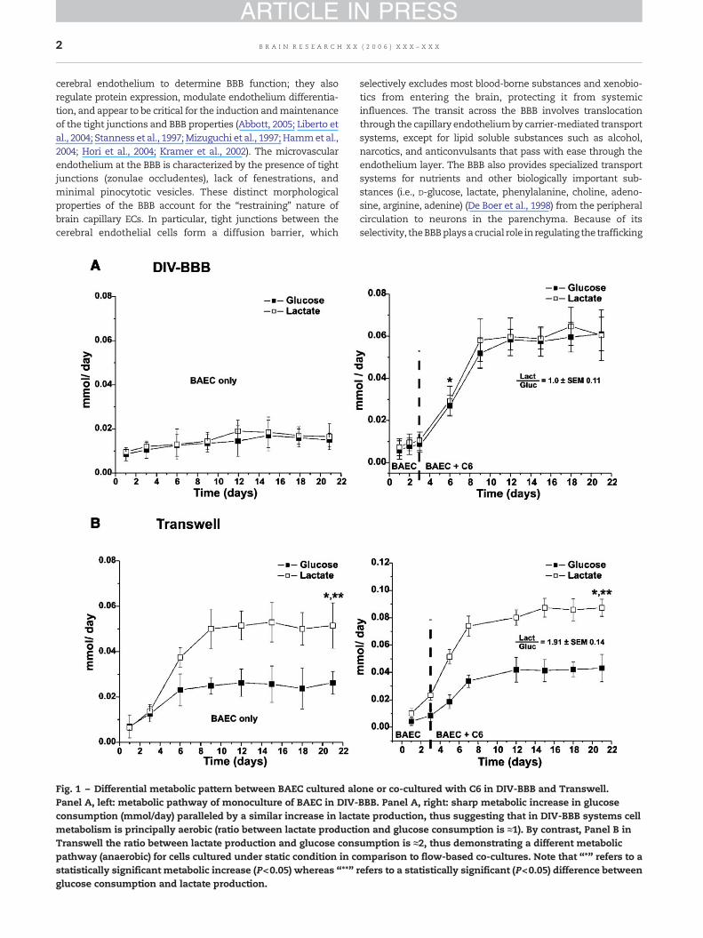
Fig. 1 – Differential metabolic pattern between BAEC cultured alPanel A, left: metabolic pathway of monoculture of BAEC in DIV-consumption (mmol/day) paralleled by a similar increase in lactametabolism is principally aerobic (ratio between lactate productiTranswell the ratio between lactate production and glucose conspathway (anaerobic) for cells cultured under static condition in cstatistically significant metabolic increase (P<0.05) whereas “**”glucose consumption and lactate production.
selectively excludes most blood-borne substances and xenobio-tics from entering the brain, protecting it from systemicinfluences. The transit across the BBB involves translocationthrough the capillary endotheliumby carrier-mediated transportsystems, except for lipid soluble substances such as alcohol,narcotics, and anticonvulsants that pass with ease through theendothelium layer. The BBB also provides specialized transportsystems for nutrients and other biologically important sub-stances (i.e., D-glucose, lactate, phenylalanine, choline, adeno-sine, arginine, adenine) (De Boer et al., 1998) from the peripheralcirculation to neurons in the parenchyma. Because of itsselectivity, theBBBplaysacrucial role in regulating the trafficking
one or co-cultured with C6 in DIV-BBB and Transwell.BBB. Panel A, right: sharp metabolic increase in glucosete production, thus suggesting that in DIV-BBB systems cellon and glucose consumption is ≈1). By contrast, Panel B inumption is ≈2, thus demonstrating a different metabolicomparison to flow-based co-cultures. Note that “*” refers to arefers to a statistically significant (P<0.05) difference between
3B R A I N R E S E A R C H X X ( 2 0 0 6 ) X X X – X X X
ARTICLE IN PRESS
between blood and CNS (Hagenbuch et al., 2002) and thedetermination of neuroimmunology and neuropathology(Banks, 2005).
BBB dysfunction has been observed in the majority ofneurological diseases, but the causes of aberrant vascularbehavior are generally unknown. Genomic and proteomicanalyses are currently being used to examine BBB function inhealthy and diseased brain to better characterize this dynamicinterface (Shusta, 2005). For example, BBB permeability changesare involved in brain edema formation and central nervoussysteminjuries, suchasstroke (Heoetal., 2005), cerebralhypoxia-reoxygenation (BrownandDavis, 2005), head injuries (Korn et al.,2005), neurodegenerative diseases, and neurological infectionsincluding bacterial and viral meningitis, encephalitis, or HIV-1infection (Toborek et al., 2005; Hawkins and Davis, 2005). Howthese permeability changes affect drug passage into the CNS islargely unknown. For example, because most CNS drugs arelipophilic, how perivascular edema or general BBB failure affectsdrug permeability is still poorly understood.
In vitro studies on reconstituted BBB models help to revealthe direct effects of pathological conditions on cerebralendothelium (Krizanac-Bengez et al., 2004), but due to some-times high “resting” permeability values, the effects ofpathogenetic factors on the impairment of barrier integritymay be obscured. CNS drug design (e.g., antineoplastics,antivirals, and antiepileptics) cannot rely entirely uponphysicochemical properties to cross the BBB and remainslargely studied in vivo. For example, lipophilicity alone is apoor predictor for drug penetration into the CNS because itrelies on a passive, diffusional uptake. Many lipophylic drugsare potential substrates for efflux carriers of the BBB (particu-larly P-glycoprotein), which can drastically reduce theirpenetration into the brain (Abbott, 2002). Investigationsperformed on reliable in vitro models of BBB can be a suitabletool for testing efficacy and brain penetration of new drugs(Cucullo et al., 2002, 2005).
The aims of the present work were (1) to address some ofthe weaknesses of existing flow-based tissue culture appara-tus (Cucullo et al., 2005); and (2) to cross compare the
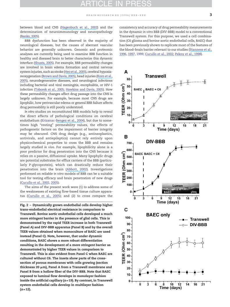
Fig. 2 – Dynamically grown endothelial cells develop highertrans-endothelial electrical resistance in comparison toTranswell. Bovine aortic endothelial cells developed a muchmore stringent barrier in the presence of glial cells. This isdemonstrated by the rapid TEER increase in both Transwell(Panel A) and DIV-BBB apparatus (Panel B) and by the overallTEER values obtained when monoculture of BAEC are usedinstead (Panel C). Note, however, that under dynamicconditions, BAEC shows a more robust differentiationresulting in the development of a more stringent barrier asdemonstrated by higher TEER values in comparison toTranswell. This is also evident from Panel C when BAEC arecultured without C6. The insets show parts of the cross-section of porous membranes with cells growing (sectionthickness 20 μm), Panel A from a Transwell membrane andPanel B from a hollow fiber of the DIV-BBB. Note that BAECexposed to luminal flow develops in monolayer fashioninside the artificial capillary (n=19). By contrast, in Transwellsystem endothelial cells develop in multilayer fashion(n=12).
consistency and accuracy of drug permeability measurementsin the dynamic in vitro BBB (DIV-BBB) model to a conventionalTranswell system. For this purpose, we used a cell combina-tion (C6 glioma and bovine aortic endothelial cells, BAEC) thathas been previously shown to replicate most of the features ofthe blood–brain barrier relevant to our studies (Stanness et al.,1996, 1997, 1999; Cucullo et al., 2002; Pekny et al., 1998).
4 B R A I N R E S E A R C H X X ( 2 0 0 6 ) X X X – X X X
ARTICLE IN PRESS
5B R A I N R E S E A R C H X X ( 2 0 0 6 ) X X X – X X X
ARTICLE IN PRESS
2. Results
2.1. Cell viability and BBB integrity
Monitoring of cell metabolism is a critical step in assessing theviability of dynamically grown endothelial cells or glia (Stan-ness et al., 1996, 1997). Fig. 1A shows early changes in glucoseconsumption and lactate production in bovine aortic endothe-lial cells grownalone (left panel) or in co-culturewithC6gliomacells in DIV-BBB (right panel). Note that in the DIV-BBB, theglucose consumption is always paralleled by a similar lactateproduction indicating predominance of aerobic metabolism(lactate production–glucose consumption ratio=1±SEM 0.08).The addition of glial cells to the extraluminal compartment(ECS) caused a similar robust increase in both glucose andlactate production. By contrast, in the Transwell apparatus(Fig. 1B), BAEC alone (left panel) or co-cultured with glial cells(rightpanel) displaya lactateproduction–glucoseconsumptionratio of 1.91±SEM 0.14, thus suggesting that in the absence offlow the metabolism is predominantly anaerobic (Desai et al.,2002).
Peripheral endothelial cells develop negligible barrierproperties in both DIV-BBB and Transwell when culturedalone (Stanness et al., 1997). However, a few days followingexposure to glial cells grown extraluminally and parallel tometabolic changes, trans-endothelial electrical resistanceincreased dramatically in both models. Transwell achieved astable electrical resistance of 57.2 Ω cm2±SEM 2.8 (Fig. 2A). Bycomparison, endothelial cells exposed to glia in the presenceof intraluminal flow, as in the DIV-BBB (Fig. 2B), developed ahigh stable TEER (650.7 Ω cm2±SEM 26.5). This demonstratesthat the presence of intraluminal flow is critical for thedifferentiation of endothelial cells and the development offunctional BBB properties in vivo, such us high TEER. Note thatdespite the lack of glial cells, BAEC exhibit proportionally amuch higher TEER when cultured under dynamic condition(Fig. 2C). The inserts in Fig. 2 show a cross section of aTranswell and a typical hollow fiber. Note that in contrast tothe Transwell apparatus (insert in Fig. 2A), endothelial cellsgrown under dynamic conditions (insert if Fig. 2B) develop in amonolayer fashion, thus mimicking the structural physiologyof capillaries in vivo.
2.2. Permeability experiment
The measurement of TEER across the endothelial cell mono-layer is indicative of barrier integrity, as previously reported(Cucullo et al., 2002, 2005; Krizanac-Bengez et al., 2006). Inaddition, assessment of the barrier functionwas carried out by
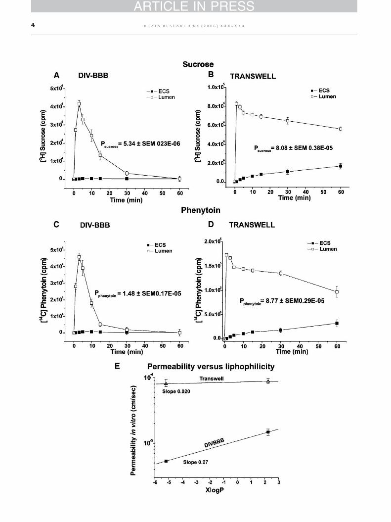
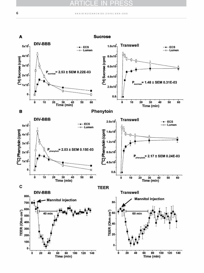
Fig. 3 – Sucrose and phenytoin permeability in DIV-BBB versusplots of counts per minute (cpm) vs. time from which permeabiliphenytoin (1.48±SEM 0.17×10⁻⁵ cm/s) in DIV-BBB was calculatedin Panels B and D) developed less stringent barriers resulting in0.38×10−5 cm/swhereas [14C] phenytoinwas 8.77±SEM 0.29×10⁻⁵
and phenytoin is 1×10⁻⁷ and 1.08×10⁻⁵ cm/s, respectively. Paneloil/water partition coefficient (XlogP) of sucrose and phenytoin ineasily discriminate between these two substances based on theiTranswell.
permeability measurements of selected molecules: phenytoin(a common antiepileptic drug) and sucrose (an establishedparacellular marker). A bolus containing [14C] phenytoin and[3H] sucrose was directly added to the medium contained inthe lumen in DIV-BBB and Transwell systems. Samples weretaken from both lumen and ECS over 60 min; [14C] phenytoinand [3H] sucrose flux were monitored by detection of theradioactive tracers. The permeability was calculated bygraphical integration of the concentration of the tracer in thelumen and in the ECS.
These molecules were chosen as paracellular markers fortheir inability to cross cell membranes. The permeabilityacross the endothelium in the DIV-BBB was 5.34±SEM0.23×10−6 cm/s for [3H]sucrose (Fig. 3A) and 1.48±SEM0.17×10−5 cm/s for [14C] phenytoin (Fig. 3C) calculated fromthe initial (60min) timeperiodof the time-transport profiles. Incomparison, in vitro BBB established in the Transwell appara-tus developed a less tight barrier as shown in Figs. 3B and D(permeability to sucrose was 8.08±SEM 0.38×10⁻⁵ cm/s and8.77±SEM 0.29×10⁻⁵ cm/s for phenytoin). Note also (Fig. 3E)that the DIV-BBB can discriminate between these substancesbased on their permeability and oil/water partition coeffi-cients, whereas this would be difficult to achieve in Transwell.
2.3. BBB integrity and permeability
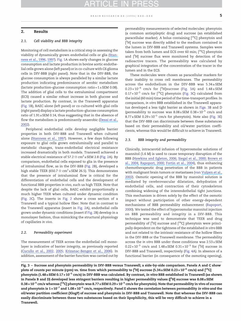
Clinically, intracarotid infusion of hyperosmolar solutions ofmannitol (1.6 M) is used to cause temporary disruption of theBBB (Hawkins and Egleton, 2006; Siegal et al., 2000; Brown etal., 2004; Rapoport, 2000; Fortin et al., 2004), thus enhancingchemotherapeutic drug penetration of the BBB in patientswith malignant brain tumors or metastases (van Vulpen et al.,2002). Osmotic opening of the BBB by mannitol solution ismediated by cerebrovascular dilatation, dehydration ofendothelial cells, and contraction of their cytoskeletoncombining widening of the interendothelial tight junctions.This mechanism is driven solely by the effect of the osmoticimpact without participation of other energy-dependentmechanisms of BBB permeability enhancement (Rapoport,2000). We tested the effect of hyperosmolarmannitol injectionon BBB permeability and integrity in a DIV-BBB. Thistechnique was used to demonstrate that TEER and drugpermeability of [3H] sucrose and [14C] phenytoin were princi-pally dependent on the tightness of the established in vitro BBBand not related to the intrinsic resistance of the hollow fibersin the DIV-BBB or the Transwell membrane. The permeabilityacross the in vitro BBB under these conditions was 2.53±SEM0.22×10−3 cm/s and 1.48±SEM 0.31×10⁻³ for [3H] sucrose inDIV-BBB and Transwell, respectively (Fig. 4A). In absence of afunctional barrier (in consequence of the osmoting opening),
Transwell; a side-by-side comparison. Panels A and C showty to [3H] sucrose (5.34±SEM 0.23×10⁻⁶ cm/s) and [14C]. By contrast, in vitro BBB established in Transwell (as shownhigher permeability values ([3H] sucrose was 8.08±SEMcm/s for phenytoin). Note that permeability in vivo of sucroseE shows the correlation between permeability in vitro and theDIV-BBB and Transwell. Note that whereas the DIV-BBB canr lipophilicity, this will be very difficult to achieve in a
6 B R A I N R E S E A R C H X X ( 2 0 0 6 ) X X X – X X X
ARTICLE IN PRESS
7B R A I N R E S E A R C H X X ( 2 0 0 6 ) X X X – X X X
ARTICLE IN PRESS
[14C] phenytoin permeability was 2.03±SEM 0.15×10⁻³ cm/s inDIV-BBB and 2.17±SEM 0.24×10⁻³ cm/s in Transwell (Fig. 4B).The permeability was calculated as previously described fromthe initial (60 min) period of the time-transport profiles. Notehow both system reach the equilibrium between the luminaland abluminal compartment. Loss of BBB integrity wasmonitored by real-time TEER measuring (Fig. 4C).
3. Discussion
The BBB is a dynamic interface between the blood and thecentral nervous system (CNS) that controls the rate of influxand efflux of biological substances needed for the brainmetabolic processes and neuronal function. The BBB pheno-type develops under the influence of associated brain cells,especially astrocytes. This leads to the formation of a muchmore stringent tight junction system (Abbott, 2005) incomparison to other capillary endothelia. A number of specifictransport and enzyme systems that regulate molecular trafficbetween the blood and the CNS comprise another particularcharacteristic of the BBB endothelium (Abbott, 2002). Thebarrier is additionally comprised of the basal lamina, which isan extension of the extracellular matrix (ECM). EC andastrocyte adhesion to the matrix are mediated by integrinreceptors (Ellison et al., 1999).
The use of cultured vascular endothelial cells as a model ofvascular permeability has significantly advanced the under-standing of microvascular physiology. However, seriouslimitations associated with the use of cultured brain endothe-lial cells have hampered the development of a reliable modelof the BBB. For example, it has been difficult to reproduce incultured cells the high electrical resistance normally foundacross BBB endothelial cells in situ. Furthermore, culturedbrain endothelial cells lose some of their specific markersfollowing prolonged in vitro culturing (Ballermann and Ott,1995). One approach used tomimic the growth environment ofin vivo brain EC has been the use of glia/endothelial co-cultures(Abbott, 2002; Boveri et al., 2005; Sobue et al., 1999; Laterra andGoldstein, 1991; Minakawa et al., 1991). Alternatively, investi-gators have attempted to imitate the physiological environ-ment of microvascular endothelial cells by exposing the cellsto flow (Ballermann and Ott, 1995; Ballermann et al., 1998).More recent attempts have led to dynamic in vitro BBB modelsthat recapitulate all these approaches (Stanness et al., 1996,1997, 1999; Cucullo et al., 2002, 2005; Ballermann and Ott, 1995;Deli et al., 2005; Grant et al., 1998; Janigro et al., 1999; Salvetti etal., 2002).
Studies from this and other laboratories have convincinglydemonstrated that the use of static models to mimic thephysiological and structural characteristic of in vivo BBB isflawed. There are several in vitro models of the BBB currentlyemployed to explore the permeability and potential efficacy of
Fig. 4 – Hyperosmolar opening of the BBB in DIV-BBB and Transwmannitol on BBB permeability to sucrose and phenytoin in DIV-Bthe permeability values of [3H] sucrose and [14C] phenytoin) in botthen previous values measured in intact BBB. Note also that the40 min following mannitol perfusion while this time window is
drugs targeting the brain. The most commonly used systemfor EC culturing consists of a porous membrane supportsubmerged in feedingmedium (e.g., Transwell apparatus). Thebi-dimensional Transwell system is characterized by a side-by-side diffusion system. Amajor disadvantage of this systemis the lack of physiologic shear stress due to the absence ofintraluminal flow. The role played by intraluminal shearstress in brain microvessel endothelial cell differentiation, aswell asmaintenance and induction of a BBB phenotype, is nowwell recognized (Desai et al., 2002; Ballermann and Ott, 1995;Krizanac-Bengez et al., 2003; Brooks et al., 2004; Wassermanand Topper, 2004; Ott and Ballermann, 1995; Ando andKamiya, 1996; Lin et al., 2000; McAllister et al., 2001).
The tightness of the barrier in Transwell, measured by bothTEER and permeability to polar molecules, is typically muchless stringent than the BBB in vivo. Thus, compounds thatpoorly penetrate across the BBB in vivo (e.g., sucrose) readilydiffuse across the endothelial monolayer in the static modelwe examined in this study (Psucrose≈8.08×10⁻⁵, permeability inTranswell was between 10−4 and 10−5 cm/s whereas in vivo itwas ≈10−7 cm/s; see Fig. 3). Because of an elevated Psucrose, BBBpermeability, values of other compounds may be overesti-mated. However, the permeability values of sucrose that wemeasured in Transwell was higher in comparison to similarsystems where instead of BAEC, porcine brain microvesselendothelial cells (PBMEC/C1-2) were used (Psucrose≈5×10⁻⁵)(Lauer et al., 2004). Nevertheless, permeability values ofsucrose measured in DIV-BBB (Psucrose≈5×10⁻⁶) were morephysiological than those reported in this more “efficient”Transwell model.
Lack of physiological conditions, such as presence ofintraluminal flow, may accelerate the dedifferentiating pro-cess that the endothelial cells experience and further enhancethe loss of the BBB characteristics with serial cell passage(Akimoto et al., 2000). Furthermore, increased cell cycle ratedue to lack of antimitotic influences by laminin and flow willcause endothelial cells to pile up in a multilayer fashion(Ziegler and Nerem, 1994). In addition, the occurrence ofirregular patterns of cell adhesion or “edge effect” in Transwellsystems can seriously hamper the measurement of BBBpermeability.
The DIV-BBB originates from amodification of a cell culturesystem used for hybridoma cell expansion. This in vitro “celldifferentiation factory” provides quasi-physiological experi-mental conditions for culturing endothelial cells and astro-cytes in a capillary-like structure and is able to functionallyand anatomically mimic the brain microvasculature. In thehollow fiber apparatus, endothelial cells develop a morphol-ogy that closely resembles the endothelial phenotype in situ,demonstrating that endothelial cells grown with flow aremore differentiated than after conventional culture (Stannesset al., 1997, 1999; Cucullo et al., 2002; Grant et al., 1998; Janigroet al., 1999; Salvetti et al., 2002).
ell models. Panels A and B show the effect of hyperosmolarBB and Transwell. Note that in absence of a functional barrier,h DIV-BBB and Transwell are similar and overall much higherDIV-BBB remains temporarily permeable for approximatelyapproximately 60 min for Transwell.
8 B R A I N R E S E A R C H X X ( 2 0 0 6 ) X X X – X X X
ARTICLE IN PRESS
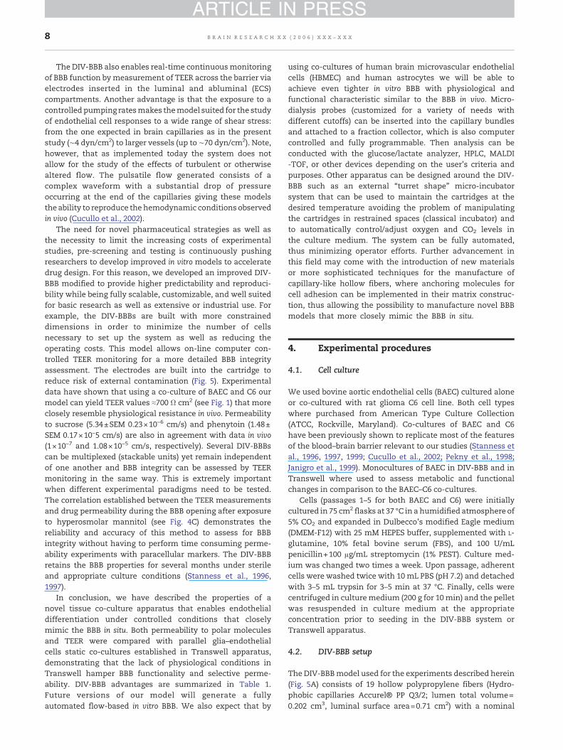
The DIV-BBB also enables real-time continuousmonitoringof BBB function bymeasurement of TEER across the barrier viaelectrodes inserted in the luminal and abluminal (ECS)compartments. Another advantage is that the exposure to acontrolled pumping ratesmakes themodel suited for the studyof endothelial cell responses to a wide range of shear stress:from the one expected in brain capillaries as in the presentstudy (∼4 dyn/cm2) to larger vessels (up to ∼70 dyn/cm2). Note,however, that as implemented today the system does notallow for the study of the effects of turbulent or otherwisealtered flow. The pulsatile flow generated consists of acomplex waveform with a substantial drop of pressureoccurring at the end of the capillaries giving these modelsthe ability to reproduce thehemodynamic conditions observedin vivo (Cucullo et al., 2002).
The need for novel pharmaceutical strategies as well asthe necessity to limit the increasing costs of experimentalstudies, pre-screening and testing is continuously pushingresearchers to develop improved in vitro models to acceleratedrug design. For this reason, we developed an improved DIV-BBB modified to provide higher predictability and reproduci-bility while being fully scalable, customizable, and well suitedfor basic research as well as extensive or industrial use. Forexample, the DIV-BBBs are built with more constraineddimensions in order to minimize the number of cellsnecessary to set up the system as well as reducing theoperating costs. This model allows on-line computer con-trolled TEER monitoring for a more detailed BBB integrityassessment. The electrodes are built into the cartridge toreduce risk of external contamination (Fig. 5). Experimentaldata have shown that using a co-culture of BAEC and C6 ourmodel can yield TEER values ≈700 Ω cm2 (see Fig. 1) that moreclosely resemble physiological resistance in vivo. Permeabilityto sucrose (5.34±SEM 0.23×10⁻⁶ cm/s) and phenytoin (1.48±SEM 0.17×10⁻⁵ cm/s) are also in agreement with data in vivo(1×10⁻⁷ and 1.08×10⁻⁵ cm/s, respectively). Several DIV-BBBscan be multiplexed (stackable units) yet remain independentof one another and BBB integrity can be assessed by TEERmonitoring in the same way. This is extremely importantwhen different experimental paradigms need to be tested.The correlation established between the TEER measurementsand drug permeability during the BBB opening after exposureto hyperosmolar mannitol (see Fig. 4C) demonstrates thereliability and accuracy of this method to assess for BBBintegrity without having to perform time consuming perme-ability experiments with paracellular markers. The DIV-BBBretains the BBB properties for several months under sterileand appropriate culture conditions (Stanness et al., 1996,1997).
In conclusion, we have described the properties of anovel tissue co-culture apparatus that enables endothelialdifferentiation under controlled conditions that closelymimic the BBB in situ. Both permeability to polar moleculesand TEER were compared with parallel glia–endothelialcells static co-cultures established in Transwell apparatus,demonstrating that the lack of physiological conditions inTranswell hamper BBB functionality and selective perme-ability. DIV-BBB advantages are summarized in Table 1.Future versions of our model will generate a fullyautomated flow-based in vitro BBB. We also expect that by
using co-cultures of human brain microvascular endothelialcells (HBMEC) and human astrocytes we will be able toachieve even tighter in vitro BBB with physiological andfunctional characteristic similar to the BBB in vivo. Micro-dialysis probes (customized for a variety of needs withdifferent cutoffs) can be inserted into the capillary bundlesand attached to a fraction collector, which is also computercontrolled and fully programmable. Then analysis can beconducted with the glucose/lactate analyzer, HPLC, MALDI-TOF, or other devices depending on the user's criteria andpurposes. Other apparatus can be designed around the DIV-BBB such as an external “turret shape” micro-incubatorsystem that can be used to maintain the cartridges at thedesired temperature avoiding the problem of manipulatingthe cartridges in restrained spaces (classical incubator) andto automatically control/adjust oxygen and CO2 levels inthe culture medium. The system can be fully automated,thus minimizing operator efforts. Further advancement inthis field may come with the introduction of new materialsor more sophisticated techniques for the manufacture ofcapillary-like hollow fibers, where anchoring molecules forcell adhesion can be implemented in their matrix construc-tion, thus allowing the possibility to manufacture novel BBBmodels that more closely mimic the BBB in situ.
4. Experimental procedures
4.1. Cell culture
We used bovine aortic endothelial cells (BAEC) cultured aloneor co-cultured with rat glioma C6 cell line. Both cell typeswhere purchased from American Type Culture Collection(ATCC, Rockville, Maryland). Co-cultures of BAEC and C6have been previously shown to replicate most of the featuresof the blood–brain barrier relevant to our studies (Stanness etal., 1996, 1997, 1999; Cucullo et al., 2002; Pekny et al., 1998;Janigro et al., 1999). Monocultures of BAEC in DIV-BBB and inTranswell where used to assess metabolic and functionalchanges in comparison to the BAEC–C6 co-cultures.
Cells (passages 1–5 for both BAEC and C6) were initiallycultured in 75 cm2 flasks at 37 °C in a humidified atmosphere of5% CO2 and expanded in Dulbecco's modified Eagle medium(DMEM-F12) with 25 mM HEPES buffer, supplemented with L-glutamine, 10% fetal bovine serum (FBS), and 100 U/mLpenicillin+100 μg/mL streptomycin (1% PEST). Culture med-ium was changed two times a week. Upon passage, adherentcells were washed twice with 10 mL PBS (pH 7.2) and detachedwith 3–5 mL trypsin for 3–5 min at 37 °C. Finally, cells werecentrifuged in culturemedium (200 g for 10min) and the pelletwas resuspended in culture medium at the appropriateconcentration prior to seeding in the DIV-BBB system orTranswell apparatus.
4.2. DIV-BBB setup
The DIV-BBBmodel used for the experiments described herein(Fig. 5A) consists of 19 hollow polypropylene fibers (Hydro-phobic capillaries Accurel® PP Q3/2; lumen total volume=0.202 cm3, luminal surface area=0.71 cm2) with a nominal
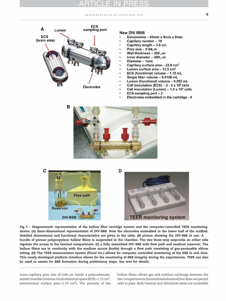
Fig. 5 – Diagrammatic representation of the hollow fiber cartridge system and the computer-controlled TEER monitoringdevice. (A) three-dimensional representation of DIV-BBB. Note the electrodes embedded in the lower half of the scaffold.Detailed dimensional and functional characteristics are given in the table. (B) picture showing the DIV-BBB in use. Abundle of porous polypropylene hollow fibers is suspended in the chamber. The two three-way stopcocks on either sideregulate the access to the luminal compartment. (C) a fully assembled DIV-BBB with flow path and medium reservoir. Thehollow fibers are in continuity with the medium source (bottle) through a flow path consisting of gas-permeable silicontubing. (D) The TEER measurement system (Flocel Inc.) allows for computer controlled monitoring of the BBB in real time.This newly developed platform interface allows for the monitoring of BBB integrity during the experiments. TEER can alsobe used to assess for BBB formation during preliminary steps. See text for details.
9B R A I N R E S E A R C H X X ( 2 0 0 6 ) X X X – X X X
ARTICLE IN PRESS
trans-capillary pore size of 0.64 μm inside a polycarbonate-sealed chamber (volumeof extraluminal space (ECS)=1.15 cm3,extraluminal surface area=1.19 cm2). The porosity of the
hollow fibers allows gas and nutrient exchange between thetwocompartments (luminal/extraluminal) but doesnot permitcells to pass. Both luminal and abluminal areas are accessible
Table 1 – Advantages of DIV-BBB
1. Glucose, oxygen, flow (shear), and temperature can beindependently varied2. Continuous monitoring of BBB integrity (TEER)3. Variable levels of Shear stress can be applied.4. Administration of Biological agents into intra- or extraluminalcompartment5. Easy separation of EC from astrocytes for analysis6. Cultures remain viable for extended period (several months)7. Easy determination of the role of individual variables modulatingBBB function8. DIV-BBB is not limited to BBB cells
10 B R A I N R E S E A R C H X X ( 2 0 0 6 ) X X X – X X X
ARTICLE IN PRESS
byports in the circuit connectedwith amediumreservoir andapulsatile pump apparatus. Four electrodes are embedded inthe bottom of each cartridge (Fig. 5B) allowing for faster andmore accurate TEERmeasurements by real-time computerizedmonitoring systems. Gaseous exchange occurs while mediaflows (≈25 mL) through gas permeable silicone tubing thatconnects the cartridge and themedium reservoir (Fig. 5C). Thepump (CellMax®QUADArtificial CapillaryCell Culture System,Spectrum Laboratories CA) is capable of generating flow ratesof 1–50 mL/min corresponding to shear stress levels ofapproximately 1–200 dyn/cm2. In order to allow for celladhesion, we used an initial flow rate of 1 mL/min for thefirst 48 h following cell inoculation and then adjusted the flowto 4 mL/min (i.e., 4 dyn/cm2). Both compartmental surfaceswere coated with 3 μg/cm2 fibronectin and the extraluminalcapillary side was precoated with poly-D-lysine (3 μg/cm2) aswell. The entire apparatus resided in a water-jacketedincubator with 5% CO2 at 37 °C. For sterile sampling, it couldbe moved into a laminar flow hood. BAEC were seededintraluminally and allowed to establish themselves for 7–15 days with or without extraluminal C6. The amount of cellsseeded was ≈1.5×10⁶ BAEC and ≈2×10⁶ C6.
4.3. Cell metabolism: lactate production and glucoseconsumption
In conjunction with TEER monitoring, indicators of cellmetabolism were used to monitor cell expansion in boththe DIV-BBB and Transwell apparatus. Depletion of the maincarbohydrate component of the growth medium (glucose)and accumulation of metabolically produced lactic acid areused as indicators of cell growth (Stanness et al., 1996, 1997,1999; Cucullo et al., 2002; Ballermann and Ott, 1995; Deli et al.,2005; McAllister et al., 2001). For this purpose, luminal andextracapillary space were sampled at daily intervals. Thecalculations for glucose consumption (mg/day) and lactateproduction rates (mg/day) are based on medium replacement,volume of non-replaced medium and previous values.Glucose consumption rate was calculated based on theconcentration of glucose in fresh and unreplaced mediumin the system, according to the following equation:
ðVn � GnÞ þ ðVo � GpÞ � ðVtotal � GcÞTc � Tp
, ð1Þ
where V represents added volumes of medium (mL), G is theglucose concentration (mg/mL), T is time of sampling (infractions of days), c and P indicate the current and previous
samples, respectively, n represents (new) fresh mediumadded after previous sampling, whereas o represents old,unreplaced medium. Lactate production rate was calculatedsimilarly:
ðVt � LcÞ � ðVn � LnÞ þ ðVu � LpÞTc � Tp
, ð2Þ
where L refers to the concentration of lactic acid in mg/mL. Adual-channel immobilized oxidase enzyme biochemistry (YSI2700 SELECT, YSI Inc., Yellow Springs, Ohio) was used tomeasure lactate and glucose in the cell culture medium. Dataobtained with the described equation were then converted inmmol/day.
4.4. DIV-BBB TEER measurement system
The trans-endothelial electrical resistance (TEER) measurementprovides aquickandeasyevaluationof the integrityof theblood–brain barrier model. We used a newly developed TEERmeasure-ment device (Flocel Inc., Cleveland, Ohio), which utilizes electro-nic multiplexing to measure multiple cartridges in quicksuccession and assess the integrity and viability of tissue culturemodels rapidly and reliably (Fig. 5D). The device uses a universalserial bus (USB) interface to a PC computer. To sample, theexcitation voltage is applied across the excitation electrodesinserted in each cartridge and the microcontroller computes theresistivity and capacitance per cm2 of the barrier from physicalparameters. TEER was measured continuously from the initialsetup throughout the course of each experiment.
4.5. Transwell systems
Cells were co-cultured using sets of 12-well Transwell-ClearPolyester Membrane apparatus (Costar cat. #3460) that feature avertical side by side diffusion system through a thin, micro-scopically transparent polyester membrane of 12 mm diameterand 0.4 μmpore sizemounted on a double chamber (Fig. 6). BAECwere seeded on the top side of the membrane and allowed toestablish themselves for 3 days. C6 were then seeded on theunderside of the filter (co-cultures only). The amount of cellsranged from 1×10⁵ (BAEC) to 1.5×10⁵ (C6). TEER was monitoredevery 2 days beginning at “day 1” of co-culture. The equipmentconsisted of a tissue resistancemeasurement chamber (Endohmchamber, WPI) that provides a reproducible electrical resistancemeasurement of endothelial tissue culture cups. In conjunctionwith TEER monitoring, BAEC monolayer integrity was alsoassessed by inspection with phase contrast microscopy.
4.6. Drug permeability: uptake of [14C]phenytoin and [3H]sucrose
A concentrated bolus of [14C] phenytoin and [3H] sucrose wasinjected into the lumen and the diffusion into the extracapillaryspace was monitored over time while maintaining a 1 mL/minintraluminal perfusion rate. Sampleswere taken from ECS or thelumen as previously described (Stanness et al., 1997; Salvetti etal., 2002), at time 0 (immediately after the injection) and at 1, 3, 5,10, 15, 30, and 60 min following the injection. Samples removedfrom ECS were replaced with equal volumes of medium. 100 μL
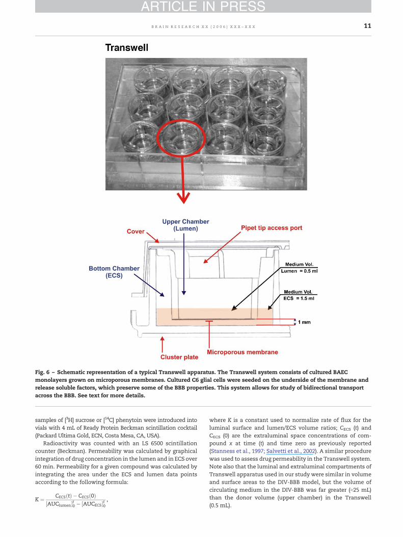
Fig. 6 – Schematic representation of a typical Transwell apparatus. The Transwell system consists of cultured BAECmonolayers grown on microporous membranes. Cultured C6 glial cells were seeded on the underside of the membrane andrelease soluble factors, which preserve some of the BBB properties. This system allows for study of bidirectional transportacross the BBB. See text for more details.
11B R A I N R E S E A R C H X X ( 2 0 0 6 ) X X X – X X X
ARTICLE IN PRESS
samples of [3H] sucrose or [14C] phenytoin were introduced intovials with 4 mL of Ready Protein Beckman scintillation cocktail(Packard Ultima Gold, ECN, Costa Mesa, CA, USA).
Radioactivity was counted with an LS 6500 scintillationcounter (Beckman). Permeability was calculated by graphicalintegration of drug concentration in the lumen and in ECS over60 min. Permeability for a given compound was calculated byintegrating the area under the ECS and lumen data pointsaccording to the following formula:
K ¼ CECSðtÞ � CECSð0Þ½AUClumen�t0 � ½AUCECS�t0
,
where K is a constant used to normalize rate of flux for theluminal surface and lumen/ECS volume ratios; CECS (t) andCECS (0) are the extraluminal space concentrations of com-pound x at time (t) and time zero as previously reported(Stanness et al., 1997; Salvetti et al., 2002). A similar procedurewas used to assess drug permeability in the Transwell system.Note also that the luminal and extraluminal compartments ofTranswell apparatus used in our study were similar in volumeand surface areas to the DIV-BBB model, but the volume ofcirculating medium in the DIV-BBB was far greater (≈25 mL)than the donor volume (upper chamber) in the Transwell(0.5 mL).
12 B R A I N R E S E A R C H X X ( 2 0 0 6 ) X X X – X X X
ARTICLE IN PRESS
4.7. BBB opening by hyperosmolar mannitol
Infusion of 10 mL of growth medium containing mannitol at aconcentration of 1.6 M was used to open the BBB (Rapoport,2000). The solution was prepared under sterile conditions andinjected intraluminally at a perfusion rate of 1 mL/min. TEERwas monitored during the course of the experiment to assessBBB failure and recovery. A concentrated bolus containingboth [14C] phenytoin and [3H] sucrose was injected into thelumen 1 min following mannitol perfusion and flux into theextracapillary space was monitored over time as previouslydescribed. In the Transwell system, the regular luminal mediaware replaced with one containing mannitol at the finalconcentration of 1.6 M and left in the luminal chamber for10 min in order to reproduce experimental parameters similarto the one established in the DIV-BBB. The medium in theluminal chamber was then removed and replaced with freshone containing [14C] phenytoin and [3H] sucrose at similarconcentrations to those tested in the DIV-BBB. Note that in theDIV-BBB apparatus the intraluminal flow removes the man-nitol from the luminal chamber (the mannitol is thenexhausted in the reservoir), lack of flow in the Transwellsystem required manual removal of the mannitol solution.
Acknowledgments
This work was supported by Philip Morris USA and PhilipMorris International external research award to Luca Cuculloand NIH-2RO1 HL51614, NIH-RO1 NS43284 NIH-RO1 NS38195to Damir Janigro.
R E F E R E N C E S
Abbott, N.J., 2002. Astrocyte–endothelial interactions andblood–brain barrier permeability. J. Anat. 200, 629–638.
Abbott, N.J., 2005. Dynamics of CNS barriers: evolution,differentiation, and modulation. Cell. Mol. Neurobiol. 25,5–23.
Akimoto, S., Mitsumata, M., Sasaguri, T., Yoshida, Y., 2000.Laminar shear stress inhibits vascular endothelial cellproliferation by inducing cyclin-dependent kinase inhibitorp21(Sdi1/Cip1/Waf1). Circ. Res. 86, 185–190.
Ando, J., Kamiya, A., 1996. Flow-dependent regulation of geneexpression in vascular endothelial cells. Jpn. Heart J. 37, 19–32.
Ballermann, B.J., Ott, M.J., 1995. Adhesion and differentiation ofendothelial cells by exposure to chronic shear stress: a vasculargraft model. Blood Purif. 13, 125–134.
Ballermann, B.J., Dardik, A., Eng, E., Liu, A., 1998. Shear stress andthe endothelium. Kidney Inter., Suppl. 67, S100–S108.
Banks, W.A., 2005. Blood–brain barrier transport of cytokines: amechanism for neuropathology. Curr. Pharm. Des 11,973–984.
Boveri, M., Berezowski, V., Price, A., Slupek, S., Lenfant, A.M.,Benaud, C., Hartung, T., Cecchelli, R., Prieto, P., Dehouck, M.P.,2005. Induction of blood–brain barrier properties in culturedbrain capillary endothelial cells: comparison between primaryglial cells and C6 cell line. Glia 51, 187–198.
Brooks, A.R., Lelkes, P.I., Rubanyi, G.M., 2004. Gene expressionprofiling of vascular endothelial cells exposed to fluidmechanical forces: relevance for focal susceptibility toatherosclerosis. Endothelium 11, 45–57.
Brown, R.C., Davis, T.P., 2005. Hypoxia/aglycemia alters expressionof occludin and actin in brain endothelial cells. Biochem.Biophys. Res. Commun. 327, 1114–1123.
Brown, R.C., Egleton, R.D., Davis, T.P., 2004. Mannitol opening ofthe blood–brain barrier: regional variation in the permeabilityof sucrose, but not 86Rb+ or albumin. Brain Res. 1014,221–227.
Cucullo, L., McAllister, M.S., Kight, K., Krizanac-Bengez, L.,Marroni, M., Mayberg, M.R., Stanness, K.A., Janigro, D., 2002. Anew dynamic in vitro model for the multidimensional study ofastrocyte-endothelial cell interactions at the blood–brainbarrier. Brain Res. 951, 243–254.
Cucullo, L., Aumayr, B., Rapp, E., Janigro, D., 2005. Drug deliveryand in vitro models of the blood–brain barrier. Curr. Opin. DrugDiscov. Dev. 8, 89–99.
De Boer, A.B., De Lange, E.L., Van der Sandt, I.C., Breimer, D.D.,1998. Transporters and the blood–brain barrier (BBB). Int. J. Clin.Pharmacol. Ther. 36, 14–15.
Deli, M.A., Abraham, C.S., Kataoka, Y., Niwa, M., 2005. Permeabilitystudies on in vitro blood–brain barrier models: physiology,pathology, and pharmacology. Cell. Mol. Neurobiol. 25, 59–127.
Desai, S.Y., Marroni, M., Cucullo, L., Krizanac-Bengez, L., Mayberg,M.R., Hossain, M.T., Grant, G.G., Janigro, D., 2002. Mechanismsof endothelial survival under shear stress. Endothelium 9,89–102.
Ellison, J.A., Barone, F.C., Feuerstein, G.Z., 1999. Matrix remodelingafter stroke. De novo expression of matrix proteins andintegrin receptors. Ann. N. Y. Acad. Sci. 890, 204–222.
Emmi, A., Wenzel, H.J., Schwartzkroin, P.A., Taglialatela, M.,Castaldo, P., Bianchi, L., Nerbonne, J., Robertson, G.A., Janigro,D., 2000. Do glia have heart? Expression and functional role forether-a-go-go currents in hippocampal astrocytes. J. Neurosci.20, 3915–3925.
Fortin, D., Salame, J.A., Desjardins, A., Benko, A., 2004. Technicalmodification in the intracarotid chemotherapy and osmoticblood–brain barrier disruption procedure to prevent the relapseof carboplatin-induced orbital pseudotumor. AJNR Am. J.Neuroradiol. 25, 830–834.
Grant, G.A., Abbott, N.J., Janigro, D., 1998. Understanding thephysiology of the blood–brain barrier: in vitro models. NewsPhysiol. Sci. 13, 287–293.
Hagenbuch, B., Gao, B., Meier, P.J., 2002. Transport ofxenobiotics across the blood–brain barrier. News Physiol. Sci.17, 231–234.
Hamm, S., Dehouck, B., Kraus, J., Wolburg-Buchholz, K., Wolburg,H., Risau, W., Cecchelli, R., Engelhardt, B., Dehouck, M.P., 2004.Astrocyte mediated modulation of blood–brain barrierpermeability does not correlate with a loss of tight junctionproteins from the cellular contacts. Cell Tissue Res. 315,157–166.
Hawkins, B.T., Davis, T.P., 2005. The blood–brainbarrier/neurovascular unit in health and disease. Pharmacol.Rev. 57, 173–185.
Hawkins, B.T., Egleton, R.D., 2006. Fluorescence imaging ofblood–brain barrier disruption. J. Neurosci. Methods 151 (2),262–267.
Heo, J.H., Han, S.W., Lee, S.K., 2005. Free radicals as triggers ofbrain edema formation after stroke. Free Radical Biol. Med.39, 51–70.
Hori, S., Ohtsuki, S., Tachikawa, M., Kimura, N., Kondo, T.,Watanabe, M., Nakashima, E., Terasaki, T., 2004. Functionalexpression of rat ABCG2 on the luminal side of brain capillariesand its enhancement by astrocyte-derived soluble factor(s).J. Neurochem. 90, 526–536.
Janigro, D., Leaman, S.M., Stanness, K.A., 1999. Dynamic modelingof the blood–brain barrier: a novel tool for studies of drugdelivery to the brain. Pharm. Sci. Technol. Today 2, 7–12.
Korn, A., Golan, H., Melamed, I., Pascual-Marqui, R., Friedman, A.,2005. Focal cortical dysfunction and blood–brain barrier

13B R A I N R E S E A R C H X X ( 2 0 0 6 ) X X X – X X X
ARTICLE IN PRESS
disruption in patients with postconcussion syndrome. J. Clin.Neurophysiol. 22, 1–9.
Kramer, S.D., Schutz, Y.B., Wunderli-Allenspach, H., Abbott, N.J.,Begley, D.J., 2002. Lipids in blood–brain barrier models in vitro:II. Influence of glial cells on lipid classes and lipid fatty acids. InVitro Cell Dev. Biol., Anim. 38, 566–571.
Krizanac-Bengez, L., Kapural, M., Parkinson, F., Cucullo, L.,Hossain, M., Mayberg, M.R., Janigro, D., 2003. Effects oftransient loss of shear stress on blood–brain barrierendothelium: role of nitric oxide and IL-6. Brain Res. 977,239–246.
Krizanac-Bengez, L., Mayberg, M.R., Janigro, D., 2004. The cerebralvasculature as a therapeutic target for neurological disordersand the role of shear stress in vascular homeostatis andpathophysiology. Neurol. Res. 26, 846–853.
Krizanac-Bengez, L., Mayberg, M.R., Cunningham, E., Hossain, M.,Ponnampalam, S., Parkinson, F.E., Janigro, D., 2006. Loss ofshear stress induces leukocyte-mediated cytokine release andblood–brain barrier failure in dynamic in vitro blood–brainbarrier model. J. Cell. Physiol. 206 (1), 68–77.
Laterra, J., Goldstein, G.W., 1991. Astroglial-induced in vitroangiogenesis: requirements for RNA and protein synthesis.J. Neurochem. 57, 1231–1239.
Lauer, R., Bauer, R., Linz, B., Pittner, F., Peschek, G.A., Ecker, G.,Friedl, P., Noe, C.R., 2004. Development of an in vitroblood–brain barrier model based on immortalized porcinebrain microvascular endothelial cells. Farmaco 59, 133–137.
Liberto, C.M., Albrecht, P.J., Herx, L.M., Yong, V.W., Levison, S.W.,2004. Pro-regenerative properties of cytokine-activatedastrocytes. J. Neurochem. 89, 1092–1100.
Lin, K., Hsu, P.P., Chen, B.P., Yuan, S., Usami, S., Shyy, J.Y., Li, Y.S.,Chien, S., 2000. Molecular mechanism of endothelial growtharrest by laminar shear stress. Proc. Natl. Acad. Sci. U. S. A. 97,9385–9389.
McAllister, M.S., Krizanac-Bengez, L., Macchia, F., Naftalin, R.J.,Pedley, K.C., Mayberg, M.R., Marroni, M., Leaman, S., Stan-ness, K.A., Janigro, D., 2001. Mechanisms of glucose transportat the blood–brain barrier: an in vitro study. Brain Res. 904,20–30.
Minakawa, T., Bready, J., Berliner, J., Fisher, M., Cancilla, P.A., 1991.In vitro interaction of astrocytes and pericytes withcapillary-like structures of brain microvessel endothelium.Lab. Invest. 65, 32–40.
Mizuguchi, H., Utoguchi, N., Mayumi, T., 1997. Preparation of glialextracellular matrix: a novel method to analyzeglial–endothelial cell interaction. Brain Res. Brain Res. Protoc. 1,339–343.
Ott, M.J., Ballermann, B.J., 1995. Shear stress-conditioned,endothelial cell-seeded vascular grafts: improved celladherence in response to in vitro shear stress. Surgery 117,334–339.
Pekny, M., Stanness, K.A., Eliasson, C., Betsholtz, C., Janigro, D.,1998. Impaired induction of blood–brain barrier properties inaortic endothelial cells by astrocytes fromGFAP-deficientmice.Glia 22, 390–400.
Rapoport, S.I., 2000. Osmotic opening of the blood–brain barrier:principles, mechanism, and therapeutic applications. Cell. Mol.Neurobiol. 20, 217–230.
Salvetti, F., Cecchetti, P., Janigro, D., Lucacchini, A., Benzi, L.,Martini, C., 2002. Insulin permeability across an in vitrodynamic model of endothelium. Pharm. Res. 19, 445–450.
Shusta, E.V., 2005. Blood–brain barrier genomics, proteomics, andnew transporter discovery. NeuroRx 2, 151–161.
Siegal, T., Rubinstein, R., Bokstein, F., Schwartz, A., Lossos, A.,Shalom, E., Chisin, R., Gomori, J.M., 2000. In vivo assessment ofthe window of barrier opening after osmotic blood–brainbarrier disruption in humans. J. Neurosurg. 92, 599–605.
Sobue, K., Yamamoto, N., Yoneda, K., Hodgson, M.E., Yamashiro,K., Tsuruoka, N., Tsuda, T., Katsuya, H., Miura, Y., Asai, K., Kato,T., 1999. Induction of blood–brain barrier properties inimmortalized bovine brain endothelial cells by astrocyticfactors. Neurosci. Res. 35, 155–164.
Stanness, K.A., Guatteo, E., Janigro, D., 1996. A dynamic modelof the blood–brain barrier “in vitro”. Neurotoxicology 17,481–496.
Stanness, K.A., Westrum, L.E., Fornaciari, E., Mascagni, P., Nelson,J.A., Stenglein, S.G., Myers, T., Janigro, D., 1997. Morphologicaland functional characterization of an in vitro blood–brainbarrier model. Brain Res. 771, 329–342.
Stanness, K.A., Neumaier, J.F., Sexton, T.J., Grant, G.A., Emmi, A.,Maris, D.O., Janigro, D., 1999. A new model of the blood–brainbarrier: co-culture of neuronal, endothelial and glial cells underdynamic conditions. NeuroReport 10, 3725–3731.
Toborek, M., Lee, Y.W., Flora, G., Pu, H., Andras, I.E., Wylegala, E.,Hennig, B., Nath, A., 2005. Mechanisms of the blood–brainbarrier disruption in HIV-1 infection. Cell. Mol. Neurobiol. 25,181–199.
van Vulpen, M., Kal, H.B., Taphoorn, M.J., El Sharouni, S.Y., 2002.Changes in blood–brain barrier permeability induced byradiotherapy: implications for timing of chemotherapy?(Review). Oncol. Rep. 9, 683–688.
Wasserman, S.M., Topper, J.N., 2004. Adaptation of theendothelium to fluid flow: in vitro analyses of gene expressionand in vivo implications. Vasc. Med. 9, 35–45.


![[side] THEORY BASE OF ACCOUNTING](https://static.fdokumen.com/doc/165x107/6316e2fb1e5d335f8d0a2c0c/side-theory-base-of-accounting.jpg)